Embed Size (px)
Citation preview
Environmental Health PerspectivesVol. 56, pp. 219-227, 1984
Studies of Pancreatic Carcinogenesisin Different Animal Modelsby Dante G. Scarpelli,* M. Sambasiva Rao* andJanardan K. Reddy*
Pancreatic carcinomas can be induced in rat, guinea pig and hamster by a variety of carcinogens. Thetypes of neoplasms which arise vary with the species of rodent. In the rat, they consist exclusively of acinarcells, in the other species the lesions are adenocarcinomas resembling those derived from pancreaticductules and ducts, those in hamster more so than in guinea pigs. Careful sequential studies in the guineapig and hamster suggest that acinar cells together with ductular and duct cells are involved in the genesis ofduct adenocarcinomas. In each rodent model, the acinar cell appears to be quite sensitive to continuedexposure to carcinogen. In each instance, acini undergo modulation, and in the guinea pig and hamster,permanent metaplastic transformation to ductlike structures. Such cells assume an enhanced capacity forcell proliferation which persists following cessation of carcinogen treatment. Other studies suggest thatadult pancreatic acinar cells possess a surprising degree of plasticity. Their involvement in thepathogenesis of neoplasms resembling pancreatic ducts is not unlike other carcinogenic sequences whereextensive cell modulation and metaplasia precede and are an integral part of the neoplastic transformation.
IntroductionIn recent years there has been considerable progress
in experimental pancreatic carcinogenesis research in-volving basically three rodent animal models, the rat,guinea pig and Syrian golden hamster. Initial carcino-genesis studies involved implantation of 7,12-dimethyl-benzanthracene (DMBA) crystals (1) or DMBA in dex-trose pellets (2) into the pancreas of Sprague-Dawleyrats. Pancreatic adenocarcinomas consisting of acinarcells developed, on the average, in about 200 daysfollowing implantation. Later workers administeredcarcinogenic substances such as 4-hydroxyaminoquino-line-i-oxide (4HAQO), a carcinogenic metabolite of4-nitroquinoline-1-oxide (3), azaserine (4), and the hypo-lipidemic drugs, nafenopin (5) and clofibrate (6,7), andN5-(N-methyl-N-nitrosocarbamoyl)-L-ornithine (MNCO),a methylnitrosourea amino acid (8), systemically, andinduced both acinar cell adenomas and adenocarci-nomas. Although the rat model has been quite useful,the pancreatic neoplasms they develop do not resemblethose that constitute the most common type encoun-tered in humans, namely ductal adenocarcinomas. Inthe case of the guinea pig, pancreatic adenocarcino-mas were induced by the prolonged administration ofN-methyl-N-nitrosourethane (MNUT) (9) in drinkingwater, or N-methyl-N-nitrosourea (MNU) by intra-
*Department of Pathology and the Cancer Center, NorthwesternUniversity Medical School, 303 E. Chicago Ave., Chicago, IL 60611.
gastric gavage weekly for 28 weeks or more (10). Avariety of N-nitrosamines derived from 13-oxidation ofN,N-dipropylnitrosamine induced pancreatic ductal ade-nocarcinomas in the Syrian golden hamster. Chemicallyinduced pancreatic cancer in both species are morpho-logically and biologically quite similar to those of man(10-17). These various models are summarized in Table1. The studies to be reviewed in this communication willdetail certain aspects of the pathogenesis of pancreaticcancer in them.
Histogenesis of ChemicallyInduced Pancreatic Cancerin Various Rodent Species
Rat and Guinea PigIn both the rat and guinea pig, the pancreatic acinar
cell appears to be singularly susceptible to carcinogene-sis regardless of the carcinogen employed. Three differ-ent carcinogens-4HAQO, (a hydroxylated metaboliteof 4-nitroquinoline-l-oxide), DMBA (a polycyclic hydro-carbon), and azaserine (a diazoketone)-all induceacinar cell adenomas and carcinomas in rat pancreas.Since the lesions induced by these carcinogens appearto be strikingly similar (1,3-5), if not identical, thehistogenetic sequence that follows may be considered asapplying to all of them. The lesions begin as localized
SCARPELLI, RAO AND REDDY
Table 1. Animal models of chemically induced pancreatic carcinoma.Route of Dosage of Carcinomas
Species Carcinogen administration carcinogen induced Reference
Rat DMBA Intrapancreatic 2-10 mg(?)a Acinar (1,2)implant
4HAQO IV 6-13 mg/kga Acinar (3)Azaserine IP 5 mg/kge Acinar (4)Nafenopin Diet 0.1% (wt/wt)b Acinar (5)Clofibrate Diet 0.1% (Wt/Wt)b Acinar (6,7)MNCO IP 10-20 mg/kgc Acinar (8)
Guinea pig MNUT In drinking water 2.5 mg/kgd Adenocarcinoma (9)MNU Gavage 10 mg/kgc Adenocarcinoma (10)
Hamster BHP SC 250-500 mg/kgc Ductal (11)BAP SC 17.5-20 mg/kg Ductal (12)BOP SC 10-30 mg/kgc Ductal, rare acinar (13)HPOP SC 9-40 mg/kg' Ductal (14)MOP SC 1.75-3.5 mg/kgc Ductal (15)DMNM Gavage 18-50 mg/kg' Ductal (16)
a Single administration.b Administered continuously.e Administered weekly.d Daily administration.
nests of acini which undergo hyperplastic growth. Whenthese are microscopic in size, they are described as"foci;" larger lesions which are grossly visible, measur-ing 1 to 10 mm in diameter, are referred to as "nodules."Acinar cells in foci and nodules, regardless of their size,are uniformly cytologically abnormal. Their nuclei arelarger and more basophilic than those of normal acinarcells; except for this alteration, the cells appear normalin every other respect, including a full complement ofzymogen granules. In larger nodules, acinar cellsshowed a somewhat greater degree of nuclear andnucleolar variability, as well as mitotic activity. Ultra-structurally, zymogen-rich cells in foci and nodulesshowed a normal complement of cell organelles andzymogen granules which were morphologically identicalto those in normal acinar cells.The classification of large nodules as adenomas is
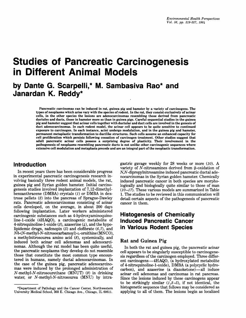
based more on their apparent potential for semiautono-mous growth rather than objective cytologic criteria.Large nodules and adenomas are usually not encap-sulated. The diagnosis of acinar cell adenocarcinomaand probable carcinomas is based on either decreasedacinar cell differentiation (Fig. 1) or evidence ofextrapancreatic spread or both. Metastases occur bothin highly differentiated zymogen granule laden acinarcell tumors as well as poorly differentiated tumors thatbear little to no resemblance to their cell of origin.
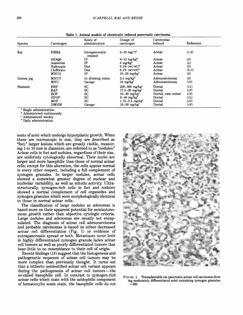
Recent findings (18) suggest that the histogenesis andpathogenetic sequence of acinar cell tumors may bemore complex than previously thought. It turns outthat a hitherto unidentified acinar cell variant appearsduring the pathogenesis of acinar cell tumors-theso-called basophilic cell. In contrast to zymogen-richacinar cells which stain with the acidophilic componentof hematoxylin eosin stain, the basophilic cells do not
FIGURE 1. Transplantable rat pancreatic acinar cell carcinoma show-ing moderately differentiated acini containing zymogen granules.x 320.
220
PANCREATIC CARCINOGENESIS MODELS
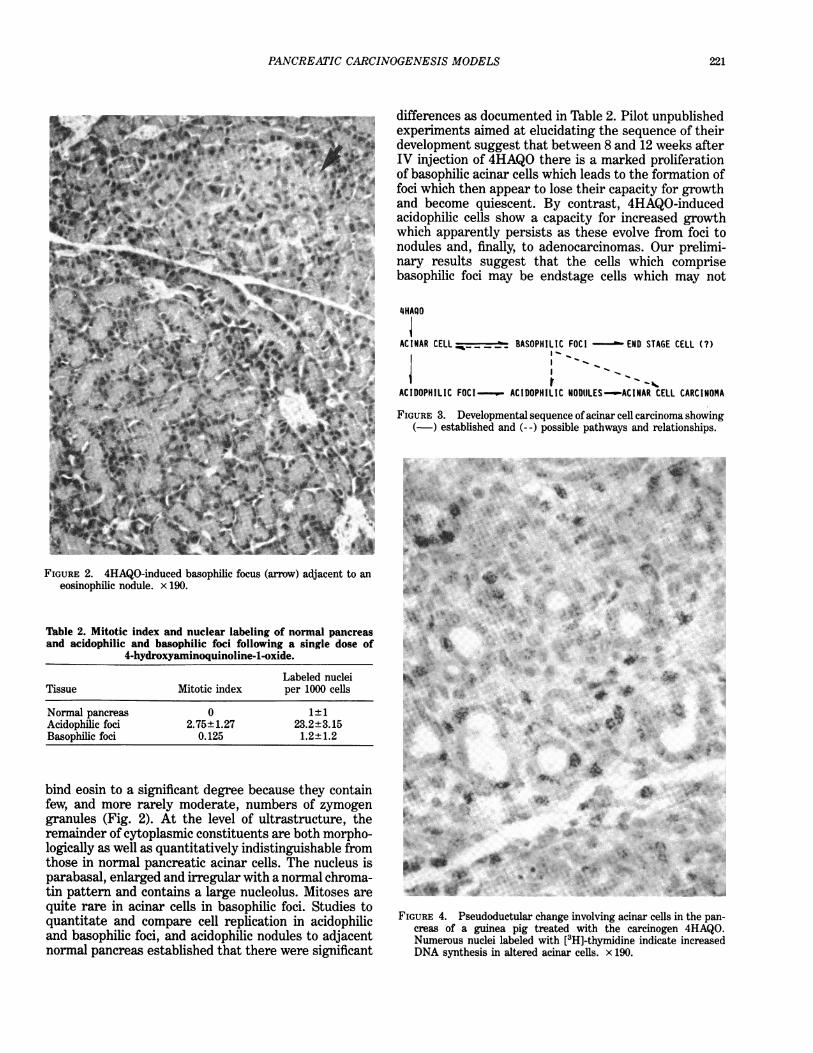
differences as documented in Table 2. Pilot unpublishedexperiments aimed at elucidating the sequence of theirdevelopment suggest that between 8 and 12 weeks afterIV injection of 4HAQO there is a marked proliferationof basophilic acinar cells which leads to the formation offoci which then appear to lose their capacity for growthand become quiescent. By contrast, 4HAQO-inducedacidophilic cells show a capacity for increased growthwhich apparently persists as these evolve from foci tonodules and, finally, to adenocarcinomas. Our prelimi-nary results suggest that the cells which comprisebasophilic foci may be endstage cells which may not
4HAQO
AC INAR CELL _ _ _ _BASOPH I L I C FOC I -END STAGE CELL C?)
ACIDOPHILIC FOCI--._w- ACIDOPHILIC NODULES--ACINAR CELL CARCINOMA
FIGURE 3. Developmental sequence of acinar cell carcinoma showing(-) established and (- -) possible pathways and relationships.
FIGURE 2. 4HAQO-induced basophilic focus (arrow) adjacent to aneosinophilic nodule. x 190.
Table 2. Mitotic index and nuclear labeling of normal pancreasand acidophilic and basophilic foci following a single dose of
4-hydroxyaminoquinoline-1-oxide.
Labeled nucleiTissue Mitotic index per 1000 cells
Normal pancreas 0 1±1Acidophilic foci 2.75±1.27 23.2±3.15Basophilic foci 0.125 1.2±1.2
bind eosin to a significant degree because they containfew, and more rarely moderate, numbers of zymogengranules (Fig. 2). At the level of ultrastructure, theremainder of cytoplasmic constituents are both morpho-logically as well as quantitatively indistinguishable fromthose in normal pancreatic acinar cells. The nucleus isparabasal, enlarged and irregular with a normal chroma-tin pattern and contains a large nucleolus. Mitoses arequite rare in acinar cells in basophilic foci. Studies toquantitate and compare cell replication in acidophilicand basophilic foci, and acidophilic nodules to adjacentnormal pancreas established that there were significant
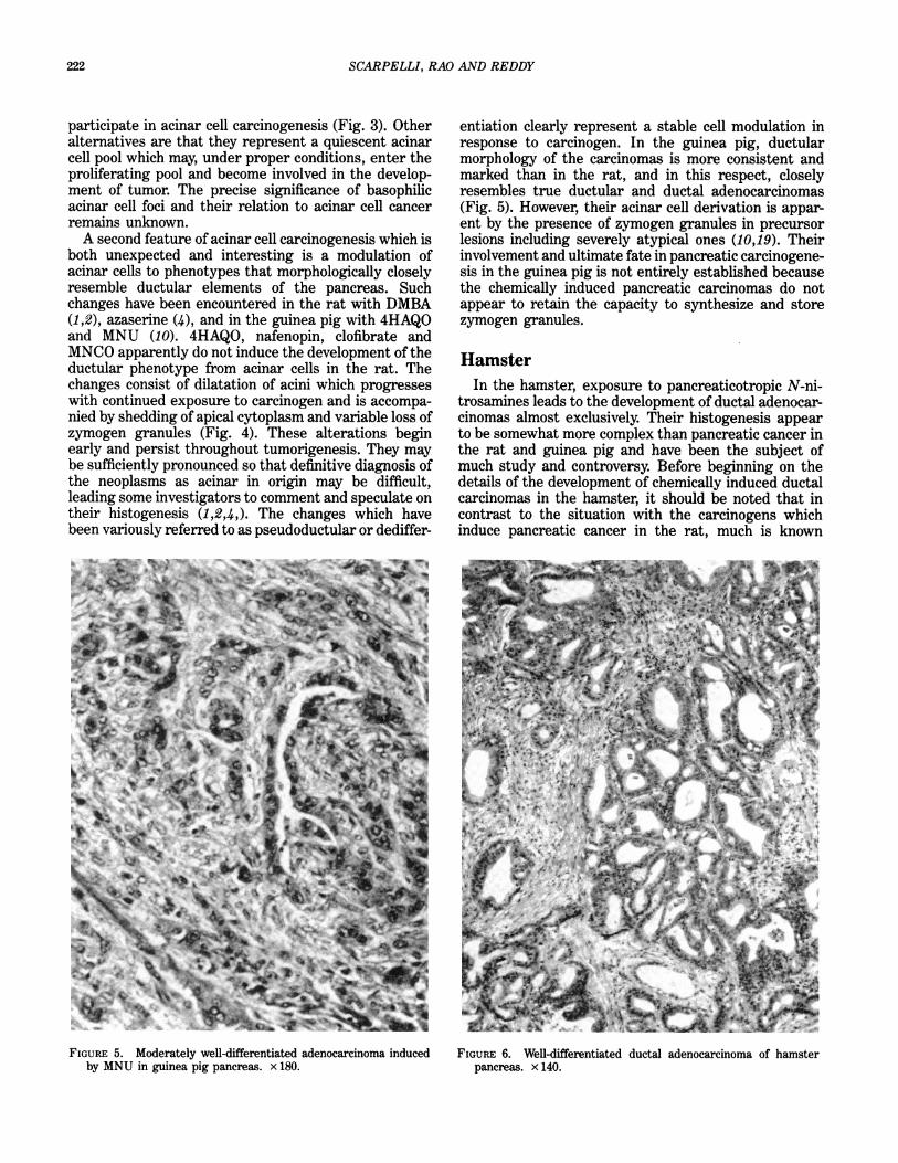
FIGURE 4. Pseudoductular change involving acinar cells in the pan-creas of a guinea pig treated with the carcinogen 4HAQO.Numerous nuclei labeled with [3H]-thymidine indicate increasedDNA synthesis in altered acinar cells. x 190.
221
SCARPELLI, RAO AND REDDY
participate in acinar cell carcinogenesis (Fig. 3). Otheralternatives are that they represent a quiescent acinarcell pool which may, under proper conditions, enter theproliferating pool and become involved in the develop-ment of tumor. The precise significance of basophilicacinar cell foci and their relation to acinar cell cancerremains unknown.A second feature of acinar cell carcinogenesis which is
both unexpected and interesting is a modulation ofacinar cells to phenotypes that morphologically closelyresemble ductular elements of the pancreas. Suchchanges have been encountered in the rat with DMBA(1,2), azaserine (4), and in the guinea pig with 4HAQOand MNU (10). 4HAQO, nafenopin, clofibrate andMNCO apparently do not induce the development of theductular phenotype from acinar cells in the rat. Thechanges consist of dilatation of acini which progresseswith continued exposure to carcinogen and is accompa-nied by shedding of apical cytoplasm and variable loss ofzymogen granules (Fig. 4). These alterations beginearly and persist throughout tumorigenesis. They maybe sufficiently pronounced so that definitive diagnosis ofthe neoplasms as acinar in origin may be difficult,leading some investigators to comment and speculate ontheir histogenesis (1,2,4,). The changes which havebeen variously referred to as pseudoductular or dediffer-
FIGURE 5. Moderately well-differentiated adenocarcinoma inducedby MNU in guinea pig pancreas. x 180.
entiation clearly represent a stable cell modulation inresponse to carcinogen. In the guinea pig, ductularmorphology of the carcinomas is more consistent andmarked than in the rat, and in this respect, closelyresembles true ductular and ductal adenocarcinomas(Fig. 5). However, their acinar cell derivation is appar-ent by the presence of zymogen granules in precursorlesions including severely atypical ones (10,19). Theirinvolvement and ultimate fate in pancreatic carcinogene-sis in the guinea pig is not entirely established becausethe chemically induced pancreatic carcinomas do notappear to retain the capacity to synthesize and storezymogen granules.
HamsterIn the hamster, exposure to pancreaticotropic N-ni-
trosamines leads to the development of ductal adenocar-cinomas almost exclusively. Their histogenesis appearto be somewhat more complex than pancreatic cancer inthe rat and guinea pig and have been the subject ofmuch study and controversy. Before beginning on thedetails of the development of chemically induced ductalcarcinomas in the hamster, it should be noted that incontrast to the situation with the carcinogens whichinduce pancreatic cancer in the rat, much is known
FIGURE 6. Well-differentiated ductal adenocarcinoma of hamsterpancreas. x 140.
222
PANCREATIC CARCINOGENESIS MODELS
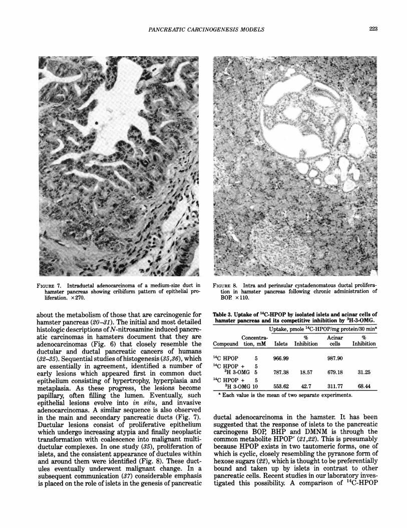
FIGURE 7. Intraductal adenocarcinoma of a medium-size duct inhamster pancreas showing cribiform pattern of epithelial pro-liferation. x 270.
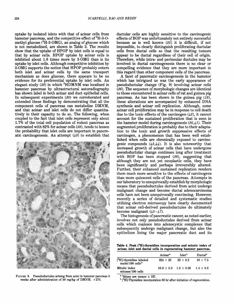
about the metabolism of those that are carcinogenic forhamster pancreas (20-31). The initial and most detailedhistologic descriptions ofN-nitrosamine induced pancre-atic carcinomas in hamsters document that they areadenocarcinomas (Fig. 6) that closely resemble theductular and ductal pancreatic cancers of humans(32-35). Sequential studies ofhistogenesis (35,36), whichare essentially in agreement, identified a number ofearly lesions which appeared first in common ductepithelium consisting of hypertrophy, hyperplasia andmetaplasia. As these progress, the lesions becomepapillary, often filling the lumen. Eventually, suchepithelial lesions evolve into in situ, and invasiveadenocarcinomas. A similar sequence is also observedin the main and secondary pancreatic ducts (Fig. 7).Ductular lesions consist of proliferative epitheliumwhich undergo increasing atypia and finally neoplastictransformation with coalescence into malignant multi-ductular complexes. In one study (35), proliferation ofislets, and the consistent appearance of ductules withinand around them were identified (Fig. 8). These duct-ules eventually underwent malignant change. In asubsequent communication (37) considerable emphasisis placed on the role of islets in the genesis of pancreatic
FIGURE 8. Intra and perinsular cystadenomatous ductal prolifera-tion in hamster pancreas following chronic administration ofBOR x 110.
Table 3. Uptake of 14C-HPOP by isolated islets and acinar cells ofhamster pancreas and its competitive inhibition by 3H-3-OMG.
Uptake, pmole "4C-HPOP/mg protein/30 minaConcentra- % Acinar %
Compound tion, mM Islets Inhibition cells Inhibition
'4C HPOP 5 966.99 987.9014C HPOP + 5
3H 3-OMG 5 787.38 18.57 679.18 31.2514C HPOP + 5
3H 3-OMG 10 553.62 42.7 311.77 68.44a Each value is the mean of two separate experiments.
ductal adenocarcinoma in the hamster. It has beensuggested that the response of islets to the pancreaticcarcinogens BOP, BHP and DMNM is through thecommon metabolite HPOP' (21,22). This is presumablybecause HPOP exists in two tautomeric forms, one ofwhich is cyclic, closely resembling the pyranose form ofhexose sugars (22), which is thought to be preferentiallybound and taken up by islets in contrast to otherpancreatic cells. Recent studies in our laboratory inves-tigated this possibility. A comparison of 14C-HPOP
223
SCARPELLI, RAO AND REDDY
uptake by isolated islets with that of acinar cells fromhamster pancreas, and the competitive effect of 3H-3-0-methyl glucose (3H-3-OMG), an analog of glucose whichis not metabolized, are shown in Table 3. The resultsshow that the uptake of HPOP by islet cells is equal tothat by acinar cells. HPOP uptake by acinar cells isinhibited about 1.6 times more by 3-OMG than is itsuptake by islet cells. Although competitive inhibition by3-OMG supports the notion that HPOP probably entersboth islet and acinar cells by the same transportmechanism as does glucose, there appears to be noevidence for its preferential uptake by islet cells. Anelegant study (38) in which 3HDMNM was localized inhamster pancreas by ultrastructural autoradiographyhas shown label in both acinar and duct epithelial cells.In subsequent experiments (30) we corroborated andextended these findings by demonstrating that all thecomponent cells of pancreas can metabolize DMNM,and that acinar and islet cells do not differ quantita-tively in their capacity to do so. The following, whencoupled to the fact that islet cells represent only about1.7% of the total cell population of rodent pancreas as
contrasted with 82% for acinar cells (39), tends to lessenthe probability that islet cells are important in pancre-atic carcinogenesis. An attempt (40) to establish that
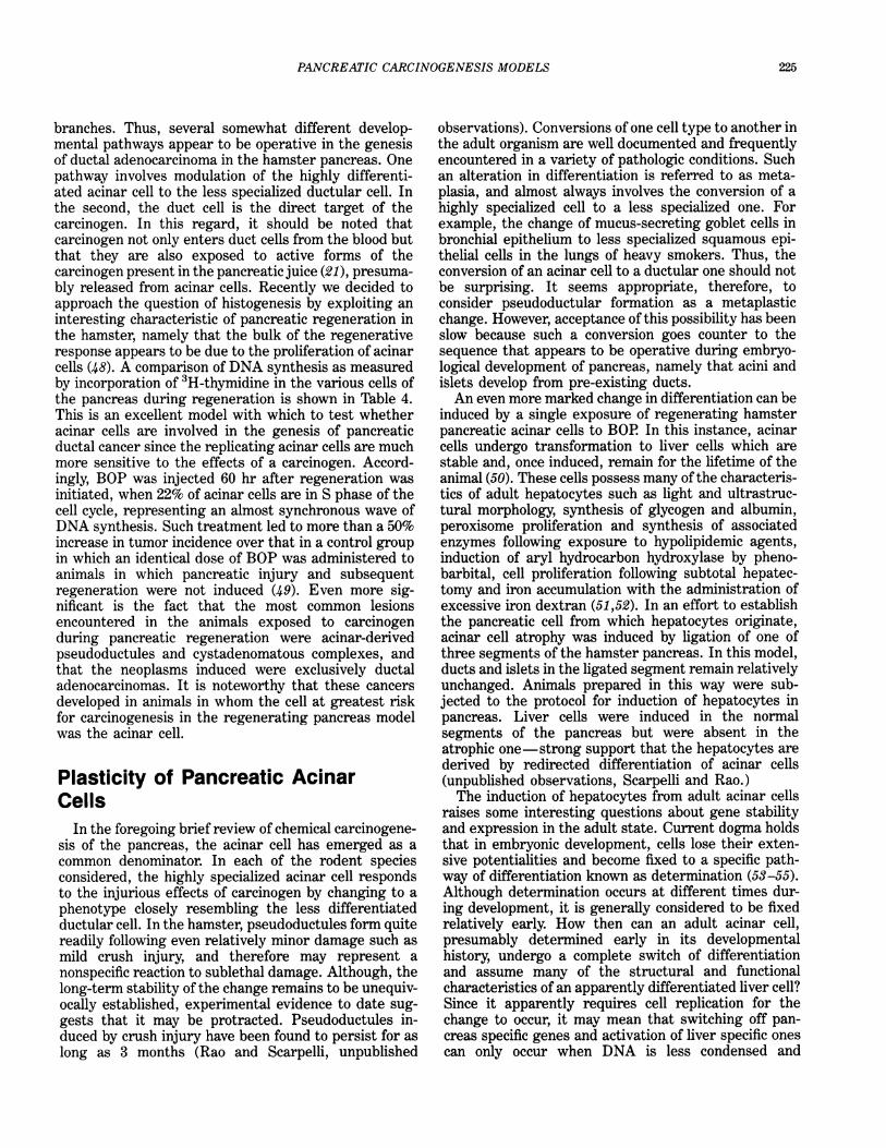
FIGURE 9. Pseudoductules arising from acini in hamster pancreas 3weeks after administration of 50 mg/kg of DMNM. x 270.
ductular cells are highly sensitive to the carcinogeniceffects ofBOP was unfortunately not entirely successfulbecause as is well known (35) it is difficult, if notimpossible, to clearly distinguish proliferating ductularcells from ductal cells so that the resulting tumorsappear to be ductal regardless of their cell of origin.Therefore, while intra- and perinsular ductules may beinvolved in ductal carcinogenesis there is no clear orcompelling evidence that they are more important inthis regard than other component cells of the pancreas.A facet of pancreatic carcinogenesis in the hamster
which has intrigued us was the early appearance ofpseudoductular change (Fig. 9) involving acinar cells(36). The sequence of morphologic changes are identicalto those encountered in acinar cells of rat and guinea pigpancreas. As has been shown in the guinea pig (19),these alterations are accompanied by enhanced DNAsynthesis and acinar cell replication. Although, someacinar cell proliferation may be in response to cell deathdue to the toxic effects of the carcinogen (42), it cannotaccount for the sustained proliferation that is seen inthe hamster model during carcinogenesis (36,41). Theiraugmented proliferation is probably due to their adapta-tion to the toxic and growth suppressive effects ofcarcinogen, a phenomenon that has been well estab-lished when cells are chronically exposed to carcino-genic compounds (43,44). It is also noteworthy thatincreased growth of acinar cells that have undergonepseudoductular change continues long after treatmentwith BOP has been stopped (36), suggesting thatalthough they are not yet neoplastic cells, they havebeen significantly and perhaps irreversibly altered.Further, their enhanced sustained replication rendersthem much more sensitive to the effects of carcinogensthan more quiescent cells of the pancreas. Attempts inour laboratory to unequivocally establish by morphologicmeans that pseudoductules derived from acini undergomalignant change and become ductal adenocarcinomacells have not been unequivocally convincing. However,recently a series of detailed and systematic studiesutilizing electron microscopy have clearly documentedthat acinar cell-derived pseudoductules do ultimatelybecome malignant (45-47).The histogenesis of pancreatic cancer, as noted earlier,
involves not only pseudoductules derived from acinarcells which coalesce into adenocystic complexes thatsubsequently undergo malignant change, but also theepithelium lining the major pancreatic duct and its
Table 4. Peak [3H]-thymidine incorporation and mitotic index ofacinar, islet and ductal cells in regenerating hamster pancreas.
Acinara Isleta Ductala[3H]-thymidine labeled 224 ± 25 23 ± 9.2 19 ± 7.5
nuclei/100 cellsbMitotic index 16.8 ± 5.0 1.8 ± 0.86 1.4 ± 0.6
mitoses/100 cellsa Values are means ± SE.b [3H]-Thymidine incorporation 60 hr after initiation of regeneration.
224
PANCREATIC CARCINOGENESIS MODELS
branches. Thus, several somewhat different develop-mental pathways appear to be operative in the genesisof ductal adenocarcinoma in the hamster pancreas. Onepathway involves modulation of the highly differenti-ated acinar cell to the less specialized ductular cell. Inthe second, the duct cell is the direct target of thecarcinogen. In this regard, it should be noted thatcarcinogen not only enters duct cells from the blood butthat they are also exposed to active forms of thecarcinogen present in the pancreatic juice (21), presuma-bly released from acinar cells. Recently we decided toapproach the question of histogenesis by exploiting aninteresting characteristic of pancreatic regeneration inthe hamster, namely that the bulk of the regenerativeresponse appears to be due to the proliferation of acinarcells (48). A comparison of DNA synthesis as measuredby incorporation of 3H-thymidine in the various cells ofthe pancreas during regeneration is shown in Table 4.This is an excellent model with which to test whetheracinar cells are involved in the genesis of pancreaticductal cancer since the replicating acinar cells are muchmore sensitive to the effects of a carcinogen. Accord-ingly, BOP was injected 60 hr after regeneration wasinitiated, when 22% of acinar cells are in S phase of thecell cycle, representing an almost synchronous wave ofDNA synthesis. Such treatment led to more than a 50%increase in tumor incidence over that in a control groupin which an identical dose of BOP was administered toanimals in which pancreatic injury and subsequentregeneration were not induced (49). Even more sig-nificant is the fact that the most common lesionsencountered in the animals exposed to carcinogenduring pancreatic regeneration were acinar-derivedpseudoductules and cystadenomatous complexes, andthat the neoplasms induced were exclusively ductaladenocarcinomas. It is noteworthy that these cancersdeveloped in animals in whom the cell at greatest riskfor carcinogenesis in the regenerating pancreas modelwas the acinar cell.
Plasticity of Pancreatic AcinarCells
In the foregoing brief review of chemical carcinogene-sis of the pancreas, the acinar cell has emerged as acommon denominator. In each of the rodent speciesconsidered, the highly specialized acinar cell respondsto the injurious effects of carcinogen by changing to aphenotype closely resembling the less differentiatedductular cell. In the hamster, pseudoductules form quitereadily following even relatively minor damage such asmild crush injury, and therefore may represent anonspecific reaction to sublethal damage. Although, thelong-term stability of the change remains to be unequiv-ocally established, experimental evidence to date sug-gests that it may be protracted. Pseudoductules in-duced by crush injury have been found to persist for aslong as 3 months (Rao and Scarpelli, unpublished
observations). Conversions of one cell type to another inthe adult organism are well documented and frequentlyencountered in a variety of pathologic conditions. Suchan alteration in differentiation is referred to as meta-plasia, and almost always involves the conversion of ahighly specialized cell to a less specialized one. Forexample, the change of mucus-secreting goblet cells inbronchial epithelium to less specialized squamous epi-thelial cells in the lungs of heavy smokers. Thus, theconversion of an acinar cell to a ductular one should notbe surprising. It seems appropriate, therefore, toconsider pseudoductular formation as a metaplasticchange. However, acceptance of this possibility has beenslow because such a conversion goes counter to thesequence that appears to be operative during embryo-logical development of pancreas, namely that acini andislets develop from pre-existing ducts.An even more marked change in differentiation can be
induced by a single exposure of regenerating hamsterpancreatic acinar cells to BOP In this instance, acinarcells undergo transformation to liver cells which arestable and, once induced, remain for the lifetime of theanimal (50). These cells possess many of the characteris-tics of adult hepatocytes such as light and ultrastruc-tural morphology, synthesis of glycogen and albumin,peroxisome proliferation and synthesis of associatedenzymes following exposure to hypolipidemic agents,induction of aryl hydrocarbon hydroxylase by pheno-barbital, cell proliferation following subtotal hepatec-tomy and iron accumulation with the administration ofexcessive iron dextran (51,52). In an effort to establishthe pancreatic cell from which hepatocytes originate,acinar cell atrophy was induced by ligation of one ofthree segments of the hamster pancreas. In this model,ducts and islets in the ligated segment remain relativelyunchanged. Animals prepared in this way were sub-jected to the protocol for induction of hepatocytes inpancreas. Liver cells were induced in the normalsegments of the pancreas but were absent in theatrophic one-strong support that the hepatocytes arederived by redirected differentiation of acinar cells(unpublished observations, Scarpelli and Rao.)The induction of hepatocytes from adult acinar cells
raises some interesting questions about gene stabilityand expression in the adult state. Current dogma holdsthat in embryonic development, cells lose their exten-sive potentialities and become fixed to a specific path-way of differentiation known as determination (53-55).Although determination occurs at different times dur-ing development, it is generally considered to be fixedrelatively early. How then can an adult acinar cell,presumably determined early in its developmentalhistory, undergo a complete switch of differentiationand assume many of the structural and functionalcharacteristics of an apparently differentiated liver cell?Since it apparently requires cell replication for thechange to occur, it may mean that switching off pan-creas specific genes and activation of liver specific onescan only occur when DNA is less condensed and
225
226 SCARPELLI, RAO AND REDDY
exposed as it is in S phase of the cell cycle. Is it possiblethat given the proper stimulus, any fully differentiatedcell can be induced to redirect its differentiation along anew pathway? Finally, is the surprising plasticity andease with which adult acinar cells in hamster and guineapig undergo pseudoductular formation related to theabsence of pancreatic acinar cell carcinomas in bothspecies? Since carcinogenesis results in a defect ofdifferentiation, it seems reasonable to study initiationand regulation of differentiation in acinar cells whichappear to be an important target cell in pancreaticcarcinogenesis.
This work was supported by the Marie A. Fleming and EdithPatterson Cancer Research Funds, USPHS Grants CA 34051, CA16954, and Contract NCI-E-72-3271. The authors gratefully acknowl-edge the excellent technical assistance of Matthew Conners andthank Nancy Schwank for preparing the manuscript.
REFERENCES
1. Dissin, J., Mills, L. R., Mains, D. L., Black, O., Jr., and Webster,P D., III. Experimental induction of pancreatic adenocarcinomain rats. J. Natl. Cancer Inst. 55: 857-864 (1975).
2. Bockman, D. E., Black, O., Jr., Mills, L. R., and Webster, P D.Origin of tubular complexes developing during induction ofpancreatic adenocarcinoma by 7,12-dimethylbenz(a)anthracene.Am. J. Pathol. 90: 645-651 (1978).
3. Hayashi, Y., and Hasegawa, T. Experimental pancreatic tumor inrats after intravenous injection of 4-hydroxyaminoquinoline1-oxide. Gann 62: 329 (1971).
4. Longnecker, D. S., and Curphey, T. J. Adenocarcinoma of thepancreas in azaserine-treated rats. Cancer Res. 35: 2249-2258(1975).
5. Reddy, J. K., and Rao, M. S. Malignant tumors in rats fednafenopin, a hepatic peroxisome proliferator. J. Natl. CancerInst. 59: 1645-1650 (1977).
6. Svoboda, D. J., and Azarnoff, D. L. Tumors in male rats fedethylchlorophenoxyisobutyrate, a hypolipidemic drug. CancerRes. 39: 3419-3428 (1979).
7. Reddy, J. K., and Qureshi, S. A. Tumorigenicity of thehypolipidaemic peroxisome proliferator ethyl-a-p-chlorophen-oxyisobutyrate (clofibrate) in rats. Brit. J. Cancer 40: 476-482(1979).
8. Longnecker, D. S., Curphey, T. J., Lilja, H. S., French, J. I., andDaniel, D. S. Carcinogenicity of a methylnitrosourea amino acidin rats. Fed. Proc. 37: 231 (1978).
9. Druckery, H., Ivankovic, S., Bucheler, J., Preussmann, R., andThomas, C. Erzeugung von Magen-und Pankrease-Krebs beimMeerschweinchen durch Methylnitroso-harnstoff und -urethan.Z. Krebsforsch. 71: 167-182 (1968).
10. Reddy, J. K., and Rao, M. S. Pancreatic adenocarcinoma in inbredguinea pigs induced by N-methyl-N-nitrosourea. Cancer Res. 35;2269-2276 (1975).
11. Pour, P, Kruger, F. W, Althoff, J., Cardesa, A., and Mohr, U.Cancer of the pancreas induced in the Syrian golden hamster.Am. J. Pathol. 76: 349-354 (1974).
12. Pour, P, Althoff, J., Gingell, R., Kupper, R., Kruger, F, andMohr, U. N-Nitroso-bis(2-acetoxypropyl)amine as a further pan-creatic carcinogen in Syrian golden hamsters. Cancer Res. 36:2877-2884 (1976).
13. Pour, R, Althoff, J., Kruger, F W, and Mohr, U. A potentpancreatic carcinogen in Syrian hamsters: N-nitroso-bis(2-oxo-propyl)amine. J. Natl. Cancer Inst. 58: 1449-1452 (1977).
14. Pour, R, Wallcave, L., Gingell, R., Nagel, D., Lawson, T.,Salmasi, S., and Tines, S. Carcinogenic effect of N-nitroso(2-hyd-roxypropyl) (2-oxopropyl)amine, a postulated proximate pancre-
atic carcinogen in Syrian hamsters. Cancer Res. 39: 3828-3833(1979).
15. Pour, P, Gingell, R., Langenbach, R., Nagel, D., Grandjean, C.,Lawson, T., and Salmasi, S. Carcinogenicity of N-nitrosome-thyl(2-oxopropyl)-amine in Syrian hamsters. Cancer Res. 40:3585-3590 (1980).
16. Mohr, U., Reznik, G., Emminger, E., and Lijinsky, W Inductionof pancreatic duct carcinomas in the Syrian hamster with2,6-dimethylnitrosomorpholine. J. Natl. Cancer Inst. 58: 429-432(1977).
17. Longnecker, D. S., Curphey, T. J., French, J. I., and Lilja, H. S.Response of the Syrian hamster to a nitrosourea amino acidcarcinogen. Cancer Letters 8: 163-168 (1979).
18. Rao, M. S., Upton, M. P, Subbarao, V, and Scarpelli, D. G. Twopopulations of cells with differing proliferative capacities inatypical acinar cell foci induced by 4-hydroxyaminoquinoline-1-oxide in the rat pancreas. Lab. Invest. 46: 527-534 (1982).
19. Rao, M. S., and Reddy, J. K. Histogenesis of pseudo-ductularchanges induced in the pancreas of guinea pigs treated withN-methyl-N-nitrosourea. Carcinogenesis 1: 1027-1037 (1980).
20. Gingell, R., Wallcave, L., Nagel, D., Kupper, R., and Pour, PCommon metabolites of N-nitroso-2,6-dimethylmorpholine andN-nitrosobis(2-oxo-propyl)amine in Syrian hamster. Cancer Let-ters 2: 47-52 (1976).
21. Gingell, R., and Pour, P Metabolism of the pancreatic carcinogenN-nitrosobis(2-oxopropyl)amine after oral and intraperitonealadministration to Syrian golden hamsters. J. Natl. Cancer Inst.60: 911-913 (1978).
22. Gingell R., Wallcave, L., Nagel, D., Kupper, R., and Pour, PMetabolism of the pancreatic carcinogens N-nitrosobis(2-oxo-propyl)amine and N-nitrosobis(2-hydroxypropyl)amine in Syrianhamster. J. Natl. Cancer Inst. 57: 1175-1178 (1976).
23. Gingell, R., Nagel, D., and Kupper, R. Differential metabolism ofgeometrical isomers of N-nitroso-2,6-dimethylmorpholine in thehamster. Xenobiotica 8: 439-443 (1978).
24. Gingell, R., Brunk, G., Nagel, D., and Pour, P Metabolism ofthree radiolabeled pancreatic carcinogenic nitrosamines in ham-sters and rats. Cancer Res. 39: 4579-4583 (1979).
25. Gingell, R., Brunk, G., Nagel, D., Wallcave, L., Walker, B., andPour, P Metabolism and mutagenicity of N-nitroso-2-methoxy-2,6-dimethylmorpholine in hamsters. J. Natl. Cancer Inst. 64:157-161 (1980).
26. Scarpelli, D. G., Rao, M. S., Subbarao, V, Beversluis, M., Gurka,D. P, and Hollenberg, P F Activation of nitrosamines tomutagens by postmitochondrial fraction of hamster pancreas.Cancer Res. 40: 67-74 (1980).
27. Whalley, C. E., Iqbal, Z. M., and Epstein, S. S. In vivo andmicrosomal metabolism of the pancreatic carcinogen N-nitro-sobis(2-oxopropyl)amine by the Syrian golden hamster. CancerRes. 41: 482-486 (1980).
28. Lawson, T. A., Hegelson, A. S., Grandjean, C. J., Wallcave, L.,and Nagel, D. The formation of N-nitrosomethyl(2-oxopropyl)a-mine from N-nitrosobis(2-oxopropyl)amine in vivo. Carcinogene-sis 2: 845-849 (1981).
29. Rao, M. S., Scarpelli, D. G., and Lijinsky, W Carcinogenesis inSyrian hamsters by N-nitroso-2,6-dimethylmorpholine, its cis andtrans isomers, and the effect of deuterium labeling. Carcinogene-sis 2: 731-735 (1981).
30. Scarpelli, D. G., Kokkinakis, D. M., Rao, M. S., Subbarao, V,Luettke, N., and Hollenberg, P F. Metabolism of the pancreaticcarcinogen N-nitroso-2,6-dimethylmorpholine by hamster liverand component cells of pancreas. Cancer Res. 42: 5089-5095(1982).
31. Underwood, B., and Lijinsky, W Comparative metabolism of2,6-dimethylnitrosomorpholine in rats, hamsters, and guineapigs. Cancer Res. 42: 54-58 (1982).
32. Pour, P, Kruger, F W, Althoff, J., Cardesa, A., and Mohr, U.Cancer of the pancreas induced in the Syrian golden hamster.Am. J. Pathol. 76: 349-358 (1974).
33. Pour, P, Mohr, U., Cardesa, A., Althoff, J., and Kruger, F WPancreatic neoplasms in an animal model: morphological, biologi-cal and comparative studies. Cancer 36: 379-389 (1975).
PANCREATIC CARCINOGENESIS MODELS 227
34. Pour, P, Althoff, J., Kruger, F W, and Mohr, U. A potentpancreatic carcinogen in Syrian hamsters N-nitrosobis(2-oxo-propyl)amine. J. Natl. Cancer Inst. 58: 1449-1453 (1977).
35. Pour, P, Althoff, J., and Takahasi, M. Early lesions of pancreaticductal carcinoma in the hamster model. Am. J. Pathol. 88:291-308 (1977).
36. Scarpelli, D. G., and Rao, M. S. Pathogenesis of pancreaticcarcinoma in hamsters induced by N-nitrosobis(2-oxopropyl)amine(BOP). Fed. Proc. 37: 232 (1978).
37. Pour, P Islet cells, a component of pancreatic ductal neoplasms.Am. J. Pathol. 90: 295-310 (1978).
38. Reznick-Schuller, H. M., Lijinsky, W, and Hogue, B. F, Jr.Electron microscopic autoradiography of the pancreas in thehamster treated with tritiated N-nitroso-2,6-dimethylmorpholine.Cancer Res. 40: 2245-2251 (1980).
39. Bolender, R. P Stereological analysis of the guinea pig pancreas1. Analytical model and quantitative description of nonstimulatedpancreatic exocrine cells. J. Cell Biol. 61: 269-287 (1974).
40. Takahasi, M., Pour, P, Althoff, J., and Donnelly, T. Sequentialalteration of the pancreas during carcinogenesis in Syrian ham-sters by N-nitrosobis(2-oxopropyl)amine. Cancer Res. 37: 4602-4607 (1977).
41. Levitt, M. H., Harris, C. C., Squire, R., Springer, S., Wenk, M.,Mollelo, C., Thomas, D., Kingsbury, E., and Newkirk, C. Experi-mental pancreatic carcinogenesis 1. Morphogenesis of pancreaticadenocarcinoma in the Syrian golden hamster induced byN-nitroso-bis(2-hydroxypropyl)amine. Am. J. Pathol. 88: 5-15(1977).
42. Farber, E., Parker, S., and Gruenstein, M. The resistance ofputative pre-malignant liver cell populations, hyperplastic nod-ules to the acute cytotoxic effects of some hepatocarcinogens.Cancer Res. 36: 3879-3887 (1976).
43. Laishes, B. A., Roberts, E., and Farber, E. In vitro measure-ment of carcinogen-resistant liver cells during hepatocarcino-genesis. Int. J. Cancer 21: 186-193 (1978).
44. Solt, D., Medline, A., and Farber, E. Rapid emergence ofcarcinogen-induced hyperplastic lesions in a new model for thesequential analysis of liver carcinogenesis. Am. J. Pathol. 88:595-610 (1977).
45. Flaks, B., Moore, M. A., and Flaks, A. Ultrastructural analysisof pancreatic carcinogenesis: morphological characterization ofN-nitrosobis(2-hydroxypropyl)amine-induced neoplasms in theSyrian hamster. Carcinogenesis 1: 423-437 (1980).
46. Flaks, B., Moore, M. A., and Flaks, A. Ultrastructural analysisof pancreatic carcinogenesis. III. Multifocal cystic lesions inducedby N-nitrosobis(2-hydroxypropyl)amine in the hamster exocrinepancreas. Carcinogenesis 1: 693-705 (1980).
47. Flaks, B., Moore, M. A., and Flaks, A. Ultrastructural analysisof pancreatic carcinogenesis. IV Pseudoductular transformationof acini in the hamster pancreas during N-nitrosobis(2-hydroxy-propyl)amine carcinogenesis. Carcinogenesis 2: 1241-1253 (1980).
48. Scarpelli, D. G., Rao, M. S., Subbarao, V, and Beversluis, M.Regeneration of Syrian golden hamster pancreas and covalentbinding of N-nitroso-2,6-3H-dimethylmorpholine. Cancer Res. 41:1051-1057 (1981).
49. Scarpelli, D. G., Rao, M. S., and Subbarao, V Augmentation ofcarcinogenesis by N-nitrosobis(2-oxopropyl)amine administeredduring S phase of the cell cycle in regenerating hamster pancreas.Cancer Res. 43: 611-616 (1983).
50. Scarpelli, D. G., and Rao, M. S. Differentiation of regeneratingpancreatic cells into hepatocyte-like cells. Proc. Natl. Acad. Sci.(U.S.) 78: 2577-2581 (1981).
51. Rao, M. S., Reddy, M. K., Reddy, J. K., and Scarpelli, D. G.Response of chemically-induced hepatocyte-like cells in hamsterpancreas to methyl clofenapate, a peroxisome proliferator. J. CellBiol. 95: 50-56 (1982).
52. Rao, M. S., Subbarao, V, Luetteke, N., and Scarpelli, D. G.Further characterization of carcinogen-induced hepatocyte-likecells in hamster pancreas. Am. J. Pathol. 110: 89-94 (1983).
53. Lewis, W H. Transplantation of the lips of the blastopore inRana palustris. Am. J. Anat. 7: 137-143 (1907).
54. Spemann, H. Uber die Determination der ersten Organanlagendes Amphibienembryo. I-IV Arch. Entwicklungsmech. Organ-ismen 43: 448-555 (1918).
55. Grobstein, C. Differentiation of vertebrate cells. In: The Cell:Biochemistry, Physiology, Morphology (J. Brachet and A. E.Mirsky, Eds.), Vol. 1, Academic Press, New York, 1959, pp.437-496.