Embed Size (px)
Citation preview
ACTAUNIVERSITATISUPSALIENSISUPPSALA2006
Digital Comprehensive Summaries of Uppsala Dissertationsfrom the Faculty of Medicine 198
Studies of the ElementalComposition of Airway SurfaceLiquid with Relevance to CysticFibrosis
VIENGPHET VANTHANOUVONG
ISSN 1651-6206urn:nbn:se:uu:diva-7245

To my loving family
Try to do the best you can. Life is too short
and full of surprises,
anddo not try to be someone who you are not

List of Papers
This thesis is based on the following papers, reproduced with the permission of the publishers, to which reference will be made by their Roman numerals:
I Kozlova I, Vanthanouvong V, Almgren B, Högman M, Roomans GM (2004) Elemental composition of airway surface liquid in the pig determined by X-ray microanalysis. Am J Resp Cell Mol Biol 32: 59-64.
II Vanthanouvong V, Kozlova I, Roomans GM (2005) Ion composition of rat airway surface liquid by X-ray microanalysis. Microsc Res Techn 68: 6-12.
III Nilsson H, Kozlova I, Vanthanouvong V, Roomans GM (2004) Collection and X-ray microanalysis of airway surface liquid in the mouse using ion exchange beads. Micron 35: 701-705.
IV Vanthanouvong V, Roomans GM (2004) Methods for determining the composition of nasal fluid by X-ray microanalysis. Microsc Res Techn 63: 122-128.
V Vanthanouvong V, Kozlova I, Johannesson M, Nääs E, Nordvall SL, Dragomir A, Roomans GM (2006) Composition of nasal airway surface liquid in cystic fibrosis and other airway diseases. Microsc Res Techn 69: 271-276.

Table of Contents
Preface..........................................................................................................11
1. Introduction.............................................................................................131.1. Cystic fibrosis .......................................................................... 13 1.2. Symptoms of CF...................................................................... 17 1.3. Diagnosis ................................................................................. 19 1.4. Prognosis ................................................................................. 20 1.5. Epithelial ion transport in cystic fibrosis ................................. 21 1.6. The respiratory system............................................................. 22
1.6.1. The airway epithelium ...................................................... 23 1.6.2. Ion and water transport in the respiratory epithelium....... 24
1.7. The airway surface liquid (ASL) ............................................. 25 1.7.1. The low volume/hydration hypothesis ............................. 27 1.7.2. The high salt concentration hypothesis ............................ 28 1.7.3. The low pH hypothesis ..................................................... 29 1.7.4. The low oxygenation hypotheses ..................................... 29 1.7.5. The defective gland hypothesis ........................................ 30
1.8. Animal models for cystic fibrosis............................................ 31
2. Aims..........................................................................................................33
3. Materials and Methods...........................................................................353.1. Experimental animals (papers I, II & III) ................................ 35
3.1.1. Pigs (paper I) .................................................................... 35 3.1.2. Rats (paper II) ................................................................... 36 3.1.3. Mice (paper III) ................................................................ 36
3.2. Frozen hydrated tissue samples (papers I and II) .................... 36 3.2.1. Pigs (paper I) .................................................................... 36 3.2.2. Rats (paper II) ................................................................... 36
3.3. Collection of ASL with Sephadex beads ................................. 37 3.3.1. Pigs (paper I) .................................................................... 37 3.3.2. Rats (paper II) ................................................................... 37 3.3.3. Mice (paper III) ................................................................ 37
3.4. Pharmacological stimulation of fluid secretion (paper II)....... 38 3.5. Human subjects (papers IV and V).......................................... 38
3.5.1. Healthy volunteers (paper IV) .......................................... 38
3.5.2. Patients with airway diseases and CF-heterozygotes (paper V) ................................................................................................ 38 3.5.3. Collection of nasal fluid (paper IV).................................. 39
3.6. X-ray microanalysis................................................................. 41 3.6.1. Frozen-hydrated tissue samples (papers I and II) ............. 41 3.6.2. Frozen droplets (paper IV) ............................................... 42 3.6.3. Analysis of Sephadex beads (papers I-V)......................... 42 3.6.4. Dried droplets on carbon planchets or filter paper (paper IV)............................................................................................... 42
3.7. Storage of ion exchange beads in silicon oil (paper II) ........... 42 3.8. Preparation for light and electron microscopy (papers I and II)........................................................................................................ 43 3.9. Light and electron microscopy (papers I and II) ..................... 43 3.10. Statistical analysis (papers I-V) ............................................. 43
4. Results ......................................................................................................444.1. Elemental composition of the ASL in experimental animals .. 44 4.2. Analysis of the composition of human nasal fluid .................. 45
5. Discussion ................................................................................................485.1. Elemental composition of the ASL in experimental animals .. 48 5.2. Analysis of the composition of human nasal fluid .................. 53
6. Conclusions ..............................................................................................56
Acknowledgements .....................................................................................58
References ....................................................................................................60
Abbreviations
ABC ATP binding cassette
ANOVA analysis of variance
ASL airway surface liquid
ATP adenosine triphosphate
CaCC calcium-activated chloride channel
cAMP 3’, 5’ cyclic adenosine
ClC chloride channel
CF cystic fibrosis
CFTR cystic fibrosis transmembrane conductance regulator
ENaC epithelial sodium channel
ER endoplasmic reticulum
MCC mucociliary clearance
PCL periciliary layer
SEM scanning electron microscope
TEM transmission electron microscope
XRMA X-ray microanalysis
F508 Deletion of phenylalanine at position 508 in CFTR


11
Preface
“Woe to that child which when kissed on the forehead tastes salty. He is bewitched and soon must die”. This adage is an early reference from northern European folklore to the genetic disease which presently is called cystic fibrosis (CF). As the saying implies, the disorder once normally killed children in infancy and is often identifiable by excessive salt in sweat.
In 1936, the first description of CF as a distinct disease was published by a Swiss physician, professor Fanconi (Fanconi et al.1936).
In 1938, the first pathological description was published by Dr Dorothy Andersen of Columbia University in New York (Andersen 1938). Andersen provided the first comprehensive description of symptoms of CF and of the changes that the disease produced in organs. She noted, that these almost always included destruction of the pancreas (even in infants) and, often, infection of and damage to the airways. Andersen gave the disease its name, calling it "cystic fibrosis of the pancreas," on the basis of microscopic features she observed in pancreatic tissue. Patients had cysts (fluid-filled sacs) and scar tissue (fibrosis) which replaced the majority of normal tissue in the pancreas.
In 1943, Farber described obstruction of the airways by abnormal mucus (thickened secretions) as the principal cause of the progressive lung disease (Farber 1943) and coined the name “mucoviscidosis”.
In 1946, studies had revealed information about the genetics of CF. It was concluded that cystic fibrosis was a recessive disease, probably caused by mutation of a single gene. If an infant inherited a damaged copy of the gene from both parents and therefore made no normal molecules of the protein specified by the gene, the child became ill. However, presence of one good copy and one damaged copy did not lead to disease.
In 1953, DiSant’ Agnese discovered the excess of sodium and chloride in the sweat, and realized its significance. This became the basic diagnostic test for CF (DiSant’ Agnese et al. 1953).
12
In 1959, the standardized sweat test by pilocarpine iontophoresis was developed by Gibson and Cooke (Gibson and Cooke 1959).
In 1989, the gene responsible for causing CF which coded for a protein called the cystic fibrosis transmembrane conductance regulator (CFTR), was discovered by Dr Jack Riordan and colleagues (Riordan et al. 1989).
CF is a genetic disease that particularly affects the Caucasian population. CF occurs rarely in African or in Asian populations. CF is also called “65 roses” The “65 roses” name came from a boy who overheard his mother talking about the disease on the phone and the 65 roses has become a nickname for CF for children who have difficulties to pronounce the scientific name. In Sweden, there are about 600 CF patients who are members of the Swedish CF Association (www.rfcf.se). About 20 people (mostly children) per year get diagnosed as having CF (the frequency of the disease is about 1:4,000-1:5,000). Most patients get their diagnosis before the age of two years. The life expectancy of CF patients in Sweden is about 40-45 years (Kollberg 1999).
13
1. Introduction
1.1. Cystic fibrosis
Cystic fibrosis (CF) is a fatal genetic disease affecting mainly the Caucasian population. CF is characterized by abnormal ion and water transport across epithelial cells. The disease affects the epithelium of many organs, such as the sweat glands, airways, pancreas, intestine, liver, salivary glands, as well as the reproductive epithelium. The most severe symptoms occur in the respiratory tract. The respiratory epithelium is important for the protection of the human body from harmful bacteria and particles from the environment. The transepithelial ion transport is defective in CF, because cAMP-stimulated chloride (Cl-) secretion is abolished due to the loss of a Cl-channel in the apical membrane, the cystic fibrosis transmembrane conductance regulator (CFTR). In normal airways, CFTR is an anion channel that controls the volume of airway surface liquid (ASL) in two ways: (1) by attenuation of ASL absorption through inhibition of the epithelial sodium channel (ENaC) in the apical membrane (Stutts et al. 1995) and (2) by forming a conduction pathway for Cl- and HCO3
- ions in the apical membrane to support liquid secretion across the glandular and surface epithelia of the airways (Ballard et al. 1999)
The altered epithelial transport of salt and water in CF results in the production of viscous mucus, impaired mucociliary transport, chronic airway infections, and bronchiectasis. The bacterial infections and the inflammation in the lungs result in a progressive loss of lung function. The chronic bacterial infection, predominately with Pseudomonas aeruginosa and Staphylococcus aureus (Gilligan 1991) is the major cause of morbidity and mortality in CF patients. There might be a direct correlation between CFTR dysfunction and the development of airway inflammation and infection. Progress in the treatment of CF has come from marked improvement of antibiotics therapy, physical chest therapy to move mucus out of the lungs,
14
pancreatic enzyme replacement, and nutritional supplementation. However, despite improved treatment of CF, there is still no cure for the disease.
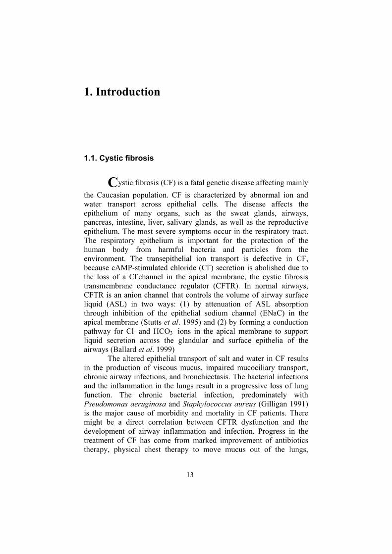
CF occurs in 1:2,500 European newborn babies (although the frequency varies between countries) and in 1:14,000 African-Americans (http://gslc.genetics.utah.edu). CF is not common in Asiabut it has been found that CF patients of southern Asian origin have a more severe clinical course compared to European CF patients (Mei-Zahav et al. 2005). The frequency of having CF or of carrying the mutation is independent of sex. Nearly 5 percent of the Europeans is born with one copy of the gene mutation that causes CF. Since CF is an autosomal recessive disease, the offspring of two carriers has a 25 percent chance of having CF. Carriers have no clinical symptoms of the disease.
CF Cr Cr Hth
Genetics of CF
Apart from being responsible for Cl- (and HCO3-) transport
across the apical membrane of airway epithelial cells, CFTR also regulates an epithelial Na+ channel (ENaC) and other ion channels (Wine et al. 1994). It is still not entirely clear how the defect in the epithelial ion transport is related to the symptoms of CF. CFTR is present in the apical membrane of most epithelial cells, such as the respiratory epithelium, the intestinal epithelium, the pancreas, and the sweat gland (Riordan et al. 1989; Harris et al. 1991; Crawford et al.1991). The CFTR gene is located on the long arm of chromosome 7 (7q31) and more than 1,500 mutations causing CF have been identified (www.genet.sickkids.on.ca/cftr). The mutations in CFTR
+
CF carrier parents Figure 1. A child born to two carrier parents has a 25% chance of having CF, a 25% chance of being healthy, and a 50% chance of being a CF carrier. Modified from www.leeds.ac.uk/lass/cf.htm
CF: Cystic fibrosis Cr: Carrier Hth: Healthy
15
can be subdivided into six different classes (Vankeerberghen et al.2002)
Class I: Defective protein production: mutations that do not produce CFTR protein due to a stop mutation in the code for CFTR. Class II: Defective protein processing: mutations in which CFTR fails to reach the apical membrane due to defective processing. In the most common mutation,
F508, CFTR is not folded correctly and is trapped in the ER and then destroyed by the ubiquitin/proteasome pathway (Ward et al. 1995). Class III: Defective regulation: mutations that produce a protein that reaches the plasma membrane but does not respond to cAMP stimulation. Class IV: Defective conductance: mutations that produce a cAMP-responsive channel with reduced conductance; these mutations often occur in the plasma membrane spanning domains. Class V: Decreased abundance: mutations which have a normal CFTR but reduced abundance due to incorrect splicing.Class VI: Defective regulation of other proteins: mutations that produce defective regulation of other channels, which affects CFTR.
Class I and II mutations are often connected with a severe form of the disease, whereas Class IV and V mutations are often connected with a milder form of the disease.
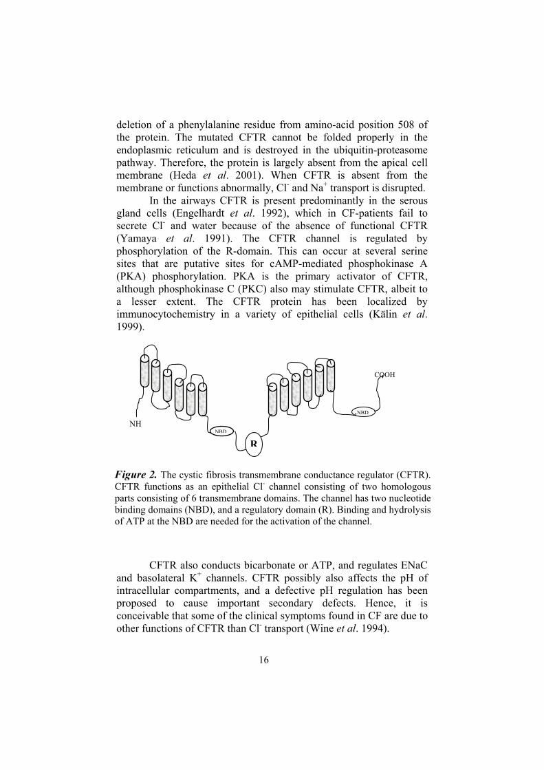
The CFTR gene was cloned in 1989 (Riordan et al. 1989) and this has opened the way to use gene targeting in mouse embryonic stem cells to create transgenic CF mice. The CFTR gene has 27 exons, which encode a 1,480 amino-acid protein. The gene consists of two putative membrane spanning domains, each consisting of six -helices, two nucleotide-binding domains (NBD) and one regulatory domain R (Riordan et al 1989). CFTR is a member of the ATP-binding cassette (ABC) membrane transporter superfamily, but its R-domain is unique.
The most common mutation, F508, is present in 70% of CF chromosomes and 90% of the CF patients have this mutation in at least one allele (Kerem et al. 1989). The mutation is due to the
16
deletion of a phenylalanine residue from amino-acid position 508 of the protein. The mutated CFTR cannot be folded properly in the endoplasmic reticulum and is destroyed in the ubiquitin-proteasome pathway. Therefore, the protein is largely absent from the apical cell membrane (Heda et al. 2001). When CFTR is absent from the membrane or functions abnormally, Cl- and Na+ transport is disrupted.
In the airways CFTR is present predominantly in the serous gland cells (Engelhardt et al. 1992), which in CF-patients fail to secrete Cl- and water because of the absence of functional CFTR (Yamaya et al. 1991). The CFTR channel is regulated by phosphorylation of the R-domain. This can occur at several serine sites that are putative sites for cAMP-mediated phosphokinase A (PKA) phosphorylation. PKA is the primary activator of CFTR, although phosphokinase C (PKC) also may stimulate CFTR, albeit to a lesser extent. The CFTR protein has been localized by immunocytochemistry in a variety of epithelial cells (Kälin et al.1999).
CFTR also conducts bicarbonate or ATP, and regulates ENaC and basolateral K+ channels. CFTR possibly also affects the pH of intracellular compartments, and a defective pH regulation has been proposed to cause important secondary defects. Hence, it is conceivable that some of the clinical symptoms found in CF are due to other functions of CFTR than Cl- transport (Wine et al. 1994).
Figure 2. The cystic fibrosis transmembrane conductance regulator (CFTR). CFTR functions as an epithelial Cl- channel consisting of two homologous parts consisting of 6 transmembrane domains. The channel has two nucleotide binding domains (NBD), and a regulatory domain (R). Binding and hydrolysis of ATP at the NBD are needed for the activation of the channel.
RNBD
NHNBD
COOH
17
CFTR transports Cl- ions in the direction of the electrochemical gradient. Cl- transport can therefore be both efflux and influx, depending on the actual electrochemical gradient. An epithelium functioning in the absorptive mode, e.g., in the sweat duct, will normally absorb both Cl- via CFTR channels and Na+ via ENaC, both of which are present in the apical membrane of the duct epithelial cells.
The situation in the airways is complicated. Over the tracheal epithelium a net influx of water takes place. This is necessary, because the fluid-covered surface of the alveoli combined is about 105 times larger than the cross-sectional surface of the trachea (Widdicombe, 2002), so the ASL has to be absorbed to avoid that the human (or animal) drowns in his own ASL. The process underlying the absorption of water is Na+ influx mediated by ENaC. Net Cl-
movement across the cells of the surface epithelium is secretory, passing through CFTR in the apical membrane. After metacholine stimulation of submucosal airway glands, the depth of the ASL layer was increased but this increase was inhibited by bumetanide, an inhibitor of active Cl- secretion. The slow return of the thickness of the ASL layer to baseline levels was inhibited by amiloride, an inhibitor of the ENaC channel (Wu et al. 1998).
1.2. Symptoms of CF
CF infants are born with relatively normal lungs (Tomashefski et al. 1993), and it can take months to years for chronic infections to become a feature of the CF airways, but breathing problems can develop at any time after birth. Inflammation is present in airways of a patient with CF already in the first months of life even before infection occurs (Khan et al. 1995). Not much is known about how these early changes in the airways affect lung function. Thick bronchial secretions eventually block the small airways with inspissated mucus, which then becomes infected mainly with mucoid forms of Pseudomonas aeruginosa, Staphylococcus aureus and Hemophilus influenzae. The mucus plugging in the lungs that often makes breathing difficult, is due to abnormal electrolyte transport, in particular of Cl- and Na+ ions. The destruction of lung tissue by the inflammatory reaction then leads to severe respiratory disease and eventually to death from a combination of respiratory failure and a failing heart caused by the underlying lung disease.
18
Meconium ileus, a form of intestinal obstruction in newborns that causes belly ache, occurs in 17 percent of those with CF (Pencharz 2003). Babies that have meconium ileus almost always develop other symptoms of CF later. The earliest symptom of CF in a newborn child that does not have meconium ileus is often poor weight gain at 4 to 6 weeks. This is due to lack of pancreatic enzymes essential for proper digestion of fat and to lack of vitamins (A, D, E, and K). About 2 to 3 percent of the patients with CF develop insulin-dependent diabetes because the scarred pancreas can no longer produce enough insulin. Both male and female CF patients often have an impaired reproductive function. Males produce no spermatozoa or have a low sperm count because the vas deferens has developed abnormally. However, male CF patients can become fathers with techniques such as microscopic epididymal sperm aspiration and intracytoplasmic sperm injection. In women, cervical secretions are abnormally thick, causing decreased fertility (Kopito 1973). The life expectancy of CF patients has increased because CF patients get better health care and support. Many CF women have reached child-bearing age and have successfully become mothers. Particular care must be taken in medical management of pregnancy. The best care is provided by a specialist CF unit (Geddes 1992; Johannesson 2002). With reasonable health women with CF who wish to have a family can be encouraged to have children and the outcome in general is good. If a woman with CF plans to have a child, some important considerations are timing, family support and the genetic background of the father. It can happen that these women get medical problems and need to be hospitalized during pregnancy and/or after giving birth. In brief, a summary of the symptoms of CF (www.ccff.ca) is: difficulty of breathing constant cough which expels thick mucus excessive appetite, with weight loss bowel disturbances skin which tastes salty repeated or prolonged bouts of pneumonia failure to thrive.
Diseases that have some symptoms in common with CF, but are not CF, are (www3.nbnet.nb.ca): Shwachman-Diamond Syndrome celiac disease (celiac sprue)
19
bronchiectasis primary ciliary dyskinesia congenital or acquired immunodeficiency alpha-1 antitrypsin deficiency intestinal lymphangiectasia (idiopathic hypoproteinemia) laryngeal cleft asthma chronic bronchitis
1.3. Diagnosis
About 70 percent of CF patients are diagnosed before the age of one (Riordan et al. 1989), and 90 percent are diagnosed before the age of eight. However, the diagnosis of some patients with milder clinical symptoms still continues to be made throughout life. In those patients, pulmonary function tests may show that respiration is compromised, or a chest X-ray may suggest the diagnosis. If also the digestive system is affected, pancreatic enzyme levels are reduced; the digestive enzymes trypsin and chymotrypsin are decreased or absent in stool (but elevated in blood), or high levels of fat in the stool are observed, the diagnosis of CF is strengthened. Intestinal obstruction may also occur. If insulin secretion is reduced, blood sugar levels are increased and patients can develop diabetes. If the patients are diagnosed at a young age, they tend to live longer because of the early care and treatment. Cystic fibrosis at an early stage can have symptoms in common with other airway diseases such as asthma and chronic bronchitis, which leads to misdiagnosis or delays the correct treatment. The basic diagnostic test for CF is the sweat test and in some countries, for example in France and the United Kingdom, a test for CF is conducted at an early stage. The diagnostic test includes (www.ccne-ethique.fr; www.cftrust.org.uk):
1. Screening test of newborn children; the test is a heel-prick to sample blood as part of the normal Guthrie test carried out on all children. To screen for CF, the levels of pancreatic trypsin in the blood, which are abnormally high in CF (Wilcken et al.1983; Wilcken and Wile 2003) are tested. The British National Screening Committee recommended that all babies should be
20
screened for CF. It is planned that this screening programme will be in place across the whole of the UK by April 2007.
2. Sweat test, if a baby has an abnormal screening test, the sweat test is conducted to measure the concentration of salt in the sweat. Children with CF have more salt in their sweat than normal (>60 mM). If a baby is diagnosed with CF, the other children in the family also should have a sweat/genetic test.
3. Antenatal testing, this test is carried out early in pregnancy; it is a genetic test to see if the mother-to-be carries the F508-CFTR mutation; if this is the case, the father-to-be is also tested (Cuckle et al. 1996).
4. Carrier testing, which is carried out by a simple mouthwash test, which yields cells from the buccal epithelium (as a DNA resource for mutation detection, because collection and DNA isolation is simple and cheap). A similar test is carried out by gently rubbing the inside of the cheek with a brush. The test is carried out to find the F508-CFTR mutation (de Vries et al.1996). This is important if a relative has CF or is a known carrier. It is especially important to have the test if one of the partners is a known carrier.
In Sweden, at present no antenatal or neonatal screening is routinely carried out. If suspicion arises that a child has CF (e.g., because of meconium ileus or failure to thrive) a sweat test and/or genetic testing for the most common mutations in CFTR (including
F508) are carried out. Because of the many different mutations in CFTR that can
give rise to CF, the severity of the symptoms may vary considerably between patients, and a person can never look too well to have CF.
1.4. Prognosis
As stated above, the severity of CF varies greatly from patient to patient, regardless of age. The clinical condition of the patient is determined largely by how much the lungs are affected. However, deterioration is inevitable, leading to disability and eventually death. Nonetheless, the outlook has improved steadily over the past 25 years, mainly because of treatment strategies, especially treatment of lung infection. Treatment can now postpone some of the changes that occur in the lungs. In the United States, half of the patients with cystic

21
fibrosis live until 32 years (Cystic Fibrosis Foundation 2000), whereas in Sweden, the average life expectancy is estimated at over 40 years (Kollberg 1999). Long-term survival is somewhat better in males, in patients that do not have pancreatic problems, and in patients in which the initial symptoms are restricted to the digestive system. Despite their many problems, patients with CF usually attend school or work until shortly before death. Since the early 1990s, more than 1000 CF patients have undergone transplantation of the lungs or other organs. Survival of patients after lung transplantation is poorer than after other types of organ transplantation with a survival of about 70% at 1 year and 45% at 4 years. Because of this limitation, patients with CF are eligible for organ transplant if they have a life expectancy of about 2 years or less, and a poor quality of life (Aurora et al.1999). In addition to the development of better methods to treat the symptoms of CF, efforts are directed at developing methods to study the role of ion channels in the pathology of the disease, which may lead to a pharmacological therapy for CF (Kerem 2005; Roomans 2001, 2003). In addition, the possibility to use gene therapy to cure the disease is being investigated (Anson et al. 2006; Griesenbach et al.2006).
1.5. Epithelial ion transport in cystic fibrosis
Ion transport across the cell membranes of epithelial cells is carried out by a number of ion transporters and ion channels. In the apical membrane, chloride transport is mainly mediated by the cAMP dependent CFTR chloride channel, which can move Cl- into or out of cells. There are other chloride channels in the apical membrane, such as Ca2+-activated chloride channels (CaCC), and volume-regulated chloride channels. In the apical membrane, an epithelial Na channel (ENaC) is present to transport Na+ ions into the cells. This Na+
channel may be inhibited by amiloride and nitric oxide (NO) (Eisenhut 2006). Across the basolateral membrane, Na+ ions are actively transported out of the cells in exchange for K+ ions by the Na+-K+-ATPase, which is a membrane protein that pumps 3 Na+ ions out of the cell in exchange for 2 K+ ions in a reaction that hydrolyses ATP. This is critical for the maintenance of low intracellular Na+
concentrations and the resting membrane potential. In the basolateral
22
membrane there are also K+ channels, and a Na+-K+-2Cl- co-transport mechanism. That CF was due to an abnormality in chloride transport was first demonstrated by Quinton (1983) in the sweat gland. In the normal sweat gland, the secretory coil produces an iso-osmotic fluid, and reabsorption of Na+ and Cl- takes place in the duct. From the lumen of the gland, Na+ ions enter into the duct cells passively through the apical membrane via ENaC and the Na+ ions leave the cell through the basolateral membrane via the Na+-K+-ATPase. Chloride enters into cells from the lumen passively via two Cl- channels, CFTR, and Ca2+-activated chloride channels. Dysfunction of CFTR and ENaC channels leads to elevated concentrations of NaCl in the final sweat. Defective CFTR has also been demonstrated in the respiratory epithelium (Knowles et al. 1981, 1983) and in the small intestine (Berschneider etal. 1988, Taylor et al. 1988, Baxter et al. 1989).
It should be remembered, that CF airways display chronic inflammation, which is associated with widening of the lateral intercellular spaces (LIS) and may result in collapse of the tight junctions, which could be partly due to the action of proinflammatory cytokines (Coyne et al. 2002). This would drive fluid from the interstitium into the lumen (Widdicombe and Widdicombe 1995). Hence, fluid transport across the CF respiratory epithelium may be more complex than originally thought.
1.6. The respiratory system
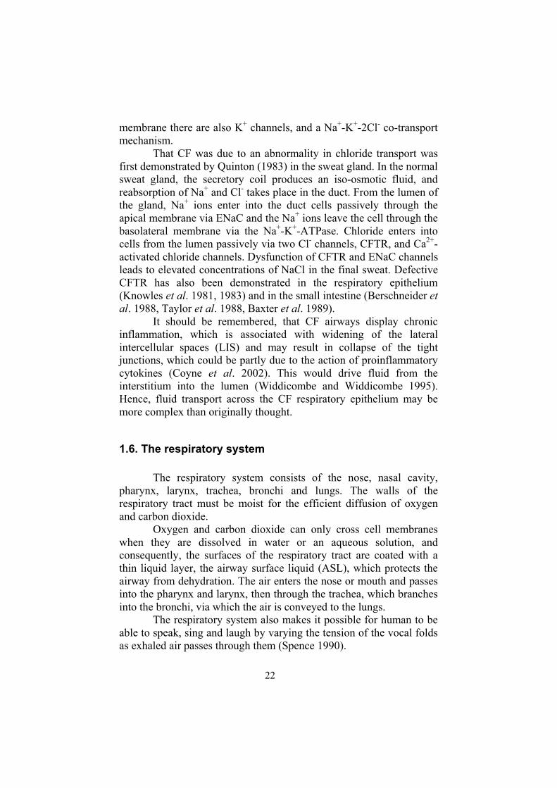
The respiratory system consists of the nose, nasal cavity, pharynx, larynx, trachea, bronchi and lungs. The walls of the respiratory tract must be moist for the efficient diffusion of oxygen and carbon dioxide.
Oxygen and carbon dioxide can only cross cell membranes when they are dissolved in water or an aqueous solution, and consequently, the surfaces of the respiratory tract are coated with a thin liquid layer, the airway surface liquid (ASL), which protects the airway from dehydration. The air enters the nose or mouth and passes into the pharynx and larynx, then through the trachea, which branches into the bronchi, via which the air is conveyed to the lungs. The respiratory system also makes it possible for human to be able to speak, sing and laugh by varying the tension of the vocal folds as exhaled air passes through them (Spence 1990).
23
Figure 3. Respiratory system from the nose to the lungs (left) and the structure of the tracheal wall (right) that shows the airway epithelium; on its top is the thin liquid layer (ASL).
1.6.1. The airway epithelium
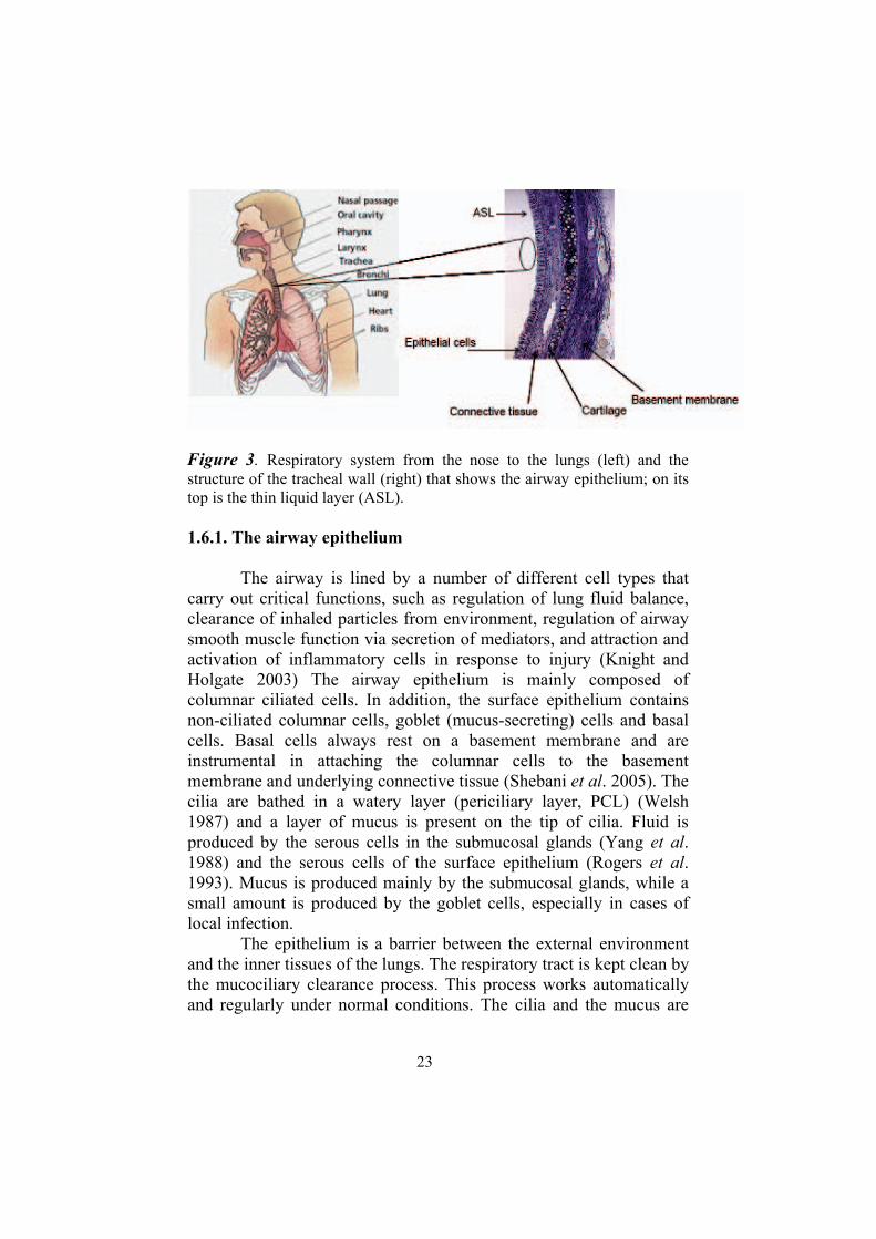
The airway is lined by a number of different cell types that carry out critical functions, such as regulation of lung fluid balance, clearance of inhaled particles from environment, regulation of airway smooth muscle function via secretion of mediators, and attraction and activation of inflammatory cells in response to injury (Knight and Holgate 2003) The airway epithelium is mainly composed of columnar ciliated cells. In addition, the surface epithelium contains non-ciliated columnar cells, goblet (mucus-secreting) cells and basal cells. Basal cells always rest on a basement membrane and are instrumental in attaching the columnar cells to the basement membrane and underlying connective tissue (Shebani et al. 2005). The cilia are bathed in a watery layer (periciliary layer, PCL) (Welsh 1987) and a layer of mucus is present on the tip of cilia. Fluid is produced by the serous cells in the submucosal glands (Yang et al.1988) and the serous cells of the surface epithelium (Rogers et al.1993). Mucus is produced mainly by the submucosal glands, while a small amount is produced by the goblet cells, especially in cases of local infection.
The epithelium is a barrier between the external environment and the inner tissues of the lungs. The respiratory tract is kept clean by the mucociliary clearance process. This process works automatically and regularly under normal conditions. The cilia and the mucus are
24
essential in transporting trapped pathogens and inhaled particles and propelling them towards the pharynx to keep the lungs clean. This transport system may be disrupted by viral and bacterial infections, and by inhaled toxins.
Airway epithelium from different species, for example, human, pig, rat and mouse, has some similarities. Pig airways share many structural and physiological similarities with human airways (Ballard et al. 1999, Cunningham et al. 2002) and are more similar to human airways than airways of other species.
1.6.2. Ion and water transport in the respiratory epithelium
In epithelia, cells are held tightly together by specialized cell junctions to form a continuous sheath. Cell junctions typical for epithelial cells are the tight junctions that limit paracellular transport, intermediate junctions and desmosomes that provide mechanical attachment between epithelial cells, and hemidesmosomes that connect basal cells to the basement membrane. Although there are different types of epithelia, they have at least one important function in common, namely that they serve as selective permeability barriers, separating compartments with a different composition on each side. Different cells perform different ion transport functions.
Polarized cells with segregated channels, cotransporters and pumps in the apical and basolateral membranes are essential for this function. Transepithelial ion and water transport in the respiratory epithelium depends on two main ion fluxes: Na+ absorption and Cl-
secretion. These two mechanisms have opposite characteristics, for example, stimulation of Cl- secretion often inhibits Na+ absorption (Al-Bazzaz 1981; Al-Bazzaz and Jayaram 1981).
It is important to understand that the epithelium of the conductive airways consists of at least three different cell types that are involved in the bulk of ion and water transport. The serous cells of the submucosal glands secrete Cl- and Na+ ions in much the same manner as the intestinal crypt cells, and contain high levels of CFTR. The surface epithelial cells absorb Na+ via an amiloride-sensitive apical epithelial Na+ channel, which controls the rate of transepithelial water absorption. In the apical membrane of these cells, Ca2+-activated Cl- channels and, to a lesser extent, CFTR are present, but most of the transepithelial chloride absorption here is thought to occur by the paracellular pathway. Also water is thought to be absorbed (or secreted) by the paracellular pathway. Finally, there are the epithelial
25
cells of the ducts of the submucosal glands. Few data are available on ion transport in these cells. They may or may not absorb Na+ and Cl-,and in one model they function similar to the cells lining the duct of the eccrine sweat glands (Lee et al. 1986).
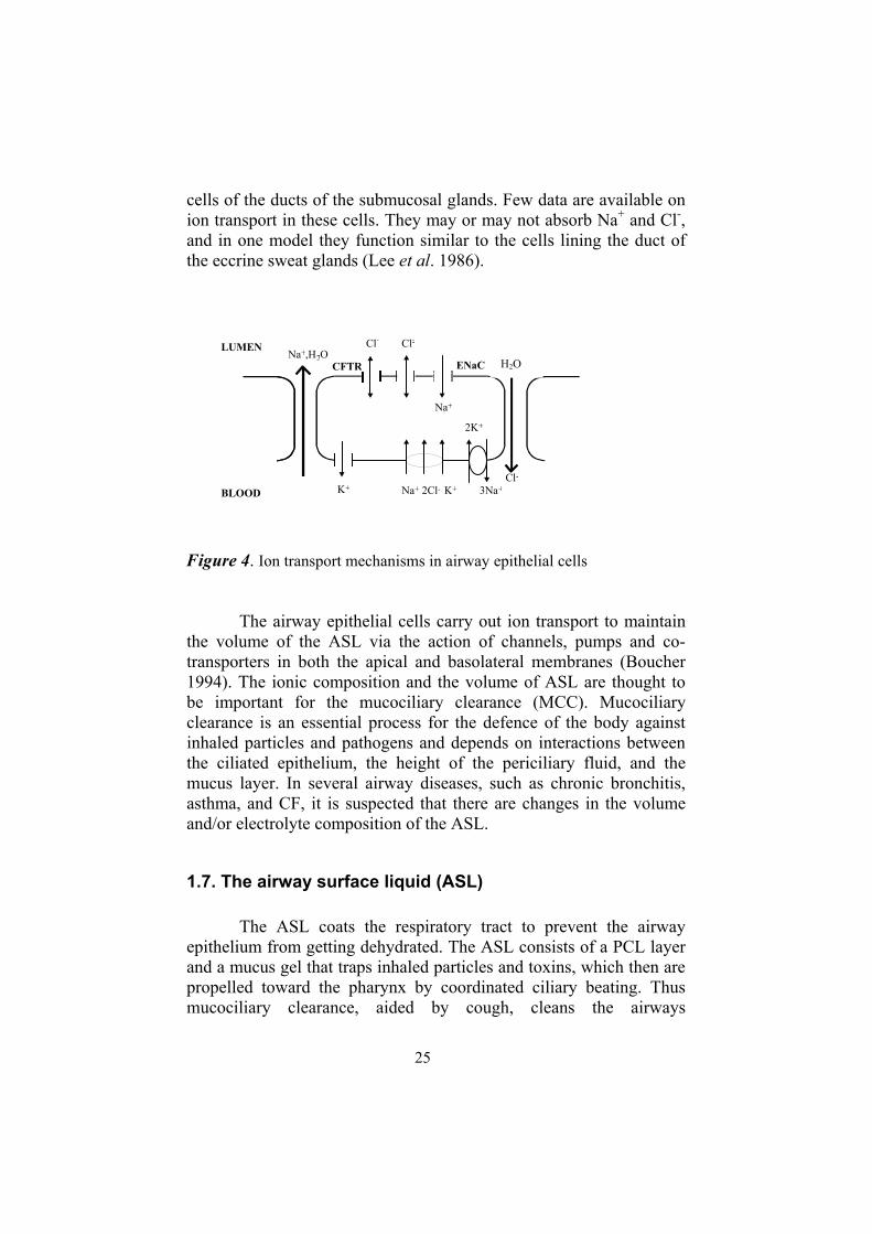
Figure 4. Ion transport mechanisms in airway epithelial cells
The airway epithelial cells carry out ion transport to maintain the volume of the ASL via the action of channels, pumps and co-transporters in both the apical and basolateral membranes (Boucher 1994). The ionic composition and the volume of ASL are thought to be important for the mucociliary clearance (MCC). Mucociliary clearance is an essential process for the defence of the body against inhaled particles and pathogens and depends on interactions between the ciliated epithelium, the height of the periciliary fluid, and the mucus layer. In several airway diseases, such as chronic bronchitis, asthma, and CF, it is suspected that there are changes in the volume and/or electrolyte composition of the ASL.
1.7. The airway surface liquid (ASL)
The ASL coats the respiratory tract to prevent the airway epithelium from getting dehydrated. The ASL consists of a PCL layer and a mucus gel that traps inhaled particles and toxins, which then are propelled toward the pharynx by coordinated ciliary beating. Thus mucociliary clearance, aided by cough, cleans the airways
BLOOD
LUMEN
Na+ 2Cl- K+ 3Na+K+
CFTR
Na+
ENaC
Cl--Cl-
2K+
Na+,H2O
Cl-
H2O
26
mechanically (Knowles and Boucher 2002). ASL is not simply salt water, but is instead a rich broth of proteases/antiproteases, oxidants/anti-oxidants, antibiotics, and antibodies that work together to inactivate or destroy pathogens without undue collateral damage to the lungs. These mucosal mechanisms are supported by cellular immune mechanisms that are recruited and coordinated by signaling molecules released into the ASL (Wine 1991). It is believed that ASL volume and composition plays a crucial role in the pathology of CF and there are many papers that have actively focused on how the ASL contributes to lung infection and inflammation in CF patients (Coakley and Boucher 2001; Davies 2002; Widdicombe 2002; Boucher 2002; Donaldson and Boucher 2003; Chinet and Blouquit 2003; Kunzelmann and Mall 2003; Huang et al. 2004; Tarran 2004; Tarran et al. 2006; Wills and Greenstone 2006; Machen 2006).
In respiratory bronchioles, mucus secreting cells are absent and being replaced by Clara cells. The function of Clara cells is to neutralize toxins dissolved in the ASL (Plopper et al. 1997). Water is secreted into the airway by serous cells in the surface epithelium (Rogers et al. 1993), and by serous cells in the submucosal glands (Yang et al. 1988). The mechanism that drives serous cell liquid secretion is active secretion of Cl- (Yang et al. 1988). The balance between water and mucus secretion is dramatically altered in CF.
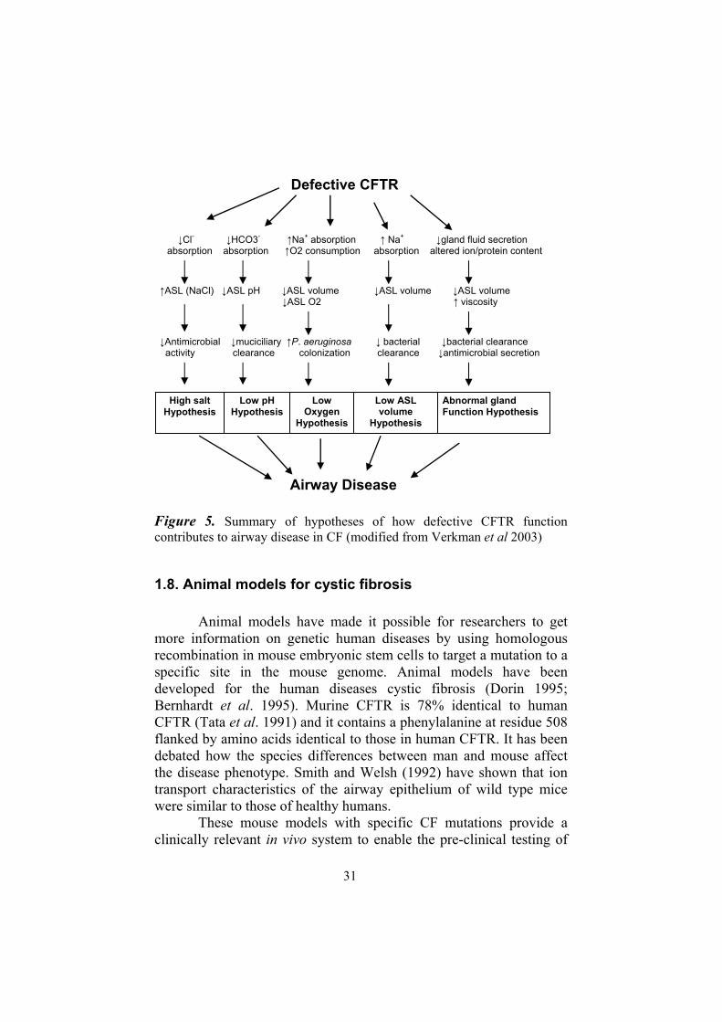
The composition, volume, and physical properties of the ASL depend on secretions from airway submucosal glands, the movement of the fluid, and the transporting properties of surface epithelial cells. Several hypotheses have tried to find a connection between abnormal ASL composition (volume, ionic content, pH, or oxygenation of the ASL) due to defective CFTR function and chronic bacterial infection of the airway.
The exact composition of the ASL and/or the depth of the ASL has been debated for decades. It has been difficult to sample an adequate volume of ASL because the layer is thin (around 5-100µm). The depth of the ASL varies from species to species (mouse < rat < ferret < dog < pig < human) and could vary from region to region (Boucher et al. 1981). The depth of the ASL varies from 5-20 µm in cell cultures (Lucas and Douglas 1934; Yoneda 1976; Sanderson and Sleig 1981; Puchelle et al. 1991) and in vivo it varies from 15 µm in cultures of dog tracheal epithelium (Johnson et al. 1993); to 35-50 µm in sheep trachea in vitro (Seybold et al. 1990), 87 µm in guinea pig trachea in vivo (Rahmoune and Shephard 1995); and 200 µm in guinea pig trachea in vitro (Rahmoune and Shephard 1994). However,

27
shrinkage artifacts due to chemical fixation, dehydration, and/or freeze-drying have prevented reliable estimates of the depth of the ASL.
Published data on the composition of the ASL are very divergent and vary from very hypotonic to hypertonic. In ASL from human trachea, the chloride concentration was found to be around 80-110 mM (Gilljam et al. 1989; Joris et al. 1993; Knowles et al. 1997; Hull et al. 1998), but in ASL from mouse airway data varied from 1 mM (Baconnais et al. 1998) to over about 60 mM (Baconnais et al.1999; Cowley et al. 2000) to 113-115 mM (Verkman. 2001; Caldwell et al. 2002). Low concentrations also have been reported for the rat (45 mM)(Govindaraju et al. 1997), but higher concentrations have been reported for the monkey bronchus (113 mM), rabbit trachea (114 mM) and bronchus (127 mM) (Caldwell et al. 2002) and even higher values have been reported for the ferret (342 mM) (Robinson et al.1989). According to several studies, nasal fluid in humans appears to have a NaCl concentration close to isotonic (Smith et al. 1996; Knowles et al. 1997, Hull et al. 1998; Grubb et al. 2002). Furthermore, studies have been carried out on cell cultures of airway epithelial cells, but these studies did not result in agreement. According to Matsui et al. (1998a,b) values for Na and Cl in the apical fluid were nearly isotonic, whereas Zabner et al. (1998) found values for Na and Cl around 50 mM, and McCray et al. (1999) found a value for Cl of 18 mM. However, recently Kozlova et al. (2006) found that the apical fluid of cultures of airway epithelial cells was hypotonic (80-100 mM NaCl), but that the concentrations of Na and Cl were higher in the apical fluid of CF airway epithelial cells.
Many different techniques for sampling and analysis have been used in these studies, and it is likely that the main source of variation between the results is due to the sampling technique.
1.7.1. The low volume/hydration hypothesis
The theory of isotonic volume transport by Boucher and collaborators (Boucher 1994), suggests that under normal conditions, the ASL is isotonic and the PCL has the same height as the outstretched cilia (~7 µm), which keeps mucus at a sufficient distance from the epithelium, while the mucus layer varies in height from cell to cell. For example, on top of the epithelial cells the height of the mucus layer is more than over the goblet cells (Rahmoune and Shephard 1995; Sims and Horne 1997). The water transport controls
28
the depth of the PCL which regulates the ciliary beat frequency (Widdicombe and Widdicombe 1995). Meanwhile cilia beat and the PCL moves at the same airflow rate in the same direction as the mucus layer (Matsui et al. 1998a, b). In the isotonic volume transport theory it is also predicted that the salt concentration in healthy persons and in CF patients is similar and close to that of the concentration in plasma (Knowles et al. 1997). In a patient with CF, the defective Cl-
transport results in a decreased water transport, which causes a reduction in the volume of the PCL (Boucher 1999, 2002), which interferes with ciliary function and leads to impaired mucus clearance. This promotes infection of stationary mucus adherent to the airway surface by P. aeruginosa and other pathogens.
1.7.2. The high salt concentration hypothesis
The high salt (compositional) hypothesis by Welsh and colleagues (Smith et al. 1996), has proposed that it is the regulation of the salt concentration in the ASL rather than its volume that is abnormal in CF. According to this hypothesis, the surface liquid of normal airways has low levels of salt (<50 mM), because salt is absorbed in excess of water. In CF airways, missing or defective CFTR causes reduced transepithelial Cl- conductance, leading to higher Cl- concentrations in the ASL, which promotes bacterial colonization resulting in early infection and inflammation, leading to lung disease (Kunzelmann and Mall 2001; Boucher 2002). By analogy with the sweat duct the reduced transepithelial Cl- conductance causes salt levels in the airway surface liquid in CF patients to remain at levels similar to that in plasma. This hypothesis proposes that human airway has the ability to restrict water absorption while salt is absorbed. Epithelia with a low water permeability transport salt in excess of water, while those with high permeability transport fluid isotonically (Nielsen et al. 1996).
Defensins are the innate immune system, which keeps the airway free from bacteria. It has been postulated that the high salt concentration in the ASL of the lung in CF inhibits the microbicidal activity of -defensins (Goldman et al. 1997); especially HBD (human
-defensin)-1 and HBD-2 (Bals et al. 1998) have been linked to the lung pathogenesis of CF. Many antimicrobial proteins and peptides present in the respiratory tract have been shown to be salt-sensitive and be inhibited by high salt concentrations in the ASL (Travis et al.1999). A similar peptide in mouse, mouse -defensin-1 (mBD-1) has
29
also been identified as a salt-sensitive antimicrobial peptide (Bals etal. 1998).
1.7.3. The low pH hypothesis
The low pH hypothesis (Coakley and Boucher 2001; Coakley et al. 2000) proposes that the pH of the ASL (pHASL) reflects a balance between active transcellular ion transport and passive paracellular ion movement. ASL acidification is, at least in part, due to the activity of the H+,K+-ATPase in the apical membrane. The localization of the H+,K+-ATPase in the apical membrane was verified by immunocytochemistry (Coakley et al. 2000). The low pH hypothesis postulates that the ASL is abnormally acidic in CF, inhibiting mucociliary clearance mechanisms because of absent CFTR-dependent bicarbonate (HCO3
-) secretion. Early studies of cultured human airway epithelial cells had proposed the presence of a CFTR-dependent apical HCO3
- transport (Smith and Welsh 1992; Devor et al. 2000). Hence, the difference in pH of the ASL between CF and normal airways reflects the lack of HCO3
- secretion by CF airway epithelia, and is not caused by increased H+,K+-ATPase-mediated H+ secretion. Reducing the pH in the ASL will reduce the electrostatic repulsive forces between mucins and increase ASL viscosity and will also promote interactions between gel-forming mucins and the mucins tethered to the membrane surface (Matsui etal. 1998a). The regulation of the pH of the ASL in response to a luminal acid challenge is likely to be important in lung defense.
Several groups have reported that the ASL in cultured human airway epithelium is mildly acidic compared with serum (Kyle et al.1990, Jayaraman et al. 2001, Fischer et al. 2002), but the mechanism which is responsible for generation of the mildly acidic ASL is not known. A low pH of the ASL has been shown to inhibit the detachment of epithelial cells from the basement membrane (Holma et al. 1977), ciliary beating (Clary–Meinesz et al. 1998), cause bronchoconstriction (Aris et al. 1990), and induce cough (Worlitzsch et al. 2002).
1.7.4. The low oxygenation hypotheses
The low oxygenation hypothesis (Worlitzsch et al. 2002),predicts that the oxygen content of the ASL is low in CF because of increased oxygen consumption in CF airway epithelial cells and
30
possibly slowed oxygen diffusion in the ASL, resulting in enhanced P.aeruginosa growth and biofilm formation and impaired clearance. The airway mucus hypoxia may be caused by thickening of the CF mucus prior to infection. This hypothesis also proposes that bacterial infection of CF airways occurs by bacteria binding to the mucus layer rather than to the epithelial cell surface. The diffusion of O2 through liquid is slower than through air (Logan 1998). It appears that the ability to generate steep O2 gradients within the ASL reflects a unique feature of CF airway epithelia (Worlitzsch et al. 2002)
1.7.5. The defective gland hypothesis
The defective gland hypothesis (Nadel et al. 1979; Trout et al.1998; Jayaraman et al. 2001), proposes that reduced fluid secretion and altered secretion of mucous glycoproteins from the airway submucosal glands is the primary defect in CF. It also predicts a reduced ASL volume and increased protein concentration and viscosity in gland fluid secretions in CF, and also predicts an altered ionic content, pH and protein composition of the gland fluid. However, it is still unclear how this hypothesis explains the airway disease in CF. Several studies have shown that submucosal glands plays an important role in the progression of airway disease in CF. One argument to support this hypothesis is that there is higher CFTR expression in epithelial cells lining serous glandular acini than in other tissues in the airway and lung (Engelhardt et al. 1992; Jacquot et al.1993; Sehgal et al. 1996). Submucosal glands become massively hypertrophied with mucus plugging of airway as CF airway disease progresses (Bedrossian and Greenberg 1976; Sobonya and Taussig 1986).
Most innate defense molecules are secreted by the submucosal glands. These glands are thus important for airway health, and CFTR is important for proper submucosal gland function (Ballard and Inglis 2004).
31
Figure 5. Summary of hypotheses of how defective CFTR function contributes to airway disease in CF (modified from Verkman et al 2003)
1.8. Animal models for cystic fibrosis
Animal models have made it possible for researchers to get more information on genetic human diseases by using homologous recombination in mouse embryonic stem cells to target a mutation to a specific site in the mouse genome. Animal models have been developed for the human diseases cystic fibrosis (Dorin 1995; Bernhardt et al. 1995). Murine CFTR is 78% identical to human CFTR (Tata et al. 1991) and it contains a phenylalanine at residue 508 flanked by amino acids identical to those in human CFTR. It has been debated how the species differences between man and mouse affect the disease phenotype. Smith and Welsh (1992) have shown that ion transport characteristics of the airway epithelium of wild type mice were similar to those of healthy humans.
These mouse models with specific CF mutations provide a clinically relevant in vivo system to enable the pre-clinical testing of
Abnormal gland Function Hypothesis
Low ASL volume
Hypothesis
Low Oxygen
Hypothesis
Low pH Hypothesis
High salt Hypothesis
ASL (NaCl) ASL pH ASL volume ASL volume ASL volume ASL O2 viscosity
Antimicrobial muciciliary P. aeruginosa bacterial bacterial clearance activity clearance colonization clearance antimicrobial secretion
Airway Disease
Cl- HCO3- Na+ absorption Na+ gland fluid secretionabsorption absorption O2 consumption absorption altered ion/protein content
Defective CFTR

32
compounds that emerge from large scale screening programs and mutation-specific therapeutic approaches. However, CF mouse models have limitations as a model for CF lung disease because normal mice trachea exhibits low levels of CFTR (Rochelle et al. 2000), and has relatively higher levels of Ca2+-dependent chloride channels. Ion and water transport in the airways of CF mice therefore differs from the situation in humans. On the one hand, CFTR is expressed predominately in the submucosal glands in man, while mice have a relatively paucity of these glands. This may be the reason that CF mice have much milder lung symptoms than CF patients. On the other hand, the CF mice have more marked intestinal symptoms (Snouwaert et al. 1992; Grubb and Boucher 1999). Sodium ion hyperabsorption is characteristic for the nasal tissue of CFTR knockout mice, and for the nasal and tracheal tissues in CF patients. However, in the tracheal epithelium of CF mice hypoabsorption of sodium ions has been noted (Stotland et al. 2000). Reduction of goblet cell hyperplasia, increased mucin gene expression, and increased production of mucus was induced by allergic airway disease both in wild-type and CF mice, but did not lead to chronic lung disease in CFTR-deficient mice (Cressman et al. 1998). Up till now almost all CF mouse models neither develop spontaneous lung inflammation nor chronic bacterial infection and/or inflammation as observed in human CF patients (Davidson and Rolfe 2001; Guilbault et al. 2006). Of particular interest is, however, a mouse model overexpressing ENaC, in which lung disease occurs that shares features with CF, including mucus obstruction, goblet cell metaplasia, neutrophilic inflammation and poor bacterial clearance (Mall et al. 2004)
As a consequence, other animal models are being studied, in particular sheep, pig and ferret. One advantage of these animal models is that the lung function, size and architecture bear greater resemblance to the human lung. Production of cloned animals derived from somatic cells was successfully demonstrated in sheep (Campbell et al. 1996). Transgenic pigs (Polejaeva et al. 2000), transgenic calves (Cibelli et al. 1998), gene-targeted sheep (McCreath et al. 2000), and
-1,3-galactosyltransferase knockout pigs (Lai et al. 2002; Dai et al.2002) have also been obtained by nuclear transfer from somatic cells.
33
2. Aims
At the start of this study, it was still unclear what the ionic composition of the thin layer of liquid that covers the airway epithelium was (in healthy airways and in CF patients), and how it contributed to the CF pathology. We therefore aimed at developing an accurate and efficient method to measure the ionic composition of the ASL both in laboratory animals and in man.
The aims of this study were:
1. to determine the ionic composition of the ASL in pig airways.
2. to determine the ionic composition of the ASL in rat and mouse airways.
3 to develop a reproducible method for sampling and analysis of nasal fluid in man
4. to compare the ionic composition of the ASL between CF patients, CF heterozygotes, and patients with other airway diseases with an inflammatory character.

34
35
3. Materials and Methods
3.1. Experimental animals (papers I, II & III)
The animal studies were approved by the Regional Committee on Animal Experiments, Uppsala.
3.1.1. Pigs (paper I)
Twenty pigs (body weight 20-30 kg) of mixed breed (Hamshire, Yorkshire, and Swedish landrace; Skaggesta Gård, Uppsala, Sweden) were used in this study. The pigs received an intramuscular injection with a tranquillizer (40 mg azaperon; Stresnil, Jansen Pharmaceuticals, Beerse, Belgium) prior to transport to the laboratory. The animals were fully anesthetized with 0.5 mg atropine (Atropin, NM Pharma AV, Stockholm, Sweden) in a mixture of 100 mg tiletamin and 100 mg zolazepam (Zoletile forte vet., Virbac Laboratories, Carros, France) diluted in 5 ml medetomidin 1 mg/ml (Dormitor, Orion Corp., Farmos, Finland); (0.05 mg/kg body weight) intramuscularly. The animals were placed in a supine position on a heating pad, and ventilated mechanically by intubation. A bolus injection of 0.2 mg fentanyl (Fentanyl, Antigen Pharmaceuticals, Roscrea, Ireland) was given intravenously after the intubation. Anesthesia was maintained by infusion of 5 ml/kg/h of 4 g ketamin (Ketamin Veterinaria, Zürich, Switzerland), 1 mg fentanyl in 1000 ml Rehydrex with glucose (Pharmacia and Upjohn, Stockholm, Sweden). The thorax of the animals was opened and the trachea and principal bronchi removed from the area where there was no damage from the ventilation tube. The tissue was dissected out and cut in pieces in a specially constructed chamber, in which the humidity was kept by means of warm water at close to 100%. The chamber consisted of a Perspex box with a retractable sheath on one side that could be opened for handling of the specimen. A water reservoir at 37 C was placed at
36
the bottom of the chamber and the specimen was placed on a perforated shelf. Humidity was monitored with a hygrometer.
3.1.2. Rats (paper II)
Male and female (7- 8 weeks old) Sprague-Dawley rats (B&K Universal, Sollentuna, Sweden) weighing 250-300 g were used. The rats were kept in a conventional animal care facility with free access to food and drinking water until the time of the experiment. At the beginning of the experiments, the rats were anaesthetized by an intraperitoneal injection of sodium pentobarbital (9 mg/100g).
3.1.3. Mice (paper III)
Female NMRI mice (B&K Universal, Sollentuna, Sweden) weighing 20-25 g were used. The mice were deeply anesthetized with sodium pentobarbital (0.025 mg/20g) and the trachea was dissected out and cut in pieces in a specially constructed chamber as described above.
3.2. Frozen hydrated tissue samples (papers I and II)
3.2.1. Pigs (paper I)
The tissue pieces were immediately frozen in liquid propane cooled by liquid nitrogen. The pieces were stored in liquid nitrogen until analysis. For analysis, the tissue pieces were placed with the mucosal side up or pointing sideways onto a specially designed holder and transferred to a Philips (Philips Electron Optics, Eindhoven, The Netherlands) 525 scanning electron microscope equipped with a Biorad (Hemel Hempstead, UK) Polaron 7500E cold stage.
3.2.2. Rats (paper II)
The trachea was removed from the anaesthetized animal and immediately frozen in liquid propane cooled by liquid nitrogen, without prior dissection, to avoid compression during dissection. The trachea was then dissected into tracheal rings under liquid nitrogen. The pieces were stored in liquid nitrogen until analysis, when they were mounted on a specimen holder as described above.
37
3.3. Collection of ASL with Sephadex beads
3.3.1. Pigs (paper I)
Sephadex G-25 (Pharmacia, Uppsala, Sweden) beads were spread evenly on the surface of the dissected pieces of the pig trachea and left during 30 min in the humidity chamber described above. After absorption of the ASL, the beads were recovered by flushing with hydrophobic volatile silicone oil (Dow Corning 200/1cS, BDH, Poole, UK) and collecting the beads in a watch-glass (Nilsson and Ljung 1979, 1985). Under a preparation microscope, all adhering fluid was removed from the beads, and single beads were transferred onto nylon specimen grids (Agar Scientific, Stansted, UK). The grid with beads was slowly lifted out of the oil bath and mounted onto an aluminum holder covered with round carbon adhesive tape and left at room temperature for evaporation of the oil. The grids with Sephadex beads were carbon coated prior to analysis.
3.3.2. Rats (paper II)
Sephadex G-25 beads were spread out evenly on the surface of the dissected trachea, and allowed to equilibrate for 20 min in the humidity chamber described above. After absorption of the ASL, the beads were recovered, collected, and transferred to grids as described above. Alternatively, the Sephadex G-25 beads were applied to double-sided tape (3M, Minneapolis, MN; USA) attached to small (about 1x1 mm2) pieces of filter paper (Whatman, Spring Mills, UK). The filter papers with the beads were placed onto the tracheal wall with the beads facing downwards for 20 minutes. Preliminary experiments showed that 10 min was not enough time in all rats in contrast to the situation in mice. Then, the filter paper with saturated beads was removed and washed with hydrophobic volatile silicone oil to remove adhering fluid and debris. Each bead was individually moved to a nylon electron microscopy grid.
3.3.3. Mice (paper III)
For the determination of the ionic composition of the airway surface liquid in mouse trachea, the Sephadex G-25 beads were equilibrated for 10 min with the ASL. To avoid the risk of formation
38
of clumps of beads, three techniques were compared: (a) the beads were spread “at random” over the surface of the airway epithelium using a spatula, (b) a small amount of beads was placed in the opening at the base of a Microlance 3 needle, (c) The beads were mounted with double-sided tape on a small piece of filter paper, which was then placed with the beads downwards on the surface of the epithelium. After the exposure to the ASL, the beads were collected and transferred to electron microscopy grids as described above.
3.4. Pharmacological stimulation of fluid secretion (paper II)
To stimulate fluid secretion in the rat airways, anesthetized animals received an intraperitoneal injection with the cholinergic agonist pilocarpine (50 mg/kg body weight), the beta-adrenergic agonist isoproterenol (10 mg/kg body weight), or the alpha-adrenergic agonist phenylephrine (10 mg/kg body weight). After 10-15 min, the tracheal ASL or the nasal ASL were collected as described above.
3.5. Human subjects (papers IV and V)
3.5.1. Healthy volunteers (paper IV)
Healthy volunteers (six females and two males; mean age 37 4yr) with no symptoms of airway disease were included in the study. In addition, one sample was taken from a subject suffering from chronic rhinitis, and two samples were taken from subjects with mild respiratory disease (common cold). None of subjects took any kind of medication for respiratory disease.
Stimulation of nasal secretion with chilli pepper: Subjects were asked to chew chilli pepper to stimulate nasal gland secretion.
3.5.2. Patients with airway diseases and CF-heterozygotes (paper V)
CF patients (six males and eleven females, age ranging from 12 to 43 yrs, mean age 25 ± 2 yr), CF heterozygotes (twelve mothers of CF- patients, age ranging from 28 to 53 yr, mean age 43 ± 2 yr), patients with primary cilia dyskinesia (PCD) (seven males and three

39
females, ages ranging from 6 to 40 yrs, mean age 24 ± 5 yr), and allergy/rhinitis patients (fifteen females and thirteen males, age ranging from 4 to 55 yrs, 24 ± 3 yr) were included in the study. All CF and PCD patients were patients of the Cystic Fibrosis Center, Uppsala University Hospital, and the CF heterozygotes were mothers of CF patients treated there. Allergy/rhinitis patients were patients of the Allergy Clinic, Children’s Hospital, Uppsala University Hospital.
As a control group in this study, a group of healthy volunteers (non-smokers) was included: five males and fourteen females, age ranging from 7 to 54 yrs, mean age 32 ± 3 yr).
The study protocol was approved by the Ethics Board of Uppsala University and all subjects and/or parents gave informed consent.
3.5.3. Collection of nasal fluid (paper IV)
Nasal fluid was collected with one of the following techniques:(1) Direct collection of 1 l fluid with a micropipette (Jencons, Leighton Buzzard, UK) with a plastic tip (Treff, Degersheim Switzerland) onto a carbon planchette, (2) Insertion of a piece of filter paper (3 mm), (3) Insertion of cotton wool, (4) Insertion of dextran (Sephadex) beads on filter papers. Details about each method are given below.
The fluid was collected in the vestibule of the nose, where there are no submucosal glands, to avoid direct stimulation of secretion from submucosal glands.
3.5.3.1. Collection with pipettes
Carbon planchets were cleaned carefully in distilled water, dried at room temperature, and then stored. Empty carbon planchets were checked for ionic contamination by X-ray microanalysis. For analysis of droplets in the frozen-hydrated state, 1µl droplets of nasal fluid were placed onto a cold (-30˚C) carbon planchet, and transferred to the Polaron E7400 (Hemel Hempstead, UK) cold stage of the electron microscope. For the analysis of dried droplets, nasal secretions (1 µl) were pipetted onto a carbon planchet. To reduce the size of the crystals that form after drying, nasal fluid (1 µl) was mixed with an equal amount of 30% glycerol or 20% mannitol in water, and then air-dried or freeze-dried in an Emitech K776 freeze-dryer
40
(Balzers, Asslar, Germany) overnight with a starting temperature of -120˚C, which was gradually raised to 25˚C. Droplets of a standard solution with known concentrations of NaCl were treated in the same way.
3.5.3.2. Collection with filter papers
Small strips (3x3 mm2) of dry filter paper (Whatman no. 2, Whatman, Springfield Mill, UK) were used. The filter papers were washed twice in distilled water and dried at room temperature overnight, and stored. Empty filter papers were analyzed to check for contamination. Filter papers were used either to collect nasal fluid directly or for indirect collection after fluid had been collected with a micropipette. In the case of direct collection, the filter paper was kept in the vestibule of the nose for 1 min. Filter papers with nasal fluid were dried at room temperature.
3.5.3.3. Collection with cotton wool
After subjects kept the nostril closed for about 10 min, a small amount (about 7 mg) of cotton wool was inserted into the nose, while the subjects held their breath, and kept there for about 30 seconds. >The cotton plug was then inserted into a micropipette tip (Treff) which was placed in a microcentrifuge tube (Elkay, Costello, Ireland) which contained a drop of mineral oil (Sigma, St. Louis, MO, USA; cat. No M-3516). The tube was centrifuged at 4000 rpm for 2 min. This separated the watery component of the nasal fluid, which became located below the drop of mineral oil, from the mucus, which remained in the cotton wool. The watery phase was collected with a micropipette and 1 µl of fluid was placed either on a filter paper or onto a carbon planchet. The cotton plug with the mucus was air-dried.
3.5.3.4. Collection with Sephadex beads
Nasal fluid was collected by inserting ion exchange beads (Sephadex G-25; 20-40 µm; Pharmacia and Upjohn, Uppsala, Sweden)) which were mounted with double-sided adhesive tape (3M, Minneapolis, MN, USA) on filter paper, in the nostril of the subject. During the insertion of the filter paper with the beads, subjects were asked to either hold their breath or to breathe through their mouth to prevent the warm expired air from causing evaporation of the nasal
41
fluid. The nostrils were kept closed for another 10 min. Usually, this is sufficient to saturate the filter paper and the beads with nasal fluid. Preliminary experiments showed that the beads were saturated after 5 min, so a period of 10 min was chosen to make certain that sufficient fluid was obtained. For some cystic fibrosis patients, the nostrils were kept closed for a longer time (15-20 min), because of reduced nasal fluid secretion in these patients. In some of the allergy/rhinitis patients also this longer time was necessary, if use of a nasal decongestant had dried out the mucous membranes of the nose. At the end of the experiment, the beads were removed from the filter paper, washed in hydrophobic volatile silicon oil, and individual beads were carefully transferred onto nylon specimen grids. The grid with beads was slowly lifted out of the oil bath and mounted onto an aluminum holder covered with round carbon adhesive tape and left at room temperature overnight for evaporation of the oil.
3.6. X-ray microanalysis
3.6.1. Frozen-hydrated tissue samples (papers I and II)
The samples were coated with a thin carbon layer on the cold stage, at a temperature of -190ºC, and kept at this temperature throughout analysis. After preliminary experiments, an accelerating voltage of 9 or 10 kV was chosen to minimize overpenetration of the beam. The samples were analyzed in a Philips 525 (Philips Electron Optics, Eindhoven, The Netherlands) scanning electron microscope (SEM) by a LINK. AN 10000 (Oxford Instruments, Oxford, UK) energy-dispersive spectrometer system. Analysis was carried out for 500 seconds with a beam size of 200 nm, a beam current of about 15 µA, a count rate of 230-235 counts/second and a detector dead time of 5%. Typically, 8-10 analyses were carried out per sample. For quantitative analysis, the data were compared with the results obtained on a standard consisting of a salt solution of known composition to which 5% albumin had been added. The salt solution was spread out in a thin layer over an aluminum planchet, shock-frozen, transferred to the cold stage of the SEM and analyzed under the same conditions as the specimen. Quantitative analysis was carried out using the ratio of characteristic to continuum intensity and by comparing this ratio for the specimen with that obtained by analysis of the standard salt solution (Roomans. 1988).

42
3.6.2. Frozen droplets (paper IV)
The frozen droplets were coated with a thin carbon layer on the cold stage at a temperature of -190˚C, and kept at this temperature throughout analysis. The droplets were analyzed for 100 sec at 15 kV.
3.6.3. Analysis of Sephadex beads (papers I-V)
Prior to analysis, the beads were coated with a thin carbon layer to prevent charging in the SEM. X-ray microanalysis of the beads was carried out with the instrumentation described above, at 20 kV for 100 sec with a beam size of 100 nm. Typically 10-12 beads were analyzed from each sample. For quantitative analysis, the data were compared to the results obtained on beads soaked for 10 min in salt solutions (NaCl, KCl) of different concentrations (50mM-250mM), and with beads soaked in serum or plasma from the same animals (pigs in paper I, rats in paper II) and analyzed by colorimetry (Konelab 30 analyzer, Thermo-Electron Corp., Espoo, Finland).
The accuracy of the method and the standard curves were verified by testing the results obtained on beads soaked in human blood serum, where they returned physiological values: [Na]: 130±22, [Cl]: 105±14, [K]: 8±1 (mM).
3.6.4. Dried droplets on carbon planchets or filter paper (paper IV)
Prior to analysis, the dried droplets were coated with a thin carbon layer to prevent charging in the SEM. The droplets were analyzed at 15 kV for 300 sec, using a beam with a spot size of 200 nm. The beam was scanned so that the entire droplet was irradiated.
3.7. Storage of ion exchange beads in silicon oil (paper II)
To determine whether the absorbed fluid was lost from the ion exchange beads during storage, beads were saturated with 150 mM NaCl for 20 min. Then the beads were collected in silicon oil as described above, and either taken out immediately, transferred to nylon grids and dried as described (control), or kept in oil for 1-5 days and then removed and treated as described above.
43
3.8. Preparation for light and electron microscopy (papers I and II)
Small pieces of tracheal wall were removed from the anaesthetized animal and immediately fixed in 2.5% glutaraldehyde in water or different concentrations of sodium cacodylate buffer (0.025, 0.05, 0.1, or 0.15 M).
All tissues were kept in fixative for 24 hours at 4ºC and then postfixed with osmium tetroxide, dehydrated in a graded ethanol series, and embedded in epoxy resin. Sections (2 µm thick) were cut perpendicularly to the epithelium and stained with toluidine blue for light microscopy. Ultrathin sections (50 nm) were cut for electron microscopy, and contrasted with uranyl acetate and lead citrate.
Pieces of trachea exposed for 30 min to Sephadex beads as described above were fixed in 2.5% glutaraldehyde in 0.1 M sodium cacodylate overnight. Tissues were postfixed and embedded as described above. Thin sections (2µm) were cut and stained with toluidine blue for light microscopy.
Some pieces of pig trachea were frozen in liquid propane cooled in liquid nitrogen and were kept in liquid nitrogen for analysis of frozen hydrated specimens.
3.9. Light and electron microscopy (papers I and II)
Light microscopy was carried out with a Leica (Wetzlar, Germany) microscope and the height of the epithelium in perpendicular sections was determined using a semi-automatic image analysis system (VIDS, Synoptics, Cambridge, UK).
Transmission electron microscopy (TEM) was carried out at 75 kV with a Hitachi (Tokyo, Japan) 7100 transmission electron microscope.
3.10. Statistical analysis (papers I-V)
Data are presented as mean standard error. Differences between more than two groups were determined by analysis of variance (ANOVA). Dunnet's Comparison Test was used to determine the significance of the differences. Differences between two groups were determined using Student's t-test. Significance was attributed to probability values (*) p<0.05, and (**) p<0.01.
44
4. Results
4.1. Elemental composition of the ASL in experimental animals
Paper I: X-ray microanalysis of frozen-hydrated pig trachea with the epithelial surface (and the ASL) pointing upwards or sideways showed a concentration of Na of about 120 mM and of Cl of about 60-80 mM. We extended our study also to the principal bronchi, where the ASL has a slightly higher concentration of Na, Cl and K compared to the trachea. The composition of the ASL in pig trachea was also measured with the Sephadex bead method. Results obtained with Sephadex G-25 beads on pig ASL were compared with data from serum from the same pig. The concentrations of Na and Cl in the ASL were slightly lower than in serum. The concentration of K was higher in the ASL than in serum but lower than that in the frozen-hydrated ASL. The concentrations of P and S were similar to the values in serum but lower than in the measurements on frozen-hydrated ASL.
Morphological studies of pig trachea fixed in solutions of different osmolarity showed a clear dependency of the cell size on the osmolarity of the fixative solution. At concentrations of 50 mM sodium cacodylate and less, evident damage to cellular organelles was observed by electron microscopy, in comparison to normal morphology at the highest concentrations (100-150 mM) of sodium cacodylate. At 50 mM sodium cacodylate, fluid filled vesicles were formed in the epithelial cells and at even lower osmolarity, the mitochondria showed clear signs of damage.
Paper II: Under unstimulated conditions, X-ray microanalysis of rat tracheal fluid and nasal fluid showed that the elemental composition of these fluids was markedly different from that of plasma. Concentrations of Na and Cl were significantly lower in both tracheal fluid and nasal fluid compared to plasma. In tracheal fluid, K was slightly, but significantly higher than in plasma, while the Ca
45
concentration was around 5 mM (close to the detection limit of the method), lower than that of plasma, and the Mg concentration was around 16 mM. In nasal fluid extremely high K values were found. The very high K concentration was the main difference between tracheal and nasal fluid.
To see if the periciliary nasal or tracheal fluid could be hypotonic as indicated by our results, a morphological study was conducted. The trachea was fixed in glutaraldehyde in different concentrations of sodium cacodylate. Trachea fixed in 50 mM sodium cacodylate did not show significant damage compared to tissue fixed in glutaraldehyde in higher (physiological) concentrations of sodium cacodylate, (100 or 150 mM), whereas tissue fixed in lower concentrations (25 mM) sodium cacodylate showed cell damage (vacuolization).
Paper III: To minimize the risk for formation of clumps, the method of randomly spreading the beads over the epithelial surface of the mouse trachea was compared to spreading the beads with a syringe, and placing a small piece of filter paper with mounted beads on the surface of the epithelium. No significant differences between the three techniques were found. However, some beads collected using the “random spreading” method had very low values for the elemental concentrations. A possible reason for this could be that some beads had been lying on top of other beads. The low values from such beads were not considered.
The beads did absorb the fluid component of the ASL, equivalent to the periciliary liquid, which is the physiologically most important component of the ASL. If the beads were not cleaned of mucus, X-ray microanalysis showed significantly higher concentrations of P and S, and significantly lower concentrations of K and Cl.
4.2. Analysis of the composition of human nasal fluid
Paper IV: Analysis of 1 µl droplets of NaCl solution after air-drying gave poorly reproducible results. This was due to the large crystals formed upon drying. In these crystals, absorption of Na and Cl X-rays occurs, which reduces the signals. The reduction is dependent on the size and orientation of the crystals and difficult to control. Crystal size can be reduced by mixing the NaCl solution with
46
either glycerol or mannitol, and the crystals become even smaller when the droplet is freeze-dried instead of air-dried. The filter paper technique is a useful technique because here the NaCl solution does not form large crystals. A good calibration line could be obtained with the filter paper method, but the filter paper "diluted" the salt, and thus the method resulted in a relatively weak signal. Therefore the counting time had to be longer. Good calibration lines must have a goodness of fit (r2) of 0.9 or more, and the filter paper method meets this limit.
The data obtained from nasal fluid after collection with a pipette and application onto a filter paper were similar to those obtained on nasal fluid mixed with glycerol or mannitol, or unmixed. Stimulation of nasal gland secretion with chilli pepper gave similar results with regard to elemental composition compared to samples obtained without stimulation. It turned out that the method of direct collection of nasal fluid was difficult to carry out in CF patients, because their mucus was so viscous. Therefore, a technique was developed where Sephadex G-25 beads were mounted on double-sided tape on filter paper, and left to equilibrate with the nasal fluid in the vestibule of the nose.
Paper V: The results from beads with nasal fluid in healthy subjects were similar to those reported previously using the same technique (paper IV). In CF patients, CF heterozygotes, and in rhinitis and PCD patients the levels of Na and Cl in the nasal fluid were significantly higher than in healthy controls. In CF and PCD patients also the levels of K were higher than in healthy controls. No significant difference in Na, Cl or K could be observed between healthy males and females. However, females with CF had significantly higher concentrations of Na, Cl and K in their nasal fluid compared to CF-males.
There was a significant difference (p<0.05) between male and female patients when all patients with airway diseases (CF, PCD, and rhinitis) were pooled. For PCD and rhinitis patients, the different between male and female patients was not significant. Differences in ion concentrations between male and female CF patients have not previously been studied. The CF patients were classified as severe, medium and mild based on the clinical condition of the patients when the samples were taken. The severe female patients had significantly higher K concentrations in their nasal fluid than the female patients in mild or medium diseased condition. Within the group of cystic fibrosis patients, there was no significant correlation between the

47
elemental concentrations and FEV1% or FVC%. There was also no significant correlation between age and elemental content of the nasal fluid. This was also the case for the other groups.
48
5. Discussion
5.1. Elemental composition of the ASL in experimental animals
Paper I: Pigs were chosen for this study because the respiratory system of the pig has, with respect to fluid transport, many similarities with the human respiratory system. There are major differences between the results of the ASL measured in situ in frozen-hydrated specimens, and the results obtained on the Sephadex beads. The ASL in situ contains substantially higher concentrations of e.g., P, S, and K than the ASL in the Sephadex beads. One could suspect that in the measurements on the ASL in the frozen-hydrated state, underlying tissue, or at least cilia, are excited by the electron beam, and thus contribute to the spectrum. There are, however, several arguments against this notion.
The spatial resolution of analysis at 9 or 10 kV in a frozen-hydrated specimen can be calculated to be in the order of 2-3 m(Reed 1975), which is much less than the actual thickness of the ASL that can be measured from micrographs where the trachea is mounted sideways. Furthermore, the results from the analysis where the trachea is mounted sideways rather than with the ASL pointing upwards, give the same result with regard to the elemental analysis, and in this situation, overpenetration would not excite underlying tissue. Finally, if the results were due to overpenetration of the electron beam and excitation of the underlying epithelium, one would expect a negative correlation between e.g., Na and P, or Na and K, since a measurement with little or no overpenetration would sample the overlying ASL, and show high Na and low P and K, whereas a measurement with much overpenetration would mainly excite the epithelium and show high P and K and lower Na. Analysis of the data, however shows that the correlation between Na and P is not negative and the same result was obtained for the relation between Na and K.
49
The difference, then, between the measurements of ASL in situin frozen hydrated specimens and in the beads must be due to the fact that actually different things are measured. The measurements of the ASL in the frozen-hydrated specimens measure the upper mucous layer of the fluid, whereas it is likely that the beads absorb the watery component of the layer. The upper part of the ASL likely contains glycoproteins, shed cells and cell debris that could add "cellular" elements such as P, S and K to the layer.
The morphological data argue strongly against the notion that the ASL could be very hypotonic. Already at a buffer concentration of 50 mM, damage to the epithelial cells is evident. In the extreme case represented by the values of Baconnais et al. (1998) of less than 10 mM, the epithelial cells could not possibly survive.
The elemental composition of the watery layer of the ASL does, however, differ from that of serum in a number of respects. Concentrations of Mg, P and K are higher than in serum. For K, where we find a concentration of 20 mM, some literature data are available. In human ASL, Joris et al. (1993) found 29 mM and Knowles et al.(1997) found 18 mM, which agrees well with our data. Published values for rat (Cowley et al. 1997) and mouse (Baconnais et al., 1998) are much lower, but these papers also give very low values for the other ions. This agrees with the data presented in papers II and III, and there is thus a clear species difference in the ionic composition of the ASL. Possibly, the source of the K in the ASL is the cell debris in the mucous layer that could leak K+ ions.
The results unequivocally confirm the notion that the Na and Cl concentrations in the watery phase of the ASL in the pig are close to those found in plasma. The Cl value of around 90 mM in the ASL in the beads agrees reasonably well with the data of Gilljam et al.(1989); Joris et al. (1993); Knowles et al. (1997) and Hull et al.(1998) for the human and confirms that the pig is a good model for the human airways. The fact that the chloride concentration in the upper layer is somewhat lower might be due to the presence of negatively charged macromolecules in this layer that would tend to attract cations and repel anions.
The notion that the ASL in the pig is near isotonic, but with a somewhat higher K content compared to serum also agrees with our data on human nasal fluid (paper IV). In both cases the periciliary fluid has a composition that is different from the mucus. The differences are similar in nose and trachea, but more pronounced in the trachea.
50
Paper II: The ionic concentrations in rat ASL in our study (paper II) showed slightly higher values compared to an earlier report on the same animal (Cowley et al. 1997). However, both studies are comparable and show that the rat ASL is hypotonic. The composition of the nasal and tracheal ASL can, however, be manipulated by pharmacological agents. Pharmacological stimulation resulted in changes in the composition of the ASL lining the trachea. Isoproterenol significantly increased the Na content of the fluid, and pilocarpine and phenylephrine the K content. Pilocarpine stimulation also increased the Mg concentration. The effects of stimulation on nasal fluid were somewhat different: pilocarpine significantly increased Na and Cl, isoproterenol significantly increased K and phenylephrine significantly decreased K but all the stimulations significantly increased the Ca concentration.
The ionic composition of the ASL in the rat was tested with and without stimulation of submucosal gland secretion. There was a rapid glandular fluid secretion in mouse after intraperitoneal stimulation with pilocarpine (Verkman. 2001). Phenylephrine has been observed to have a slight effect on glandular secretion in cats but not in humans or pigs (Joo et al. 2001) but there is no data on the rat. Isoproterenol and phenylephrine did not show any effect on porcine bronchi (Trout et al. 2001).
The change in the volume of the ASL can be critical and is affected by many factors. Changes in ASL volume can also change the ionic concentrations. Many substances can increase or reduce the production of fluid at the airway surface. Small disturbances, e.g., touching the airway surface, can cause volume changes in the ASL by mechanical stimulation of airway epithelial cells or submucosal glands, which can cause changes in the ion concentrations in the ASL. According to Joris et al. (1993) human ASL was hypotonic under normal conditions but the Na and Cl concentrations increased and the ASL became nearly isotonic after airway irritation or acute infection.
The analysis of the data from the ASL from both nasal fluid and trachea showed that the ASL in the rat was hypotonic, although the nasal fluid had a high K concentration. The ASL is a thin liquid film consisting of two separate layers. The height of these layers varies markedly from species to species depending on the size of the species. It is critical in the collection of samples from such small volume that this is done without disturbing the cells underneath the fluid layer.

51
The results from the frozen hydrated rat trachea shows somewhat higher elemental content of Na, Cl and K compared to the data obtained from the Sephadex beads. As in the study on pig (paper I) this can be explained by the fact that the analysis of frozen hydrated samples gives results from the mucus layers. In contrast, the Sephadex beads give results mainly from the PLC layer.
The frozen hydrated samples also showed high concentrations of P and S. This is likely to be due to the cell debris in the mucus layer. As in paper I, we have considered the alternative explanation, namely that the electron beam would penetrate the thin ASL layer, and excite the cells, but found that this was not the case.
Remarkably, the K concentration in the rat nasal fluid was very high. Possibly, the submucosal glands in the nose of the rat resemble salivary glands. In these glands, it has been shown that radiation damage can cause elevated K+ levels in the secreted fluid (Vissink et al. 1990, 1991). It has also been shown that inhibition of the Na+-K+ ATPase in salivary glands by ouabain can cause high levels of K+ in the secreted fluid (Dehpour et al. 1995). Therefore, the high K+ levels in the nasal secretion may point to the fact that these nasal glands function in a suboptimal way in the rat, and that therefore the K+ level in the secreted fluid is abnormally high. Analysis of beads stored in oil for different times (minutes up to days) showed that no significant changes in Na and Cl content occurred during up to 5 days of storage. Possibly, a minor decrease occurs after the third day but this was not significant.
Ion exchange beads can collect small volumes of fluid; moreover, X-ray microanalysis requires only very little material and can also be used for in situ analysis. Therefore, this technique would appear the method of choice for such investigations.
Paper III: The three different methods of ASL collection tested ((1) beads were mounted on filter paper, (2) beads were randomly spread over the airway epithelium, and (3) beads were spread over the epithelium with a syringe) can all be used to study the ionic composition of the ASL. All three methods are easy to use and are neither complicated nor expensive.
The analysis of the ASL in the mouse showed that the periciliary liquid was hypotonic. This result is comparable to earlier reports (Baconnais et al.1999). Nevertheless, there is a considerable difference between our study and the studies by Baconnais et al.(1998, 1999) with regard to the methods. In the study by Baconnais et
52
al. (1998) the ASL was collected by holding a cold (liquid nitrogen temperature) metal probe against the ASL layer. The disagreement between the data of Baconnais et al. (1998) and our data can be explained by assuming that in the cold probe technique, a condensate is sampled rather than the actual ASL.
The Sephadex beads absorb more K+ than Na+ ions. This might be related to the size of the pores relative to the radius of the hydrated ions, or to the position of the oxygen atoms in the dextran, which may be more favourable for K+ than for Na+ absorption. However, a correction can be made for the different ability to absorb different ion by making separate calibration lines for different elements.
To avoid obtaining results “contaminated” by the overlying mucus layer the beads that have collected fluid from the PCL must be cleaned properly to remove the mucus adhering to the beads. The mucus layer also contains cell debris, which can give rise to S and P.
The mouse has been used as an animal model for CF, since scientists have succeeded to generate transgenic mice by gene targeting (Koller and Smithies 1992). Using animal models in research has given interesting results, but it has to be taken into account that the anatomy or physiology of some animals can have more or less similarity compare to humans. The mouse models have been instructive with respect to CF pathogenesis in some organs, but unfortunately only little CFTR protein is expressed in the mouse lung and it appears that ion and water transport in the mouse lung functions in a different way compared to the human lung. Moreover, it has been shown that transgenic CF mice do not have the same airway symptoms as human CF patients (Grubb and Boucher 1999). However, Harkema (1991) has shown that the murine nasal septal mucosa has a cellular composition similar to the human nasal mucosa, so that the differences still may be relatively minor.
The difference in ionic composition between mouse ASL and human ASL (paper IV) could be due to the structural differences between mouse and human airways, namely the fact that the mouse has fewer submucosal glands compared to the pig that has an airway structure that is more similar to the human airway (Choi et al. 2000). Since the glands produce most of the mucus that is present in the airway, and forms a defense system against both inflammatory and physical environmental stimuli, this structural difference between mice and man is potentially of great importance.
53
5.2. Analysis of the composition of human nasal fluid
Paper IV: In humans, nasal fluid has many similarities to the fluid lining the walls of the bronchi but is easier to collect, and hence we focused our study on the nasal fluid. As ASL in general, nasal fluid consists of a periciliary fluid phase and a more superficial mucous phase, and contains in addition to water and ions, glycoproteins, such as mucins, and other proteins (Verkman et al.2003). Direct collection of nasal fluid gives results based on both the periciliary and the mucous phases, while collecting nasal fluid with Sephadex beads is likely to give results mainly based on the periciliary fluid. We attempted to collect nasal fluid by placing cotton wool in the nostril and centrifuging the cotton to remove the nasal fluid. It appears that (part of) the mucous phase remains in the cotton wool, and that this phase is richer in P, S and Ca than the fluid, which has relatively higher levels of monovalent ions. This finding can be expected since the mucus in addition will contain cell debris and bacteria that contribute P and S.
Our results from nasal fluid collected with the direct method and with the Sephadex bead method show that the concentrations of Na and Cl are similar to those in serum, so that the ASL in the human airways is likely to be close to isotonic, which agrees with published reports (Smith et al. 1996; Knowles et al. 1997; Hull et al. 1998; Grubb et al. 2002). The differences in elemental content between mucous phase and fluid phase are not very large. Hence, it is not surprising that the differences between the filter method (containing both phases) and the Sephadex bead method (containing predominantly the fluid phase) are relatively small, and that the differences consist of higher concentrations of P, S and Ca and lower concentrations of Na, Cl and K in the filter paper compared to the beads. This notion is also in line with the fact that chewing of chili pepper, which is likely to mainly increase the fluid phase, does not affect the ionic composition of the total nasal fluid significantly.
Paper V: Collecting samples of nasal fluid by using ion-exchange beads does not appear to cause any disturbance to the airway epithelium. The nose is a good model system to study the ion composition of the ASL since the nose has similar ion transport characteristics and a similar epithelium compared to the lower airways. It has been shown that the ion composition of nasal fluid
54
resembles that of ASL taken from the lower airways, even though there are small differences (Hull et al. 1998; Knowles et al. 1997). The sampling technique can affect the ionic composition in the ASL, therefore all materials that are used to collect samples must be checked to make sure that there are no ions left in the materials. It could be shown that the beads have the capacity to hold ions without changes in concentration for at least five days (paper V), which means that sample of nasal fluid could be taken at one location and mailed to a central laboratory elsewhere for analysis.
CF heterozygotes have Na and Cl concentrations that are intermediate between healthy controls and CF patients, which might be due to a reduced level of CFTR, and is in line with findings that the nasal potential difference in CF heterozygotes is intermediate between healthy controls and CF-patients (Sermet-Gaudelus et al. 2005).
Patients with rhinitis or PCD have higher Na and Cl concentrations than healthy controls but lower than CF-patients. Possibly, the higher Na and Cl concentrations are the result of opening of tight junctions (e.g., by proinflammatory cytokines (Coyne et al.2002)) which could increase the flux of fluid from the connective tissue to the ASL. The higher Na and Cl concentrations in the nasal fluid of CF patients could be in part due to the inflammatory process, and in part to the basic defect in CFTR. That the basic defect in CFTR can give rise to higher Na and Cl concentrations in the ASL is in line with the findings of Kozlova et al. (2006) that the apical fluid in cultured CF respiratory epithelial cells is higher than the concentration of Na and Cl in the apical fluid covering cultured control respiratory epithelial cells. Since in these cell cultures, there is no inflammation, and the only difference is in the CFTR, this means that there is a direct link between the defect in CFTR and the ion content of the apical fluid.
Female CF patients have a higher Na, Cl and K concentration in their nasal fluid than male patients. This may have a connection to the fact that female CF patients are earlier colonized by mucoid strains of Pseudomonas aeruginosa (Demko et al. 1995) and that life expectancy for female CF patients is shorter than for male patients (Dodge et al. 1997)
The increase in the NaCl concentration in the nasal fluid of CF patients may be clinically relevant, because high NaCl concentrations inhibit the activity of antibacterial factors (Smith et al. 1996, Wine 1997; Travis et al. 1999).

55
Relatively high K concentrations were observed in the nasal fluid of healthy controls and even higher K concentrations were found in the nasal fluid of CF patients. K concentrations higher than those normally found in extracellular fluid were also observed in the ASL of mice, rats and pigs. High concentrations of K (although not as high as the K concentrations that we found) have also been found in ASL from dog trachea, human trachea or bronchi and human nasal fluid (Boucher et al. 1981, Joris et al. 1993, Knowles et al. 1997). Of interest is that high K concentrations were also found in the fluid covering the uterine epithelium (endometrium) in mice (Jin and Roomans 1997). Possibly, high concentrations of K (higher than normal in extracellular fluid) are a general rule for fluid covering epithelia.
56
6. Conclusions
1. Several methods were developed to collect ASL without causing irritation to the respiratory epithelial cells. The collection method where Sephadex beads are used can (with variations) be applied to collect nasal and tracheal fluid in laboratory animals and humans. X-ray microanalysis is a technique that is suitable to analyze small samples of fluid. Differences between results on ASL collected with Sephadex beads and ASL analyzed directly in frozen-hydrated samples can be explained by the fact that the Sephadex beads collect mainly the periciliary liquid, whereas in the analysis of frozen-hydrated samples, mainly the upper mucous layer is analyzed.
2. Animal models have given results that are helpful for the understanding of human diseases, and can be used in the development of a pharmacological treatment. However, species differences may make a direct comparison between the animal model and the human disease difficult. The mouse has been used as a model for CF, but fluid transport in mouse airways is different from human airways, and CF mice do not show the same respiratory symptoms as CF patients. However, with this limitation, CF mice can still be useful for CF research.
3. The ASL in pig trachea is about isotonic, but has a somewhat higher K content than the K content in serum. The upper mucous layer of the ASL contains more P and S than the periciliary fluid. The physiologically most relevant data would be those from the periciliary fluid. Mouse and rat ASL are in contrast markedly hypotonic, but also here a higher K content than in serum is found. The ionic composition of the ASL may be influenced by pharmacological stimulation. The ionic composition of the nasal fluid is not always identical to that of

57
the tracheal fluid, e.g., in the rat the nasal fluid contained much higher concentrations of K than the tracheal fluid.
4. A reproducible method was developed to determine the ionic composition of small volumes of human nasal fluid by X-ray microanalysis. In healthy individuals, the Na and Cl content of nasal fluid is close to that in serum (which agrees with the situation in pigs, but not in rodents), and the K content is somewhat higher. In subjects with respiratory diseases (common cold, rhinitis) the Na and Cl concentrations may be higher than normal.
5. CF patients have higher concentration of Na, Cl and K in their nasal fluid than healthy controls. This may in part be due to the inflammatory process in the nasal epithelium (since also patients with rhinitis or PCD have elevated Na and Cl concentrations) and in part to the basic defect in CFTR (since also CF heterozygotes) have elevated Na and Cl concentrations. The elevated salt concentrations may have clinical significance, since the activity of bactericidal substances in the ASL appears to be reduced by high salt concentrations.
58
Acknowledgements
This work was performed at the Department of Medical Cell Biology, Uppsala University.
I wish to express my deep and sincere gratitude to all people whom I have met and who have taught me during the period that I was a graduate student. I would like to thank in particular:
My supervisor, Godfried M. Roomans, for making it possible for me to become a graduate student and finish my PhD degree. It has been a big adventure to come this far learning about cystic fibrosis. With all your support you have made me strong and you made me dare to do many things that I want to do. It has not been easy but it has been worth the effort. I can never thank you enough for endless support, and being so patient.
Jarin Hongpasan and family, for introducing me to Sweden and giving me the opportunity to be where I am today.
My co-authors and colleagues Birgitta Almgren, Anca Dragomir, Marian Högman, Inna Kozlova and Harriet Nilsson,for pleasant cooperation and fruitful discussions.
Marie Johannesson, and all the people in Uppsala CF Center and the Allergy Clinic at Akademiska Barnsjukhuset in Uppsala, in particular Lennart Nordvall and Elisabeth Nääs, for being kind and helpful, in particular with the clinical study.
Anders Ahlander, for helping with many practical things, for being patient, friendly and sharing lots of jokes and laughs.
Leif Ljung and Marianne Ljungkvist, for all the help in the lab and being so kind.
My colleagues in the Anatomy corridor, FaranakAzarbayani, Kärstin Flink, Dieter Fuchs, Andrei Malinovschi, Jacqueline Relova, Zhanna Servetnyk, for friendly discussions, and Barbro Einarsson, for nice Swedish practice talk during lunch breaks.

59
The administrative staff, Agneta Bäfwe, Karin Öberg, and the late Gun-Britt Lind, for being kind and friendly.
Also thanks to the former graduate students in the Department for good advice.
The staff of the Lao embassy for their help throughout these years.
My friends and their family members and children, here and abroad, especially those in Laos for their endless love and friendship, and for sharing laughs and talk even though I am so far away, but you still make me feel like I were home.
Evi Marklund, my very special friend, for loving and caring. You are always there for me when I need someone. You are more like a family member to me than just a friend.
Ib Christian Rasmussen, for being so kind and friendly. Michael Söderman, Anna Lindahl and Ilon Michaelsdotter
Lindahl, for sharing laughs, and love and being friendly. My family, especially my mother, for all support, love and
for believing in me, because I believe that is why I have become who I am today. You are a strong woman and you have passed this strength to me. Thank you from all my heart for always encouraging me to go through all the difficulties. You are the best mother ever.
Einar and Gudrun Söderman, for their love, care, and encouragement, and for being a big part of my life, especially for being my parents.
Magnus, for being so patient, loving, making me strong and sharing sadness and happiness.
Johan Saysakda, for giving me endless love and making me happy with your smile as I had never felt before. You are the sunshine of my life even in the dark winter days.
This thesis was financially supported through grants from the Cystic Fibrosis Association (U.S.A.), the Swedish Science Research Council, the Swedish Heart Lung Association, and the Swedish Association for Cystic Fibrosis.
60
References
Al-Bazzaz FJ. Role of cyclic AMP in regulation of chloride secretion by canine tracheal mucosa. Am Rev Respir Dis 1981; 123: 295-298.
Al-Bazzaz F, Jayaram T. Ion transport by canine tracheal mucosa: effect of elevation of cellular calcium. Exp Lung Res 1981; 2: 121-130.
Andersen DH, Cystic fibrosis of the pancreas and its relation to celiac disease: a clinical and pathological study. Am J Dis Child 1938; 56:344-399
Anson DS, Smith GJ, Parsons DW. Gene therapy for cystic fibrosis disease- is clinical success imminent? Curr Gene Ther 2006; 6: 161-179
Aris R, Christian D, Sheppard D, Balmes JR. Acid fog-induced bronchoconstriction. The role of hydroxymethanesulfonic acid. Am Rev Respir Dis 1990; 141: 546-551.
Aurora. P, Whitehead. B, Wade. A, Bowyer J, Whitmore P, Rees PG, Tsang VT, Elliott MJ, de Leval M. Lung transplantation and life extension in children with cystic fibrosis. Lancet 1999; 354: 1591–1593.
Baconnais S, Zahm JM, Kilian L, Bonhomme P, Gobillard D, Perchet A, Puchelle E, Balossier G. X-ray microanalysis of native airway surface liquid collected by cryotechnique. J Microsc 1998; 191: 311-319.
Baconnais S, Tirouvanziam R, Zahm JM, de Bentzmann S, Peault B, Balossier G, Puchelle E. Ion composition and rheology of airway liquid from cystic fibrosis fetal tracheal xenografts. Am J Respir Cell Mol Biol 1999; 20: 605-611.
Ballard ST, Inglis SK. Liquid secretion properties of airway submucosal glands. J Physiol 2004; 556: 1–10
Ballard ST, Trout L, Bebok Z, Sorscher EJ, Crews A. CFTR involvement in chloride, bicarbonate, and liquid secretion by airway submucosal glands. Am J Physiol 1999; 277: L694-L699.
61
Bals R, Wang X, Wu Z, Freeman T, Bafna V, Zasloff M, Wilson JM. Human beta-defensin 2 is a salt-sensitive peptide antibiotic expressed in human lung. J Clin Invest 1998; 102: 874-880.
Baxter PS, Wilson AJ; Read NW, Hardcastle J, Hardcastle PT, Taylor CJ. Abnormal jejunal potential difference in cystic fibrosis. Lancet 1989; 1: 464-466.
Bedrossian CW, Greenberg SD. The lung in cystic fibrosis. A quantitative study including prevalence of pathologic findings among different age groups. Hum Pathol 1976; 7: 195-204.
Bernhardt GZ, Eichwald E, Zabner J, Smith JJ, Puga AP, McCray Jr PB, Capecchi MR, Welsh MJ, Thomas KR. A mouse model for the F508 allele of cystic fibrosis. J Clin Invest 1995; 96: 2051-2064.
Berschneider HM, Knowles MR, Azizkhan RG, Boucher RC, Tobey NA, Orlando RC, Powell DW. Altered intestinal chloride transport in cystic fibrosis. FASEB J 1988; 2: 2625-2629.
Boucher RC. Human airway ion transport. Am J Respir Crit Care Med1994; 150: 271-281.
Boucher RC. Molecular insights into the physiology of the 'thin film' of airway surface liquid. J Physiol (Lond) 1999; 516: 631-638.
Boucher RC. An overview of the pathogenesis of cystic fibrosis lung disease. Adv Drug Deliv Rev 2002; 54: 1359-1371.
Boucher RC, Stutts MJ, Bromberg PA, Gatzy JT Regional differences in airway surface liquid composition. J Appl Physiol 1981; 50: 613-620.
Caldwell RA, Grubb BR, Tarran R, Boucher RC, Knowles MR, Barker PM. In vivo airway surface liquid Cl- analysis with solid-state electrodes. J Gen Physiol 2002; 119: 3-14.
Campbell KH, McWhir J, Ritchie WA, Wilmut I. Sheep cloned by nuclear transfer from a cultured cell line. Nature; 1996; 380: 64–66.
Chinet T, Blouquit S. Genetics and cellular biology of cystic fibrosis. Rev Prat 2003; 53: 130-134.
Choi HK, Finkbeiner WE, Widdicombe JH. A comparative study of mammalian tracheal mucous glands. J Anat 2000; 197: 361-372.
Cibelli JB, Stice SL, Golueke PJ, Kane JJ, Jerry J, Blackwell C, Ponce de Leon FA, Robl JM. Cloned transgenic calves produced from nonquiescent fetal fibroblasts. Science 1998; 280: 1256-1258.
62
Clary-Meinesz C, Mouroux J, Cosson J, Huitorel P, Blaive B. Eur Respir J. Influence of external pH on ciliary beat frequency in human bronchi and bronchioles 1998; 11: 330-333.
Cowley EA, Govindaraju K, Lloyd DK, Eidelman DH. Airway surface fluid composition in the rat determined by capillary electrophoresis. Am J Physiol 1997; 273: L895-L899.
Coakley RD, Boucher RC. Regulation and functional significance of airway surface liquid pH. JOP 2001; 2: 294-300.
Coakley RD, Grubb BR, Gatzy JT, Chadburn JL, Boucher RC. Differential airway surface liquid pH, HCO3
- and K+
homeostasis in cultured human and dog bronchial epithelium. Pediatr Pulmonol Suppl 2000; 20: 194 (abstr).
Cowley EA, Govindaraju K, Guilbault C, Radzioch D, Eidelman DH. Airway surface liquid composition in mice. Am J Physiol Lung Cell Mol Physiol 2000; 278: L1213-L1220.
Coyne CB, Vanhook MK, Gambling TM, Carson JL, Boucher RC, Johnson LG. Regulation of airway tight junction by proinflammatory cytokines 2002; 13: 3218-3234.
Crawford I, Maloney PC, Zeitlin PL, Guggino WB, Hyde SC, Turkey H, Gatter KC, Harris A, Higgins CF. Immunocytochemiscal localization of the cystic fibrosis gene product CFTR. Proc Natl Acad Sci USA 1991; 88: 9262-9266.
Cressman VL, Hicks EM, Funkhouser WK, Backlund DC, Koller BH. The relationship of chronic mucin secretion to airway disease in normal and CFTR-deficient mice. Am J Respir Cell Mol Biol 1998; 19: 853-866.
Cuckle H, Quirke P, Sehmi I, Lewis F, Murray J, Cross D, Cuckle P, Ozols B. Antenatal screening for cystic fibrosis. Br J Obstet Gynaecol 1996; 103: 795-799.
Cunningham S, Meng QH, Klein N, McAnulty RJ, Hart SL. Evaluation of a porcine model for pulmonary gene transfer using a novel synthetic vector. J Gene Med 2002; 4: 438-446.
Cystic Fibrosis Foundation. Cystic Fibrosis Foundation Patient Registry Annual Report, CF Foundation, Bethesda, MD. 2000. 2001 pp.
Dai Y, Vaught TD, Boone J, Chen SH, Phelps CJ, Ball S, Monahan JA, Jobst PM, McCreath KJ, Lamborn AE, Cowell-Lucero JL, Wells KD, Colman A, Polejaeva IA, Ayares DL. Targeted disruption of the alpha 1,3-galactosyltransferase gene in cloned pigs. Nat Biotechnol 2002; 20: 251-255.
63
Davies JC. Pseudomonas aeruginosa in cystic fibrosis: pathogenesis and persistence. Paediatr Respir Rev 2002; 3: 128-134.
Davidson DJ, Rolfe M. Mouse models of cystic fibrosis. Trends Genet 2001; 17: S29-S37.
Dehpour AR, Ghafourifar P, Juibari AM, Mousavizadeth K. Alteration by oubain of rat submandibular glands function. Gen Pharmacol 1995; 26: 1003-1007.
Demko CA, Byard PJ, Davis PB. Gender differences in cystic fibrosis: Pseudomonas aeruginosa infection. J Clin Epidemiol 1995; 48: 1041-1049.
Devor DC, Bridge RJ, Pilewski JM. Pharmacological modulation of ion transport across wild-type and Delta F508 CFTR-expressing human bronchial epithelia. Am J Physiol Cell Physiol 2000; 279: C461-C479.
de Vries HG, Collee JM, van Veldhuizen MH, Achterhof L, Smit Sibinga CT, Scheffer H, Buys CH, ten Kate LP. Validation of the determination of deltaF508 mutation of the cystic fibrosis gene in over 11000 mouthwashes. Hum Genet 1996; 97: 334-336.
DiSant’ Agnese PA, Darling RC, Perera GA, Shea E. Abnormal Electrolyte composition of sweat in cystic fibrosis of the pancreas, its clinical significance and relationship to the disease. Pediatrics 1953; 12: 549-563.
Dodge JA, Morison S, Lewis PA, Cole EC, Geddes D, Russell G, Littlewood JM, Scott MT. Incidence, population, and survival of cystic fibrosis in the UK, 1968-95. Arch Dis Child 1997; 77: 493-496.
Donaldson SH, Boucher RC. Update on pathogenesis of cystic fibrosis lung disease. Curr Opin Pulm Med 2003; 9: 486-491.
Dorin JR. Development of mouse models for cystic fibrosis. J Inherit Metab Dis 1995; 18: 495-500
Eisenhut M. Changes in ion transport in inflammatory disease. J Inflamm 2006; 29: 3-5.
Engelhardt JF, Yankaskas JR, Ernst S, Yang Y, Marino CR, Boucher RC, et al. Submucosal glands are the predominent site of CFTR expression in the human bronchus. Nature Genet 1992; 2: 240-247.
Fanconi G, Uehlinger E, Knauer C. Das Coeliakiesyndrom bei angeborener zysticher Pankreasfibromatose und Bronchiek-tasien. Wien Med Wschr 1936; 86: 753-756.
64
Farber S. Pancreatic insufficiency and the celiac syndrome. N Engl J Med 1943; 229: 653-682.
Fischer H, Widdicombe JH, Illek B. Acid secretion and proton conductance in human airway epithelium. Am J Physiol Cell Physiol 2002; 282: C736-C743.
Geddes DM Cystic fibrosis and pregnancy. J R Soc Med 1992; 85 Suppl 19: 36-37.
Gibson LE, Cooke RE. A test of concentration of lelctrolyte in cystic fibrosis using pilocarpine by eletrophoresis 1959; 23: 549-563.
Gilligan PH. Microbiology of airway disease in patients with cystic fibrosis, Clin Microbiol Rev 1991; 4: 35-51.
Gilljam H, Ellin A, Strandvik B. Increased bronchial chloride concentration in cystic fibrosis. Scand J Clin Lab Invest 1989; 49: 121-124.
Goldman MJ, Anderson GM, Stolzenberg ED, Kari UP, Zasloff M, Wilson JM. Human beta-defensin-1 is a salt-sensitive antibiotic in lung that is inactivated in cystic fibrosis. Cell 1997; 88: 553-560.
Govindaraju K, Cowley EA, Eidelman DH, Lloyd DK. Microanalusis of lung airway surface fluid by capillary electrophoresis with conductivity detection. Anal Chem 1997; 69: 2793-2797.
Griesenbach U, Geddes DM, Alton EW. Gene therapy progress and prospects: cystic fibrosis. Gene Ther 2006; 13: 1061-1067.
Grubb BR, Boucher RC. Pathophysiology of gene-targeted mouse models for cystic fibrosis. Physiol Rev 1999; 79: S193-S214.
Grubb BR, Chadburn JL, Boucher RC. In vivo microdialysis for determination of nasal liquid ion composition. Am J Physiol Cell Physiol 2002; 282: C1423-C1431.
Guilbault C, Saeed Z, Downey GP, Radzioch D. Cystic fibrosis mouse models. Am J Resp Cell Mol Biol, 2006, in press (doi 10.1165).
Harkema JR. Comparative aspects of nasal airway anatomy: relevance to inhalation toxicology. Toxicol Pathol 1991; 19: 313-320.
Harris A, Chalkley G, Goodman S, Coleman L. Expression of the cystic fibrosis gene in human development. Development 1991; 113: 305-310.
Heda GD, Tanwani M, Marino CR. The Delta F508 mutation shortens the biochemical half-life of plasma membrane CFTR in polarized epithelial cells. Am J Physiol Cell Physiol 2001; 280: C166-C174.
65
Holma B, Lindegren M, Andersen JM. pH effects on ciliomotility and morphology of respiratory mucosa. Arch Environ Heath 1977; 32: 216-226.
Huang P, Gilmore E, Kultgen P, Barnes P, Milgram S, Stutts MJ. Local regulation of cystic fibrosis transmembrane regulator and epithelial sodium channel in airway epithelium. Proc Am Thorac Soc 2004; 1: 33-37.
Hull J, Skinner W, Robertson C, Phelan P. Elemental content of airway surface liquid from infant with cystic fibrosis. Am J Respir Crit Care Med 1998; 157: 10-14.
Jacquot J, Puchelle E, Hinnrasky J, Fuchey C, Bettinger C, Spilmont C, Bonnet N, Dieterle A, Dreyer D, Pavirani A, et al. Localization of the cystic fibrosis transmembrane conductance regulator in airway secretory glands. Eur Respir J 1993; 6: 169-176.
Jayaraman S, Song Y, Verkman AS. Airway surface liquid pH in well-differntialted airway epithelial cell cultures and mouse trachea. Am J Physiol Cell Physiol 2001; 281: C1504-C1511.
Jin Z, Roomans GM. X-ray microanalysis of uterine secretion in the mouse. J Submicrosc Cytol Pathol 1997; 29: 173-177.
Johannesson M. Effects of pregnancy on health: certain aspects of importance for women with cystic fibrosis. J Cyst Fibros 2002; 1: 9-12.
Johnson LG, Dickman KG, Moore KL, Madel LJ, Boucher RC. Enhanced Na+ transport in an air-liquid interface culture system. Am J Physiol 1993; 264: L560-L565.
Joo NS, Krouse ME, Wu JV, Saenz Y, Jayaraman S, Verkman, AS, Wine JJ. HCO3
- transport in relation to mucus secretion from submucosal glands. JOP 2001; 2: 280-284.
Joris L, Dab I, Quinton PM. Elemental composition of human airway surface liquid in healthy and diseased airways. Am Rev Respir Dis 1993; 148: 1633-1637.
Kälin N, Claass A, Sommer M, Puchelle E, Tümmler B. DeltaF508 CFTR protein expression in tissues from patients with cystic fibrosis. J Clin Invest 1999; 103, 1379-1389.
Kerem B, Rommens JM, Buchanan JA, Markiewicz D, Cox TK, Chakravarti A, Buchwald M, T Sui LC. Identification of the cystic fibrosis gene: genetic analysis. Science 1989; 245: 1073-1080.
66
Kerem E. 2005. Pharmacological induction of CFTR function in patients with cystic fibrosis: mutation-specific therapy. Pediatr Pulmon 2005; 40: 183-196.
Khan TZ, Wagener JS, Bost T, Martinez J, Accurso FJ, Riches DW.Early pulmonary inflammation in infants with cystic fibrosis. Am J Respir Crit Care Med 1995; 155: 1075-1082.
Knight DA, Holgate ST. The airway epithelium: structural and functional properties in health and disease. Respirology 2003; 8: 432-446.
Knowles MR, Robinson JM, Wood RE, Pue CA, Mentz WM, Wager GC, Gatzy JT, Boucher RC. Ion composition of airway surface liquid of patients with cystic fibrosis as compared with normal and disease-control subjects. J Clin Invest 1997; 100: 2588-2595.
Knowles MR, Boucher RC. Mucus clearance as a primary innate defense mechanism for mammalian airways. J Clin Invest 2002; 109: 571-577.
Knowles MR, Carson JL, Collier AM, Collier AM, Gatzy JT, Boucher RC. Measurements of nasal transepithelial electric potential differences in normal human subjects in vivo. Am Rev Respir Dis 1981; 124: 484-490.
Knowles MR, Stutts MJ, Spock A, Fischer N, Gatzy JT, Boucher RC. Abnormal ion permeation through cystic fibrosis repiratory epithelium. Science 1983; 221: 1067-1070.
Koller BH, Smithies O. Altering genes in animals by gene targeting. Annu Rev Immunol 1992; 10: 705-730.
Kollberg H. Cystisk fibros urgammal sjukdom med ny aktualitet. Historik och klinik. Läkartidningen 1999; 96: 3324-3329.
Kopito LE, Kosasky HJ, Shwachman H. Water and electrolytes in cervical mucus from patients with cystic fibrosis. Fertil Steril 1973; 24: 512–516.
Kozlova I, Nilsson H, Henriksnas J, Roomans GM. X-ray microanalysis of apical fluid in cystic fibrosis airway epithelial cell lines. Cell Physiol Biochem 2006; 17: 13-20.
Kunzelmann K, Mall M. Pharmacotherapy of the ion transport defect in cystic fibrosis. Clin Exp Pharmacol Physiol 2001; 28: 857-867.
Kunzelmann K, Mall M. Pharmacotherapy of the ion transport defect in cystic fibrosis: role of purinergic receptor agonists and other potential therapeutics. Am J Respir Med 2003; 2: 299-309.
67
Kyle H, Ward JP, Widdicombe JH, Wine JJ. Evidence that Calu-3 human airway cells secrete bicarbonate. J Appl Physiol 1990; 68: 135-140.
Lai L, Kolber-Simonds D, Park KW, Cheong HT, Greenstein JL, Im GS. Samuel M, Bonk A, Rieke A, Day BN, Murphy CN, Carter DB, Hawley RJ, Prather RS. Production of alpha-1,3-galactosyltransferase knockout pigs by nuclear transfer cloning. Science 2002; 295: 1089-1092.
Lee CM, Carpenter F, Coaker T, Kealey T. The primary culture of epithelia from the secretory coil and collecting duct of normal human and cystic fibrotic eccrine sweat glands. J Cell Sci 1986; 83: 103-118.
Logan SR. Physical chemistry for the biomedical sciences. Taylor& Francis. London, United Kingdom 1998; 251 pp.
Lucas AM, Douglas LC. Principles underlying ciliary activity in the respiratory tract. II. A comparison of nasal clearance in man, monkey, and other mammals. Arch Otolaryngol 1934; 20: 518-541.
Machen TE. Innate immune response in CF airway epithelia: hyperinflammatory? Am J Physiol Cell Physiol 2006; 291: C1218-C1230.
Matsui H, Grubb BR, Tarran R, Randell SH, Gatzy JT, Davis CW, Boucher RC. Evidence for periciliary liquid layer depletion, not abnormal ion composition, in the pathogenesis of cystic fibrosis airway disease. Cell 1998a; 95: 1005-1015.
Matsui H, Randell SH, Peretti SW, Davis CW, Boucher RC. Coordinated clearance of periciliary liquid and mucus from airway surfaces. J Clin Invest 1998b; 102: 1125-1131.
McCray PB Jr, Zabner J, Jia HP, Welsh MJ, Thorne PS. Efficient killing of inhaled bacteria in DeltaF508 mice: role of airway surface liquid composition. Am J Physiol 1999; 277: L183-L190.
McCreath KJ, Howcroft J, Campbell KH, Colman A, Schnieke AE, Kind AJ. Production of gene targeted sheep by nuclear transfer from cultures somatic cells. Nature 2000;405: 1066-1069.
Mei-Zahav M, Durie P, Zielenski J, Solomon M, Tullis E, Tsui LC, Corey M. The prevalence and clinical characteristics of cystic fibrosis in South Asian Canadian immigrants. Arch Dis Child 2005; 90: 675-679.
Nadel JA, Davis B, Phipps RJ. Control of mucus secretion and ion transport in airways. Annu Rev Physiol 1979; 41: 369-381.
68
Nielsen S, Marples D, Frokiaer J, Knepper M, Agre P. Kidney Intl 1996; 49: 1718-1723.
Nilsson BO, Ljung L. Electron probe micro-X-ray analyses of electrolyte composition of fluid microsamples by use of Sephadex bead. Ups J Med Sci 1979; 84: 1-2.
Nilsson BO, Ljung L. X-ray micro analyses of cations (Na, K, Ca) and anions (S, P, Cl) in uterine secretions during blastocyst implantation in the rat. J Exp Zool. 1985; 234: 415-421.
Pencharz PB. Cystic fibrosis. Best Pract Res Clin Gastroenterol 2003; 2: 213-235.
Plopper CG, Hyde DM, Buckpitt AR. Clara cells. In: The Lung: Scientific Foundations 1997 (eds Crystal RG, West JB, Weibel E, Barnes PJ), Phipadelphia: Lippincott-Raven, pp. 517–534.
Polejaeva IA, Chen SH, Vaught TD, Page RL, Mullins J, Ball S, Dai Y, Boone J, Walker S, Ayares DL, Colman A, Campbell KH. Cloned pigs produced by nuclear transfer from adult somatic cells. Nature 2000; 407: 29-30.
Puchelle E, Beorchia A, Menager M, Zahm JM, Ploton D. Three-dimensional imaging of the mucus secretory epithelium. Biol Cell 1991; 72: 159-166.
Quinton PM. Chloride impermeability in cystic fibrosis. Nature 1983; 301: 421-422.
Rahmoune H, Shephard KL. Regulation of airway surface liquid on the isolated guinea-pig trachea. Pulm Pharmacol 1994; 7: 265-269.
Rahmoune H, Shephard KL. State of airway surface liquid on guinea pig trachea. J. Appl. Physiol 1995; 78: 2020-2024.
Reed SJB. Electron Microprobe Analysis 1975. Cambridge University Press, Cambridge.
Riordan JR, Rommens JM, Kerem B, Alon N, Rozmahel R, Grzelczac Z et al. Identification of the cystic fibrosis gene: cloning and characterization of complementary DNA, Science 1989; 245: 1066-1073.
Robinson NP, Kyle H, Webber SE, Widdicombe JG. Electrolyte and other chemical concentrations in tracheal airway surface liquid and mucus. J Appl Physiol 1989; 66: 2129-2135.
Rochelle LG, Li DC, Ye H, Lee E, Talbot CR, Boucher RC. Distribution of ion transport mRNAs throughout murine nose and lung. Am J Physiol Lung Cell Mol Physiol 2000; 279: L14-L24.
69
Rogers AV, Dewar A, Corrin B, Jeffery PK. Identification of serous-like cells in the surface epithelium of human bronchioles. EurRespiratory J 1993; 6: 498-504.
Roomans GM. Quantitative X-ray microanalysis of biological specimens. J Electron Microsc Tech. 1988; 9: 19-43.
Roomans GM. Pharmacological treatment of the ion transport defect in cystic fibrosis. Expert Opin Investig Drug 2001; 10: 1-19.
Roomans GM. Pharmacological approaches to correcting the ion transport defect in cystic fibrosis. Am J Respir Med 2003; 2: 413-431.
Sanderson MJ, Sleigh MA. Ciliary activity of cultures rabbit tracheal epithelium: beat pattern and metachrony. J Cell Sci 1981; 47: 331-347.
Sehgal A, Presente A, Engelhardt JF. Developmental expression patterns of CFTR in ferret tracheal surface airway and submucosal gland epithelia. Am J Respir Cell Mol Biol 1996; 15: 122-131.
Sermet-Gaudelus I, Dechaux M, Vallee B, Fajac A, Girodon E, Nguyen-Khoa T, Marianovski R, Hurbain I, Bresson JL, Lenoir G, Edelman A. Chloride transport in nasal ciliated cells of cystic fibrosis heterozygotes. Am J Respir Crit Care Med 2005; 171: 1026-1031.
Seybold ZV, Mariassy AT, Stroth D, Kim CS, Gazeroglu H, Wanner A. Mucociliary interaction in vitro effects of physiological and inflammatory stimuli. J Appl Physiol 1990; 68: 1421-1426.
Shebani E, Shahana S, Janson C, Roomans GM; BHR group. Attachment of columnar airway epithelial cell in asthma. Tissue Cell 2005; 37: 145-152.
Sims DE, Horne MH. Heterogeneity of the composition and thickness of tracheal mucus in rats. Am J Physiol 1997; 273: L1036-L1041.
Smith JJ, Welsh MJ. cAMP stimulates bicarbonate secretion across normal, but not cystic fibrosis airway epithelia. J Clin Invest 1992; 89: 1148-1153.
Smith JJ, Travis SM, Greenberg EP, Welsh MJ. Cystic fibrosis airway epithelial fails to kill bacteria because of abnormal airway surface fluid. Cell 1996; 85: 229-236.
Snouwaert JN, Brigman KK, Latour AM, Malouf NN, Boucher RC, Smithies O, Koller BH. Animal model for cystic fibrosis made by gene targeting. Science 1992; 257: 1083-1088.

70
Sobonya RE, Taussig LM. Quantitative aspects of lung pathology in cystic fibrosis. Am Rev Respir Dis 1986; 134: 290-295.
Spence AP. Basic Anatomy 3rd ed. 1990, Benjamin Cummings, San Francisco, CA.
Stotland PK, Radzioch D, Stevenson MM. Mouse models of chronic lung infection with Pseudomonas aeruginosa: Models for study of cystic fibrosis. Pediatric Pulmonol 2000; 30: 413-424.
Stutts MJ, Canessa CM, Olsen JC, Hamrick M, Cohn JA, Rossier BC, Boucher RC. CFTR as a cAMP-dependent regulator of sodium channels. Science 1995; 269: 847-850.
Tarran R. Regulation of airway surface liquid volume and mucus transport by active ion transport. Proc Am Thorac Soc 2004; 1: 42-46.
Taran R, Button B, Boucher RC. Regulation of normal and cystic fibrosis airway surface liquid volume by phasic shear stress. Annu Rev Physiol 2006; 68: 543-561.
Tata F, Stanier P, Wicking C, Halford S, Kruyer H, Lench NJ, Scambler PJ, Hansen C, Braman JC, Williamson R, et al. Cloning the mouse homolog of the human cystic fibrosis transmembrane conductance regulator gene. Genomics 1991; 10: 301-307.
Taylor CJ, Baxter PS, Hardcastle J, Hardcastle PT. Failure to induce secretion in jejunal biopsies from children with cystic fibrosis. Gut 1988; 29: 957-962.
Tomashefski JF Jr, Dahms BB, Abramowsky CR. The pathology of cystic fibrosis. In: Davis PB, ed. Cystic Fibrosis 1993. Marcel Dekker Inc, New York. pp 435–489.
Travis SM, Conway BA, Zabner J, Smith JJ, Anderson NN, Singh PK, Greenberg EP, Welsh MJ. Activity of abundant antimicrobial of the human airway. Am J Respir Cell Mol Biol 1999; 20: 872-879.
Trout L, King M, Feng W, Ingrlis SK, Ballard ST. Inhibition of airway liquid secretion and its effect on the physical properties of airway mucus. Am J Physiol Lung Cell Mol Physiol 1998; 274: L258-L263.
Trout L, Corboz MR, Ballard ST. Mechanism of substance P-induced liquid secretion across bronchial epithelium. Am J Physiol Lung Cell Mol Physiol 2001; 281: l639-1645.
Vankeerberghen A, Cuppens H, Cassiman JJ. The cystic fibrosis transmembrane conductance regulator: an intriguing protein with pleiotropic functions. J Cyst Fibros 2002; 1: 13-20.
71
Verkman AS. Lung disease in cystic fibrosis: is airway surface liquid composition abnormal? Am J Phsyiol Lung Cell Mol Physiol 2001; 281: L306-L308.
Verkman AS, Song Y, Thiagarajah JR. Role of airway surface liquid and submucosal glands in cystic fibrosis lung disease. Am J Physiol Cell Physiol 2003; 284: C2-C15.
Vissink A, Konings AW, Ligeon EE, ‘s-Gravenmade EJ. Irradiation-induced changes in secretion and composition of rat saliva. J Biol Buccale. 1990; 18: 3-8.
Vissink A, Kalicharan D, S-Gravenmade EJ, Jongebloed WL, Ligeon EE, Nieuwenhuis P, Konings AW. Acute irradiation effect on morphology and function of rat subandibular glands. J Oral Pathol Med 1991; 20: 449-456.
Ward CL, Omura S, Kopito RR. Degradation of CFTR by the ubiquitin-proteasome pathway. Cell 1995; 83: 121-127.
Welsh MJ. Electrolyte transport by airway epithelia. Physiol Rev 1987; 67: 1143-1184.
Widdicombe JH. Regulation of depth and composition of airway surface liquid. J Anat 2002; 201: 313-318.
Widdicombe JH, Widdicombe JG. Regulation of human airway surface liquid. Respir Physiol 1995; 99: 3-12.
Wilcken B, Brown AR, Urwin R, Brown DA. Cystic fibrosis screening by dried blood spot trypsin: results in 75000 newborn infants. J Pediatr 1983; 102: 383-387.
Wilcken B, Wiley V. Newborn screening methods for cystic fibrosis. Paediatr Respir Rev 2003; 4: 272-277.
Wills P. Greenstone M. Inhaled hyperosmolar agents for bronchiectasis. Conchrane Database Syst Rev 2006; 19: CD002996
Wine JJ. Basic aspects of cystic fibrosis. Clin Rev Allergy 1991; 9: 1-28.
Wine JJ. A sensitive defense: salt and cystic fibrosis. Nat. Med 1997; 3: 494-495.
Wine JJ, Finkbeiner WE, Haws C, Krouse ME, Moon S, Widdicombe JH, Xia Y. CFTR and other Cl- channels in human airway cells. Jpn J Physiol 1994. 44 Suppl 2: S199-S205.
Worlitzsch D, Tarran R, Ulrich M, Schwab U, Cekici A, Meyer KC, Birrer P, Bellon G, Berger J, Weiss T, Botzenhart K, Yankaskas JR, Randell S, Boucher RC, Doring G. Effects of reduced mucus oxygen concentration in airway Pseudomonas

72
infections of cystic fibrosis patients. J Clin Invest 2002; 109: 317-325.
Wu DX, Lee CY, Uyekubo SN, Choi HK. Bastacky SJ, Widdicombe JH. Regulation of the depth of surface liquid in bovine trachea. Am J Physiol 1998; 274: L388-L395.
Yamaya M, Finkbeiner WE, Widdicombe JH. Altered ion transport by tracheal glands in cystic fibrosis. Am J Physiol 1991; 261: L491-L494.
Yang CM, Farley JM, Dwyer TM. Acetylcholine-stimulated chloride flux in tracheal submucosal gland cells. J. Appl. Physiol 1988; 65: 1891-1894.
Yoneda K. Mucous blanket of rat bronchus: an ultrastructural study. Am Rev Respir Dis 1976; 114: 837-842.
Zabner J, Smith JJ, Karp PH, Widdicombe JH, Welsh MJ. Loss of CFTR chloride channels alters salt absorption by cystic fibrosis airway epithelia in vitro. Mol Cell 1998; 2: 397-403.

Acta Universitatis UpsaliensisDigital Comprehensive Summaries of Uppsala Dissertationsfrom the Faculty of Medicine 198
Editor: The Dean of the Faculty of Medicine
A doctoral dissertation from the Faculty of Medicine, UppsalaUniversity, is usually a summary of a number of papers. A fewcopies of the complete dissertation are kept at major Swedishresearch libraries, while the summary alone is distributedinternationally through the series Digital ComprehensiveSummaries of Uppsala Dissertations from the Faculty ofMedicine. (Prior to January, 2005, the series was publishedunder the title “Comprehensive Summaries of UppsalaDissertations from the Faculty of Medicine”.)
Distribution: publications.uu.seurn:nbn:se:uu:diva-7245
ACTAUNIVERSITATISUPSALIENSISUPPSALA2006