Embed Size (px)
Citation preview

[Chakraborty et. al., Vol.5 (Iss.10): October, 2017] ISSN- 2350-0530(O), ISSN- 2394-3629(P)
DOI: 10.5281/zenodo.1040675
Http://www.granthaalayah.com ©International Journal of Research - GRANTHAALAYAH [197]
Science
STUDIES ON ANTIMICROBIAL ACTIVITY, PHYTOCHEMICAL
SCREENING TESTS, BIOCHEMICAL EVALUATION OF CLITOREA
TERNATEA LINN. PLANT EXTRACTS
Sriyeta Chakraborty1, Souvagyalaxmi Sahoo
*1, Anjana Bhagat
2, Sangita Dixit
2
1, 2 Department of Biotechnology, Tectona Biotech Resource Centre,
Bhubaneswar-751002, Odisha, India
Abstract
The Clitoria ternatea medicinal plant deserves multipotent bioactive secondary metabolites
potentials in a great deal. The aim of this study is to analyze the phytochemical, biochemical and
antimicrobial activities of the different plant extracts. Extracts from the leaves and seed of
Clitoria ternatea were extracted with water and methanol. Phytochemical analysis observed the
presence of flavonoids, Carbohydrates, phenols, saponins, tannins, quinines, terpenoids and
oxalate components in leaves and seed extract of methanol. In seeds aqueous and methanol
extracts, alkaloids, carbohydrates, glycosides, flavonoids, tannins, saponins, amino acids,
proteins, terpenoids were present. Quantitatively, seed and leaves methanol extract have good
quantity of phenol, carbohydrates, tannin, flavonoid and terpenoid. Two bacterial and fungal
strains were taken for antimicrobial analysis. The antibacterial study against E.coli and B.subtlis,
result in the zone of inhibition which was more in case of methanol extracts. In antifungal
analysis, the extracts were showed equally effective against A.niger whereas the seeds methanol
extracts were little more effective against in case of P.chrysogenum. Hence, C. ternatea can be
used to discover bioactive natural products that may serve as a base in the development of new
natural plant-based medicine.
Keywords: Clitoria Ternatea; Quantitative; Antibacterial; Antifungal; Secondary Metabolites.
Cite This Article: Sriyeta Chakraborty, Souvagyalaxmi Sahoo, Anjana Bhagat, and Sangita
Dixit. (2017). “STUDIES ON ANTIMICROBIAL ACTIVITY, PHYTOCHEMICAL
SCREENING TESTS, BIOCHEMICAL EVALUATION OF CLITOREA TERNATEA LINN.
PLANT EXTRACTS.” International Journal of Research - Granthaalayah, 5(10), 197-208.
https://doi.org/10.5281/zenodo.1040675.
1. Introduction
Clitoria ternatea Linn. Is an appealing perennial climber with conspicuous blue or white flower.
It is commonly known as “Aparajita”, “butterfly pea”, “shankhapuspi” and belongs to the
Fabaceae family. It is traditionally used to deal with diverse illnesses (1). The plant is native to
south-east Asia and allotted in tropical Asia including India, the Philippines and Madagascar.

[Chakraborty et. al., Vol.5 (Iss.10): October, 2017] ISSN- 2350-0530(O), ISSN- 2394-3629(P)
DOI: 10.5281/zenodo.1040675
Http://www.granthaalayah.com ©International Journal of Research - GRANTHAALAYAH [198]
Seeds 6-10 easy yellowish brown. Useful elements are roots, leaves and seeds. Clitoria ternatea
has diuretic and laxative effects. Seeds are used in belly cramps, sell mind and the leaves and
flowers have the cooling effects (2). This whole plant extract has potential medicinal values such
as anti-helmintic (3), anti-inflammatory, antipyretic, antibacterial (4), analgesic (5),
antidepressant, anxiolytic, sedative, anticonvulsant, anticancer, hypoglycemic, properties (6,7).
In conventional Ayurvedic medication, it's been used for hundreds of years as a memory
enhancer, nootropic, antistress, anxiolytic, antidepressant, anticonvulsant and sedative agent. The
active constituents include resin, tannins, taraxerone and starch and taraxerol (8). The plant
includes numerous secondary metabolites consisting of kaempferol and its glucoside–clitorin,
taraxerol and a lactone aparajitin (9). Seeds contain - hexacosanal, Sistosterol, and anthoxanthin
(10). The present study deals with the Quantitative and qualitativ analysis of leaf and seed of
Clitoria ternatea for the presence of Alkaloids, Tannins, Glycosides, Steroids, Saponins,
Flavonoids and Phenols.
Clitoria ternatea belong to the Fabaceae family and activities studies show that Clitoria ternatea
display potent antimicrobial activity against E. coli, K. pneumonia, P. aeruginosa. These
peptides may have potential to be developed as antimicrobial and anti-cancer agents. In animal
tests the methanolic extracts of C. ternatea demonstrated anxiolytic, antidepressant,
anticonvulsant and antistress activity (11). The active constituents include tannins, resins, starch,
taraxerol, and taraxerone. This work attempts to find out the anti microbial properties of Clitoria
ternatea, against select list of microbes and extraction, isolation and characterization of
compounds that give these properties to these plants.
2. Materials and Methods
2.1. Selection and Procurement of Plants
Healthy and disease free leaves and seeds of Clitoria ternatea were collected from the
greenhouse nursery of Tectona Biotech Resource Center, Bhubaneswar, Odisha. Among the two
varieties, white and blue variety flowering plant, the blue variety flowering plants leaves and
seeds were taken for further studies.
2.2. Preparation of Extracts
The collected Mature, healthy and fresh leaves and seeds (all seed coats were evacuated) of C.
ternatea were washed in tap water for ten minutes and rinsed with sterile distilled water and
completely air dried. The dried leaves and seeds were grinded into fine powder. For methanol
and aqueous extracts, 10g of two powder sample was dissolved in 100ml of distilled water and
60% methanol solution. The flasks were kept in room temperature in rotary shaker at 100 rpm
for 72 hrs. Then this extracts were filtered separately by using Whatman No.1 filter paper and
stored refrigerator at 4°C.

[Chakraborty et. al., Vol.5 (Iss.10): October, 2017] ISSN- 2350-0530(O), ISSN- 2394-3629(P)
DOI: 10.5281/zenodo.1040675
Http://www.granthaalayah.com ©International Journal of Research - GRANTHAALAYAH [199]
2.3. Phytochemical Qualitative Screening
The samples were screened for alkaloids, carbohydrates, glycosides, flavonoids, phenols,
tannins, amino acids, proteins, saponins, sterols, terpenoids, quinones, oxalate which helps to
confirm the presence of the secondary metabolites in the prepared extracts.
Test for alkaloids: Wagner’s Test: About 0.5ml of plant sample was treated with Four to five
drops of Wagner’s reagent (2g of potassium iodide and 1.27g of iodine taken in 100ml of water)
and formation of reddish brown precipitate (or coloration) was observed.
Test for Carbohydrates (Molisch’s test): Four to five drops of Molisch’s reagent (dissolve 3.75
gram of 1-α naphthol was dissolved in 25ml of 99% Ethanol) were added to 2ml of extracts.
Then 2ml of conc. H₂SO₄ was added down the side of the test tube. Then this mixture sample
was allowed to stand for 2-3 mins. Formation of a red or dull violet colour at the interphase of
the two layers was a positive test.
Test for Glycosides (Keller Kelliani’s test): About 5ml of plant sample and 2ml of glacial
acetic acid was taken in a test tube and then one drop of ferric chloride solution was added to it.
This was carefully under layer with 1ml concentrated sulphuric acid. A brown ring at the
interface indicated the presence of Glycosides.
Test for Flavonoids (Alkaline reagent test): 2ml of extracts was treated with few drops of 20%
sodium hydroxide solution. Formation of intense yellow colour, that becomes colourless on
addition of dilute hydrochloric acid, indicates the presence of flavonoids.
Test for Phenols (Ferric chloride test): A fraction of the extracts was treated with aqueous 5%
ferric chloride and observed for formation of deep blue or black colour.
Test for tannins (Precipitate test): Deposition of a red precipitate when 2ml of extract was
boiled with 1ml of 1% aqueous hydrochloric acid was taken as evidence for the presence of
tannins.
Test for Amino acids and Proteins (Ninhydrin test): 2ml of filtrate was treated with 2-5 drops
of ninhydrin solution (1% ninhydrin solution in acetone) placed in a boiling water bath for 1-2
minutes and observed for the formation of purple color.
Test for Saponins (Foam test): To 2ml of plant extract was treated with 6ml of water in a test
tube. The mixture was shaken vigorously and observed for the formation of persistent foam for 5
mins that confirms the presence of saponins.
Test for Sterols (Liebermann-Burchard test): 1ml of extract was treated with drops of
chloroform, acetic anhydride and conc. sulphuric acid and examined for the colour change in red
or dark pink.
[Chakraborty et. al., Vol.5 (Iss.10): October, 2017] ISSN- 2350-0530(O), ISSN- 2394-3629(P)
DOI: 10.5281/zenodo.1040675
Http://www.granthaalayah.com ©International Journal of Research - GRANTHAALAYAH [200]
Test for Terpenoids (Salkowki’s test): 1ml of chloroform was added to 2ml of each extract
followed by a few drops of concentrated sulphuric acid and observed immediately formation of a
reddish brown precipitate which indicated presence of terpenoids.
Test for Quinones: A small amount of extract was treated with concentrated HCl and observed
for the formation of yellow precipitate (or coloration).
Test for Oxalate: To 3ml of extracts a few drops of glacial acetic acid were added. A greenish
black coloration indicates presence of oxalates.
Test for Resins: To 3ml extracts 5ml of H₂O was added and mixed. Presence of turbidity
indicates the resin is present in the plant extract.
3. Phytochemical Quantitative Screening
3.1. Determination of Total Phenol Content
The total phenol content was determined by the procedure of Skerget et al. 2005. In brief,
different conc. of the plant extracts were treated to 0.1 ml of F.C. reagent and 2.5 ml of 0.2 N
Na2CO3 were added and incubated for 30 min at room temperature. Absorbance was taken using
UV-spectrophotometer at 760 nm. Gallic acid became used have been expressed as μg of gallic
acid equivalents according to gram dry mass of extract (μg GAE/gDM).
3.2. Determination of Total Flavonoid Content
The total flavonoid content was determined by the aluminium chloride calorimetric assay. 0.3 ml
of plant extracts was mixed with 0.15ml of AlCl3.6H2O(0.3M), 0.15 ml of NaNO2 (0.5 M), and
3.4 ml of 30% methanol in a test tube, After 5 min, 1 ml of NaOH was added. Then the O.D. was
measure at 506 nm. Rutin was used as a standard solution of flavonoid. The total flavonoid
content was examined with the rutin equivalents consistent with g of dried fraction.
3.3. Determination of Total Carbohydrate by Anthrone Method
100 mg of the sample was weighed into a boiling tube. Then the sample was hydrolyzed by
putting in a boiling water bath for 3 hours with five ml of 2.5 N hydrochloric acid then cooled to
room temperature. Neutralized it with solid Na2Co3 until the effervescence ceases. The volume
was made up to 100 ml and centrifuge at 10,000rpm for 20 mins. Then the 0.5 and 1 ml aliquots
were taken for further analysis. The standard was prepared by 0, 0.2, 0.4, 0.6, 0.8 and 1 ml of the
sucrose. The volume was made up to 1 ml in all the tubes by adding distilled water. Then 4 ml of
anthrone reagent was added (Dissolve 200 mg anthrone in 100 ml of ice‐cold 95% H2SO4.
Prepare fresh before use.). Then the mixture was heated for 8 minutes by using boiling water
bath. Cooled rapidly and read the green to dark green color at 630 nm.
[Chakraborty et. al., Vol.5 (Iss.10): October, 2017] ISSN- 2350-0530(O), ISSN- 2394-3629(P)
DOI: 10.5281/zenodo.1040675
Http://www.granthaalayah.com ©International Journal of Research - GRANTHAALAYAH [201]
3.4. Estimation of tannins
Estimation of tannins was carried out by using Folin-Denis reagent. Folin-Denis reagent (5 ml)
was added to an aliquot (1ml) of the 1: 100 weakening depicted previously. The solution was
shaken vigorously and permitted to remain for three minutes. Sodium carbonate solution (10 ml)
was added and the sample again was shaken and permitted to remain for two hours. Around then,
the sample was centrifuged until the particulate materials have been evacuated. The absorbance
was measured at 700 nm by UV/VIS Spectrophotometer (Perkin-Elmer 551). A blank was also
analyzed in every sample. By a series of dilutions of the tannin solutions prepared (1: 250, 1: 50,
1: 40, 1: 25, and 1: 10), a standard curve was plotted for tannic acid.
3.5. Estimation of Terpenoids
To 1ml of plant extract was treated with 3 ml of chloroform. The sample mixture was thoroughly
vortexed and left for 3 mins. Then 200ul of conc. H₂SO₄ was added and mixture was incubated
for 1.5-2 hrs in dark condition and during incubation a reddish brown precipitation was formed.
Then carefully and gently all the supernatant of the reaction mixture was decanted without
disturbing the precipitation. 3 ml of 95% (v/v) methanol was added and vortexed thoroughly
until all the precipitation dissolve in methanol completely. The absorbance was reading was
taken at 538 nm using UV visible spectrophotometer. The total terpenoid content was calculated
was calibration curve of linalool and the results were expressed as linolool equivalent (mg/g).
3.6. Antimicrobial Activity of Leaf and Seed Extracts of Clitoria Ternatea
Pure bacterial and fungal cultures were obtained from the Tectona Biotech Resource Center,
Bhubaneswar. Two bacterial culture Escherichia coli, Bacillus subtilis, were maintained in
nutrient agar slants at 4o
C and were subculture into newly prepared nutrient agar slants, every
two-week. Two fungal cultures of Penicillium chrysogenum, Aspergillus niger were maintained
in Potato dextrose agar (PDA) medium. Antimicrobial activity was determined using the
standard disc diffusion method. The crude methanol extracts were used for bioassay against both
bacteria and fungi. Sterile discs having six mm diameter prepared from Whatmann No.1 filter
paper were loaded with 100, 200, 400 µg/ml of gel methanol and aqueous extracts and
introduced into the sterile Muller – Hinton agar medium for bacterial and PDA medium for
fungal test organisms. The plates were incubated 24 hours at 37°C for bacteria and 48 hours at
28°C for fungus. By measuring the diameter of zone of inhibition (mm) produced after
incubation the antimicrobial activity was examined.
4. Results and Discussions
4.1. Qualitative Phytochemical Analysis
Phytochemical screening of medicinal plants is important for identification of new sources of
therapeutical medicine and industrial importance (12). In the present analysis, the methanolic
leaf extract of C. ternatea observed the presence of various phytochemicals such as proteins,
carbohydrates, glycoside, resins, alkaloid, steroid, tannin, and phenols. The presences of steroidal
[Chakraborty et. al., Vol.5 (Iss.10): October, 2017] ISSN- 2350-0530(O), ISSN- 2394-3629(P)
DOI: 10.5281/zenodo.1040675
Http://www.granthaalayah.com ©International Journal of Research - GRANTHAALAYAH [202]
compounds are important and interest in pharmacy because of their relationship with compounds
as sex hormones (Okwu et al., 2001).
Phytochemical analysis of Clitoria ternatea leaves and seeds were carried out in aqueous and
methanol extract respectively using different methods and the results were shown below in the
Table 1. In our result, the leaves aqueous extract showed positive result against alkaloids,
carbohydrates, flavonoids, phenols, tannins, saponins, terpenoids, quinones components whereas
components like glycosides, aminoacids, proteins, resins and oxalate were absent. Alkaloids are
produced by large variety of organisms including bacteria, fungi, plants and animals and many
alkaloids are toxic to some organisms while some have bitter taste (14) it was attributed that
tannins contributed the property of astringency leading to faster healing of wounds and inflamed
mucous membranes (13). Terahara et al. (15), Uma (16) and Manalisha, and Chandra (17)
reported the presence of Tannins and Resins in the roots, we found that they are absent in roots.
Carbohydrates, flavonoids, phenols, tannins, saponins, terpenoids, quinines and oxalate
components were present in leaves methanol. In the case of seeds extracts, alkaloids,
carbohydrates, glycosides, flavonoids, tannins, saponins, amino acids, proteins, terpenoids were
present in aqueous extract. Except oxalate all the other components were present in the methanol
seeds extract. Resins was absent in all the extracts.
Table 1: Qualitative phytochemical analysis for different secondary metabolites in the leaves and
seeds extracts of Clitorea ternatea
Secondary
Metabolites
Leaves aqueous
extract
Leaves methanol
extract
Seeds aqueous
extract
Seeds methanol
extract
Alkaloids ++ - + ++
Carbohydrates ++ +++ +++ ++
Glycosides - - +++ +
Flavonoids +++ + +++ ++
Phenols ++ ++ - +++
Tannins ++ ++ - ++
Amino acids and
proteins
- - ++ +++
Saponins ++ ++ +++ ++
Terpenoids + + + +++
Quinones ++ ++ - +
Oxalate - + - -
Resins - - - -
4.2. Quantitative Phytochemical Analysis
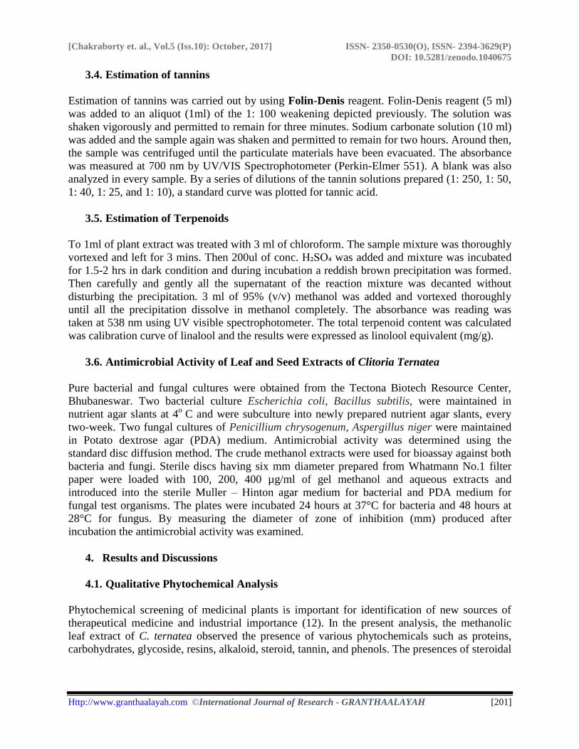
4.2.1. Estimation of Phenol
The results of total phenol content in the examined plant extract using the Folins Ciocalteu’s
method using Gallic acid to plot the standard curve (graph: 1 ). These were expressed in terms
of Gallic acid equivalent (the standard curve equation: y = 0.0102x ;R² = 0.9494). The results of
total phenol content of Clitoria ternatea extract is showing in Table 2. Our results with
methanolic leaf and seed extract in Clitoria ternatea extract showed a significant increase in
phenol content (#P < 0.001). Total phenol content in Clitoria ternatea leaf and seed was 67.25
[Chakraborty et. al., Vol.5 (Iss.10): October, 2017] ISSN- 2350-0530(O), ISSN- 2394-3629(P)
DOI: 10.5281/zenodo.1040675
Http://www.granthaalayah.com ©International Journal of Research - GRANTHAALAYAH [203]
µg/mL and 79.70 µg/mL. Our results indicated higher total phenol content in methanolic extract
of Clitoria ternatea plant compare to aqueous extract.
Graph 1: Standard curve for Gallic acid
Table 2: Estimated Phenol content present in the extracts of Clitoria ternatea
Extracts Total phenolic content
(mg of GAE/100 g of dry mass).
Leaves Aqueous (S1) 54.01960784
Leaves methanol (S2) 67.25490196
Seed aqueous (S3) 45.39215686
Seed methanol (S4) 79.70588235
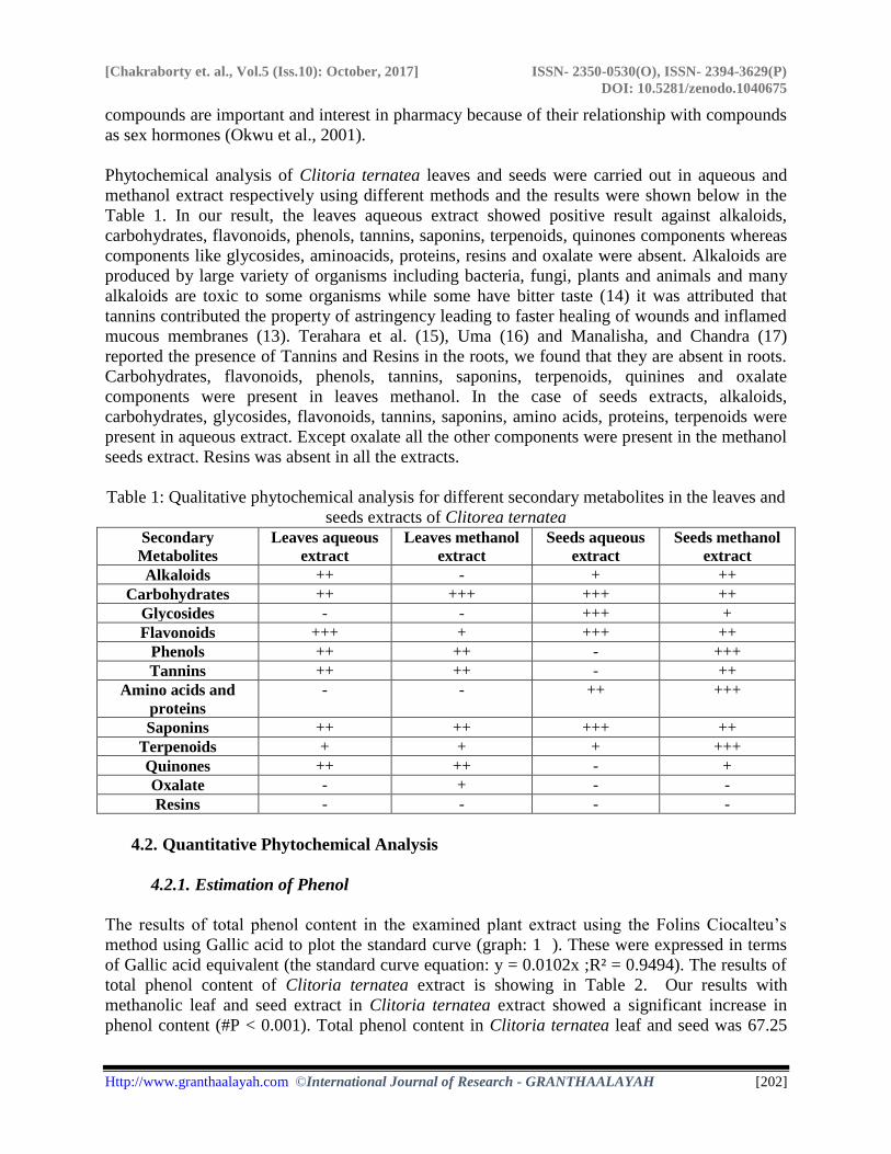
4.2.2. Estimation of Flavonoid
The total flavonoid contents in the examined different plant extract using the Aluminium
Chloride Colorimetric Assay were expressed in terms of Quercetin equivalent (the standard
curve equation: y = 0.0007x; R² = 0.916). The results of total flavonoid contents of Clitoria
ternatea leaf and seed extract is showing in Table 3. Quercetin was used to plot the standard
curve (graph 2). The values obtained for the concentration of total flavonoid were expressed as
mg of quercetin equivalents/100 g of dry mass .In this case leaves methanol extract have a high
content of flavonoid.
Graph 2: Standard curve for Quercetin
[Chakraborty et. al., Vol.5 (Iss.10): October, 2017] ISSN- 2350-0530(O), ISSN- 2394-3629(P)
DOI: 10.5281/zenodo.1040675
Http://www.granthaalayah.com ©International Journal of Research - GRANTHAALAYAH [204]
Table 3: Estimated flavonoid content present in the extracts of Clitoria ternatea
Extracts Total flavonoid content
(mg of Quercetin /100 g of dry mass).
Leaves Aqueous (S1) 147.1428571
Leaves methanol (S2) 271.4285714
Seed aqueous (S3) 85.71428571
Seed methanol (S4) 5.571428571
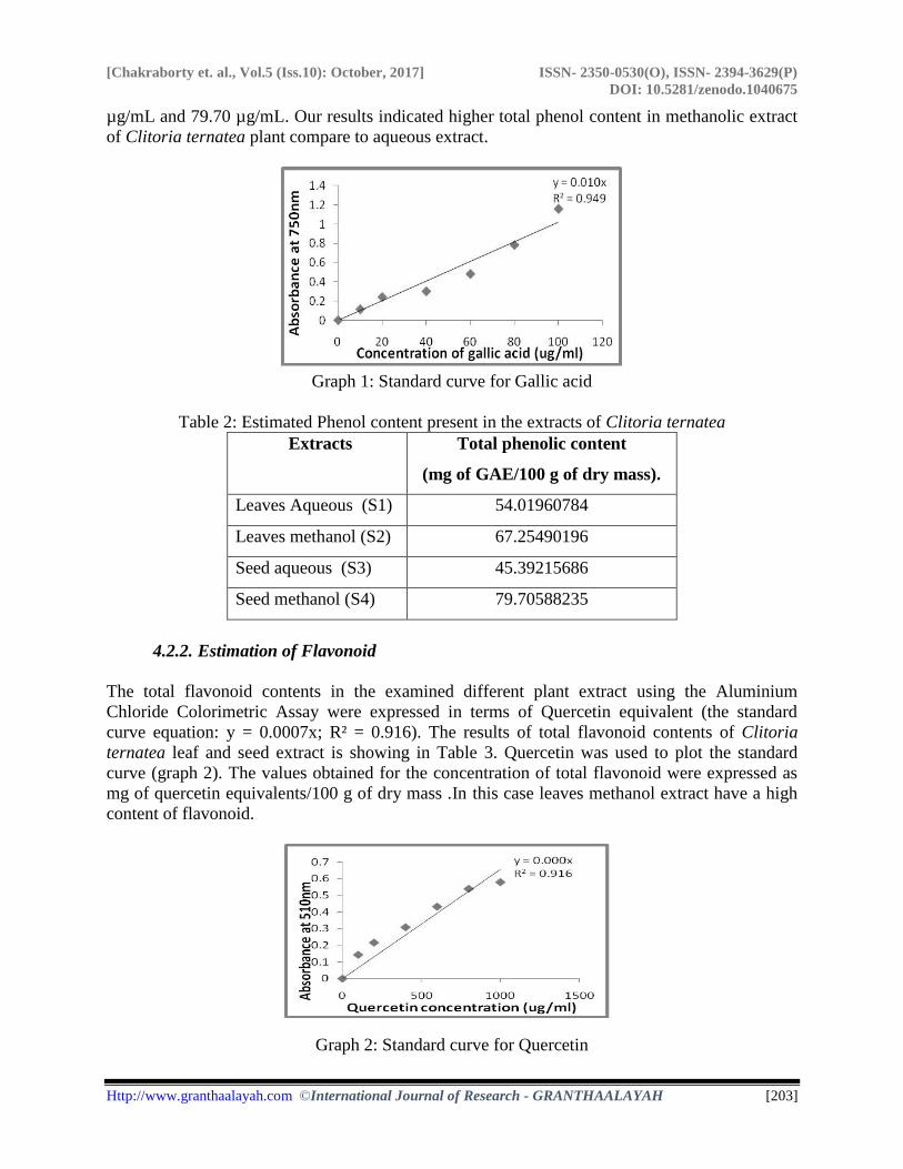
4.2.3. Estimation of Carbohydrate
Graph 3: Standard curve for Glucose
The total carbohydrate content in the different plant extract using the Anthrone method were
expressed in terms of glucose equivalent (the standard curve equation : y = 1.1966x ;R² =
0.9181) (graph. 3). The values obtained for the concentration of total glucose were expressed as
mg of glucose equivalents/100 mg of dry mass. The total carbohydrates contents in the examined
extracts were tabulated in Table 4 where all the extracts almost have a same amount of
carbohydrates but as calculated seed methanol showed (78.22) a high content.
Table 4: Estimated carbohydrates content present in the extracts of Clitoria ternatea
Extracts Total glucose content
(mg /100 mg of sample)
Leaves Aqueous (S1) 77.6784222
Leaves methanol(S2) 76.96807622
Seed aqueous (S3) 77.05164633
Seed methanol(S4) 78.22162795
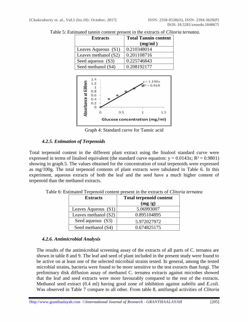
4.2.4. Estimation of Tannins
The total tannin content in the different plant extract using the Folin-Denis reagent method were
expressed in terms of tannic acid equivalent (the standard curve equation : y = 0.3247x ;R² =
0.9678) (graph. 4). The values obtained for the concentration of total tannic acid were expressed
as mg/m. The total tannin contents in the examined extracts were tabulated in Table 5. The
content of tannin was observed low in all the extracts.
[Chakraborty et. al., Vol.5 (Iss.10): October, 2017] ISSN- 2350-0530(O), ISSN- 2394-3629(P)
DOI: 10.5281/zenodo.1040675
Http://www.granthaalayah.com ©International Journal of Research - GRANTHAALAYAH [205]
Table 5: Estimated tannin content present in the extracts of Clitoria ternatea.
Extracts Total Tannin content
(mg/ml )
Leaves Aqueous (S1) 0.210348014
Leaves methanol (S2) 0.201108716
Seed aqueous (S3) 0.225746843
Seed methanol (S4) 0.208192177
Graph 4: Standard curve for Tannic acid
4.2.5. Estimation of Terpenoids
Total terpenoid content in the different plant extract using the linalool standard curve were
expressed in terms of linalool equivalent (the standard curve equation: y = 0.0143x; R² = 0.9801)
showing in graph.5. The values obtained for the concentration of total terpenoids were expressed
as mg/100g. The total terpenoid contents of plant extracts were tabulated in Table 6. In this
experiment, aqueous extracts of both the leaf and the seed have a much higher content of
terpenoid than the methanol extracts.
Table 6: Estimated Terpenoid content present in the extracts of Clitoria ternatea
Extracts Total terpenoid content
(mg /g)
Leaves Aqueous (S1) 5.06993007
Leaves methanol (S2) 0.895104895
Seed aqueous (S3) 5.972027972
Seed methanol (S4) 0.674825175
4.2.6. Antimicrobial Analysis
The results of the antimicrobial screening assay of the extracts of all parts of C. ternatea are
shown in table 8 and 9. The leaf and seed of plant included in the present study were found to
be active on at least one of the selected microbial strains tested. In general, among the tested
microbial strains, bacteria were found to be more sensitive to the test extracts than fungi. The
preliminary disk diffusion assay of methanol C. ternatea extracts against microbes showed
that the leaf and seed extracts were more favourably compared to the rest of the extracts.
Methanol seed extract (0.4 ml) having good zone of inhibition against subtilis and E.coli.
Was observed in Table 7 compare to all other. From table 8, antifungal activities of Clitoria
[Chakraborty et. al., Vol.5 (Iss.10): October, 2017] ISSN- 2350-0530(O), ISSN- 2394-3629(P)
DOI: 10.5281/zenodo.1040675
Http://www.granthaalayah.com ©International Journal of Research - GRANTHAALAYAH [206]
ternatea was to be found less effective to the positive controls. The different extract of
Clitoria ternatea in different concentration showed different spectrum of activities, especially
by the disk diffusion method where the microorganisms tested produced difference zones of
inhibition. It was observed that, the extracts of difference parts of Clitoria ternatea have
different efficacy against the selected microorganisms. These differences could be due to the
nature and level of the antimicrobial agents present in the extracts and their mode of action
on the different test microorganisms (18). Haripriya et.al. (19) Observed that petroleum ether
extracts of S. involvens showed higher antibacterial activity against E. coli and
Pseudomonas. Similarly in the present study also, Methanol extracts of leaf and seed showed
the maximum zone of activity against all bacteria and fungus pathogen.
Table 7: Formation of zone of inhibition against some selected bacteria
Concentration
of extracts
(ml)
Zone of Inhibition (mm)
Leaves Methanol Leaves water Seeds methanol Seeds water
E.coli. B.subtilis. E.coli. B.subtilis. E.coli. B.subtilis. E.coli. B.subtilis.
0.1 14 17 23 12 16 16 16 15
0.2 30 22 15 14 36 19 17 17
0.4 24 23 12 15 38 33 20 18
0.6 26 27 -ve 16 22 26 17 16
In some cases, it was observed that the zone of inhibition decreases with the increase of extracts
which indicates that the microbes/ pathogen have adapted a resistivity towards the extracts that
were used. Hence small quantities of plant extracts also are effective to effective to control the
growth of bacteria and fungus. Otherwise there was a chance that the organisms become resistant
towards the extracts.
5. Conclusion
Natural plant compounds have also been shown to antimicrobial properties and may be an
alternative to synthetic chemical agents. The present study concluded that the presence of
multiactive secondary metabolites which was present in the leaf and seed extracts of the Clitoria
ternatea have made the plant a very important medicinal plant. Seed methanol extract have high
content of phenol whereas leaves methanol have high content of flavonoid. Carbohydrate content
was almost equal in all the extract. Tannin was of low content in all the extract. Leaves aqueous
extract contain terpenoid. The positive results of antimicrobial activity against the bacteria and
the fungus also revealed the importance of this plant. In conclusion, all plant extracts of Clitoria
ternatea possessed activity against at least one strain of bacteria and/or fungi. Further studies
aimed at the isolation and identification of active substances from the methanol extracts of C.
ternatea could also disclose compounds with better value for food preservation as well as natural
plant based medicine. It would become a base for the development of new drug which could be
useful in treatment of diseases.
[Chakraborty et. al., Vol.5 (Iss.10): October, 2017] ISSN- 2350-0530(O), ISSN- 2394-3629(P)
DOI: 10.5281/zenodo.1040675
Http://www.granthaalayah.com ©International Journal of Research - GRANTHAALAYAH [207]
Acknowledgements
The authors are very much thankful to Dr Shovan Kumar Mishra, Director of Tectona Biotech
Resource Centre, Bhubaneswar, Odisha for providing the necessary facilities to carry out the
research work. The authors are also grateful to research centre realizing the need of research,
training and education for generating interdisciplinary human resource relevant to biotechnology
for creating best skill development and research in biotechnology through co-operation and help
from all sectors providing a place where innovation enterprise and Industrial development will
generate as far as practicable.
References
[1] Darsini, I. P., Shamshad, A. S. 2013. Antimicrobial Activity and Phytochemical Evaluation of
Clitoria ternatea. International Journal of Science and Research 4:823-825.
[2] Esmail, A., Snafi A. 2016. Pharmacological Importance of Clitoria ternatea – A review. Journal
of Pharmacy 6:68-83.
[3] Gupta, G.K., Chahal J., Bhatia M. 2010. Clitoria ternatea (L.): Old and new aspects. Journal of
Pharmacy Research 3:610-2614.
[4] Haripriya, D., Selvan, N., Jeyakumar, N., Periasamy, R., Johnson, M., Irudayaraj, V. 2010. The
effect of extracts of Selaginella involvens and Selaginella inaequalifolia leaves on poultry
pathogens. Asian Pac J Trop Med 3:723-726.
[5] Indumathi, C., Durgadevi, G., Nithyavani, S., Gayathri, P.K. 2014. Estimation of Terpenoid
Content and Its Antimicrobial Property in Enicostemma litorrale International Journal of
ChemTech Research 6:4264-4267.
[6] Kamtekar, S., Keer, V., Patil, V. 2014. Estimation of Phenolic Content, Flavonoid Content,
Antioxidant and Alpha-amylase Inhibitory Activity of Marketed Polyherbal Formulation . Journal
of Applied Pharmaceutical Science 4:061-065.
[7] Kavitha, L.R., and Premalakshmi, V. 2013. Phytochemical Analysis of Ethanolic Extract of
Leaves of Clitoria ternatea. International Journal of Pharma and BioSciences 4:236–242.
[8] Lakshmia, D. M., Mahithaa, B., Madhavia, T., Sushma, J. 2015. N. Phytochemical Screening and
Ftir Analysis of Clitoria ternatea Leaves. International Journal of Scientific & Engineering
Research 6:287-290.
[9] Barik, D.P., Naik, S.K., Mudgal, A. and Chand, P.K. 2007. Rapid Plant regeneration through in
vitro auxiliary shoot proliferation of butterfly pea (Clitoria ternatea L.) a twining legume. In vitro
cell. Dev. Biol. Plant 43:144-148.
[10] Yogendrasinh, B., Solanki., and Sunita. M. 2010. Immunomodulatory activity of ayurvedic plant
aparajita in male albino rats. Glo. j. science frontier res. 10: 2-8.
[11] Pahune, B., Niranjane K., Danao, K., Bodhe, M., and Rokade, V. 2013. Antimicrobial Activity of
Clitoria ternatea L. flower extract and use as a natural indicator in acid base titration. J. Nat. Prod.
Plant Resour. 3:48-51.
[12] Salhan, M., Kumar, B., Tiwari, P., Sharma, P., Sandhar, H.K., and Gautam, M. 2011.
Comparative Anthelmintic Activity of aqueous and ethanolic leaf extracts of Clitoria ternatea.
International Journal of Drug Development and Research 3: 68-69.
[13] Okwu, D.E. and Josiah, C. 2006. Evaluation of the chemical composition of two Nigerian
medicinal plants. Afri. J. Biotech 5:357-361.
[14] Gupta, G.K., Chahal, J., and Bhatia, M. 2010. Clitoria ternatea (L.) Old and new aspects. Journal
of Pharmacy Research 3 (11) 2610-2614.
[15] Terahara, N., Saito, N., Matsui, T., Osmajima, Y., and Saito. N., 1996. Five New Anthocyanins
A3, B4, B3, B2 and D2 from Clitoria ternatea. J Nat Prod 59: 139-144.
[Chakraborty et. al., Vol.5 (Iss.10): October, 2017] ISSN- 2350-0530(O), ISSN- 2394-3629(P)
DOI: 10.5281/zenodo.1040675
Http://www.granthaalayah.com ©International Journal of Research - GRANTHAALAYAH [208]
[16] Uma, B. 2009. Phytochemical analysis and antimicrobial activity of Clitoria ternatea Linn.
against extended spectrum beta lactamase producing enteric and Urinary Pathogens. Asian
Journal of Pharmaceutical and Clinical Research 2:0974-2441.
[17] Manalisha, D., and Chandra, K.J. 2011. Preliminary phytochemical analysis and acute oral
toxicity study of Clitoria ternatea Linn. in albino mice. International Research Journal of
Pharmacy 2:139-140.
[18] Manjula, P., Sreekanth, M.C., Keerthi, B., Devi, B.P. 2013. Phytochemical Analysis of Clitoria
ternatea linn., A valuable medicinal plant. Journal of Indian Botanical Society 92: 173-178.
[19] Haripriya, D., Selvan, N., Jeyakumar, N., Periasamy, R,, Johnson, M., and Irudayaraj, V. 2010.
The effect of extracts of Selaginella involvens and Selaginella inaequalifolia leaves on poultry
pathogens. Asian Pac. J Trop Med.
*Corresponding author.
E-mail address: souvagya.tbrc@ gmail.com


![EXPERIMENTAL STUDY OF FLAT PLATE SOLAR COLLECTORS …granthaalayah.com/Articles/Vol5Iss10/15_IJRG17_A09_656.pdf[Shukla et. al., Vol.5 (Iss.10): October, 2017] ISSN- 2350-0530(O), ISSN-](https://img.pdfslide.net/doc/110x75/605209752259a936a171cba7/experimental-study-of-flat-plate-solar-collectors-shukla-et-al-vol5-iss10.jpg)