Embed Size (px)
Citation preview
STUDIES ON GRAMINICOLOUS SPECIES OF PHYLLACHORA NKE. in FCKL.
V.* A TAXONOMIC MONOGRAPH
[Manuscript submitted November 9, 19661
Summary
This monograph describes the genus Phyllachora as it occurs on members of the Gramineae, discusses the reliability of criteria for delimiting species, and gives a taxonomic account of the Phyllachora species found on grasses.
A world-wide survey of graminicolous Phyllachora specimens has led to the conclusion that of the 278 species and varieties named in the literature, only 95 are valid species and a further 21 are doubtful species.
Of the 95 valid species, three (P. americana, P, longinaviculata, and P. microsperma) have been given new names, and five (P, bulbosa, P. koondrookensis, P. platyelliptica, P. polytocae, and P, rostellispora) are new species.
Two species complexes are discussed. The P. fallax complex includes four species which, although fairly readily separated, have several features in common and may be better delimited in future as varieties. The P. shiraiana complex includes seven bambusicolous species, few of which have been seen by the author, and the published descriptions of which are closely similar. It is possible that six of these species are synonyms of P. shiraiana s. str.
A key to graminicolous Phyllachora species and a guide to its uses are provided as well as a host index, including 135 grass genera, and an index to Phyllachora species, their synonyms, and doubtful species.
Since Phyllachora graminis (Pers. ex Fries) Nke. in Fckl. was described by Nitschke in Fuckel's "Symbolae Mycologicae" (1869), almost 300 "species" have been recorded on hosts in the Gramineae and many more$ on hosts in many other plant families. When descriptions of species and groups of specimens are compared, it is evident that far fewer graminicolous species exist than the 300 named. Most likely the same is true of the non-graminicolous species of Phyllachora.
There are various explanations of this situation. One of the more apparent is that since Phyllachora species are obligate parasites, species delimitation has been strongly influenced by the concept of host specialization in the strictest sense. Para- doxically, P. graminis has been used as a "dustbin" species by many who ascribed to it any Phyllachora found on grass. In fact many species do have wide host ranges and are widely distributed geographically, so that some species names have many
* Part IV, Aust. J. Bot., 1964, 12, 265. t Department of Botany, University of Melbourne. f; The total number of names of Phyllachora spp. known to the author at present, including
homonyms, is 1023, of which 278 refer to graminicolous species.
Aust. J. Bot., 1967, 15, 271-375
272 D. G. PARBERY
synonyms. Another reason for the multiplicity of names is that several unstable and extremely variable characters have been relied on previously for delimiting species (Parbery and Langdon 1964). Consequently some grass genera are listed as hosts for many Phyllachora species; e.g. Panicum has 29, Paspalurn 16, and Andvopogon 15.
Previous work (Parbery 1963a) showed that the anatomy of the host and the interaction between the developing host and parasite have a marked effect on the ultimate morphology of the fungus. A study of the criteria used to delimit species of Phyllachora (Parbery and Langdon 1964) showed that most of the criteria previously used were unreliable. A complete revision of the genus Phyllachora, based on reliable criteria, is therefore necessary. In this monograph the scope has been limited to graminicoious species.
Taxonomic accounts often include repetitive descriptive detail of the genus in each species. This practice is not adopted here. Full descriptions are given for only the generic characters of Phyllachora and the details of each species which distinguish it from the others. For further detail of species descriptions, reference may be made to the original text.
The revision of the graminicolous species has been made after examining type material and other specimens of over two-thirds of the species and synonyms. Descriptions only were available for the rest. Comparisons have been made primarily by using the shapes of ascospores, appressoria, and spermatiophores and the relative length of the ascus pedicel. Characters which were useful but not wholly reliable were ascospore and spermatiophore size.
When descriptions only of species were available, details of the above criteria alone were considered. Although some species have been reduced to synonyms when only descriptions have been examined, many have been regarded as valid. All of these, however, should be reconsidered if their type specimens can be found.
11. THE GENUS PHYLLACHORA
(a) Biology and Life Cycle
Phyllachora species live as biotrophs, usually in the leaf tissue of their hosts where they produce small, dark brown, shiny colonies referred to as "tar-spots". They differ from other biotrophic fungal parasites in that their mycelium is intra- cellulari~h a w n e x skikir te &-zit of endotrophic mycorrhizal fungi (Parbery 1963a), instead of intercellular in the manner of downy mildew, white rust, and rust fungi. Their biotrophic nature explains the failure of various authors to grow them in artificial culture.
Generally Phyllachora species do not cause marked damage to their hosts so that they are rarely of economic importance. The relationship between Phyllachora species and their hosts is such that the host tissue does not usually die, at least until maturation of the fungal fructifications. During the early, and sometimes later, stages of infection, a chlorotic zone of host tissue surrounding the fungal colony may appear. As the colony reaches maturity, however, this zone usually disappears. In senescing leaves the green island phenomenon (Allen 1942; Thrower 1965) is commonly observed.
GRAMINICOLOUS PHYLLACHORA SPP. V 273
There is evidence that some caulicolous Phyllachora species may be able ta complete their development in dying or dead tissue, i.e. as necrotrophs. During the present investigation, observations of preserved material suggested that P. atrojigurans may be able to complete the development of its fructifications in dying or dead host tissue (it is caulicolous). Von Arx (personal communication) claims that in Europe some species, such as P. helvetica Fckl. and P. therophila (Desm.) von Arx & Muller, grow or ripen only in dead tissue. These species, he believes, are intermediate between other species of Phyllachora which are biotrophs (Parbery 1963a) and species of Glomerella which are necrotrophs. This is of interest, since Glomerella species such as G. montana (Sacc.) von Arx & Muller (Phyllachora montana Sacc.) do have rudi- mentary ciypei (:.E Prx & ?iln!!er 19.54) and are p~ssib!y related to LDhj?ll~chora species.
The observation that leaf-inhabiting Phyllachora species are not normally found in necrotic tissue is of importance to the taxonomist, because when colonies are occasionally found surrounded by a zone of dead tissue in an otherwise healthy leaf, it is a reliable indication that the Phyllachora colony has been attacked by a hyper- parasite. There are several reported conidial states of Phyllachora species (Orton 1944). There is strong justification for believing, however, that almost all if not all such reports relate to spores of various hyperparasites (Parbery and Langdon 1963~).
For no graminicolous Phyllachora species has the life cycle been fully determined. Orton (1956) discussed the life cycle of P. punctum fairly fully, but was unable to say if the spermatial state was essential for its completion. Parbery (1963~) studied the pattern and timing of stages in the life cycles of P. ischaemi and P. paspalicola (as P. parilis) but was unable to determine the means of survival. The life cycle of the leguminicolous species P. lespedezae is well documented (Miller 1954), but it is unlikely that it would be typical of the genus, in view of an overwintering stromatic state apparently absent in other species.
(b) Taxonomy
The genus Phyllachora is one of a group of ascomycete genera which are included in either the Sphaeriales (Orton 1944; Miller 1949; von Arx and Muller 1954) or the Xylariales (Luttrell 1951). Doidge (1942) gives the details of the early fortunes of the genus, including its recognition as being not dothideal by Petrak (1924) and Orton (1924). She also gives an account of how the genus was artificially divided into three, Catacauma, Phyllachora, and Trabutia, according to the position of the clypeus in the host, and points out that while Catacauma was untenable as a genus, Trabutia was a mixed genus containing some true Phyllachora species and some truly dothideal fungi for which the name has been retained.
A genus which sometimes causes confusion in literature dealing with Phyllachora is Endodothella. Theissen and Sydow (191.5)" transferred several Phyllachora species
* In earlier publications (Parbery and Langdon 1963a, 1964) one reference was quoted erroneously. Theissen, F., Sydow, S. J., and Sydow, H. (1915) should read: "Theissen, F., and Sydow, H. (1915)". The explanation is worth publishing as it may save others from the same mistake. In the original text the authors are given as "F. Theissen S.J. and H. Sydow". Apparently this does not refer to two Sydows. There was no "S. J. Sydow". The "S.J." after Theissen's name stands for "Society of Jesus". Theissen was a Jesuit priest.
274 D. G. PARBERY
to that genus, believing that they had two-celled ascospores. Re-examination of some of these by von Arx (1958) and Muller and von Arx (1962) has shown, however, that what was mistaken for a septum was a densely staining band in the equatorial region of the ascospore. Consequently Endodothella is now a synonym of Phyllachora (von Arx 1958). Other synonyms of the genus Phyllachora are listed by von Arx and Muller (1954). The name Discomycopsella should be deleted from it, however, and the name Lophiella should be added. The reasons for this are discussed in relation to Phyllachora longinaviculata.
The authority for the name of the genus Phyllachora is sometimes given as "Nke." (Theissen and Sydow 1915) and other times as "Fckl." (Parbery 1963; Parbery and Langdon 1963a, 19636, 1964). It is apparent, however, that according to the International Code of Botanical Nomenclature (Recommendation 46D, Montreal 1959) that neither of these citations is strictly correct. The correct one, Phyllachora Nke. in Fckl., is given by von Arx and Muller (1954) and has been adopted here. The reason is that Nitschke was the mycologist who described the genus and P. graminis, but Fuckel was the author of the book in which it was published, and according to the above rules his name must be included in the authority for the name. This also applies to the name Phyllachora graminis, which may be correctly given as Phyllachora graminis (Pers.) Nke. in Fckl. or Phyllachora graminis (Pers. ex. Fr.) Nke. in Fckl.
The type species for the genus is a graminicolous species, P. graminis, and all other graminicolous species fit the generic concept remarkably well. P. graminis is distinguished from all other species by its relatively small and predominantly ovoid to ovate-truncate ascospores, which are invariably monostichously arranged. The tar-spotted appearance it gives to its hosts is typical of all graminicolous species. In fact so similar are the characters of the clypeus and perithecia in all species, within the limits discussed in the following pages, that a repeated description of these in re-describing old or describing new species is unnecessary unless they are quite different, e.g. if the perithecia are stromatic.
The following subsection (c) describes the characteristics of the genus, as worked out for P. graminis by Petrak (1924), Orton (1924), Karbush (1927), and Miller (1949), and shown to be similar for other species by various other workers, particularly Miller (1949), Orton (1956), Parbery (1963a), and Lopez-Rosa and Sherwood (1966).
(c) Morphology
Many features of the colonies of species of Phyllachora have been used in the past to separate species. Parbery and Langdon (1964) examined and evaluated the various characters of Phyllachora for their use in delimiting species. In the following sections the characters are described and their value in taxonomic studies is assessed.
(i) Colonies Colonies usually develop in the tissue of the leaf blade, and occasionally in the
leaf sheath as well. In very few species do the colonies develop on the culms of their hosts. Among graminicolous species it is rare to find colonies developed elsewhere than in the leaf mesophyll, although they may be restricted to just beneath the
GRAMINICOLOUS PHYLLACHORA SPP. V 275
epidermis or even the cuticle. Colonies may be few or many, scattered or congregated, discrete or confluent, and epiphyllous, hypophyllous, or amphigenous. The position and arrangement of colonies depend on the initial intensity of the inoculum and the points of penetration. These characters are of no taxonomic significance (Parbery and Langdon 1964). Similarly colony size and shape depend on the influence of the host and not on the species of fungus.
(ii) Clypeus The only visible parts of a Phyllachova colony are the surfaces of the clypeus,
and these may appear on only one or on both sides of the leaf. The clypeus is the only stromatic structure developed by the great majority of species, and only two species are known to the author -P. themedae (Ananthanarayanan 1964) and P. lespedezae (Miller 1949, 1954) (which is leguminicolous) -which produce stromatic tissue deep in the leaf.
The clypeus of most graminicolous species is produced in the epidermal cells, prior to or concurrent with spermagonium and perithecium development (Parbery 1963~). It consists of densely packed dark hyphae, 2-8 p in diameter, confined to the epidermal cells. The depth of the epidermal cells determines the thickness of the clypeus. Consequently no characteristic of the clypeus is useful for separating species, even though this structure is a typical feature of the genus.
(iii) Vegetative Hyphae These are completely intracellular, septate, smooth, generally hyaline, and
vary in diameter from 2 to 6 p. They are most common in the palisade and spongy mesophyll cells of the host (Parbery 1963a) and pass from one cell to another by means of fine penetration pegs approximately 0.5 p in diameter, which are pro- duced from appressorium-like structures.
(iv) Perithecia The number of perithecia produced in any one colony is related to the size of
the colony, and may vary from one to many. Size and shape of perithecia are also variable, according to host and area of development (Parbery 1963a), but if un- restricted they are generally spherical, with the ostiolar region becoming fused to the clypeus. Perithecia frequently open through only one side of the leaf, but it is not uncommon to find that they open to both dorsal and ventral sides, even in a single colony. This also appears to be related only to the extent of development of the host, and not to specific identity. They are both paraphysate and periphysate, at least in the early stages of development, although these structures usually persist. Perithecia become ostiolate and the ostiole extends right through the clypeus. The perithecial wall consists of four to six layers of cells, and varies from 7 to 23 p thick, a range of 7-20 p being noted in single specimens.
(v) Pavaphyses Petrak (1948, 1955) refers to these structures as metaphyses, a term which has
not gained general acceptance and is not used in this monograph. Although no critical analysis of the usefulness of characters of paraphyses as
taxonomic criteria is available, it does not seem that they would be sufficiently reliable as aids to delimiting species.
276 D. G. PARBERY
Paraphyses have always been found in fresh specimens, where they are usually longer than the asci, and in some species they may be much longer. The degree to which the relative lengths of asci and paraphyses vary within a species is unknown. In the fungi examined fresh (Parbery 1962) the paraphysis width was usually 2-4 p, so that this character seems similar in all species.
Some species, such as P. oryzopsidis, produced a proportion of branched paraphyses, but most species do not. There is, however, insufficient knowledge about the reliability of this character, which is not recorded for most species. Two other characters which may be useful, but for which no information is available are: septation (some authors report them as septate, others as continuous) and apices. Some apices are tapered, others are rounded, and occasionaiiy they are swollen. Further studies of fresh material from a large number of specimens would need to be conducted before any judgment could be made on the usefulness of these characters for taxonomic purposes.
During the present study, paraphyses have been noted as filiform and con- tinuous, sometimes guttulate, rarely branched, and with somewhat variable apices. The paraphyses of most species examined soon after being collected, have been similar and could usually be described in these terms. Unfortunately no critical appraisal of the usefulness of paraphyses of Phyllachora species as taxonomic criteria has been made.
(vi) Ascus The ascus has a single wall which, although rather thick in the early stage of
development, becomes thinner as it stretches to accommodate the developing spores. It has an annular thickening around the apical pore, giving the ascus apex a coronate appearance when viewed laterally. This is referred to as the "ascus crown". The ascus crown is a feature of all Phyllachora species and is easily seen in developing and near-mature asci. All asci are pedicellate. The pedicel length varies but in almost all species is less than half the length of the spore-containing portion. This is considered short. Only one species has long pedicels - P. epicampis, where they are often equal in length to the ascosporific part. Ascus size is very variable, the largest frequently being twice as long as the shortest. Most specimens have asci within the length and breadth range of 40-80 by 8-10 p. There are some species with asci of larger or smaller dimensions.
Ascospores are usually at an oblique angle to the length of the ascus. They may be monostichous, distichous, or inordinate in arrangement. Some species, e.g. P, gmminis and P. punctum, always exhibit monostichous arrangements, but no species are known in which spores are always distichous or inordinate, monostichous spores always being found in asci of some specimens.
(vii) Ascospores These are usually hyaline, but are occasionally pale yellow. They are one-celled,
smooth-walled, and vary greatly in size and shape. Some species have very long narrow spores, e.g. P. bambusae, others almost spherical spores, e.g. P. sphaerosperma.
Shape, however, is constant and useful. Some species exhibit a variety of spore shapes, whereas other species produce very uniform spores. Ascospore shapes
GRAMINICOLOUS PHYLLACHORA SPP. V 277
vary from completely symmetrical to quite asymmetrical, so that it is often necessary to rotate spores in order to determine their actual shape.
Most species produce eight spores per ascus, a few only four, while P. quadra- spora can produce either eight- or four-spored asci.
Spore size is a useful guide in identifying species but is not very useful for the initial delimitation of species. This may appear paradoxical, but what is meant is this. Ideally it is possible to know the size limits of spores of a species only after all individuals of the species have been examined. This ideal implies two things: (1) that it is unlikely that the full range of spore size will ever be known for any species and therefore it is unwise to delimit new species because of unusual spore measurements ai"iiel (2) illat loeca-use there is a iciiowii raiige of spoi;e sizes for eac;i species
because there are differences in these ranges between some species, it is wise to use this existing knowledge to help identify species which have previously been delimited by other criteria. Too often it is expected that a consideration of spore size will lead directly to species identification when in fact it will only cut down the number of species possibilities and thus aid identification.
The ascospores of many species contain a dense band of cytoplasm in the equatorial region, from which dense strands radiate to the walls. Sometimes spores are found which are almost filled with a very dense yellow body of unknown nature. Others contain two or more such yellow bodies, which are regularly present in some species, e.g. P. orbiculata.
(viii) Appressoria
The shape of appressoria produced on and attached to a leaf surface is characteristic of a number of species (Parbery 1963b). Unfortunately the development of germinating spores of only a few species has been examined, so that appressorium shape is known only for very few and is therefore of little aid to taxonomy yet. This position should improve as more work is done.
(ix) Spermogonia
Many species of Phyllachora produce spermogonia. In graminicolous species the spermatia are scolecospores, rather than the classical amerosporic bacilloid spermatium shape, and the spermatial state has been placed in the form genus Linochora (von Hohnel 1910) (referred to earlier as Leptostromella by Parbery (1963~) and Parbery and Langdon (1963a, 1964)). Since some non-graminicolous Phyllachora species such as P. actinodaphnes, P. ambrosiae, and P. lespedeza produce spermatia of classical form, while other species such as P . langdonii and P. leptospermi produce the scolecosporic type, it is possible that some graminicolous species may produce bacilloid spermatum. So far only one, P. gracilis, has been found, in which bacilloid spermatia may be produced.
The spermatia are variable in length from about 7 to 40 p, and from 0.5 to 1.5 p in width. In some species, e.g. P. punctum, their length is fairly uniform, 7-12 p. In others, however, it is extremely variable. The spermatia are broadest near the base, and narrow slightly to the point of attachment. They taper gradually to the apex.
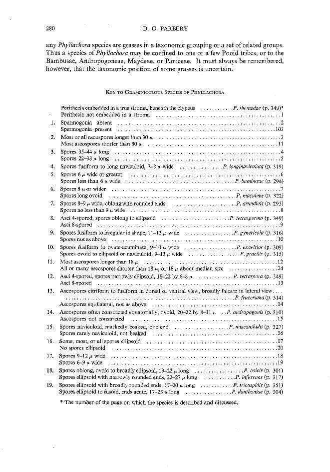
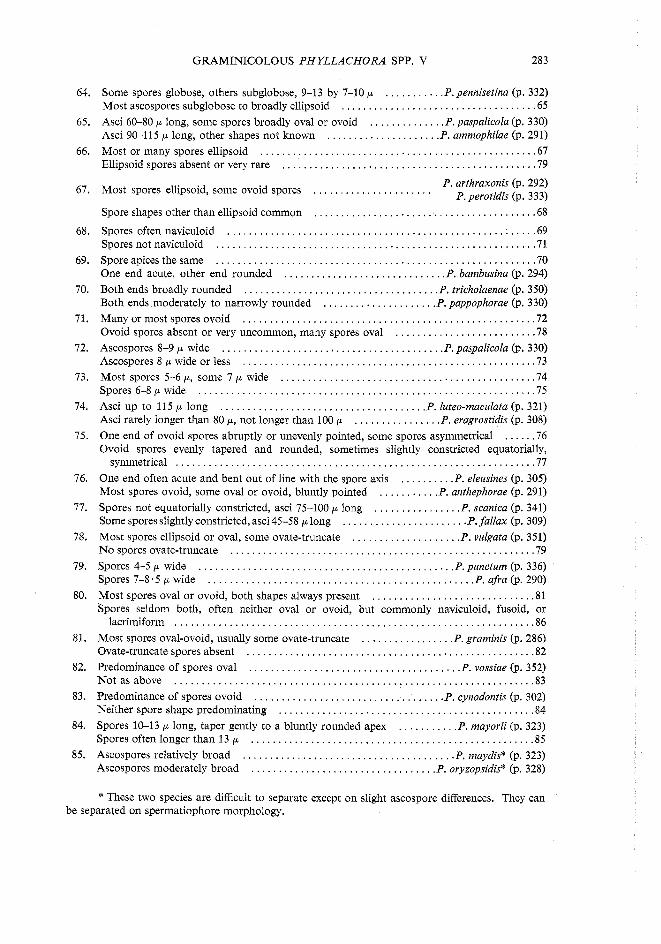
The shape of spermatiophores is important, being consistent for a particular species. These structures are usually unbranched and are produced in dense layers
D. G. PARBERY
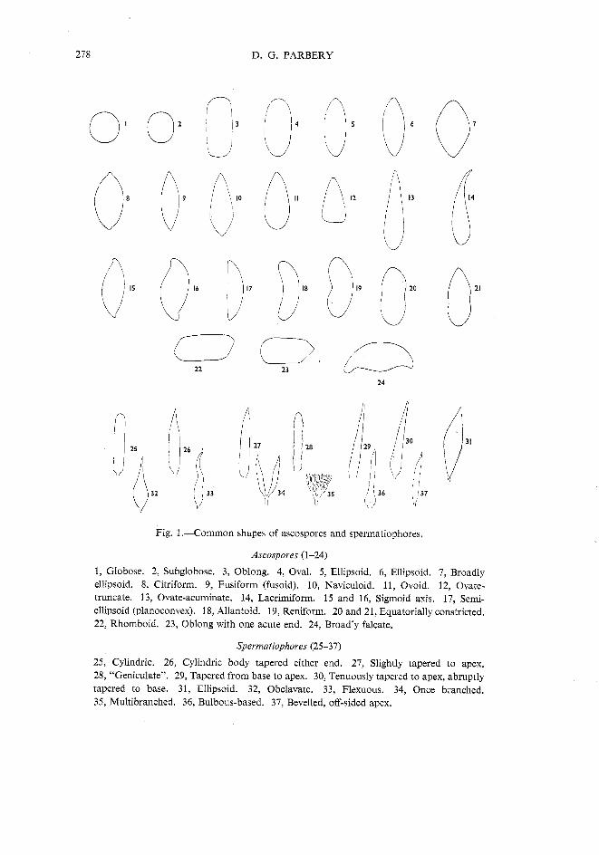
Fig. 1 .-Common shapes of ascospores and spermatiophores.
Ascospoves (1-24)
1, Globose. 2, Subglobose. 3, Oblong. 4, Oval. 5, Ellipsoid. 6, Ellipsoid. 7, Broadly ellipsoid. 8, Citriform. 9, Fusiform (fusoid). 10, Naviculoid. 11, Ovoid. 12, Ovate- truncate. 13, Ovate-acuminate. 14, Lacrimiform. 15 and 16, Sigmoid axis. 17, Semi- ellipsoid (planoconvex). 18, Allantoid. 19, Reniform. 20 and 21, Equatorially constricted. 22, Rhomboid. 23, Oblong with one acute end. 24, Broadly falcate.
Spevmatiophoves (25-37)
25, Cylindric. 26, Cylindric body tapered either end. 27, Slightly tapered to apex. 28, "Geniculate". 29, Tapered from base to apex. 30, Tenuously tapered to apex, abruptly tapered to base. 31, Ellipsoid. 32, Obclavate. 33, Flexuous. 34, Once branched. 35, Multibranched. 36, Bulbous-based. 37, Bevelled, off-sided apex.
GRAMINICOLOUS PHYLLACHORA SPP. V 279
from the floor of the spermagonium, but in some species, e.g. P. paspalicola and P. sylvatica, they are branched.
Some young Phyllachora colonies contain only spermogonia, beneath the clypeus, and infection by hyperparasites at this stage of development has often led to taxonomic confusion (Parbery and Langdon 1963~). Fertilization of ascogonia by the scolecosporic spermatia has not been observed, although there are three reports of fertilization by bacilloid spermatia. These reports relate to P. ambvosiae (Miller 1951), P. lespedezae (Miller 1954), and P. actinodaphnes (Tilak 1960).
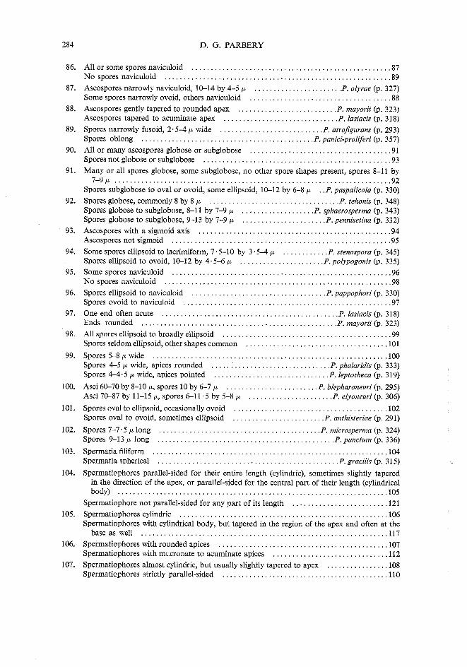
(x) Conidial State One species, P. quadraspora, is known to have a conidial state. The conidia of
this species are produced in colonies similar in appearance to the ascal state, and have been shown to be connected genetically to the ascal state (Parbery and Langdon 1963b).
(d) Use of the Key
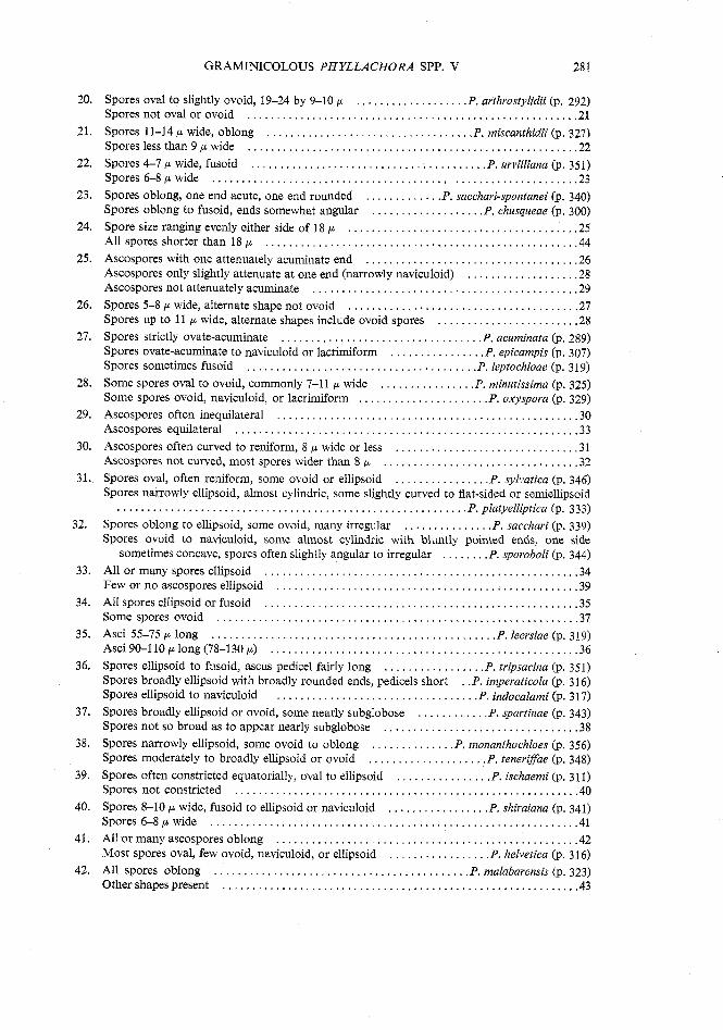
In order to facilitate the use of the key, the reader's attention is directed to the following points.
Since many Phyllachora species have spore shapes which show various outlines at different angles, and since some species produce spores with a range of shapes, it has been necessary for the sake of simple key construction to bring out some species in more than one place. The same applies to ascospore size. It is essential to realize that spore sizes found in specimens will frequently only approximate those given here. They will be in the same order, but will often have different limits.
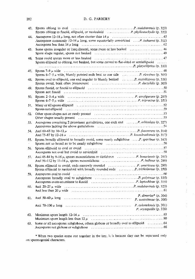
Some Phyllachora species cannot be separated satisfactorily on ascospore characters alone. At the same time, spermatial states are not found in every specimen, so that in the first 102 dichotomies, only ascospore and ascus characters are used. Consequently some species have been separated by only small and not very reliable differences, for the sake of including all species in this section of the key. Such species may be separated by reference to other less reliable characters, such as ascus size and spore arrangement as given in the description. Also, species which do not separate readily in the first part of the key are distinguished on spermatial states. It should be noted that in species which produce spermatia, the spermagonia will usually be found if the younger colonies are examined.
Figure 1 is provided as a ready definition of the shapes of ascospores and spermatiophores referred to in the key. In addition each species is illustrated in the plates.
Special mention is needed regarding use of the word "oval". In mycological literature, spores with broadly rounded ends and convex sides are commonly referred to as oval, in both outline and solid form. Mycologists generally accept this term and know its meanings. Ainsworth and Bisby (1961), however, point out that "oval" refers only to outline, and that the solid form should be called "broadened ellipsoid". It is considered here, however, that the use of the latter term would cause confusion; hence the term "oval" is used to refer to both outline and shape.
Finally, it may be helpful to consider the host genus in the context of its relationship to other groups of grasses, since this study has shown that the hosts of
280 D . G . PARBERY
any Phyllachora species are grasses in a taxonomic grouping or a set of related groups . Thus a species of Phyllachora may be confined to one or a few Pooid tribes. or to the Bambusae. Andropogoneae. Maydeae. or Paniceae . It must always be remembered. however. that the taxonomic position of some grasses is uncertain .
Perithecia embedded in a true stroma. beneath the clypeus . . . . . . . . . . . . P. themedae (p . 349)* Perithecia not embedded in a stroma . . . . . . . . . . . . . . . . . . . . . . . . . . . . . . . . . . . . . . . . . . . . . . 1 Spermogonia absent . . . . . . . . . . . . . . . . . . . . . . . . . . . . . . . . . . . . . . . . . . . . . . . . . . . . . . . . . . . . 2 Spermogonia present . . . . . . . . . . . . . . . . . . . . . . . . . . . . . . . . . . . . . . . . . . . . . . . . . . . . . . . . . . 103 Most or all ascospores longer than 30 p . . . . . . . . . . . . . . . . . . . . . . .. .. . . . . . ... . . . . . . . . 3 Most ascospores shorter than 30 p . . . . . . . . . . . . . . . . . . . . . . . . . . ... . . . . . . . . . . . . . . . . . . 11
. . . . . . . . . . . . . . . . . . . . . . . . . . . . . . . . . . . . . . . . . . . . . . . . . . . . . . . . . . . . . Spores 35-44plong 4 Spores 22-38 p long . . . . . . . . . . . . . . . . . . . . .. . . . . . . . . . . . . .. . . . . . . . . . . . . . . . . . . . . . . . 5
. . . . . . . . . . . . . . . . . . Spores fusiform to long naviculoid. 7-8 p wide P longinaviculata (p 319) Spores 6 p wide or greater . . . . . . . . . . . . . . . . . . . . . . . . . . . . . . . . . . . . . . . . . . . . . . . . . . . . . . . . 6 Spores less than 6 p wide . . . . . . . . . . . . . . . . . . . . . . . . . . . . . . . . . . . . . . . . P . bambusae (p . 294) Spores 8 p or wider . . . . . . . . . . . . . . . . . . . . . . . . . . . . . . . . . . . . . . . . . . . . . . . . . . . . . . . . . . . . . . 7 Spores long ovoid . . . . . . . . . . . . . . . . . . . . . . . . . . . . . . . . . . . . . . . . . . . . . . . P . maculans (p . 322) Spores 8-9 p wide. oblong with rounded ends . . . . . . . . . . . . . . . . . . . . . . . . P . arundinis (p . 293) Spores no less than 9 p wide . . . . . . . . . . . . . . . . . . . . . . . . . . . . . . . . . . . . . . . . . . . . . . . . . . . . . . . 8
. . . . . . . . . . . . . . . . . . . . . . . . . . . Asci 4.spored. spores oblong to ellipsoid P tetvasperma (p 349) Asci 8-spored . . . . . . . . . . . . . . . . . . . . . . . . . . . . . . . . . . . . . . . . . . . . . . . . . . . . . . . . . . . . . . . . . . . . 9 Spores fusiform to irregular in shape. 11-13 p wide . . . . . . . . . . . . . . . . . . P. gynevicola (p . 316)
. . . . . . . . . . . . . . . . . . . . . . . . . . . . . . . . . . . . . . . . . . . . . . . . . . . . . . . . . . . . . Sporesnotasabove 10 . . . . . . . . . . . . . . . . . . . . Spores fusiform to ovate.acuminate. 9-10 p wide P. excelsior (p 309) . Spores ovoid to ellipsoid or naviculoid. 9-1 3 p wide . . . . . . . . . . . . . . . . . . . P. gracilis (p 315)
Most ascospores longer than 18 p . . . . . . . . . . . . . . . . . . . . . . . . . . . . . . . . . . . . . . . . . . . . . . . . . 12 All or many ascospores shorter than 18 p. or 18 p about median size . . . . . . . . . . . . . . . . . . 24
. . . . . . . . . . . . . . Asci 4.spored. spores narrowly ellipsoid. 18-22 by 6-8 p P. tetraspova (p 349) Asci 8-spored . . . . . . . . . . . . . . . . . . . . . . . . . . . . . . . . . . . . . . . . . . . . . . . . . . . . . . . . . . . . . . . . . . 13 Ascospores citriform to fusiform in dorsal or ventral view. broadly falcate in lateral view . . . .
P frazeriana (p 314) . . . . . . . . . . . . . . . . . . . . . . . . . . . . . . . . . . . . . . . . . . . . . . . . . . . . . . . . . . . . . . . . Ascospores equilateral. not as above . . . . . . . . . . . . . . . . . . . . . . . . . . . . . . . . . . . . . . . . . . . . . . 14 Ascospores often constricted equatorially. ovoid. 20-22 by 8-11 p . . P. andropogonis (p . 310) Ascospores not constricted . . . . . . . . . . . . . . . . . . . . . . . . . . . . . . . . . . . . . . . . . . . . . . . . . . . . . . 15 Spores naviculoid. markedly beaked. one end . . . . . . . . . . . . . . . . . . . . P. miscanthidii (p . 327) Spores rarely naviculoid. not beaked . . . . . . . . . . . . . . . . . . . . . . . . . . . . . . . . . . . . . . . . . . . . . . 16 Some. most. or all spores ellipsoid . . . . . . . . . . . . . . . . . . . . . . . . . . . . . . . . . . . . . . . . . . . . . . . . 17 No spores ellipsoid . . . . . . . . . . . . . . . . . . . . . . . . . . . . . . . . . . . . . . . . . . . . . . . . . . . . . . . . . . . . . 20 Spores 9-12 p wide . . . . . . . . . . . . . . . . . . . . . . . . . . . . . . . . . . . . . . . . . . . . . . . . . . . . . . . . . . . . . 18 Spores 6-9 p wide . . . . . . . . . . . . . . . . . . . . . .. .. . . . . . . . . . . . . . . . . . . . . . . . . . . . . . . . . . . . . 19 Spores oblong. ovoid to broadly ellipsoid. 19-22 p long . . . . . . . . . . . . . . . . . . P . coicis (p . 301) Spores ellipsoid with narrowly rounded ends. 22-27 p long . . . . . . . . . . . . P. infiscans (p . 317)
. . Spores ellipsoid with broadly rounded ends. 17-20 p long . . . . . . . . . . . . P tricuspidis (p 351) Spores ellipsoid to fusoid. ends acute. 17-25 p long . . . . . . . . . . . . . . . . . P. danthoniae (p . 304)
* The number of the page on which the species is described and discussed .
GRAMINICOLOUS PHYLLACHORA SPP . V 281
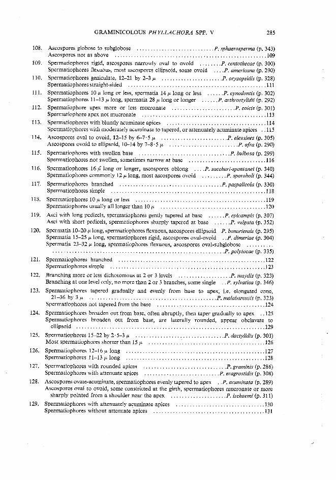
. Spores oval to slightly ovoid. 19-24 by 9-10 p . . . . . . . . . . . . . . . . . . . P. arthrostylidii (p 292) Spores not oval or ovoid . . . . . . . . . . . . . . . . . . . . . . . . . . . . . . . . . . . . . . . . . . . . . . . . . . . . . . . . 21
. Spores 11-14 p wide. oblong . . . . . . . . . . . . . . . . . . . . . . . . . . . . . . . . . . . P . miscanthidii (p 327) Spores less than 9 p wide . . . . . . . . . . . . . . . . . . . . . . . . . . . . . . . . . . . . . . . . . . . . . . . . . . . . . . . 22
. Spores 4-7 p wide. fusoid . . . . . . . . . . . . . . . . . . . . . . . . . . . . . . . . . . . . . . . . P. urvilliana (p 351) Spores 6-8 p wide . . . . . . . . . . . . . . . . . . . . . . . . . . . . . . . . . . . . . . . . . . . . . . . . . . . . . . . . . . . . . 23
. . . . . . . . . . . . . . Spores oblong. one end acute. one end rounded P. sacchari-spontanei (p 340) . Spores oblong to fusoid. ends somewhat angular . . . . . . . . . . . . . . . . . . . P. chusqueae (p 300)
Spore size ranging evenly either side of 18 p . . . . . . . . . . . . . . . . . . . . . . . . . . . . . . . . . . . . . . . 25 All spores shorter than 18 p . . . . . . . . . . . . . . . . . . . . . . . . . . . . . . . . . . . . . . . . . . . . . . . . . . . . . 44 Ascospores with one attenuately acuminate end . . . . . . . . . . . . . . . . . . . . . . . . . . . . . . . . . . . . 26 Ascospores only slightly attenuate at one edd (narrowly naviculoid) . . . . . . . . . . . . . . . . . . . 28 Ascospores not attenuately acuminate . . . . . . . . . . . . . . . . . . . . . . . . . . . . . . . . . . . . . . . . . . . . . 29 Spores 5-8 p wide. alternate shape not ovoid . . . . . . . . . . . . . . . . . . . . . . . . . . . . . . . . . . . . . . . 27 Spores up to 11 p wide. alternate shapes include ovoid spores . . . . . . . . . . . . . . . . . . . . . . . . 28
. Spores strictly ovate-acuminate . . . . . . . . . . . . . . . . . . . . . . . . . . . . . . . . . . P . acuminata (p 289) . . Spores ovate-acuminate to naviculoid or lacrimiform . . . . . . . . . . . . . . . . P epicampis (p 307)
. . Spores sometimes fusoid . . . . . . . . . . . . . . . . . . . . . . . . . . . . . . . . . . . . . . . P leptochloae (p 319) . Some spores oval to ovoid. commonly 7-11 p wide . . . . . . . . . . . . . . . . P. minutissima (p 325) . Some spores ovoid. naviculoid. or lacrimiform . . . . . . . . . . . . . . . . . . . . . . P. oxyspora (p 329)
Ascospores often inequilateral . . . . . . . . . . . . . . . . . . . . . . . . . . . . . . . . . . . . . . . . . . . . . . . . . . 30 Ascospores equilateral . . . . . . . . . . . . . . . . . . . . . . . . . . . . . . . . . . . . . . . . . . . . . . . . . . . . . . . . . . 33 Ascospores often curved to reniform. 8 p wide or less . . . . . . . . . . . . . . . . . . . . . . . . . . . . . . 31 Ascospores not curved. most spores wider than 8 p . . . . . . . . . . . . . . . . . . . . . . . . . . . . . . . . 3 2
. Spores oval. often reniform. some ovoid or ellipsoid . . . . . . . . . . . . . . . . P. sylvatica (p 346) Spores narrowly ellipsoid. almost cylindric. some slightly curved to flat-sided or semiellipsoid
P platyelliptica (p 333) . . . . . . . . . . . . . . . . . . . . . . . . . . . . . . . . . . . . . . . . . . . . . . . . . . . . . . . . . . . . . . Spores oblong to ellipsoid. some ovoid. many irregular . . . . . . . . . . . . . . . P. sacchari (p 339)
Spores ovoid to naviculoid. some almost cylindric with bluntly pointed ends. one side sometimes concave. spores often slightly angular to irregular . . . . . . . . P. sporoboli (p . 344)
All or many spores ellipsoid . . . . . . . . . . . . . . . . . . . . . . . . . . . . . . . . . . . . . . . . . . . . . . . . . . . . . 34 Few or no ascospores ellipsoid . . . . . . . . . . . . . . . . . . . . . . . . . . . . . . . . . . . . . . . . . . . . . . . . . . . 39 A11 spores ellipsoid or fusoid . . . . . . . . . . . . . . . . . . . . . . . . . . . . . . . . . . . . . . . . . . . . . . . . . . . . . 35 Some spores ovoid . . . . . . . . . . . . . . . . . . . . . . . . . . . . . . . . . . . . . . . . . . . . . . . . . . . . . . . . . . . . . 37
. Asci 55-75 p long . . . . . . . . . . . . . . . . . . . . . . . . . . . . . . . . . . . . . . . . . . . . . . . . P. leersiae (p 319) Asci 90-110 p long (78-130 p) . . . . . . . . . . . . . . . . . . . . . . . . . . . . . . . . . . . . . . . . . . . . . . . . . . . . 36
. Spores ellipsoid to fusoid. ascus pedicel fairly long . . . . . . . . . . . . . . . . . P. tripsacina (p 351) Spores broadly ellipsoid with broadly rounded ends. pedicels short . . P. imperaticola (p . 316) Spores ellipsoid to naviculoid . . . . . . . . . . . . . . . . . . . . . . . . . . . . . . . . . . P . indocalami (p . 317)
. Spores broadly ellipsoid or ovoid. some nearly subglobose . . . . . . . . . . . . P. spartinae (p 343) Spores not so broad as to appear nearly subglobose . . . . . . . . . . . . . . . . . . . . . . . . . . . . . . . . . 38
. . . . . . . . . . . . . . . Spores narrowly ellipsoid. some ovoid to oblong P. monanthochloes (p 356) . Spores moderately to broadly ellipsoid or ovoid . . . . . . . . . . . . . . . . . . . . P. teneriffae (p 348)
. . Spores often constricted equatorially. oval to ellipsoid . . . . . . . . . . . . . . . . P ischaemi (p 311) Spores not constricted . . . . . . . . . . . . . . . . . . . . . . . . . . . . . . . . . . . . . . . . . . . . . . . . . . . . . . . . . . 40
. . Spores 8-10 p wide. fusoid to ellipsoid or naviculoid . . . . . . . . . . . . . . . . . P shiraiana (p 341) Spores 6-8 p wide . . . . . . . . . . . . . . . . . . . . . . . . . . . . . . . . . . . . . . . . . . . . . . . . . . . . . . . . . . . . . . 41 All or many ascospores oblong . . . . . . . . . . . . . . . . . . . . . . . . . . . . . . . . . . . . . . . . . . . . . . . . . . . 42
. . Most spores oval. few ovoid. naviculoid. or ellipsoid . . . . . . . . . . . . . . . . . P helvetica (p 316) . All spores oblong . . . . . . . . . . . . . . . . . . . . . . . . . . . . . . . . . . . . . . . . . . . P . malabarensis (p 323)
Other shapes present . . . . . . . . . . . . . . . . . . . . . . . . . . . . . . . . . . . . . . . . . . . . . . . . . . . . . . . . . . . . 43
282 D . G . PARBERY
43 . Spores oblong to oval . . . . . . . . . . . . . . . . . . . . . . . . . . . . . . . . . . . . . . . P. malabarensis (p . 323) Spores oblong to fusoid, ellipsoid. or naviculoid . . . . . . . . . . . . . . . P . phyllostachydis (p . 333)
44 . Ascospores 12-18 p long. not often shorter than 14 p . . . . . . . . . . . . . . . . . . . . . . . . . . . . . . . . 45 Ascospores commonly 12-16 p long. some equatorially constricted . . . . P . ischaerni (p . 311) Ascospores less than 14 p long . . . . . . . . . . . . . . . . . . . . . . .. . . . . . . . . . . . . . . . . . . . . . . . . . . 62
45 . Some spores irregular or inequilateral. some more or less beaked . . . . . . . . . . . . . . . . . . . . . 46 . . . . . . . . . . . . . . . . . Spore shape regular. spores not beaked . . . . . . . . . . . . . . . . . . . . . . ... 49
. . . . . . . . . . . . . . . . . . . . . . . . . . . . . . . . . . . . . . . . . . . 46 . Some ovoid spores more or less beaked 47 Spores ellipsoid to oblong. not beaked. but some curved to flat-sided or semiellipsoid . . . . . .
. . . . . . . . . . . . . . . . . . . . . . . . . . . . . . . . . . . . . . . . . . . . . . . . . . . . . . . . . . . P . platyelliptica (p . 333) . . . . . . . . . . . . . . . . . . . . . . . . . . . . . . . . . . . . . . . . . . . . . . . . . . . . . . . . . . . . . . 47 . Spores 7-9 p wide 48
Speres 6-7.5 ;1 wide. blmt!y pointed ends bent to one side . . . . . . . . . . . P. eleusines (p . 105)
48 . Spores oval to ellipsoid. one end angular to bluntly beaked . . . . . . . .P. rostellispora (p . 338) . . . . . . . . . . . . . . . . . . . . . . . . . . . . . . Spores ovoid. beak often pronounced P. dactylidis (p 303)
Spores fusoid. or fusoid to ellipsoid . . . . . . . . . . . . . . . . . . . . . . . . . . . . . . . . . . . . . . . . . . . . . . . 50 . . . . . . . . . . . . . . . . . . . . . . . . . . . . . . . . . . . . . . . . . . . . . . . . . . . . . . . . . . . . . . Spores not fusoid 51
. Spores 2.5-4 p wide . . . . . . . . . . . . . . . . . . . . . . . . . . . . . . . . . . . . . . . . . . P . atrojigurans (p 293)
. Spores 6-7.5 p wide . . . . . . . . . . . . . . . . . . . . . . . . . . . . . . . . . . . . . . . . . . . . P. tripsacina (p 351) . . . . . . . . . . . . . . . . . . . . . . . . . . . . . . . . . . . . . . . . . . . . . . . . . . . . . . Manyorallsporesellipsoid 52
. . . . . . . . . . . . . . . . . . . . . . . . . . . . . . . . . . . . . . . . . . . . . . . . . . . . . . . . . . . . . Spores not ellipsoid 59 Other spore shapes not or rarely present . . . . . . . . . . . . . . . . . . . . . . . . . . . . . . . . . . . . . . . . . . . 53
. . . . . . . . . . . . . . . . . . . . . . . . . . . . . . . . . . . . . . . . . . . . . . . . . . . . . Othershapesusuallypresent 55 Ascospores containing 2 prominent guttulations. one each end . . . . . . . P. orbiculata (p . 327) Ascospores lacking the above guttulations . . . . . . . . . . . . . . . . . . . . . . . . . . . . . . . . . . . . . . . . . 54
Asci 60-65 by 10-12 p . . . . . . . . . . . . . . . . . . . . . . . . . . . . . . . . . . . . . . . . . . P. fuscescens (p . 314) Asci 75-87 by 12-1 6 p . . . . . . . . . . . . . . . . . . . . . . . . . . . . . . . . . . . . . . P . koondrookensis (p . 317) Spores broadly ellipsoid to broadly ovoid. some nearly subglobose . . . . P. spartinae (p . 343)
. . . . . . . . . . . . . . . . . . . . . . . . . . . . . . . . . . . . Spores not so broad as to be nearly subglobose 56
Spores ellipsoid to oval or ovoid . . . . . . . . . . . . . . . . . . . . . . . . . . . . . . . . . . . . . . . . . . . . . . . . . 57 Ascospores not oval but ovoid to naviculoid . . . . . . . . . . . . . . . . . . . . . . . . . . . . . . . . . . . . . . . 58
. . . . . . . . . . . . Asci 45-84 by 9-16 p. spores monostichous or distichous P . bonariensis (p . 295) . . . . . . . . . . . . . . . . . . . . . . . . Asci 96-1 12 by 11-16 p. spores monostichous P . bulbosa (p . 299)
Spores ellipsoid to ovoid. ends narrowly rounded . . . . . . . . . . . . . . . . . . P . americana (p . 290) Spores ellipsoid to naviculoid with broadly rounded ends . . . . . . . . . . P. tricholaenae (p . 350)
. . . . . . . . . . . . . . . . . . . . . . . . . . . . . . . . . . . . . . . . . . . . . . . . . . . . . . . . Ascospores oval to ovoid 60 Ascospores broadly oval to subglobose . . . . . . . . . . . . . . . . . . . . . . . . . . . P.polytocae (p . 335) Ascospores ovate-acuminate to fusoid . . . . . . . . . . . . . . . . . . . . . . . . . . . . P. leptochloae (p . 319)
. . . . . . . . . . . . . . . . . . . . . . . . . . . . . . . . . . . . . . . . . . . Asci 20-25 p wide P. malabarensis (p . 323) . . . . . . . . . . . . . . . . . . . . . . . . . . . . . . . . . . . . . . . . . . . . . . . . . . . . . . . . . Asci less than 20 p wide 61
61 . Asci 50-60 p long . . . . . . . . . . . . . . . . . . P . dimeriae* (p . 304) P . centothecae (p . 300)
. . . . . . . . . . . . . . . . . Asci 70-100 p long . . . . . . . . . . . . . . . . . . . . . .. P . coloradensis (p . 301) P . oryzopsidis (p . 328)
. . . . . . . . . . . . . . . . . . . . . . . . . . . . . . . . . . . . . . . . . . . . . . . . . . 62 . Maximum spore length 12-14 p 63 Maximum spore length less than 12 . . . . . . . . . . . . . . . . . . . . . . . . . . . . . . . . . . . . . . . . . . . . . 90
63 . Some or all ascospores subglobose. others globose or broadly oval to ellipsoid . . . . . . . . . . 64 Ascosporesnotgloboseorsubglobose . . . . . . . . . . . . . . . . . . . . . . . . . . . . . . . . . . . . . . . . . . . . . 66
* When two species come out together in the key. it is because they can be separated only on spermogonial characters .
GRAMINICOLOUS PHYLLACHORA SPP . V 283
. Some spores globose. others subglobose. 9-13 by 7-10 p . . . . . . . . . . . P.pennisetina (p 332) Most ascospores subglobose to broadly ellipsoid . . . . . . . . . . . . . . . . . . . . . . . . . .. . . . . . . . . 65 Asci 60-80 p long. some spores broadly oval or ovoid . . . . . . . . . . . . . . P . paspalicola (p . 330) Asci 90-115 p long. other shapes not known . . . . . . . . . . . . . . . . . . . . . P. ammophilae (p . 291) Most or many spores ellipsoid . . . . . . . . . . . . . . . . . . . . . . . . . . . . . . . . . . . . . . . . . . . . . . . . . . . 67 Ellipsoid spores absent or very rare . . . . . . . . . . . . . . . . . . . . . . . . . . . . . . . . . . . . . . . . . . . . . . . 79
P . arthvaxonis (p . 292) Most spores ellipsoid. some ovoid spores . . . . . . . . . . . . . . . . . . . . . . P . pevotidis (p . 333) Spore shapes other than ellipsoid common . . . . . . . . . . . . . . . . . . . . . . . . . . . . . . . . . . . . . . . . . . 68
. . . . . . . . . . . . . . . . . . . . . . . . . . . . . . . . . . . . . . . . . . . . . . . . . . . . . . . . . Spores often naviculoid 69 Spores not naviculoid . . . . . . . . . . . . . . . . . . . . . . . . . . . . . . . . . . . . . . . . . . . . . . . . . . . . . . . . . . . 71 Spore apices the same . . . . . . . . . . . . . . . . . . . . . . . . . . . . . . . . . . . . . . . . . . . . . . . . . . . . . . . . . . . 70 One end acute. other end rounded . . . . . . . . . . . . . . . . . . . . . . . . . . . . . . P. bambusina (p . 294) Both ends broadly rounded . . . . . . . . . . . . . . . . . . . . . . . . . . . . . . . . . . . . P. tricholaenae (p . 350) Both ends moderately to narrowly rounded . . . . . . . . . . . . . . . . . . . . . P. pappophorae (p . 330) Many or most spores ovoid . . . . . . . . . . . . . . . . . . . . . . . .. .. . . . . . . . . . . . . . . . . . . . . . . . . . . 72 Ovoid spores absent or very uncommon. many spores oval . . . . . . . . . . . . . . . . . . . . . . . . . . 78 Ascospores 8-9 p wide . . . . . . . . . . . . . . . . . . . . . . . . . . . . . . . . . . . . . . . . .P. paspalicola (p . 330) Ascospores 8 p wide or less . . . . . . . . . . . . . . . . . . . . . . . . . . . . . . . . . . . . . . . . . . . . . . . . . . . . . . 73 Most spores 5-6 p. some 7 p wide . . . . . . . . . . . . . . . . . . . . . . . . . . . . . . . . . . . . . . . . . . . . . . . 74 Spores 6-8 p wide . . . . . . . . . . . . . . . . . . . . . . . . . . . . . . . . . . . . . . . . . . . . . . . . . . . . . . . . . . . . . . 75 Asci up to 115 p long . . . . . . . . . . . . . . . . . . . . . . . . . . . . . . . . . . . . . .P . luteo-maculata (p . 321) Asci rarely longer than 80 p. not longer than 100 p . . . . . . . . . . . . . . . . P . eragrostidis (p . 308) One end of ovoid spores abruptly or unevenly pointed. some spores asymmetrical . . . . . . 76 Ovoid spores evenly tapered and rounded. sometimes slightly constricted equatorially.
symmetrical . . . . . . . . . . . . . . . . . . . . . . . . . . . . . . . . . . . . . . . . . . . . . . . . . . . . . . . . . . . . . . . . . . 77 One end often acute and bent out of line with the spore axis . . . . . . . . . . P. eleusines (p . 305) Most spores ovoid. some oval or ovoid. bluntly pointed . . . . . . . . . . . P. anthephorae (p . 291) Spores not equatorially constricted. asci 75-100 p long . . . . . . . . . . . . . . . . P. scanica (p . 341) Some spores slightly constricted. asci 45-58 p long . . . . . . . . . . . . . . . . . . . . . . . P. fallax (p . 309) Most spores ellipsoid or oval. some ovate-truncate . . . . . . . . . . . . . . . . . . . . P . vulgata (p . 351) Nosporesovate-truncate . . . . . . . . . . . . . . . . . . . . . . . . . . . . . . . . . . . . . . . . . . . . . . . . . . . . . . . . 79 Spores 4-5 p wide . . . . . . . . . . . . . . . . . . . . . . . . . . . . . . . . . . . . . . . . . . . . . . . P. punctum (p . 336)
. Spores 7-8.5 p wide . . . . . . . . . . . . . . . . . . . . . . . . . . . . . . . . . . . . . . . . . . . . . . . . . P . afra (p 290) Most spores oval or ovoid. both shapes always present . . . . . . . . . . . . . . . . . . . . . . . . . . . . . . 81 Spores seldom both. often neither oval or ovoid. but commonly naviculoid. fusoid. or
lacrinliform . . . . . . . . . . . . . . . . . . . . . . . . .. . . . . . . . . . . . . . . . . . . . . . . . . . . . . . . . . . . . . . . . 86 Most spores oval.ovoid. usually some ovate-truncate . . . . . . . . . . . . . . . . . P. graminis (p . 286) Ovate-truncate spores absent . . . . . . . . . . . . . . . . . . . . . . . . . . . . . . . . . . . . . . . . . . . . . . . . . . . . . 82
. Predominance of spores oval . . . . . . . . . . . . . . . . . . . . . . . . . . . . . . . . . . . . . . . P. vossiae (p 352) Not as above . . . . . . . . . . . . . . . . . . . . . . . . . . . . . . . . . . . . . . . . . . . . . . . . . . . . . . . . . . . . . . . . . . 83
. Predominance of spores ovoid . . . . . . . . . . . . . . . . . . . . . . . . . . . . . . . . . . . P. cynodontis (p 302) . . . . . . . . Neither spore shape predominating . . . . . . . . . . . . . . . . . . . . . . . . . . . . . . . . . . . . ... 84
Spores 10-13 p long. taper gently to a bluntly rounded apex . . . . . . . . . . . P. mayovii (p . 323) Spores often longer than 13 p . . . . . . . . . . . . . . . . . . . . . . . . . . . . . . . . . . . . . . . . . . . . . . . . . . . . 85 Ascospores relatively broad . . . . . . . . . . . . . . . . . . . . . . . . . . . . . . . . . . . . . . . P. maydis* (p . 323) Ascospores moderately broad . . . . . . . . . . . . . . . . . . . . . . . . . . . . . . . . . . P. ovyzopsidis* (p . 328)
* These two species are difficult to separate except on slight ascospore differences . They can be separated on spermatiophore morphology .
284 D . G . PARBERY
86 . All or some spores naviculoid . . . . . . . . . . . . . . . . . . . . . . . . . . . . . . . . . . . . . . . . . . . . . . . . . . . . 87 . . . . . . . . . . . . . . . . . . . . . . . . . . . . . . . . . . . . . . . . . . . . . . . . . . . . . . . . . . . No spores naviculoid 89
. 87 . Ascospores narrowly naviculoid. 10-14 by 4-5 p . . . . . . . . . . . . . . . . . . . . . . . P. olyrae (p 327) Some spores narrowly ovoid. others naviculoid . . . . . . . . . . . . . . . . . . . . . . . . . . . . . . . . . . . . . 88
. 88 . Ascospores gently tapered to rounded apex . . . . . . . . . . . . . . . . . . . . . . . . . . P. mayorii (p 323) . . Ascospores tapered to acuminate apex . . . . . . . . . . . . . . . . . . . . . . . . . . . . . . P lasiacis (p 318)
. 89 . Spores narrowly fusoid, 2.5-4 p wide . . . . . . . . . . . . . . . . . . . . . . . . . . . P. atrofigurans (p 293) . . Spores oblong . . . . . . . . . . . . . . . . . . . . . . . . . . . . . . . . . . . . . . . . . . . . . P panici-proliferi (p 357)
90 . All or many ascospores globose or subglobose . . . . . . . . . . . . . . . . . . . . . . . . . . . . . . . . . . . . . 91 Spores not globose or subglobose . . . . . . . . . . . . . . . . . . . . . . . . . . . . . . . . . . . . . . . . . . . . . . . . 93
91 . Many or all spores globose. some subglobose. no other spore shapes present, spores 8-11 by - - . . . . . . . . . . . . . . . . . . . . . . . . . . . . . . . . . . . . . . . . . . . . . . . . . . . . . . . . . . . . . . . . . . . . . . . . 7-9p YL
Spores subglobose to oval or ovoid, some ellipsoid, 10-12 by 6-8 p . . P. paspalicola (p . 330) . 92 . Spores globose, commonly 8 by 8 p . . . . . . . . . . . . . . . . . . . . . . . . . . . . . . . . . . P. tehonis (p 348)
. . Spores globose to subglobose. 8-11 by 7-9 p . . . . . . . . . . . . . . . . . . . P sphaerosperma (p 343) . Spores globose to subglobose, 9-13 by 7-9 p . . . . . . . . . . . . . . . . . . . . . . P. pennisetina (p 332)
. 93 Ascospores with a sigmoid axis . . . . . . . . . . . . . . . . . . . . . . . . . . . . . . . . . . . . . . . . . . . . . . . . . . 94 Ascospores not sigmoid . . . . . . . . . . . . . . . . . . . . . . . . . . . . . . . . . . . . . . . . . . . . . . . . . . . . . . . . . 95
. 94 . Some spores ellipsoid to lacrimiform, 7.5-10 by 3.5-4 p . . . . . . . . . . . . P. stenospora (p 345)
. Spores ellipsoid to ovoid. 10-12 by 4.5-6 p . . . . . . . . . . . . . . . . . . . . . . P.polypogonis (p 335) 95 . Some spores naviculoid . . . . . . . . . . . . . . . . . . . . . . . . . . . . . . .... ... . . . . . . . . . . . . . . . . . . 96
. . . . . . . . . . . . . . . . . . . . . . . . . . . . . . . . . . . . . . . . . . . . . . . . . . . . . . . . . . . No spores naviculoid 98 . 96 . Spores ellipsoid to naviculoid . . . . . . . . . . . . . . . . . . . . . . . . . . . . . . . . . . . P. pappophovi (p 330)
. . . . . . . . . . . . . . . . . . . . . . . . . . . . . . . . . . . . . . . . . . . . . . . . . . . Spores ovoid to naviculoid .. 97 . 97 . One end often acute . . . . . . . . . . . . . . . . . . . . . . . . . . . . . . . . . . . . . . . . . . . . . . P lasiacis (p . 318)
Ends rounded . . . . . . . . . . . . . . . . . . . . . . . . . . . . . . . . . . . . . . . . . . . . . . . . . . . P.mayorii(p.323) 98 . All spores ellipsoid to broadly ellipsoid . . . . . . . . . . . . . . . . . . . . . . . . . . . . . . . . . . . . . . . . . . . . 99
Spores seldom ellipsoid, other shapes common . . . . . . . . . . . . . . . . . . . . . . . . . . . . . . . . . . . . . 101
99 . Spores 5-8 p wide . . . . . . . . . . . . . . . . . . . . . . . . . . . . . . . . . . . . . . . . . . . . . . . . . . . . . . . . . . . . . 100 . Spores 4-5 p wide, apices rounded . . . . . . . . . . . . . . . . . . . . . . . . . . . . . . . P. phalaridis (p 333)
. Spores 4-4.5 p wide. apices pointed . . . . . . . . . . . . . . . . . . . . . . . . . . . . . . P leptotheca (p . 319)
. 100 . Asci 60-70 by 8-10 p. spores 10 by 6-7 p . . . . . . . . . . . . . . . . . . . . . . . . P . blepharoneuri (p 295) . . . . . . . . . . . . . . . . . . . . . . Asci70-87by 11-15p,spores6-11.5by 5-8p P.elyoneuri(p.306)
101 . Spores oval to ellipsoid, occasionally ovoid . . . . . . . . . . . . . . . . . . . . . . . . . . . . . . . . . . . . . . . . 102 . Spores oval to ovoid. sometimes ellipsoid . . . . . . . . . . . . . . . . . . . . . . . . P. anthisteriae (p 291)
102 . Spores 7-7.5 p long . . . . . . . . . . . . . . . . . . . . . . . . . . . . . . . . . . . . . . . . . . P . microsperma (p . 324) . Spores 9-13 p long . . . . . . . . . . . . . . . . . . . . . . . . . . . . . . . . . . . . . . . . . . . . . . P. punctum (p 336)
103 . Spermatia filiform . . . . . . . . . . . . . . . . . . . . . . . . . . . . . . . . . . . . . . . . . . . . . . . . . . . . . . . . . . . . . 104 . . Spermatia spherical . . . . . . . . . . . . . . . . . . . . . . . . . . . . . . . . . . . . . . . . . . . . . . . P gracilis (p 315)
104 . Spermatiophores parallel-sided for their entire length (cylindric). sometimes slightly tapered in the direction of the apex, or parallel-sided for the central part of their length (cylindrical body) . . . . . . . . . . . . . . . . . . . . . . . . . . . . . . . . . . . . . . . . . . . . . . . . . . . . . . . . . . . . . . . . . . . . . . 105
Spermatiophore not parallel-sided for any part of its length . . . . . . . . . . . . . . . . . . . . . . . . . 121 105 . Spermatiophores cylindric . . . . . . . . . . . . . . . . . . . . . . . . . . . . . . . . . . . . . . . . . . . . . . . . . . . . . . 106
Spermatiophores with cylindrical body, but tapered in the region of the apex and often at the base as well . . . . . . . . . . . . . . . . . . . . . . . . . . . . . . . . . . . . . . . . . . . . . . . . . . . . . . . . . . . . . . . . 117
106 . Spermatiophores with rounded apices . . . . . . . . . . . . . . . . . . . . . . . . . . . . . . . . . . . . . . . . . . . . 107 Spermatiophores with mucronate to acuminate apices . . . . . . . . . . . . . . . . . . . . . . . . . . . . . . 112
107 . Spermatiophores almost cylindric, but usually slightly tapered to apex . . . . . . . . . . . . . . . . 108 Spermatiophores strictly parallel-sided . . . . . . . . . . . . . . . . . . . . . . . . . . . . . . . . . . . . . . . . . . . 110
GRAMINICOLOUS PHYLLACHORA SPP . V 285
. Ascospores globose to subglobose . . . . . . . . . . . . . . . . . . . . . . . . . . . . P. sphaerosperma (p 343) Ascospores not as above . . . . . . . . . . . . . . . . . . . . . . . . . . . . . . . . . . . . . . . . . . . . . . . . . . . . . . . 109
. Spermatiophores rigid. ascospores narrowly oval to ovoid . . . . . . . . P. centothecae (p 300) Spermatiophores flexuous. most ascospores ellipsoid. some ovoid . . . . P. americana (p . 290)
. Spermatiophores geniculate. 12-21 by 2-3 p . . . . . . . . . . . . . . . . . . . . . . P. oryzopsidis (p 328) Spermatiophores straight-sided . . . . . . . . . . . . . . . . . . . . . . . . . . . . . . . . . . . . . . . . . . . . . . . . . . 111
. Spermatiophores 10 p long or less. spermatia 14 p long or less . . . . . . P. cynodontis (p 302) . . . . . . . Spermatiophores 11-13 p long. spermatia 28 p long or longer P. arthrostylidii (p 292)
. Spermaliophore apex more or less mucronate . . . . . . . . . . . . . . . . . . . . . . . P. coicis (p 301) . . . . . . . . . . . Spermatiophore apex not mucronate . . . . . . . . . . . . . . . . . . . . . . . . . . . . . . . .. 113
Spermatiophores with bluntly acuminate apices . . . . . . . . . . . . . . . . . . . . . . . . . . . . . . . . . . . . 114 Speriiiatiopiiores with modcraiciy acuminatc to tapered. or attenuately acuminate apices . . 1 i 5
. Ascospores oval to ovoid. 12-15 by 6-7.5 p . . . . . . . . . . . . . . . . . . . . . . . . P. eleusines (p 305) . . Ascospores ovoid to ellipsoid. 10-14 by 7-8.5 p . . . . . . . . . . . . . . . . . . . . . . . . . P afra (p 290)
. Spermatiophores with swollen base . . . . . . . . . . . . . . . . . . . . . . . . . . . . . . . . . P. bulbosa (p 299) Spermatiophores not swollen. sometimes narrow at base . . . . . . . . . . . . . . . . . . . . . . . . . . . . 116
Spermatiophores 16 long or longer. ascospores oblong . . . . P. sacchari-spontanei (p . 340) . Spermatiophores commonly 12 p long. most ascospores ovoid . . . . . . . . .P. sporoboli (p 344)
. . Spermatiophores branched . . . . . . . . . . . . . . . . . . . . . . . . . . . . . . . . . . . . . P paspalicolu (p 330) . . . . . . . . . . . . . . . . Spermatiophores simple . . . . . . . . . . . . . . . . . . . . . . . . . . . . . . . . . . . . .... 118
Spermatiophores 10 p long or less . . . . . . . . . . . . . . . . . . . . . . . . . . . . . .. ... . . . . . . . . . . . . 119 Spermatiophores usually all longer than 10 p . . . . . . . . . . . . . . . . . . . . . . . . . . . . . . . . . . . . . 120
Asci with long pedicels. spermatiophores gently tapered at base . . . . . . P. epicampis (p . 307) Asci with short pedicels. spermatiophores sharply tapered at base . . . . . . P. vulgata (p . 352)
Spermatia 10-20 p long. spermatiophores flexuous. ascospores ellipsoid P . bonariensis (p . 295) Spermatia 15-25 p long. spermatiophores rigid. ascospores oval-ovoid . . P. dimeriae (p . 304) Spermatia 23-32 p long. spermatiophores flexuous. ascospores oval-subglobose . . . . . . . . . .
. . P polytocae (p 335) . . . . . . . . . . . . . . . . . . . . . . . . . . . . . . . . . . . . . . . . . . . . . . . . . . . . . . . . . . . . . . Spermatiophores branched . . . . . . . . . . . . . . . . . . . . . . . . . . . . . . . . . . . . . . . . . . . . . . . . . . . . . 122 Spermatiophores simple . . . . . . . . . . . . . . . . . . . . . . . . . . . . . . . . . . . . . . . . . . . . . . . . . . . . . . . . 123
. Branching more or less dichotomous at 2 or 3 levels . . . . . . . . . . . . . . . . . . . P. maydis (p 323) Branching at one level only. no more than 2 or 3 branches. some single . . P. sylvatica (p . 346)
Spermatiophores tapered gradually and evenly from base to apex. i.e. elongated cone. . 21-36 by 3 p . . . . . . . . . . . . . . . . . . . . . . . . . . . . . . . . . . . . . . . . . . . . . . P. malabarensis (p 323)
Spermatiophores not tapered from the base . . . . . . . . . . . . . . . . . . . . . . . . . . . . . . . . . . . . . . . 124
Sperrnatiophores broaden out from base. often abruptly. then taper gradually to apex . . 125 Sperrnatiophores broaden out from base. are laterally rounded. appear obclavate to
ellipsoid . . . . . . . . . . . . . . . . . . . . . . . . . . . . . . . . . . . . . . . . . . . . . . . . . . . . . . . . . . . . . . . . . . . . 129
. Spermatiophores 15-22 by 2.5-3 p . . . . . . . . . . . . . . . . . . . . . . . . . . . . . . . . P. dactylidis (p 303) Most spermatiophores shorter than 15 p . . . . . . . . . . . . . . . . . . . . . . . . . . . . . . . . . . . . . . . . . . 126
Spermatiophores 12-16 p long . . . . . . . . . . . . . . . . . . . . . . . . . . . . . . . . . . . . . . . . . . . . . . . . . . 127 Spermatiophores 11-13 p long . . . . . . . . . . . . . . . . . . . . . . . . . . . . . . . . . . . . . . . . . . . . . . . . . 128
Spermatiophores with rounded apices . . . . . . . . . . . . . . . . . . . . . . . . . . . . . . P. graminis (p . 286) . Spermatiophores with attenuate apices . . . . . . . . . . . . . . . . . . . . . . . . . . . P . eragrostidis (p 308)
Ascospores ovate-acuminate. spermatiophores evenly tapered to apex . . P . acuminata (p . 289) Ascospores oval to ovoid. some constricted at the girth. spermatiophores mucronate or more
. sharply pointed from a shoulder near the apex . . . . . . . . . . . . . . . . . . . . P. ischaemi (p 311)
Spermatiophores with attenuately acuminate apices . . . . . . . . . . . . . . . . . . . . . . . . . . . . . . . . 130 Spermatiophores without attenuate apices . . . . . . . . . . . . . . . . . . . . . . . . . . . . . . . . . . . . . . . . 13 1
286 D. G. PARBERY
130. Ascospores fusoid to long naviculoid . . . . . . . . . . . . . . . . . . . . . . . . . P. longinaviculata (p. 3 19) Ascospores oval, ovoid to ovate-acuminate . . . . . . . . . . . . . . . . . . . . . .P. minutissima (p. 325)
131. Spermatiophores longer than 10 p, sometimes branched . . . . . . . . . . . . . .P. sylvatica (p. 346) Spermatiophores shorter than 10 p . . . . . . . . . . . . . . . . . . . . . . . . . . . . . . . . . . . . . . . . . . . . . .I32
132. Ascospores 9-13 p, oval to ellipsoid, not guttulate . . . . . . . . . . . . . . . . . . .P, punctum (p. 336) Ascospores 12-18 p, ellipsoid, biguttulate . . . . . . . . . . . . . . . . . . . . . . . . .P, ovbiculata (p. 327)
IV. TAXONOMIC ACCOUNT
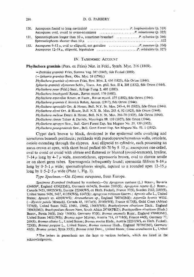
Phyllachora graminis (Pers. ex Fries) Nke. in Fckl., Symb. Myc. 216 (1869).
= Dothidea graminis Fries, Summa Veg. 387 (1845), fide Fuckel (1869). (=Sphaeria graminis Pers., Obs. Myc. 18 (1796).) Phyllachora gvaminis elymovum Fries, Syst. Myc. 2, 434 (1823), fide Orton (1944). Sphaeria graminis elymorum Schiv., Trans. Am. phil. Soc. 11 4, 208 (1832), fide Orton (1944). Phyllachora poae (Fckl.) Sacc., Sylloge Fung. 2, 603 (1883). Phyllachora brachypodii Roum., Revue mycol. 170 (1885). Phyllachora asperellae Roum. et Fautr., Revue mycol. 175 (1892), fide Orton (1944). Phyllachora gvaminis f. histvicis Rehm, Ascom. (1917), fide Orton (1944). Phyllachova agvostidis Ort. & House, Bull. N.Y. St. Mus. 243-4, 91 (1923), fide Orton (1944). Phyllachora elymi Ort. & House, Bull. N.Y. St. Mus. 243-4, 92 (1923), fide Orton (1944). Phyllachora melicae Dearn & House, Bull. N.Y. St. Mus. 266-70 (1925), fide Orton (1944). Phyllachora cinnae Tehan & Daniels, Mycologia 19, 110 (1927), fide Orton (1944). Phyllachora agvopyri Saw., Bull. Govt Forest Exp. Stn Meguro No. 53, 159 (1952). Phyllachorapoae-pvatensis Saw., Bull. Govt Forest Exp. Stn Meguro No. 53, 1 (1952).
Clypei dark brown to black, developed in the epidermal cells overlying and sometimes beneath perithecia; perithecia with pseudoparenchymatous walls, ostiolate, ostiole extending through the clypeus. Asci ellipsoid to cylindric, each possessing an ascus crown at apex, with short basal pedicel 60-70 by 8-10 p; ascospores one-celled, oval to ovoid or ovoid with obtuse end flattened or blunted (ovoid-truncate), hyaline, 7-14 p long by 4-7 p wide, monostichous, appressoria brown, oval to clavate sessile or on short germ tubes. Spermagonia infrequently found; spermatia filiform 9-14 p long by 0.5-1 p wide; spermatiophores simple, tapered to a rounded apex 12-15 p long by 1.5-2.5 p wide (Plate 1, Fig. 1).
Type Specimen.-On Elymus europaeus, from Europe.
Specimens Examined (indicated b y numbers).-On Agvopyron caninum (L.) Beauv., Bavaria 6346(S)*, England 63562(IMI), Germany 6634(S), Sweden 2181(S); Agvopyron vepens (L.) Beauv., Canada 5423, 18925(WS), Europe 22249(WS, ex Herb. Fuckel), France 37(S), Sweden 2182,2183(S), United States 5426, 5427,41359(WS), 6147(S); Agropyron triticeurn Gaertn.; Agrostis alba L., United States; Agrostis sp. 42938(WS); Arrenathevum sp., England 79074(IMI); Asperella hystrix Willd. (=Hystrix patula Moench), Canada 45, 1917a(S), 29169(WS), France 6173(S), Gold Coast (Africa) 7376(S), United States 5422, 13661, 13662, 13663(WS); Brachyelytvum silvaticum Hack., England 78943(IMI); Brachypodium Jlexum Nees, South Africa 29749(PRE) ; Brachypodium silvaticum (Huds.) Beauv., Persia 36(S), Italy 1309(S), Germany 87(S); Bvomus anomalis Rupr., England 97090(IMI), United States 34011(WS); Bromus asper Murray, Austria 718, 61718(S), France 44(S), Germany 77, 269(S); Bromus ciliatus L., United States; Bromus erectus Huds., Austria 22251(WS, ex Herb. Fuckel), 723(S); Bromus purgans L., United States; Bvomus ramosus Huds., England 70799(IMI), Germany 43(S); Bromus syriaci, Syria 35(S); Bvomus tvinii Desv., United States; Cinna avundinacea L., United
* The letters in parenthesis are the keys to various herbaria, which are listed in the acknowledgments.
GRAMINICOLOUS PHYLLACHORA SPP. V 287
States 39632, 46351(WS); Elymus canadensis L., United States 5420, 25743(WS); Elymus euvopaeus L., Germany 6212(S); Elymus glaucus Buckl., Canada 42093,42094(WS); Elymus mollis Trin., Japan 46(S); Elymus robustus Scribn. & Smith (=E. canadensis var. robustus), United States 5425(WS); Elymus sp., Ohio (U.S.A.) 5543(WS); Elymus villosus Muhl., United States 48231(WS); Elyrnus vivginicus L., United States 5425(WS); Poa annua L., Germany 1886(S); Poa nemoralis L., Bavaria 15186(WS), Germany 2079(S), United States 148765(WS); Poa sudetica (no authority given), Austria 22266(WS, ex Herb. Fuckel).
Discussion.-Unfortunately the type specimen of Phyllachora graminis has not been available to the present author, who was unable to trace its whereabouts. There seems little doubt, however, that the present concept of the species is an accurate one, for the problem of what constituted the type of P. graminis and thus for the genus Phyllachora was thoroughly investigated by Theissen and Sydow (1915). They found six sheets in the Leiden Herbarium with specimens labelled Sphaeria graminis. They were reasonably satisfied that Persoon had labelled these sheets. After discarding four of the sheets for various reasons, they were left with two, sheets 12 and 13, to choose from. They decided the specimen on sheet 12 was the type of P. graminis, since Persoon evidently received it first and also because the greater number of specimens from other herbaria labelled P. graminis corresponded with this. It seems logical to suppose, therefore, that these other specimens from various herbaria had been identified by either direct or indirect comparison with the specimen in sheet 12.
Specimen No. 12 was described as follows:
"Stromata through full leaf depth, longitudinal, dull black, slightly arched in long axis of the leaf, approximately 4 mm long and 1 mm wide, tapering at both ends, flat and dull coloured on the underside of the leaf, on the top side raised in between 3 veins (ribs), and noticeable by the slightly glistening raised parts.
"On both sides of the leaf the epidermis is permeated by brown black stromata, as clypei; between these, occupying the entire thickness of the leaf lie subglobose perithecia crowded together. The remaining leaf tissue is invaded fairly densely by bright brown small celled parenchymatous stroma tissue." [This should be evaluated in the light of later findings by Petrak (1924), Orton (1924), Miller (1949), and Parbery (1963a).]
"The perithecia average 180-220 p wide, 145-170 p high, are narrowly constricted at the apex and grow together with the clypeus. The epidermis splits when ripe and with it also the perithecial apex forming an opening. The nucleus (centrum) is surrounded by a brown wall 10-12 p thick formed from the stroma.
"The asci are paraphysate with very short stalks, cylindric, without a blue reaction with iodine, 60-70 x 8-10 p.
"Spores up to 8, monostichous, ellipsoid 9-11 p long 4 p thick and 5-5.5 p wide."
It must be realized that this description was made with a different concept of the biology of the species in mind, so that it needs to be evaluated in the light of what is said earlier in this monograph in relation to morphology.
During the present study nine specimens identified as Phyllachora graminis occurring on E. europeus from Germany, on other species of Elymus, and on Agropyron repens, a host recognized by Fuckel, have been available. One specimen from Fuckel's herbarium (22249, WS), collected in Europe and identified by him as P. graminis, has been accepted as an authentic specimen of this species. The spores of this specimen were oval to ovoid or ovate-truncate, monostichous in arrangement, and measured 8-12 p by 4-7 p. An ascus crown was visible in each ascus, which was unitunicate
288 D. G. PARBERY
and had a short basal pedicel. No spermatia were found. In a second specimen on Agvopyvon vepens from Canada (5423, WS) spermatia were found. In this specimen the ascospores were identical in shape and size with those in other specimens on Elymus and Agropyvon spp., so that there was no doubt of its being P. gvaminis. Each spermatium was filiform, gradually tapered to its apex, briefly tapered to its base, hyaline, non-septate, and measuring 11-18 p by 0.5 p (most commonly 15 p
long). The spermatiophores were simple, very gradually tapering to a rounded though fairly fine apex, and measured 10-19 p long by 1.5-2 p wide. The spermatia and spermatiophores were very similar to those found in specimens on Bvomus spp. (36+37, S). Saccardo (1883) also described filiform spores for P. gvaminis, but the host was not stated.
Grove (1937) applied the name Leptostromella graminis to the spermatial state of P. graminis, which he found on Agropyron repens. Von Arx and Muller (1954) made a study of P. gvaminis and also found the spermatial state (which is well illus- trated) on Agropyvon repens. Their description of the spermatia gives sizes twice those given here; however, there is little doubt that it is the same since the morphology is similar to that described here. Petrak (1956) claims that the spermatial state is not a Leptost~omella, but is a Linochova. The conclusion from the present study is that Petrak is correct, and the correct name should be Linochova gvaminis (Grove) nom. nov. = Leptostromella graminis Grove, British Stem and Leaf Fungi (Cambridge), p. 194 (1937).
I t has been possible to give a fuller and more accurate description of P. gvaminis now that the above specimens have been examined.
Synonymy
Ten of the synonyms were listed by Orton (1944) and have been accepted, since the original descriptions were close to P. graminis; also the type specimens of several of the species have been examined by the author, who agrees with Orton's conclusions. The following names were also reduced to synonyms for the reasons given.
Phyllachora poae (Fckl.) Sacc. One specimen (22266, WS) on Poa sudetica originally identified by Fuckel was examined, and although in poor condition, the few spores found were similar to those of Phyllachora graminis. Other specimens of P. graminis have also been found on Poa nemoralis from Europe and North America, and on P. sudetica from Europe. The original description of Phyllachorapoae, which did not include a range of ascospore size but gave them only as 6 by 3 p, was otherwise no different from that of P. graminis.
Phyllachora brachypodii Roum. Although the type specimen of this fungus has not been seen, it is regarded as P. graminis. Three other specimens of Phyllachora on Brachypodium species were examined, and all were P. graminis. Theissen and Sydow (1915) examined a specimen on Brachypodium sp. which they called P. bromi, so that it is possible, allowing for the doubt about the validity of P. bromi, that two species occur on Brachypodium. Apart from giving spore dimensions slightly broader than for P, graminis, the original description does not distinguish the above from P. graminis.
Phyllachora agropyri Saw. No specimen was seen, but the description is very similar to P. graminis. Agropyron spp. are common hosts of P. graminis.
Phyllachora poae-pratensis Saw. The illustrations of the spores of this species are identical with those of P. graminis, including the ovate-truncate type which occurs only in P. graminis and P. vulgata. The description also agrees with that of P. graminis.
GRAMINICOLOUS PHYLLACHORA SPP. V 289
Host Range.-Agropyron, Agrostis, Arrenatherum, Brachyelytrum, Brachypodium, Bromus, Calamagrostis, Cinna, Elymus, Festuca, Hordeum, Hystrix, Melica, Phleum, Poa, Tritium, Uniola.
There are many reports of P. graminis occurring on other grasses. Specimens of many of these have been examined, but were discovered to be other species. It is noteworthy that no specimen of Phyllachora found on hosts in the Bambusae or the Panicoideae has been P. graminis, although many such claims have been made. Among the tribes of the Pooideae only four, Agrostidae, Avenae, Festucae, and Hordeae, contain genera which act as hosts of P. graminis.
Geographical Distribution.-Austria, Bavaria, Canada, England, France, Germany, Gold Coast of Africa, Italy, Japan, Persia, South Africa, Sweden, Taiwan, and northern United States. This species is widespread in Europe, Canada, and the northern United States. It has been found in Japan and Taiwan, but not in southern Asia. One specimen from Persia has been seen. Only two authentic identifications have been made in the southern hemisphere; these were specimens from the Gold Coast of Africa and South Africa. P. graminis has not been found in South America, the southern Pacific, south-east or central Asia, or in Australia.
Phyllachora acuminata Starb., Archiv. Bot. 51, 11 (1905).
Phyllachora cornispora-necrotica Chard., Boln R. Soc. esp. Hist. nat. 28, 116 (1928). Phyllachora ortonii Chard., J. Dep. Agric. P. Rico 13, 11 (1929). Phyllachora murilloi Garces, Caldasia No. 2, 86 (1941).
Asci cylindrical-clavate, briefly stipitate, 90-140 by 10-18 p, paraphysate; ascospores monostichous or distichous, commonly ovate-acuminate with tenuous often bent apices, other end rounded or acute, some spores ovoid, 14-17 by 5-7 p ;
spermatia filiform, hyaline, 12-20 by 0.5-1 p ; spermatiophores broadening out from their bases, then tapering fairly evenly to their apices, 8.5-10.5 by 1.5 p (Plate 3, Fig. 27).
Type Specimen.-On Paspalum sp., from Salta, Argentina, 4(S).
Specimens Examined.-On Paspalum elongatum Griseb., Brazil 12676(S); Paspalum sp. Argentina 4(S).
Discussion.-Phyllachora acuminata and its three synonyms were all listed as synonyms of P. cornispora by Orton (1944). All these fungi had been found on species of Paspalum in South America. The specimens examined were of Phyllachora acuminata, which is different from P . cornispora (=P. minutissima) in ascospore shape and width, as well as in spermatiophore shape and size. The general shape of each of these characters is similar in each species but whereas P. acuminata has narrow elongate spores, P. minutissima has broader, shorter ones. Similarly, the spermatio- phores of the two species are generally similar, but there are slight differences in shape and marked differences in size between them.
P. cornispora-necrotica was found on Paspalum virgatum L. and was described in similar terms to Phyllachora acuminata. P. ortonii was found on Paspalum millegrana Schrad. in Puerto Rico, and has slightly smaller spores than the type specimen of P. acuminata, 13-15 by 5-6 p, but similar in shape. Phyllachora ortonii Tilak (1959)
290 D. G. PARBERY
is a homonym of P. ortonii Chard., but is a different species. Phyllachora murilloi was found on Paspalum virgatum and is the same as P. acuminata. This species is distributed from Guatemala down to Argentina in South America.
Phyllachora afra Syd., Annls mycol. 37, 220 (1939).
Asci cylindric, briefly stipitate; ascospores monostichous, ellipsoid to ovoid, more or less pointed, but broadly rounded, 10-14 by 7-8.5 p; spermatia 12-15 by 0 .5 p, filiform; spermatiophores long, nearly flexuous, pointed, 12-14 by 1-1.5 p (Plate 5, Fig. 51).
Type Specimen.-On Sporobolus pyramidalis Beauv., from Sierra Leone. Specimen Examined.-On Sporobolus myrianthus Benth., Nigeria 99380(IMI).
This species has been found only in the central-west African region. It is quite distinct in both ascospore and spermatial characters from other species of Phyllachora occurring on Sporobolus species. The type specimen has not been seen, but the morphology of the ascospores in specimen 99380 agreed with the description. Spermatia were not found by Sydow (1939).
Phyllachora americana nom. nov.
~Sphaer ia nervisequia Schw., Trans. Am. phil. Soc. 11 4, 208 (1832). --Phyllachora nervisequia (Schw.) Ort., Mycologia 36, 25 (1944). Sphaeria andropogonis Schw., Trans. Am. phil. Soc. 11 4, 209 (1832). Phyllachora andropogonis Ell. & Ev., N. Am. Fungi 2828 (1893).
Asci cylindric to narrowly ellipsoid, briefly stipitate, 95-125 by 15-19 p; ascospores ellipsoid, occasionally nearly ovoid, 12-17 by 6-8 p ; spermatiophores cylindric-flexuous, slightly tapered to the apex which is rounded, tapered to the base, 13-15 by 1.5-2 p (Plate 5, Fig. 56).
Type Specimen.-On Andropogon sp., from Pennsylvania, U.S.A. Specimen Examined.-On Andropogon elliottii Chapm., Georgia (U.S.A.) 6163(S).
Synonymy
Sphaeria nervisequia, S. andropogonis, and Phyllachora andropogonis were listed as synonyms of P. nervisequia (Schw.) Ort. (Orton 1944) and have been accepted as such since the descriptions are similar.
Phyllachora nervisequia. This name is a homonym of P. nervisequia Wint., the name of a non- graminicolous species published in February 1885 (Winter 1885) and predating P. nervisequia Cooke, also a non-graminicolous species, published in March 1885 (Cooke 1885).
Host Range.-Andropogon s. str.
Distribution.-South-eastern United States, the West Indies, and possibly South America (see remarks on distribution of P. fallax).
Discussion.-The name Phyllachora americana was chosen because this species is found only on the American continent and the nearby West Indies.
Phyllachora americana is quite similar to P. dimeriae, except that the ascospores are ellipsoid rather than ovoid, the spermatiophores are smaller than and lack the
GRAMINICOLOUS PHYLLACHORA SPP. V 291
acuminrde apices of P. dimeriae, and the spermatia also are much smaller than those of P. dimeriae.
P. americana may also be difficult to distinguish from some isolates of P. .fallax if ascospores only are present. The spermatiophores of P. fallax are smaller than those of P. americana, and have a different shape and acuminate apices. Most isolates of P. ,fallax exhibit some ascospores with median constrictions, a character not present in P. americana. It is unlikely, however, that this possible confusion would arise since P. fallax is believed not to occur in the American continent or the West Indies, whereas P. americana is known only from those regions.
A third species with which this fungus is likely to be confused is P. luteo- maculata, which has slightly broader and shorter spores that are ovoid to ellipsoid. There is no known spermatial state of P. luteo-maculata.
Phyllachora ammophilae Ort., Mycologia 36, 39 (1944).
Phyllachora calamagrostis Saw., Bull. Govt Forest Exp. Stn Meguro No. 53, 135-94 (1952).
Asci cylindrical, 90-115 by 10-15 p ; ascospores broadly ellipsoid, often sub- spherical, monostichous, 10-1 5 by 6-9 p.
Type Specimen.-On Ammophila arenaria (L.) Link, from New York State, U.S.A.
Host Range.-Ammophila and Calamagrostis.
Distribution.-North America, Taiwan.
Specimens of neither of the above fungi have been examined. The descriptions, however, are very similar, and since the two host genera are closely related, P. calamagrostis is given as a synonym of P. ammophilae.
Phyllachora anthephorae Syd., Annls mycol. 13, 39 (1915).
Asci cylindrical, briefly stipitate, paraphysate, 60-70 by 10-14 p ; ascospores ovoid, having a bluntly acute end (which is sometimes slightly beaked) or oval to ellipsoid, monostichous, 1 1-14 by 6.5-7 5 p (Plate 4, Fig. 36).
Type Specimen.-On Anthephora hermaphrodita (L.) Kuntze, from Jamaica, 6(S).
S~ecimen Examined.-On Anthephora hermaphrodita: Jamaica 6@).
Discussion.-An examination of the type material has shown that the ascospores of this species are longer and narrower than described by Sydow (1915).
This species is known on the type host only, but Orton (1944) found it in the Dominican Republic and Puerto Rico, as well as Jamaica. He found ascospores similar to those described here and asci up to 100 p long.
Phyllachora anthistiriae Racib. in Theiss. and Syd., Annls mycol. 13, 439 (1915).
Asci cylindric, briefly stipitate, 70-80 by 7-9 p ; ascospores oval, ovoid, or ellipsoid, 8-12 by 4-5.5 p (Plate 4, Fig. 33).
Type Specimen.-On Anthistiria ciliata.
Specimen Examined.-On Anthistiria arguens, 15(S).
292 D. G. PARBERY
Discussion.-Neither of the two above specimens have any recorded place of origin, consequently the place of origin is unknown, except that these hosts are common in south-east Asia (Bor 1960).
No authority for the name A. ciliata was given by Theissen and Sydow (1915), so that it could either be A. ciliata L., which is Themeda quadrivalvis (L.) 0. Ktze, or A. ciliata Nees, which is Themeda triandra Forssk., common in South Africa as well as in south-east Asia.
The specimen examined came from Sydow's collection and agreed with the description. No authority was given for this host either. It could be A. arguens Nees, which is Themeda triandra Forssk., or it could be A. arguens Willd., which is Themeda arguens (L.) Hack. If the former then it is quite possible that the specimen examined was the type for P. anthisteriae. The ascospores are unlike those of any other Phyllachora.
Phyllachora arthraxonis P. Henn., Hedwigia 43, 142 (1904).
Phyllachora arthraxon-hispidi Saw., Rep. Govt Res. Inst. Dep. Agric. Formosa No. 85, 25 (1943).
Asci clavate or cylindric to clavate, briefly stipitate, 36-72 by 10-15 p, paraphy- sate; ascospores mostly ellipsoid, some slightly ovoid, sometimes with slightly sigmoid axes, 11-1 3 by 4.5-5.5 p (Plate 3, Fig. 23).
Type Specimen.-On Arthraxon hispidus (Thunb.) Makino (as Arthraxon ciliare Beauv.), from Japan, 14(S).
Specimen Examined.-On Arthraxon hispidus, Japan 14(S); Arthraxon hispidis Makino var. typicus Honda, Taiwan (TNS, type o f P. arthraxonis-hispidi).
Sawada (1943) claimed that P. arthraxon-hispidi was different from P. arthraxonis because the latter had much smaller spores and asci. A re-examination has shown that the ascospores in the type material are larger than described by Hennings. Consequently, since the fungus found in Taiwan on A. hispidus var. typicus Honda is slightly larger than the Japanese material (Plate 6, Fig. 71), the size difference is insignificant. The spore shape is the same.
This species of Phyllachora has been found only on Arthraxon from Japan and Taiwan.
Phyllachora arthrostilidii Pet. & Cif., Annls mycol. 30, 232 (1932).
Sphaerodothis portoricensis Chard., J . Dep. Agric. P. Rico 16, 189 (1932). Phyllachora portoricensis (Chard.) Ort., Mycologia 36, 49 (1944).
Asci clavate, briefly stipitate, paraphysate, 90-120 by 20-25 p; ascospores usually distichous, oval to slightly ovoid, the narrower end not very different from the other, 19-24 by 83-10 p; spermatia long, filiform, flexuous, 28-37 by 0.5 p ;
spermatiophores cylindric, apex rounded or slightly narrowed becoming acuminate as spermatium develops, base not or slightly constricted, 11-1 3 by 1 -5-2.5 p (Plate 5, Fig. 49).
Type Specimen.-On Arthrostylidium multzspiculatum Pilger, from Dominican Republic, 22(S).
GRAMINICOLOUS PHYLLACHORA SPP. V 293
Specimen Examined.-On Arthvostylidium multiseptatum, Dominican Republic, 22, 3491(S).
Orton (1944) called this species P. portoricensis, a name combination given to a specimen collected on Arthrostylidium sarmentosum Pilger in Puerto Rico. Orton based his new combination on the name Sphaerodothis portoricensis, which was apparently published prior to P. arthrostylidii but in the same year, In consequence, Orton listed P. arthrostylidii as a synonym of P. portoricensis. ~ e t r a k (1955) points out, however, that the specific epithet, "portoricensis" has been applied to four Phyllachora species, and that Phyllachora portoricensis (Stev.) Pet., based on Trabutia portoricensis Stev. (Stevens, 1920), is the valid combination. This makes P. portori- censis (Chard.) Ort. a homonym of the valid name, so that it becomes a synonym of P. arthristylidii, the correct name for the above species.
The morphology of ascospores found in the type specimen of P. arthrostylidii during the present study agreed with the descriptions given by Petrak and Ciferri (1932) and Orton (1944). The spermatial state was found during the present study. It is noteworthy that the spermatia develop by an elongation of the spermatiophore apex, instead of beginning as an apical swelling as has been seen in other species (Parbery and Langdon 1963a).
Phyllachora arundinis Saw., Rep. Govt Res. Inst. Formosa No. 87, 12 (1944).
Phyllachora phragmites-karkae Saw., Rep. Govt Res. Inst. Formosa No. 87, 19 (1944).
Asci cylindric, rounded apex, briefly stipitate, 80-120 by 18-26 p ; paraphysate; ascospores distichous, oblong to elongate, rounded both ends, 27-32 by 8-9 p.
Type Specimen.-On Arundo ,formosana Hack., from Taiwan.
Specimens of neither of the above fungi have been examined. In essential characters, however, the descriptions are very similar. There are major differences in ascus length, 51-81 and 80-125 p respectively, and arrangement, distichous and inordinate respectively, but neither of these characters is a useful criterion (Parbery and Langdon 1964). Ascus width is the same in each case; so is ascospore shape and length, although P. arundinis has narrower spores than P. phragmites-karkae in which they are 9-12 p. This difference, however, is not great in large spores, and since shape is similar and the hosts belong in one tribe, Arundineae (Bor 1960), there seems little doubt concerning the validity of this listing. The type host for P. phragmites- karkac! was Phragmites karkrr Trin, from Taiwan;
One of the main dangers in trying to compare descriptions by Sawada is that "oblong" is a term used too frequently and rather meaninglessly by him. It seems to be applied to any spore which is non-spherical, almost regardless of shape.
Phyllachora atrofigurans Rehm, Ascom. Philipp. I, Philipp. J. Sci. 8, 183 (1913). Phyllachora atronitens Rehm, Ascom. Philipp. V , Leafl. Philipp. Bot. 6, 2221 (1914). Phyllachora donacina Rehm, Ascom. Philipp. V , Leafl. Philipp. Bot. 6 , 2222 (1914).
The ascospores of this species are narrowly fusiform, 9-18 by 2.5-4 p (Plate 4, Fig. 32).
Type Specimen.-On Donax cannaefolia, from the Philippines. Specimens Examined.-On Donax cannaefolia, Malaya 73a, Philippines 1018, 55a, 1989(S).
294 D. G. PARBERY
Synonymy The descriptions of the three are all identical except for spore length. In fact only one
specimen, 1989, was good and it was of P. atuonitens, showing spores to cover a wider range than described. Thus there is no doubt that all of these are the same.
Discussion.-The host range of this species is apparently limited to Donax (=Arundo) on which the fungus seems able to spore in the dead culms. P. atrojigurans has not been found outside Malaya and the Philippines area.
Phyllachora bambusae (Syd. & Butl.) Syd. & Butl. in Theiss. & Syd., Annls mycol. 13, 441 (1915). =Metachora bambusae Syd. & Butl., Annls mycol. 9, 400 (1911).
Asci cylindric, briefly stipitate, 100-150 by 10-16 p ; paraphysate; ascospores elongate fusiform often slightly curved, distichous to tristichous, 22-35 by 4-5 (rarely 6) p (Plate 4, Fig. 37).
Type Specimen.-On leaves of bamboo, Kanouth, Malabar, India, 1246(S). Specimens Examined.-On bamboo leaves, Malabar 1246(S), Philippines 398, 1447, 21832(S).
The colonies of this species are usually raised abruptly above the surface of the leaf, so that the clypeus over the older colonies cracks. This cracking may be the result of drying and age, but since no fresh specimens have been seen, no comparison with fresh material has been possible.
The synonym of P. bambusae was listed by Theissen and Sydow (1915) and was an earlier name for it.
Specimens of Phyllachora on species of Bambzisa and Thysanolaena maxima (Roxb.) 0. Ktze., identified as P. bambusae, have been examined. The ascospores are fairly similar to those of P. bambusae, but are consistently broader, longer, and elongate-naviculoid, rather than elongate-fusoid. Since spermatia have been found in this latter group of specimens, they are being regarded as a new species, P. longi- naviculata, and not P. bambusae as originally believed (Parbery 1962).
Phyllachora bambusina Speg., F. Guar. I. No. 265, An. Soc. cient, argent. 19,52 (1885).
Phyllachora tonkinensis Sacc., Sylloge Fung. 14, 669 (1899). ~Trabu t ia tonkinensis (Sacc.) Theiss. & Syd., Annls mycol. 13, 355 (1915). Phyllachora bonaviensis Speg. var. tonkinensis Roum, Revue mycol. 13, 77 (1891). Phyllachora graminis (Pers. ex Fries) Fckl. f, bambusae Har. & Pat., Bull. natn. Mus. Hist.
nat., Paris 368 (191 1). Phyllachora caespiticia Theiss. & Syd., Annls mycol. 13, 441 (1915).
Asci cylindric, apex flatly rounded, thick-walled, briefly stipitate, 55-60 by 7-8 p, paraphysate; spores monostichous, ellipsoid or clavate (naviculoid) ends rounded- acute, 12-13 by 5-5.5 p, middle lightly constricted sometimes appearing 1-septate.
Type Specimen.-On bamboo leaves from Brazil. Specimen Examined.-On bamboo leaves from Paraguay (Brazil) 21(S).
Synonymy P. tonkinensis. This fungus was found on dying or dead leaves of an unknown plant. The ascospores
were 12-15 by 9 p and ellipsoid. No specimen has been seen, but Theissen and Sydow (1915) considered that it was not P, caespiticia only because it was a Trabutia, suggesting that the ascospores were in fact narrower than Saccardo (1899) stated.
GRAMINICOLOUS PHYLLACHORA SPP. V 295
P. bonariensis var. tonkinensis. This name was listed as a synonym of P, caespiticia by Theissen and Sydow (1915).
P. graminis f. bambusae. This name was given to a fungus found on bamboo leaves from the Congo. The spores were 12-14 by 6 p (Saccardo 1926). Consequently it is reasonably certain that this was a specimen of P. bambusina.
P. caespiticia. A specimen found in Tonkin-China was given this name. Ascospores were long- ellipsoid, monostichous or distichous, 12-15 by 5-6 p. Asci were also very similar to those of P. bambusina.
T. tonkinensis. This was a later name given by Theissen and Sydow (1915) to P. tonkinensis because the colonies were subcuticular instead of subepidermal. This character is not a valid criterion of genera (Doidge 1942), nor of species since in several species on bamboos the one species can be either subcuticular or subepidermal. The specimen of P. bambusina (21, S) was subepidermal.
Discussion.-The specimen examined was not satisfactory. Only one ascospore was seen, which was similar to the description by Spegazzini. Otherwise the specimen was heavily infected with a hyperparasite with dark, 3-septate ascospores. The description, however, has been accepted on the evidence of the one Phyllachora ascospore and the other descriptions have been compared with it. Specimens of none of the other fungi were seen. However, with the possible exception of P. tonkinensis, the descriptions are so near that of P. bambusina that there is little doubt of the validity of the listing.
This species has been found only on unidentified bamboos from China, Africa, and South America. It may be more common throughout Asia than the evidence shows; for example, one small-spored specimen of P. shiraiana on Schizostachyia acutij?ora from the Philippines, with ascospores 14-17 by 6-7.5 p, may belong in this species.
Phyllachora blepharoneuri Fairm., Mycologia 10, 25 1 (19 18). Asci paraphysate, similar to those of P. graminis, spores monostichous, ellipsoid,
eguttulate, 10 by 6-7 p.
Type Specimen.-On Blepharoneuron tricholopsis (Torr.) Nash, from New Mexico.
Discussion.-The description of this species is inadequate, so that it is difficult to compare it with other fungi. Orton (1944) gave Blepharoneuron tricholopsis as a host of P. yzllg~tcz, but that fungus dees mt usually have such wide spmes. It seems likely, however, that an examination of the type material of this fungus would show that it is P. vulgata.
Phyllachora bonariensis Speg., F. Argent. 1, 185 (1880). Phyllachora setariaecola Speg., F . Guar. I No. 279, in Sylloge Fung. 9, 1026 (1891). Phyllachora cordobensis Rehm., Hedwigia 36, 374 (1897). Phyllachora pazschkeana Syd., Bull. Herb. Boissier 80 (1901). Phyllachora heterospora P. Henn., apud De Wildman, Mission E. Laurent fasc. 4, 362 (1907). Phyllachora oplismeni Syd., Annls mycol. 5, 339 (1907), non fide Orton (1944). Phyllachora evansii Syd., Annls mycol. 10, 40 (1912). Phyllachora congruens Rehm, Leafl. Philipp. Bot. 6, 2220 (1914). Phyllachovapanici-sulcati (P. Henn.) Theiss. & Syd., Annls mycol. 13, 453 (1915). -Phyllachoragraminis (Pers. exFries) Fckl.var,panici-sulcata P. Henn., Hedwigia41,103 (1902).
296 D. G. PARBERY
Phyllachora raciborskii Theiss. & Syd., Annls mycol. 13, 453 (1915). Phyllachora sanguinolenta Theiss. & Syd., Annls mycol. 13, 455 (1915). Phyllachora sanguinolenta var. microspora Theiss. & Syd., Annls mycol. 13, 455 (1915). Phyllachora seriata Theiss. & Syd., Annls mycol. 13, 453 (1915). Phyllachora striatula Theiss. & Syd., Annls mycol. 13, 440 (1915). Phyllachora vanderystii Theiss. & Syd., Annls mycol. 13, 455 (1915). Phyllachora melinicola Syd., Annls mycol. 22, 429 (1924). Phyllachora oplismeni-compositi Saw., Rep. Govt. Res. Inst. Formosa No. 87, 18 (1944). Phyllachora oplismeni Syd. var. major Batista, Revta biol. Lisb. 1, 310 (1958).
Asci cylindric, ellipsoid or saccate, briefly stipitate, 45-84 by 9-16 p; ascospores most commonly ellipsoid, sometimes ovoid, rarely oval, 11-19 by 5-8.5 p (most commonly 13-17 by 5-7 p), monostichous or distichous; spermatia filiform, 10-20 by 0.5-0.7 p ; spermatiophores simple, almost cylindric but usually tapered body, narrowed at base and tapered to apex, flexuous, 9-16 by 1.5-3 p (Plate 1, Fig. 7).
Type Specimen.-On Lasiacis divaricatis (L.) Hitch. (as Panicum bambusoides Desv. ex Ham) from Argentina, 5643(S).
Specimens Examined.-On Alloteropsis semialata (R. Br.) Hitch., South Africa (PRE); Axonopus semialatus (R. Br.) Hook. (=Alloteropsis semialata), South Africa 59(S); grass from Congo 100(S), Papua 266(TPNG); Lasiacis divaricatis (as P. bambusoides), Argentina 5643(S); Melinis tenuinervis Stapf, South Africa 26037(PRE); Oplismenus aemulus Kunth, Australia (Qd) 2022, 2024(BRIU); Oplismenus burmannii (Retz.) Beauv., Ecuador (S, ex Herb. Syd.), Japan (S, ex Herb. Syd.); Oplismenus compositus (L.) Beauv., Australia 3539(DAR); Philippines 78a(S); Oplismenus Jlaccidus Kunth, Australia (Qd) 2023(BRIU); Oplismenus hirtellus (L.) Beauv., Dominican Republic NH15473(WS); Oplismenus humboldtianus Nees, Costa Rica (S); Oplismenus imbecillus Kunth, Australia (Qd) (BRIU); Oplismenus undulatifolius (Ard.) Beauv., Japan (S); Panicum carinatum Presl. (=Cyrtococcum patens (L.) A. Camus), Philippines 71, 1819(S); Panicum deustum Thunb., South Africa 33148(PRE); Panicum maximum Jacq., Congo 50(S), Mauritzburg (Africa) 1442(S), Ruanda Urundi (as West Usamburu) 49(S), South Africa 33937(PRE); Panicum nepalensis Spreng., Nepal (S) ; Panicum palmaefolium Koen. (=Setaria palmifolia (Koen.) Stapf), Philippines (S); Panicum plicatum Willd. (=S , palmifolia), Congo (S, type of Phyllachora vanderystii); Panicum pygmaeum R. Br., Australia (Qd) 204(BRIU); Panicum sanguinale L. (=Digitaria sanguinalis (L.) Scop.), Congo (S); Panicum sp., Brazil 55(S); Panicum sp. (as grass), Congo (S); Panicum sp., Philippines 17993(WS); Setaria chevalieri (Stapf & Hubbard) Stapf, South Africa 137+138 (as Setaria sulcata Raddi) 12231, 26028(PRE); Setaria italica (L.) Beauv., Brazil 19(S); Setaria lindenbergiana Stapf, South Africa 23464(PRE); ?Setaria sp. (as grass leaves), Argentina 18(S); Setaria sp., Brazil 60(S); Urochloa helopus Stapf, South Africa 17015(PRE).
Host Range.-Alloteropsis, Brachiaria, Cyrtococcum, Digitaria, Lasiacis, Melinis, Oplismenus, Yanicum, Setaria, and Urochloa.
Distribution.-Australia, central and southern Africa, central and south America, Japan, Java, Nepal, Taiwan, and West Indies.
Synonymy P. setariaecola. Although the type material of P. setariaecola (60, S ) was too heavily parasitized to be
of much value, the circumstantial evidence for it being P. bonariensis is strong enough to support this view. P. setariaecola was found in Brazil on an unidentified species of Setaria. The description given by Saccardo (1891) is very similar to that of P. bonariensis except that although the ascospores were ellipsoid, they were described as having acute ends. The significance of this is doubtful, since some spores may be quite tapered to the ends, which are still rounded but could be referred to by some as acute; the other evidence, however, shows that the fungi have so much in common, i.e. spore size and shape, spermatia (only), host range, distribution, and the hyperparasite Cryptodidymosphaeria clandistina, that they have been accepted as the same.

GRAMINICOLOUS PHYLLACHORA SPP. V 297
P. cordobensis. The type material of this fungus (18, S) was collected on grass leaves (thought to be Setaria sp.) in Argentina in 1876. The species was subsequently recorded on species of Setaria and Panicum (Saccardo 1897). The type material (18, S) and another specimen (19, S) were examined, but only one spore type was found in each. The former had ascospores the same as, although not as big as the largest of, those of P. bonariensis, whereas the latter had spermatia and spermatiophores the same as P. bonariensis. Consequently there is no doubt of the identity of this fungus.
P. pazschkeana. This fungus was found on a species of Panicurn in Brazil. For reasons discussed in connection with Phyllachora microsperma, it is believed that the host species was not Panicurn
. scuirotes, as stated by Sydow (1901), but some other species. A specimen of Phyllachora pazschkeana (55, S possibly the TYPE) has been examined and is indistinguishable from P. bonariensis.
P. graminis var. panici-sulcata. This fungus was found in Costa Rica on Panicum sulcata (no authority given) which could be either Setavia paniculifera (Steud.) Fourn. or Setariapoiretiana (Schult.) Kunth, (Bor 1960). This Phyllachora, which is morphologically similar to P. bonaviensis, was later named P. panici-sulcati.
P. heterospora. This name was given to a fungus found on Panicum maxicum in the Congo. The type material of Phyllachora heterospora (50, S) has been the basis of study for this species, P. bonaviensis. P. heterospora was believed to be the valid name of this species (Parbery 1962; Parbery and Langdon 19633) until the type specimens of P. bonariensis, P. setariaecola, and P. cordobensis were seen, by which time all the other specimens listed had been examined and compared with P. heterospora. Since the type material of P. bonariensis was found and examined, it has been seen that the ascospores of the two fungi are identical; and although spermogonia were not found, P. setariaecola is considered to be the same as P. bonariensis. In effect, all synonyms listed after this were made synonyms of P. heterospora, which is now regarded as a synonym of P. bonariensis.
P. oplismeni. The type material of this species occurred on 0. undulatijolius in Japan. Ascospores in the type specimen were similar in shape to those of P. bonariensis, and 10-14 by 4.5-5.5 p. Their slightly smaller dimensions are of no account. In a second specimen from Japan (on 0. burmannii) the spermatial state identical to that of P. bonariensis was found. Filiform spermatia were 15-18 by 0.5 p. Since P. bonariensis is common on Oplismenus spp. in Australia, where ascospores range in size from 11 to 19 p long by 5 to 8 p wide but spermatial size varies only between 15 and 18 p long, there is no doubt that the name P. oplismeni is a synonym of P. bonariensis. This decision, however, contradicts Orton's belief (Orton 1944) that P. oplismeni was a synonym of P, punctum. Although ascospore size of P. oplisrneni is similar to that of P. punctum, the shape of the latter is more uniformly ellipsoid, it is always monostichous in arrangement, and has a distinctly different spermatiophore, producing spermatia rarely longer than 10 p.
P. evansii. The type specimen (137 + 138, S) of this fungus was examined and found very similar to P. bonariensis. The ascospores were 13-15 by 6-8 p and broadly ellipsoid, occasionally more pointed at one end than the other. Generally, therefore, they were a little broader and longer than most P. bonariensis specimens. Ascospores found in other specimens on Setaria spp. were, however, quite similar to those of P. bonariensis. The spermatiophores found in the type material of P. evansii were no different from those of P. bonariensis. Earlier (Parbery 1962) it was believed that septation of the spermatiophore distinguished P. evansii as a separate species. It is now known that the septation seen earlier also occurs in specimens of P. bonariensis occasionally and is not always present in the specimens called P. evansii. These septa apparently cut off the main body of the spermatophore from the ground hyphae in the spermagonium, but occasionally one develops high up, so as to create what appears as a septate spermatiophore.
P. congruens. The type material (1819, S?) of this fungus, collected on Panicum carinatum (=Cyrtococcum patens (Linn.) A. Camus) in the Philippines, is indistinguishable from P. bonariensis in both ascospore and spermatial characters. Parbery (1962) had listed P. congruens as a synonym of P, punctum but had not then seen the type material, the specimen
298 - D. G. PARBERY
examined at that time having been incorrectly identified as P , punctum. Orton (1944) listed P. microstroma as a synonym of P. congruens, but this is not the case. P, microstroma is a synonym of P. punctum.
P. panici-sulcati. This is a later name for P. graminis var. panici-sulcata, which is a synonym of P. bonariensis.
P. raciborskii. This fungus was found in Java on Panicum nepalense, and has ascospores indistinguish- able from those of Phyllachora bonariensis. Unfortunately no spermatia were found in the specimen. Orton (1944) lists this as a synonym of P. heterospora.
P. sanguinolenta. This name was given to a collection of P, bonariensis found in the Congo on Digitaria sanguinalis (see Panicum sanguinale (S)). Both ascospores and spermatial characters were indistinguishable from those of P. bonariensis.
P. sanguinolenta var, microspora. Ascospores in this fungus were similar in shape to those of P. bonariensis and, although within the size range, were not as big as usual. Spores in the type material (100, S), however, were larger than in an earlier specimen (30436, PRE) examined by Parbery (1962), who then regarded it as a synonym of P. punctum. Only Doidge's specimen, not P. sanguinolenta var. microspora, however, was P. punctum.
P. seriata. A specimen of this fungus, identified by Sydow, and occurring on a grass now called Setaria palmifolia but referred to by Sydow as a species of Panicum, is the same in ascospore morphology as P. bonariensis. Spermatia were not found, but the hyperparasite Cryptodidymos- phaeria clandestina, a species common in spermogonia and perithecia of P. bonaviensis, was abundant. The specimen came from the type locality.
P. striatula. The type material of the fungus given this name was not available. The specimen examined (32282), however, had been compared with the type material by Doidge (1942) and was the same. Doidge, however, found scolecospores 25-40 by 1 p. During the present examination ascospores similar to those of P. bonariensis were found, as well as spermatiophores 8-10 by 1.5 p and spermatia 15-17 by 0.5 p.
P. vanderystii. The type specimen (unnumbered) of this species name is the same as P. bonariensis. It was collected in the Congo on Setaria palmifolia. Orton (1944) lists it as a synonym of P. heterospora.
P. melinicola Syd. This fungus, found on Melinis minutiflora Beauv. var. mutica Tzaneen, was considered the same as specimen 26037 (Doidge 1942), which is the same as P. bonariensis in both ascospore and spermatophore characters. Spermatia 15-16 by 0.5 p were abundant.
P. microstroma. Orton (1944) listed this as a synonym of P. congruens, which is given here as a synonym of P. bonariensis. However, P, microstroma is a synonym of P. punctum.
P. oplismeni-compositi. No specimen of this fungus, which was found on Oplismenus composltus in Taiwan. has been available. However, since qecimenq on Oplirmenur spp including O compositus from Japan and the Philippines were all P. bonarienszs, and since Sawada's description is close (he describes the spores as oblong to oblong-elongate, terms he applies to most species!) there is no doubt that his fungus is P. bonariensis.
P. oplismeni var. major. Batista (1958) described this variety from a specimen collected near Sydney, Australia (3539, DAR) on 0. compositus. This specimen has been examined, together with several other collections made in Australia on species of Oplismenus including 0, compositus, all of which are P. bonariensis.
Discussion.-The type host of P. bonariensis was originally identified as Bambusa vulgata (Spegazzini 1880). This host was also published at first by Saccardo (1883), who later (Saccardo 1891) stated that Phyllachora bonariensis occurred on Panicunz bambusoides and not on Bambusa species. Theissen and Sydow (1915) also give Panicum bambusoides as the type host of this species.
GRAMINICOLOUS PHYLLACHORA SPP. V 299 + "i . i
P. bonariensis is most common on species of Oplismenus, Panicum, and Setaria and has been found in several countries in and bordering on the Pacific Ocean, as well as Africa, India, South America, and the West Indies. Phyllachora congruens (P. bonariensis) has been recorded on Paspalum spp. (Orton 1944), but this was because P. microstroma (P. punctum) which occurs on species of both Panicum and Paspalum had been mistakenly listed as a synonym of Phyllachora congruens. Consequently, as P. microstroma is not a synonym of P . bonariensis, Paspalum spp. are excluded from the host range. Pennisetum spp. have not been included in the host range either, for although several Phyllachora specimens have been seen on these grasses, none have been P. bonariensis; so that it is possible that Orton (1944) mistook P. cornispora (P. minutissima), which is found on Pennisetum spp., for Phyllachora bonariensis because sometimes these two species are fairly similar, especially if the spermatial states are not compared. Doidge (1942) listed two species of Brachiaria in the host range of P. sanguinolenta (P. bonariensis).
Unfortunately no spermogonia were found in the type specimen of P. bonariensis. The ascospores, however, were so like those of P. heterospora that the chances of their being different fungi are remote. Consequently the former has been accepted as the same fungus.
Phyllachora bulbosa sp. nov.
Asci ellipsoidei, breviter stipitati, 96-112x11-16p; paraphyses quam asci duplo longiores; ascosporae uniseriatae, moderate vel anguste ovatae vel ellipsoideae interdum ovales, 13-7 x 6.5-7.5 p ; spermatiophoria cylindrica, apicibus moderate acuminatis saepe flexis, basibus leviter timidis vel manifeste bulbosis, corpore 10-13 x 1 - 5 p, et basi 1 .5-2.5 p in diametro ; spermatia filiformia, apicem versus gradatim contracta, basin versus abrupte acuminata, 12-14 x 0 - 5 p.
Status Spermatii.-Linochora bulbosa f. sp. nov.
Typus.-In filio vivo Zoiziae macranthae Desv. Wapengo Lake, N.S.W., Aus- tralia, 2804(MELU).
Colonies black, raised, scattered, discrete, commonly 1.5 by 1 mm, visible on both sides of host leaf, found only in living leaf tissue, containing 1-2 rarely more perithecia and sometimes 1 or more spermogonia; clypei developed in and beneath both epidermi usually to the same extent in each, being 36-40 p thick, but sometimes more scanty in the lower epidermis; mycelium extending 4-6 interveinal spaces away from fructifications in all host cell types, except sclerenchyma, abundant in parenchyma but often dense in individual xylem vessels, individual hyphae hyaline, septate, guttulate and branched, entirely intracellular; perithecia extending through full leaf depth, more or less globose to cuboid 120-160 p in diameter, wall more or less prosenchymatous
' 12-20 p thick. Spermogonia often alone in very young colonies where clypeus develop- ment is meagre and spermogonial wall lacking, spermogonia more or less as labyrin- thiform locules or acervuli in areas of very dense mycelium, which at first completely fills xylem and parenchyma cells around vascular bundles and later intercellular spaces as well, when adjacent to perithecia, spermogonia squeezed up and atypical. Perithecium initials appear adjacent to spermogonia.
300 D. G. PARBERY
Asci ellipsoid, briefly stipitate, 96-112 by 11-16 p; paraphyses twice as long as asci; ascospores monostichous, moderately to narrowly ovoid or ellipsoid, occasionally oval, 13-17 by 6.5-7.5 p ; spermatiophores cylindric, apices moderately acuminate often bent, bases slightly swollen to noticeably bulbose, body 10-13 by 1.5 p; base 1 5-2.5 p in diameter; spermatia filiform, taper gradually to apex, abruptly acuminate at base, 12-14 by 0.5 p (Plate 6, Fig. 67).
Type Specimen.-On Zoisia macrantha Desv., from Wapengo Lake, N.S.W., Australia, 2804(MELU).
This is the first record of a Phyllachora on a species of Zoisia, and so far the species has been collected only from the type locality.
It is the only known species which produces spermatiophores with swollen bases, the character from which the name is derived. The basal swellings are more pronounced in some sporophores than others, being almost absent from some.
The host of this species was growing beside a salt water inlet sheltered from the open sea by a large sand dune. Only three Phyllachora species are known, in Australia, to occur close to the sea. These are specimens of P. ischaemi, P. bulbosa, and an unnamed species found on Sporobolus virginicus in New South Wales.
Phyllachora centothecae Syd., Annls mycol. 13, 39 (1915).
Asci cylindrical, briefly stipitate, copiously paraphysate, 8-spored, 50-60 by 9-11 p; ascospores monostichous-distichous, narrowly oval to ovoid, 14.5-16 by 5.5-6 p ; spermatia short filiform, 12-16 by 0.5-0.75 p, borne on spermatiophores which are almost cylindrical with rounded apices 10-1 1 by 1-1.5 p (Plate 4, Fig. 38).
Type Specimen.-On Centotheca lappacea Desv., from Moulmein, Burma, 1260(S).
Discussion.-The type specimen of this species has been examined. It is distinct from any other species, being closer to P. cynodontis than any other but differing from it in that the ascospores are generally not as markedly ovoid, and the spermatiophores lack any suggestion of a swollen apex. This species is known only from Burma.
PhylIachora chusqueae P. Henn. & Lind., Hedwigia 36,224 (1897).
Asci cylindrical, briefly stipitate, 80-90 by 16-20 p, paraphysate, ascospores monostichous or distichous, oblong to cylindric, long ellipsoid to fusoid, ends acute, almost angular, 18-24 by 6-7 p (Plate 2, Fig. 16).
Type Specimen.-On Chusquea sp., from Concepcion, Chile. Specimen Examined.-On Chusquea sp., Chile 420(S).
The specimen examined was probably the type for the species. Unfortunately many of the early type specimens from Sydow's herbarium are unmarked, so that it is possible to be sure only when packets bear numbers referred to in descriptions. This is rarely found.
The ascospores of this species, although having dimensions and general shape which suggest affinity with the P. shiraiana complex, are sufficiently different from the ascospore of P. shiraiana to indicate that they are different species. The near parallel- sidedness of the ascospore walls and the almost angular, rather than rounded, nature of
GRAMINICOLOUS PHYLLACHORA SPP. V 301
the apices distinguish the spores ofP. chusqueae from those of P. shiraiana, which are more gently rounded.
Conidia, which were hyaline, 3-septate, cylindric, and 33-35 by 2.5-3 p in size, were found in some of the apparently undiseased colonies. Conidiophores, which were small, short clavate to clavate-acuminate, and 6-7.5 by 2-2.5 p, were also noted. It is thought most likely that these conidia belong to a hyperparasitic Stagonospora sp. which has possibly attacked young spermogonia (Parbery and Langdon 1963~). It would be valuable, therefore, to look for fresh material of t h i ~ species and see if spermogonia are produced.
Phyllachora coicis P. Henn., Hedwigia 36, 12 (1895).
Asci cylindric, briefly stipitate, paraphysate, 90-100 by 18-20 p; ascospores distichous, oblong to broadly ellipsoid or ovoid, often inequilateral, 19-22 by 9-1 1.5 p ; spermatia short, filiform with bluntly acuminate apices, 6-8 by 1 p, spermatiophores 10-14 by 1.5-2.5 p, cylindric with mucronate apices. (Plate 2, Fig. 17).
Type Specimen.-On Coix lacryma-jobi L. (as Coix agrestis Lour.), from Tonkin, North Vietnam.
Specimens Examined.-On C. lacryma-jobi, Bogar (Java) 17a, Malabar Coast (India) 1262, Mambu (Congo) 19a, Philippines 15793, 421(S), New Guinea 1382(TPNG, IMI 74099), Papua 1297(TPNG).
Distribution.-Congo, India, Java, North Vietnam, Papua-New Guinea, Philippines.
Discussion.-This species, which has a spermatial state distinct from any other Phyllachora, has been found on only the one host species in tropical regions.
A new species, P. coorgiana, has been described from a specimen collected on Coix aquatica Roxb. at Coorg, Mysore, India (Seshadri 1965). This new species, however, is said to differ from P. coicis only in having longer asci. The present study has confirmed Theissen and Sydow's (1915) finding that P. coicis has asci equally as long as those found in P. coorgiana by Seshadri (1965). Consequently it does not seem that there is any real difference between the ascus lengths of the two species, although there was a difference found between the two specimens collected in India by Seshadri. Differences of this nature were often found in other Phyllachora species (Pzrbery and Lzngdo2 1964) 2nd for this reason ~~~~~~s length is considered a n unreliable criterion of species.
Since the illustrations of ascospore shape of P. coorgiana (Seshadri 1965) are similar to'those of the spore shape of P. coicis given in this text, it seems most improbable that P. coorgiana is a valid species. There seems little doubt of it being a synonym of P. coicis. Unfortunately time does not allow a specimen of P. coorgiana to be obtained and examined before this monograph is published.
Phyllachora coloradensis Ort., Mycologia 36, 42 (1944).
Asci cylindric, 70-85 by 9-12p, briefly stipitate; ascospores monostichous, oval to ovoid, 12-15.5 by 5.5--7 p ; spermatia filiform, 8-1 1 by 0.5 p, spermatiophores unknown.
302 D. G. PARBERY
Type Specimen.-On Muehlenbergia montana (Nutt.) Hitch. from Colorado, U.S.A., 17077(US).
Specimen Examined.-On Muehlenbergia montana, Colorado, 17077(WS).
Discussion.-This species of Phyllachora has been recorded on one host only in one locality. It is very similar to P. vulgata, from which it differs only in having slightly larger ascospores. If it were not that P. vulgata has spores which are particularly stable in size and that the spermatiophores are unknown (not seen) this would probably be regarded as a synonym. In some respects this fungus is intermediate between P. vulgata and P. epicampis, an observation which offers the possibility that P. coloradensis could be a hybrid. This is certainly possible, because both the suggested parent species share a common host species M. longiligula, and collections of both have been made on this host in Arizona, an adjacent State to Colorado. P. vulgata itself has also been found on M. montana in Colorado. M. reverschoni is also a common host of the parental types. Consequently there has been plenty of opportunity for hybridization to occur. It would be a worth-while addition to experimental taxonomy for someone to collect and grow both the supposed parents and study any offspring.
Orton's description of P. coloradensis (Orton 1944) did not quite fit this specimen, which has slightly larger spores (Orton gave them as ellipsoid and up to 14 p). Also, Orton described large phragmospores, possibly those of Davisiella elymina (Davis) Petrak or D. domingensis Pet. & Cif., as conidia of P. coloradensis. These were not found, and are believed to be those of a hyperparasite.
Phyllachora cynodontis (Sacc.) Niessl., Not. Pyren. 54 (1882). Phyllachora gvaminis (Pers. ex Fries) Fckl. var. cynodontis Sacc., Sylloge Fung. 2, 602 (1883),
fide Theiss. et Syd. (1915). Physalspora cynodontis Delacr., Bull. Soc. mycol. Fr. 6, 183 (1 89O), fide Theiss. et Syd. (191 5). Phyllachora cynodontis var. chloridis P. Henn., Piltz Ostafr. 34 (1893). Phyllachora boutelouae Rehm, Hedwigia 36, 355 (1897). Phyllachora serialis Ell. & Ev., J. Mycol. 8, 18 (1902). Phyllachora boutelouicola Speg., An. Mus. nac. Hist. nat. B. Aires 19, 415 (1909). Phyllachora chloridicola Speg., An. Mus. nac. Hist. nat. B. Aires 19, 416 (1909). Phyllachora minima Chard., J . Dep. Agric. P. Rico 16, 175 (1932).
Asci clavate, with a stipe 20-25 p long, sometimes shorter, spore-bearing part 45-50 p by 12-15 p ; ascospores monostichous, distichous, or inordinate in arrange- glent, h v a l i n ~ --J to p& ye!!ow, predominant!y eveid, s~metimes eva!, with turgid appearance, 8-15 by 5-6 p. Spermatia filiform, hyaline, guttulate, 8-14 by 0 .5 p. Spermatiophores cylindric with rounded to slightly swollen apices, 7-10 by 1 .5 p.
Appressoria sessile, obclavate, brown. Spevmatial State.-Linochora cynodontis (Sacc.) Pet., Sydowia 10, 300 (1956)
(Plate 2, Fig. 9). Type Specimen.-On Cynodon dactylon (L.) Pers., from Italy, ?476(S). Specimens Examined.-On Bouteloua curtipendula (Michx.) Torr., Argentina 28(S); Chloris
radiata Saw. (as C. pycnothrix Trin.), United States 35020(WS); Chloris sp., Brazil 35030(WS), South Africa 1602(S), unknown 34(S); Cynodon dactylon (L.) Pers., Australia, at Victor Harbour (S.A.) 2079(ADW), Brazil? 1721(S), Egypt 32(S), France 29(S), India 31, 3776(S), 43980(WS), Italy 33, 476(S TYPE?), Japan 113(S), Philippines 48(S), Portugal 23975(S), Ruanda Urundi 1602(S), Russia 1044(S), South Africa 35163(PRE), Tanganyika 1161(S), Uruguay 8634(S).
GRAMINICOLOUS PHYLLACHORA SPP. V 303
Discussion.-Saccardo (1882) distinguished this species from P, graminis on characters of the stroma. Even though these characters are not acceptable (Parbery and Langdon 1964) the two species are readily separated. Ascospores of P. cynodontis are frequently a little larger, more turgid in appearance, and more acutely ovoid than P. graminis, while the spermatiophores of P. cynodontis are quite distinct from and more frequently produced than those of P. graminis. Finally, P, cynodontis is found only on hosts in the Chlorideae, where P. graminis has not been found.
Petrak (1956) described the spermatial state of Phyllachora cynodontis, which he called Linochova cynodontis, a new name for Leptostromella cynodontis Sacc. (1903). Petrak, however, describes spermatia (he called them conidia) up to 46 by 3 p long. These dimensions are greatly in excess of those noted during this investigation when spermatia were found in numerous specimens from world-wide sources. Spermatia described above were similar to those reported by Saccardo (1883, 1903).
Host Range.-Boutelouu, Buchloe, Chloris, Cynodon, and Spartinia. No specimen has been seen on Buchloe during the preparation of this paper.
Orton (1944) listed it as a host. The spermatial state has been found on hosts in the other three genera. Other species, however, are similar to P. cynodontis in regard to ascospore characters.
Distribution.-This species is distributed almost as widely as its major host, Cynodon dactylon, which is world wide. It has been recorded in Australia, but does not appear commonly. Langdon (1954) has regarded Cynodon dactylon as an intro- duction to Australia and if this is so, Phyllachora cynodontis is also to be considered as an introduced species.
Synonymy
P. graminis var. cynodontis and Physalospora cynodontis were listed as synonyms by Theissen and Sydow (1915).
Phyllachora boutelouae Rehm. A specimen from Argentina (28, S), believed to be the type, has been examined and found to agree in both ascospore and spermatia characters (Plate 2, Fig. 9, No. 28) with P. cynodontis.
P. boutelouicola, P. chloridicola, and P. minima. These were given as synonyms of P. boutelouae (Orton 1944) so these three are also regarded as synonyms of P, cynodontis since the original descriptions also agree with P. cynodontis.
P. cynodoniis var, chioridis P. Eenn. T ie description by Keii~iings agrccs with iiiat of r^. cynoodontis and so does the type specimen (1602).
P. serialis. The description of this species agrees closely with P. cynodontis. It was found in California, USA., on Spartinia stricta (Ait.) Roth, which Orton (1944) claims was in error for Spartinia leiantha Benth. Orton's (1944) description of this species does not tally with the earlier ones, consequently his inclusion of Elymus in the host range is disregarded here.
Phyllachora dactylidis Delacr., Bull. Soc. mycol. Fr. 8, 191 (1892).
Asci briefly stipitate, ellipsoid to saccate; ascospores broadly ovoid often with a pronounced beak, often slightly out of line with main spore axis, some spores oval, rhomboid or irregular, hyaline to yellowish when mature, 12-18 by 7-9 p ; spermatia produced rarely, filiform, 16-18 by 0.5 p ; spermatiophores long and tapered to the apex, narrowed at the base, 15-22 by 2.5-3 p (Plate 2, Fig. 13).
3 04 D . G . PARBERY
Type Specimen.-On Dactylis glomerata L., from Rouen, France.
Specimens Examined.-On Bromus sp., Italy 41(S); Bromus tormentellus Boiss. 39(S); Bromus unioloides H.B.K., Uruguay 313(S); Dactylis glomerata L., Denmark 40(S), Eire 89199(IMI), France 2871(S), Germany 42(S), Guernsey I. 18694, 33982, 33983(IMI), England 18693, 18695, 33892, 36122, 58771, 78942(IMI), 2031(BRIU ex IMI); Festuca sp., Italy 1049(S ex Herb. Sacc.).
Host Range.-Bromus, Dactylis, and Festuca. Distribution.-Eire, Europe, and United Kingdom. Discussion.-It is evident after seeing various Phyllachora specimens on Dactylis
and Festuca spp. that there has been considerable conf~~sion between P. dactylidi,c and P. sylvatica. The former species can occur on both host genera, but the latter species has not been noted on Dactylis during this investigation. P. dactylis is quite distinct from the other species of Phyllachora present in Europe, but is somewhat similar in ascospore characters to P. eleusines, which is common throughout Africa. The spermatial state of P. eleusines is, however, distinct from that of P. dactylidis, but because spermatia are produced only rarely in each species, difficulty could be experi- enced in identifying some specimens, unless the host is considered.
P. dactylidis also occurs on species of Bromus and it is quite possible that P. bromi Fckl. is the same fungus, in which case the name P. dactylidis would be invalid. No authentic specimens of P. bromi have been available for study. Consequently, since other authors (Orton 1944) have listed P. bromi as a synonym of P. graminis, which is also known to occur on Bromus spp., no decision has been possible. There is doubt about Orton's decision, however, since the description of P. bromi is nearer that of P. dactylidis, especially in ascospore shape and width.
Phyllachora danthoniae (Chard.) Cif., Quad. Lab. crittogam., Pavia 19, 122 (1961).
Trabutia danthoniae Chard., Farlowia 2, 460 (1946).
Asci clavate, 8-spored, 70-85 by 17-21 p ; ascospores distichous, hyaline, long ellipsoid with 1 end subacute, 17-25 by 7-9 p. Colonies found to date are subcuticular.
Type Specimen.-On Danthonia domingensis Hack. & Pilg., from Cordillera Central, Dominican Republic.
This species is known only from the type locality. No specimen has been seen. It is quite distinct from the Phyllachora found on Danthonia in New Zealand. It is most unusua! to find subci~ticu!ar Phyllnchorsr species on grasses,
This fungus is quite distinct from a species of Sphaerodothis which occurs on Danthonia spp. in southern Australia (i.e. South Australia and Victoria).
Phyllachora dimeriae Theiss. & Syd., Annls mycol. 13, 447 (1915).
Phyllachora ravennae Nathrass, First.List o f Cypress Fungi 7 (1937). Phyllachora erianthi Ort., Mycologia 36, 27 (1944).
Asci cylindrical, briefly stipitate, paraphysate, 60-100 by 11-14 p ; ascospores monostichous, oval to ovoid, turgid, 12-16 by 6-8 p ; spermatia filiform, broader at base than apex which is much tapered, 15-25 by 1 p ; spermatiophores cylindric to slightly tapered toward the apex which is acuminate, usually narrower at the base, 15-19 by 2-2.5 p (Plate 4, Fig. 39).

GRAMINICOLOUS PHYLLACHORA SPP. V 305
Type Specimen.-On Dimeria ornithopoda Trin. (as D. stipaejormis Miq.), from Japan, 55(S).
Specimens Examined.-On Dimeria ornithopoda, Japan 55(S); Erianthus contortus Ell., United States 6178(S).
Host Range.-Dimeria and Erianthus. Distribution.-Cyprus, Japan, and North America. Discussion.-An examination of the type material of this species revealed that
the ascospores were slightly narrower and longer than described by Theissen and Sydow (1 91 5), who gave 10-14 by 7-8 p as the measurements. It was also found that they were oval to ovoid rather than ellipsoid. Spermatia and spermatiophores were also present. Similarly the asci which possessed short stipes were longer than described and possessed short stipes.
This species is distinct from P. elyonuri, which has also been found on Dimeria in India (as P. uppalii). P. elyonuri has smaller ascospores than P. dimeriae and they are broadly ellipsoid to subglobose rather than oval to ovoid.
It is quite possible that Phyllachora erianthi Saw., which was found on Erianthus pollinoides Rendl., is also a synonym of P. dimeriae, but no specimen has been seen and Sawada's fungus is described as having larger spores, these being 17-20 by 9-1 1 p (Sawada 1944). The size difference need not be significant (Parbery and Langdon 1964), but the width is considerably greater, so that the ascospore shape appears to be different from that of P. dimeriae.
Synonymy
Phyllachora ravennae. This fungus was found on Erianthus ravennae (L.) Beauv. in Cyprus by Nattrass (1937). No specimen has been seen, but the description fits that of P. dimeriae very closely.
P. erianthii Ort. Examination of one of Orton's specimens (6178, S) showed that ascospore morphology is identical with that of the type material. Although no spermatiophores were found, Orton (1944) described spermatia which were very similar but not quite as wide.
Phyllachora eleusines P. Henn., Annls Mus. Hist. nat. Congo belge 2, 98 (1907). Phyllachora eragrostidis Doidge, Bothalia 4(2), 430 (1942). -Phyllachora eragrostidicola Doidge, Bothalia 4(4), 431 (1948). Phyllachora superba Doidge, Bothalia 4(2), 431 (1942). Phyllachora eragrostidis Saw., Rep. Govt Exp. Stn Formosa No. 87, 11 (1944). Phyllachora eragrostis Pet., Meddn Goteborgs bot. 17, 139 (1947). Phyllachora henningsii Hendrickx, Publ. Inst. natn. ~ t u d e agron. Congo belge, Ser. sci. 35,
8 (1948), fide Hendrickx (1948).
Asci cylindric, briefly stipitate, 80-90 by 10-12.5 p; ascospores monostichous or distichous, broadly ellipsoid to ovoid with 'the apex bent out of line with spore axis, 12-15 by 6-7.5 p; spermatia filiform, 10-19 by 0.5 p, spermatiophores cylindric with bluntly pointed apex, 8-16 by 1 a5 p. Appressoria brown, sessile or on very short germ tube, oval, 15-20 by 6-8 p (Plate 3, Fig. 28).
Type Specimen.-On Eleusine tristachya (Lam.) Lam., from the Congo. Specimens Examined.-On Eleusine coracana (L.) Gaertn., Sudan 45178, 59008, 59012,
59744(IMI), Tanganyika 1266(IMI), Uganda 85773, 85774, 91751(IMI); Eleusine floccifolia Spreng.,
306 D. G. PARBERY
Kenya 91151(IMI); Eleusine indica (L.) Gaertn., Congo 26(S), Gold Coast (Africa) 63093(IMI), Mauritius 18710(IMI), ?25(S), Uganda 4529, 18709(IMI); Evagrostis bvownii Nees (=E. simplex Scribn.), Kenya 47476(IMI); Eragrostis cuvvula Nees, Basutoland 32428(PRE); Evagvostis infecunda J. M. Black, Australia, from Two Wells (S.A.) 3369(ADW); Evagrostis leptocavpa Benth., Australia, from Bolivar (S.A.) 2001(ADW); Evagvostis supevba Peyr., South Africa 36063(IMI ex 29864 PRE); Eragvostis vigidiov Pilg., South Africa 32444(PRE); Eragrostis sp., Texas (U.S.A.) SA,4300(BPI).
Synonymy
Phyllachora eragrostis Doidge. Doidge realized that this name was a homonym of P. eragvostidis Chard. so renamed her fungus P. evagvostidicola (Doidge 1948). The type specimen of this fungus (32428PRE) has been seen and was indistinguishable from that of P. eleusines.
P. superba. The type specimen of this fungus (29864PRE) has also been examined, and showed that this too is a synonym of P. eleusines.
P. eragrostidis Saw. This fungus has not been examined. There is, however, no reasonable doubt of its identity. It was found on Eragvostis atrovivans Trin. in Taiwan and was later identified as P. evagvostidicola, which also occurs in China (Petrak 1955). The description of Sawada's fungus is also close to that of P. eleusines, although his description of the spore shape could fit P. evagvostidis Chardon.
P. eragrostis Pet. This fungus was found on Eragvostis fevvuginea (Thunb.) Beauv. in China by Petrak (1947), who later identified it as P. evagvostidicola.
P. eragrostidicola. This was a later name for P. evagvostidis Doidge.
P. henningsii. This name was given as a synonym of Phyllachova eleusines P. Henn. by Hendrickx (1948), who considered that Phyllachova eleusines Speg. (=P. evagvostidis) antidated P. eleusines P. Henn. That was not the case.
Host Range.-Eleusine and Eragrostidis. Distribution.-Australia, Basutoland, China, Congo, Gold Coast (Africa),
Kenya, Mauritius, South Africa, Sudan, Taiwan, Tanganyika, Texas (U.S.A.), and Uganda.
Discussion.-Although the ascospore form of P. eleusines is reasonably distinct from that of P. euagrostidis, there are occasional specimens (5A 4300, BPI) in which a mixture of the spore types may be found. This suggests, therefore, that P. eragrostidis should also be listed as a synonym of P. eleusines. The morphology of the spermatio- phores, however, is quite different; consequently, although these structures occur only occasionally, there is no reason for combining these two groups of fungi under one species name. It is not always possible, however, to separate these two species with certainty. Even geographical distribution does not resolve this difficulty, for although P. eragrostidis is restricted to central and South America and P. eleusines is most common in Africa, the latter has been found in central America as well as in Australia, China, and Taiwan. The fact that Eleusine tvistachya was imported from Africa by America does not appear an entirely satisfactory explanation of the mixture of the species in America.
Phyllachora elyonuri Doidge, Bothalia 4(2), 424 (1942).
Phyllachora indica Uppal, Pate1 & Bhide, Indian Phytopath. 2, 176 (1949). =Phyllachova uppalii (Uppal, Pate1 & Bhide) Subramanian and Ramakrishnan, J. Madras
Univ. B 26(2), 364 (1956).
GRAMINICOLOUS PHYLLACHORA SPP. V 307
Asci copiously paraphysate, cylindrical to cylindrical-clavate, briefly stipitate, 70-87 by 11-15 y ; ascospores mostly monostichous, broadly ellipsoid, 6-1 1 - 5 by 5-8 y (Plate 5, Fig. 54).
Type Specimen.-On Elyonurus glaber Phillips var. villosus Phillips, from South Africa.
Specimen Examined.-On Elyonurus argenteus Nees, South Africa 30076(PRE).
Host Range.-Dimeria and Elyonurus. Distribution.-India and South Africa.
Synonymy Phyllachora uppalii, which was found on Dimeria ornathopoda Trin. from Bombay, is described m
very similar terms to P. elyonuri. Unfortunately no specimen was seen, but the descriptions are very similar and the two species could not be distinguished from one another by the description.
Phyllachora indica is a homonym of a species occurring on Acacia sp. and so the name was changed to P. uppnlii by Subramanian and Ramakrishnan (1956).
Discussion.-Only one specimen of this fungus has been seen. Unfortunately it was heavily parasitized by Cvyptodidymosphaeria clandestina and Coniothyrium occulta. From the few ascospores found, it was concluded that Doidge's description (Doidge 1942) was accurate.
It is possible that Phyllachora dolgei Chard. (Chardon 1934) is the correct name of this species. P. dolgei was found in Venezuela on Elyonurus tripsacoides H. & B. Ascospores were ellipsoid with obtuse ends, 10-12.5 by 6-7.5 p. Thus they are slightly longer than ascospores of P. elyonuri. Consequently until the type specimens of both can be compared, no decision can be made.
Phyllachora epicampis Ort., Mycologia 36, 39 (1944).
Phyllachora texensis Ort., Mycologia 36, 41 (1944).
Asci cylindrical to clavate, 95-150 y, with long pedicels, half as long or as long as the spore-bearing part of the ascus; ascospores monostichous or distichous, com- monly naviculoid, ovate-acuminate and lacrimiform, 14-23 by 6-8 p; spermatia filiform, 10-20 by 0 .5 p, on spermatiophores basically cylindric, tapered at the base, and to the apex, which is one plane, is often offcet, 6-8.5 by 1 a5 p:
Type Specimen.-On Muehlenbergia rigens (Benth.) Hitch., California, U.S.A.
Specimens Examined.-On Muehlenbevgia emersleyi Vasey, United States 33933(WS); Muehlenbergia longiligula Hitch., United States 20036, 33935(WS); Muehlenbergia sp., Mexico 42944(WS); Muehlenbergia sp., Mexico 42934(WS).
Synonymy
Phyllachora texensis. The specimen examined was the same as that identified as P. epicampis.
Host Range.-Muehlenbergia. Distribution.-Central America. Discussion.-Generally, this species is fairly variable, except in spermatial
characters. Both spermatia and spermatiophores are remarkably stable. In one
308 D. G. PARBERY
specimen (42944, WS) the ascospores were almost entirely lacrimiform, a shape found only occasionally in other specimens. Sprague (annotation on the herbarium sheet of this specimen) suggests this was yet another species. The fact that ascospores in other specimens were sometimes lacrimiform as well as the fact that there was close agreement in spermatial characters, however, does not support Sprague's idea.
Phyllachora eragrostidis Chard., Boln Soc. venez. Cienc. nat. 5, 251 (1939).
Phyllachora eleusines Speg., Myc. Argent. IV No. 143 (1909). Phyllachora eragrostidis Viegas, Brigantia 4, 75 (1944).
Asci cylindric, 70-80 by 8-12 p, briefly stipitate; 8 ascospores monostichous, hyaline narrowly and slightly ovoid to ellipsoid, usually fairly well-rounded ends, but occasional ovoid spores with an acute end, 11-13 by 5-7 p; spermatia not common, 15 by 0.5-1 p ; spermatiophores simple, long, tapered tenuously to the apex, more abruptly to the base, 12-16 by 1.5-2 ,LL (Plate 5, Fig. 52).
Type Specimen.-On Eragrostis polytricha Nees, from Venezuela, 333(BPI). Specimens Examined.-On Eleusine hirsuta (Michx.) Nees, Georgia (U.S.A.) 6174 (S); Eleusine
tristachya Kunth, Argentina 25597 (S, ex Herb. Speg.), Uruguay 68056(S); Eragrostis palmeri S. Wats., Texas (U.S.A.) SA3930 (BPI); Eragrostis polytricha Nees, Venezuela 333 (BPI ex Chardon); Eragrostis retinans Hackel & Arech., Texas 1678 (BPI); Triodia albescens Vasey, Texas (U.S.A.) 123 (BPI) ; Triodia Java (L.) Smyth, 117 (BPI); Triodia sp. (as Tricuspis latifolia Griseb.) Paraguay 99(S).
Synonymy Phyllachora eleusines Speg. The type specimen of this fungus has been compared with that of
Chardon's fungus. They are identical in both ascal and spermatial characters. P. eleusine Speg., although an earlier name than P. eragrostidis, is invalid since it is a homonym of P. eleusine P. Henn., which antedates it and which applies to a different species.
Phyllachora eragrostidis Viegas. Petrak (1955) listed this as a synonym of P. eragrostidis Chard.
Host Range.-Eleusine, Eragrostis, and Triodia (tribe Eragrostidae). Distribution.-Argentina, Georgia (U.S.A.), Texas (U.S.A.), Uruguay, and
Venezuela. This species is confined to the American continent and occurs no farther than the 35th parallel either north or south of the equator.
Discussion.-It is possible that the correct name for this species is not P. eragrostidis, but Phyllachora tricuspidis Speg. Spegazzini (1888) gave the latter name to a fungus he found on Tricuspis latifolia in Guarapi, Brazil. The specimen which has been seen during this study (99) came from Sydow's herbarium and could be the type material of P. tricuspidis. The ascospores, however, are the same as spores of P. eragrostidis, being narrowly ellipsoid or oval to ovoid, 9-1 1 by 4-4.5 p, and not as described by Spegazzini (1888), Saccardo (1891), or Theissen and Sydow (1915), who all described them as elliptical, broadly rounded at each end, 17-20 by 6-8 p. The only significant difference between the specimen examined and the descriptions of P. tricuspidis is in the size of the ascospores. If this difference were a mistake in scale made by Spegazzini, and simply repeated by Saccardo and by Theissen and Sydow, it could be ignored. Theissen and Sydow (1915) make the statement that "the original was not seen" at the end of the description of various species in their monograph. Consequently, since no such statement occurs at the end of their description of
GRAMINICOLOUS PHYLLACHORA SPP. V 309
P. tricuspidis, it implies that they did see the specimen, and it seems unlikely that they would repeat any earlier mistake. Consequently it has been accepted that P. tricuspidis is a separate species from P. eragrostidis, although with some reservation.
Phyllachora excelsior Ort., Mycologia 36, 49 (1944).
Asci cylindrical or narrowly ellipsoid, 150-180 by 15-25 p; ascospores ovate- acuminate to fusiform, 30-38 by 9-10 p, monostichous or distichous.
Type Specimen.-On Arthrostylidium excelsum Griseb., from Guadeloupe. This fungus has not been examined. I t is known only from the type locality
(Orton 1944). It is one of the largest-spored Phyllachoras known, and is quite distinct from P. arthrostylidii.
A comparison of this species with P. gracilis may show that they are the same, since the shape and size of their ascospores are not very dissimilar.
This is a group of fungi which are common on members of the Andropogoneae and which produce some ascospores with a constricted girth. There are, however, some morphological differences, correlated with geographic distribution and host range, which suggest that it is not one large variable species but rather four fairly variable species, which are phylogenetically very close but have been geographically isolated long enough to be distinct. Fungi belonging in this complex may be described as follows :
Asci briefly stipitate; paraphysate; ascospores monostichous, less commonly distichous, oval to ovoid, occasionally ellipsoid, some spores with a median constriction, 10-22 by 5-10 p.
Ascospores generally longer than 20 p and broader than 8 p . . . . . . . . . . . . . . . . . .P. andropogonis Ascospores generally less than 18 p long and 8 p wide . . . . . . . . . . . . . . . . . . . . . . . . . . . . . . . . . . . . 1
1. Ascospores most commonly oval and often strongly constricted, seldom ovoid or ellipsoid, or less than 7 p wide . . . . . . . . . . . . . . . . . . . . . . . . . . . . . . . . . . . . . . . . . . . . . . . . . . . . . . . .P. fallax
. . . . . . . . . . . . . . . . . . . . . . . Ascospores most commonly ovoid, not commoniy wider than 7 p .2 2. Ascospores ovoid, or ellipsoid sometimes ovoid, especially in side view, spermatia sometimes
present . . . . . . . . . . . . . . . . . . . . . . . . . . . . . . . . . . . . . . . . . . . . . . . . . . . . . . . . . . . . . . . . P. ischaemi Ascospores ovoid to naviculoid, some with angular ends . . . . . . . . . . . . . . . . . . . .P. pogonotheri
Phyllachora fallax Sacc., Hedwigia 15, 1 17 (1876).
Dothidella fallax (Sacc.) Sacc., Sylloge Fung. 2, 608 (1882). Phyllachora chionachnes Syd., Annls mycol. 32, 164 (1904). Phyllachora bromi Fckl, var. andropogonis Sacc., Annls mycol. 12, 285 (1914). Phyllachora fragosoana Maire, Bull. Soc. Hist. nat. Afr. N. 8, 170 (1917) fide Petrak (1941).
Asci cylindric, with brief pedicel, 90-100 by 10-12 p; ascospores monostichous, most commonly oval, often strongly constricted around their girth, less frequently
310 D. G. PARBERY
ovoid or ellipsoid, 11-14 by 7-8.5 p. Spermatia rare, 10-20 by 0.5-1 p, spermatio- phores undescribed (Plate 1, Fig. 3; Plate 3, Fig. 21).
Type Specimen.-On Chrysopogon gryllus (L.) Trin., from Germany, 698(S).
Specimens Examined.-On Chrysopogon gryllus (as Andropogon gryllus L.), Germany 698(S), 18575(WS); Chrysopogon sp., Germany 12(S), Holland 13(S), India (M.A.C.S.); Chionachnes koenigii (Spreng.) Thw. (as C. barbata (Roseb.) R.Br.), India 16(S); Hyparrhenia hirta (L.) Stapf (as Andro- pogon hirta), Greece 51(S).
Host Range.-Chrysopogon, Chionachnes, and Hyparrhenia. Distribution.-Central and southern Europe, Middle East, northern Africa,
and India.
Synonymy
Dothidella fallax was the name given by Saccardo (1 882) to the fungus he originally called Phyllachora. The confusion probably arose from the fact that the ascospores, being constricted, and con- taining densely staining cytoplasm in the central region, can give the appearance of being 2-celled. Re-examination of the type material, however, has shown that this fungus is a true Phyllachora. Petrak (1941) also came to this conclusion.
Phyllachora chionachnes. Morphologically the type material (16, S) of this fungus is indistinguishable from that of P. fallax, although its distribution suggests alliance with P. ischaemi, which is closely related to P. fallax.
Phyllachora bromi var. andropogonis was originally listed as a synonym of P. fragosoana and later both these fungi were listed as synonyms of P. fallax by Petrak (1941).
Phyllachora fragosoana. The type material of this fungus was collected in Spain and additional material from Algeria, on Hyparrhenia hirta. Petrak (1941) lists it as a synonym of P. fallax. A specimen from Greece, identified on the herbarium sheet as P. fragosoana, has been examined and is P. fallax.
Discussion.-Petrak (1941) gave a complete history of this species when he re-established the name P, fallax. He stated that the ascospores are sometimes 16 p long and up to 10 p wide. During the present study no spores of this size were found. Petrak (1941) also found spermatia which he ascribed to the form genus Linochora and called conidia. Unfortunately no spermatiophores were found by either Petrak or the present author.
Although there are occasional reports of this species occurring in the Americas, no authentic material has been found during the present study. It is believed that P. fallax does not occur on the American continent, but that specimens of P. luteo- maculata, which is similar but lacks any constriction of the ascospores, have been wrongly identified. It has, however, been recorded from India (Ananthanarayanan 1964) on Chrysopogon montanus Trin., and since the type material of P. chionachnes (P. fallax) was found in India this species is known to occur there.
Phyllachora andropogonis Karst. & Har., Revue mycol. 7, 172 (1890).
Asci elongate, cylindric, very briefly pedicellate, 100 by 20 p; ascospores distichous, generally ovoid and strongly constricted, occasionally not constricted, 20-21.5 by 8-11 p (Plate 1, Fig. 5).
Type Specimen.-On Andropogon sp., from Timor-Incel (Indonesia), 431(S).
GRAMINICOLOUS PHYLLACHORA SPP. V 311
Specimen Examined.-On Andropogon halepensis (L.) Brot. var. propinquum, 509(S); Andropogon sp., Timor-Incel, 431(S).
Discussion.-The original description of this fungus gives the ascospores as "oblong ellipsoid, 16-20x6-8 p". No mention was made of the constriction. Unfortunately the type material was in poor condition, so this could not be checked accurately. A second specimen, on Sorghum halepense, identified by Sydow as P. andropogonis Karst. & Har., was examined and is described here.
This species has been found only in the Indonesia-Philippines region. I t has ascospores which are similar in shape to, but considerably larger especially in width than, P. ischaemi, which is not known on Sorghum spp.
Theissen and Sydow (1915) suggested that Sphaeriu andropogonis Schw. could be a synonym of P. andropogonis. This is not so, however, as Orton (1944) lists S. andropogonis as a synonym of P. nervisequia (P. americana), which occurs only in America and the West Indies. The reason for accepting Orton's decision here is discussed later in relation to P. americana. Similarly, P. andropogonis Ell. & Ev., a homonym only of P. andropogonis Karst. & Har., is a synonym of P. americana.
P. andropogonicola Speg., found in Argentina, is distinct from P. andropogonis and is a synonym of P. sacchari.
Phyllachora ischaemi Syd., Annls mycol. 13, 40 (1915). Phyllachora assimilis Theiss. & Syd., Annls mycol. 13, 439 (1915). Phyllachoua athisteriicola Syd., Bothalia 1, 219 (1924). Phyllachora chrysopogonis Syd., Bothalia 1, 219 (1924). Phyllachora doidgeae Syd., Bothalia 1, 220 (1924). Phyllachora reducta Hohnel, Mitt. bot. Inst. tech. Hochsch. Wien 9, 6 (1932). Phyllachora liebenbergii Hansf., Proc. Linn. Soc. (Lond.) 153rd Sess. (1941). Phyllachora bottomleyae Doidge, Bothalia 4(2), 425 (1942). Phyllachora schyzachryii Doidge, Bothalia 4(2), 426 (1942). Phyllachora andropogonis-micuanthi Saw., Rep. Govt Res. Inst. Dep. Agric. Formosa No. 87,
11 (1944). Phyllachora heteropogonis Saw., Rep. Govt Res. Inst. Dep. Agric. Formosa No. 87, 16 (1944). Phyllachora microstegii Saw., Rep. Govt Res. Inst. Dep. Agric. Formosa No. 87, 17 (1944). Phyllachora miscanthicola Saw., Bull. Govt Forest Exp. Stn Meguro No. 53, 153 (1952). Phyllachora andropogonis-aciculatis Saw., Spec. Publs Coll. Agric. natn. Taiwan Univ. No. 8,
55 (1959).
Asci cylindrical to cylindrical-saccate, short pedicels, 8-spored, 67-98 by 8-12 p; ascospores commonly monostichous, sometimes distichous, hyaline oval to ovoid, rarely ellipsoid, ovoid spores usually oval when rolled over and viewed in another plane, often slightly constricted around their girth giving a somewhat dumb-bell appearance, 10-21 by 5-9 p, but most commonly 12-16 by 5-7 p; appressoria ovoid- obclavate, often having a sigmoid axis, sessile or on germ tubes of varying length; spermatia rare, filiform and fairly straight, 7-20 by 0.5-1 p; spermatiophores gently tapered to a mucronate apex, 7-13 by 1.5-2 p (Plate 1, Fig. 8; Plate 5, Fig. 57B, No. 620).
Type Specimen.-On Sehima nervosum (Rottl.) Stapf, from Burma (as Ischaemum laxum R.Br.), 1258(S).
Specimens Examined.-On Bothriochloa ambigua S. T . Blake, Australia (Qd) 1974(BRIU); Bothriochloa decipiens (Hack.) C. E. Hubbard, Australia (Qd) 1973(BRIU); Bothriochloa intermedia
312 D. G. PARBERY
(R.Br.) A. Camus, Australia (Qd); Capillipedium assimile (Steud.) A. Camus (as Andropogon assimilis Steud.), India 1251(S), Himalaya 1250(S); Capillipedium parviJorum (R.Br.) Stapf, Australia 1975(BRIU), Burma (as Andvopogon micranthus Kunth) 1252(S), New Guinea 17O(TPNG, IMI 74101); Capillipedium spicigerum S. T . Blake, Australia (Qd) 1976(BRIU); Chvysopogon fulvus (Spreng.) Chiov. (as C, montanus Trin.), South Africa 9302(PRE); Cymbopogon rnavginatus (Steud.) Stapf, South Africa 29800(PRE); Cymbopogon refractus (R.Br.) A. Camus, Australia (Qd) I979,1980,205(BRIU) ; Dichanthium humilius J. M . Black, Australia (Qd) 1982(BRIU); Dichanthium sericeum (R. Br.) A. Carnus, Australia (Qd) 1983(BRIU); Dichanthium? tenue (R.Br.) A. Camus, Australia (Qd) 1983(BRIU); Heteropogon contortus Beauv., Australia (Qd) (MELU); Hetevopogon triticeus (R.Br.) Stapf, Australia (Qd) 2232(BRIU); Hypavvhenia hivta (L.) Stapf, South Africa 29801(PRE); Ischaernum arcuatum (Nees) Stapf, South Africa 31017(PRE); Ischaemum australe R.Br., Australia (Qd) 360, 1986, 1987(BRIU); Ischaemum triticeum R.Br., Australia (Qd) 1985, 1988, (N.S.W.) 1989(BRIU); Monocymbium ceresiiformis (Nees) Stapf, South Africa 26036(PRE); Sehima nervosum (Rottl.) Stapf (as Ischaemum laxum R.Br.), Burma 1258(S) ; Schizachyrium semiberbe Nees, South Africa 29747(PRE) ; Themeda australis (R.Br.) Stapf, Australia (Qd) 1991(BRIU) ; Papua 826(TPNG, IMI 74252) ; Themeda avenacea (F. Muell.) Durand & Jackson, Australia (Qd) 1996(BRIU); Themeda triandra Forsk., South Africa 30069(PRE).
Host Range.-On Anthisteria, Bothriochloa, Capillipedium, Chrysopogon, Cymbopogon, Dichanthium, Heteropogon, Hyparrlzenia, Ischaemum, Microstegium, Miscanthus, Monocymbium, Sehima, Schyzachyrium, and Themeda.
Distribution.-Australia, Burma, India, Papa-New Guinea, Indonesia (Java), Japan, South Africa, Taiwan, and Uganda.
Synonymy
Phyllachora assimilis. Type material of P. assimilis (1250, S), which was collected on Capillipediunz assimilis in the Himalayas, has been examined and compared with the type material of P. ischaemi. These fungi are morphologically indistinguishable.
Phyllachora anthisteriicola. A specimen of this fungus (30069, PRE), identified by Doidge (1942) who had access to the type material, has been seen, and is the same as P. ischaerni in both ascal and spermatial characters.
Phyllachora chrysopogonis. The type material of this fungus (9302, PRE) is indistinguishable from P. ischaemi. The ascospores were 13-1 8 by 7-8.5 p, which is larger than described by Doidge (1942) and very similar to the larger spores found on various hosts such as Ischaemum and Heteropogon spp. in Queensland. The ascospores are generally larger than but similar in shape to those of P. fallax.
Phyllachora doidgeae. Although the type material of this fungus was not examined, three other specimens (26036, 29800, 29801, PRE) identified by Doidge (1942) have been studied. They were all specimens of P. ischaemi The cnnidial state described by Doidge (1942) was not found, and it is doubtful whether it was part of the Phyllachora, it being more probably a hyperparasite (Parbery and Langdon 1963~). Doidge (1942) stated that these specimens were frequently parasitized by species of Cryptodidymosphaeria, Coniothyrium, Stagonospora, and possibly Pleospora. Spermatia 14-20 by 0.5-1 p were found in one specimen (29801) and it is interesting to note that these parasites frequently attack the spermatial state in such a way as to obscure its identity and suggest a conidial state instead (Parbery and Langdon 1963~).
Phyllachora reducta. This fungus was found on Anthisteria sp. in Java, and is described in terms which strongly suggest that it is a specimen of P. ischaemi, which is commonly found on related hosts in Australia and Burma as well as countries further removed from hdonesia.
Phyllachora liebenbergii. This fungus was found on Ischaemum bvachyatherum (Hochst.) Fenzl in Uganda and was described in terms which strongly suggest that it is P. ischaemi. Unfortunately no specimen has been seen. It is probable that the species Hansford (1941) found on Cyrnbo- pogon sp. in Uganda was also P. ischaemi.
GRAMINICOLOUS PHYLLACHORA SPP. V 313
" Phyllachora bottomleyae. The type material of this fungus has not been seen. However, another specimen (31017, PRE) identified by Doidge and from the type locality, on I. arcuatum, the type host, has been examined, and is the same as P. ischaemi. Doidge (1942) claimed that it was different from P. ischaemi but did not state in what way, and no differences could be found. Doidge (1942), however, described the ascospores as being 9-12 by 5-6.5 p, which was much smaller than they really were, viz 10.5-14 by 5.5-7.5 p, and this would probably account for her decision.
Phyllachora schyzachyrii. The type specimen of this fungus (29747, PRE) has been examined and ovoid ascospores constricted at their girth, 12.5-15 by 6-7 p, were found. These are un- doubtedly spores of P. ischaemi.
Phyllachora andropogonis-micranthi. The description of this species is the same as those given by Sawada for P. heteropogonis, P. microstegii, and P. miscanthicola. All of these descriptions are similar to P. ischaemi. The type host for P. andropogonis-micranthi was given as Andropogon micranthus Kunth; this, however, is Capillipedium parviflorum, which has been recorded as a host of Phyllachora ischaemi in Australia and Burma. Consequently although no specimen has been seen, there is no reasonable doubt that P. andropogonis-micranthi is a synonym of P. ischaemi.
Phyllachora heteropogonis. This fungus was found in Taiwan on Hefevopogon contortus Beauv. var. hispidissimus Honda. Sawada's specimen has not been examined, but the description reads very like P, ischaemi, which occurs on Heteropogon spp. in Australia.
Phyllachora microtegii. This species is described in identical terms as those used for P. andropogonis- micranthi and P. heteropogonis, and was found in the same locality.
Phyllachora miscanthicola. This species is also described in the same terms as P. andropogonis- micranthi and P. heteropogonis.
No specimens of the last three fungi have been seen.
Phyllachora andropogonis-aciculatis. This species was found in Taiwan on Chrysopogon aciculatus (Retz.) Trin. (as Rhaphis aciculatus (Retz.) Desv.=Andropogon aciculatus Retz.). The description of this species is similar to those applied to the other fungi from Taiwan listed above, and the illustrations show spores similar to P. ischaemi. The "pycnospores" referred to by Sawada could be spermatia, but are more likely spores of a hyperparasite.
Discussion.-This species occurs in South Africa, south-east Asia, and Australia. I t is believed to occur also in Taiwan, but this has not been confirmed because Sawada's specimens have not been available for study. P. ischaemi does not occur in Europe or the Americas. It is found only on hosts of the tribe Andropogoneae, which are more common in subtropical to tropical regions. Even though some hosts occur in subtropical to temperate zones, the fungus has not been found further than 32" north or south of the equator and rarely further than 30" either away.
This species is very common in Queensland, Australia, where it has been widely collected. Most specimens are very similar to the type material from Burma. Specimens found on species of Ischaemum were variable. Some collections were identical with the type material whereas other specimens, although containing some ascospores very similar to those of P. ischaemi, also contained others exhibiting curious forms. Some collections on Themeda spp. from Queensland were P. ischaemi, but others were atypical. It is believed that both P. ischaemi and P. platyelliptica occur on Themeda species, often together, so that in the past (Parbery 1962) the identity of the fungus on Themeda species was in doubt. The solution to the problem of identifying the species on Themeda in Australia is discussed further in relation to P. platyelliptica.
314 D. G. PARBERY
Phyllachora pogonotheri Syd., Annls mycol. 13, 40 (1 91 5).
Asci cylindrical, briefly stipitate, 8-spored, paraphysate, 60-80 by 10-14 p ; ascospores monostichous or distichous, more or less broadly ovoid with often an angular apex, occasional spores narrowly ovoid to ellipsoid, 12-13 by 4.5-7.5 p ;
appressoria almost sessile, brown, ovoid and long (Plate 1, Fig. 6). Type Specimen.-On Pogonotherum saccharoideum Beauv., from Kumaon,
Himalayas, India, 1254(S). Specimens Examined.-On Pogonotherum sacchavoideum (=Pogonotherum paniceum (Lank.)
Hack.), Kumaon, Himalaya 1254(S); Pogonotherum saccharoideum var. monandvum, Tosa (Japan) 35a(S); Pogonotherum sp., Philippines 22754(S).
Host Range.-Pogonotherum spp. Distribution.-India, Japan, and Philippines. Discussion.-The type material of this species (1254) contained occasional
slightly constricted spores, which suggests affinity with the P. fallax group. The Japanese and Philippine material lacked this, but was similar to the Indian material.
P. pogonatheri resembles P. anthephorae Syd., which was found in Jamaica and occurs in South America. There are, however, morphological differences which separate the two, and this difference is confirmed by the wide difference in hosts.
Phyllachora fraseriana Syd., Annls mycol. 36, 296 (1938).
Asci elliptical to cylindric, briefly stipitate, 70-80 by 11-13 p ; ascospores monostichous or distichous, asymmetrically shaped, being curved when viewed laterally to appear broadly falcate, but broadly fusiform to citriform in dorsal (or ventral) view, 17-27 p long by 7-11 p wide, being wider across the dorsal plane than the lateral plane (Plate 5, Fig. 50).
Type Specimen.-On Triodia mitchelli Benth., from Warialda, N.S.W., Australia. Specimen Examined.-On T. mitchelli, Warialda 18702(IMI, cotype).
This is a distinct species, known only on one host and from the one locality. It was one of the four* species endemic to Australia.
Phyllachora fuscescens Speg., F. Argent. 184 (1909).
Asci cylindric-clavate, 60-65 by 10-12 p; paraphyses filiform with swollen apices; ascospores distichoiis, ellipsoid, fiill and sjiiiiliieirical, 12-16 bji 5-7 p (Rate 4, Fig. 30).
Type Specimen.-On Agrostis sp. from Argentina. Specimen Examined.-On Agvostis stolonifer L., New Zealand 79(NZ); Poa anceps Forst.,
New Zealand 7753WZ).
Discussion.-This species differs from P. helvetica in the size of its ascospores, which are considerably smaller; in having paraphyses which have slightly swollen apices, a character which may not be taxonomically significant; and in geographical distribution, P. helvetica having been found only in Europe and P. fuscescens only in the southern hemisphere.
* A possible fifth species has been found on Sporobolus vivginicus in New South Wales, but too little material has been collected to enable a description to be made.
GRAMINICOLOUS PHYLLACHORA SPP. V 315
Phyllachora fusispora Chard., Mycologia 32, 191 (1 940).
Asci cylindric to cylindric-clavate, briefly stipitate, 100-130 by 17-25 p ; ascospores monostichous or distichous, long-fusoid, 23-27 by 6-7 p.
Type Specimen.-On Andropogon sp., from Brazil. This species is known only from the type locality. No specimen of it has been
seen, but the description shows that it is different from any other species. Chardon et al. (1940) stated that it is different from any other Phyllachora species occurring on species of Andropogon. It is unlike the other species occurring on hosts in the Andropogoneae, although similar in spore shape to species on hosts in the Pooideae and Bambusae. The differences in spore sizes and host relationships of these other fungi, however, suggest this is a valid species.
Phyllachora gracilis Speg., F. Guar. 11, No. 102 in An. Soc. cient. argent. 26,38 (1888). Phyllachova eximia Syd., Annls mycol. 2, 163 (1904). Phyllachova sinensis Sacc., Philipp. J . Sci. 16, 600 (1921).
Asci cylindric-saccate, briefly stipitate, 90-180 by 14-25 p ; paraphysate; ascospores monostichous or distichous, large, ovoid to ellipsoid, or naviculoid, naviculoid spores often mammiform at broad end, 24-34 by 9-13 p ; spermagonia possibly produced, doubtful, but if so, spermatia subglobose-globose 1-1.5 by 1 5 p ; spermatiophores ellipsoid body, acuminate apex, tapered base, 7-9 by 1 5-2 p (Plate 2, Fig. 14).
Type Specimen.-On bamboo leaf from Peribebuy, Brazil, 4049(S). Specimens Examined.-On Avundinavia alpina K. Schum., German East Africa 20(S); bamboo
leaves, Peribebuy (Brazil) 4049(S).
The specimens examined are most likely the type material for P. gracilis and P. eximia. The fungi in these specimens are very alike. The ascospores in specimen 4049 were slightly larger than those in specimen 20, but these differences were slight and shape was similar. The spermatial state is unusual for a graminicolous Phyllachova. This consisted of small, almost spherical spores and sporophores. These were similar to spermatial states found in some non-graminicolous Phylluchora species, and if it is spermatial then it is the only graminicolous Phyllachora which produced the classical bacilloid or coccoid type of spermatia.
If further investigations of the African fungus prove that it has a classical spernlatiai state, and that the Braziiian Phyiiachora has not or produces a different spermatial state, then they would be shown to be different species. It is possible, however, that both may yet be shown to produce a scolecosporic spermatial state. At present, however, the supposed spermatial state is in doubt, so that only ascospore shape and size have been taken into account in comparing the two, and on these characters they cannot be separated.
The original description of P. gracilis gives a much smaller spore size; Theissen and Sydow (1915) repeated this mistake.
P. sinensis is a name given to a bambusicolous Phyllachora found on an uniden- tified host in China. The description of both ascal and ascospore characters corres- ponds closely to the description of P. gracilis. Saccardo (1926) commented on the similarity of P. sinensis to P. eximia.
316 D. G. PARBERY
Phyllachora gynericola Garces, Mycologia 36, 454 (1944).
Asci clavate or saccate, paraphysate, %spored, pedicellate when young, with a spore body of 115-140 by 30-36 p; ascospores distichous or inordinate, hyaline, elliptic-elongate when young, finally angular or deformed, usually with one blunt end and the other pointed, 30-33 by 11-12.5 p ; paraphyses filiform and abundant.
Type Specimen.-On Gynerium saccharoides H.B.K., from Colombia. This species is known only on G. saccharoides from the type locality, where
Garces (1944) reported it was commonly found all through the Medellin Valley. This species has much larger ascospores and broader asci than any other Phyllachora found on festucoid hosts.
Phyllachora helvetica Fckl., Symb. Myc. 217 (1869). =Endodothella helvetica (Fckl.) Theiss. & Syd., Annls n~ycol. 13, 582 (1915), fide von Arx
(1957). =Dothidella helvetica (Fckl.) Sacc., Sylloge Fung. 2, 628 (1883). Phyllachora agrostidis Fckl., Symb. Myc. 8, 217 (1869). =Dothidella agrostidis (Fckl.) Sacc., Sjilloge Fung. 2, 628 (1883). Euryachora helvetica Rehm, Annls mycol. 6, 517 (1908), fide Theiss. et Syd. (1915).
Clypeus often extensive enough to cover only perithecia; asci short and plump, pedicel short with a broad base; ascospores distichous, long, narrowly oval to slightly ellipsoid, or ovoid to naviculoid, 17-19 by 6-7 p. Appressoria brown, globose to slightly pointed, sessile (Plate 1, Fig. 4; Plate 5, Fig. 62b, c).
Type Specimen.-On leaves of Agrostis sp., from Germany. Specimens Examined.-On grass leaves 211(S, ex Herb. Rehm, Ascom. 1928); Agrostis sp.,
223(S, ex Herb. Fuckel).
Synonymy
Dothidella helvetica and Euryachora helvetica were listed as synonyms of Endodothella helvetica by Theissen and Sydow (1915). ,Phyllachora agrostidis was given as a synonym of Dothidella agrostidis by Saccardo (1883). In fact, Phyllachora agrostidis was described in terms very similar to those used for P. helvetica by Fuckel(1869) except that the ascospores of P. agrostidis were larger. Since the ascospores of P. helvetica are bigger than originally stated, both P. agrostidis and Dothidella agrostidis have been listed as synonyms of Phyllachora helvetica. P. helvetica was re-established by von Arx (1958) and Endodothella helvetica listed as a synonym. He also lists Endodothella as a synonym of Phyllachora.
Host Range.-Agrostis spp. Distribution.-Germany and Switzerland (and probably elsewhere in Europe). ~iscu&on.-P. graminis also occurs on Agrostis spp., Phyllachora agrostidis
Ort. & House being a synonym of P. graminis, not of P. helvetica.
Phyllachora imperaticola Saw., Spec. Publs Coll. Agric. natn. Taiwan Univ. No. 8, 56 (1959).
Asci clavate-cylindrical, very shortly pedicellate, 89-1 10 by 13-18p; paraphy- sate; ascospores monostichous or irregularly distichous, ellipsoid to broadly ellipsoid, rounded at both ends, 17-19 by 9-10 p.
GRAMINICOLOUS PHYLLACHORA SPP. V 317
Type Specimen.-On Imperata cylindrica (L.) Beauv. var. koenigii (Retz.) Dur., from Taiwan.
Sawada (1959) described how this species differs from P. imperatae (a synonym of P. oxyspora) and although the differences in colony (stromatal) characters are unimportant, the ascospore shape is different. This species is confined to Taiwan.
Phyllachora indocalami Saw., Taiwan Govt Res. Inst. Dep. Agric. Rep. No. 85, 25 (1943).
Asci cylindric, 78-132 by 8-10.5 p, pedicel short, 10-20 p, tapered; ascospores ellipsoid to naviculoid, rounded at each end, containing particle-like bodies, 16-20 by 6-8 p.
Type Specimen.-On Indocalamus niitakayamensis Nak., from an island off Taiwan. An accurate translation of this island's name presented some difficulty, but it was probably Okinawa (TNS).
The type specimen of this species has been examined. The specimen was not in good order, as most colonies had been hyperparasitized by an unknown fungus with large hyaline 1-celled lunate conidia, 45-60 by 5-7 p. Some healthy and apparently mature ascospores were found. These were ellipsoid to naviculoid and of similar dimensions to those stated by Sawada (1943). The shape, however, is noteworthy. Sawada described it as "globose and oblong", so that his meaning was not clear. The "particle-like" bodies referred to by Sawada can only be interpreted as multi- tudinous guttulations which were especially evident in ascospores from parasitized colonies.
This species is known only from the type locality.
Phyllachora infuscans Wint., Grevillea 15, 89 (1887).
Asci cylindrical saccate, briefly stipitate, 100-140 by 18-25 p, paraphysate; ascospores mostly distichous, ellipsoid, either end rounded and slightly narrowed, 22-27 by 9-1 1 p.
Type Specimen.-On Paspalum sp., from Brazil. No specimen of this species has been seen. This species is described in
terms which suggest an affinity with P. minutissima, which, however, is not known to occur on Paspahm species. It w ~ u l d be vzluabk t~ be able t e c m q a r e a specimen of P. infuscans with specimens of both P. minutissima and P. acuminata. The latter species is common on Paspalum sp.
Theissen and Sydow (1915) report that Spegazzini found the same fungus on Paspalum giganteum Balw. in Argentina. Phyllachora infuscans is known only on Paspalum spp. in South America.
Phyllachora koondrookensis sp. nov.
Asci saccati, perbreviter stipitati, basibus dilatatis 75-87 x 12-16 p ; ascosporae uniseriatae vel inordinatae, anguste ellipsoideae usque ad oblongae (tunc apicibus anguste rotundatis), extremitate una quam alia acutior, interdum naviculares 14-16.5~5-5.5 p.
318 D. G. PARBERY
Typus.-In folio vivo Chloris truncatae R.Br. Koondrook, Victoria, Australia, 2800(MELU).
Colonies black, irregular with broken margins, 1-6 by 0.5-2 mm, scattered, discrete, visible on both leaf surfaces, contain several perithecia between well- developed upper and lower clypei, raised on upper surface extending normal leaf (120 p) up to 240 p thick; upper clypeus generally wider than the lower covering all perithecia, 40-60 p thick, lower clypeus much more restricted laterally by sclerenchyma associated with vascular bundles and divided laterally by them, 20-30 p thick; mycelium up to 2 interveinal spaces away from perithecia, in all host cells except sclerenchyma, intracellular, completely filling large bulliform host cells between vascular bundles; perithecial initials in the hyphal masses; perithecia oval to ellipsoid, 100-120 p wide, 120-140 p high, broader at apex than at base which tapers in between vascular bundles; ostiole epiphyllous, 15-19 p wide.
Asci saccate 75-87 by 12-16 p, very briefly stipitate with a broadened base, paraphysate; ascospores monostichous to inordinate, narrowly ellipsoid to oblong with acutely rounded ends, one end often more acute than the other, occasionally naviculoid, 14-16.5 by 5-5 5 p (Plate 6, Fig. 64; Plate 7, Fig. 76).
Type Specimen.-On Chloris truncata R.Br., from Koondrook, Victoria, Australia, 2800(MELU).
This species although similar to Phyllachora cynodontis in some respects is sufficiently distinct in ascospore morphology to be regarded as a separate species. The ascospores lack the turgid appearance of those of P. cynodontis, are narrower, and are more ellipsoid to oblong than the ovoid to oval ascospores of P. cynodontis. It has larger asci than P. cynodontis and lacks any spermatial state.
There have been specimens of P. cynodontis found on other species of Chloris in Africa and the Americas. P. cynodontis has been found at Victor Harbour in South Australia. It is, however, a species in which ascospore morphology is relatively stable, consequently P. koondrookensis is regarded as separate because of the differ- ences which have been noted.
P. koondrookensis has been recorded only from the type locality.
Phyllachora lasiacis Syd., Annls mycol. 23, 374 (1925).
Asci cylindric to eiiipsoid, 55-75 by 9-iZ p, briefly stipitate; ascospores narrowly naviculoid to narrowly ovoid, with one end much more acute than the other, the acute end being nearer the broadest region of the spore, 10-13 by 4-5 p.
Type Specimen.-On Lasiacis divaricata (L.) Hitch., from Costa Rica, 203(S).
Specimen Examined.-On Lasiacis divavicata (L.) Hitch., Costa Rica 203(S).
Host Range.-Lasiacis spp. Distribution.--Costa Rica and Venezuela. This species is limited in its host range, having been found on only two species,
and in its distribution to the Caribbean area. Orton (1944) found small ellipsoid, hyaline spores, 1-1 - 5 by 3.5 p, thought to be conidia associated with material he examined. These were not found during the present study.
GRAMINICOLOUS PHYLLACHORA SPP. V 319
Phyllachora leersiae Chard., J. Dep. Agric. P. Rico 16, 176 (1932).
Asci cylindrical-clavate, fragile, 55-75 by 14-18 p ; ascospores narrowly ellipsoid, 16-21 by 7-8.5 p.
Type Specimen.-On Leersia monandra Sw., from the Dominican Republic. This fungus is known only from the type locality. It is the only Phyllachora
occurring on a grass in the Oryzae. No specimen was seen.
Phyllachora leptochloae Chard., J. Dep. Agric. P. Rico 16, 176 (1932).
Asci cylindrical, briefly stipitate, 75-95 by 9.5-13 p ; ascospores ovate-acuminate to fusiform, 14-19 by 5-6 p ; spermatia 15-20 by 1 p, filiform; spermatiophores undescribed.
Type Specimen.-On Leptochloa virgata (L.) Beauv., from Honduras. Host Range.-Leptochloa. Distribution.-Central America to Venezuela. No specimens of this species have been available for study. Although the
spermatia are the same as those of P. eragrostidis, there is no information about the spermatiophores, and the ascospore shape and size are different from those of P. eragrostidis.
Phyllachora leptotheca Theiss. & Syd., Annls mycol. 13, 458 (1915).
Asci cylindrical, stalked, abundantly paraphysate, 55-70 by 4.5-6 p; ascospores monostichous, ellipsoid, both ends usually slightly pointed, 8-10 by 4-4.5 p.
Type Specimen. -On Microstegium vimineum (Trin.) A. Camus (as Pollinia imberbis Nees), from Tochi-mura, Tosa, Japan.
Theissen and Sydow (1915) emphasize that the narrow ascus and small spores are characteristic of this species. Certainly there is no other graminicolous Phyllachora which has such narrow asci, and few have smaller spores.
Phyllachora leptotheca is apparently quite different from the fungus which is found on Microstegium ciliatum (Trin.) A. Camus var. wallachiana Hack, in Taiwan and which is considered to be P. ischaemi.
Phyllachora longinaviculata nom. nov. Lophiella bambusae P. Henn., Hedwigia 41, 143 (1902). Non Phyllachora bambusae (Syd. & Butl.) Syd. & Butl., Annls mycol. 13, 441 (1915); nec
Discomycopsella bambusae P. Henn., Hedwigia 41, 146 (1902).
Descriptio emendata
Asci clavati usque ad saccati, breviter stipitati, apicibus rotundatis, 80-90 x 15-16 p (paraphyses adsunt); ascosporae distichae, longinaviculares, raro longifusi- formes, plerumque rectae, aliquando leviter curvatae, 35-42 x 7-8 p ; spermatia filiformia, non flexuosa, ad apicem acuminata, ad basin rotundata, 25 x 1 p; sper- matiophorii forma dubia, probabiliter obclavata, apice attenuata, basi gradatim contracta, 14-17 x 1.5-2- 5 p.
Typus.-In folio Bambusae sp. ex Malong, Java, 97(S).
3 20 D. G. PARBERY
Asci clavate to saccate, apices rounded, briefly stipitate, 80-90 by 15-16 p, paraphysate, ascospores distichous, elongate-naviculoid, rarely elongate-fusoid, mostly straight, sometimes slightly curved, 35-42 by 7-8 p ; spermatia filiform, curved, not flexuous, apex acuminate, base rounded, 25 by 1 p ; spermatiophore structure doubtful, probably obclavate, apex attenuate, base tapered, 14-17 by 1-5-23 p
(Plate 6, Fig. 66).
Type Specimen.-On Bambusa sp., from Malong, Java, 97(S). Specimens Examined.-On Bambusa sp., Malong (Java) 97(S); Thysanolaena maxima Kuntz,
Philippines 282(S), 18149(WS).
Discussion.-The type specimen of Phyllachora longinaviculata was collected in Indonesia by Zimmermann in 1900 and was described by Hennings (1902) as a new species of Lophiella, L. bambusae. Hennings (1902) also found what he believed was the conidial state of L. bambusae. He erected the new form genus Discomycopsella to accommodate the conidial state which he named Discomycopsella bambusae. The conidia were described as being globose, subglobose or ovoid, granulate-verrucose, 8-1 1 p in diameter, and were borne in pycnidia.
During the present study although the type specimen was not found a cotype specimen was examined. It was found that Lophiella bambusae is a Phyllachora species as suggested earlier by Theissen and Sydow (1915), but that it is different from any other. This fungus has been named Phyllachora longinaviculata since the name Phyllachora bambusae was given to a different species by Sydow and Butler (1915). Hennings (1902) described the ascospores of this fungus as oblong- clavate, obtuse, 20-30 by 6-8 p and smoky brown. The shape is similar to that described by Hennings, although the term "long-naviculoid" is preferred to oblong- clavate. The ascospore size given by Hennings was smaller than found during the present investigation. The light pigmentation of older Phyllachora ascospores is fairly common, although they are never dark. Consequently the name Phyllachora longinaviculata has been given to the fungus called Lophiella bambusae by Hennings (1902).
During the present study it was also found that the conidial fungus Discomy- copsella bambusae is not part of Phyllachora longinaviculata, nor is the form genus Discomycopsella a synonym of the genus Phyllachora as stated by von Arx and Muller (1954) and by Ainsworth and Bisbey (1961).
Although von Arx and Muller (1954) listed Discomycopsella as a synonym of Phyllachora, they mention in the text that it was a member of the Sphaeropsidales and state that von Hohnel (Fragm. 2, Nyk. No. 681) inspected the type specimen of Discomycopsella bambusae only to find Phyllachora colonies mixed with old uredosori of a rust fungus. Von Arx and Muller (1954) examined specimen 97(SW) and found only a Phyllachora which they identified as P. tjankorreh, the name written on the packet of specimen 97. Neither von Hohnel nor von Arx and Muller mention the genus Lophiella.
The status of the genus Discomycopsella is doubtful. I t is not related to Phyllachora, nor does it seem that the original description was necessarily correct in stating that the conidia were borne in pycnidia (called perithecia by Hennings, 1902).
GRqMINICOLOUS PHYLLACHORA SPP. V 321
All that can be said is that spores similar to those described by Hennings were found in specimen 97, although they were smaller. The so-called "perithecium", however, could well have been the old wall of a parasitized spermogonium, in which the parasite produced conidia. Consequently Hennings' description of the genus Discomycopsella was most likely a confusion of the spermatial state of P. longinaviculata and spores of an unknown fungus. For this reason, the present description of the spermatiophores of P. longinaviculata, although believed accurate, could be suspect.
P. tjankarreh, the name applied to specimen 97 by von Arx and Muller, is a different fungus (P. maculans) from this, as it has shorter, different-shaped spores.
The host range of P. longinaviculata is of interest. The two genera on which P. longinaviculata is found are Bambusa and Thysanolaena. In most Phyllachora species it has been noted that hosts usually fall into closely related groups, such as the grass tribes. In this instance, however, the relationship does not seem to be so close, but it is difficult to define. This is the only bambusicolous Phyllachora which occurs on a host probably outside the Bambusae. The genus Thysanaluena has been placed in the tribe Thysanolaeneae (Pilger 1954; Jacques-Felix 1959). Its taxonomic position, however, is not at all certain, as indicated by Prat (1960), who declined to allot it to any tribe. These authors agree, however, that it is in the Pooideae.
Phyllachora luteo-maculata (Schw.) Ort., J. Dep. Agric. P. Rico 2, 152 (1918).
=Sphaeria luteo-maculata Schw., Trans. Am. phil. Soc. 11 4, 209 (1832), fide Orton (1944). Sphaeria andropogonicola Schw., Trans. Am. phil. Soc. 11 4, 209 (1832), fide Orton (1944). Phyllachora brevifolia Chard., J . Dep. Agric. P. Rico 8, 11-12 (1929). Phyllachora cedralensis Chard., Boln Soc. venez. Cienc. nat. 5, 250 (1939). Phyllachora ischaemicola Chard., Boln Soc. venez. Cienc. nat. 5, 252 (1939).
Asci cylindric, briefly stipitate, 75-115 by 9-12 p ; ascospores monostichous, broadly ellipsoid, sometimes broadly ovoid, 9-14.5 by 5-7.5 p (Plate 4, Fig. 43).
Type Specimen.-On Andropogon sp., from South Carolina, U.S.A. Specimens Examined.-On Andropogon furcatus Muehl., Michigan (U.S.A.) 67, 91(S), Ohio
(U.S.A.) 20209(WS); Andropogon provincialis Lam., South Dakota (U.S.A.) 4537(WS); Anduopogon soparius Michx., Georgia (U.S.A.) 6193(S).
Host Range.-Andropogon and Ischaemum spp. Distribution.-Central and southern United States, South America, and the
West Indies.
Synonymy
Sphaeria luteo-maculata and S. andropogonicola were listed as synonyms by Orton (1944), who proposed the combination P. luteo-maculata.
Phyllachora brevifolia. This fungus is described in very similar terms to P. Iuteo-maculata and comes from a locality not far from that in which the latter species occurs, being found on Andropogon brevifolius L. in Puerto Rico and South America.
Phyllachora cedralensis. This species was found in Venezuela on Andropogon condensatus H.B.K. and is described in almost identical terms to P. luteo-maculata. No specimen has been seen, but the illustration accompanying the description is also similar to P. luteo-maculata.
322 D. G. PARBERY
Phyllachora ischaemicola. This fungus was recorded on Ischaemum latifolium (Spreng.) Kunth from Venezuela, and was first thought to be part of P. ischaemi (Parbery 1962; Parbery and Langdon 1964). The ascospores, however, are broadly ellipsoid and although broader than most isolates of P. luteo-maculata agree closely in other detail as well as in geographical distribution.
Discussion.-Orton (1944) attributed a conidial state to this species. The conidia were described as occurring in similar fructifications to the Phyllachora, 3-septate, cylindrical, 18-19 by 3-3.5 p. It is probable, however, that these conidia belonged to one of the various phragmosporic hyperparasites of Phyllachora spp. (Parbery and Langdon 1963b), and the relationship is discounted here.
This species, which lacks a spermatial state, is similar to both P. americana and P. dimeuiae, from which it can be distinguished only by having slightly shorter, broader ascospores, as well as by the fact that each of the latter species has distinctive spermatial organs.
Phyllachora maculans (Karst.) Theiss. & Syd., Annls mycol. 13, 442 (1915). =Physalospora maculans Karst., Revue mycol. 12, 127 (1890). Phyllachora tjangkorreh Racib., Parsit. Algen. u. Pilze Javas 111, 25 (1900).
Asci cylindric, briefly stipitate, paraphysate, 90-180 by 15-20 p; ascospores long ovoid, sometimes with a tapered apex, 24-35 by 5.5-8 p (Plate 4, Fig. 40).
Type Specimen.-On leaves of bamboo, from Tonkin, North Vietnam, 91(S).
Specimens Examined.--On bamboo leaves, Tonkin (North Vietnam) 91(S); Dinochloa scandens Kutze, Philippines 8791, 9073(S); Dinochloa sp., Philippines 576, 3304(S); Gigantochloa scribneriana Merrill, Philippines 577, 3052(S); Schizostachyum rotondifolium (no authority known), Philippines 39885(S) ; Schizostachyum sp., Philippines 98(S).
Host Range.-Dinochloa, Gigantochloa, Schizostachyum. Distribution.-Indonesia, Philippines, North Vietnarn.
Synonymy
Physalospora maculans. This name was changed by Theissen and Sydow (1915) to Phyllachora maculans.
P. tjangkorreh. The type specimen, collected in Java on Dinochloa tjangkorreh Buese, has not been seen, but eight specimens from the nearby Philippines have been examined. These eight specimens were remarkably uniform and contained ascospores which were long ovoid, 24-35 by 5.5-7 p. These resembled closely the spores found in the type specimen of P. maculans, which were aiso iong ovoid and 23.5-28.5 by 7-8 p.
Discussion.-The ascospores of this species are an unusual shape. No other Phyllachora species is like them, in that they are ovoid but very long, tapering gently and evenly from the base, which is the broadest, to the narrower apex, which may be only slightly less than the base or almost acuminate. The spore of the type material were not quite as long and narrow, but were very similar to the shorter spores found in specimens 98, 3304, 3052, and 8791. The latter specimen had been attacked by an unknown hyperparasite which affected spore production in some colonies so as to produce near-globose spores. Ascospores were normal in other colonies.) Con- sequently, although the ascospores in specimens identified as P. tjangkorreh often appeared long and lithe in comparison to those of P. maculans, they are believed to be the same species.
GRAMINICOLOUS PHYLLACHORA SPP. V 323
Phyllachora malabarensis Syd., & Butl., Annls mycol. 9, 398 (1911).
Asci saccate, briefly stipitate, paraphysate, 70-100 by 20-25 p ; ascospores disti- chous, oval to oblong, 15-19 by 6.5-8 5 p ; spermatia filiform, curved, 25-35 by 1 p ; spermatiophores broad-based, 3 p wide, tapering evenly to an acuminate apex, 21-36 p long (Plate 4, Fig. 31).
Type Specimen.-On bamboo leaves, from Wynaad (Malabar), 1270(S).
Specimen Examined.-On bamboo leaves, Wynaad (Malabar) 1270(S).
The specimen examined came from Sydow's herbarium and is believed to be the type for the species. The ascospores, however, were much smaller than described by Sydow and Butler, being 15-19 by 6.5-8.5 p instead of 27-35 by 9-14p. The shape was similar to that given in the description. The spermatiophores of this species are quite different from those of any other species, since in most species they are more or less tapered at the base and rarely exceed 20 p in length.
P. malabarensis has been reported only from the type locality.
Phyllachora maydis Maubl., Bull. Soc. mycol. Fr. 20, 72 (1904).
Asci cylindric, briefly stipitate, 80-100 by 7-8 p, paraphysate; ascospores broadly oval to ovoid, sometimes less broadly ovoid, 10-14 by 5.5-8 p ; spermatia filiform, 10-15 by 0.5 p; spermatiophores branched at 2 or 3 levels, 24 p high, branching more or less dichotomous, but sometimes 3 branches arise together, each branch gradually tapered to apex, 11-16 by 1-1 - 5 p (Plate 3, Fig. 25).
Type Specimen.-On Zea mays L., from Mexico, 70(S).
Specimens Examined.-On Z. mays, Mexico 70(S), Dominican Republic 71(S).
Discussion.-This species of Phyllachora is unique in having multibranched spermatiophores. Other species, such as P. sylvatica and P. paspalicola, which produce branched spermatiophores exhibit branching at only one level.
This species has been found only on 2. mays, often on cultivated varieties, which can suffer fairly severe leaf damage under suitable environmental conditions. Although this is one of the few graminicolous species which can cause significant damage to its host, it is rarely a serious pathogen.
P. maydis has been recorded from the Dominican Republic, Guatemala, Mexico, and Puerto Rico (Orton 1944).
Phyllachora mayorii Chard., Boln R. Soc. esp. Hist. nat. 28(2), 118(1928).
Asci ellipsoid to saccate, briefly stipitate, 51-72 by 10-11 p ; ascospores disti- chous, narrowly to moderately naviculoid or elongate ovoid, 10-12 by 4.5 p (Plate 4, Fig. 46).
Type Specimen.-On Panicum laxatum Sw., from Colombia, 56(S).
Specimen Examined.-On P. laxatum, Colombia 56(S).
This species is known only from the type locality. Had the ascospores not been naviculoid but instead ellipsoid to ovoid, this specimen would have been regarded as P. punctum. The material examined was not in good condition, so that examination
3 24 D. G. PARBERY
of a better specimen, especially fresh material, may show this species is really a synonym of P. punctum. This possibility is strengthened by the fact that P. micro- stroma, a synonym of P . punctum, was also found on Panicum laxatum in Colombia.
Phyllachora microsperma nom. nov.
Physalospova panici Rehm, Hedwigia 40, 114 (1901). Phyllachora panici (Rehm) Theiss. & Syd., Annls mycol. 13, 452 (1915).
Asci cylindrical, briefly stipitate, 45 by 7 p, paraphysate; ascospores oval to broadly ellipsoid, hyaline, monostichous, 7-7.5 by 4.5-5 p (Plate 5, Fig. 59).
Type Specimen.-On Panicum sciurotis Trin., from Brazil, 54(S).
Specimen Examined.-On P. sciurotis, Brazil 54(S).
The results of this study suggest that the type specimen of P. pazshkeana did not occur on Panicum sciurotis but instead on some other species of Panicum. Similarly it is suggested that the type host of Phyllachora panici (P. microsperma) was Panicum sciurotis and not some other species. The evidence for this is that the specimen (54, S) from Sydow's collection, identified as Phyllachora pazchkeana, contained ascospores which agreed with the description of P. panici (P. microsperma) yet occurred on Panicum sciurotis. The specimen (55, S) identified as P. panici and found on Panicum sp., however, contained ascospores which were undoubtedly those of Phyllachora pazshkeana (P. bonariensis). This suggests that, after the two specimens were collected, the wrong Phyllachora names went onto the packets, which could give the wrong host name-species name combination. Both the specimens had been collected in Brazil. On a much later collecting trip Chardon et al. (1940) collected a Phyllachora which was undoubtedly P. panici (P. microsperma) on Panicum sciurotis and found P. pazshkeana on another Panicum species. Also, as far as the author is aware Phyllachora bonariensis has not been recorded on Panicum sciurotis.
Consequently it is believed that the collection of P. pazschkeana made by Theis- sen and Sydow (1915) was wrongly recorded. For the purpose of this manuscript Panicum sciurotis is being accepted as the type host of P. panici (P. microsperma) and Panicum sp. as that for P. pazshkeana (P. bonariensis).
P. panici (Rehm) Theiss. & Syd. was listed as a synonym of P. guianensis by Orton (1944) so that if Orton's decision were accepted here, P. panici should become a synonym of P. punctum. Since the fungus in specimen 54 (S) agrees so closely with the description of P. panzci and is not P. punctum, Orton's listing is not acceptable.
The name P. panici (Rehm) Theiss. & Syd. is, however, a homonym of an earlier name, P. panici (Schw.) Sacc., which is undoubtedly a synonym of P. punctum; therefore the name P. panici cannot be applied to this species, so that the name P. microsperma has been used instead.
P. microsperma has the smallest ascospores found in any graminicolous Phylla- chora sp. and has been found only on Panicum sciurotis in Brazil.
The name Physalospova panici was changed to Phyllachora panici by Theissen and Sydow (1915), so that both these names are synonyms of P. microsperma.
GRAMINICOLOUS PHYLLACHORA SPP. V
Phyllachora minutissima (Welv. & Curr.) Sm., J. Bot., Lond. 36, 179 (1898). =Zsothea minutissima Welv. & Curr., Trans. Linn. Soc. Lond. 26, 285 (1868). Phyllachora apiculata Speg., F. Argent. No. 657 (1880). Phyllachora cornispora Atk., Bull. Cornell Univ. No. 3, 11 (1897). Phyllachora bokensis P. Henn., Ann. Mus. Hist. nat. Congo belge 2, fasc. 3, 226 (1908). Phyllachorapenniseti Syd., Annls mycol. 13, 39 (1915). Phyllachora chaetochloae Stev., Illinois biol. Monogr. 8, 19 (1923). Phyllachora transvaalensis Doidge, Bothalia 4(2), 436 (1942).
Asci cylindrical, rarely ellipsoid or saccate, briefly stipitate, 65-125 by 10-18 p ; ascospores commonly ovate-acuminate (not extreme) to ovoid or oval, 14-30 by 6-1 3 p (more commonly 17-24 by 7-11 p); appressoria sessile, dark brown, obclavate; spermatia filiform, curved, or flexuous, 15-30 by 0.5-1 p ; spermatiophores obclavate attenuate to apex, 16-22 by 1 5-3 p. Spermogonia are commonly produced (Plate 2, Fig. 18; Plate 5, Figs. 60, 62f).
Type Specimen.-On Pennisetum sp., from Angola (Africa), 58(S). Specimens Examined.-On Panicum sp., Congo 2(S); Pennisetum alopecuroides (L.) Spreng.,
Australia 2027, 2028, 2029, 2030(BRIU); Pennisetum benthami Steud. (=Pennisetum purpureum Schumack.), (S, type of Phyllachorapenniseti); Pennisetum macrourum Trin., South Africa 30071 (PRE); P. purpureum, Gold Coast (Africa) 33538(IMI); Pennisetum sphacelatum (Nees) Dur. & Sching., South Africa 14102(PRE); Pennisetum sp., Angola 58(S); Pseudoechinochlaena polystachya (H.B.K.) Stapf (as Echinochloa polystachya (H.B.K.) Hitch.), Argentina (S), 48750(WS); Setaria palmifolia (Koen.) Stapf, Papua 1083, 1922(TPNG); Setaria setosa (Swartz.) Beauv., Haiti 48746(WS), 696(S); Setaria sphacelata (Schaum.) Stapf & C. E. Hubbard, South Africa 1265(PRE).
Host Range.-Panicum, Pennisetum, Pseudoechinochlaena, Setaria. Distribution.-Africa, Australia, Papua-New Guinea, North, central, and
South America, West Indies.
Synonymy I. minutissima. This was the name given to the first specimen of P. minutissimia which was collected
on Pennisetum sp. in Angola. It was later identified as a Phyllachora by Smith (1898), who proposed the name combination, which has been accepted as the authentic name.
P. apiculata. This name was given to a fungus found on Panicum grumosum in Argentina. NO specimen has been seen, but the description suggests that it is Phyllachova minutissima, which is known to occur in Argentina on Pseudoechinochlaena polystachya.
P. cornispora. A fungus found on Panicum agrostoides Spreng, from Alabama, U.S.A., was given this name. The type m~terial for this species has not been seen but other specimens (S, 48750 WS) have been seen and agree with the description of the species. This fungus is the same as occurs on various Pennisetum spp. from Africa and Australia. Not all the synonyms of P. cornispora listed by Orton (1944) apply to Phyllachora minutissima, since it has been shown that the Phyllachora occurring on species of Paspalum is Phyllachova acuminata.
P. bokensis. The type specimen of this species (2, S) is P. minutissima. It was found on Panicum sp. in the Congo.
P. penniseti. The type host for the fungus given this name was Pennisetum benthami (=P. purpureum) which was collected in the Congo. The spores were the same shape and order of size as those of Phyllachora minutissima. The morphology of the spermatial state was also the same.
P. chaetochloae. This name was listed by Orton (1944) as a synonym of P. cornispora. This fungus was originally found on a species of Chaetochloa (=Setaria) from Trinidad. The specimen examined (696) was from nearby Haiti, and was P. minutissima. It had a spermatial state as well as ascospores, the same as those of P. minutissima.
326 D. G. PARBERY
P. transvaalensis. There is some doubt about the validity of this listing. According to Doidge (1942) the ascospores in the type specimen for this name were subglobose to broadly ellipsoid and 10-12.5 by 7.5-9 p (the specimen was found on Setaria flabellata Stapf in South Africa). The specimen examined (1265) was believed to be P. transvaalensis by Doidge, who claimed that the material was scanty and poor. The present study found spermatia similar to those of P. minutissima, but no spermatiophores and ascospores which were broadly ellipsoid and 11-16 by 6-8 p. Spores similar to this do occur in some of the Australian specimens. Consequently P. transvaalensis is considered to be P. minutissima, although this decision needs confirmation.
Discussion.-The type specimen (58, S) of this species was examined, but no spores were found. Another specimen (SW) from the Congo, was seen and was accepted as P. minutissima.
This species is quite distinct from P. pennisetina, which occurs in China and Taiwan and has oval to subglobose ascospores.
Two Phyllachora species, P. infuscans and P. acuminata, which occur on Paspalum spp. are closely related morphologically to P. minutissima. P. infuscans is described in fairly similar terms to P. minutissima, but until a specimen of P. infuscans can be seen, no decision can be made.
The ascospores of P. acuminata are much narrower and more pronouncedly acuminate (even lacrimiform) than those of P. minutissima. This species, together with its three synonyms P . cornispora-necuotica, P. ortonii, and P. murilloi, were listed as synonyms of P. cornispora by Orton (1944). A re-examination of this material, however, shows that P. cornispora has a much closer affinity to P. minutissima than does P. acuminata, which besides having different ascospores has much smaller spermatiophores.
Phyllachora miscanthi Syd., Annls mycol. 15, 227 (1917).
Phyllachora miscanthi-japonici Saw., Rep. Govt Res. Inst. Dep. Agric. Formosa No. 85, 26 (1943).
Asci clavate, briefly stipitate, 128-200 by 16-26 p; ascospores monostichous, rarely distichous, oblong to oblong elongate, rounded at each end, 18-31 by 11-14 p; paraphyses sparse.
Type Specimen.-On Miscan thus japonicus Andr., from Philippines. iiost Range.--On Miscanthus spp. Distribution.-Philippines and Taiwan. Discussion.-Sydow found this fungus on M. japonicus from the Philippines and
Miscanthus sinensis var. formosana Hack, from Taiwan. Consequently, although no specimen of either P. miscanthi or P. miscanthi-japonici has been seen, the fact that Sydow's specimen was also found on M. japonicus from Taiwan and that the descrip- tions are similar, leaves no doubt that the latter is a synonym of P, miscanthii. Sawada (1943) claimed that his fungus was a different species from that described by Sydow (19 17) because of differences in ascus size (128-200 by 20-26 p, not 160-1 80 by 16-20 p) and in ascospore characters (25-28 by 13-15 p, not 18-24 by 11-14 p). None of these differences are significant, and in fact Sawada quotes Sydow as listing slightly different measurements from those occurring in his papers.
GRAMINICOLOUS PHYLLACHORA SPP. V 327
The shape of the ascospores distinguishes this fungus from any other Phyllachora species.
Phyllachora miscanthidii Doidge, Bothalia 4(2), 425 (1942).
Asci paraphysate, usually cylindrical to cylindrical-ellipsoid, sometimes tapering somewhat to the rounded apex, narrowed below into a stalk which is usually short, but sometimes more elongated, 125-135 by 17-20 p, occasionally clavate, 90 by 30 p ; wall firm and thick, 2-2.5 p ; ascospores obliquely monostichous or distichous, naviculoid with a projection up to 8 p long and 1.5-2 p wide arising from the broader end, 20-27 by 7-1 1 p ; appressoria on a length of germ tube, brown sigmoid-fusiform with a twisted apex; spermatia abundant, filiform flexuous, 20-25 by 0 .5 p; sperma- tiophores undescribed (Plate 5, Fig. 61).
Type Specimen.-On Miscanthidium sorghum (Nees) Stapf, from South Africa. Specimen Examined.-On Miscanthidium capense (Nees) Stapf, South Africa 32675(PRE).
Discussion.-This species is known only on the above two hosts from South Africa. Unfortunately the specimen examined was parasitized by a species of Hendersonia which had destroyed any spermatial colonies to the extent that no spermatiophores were seen. Ascospores were very similar to the description given by Doidge (1942), except that the appendage occurred in most cases on the broad end and not the acute end as described (Doidge 1942). This is a distinctive species, there being no other similar to it.
Phyllachora olyrae Rehm, Hedwigia 36, 374 (1897).
Asci cylindric, 8-spored, 50 by 7 p, ascospores narrowly naviculoid with the apex of the attenuate portion bluntly rounded, 10-14 by 4.5-5 p, monostichous (Plate 5, Fig. 58).
Type Specimen.-On Olyra sp., from Brazil, 573 (S, ex Herb. Rehm). Discussion.-The type specimen of this species was examined. The ascospores
are distinct from those of any other species. Rehm (1897) described the spores as ellipsoid with dimensions of 9-10 by 4 .5 p. However, ascospores observed during my examination of the type specimen were larger (10-14 by 4.5-5 p) and the shape was different from that described by Rehm. The description of P. olyrae has been amended in the light of recent stcdies.
Phyllachora orbiculata Rehm, Leafl. Philipp. Bot. 6 , 2221 (1914).
Asci cylindric-saccate, briefly stipitate, paraphysate, 55-70 by 10-13 p; asco- spores ellipsoid, usually containing 2 large guttulations, 12-18 by 5-7.5 p; spermatia (not fully described) filiform short, approx. 8 by 0.5 p ; spermatiophores tapered at base and apex, body more or less ellipsoid, 6.5-8 by 1-1 a5 p.
Type Specimen.-On Bambusa blumeana (no authority given)," from Los Banos, Philippines.
* Two authors applied this name to different bamboos. B. blumeana Hook. & Am is a synonym of B. tuldoides Munro, whereas B. blumeana Schultz is a valid name. The first was found in China, the second in Java, so is the more likely species.
328 D. G. PARBERY
Specimens Examined. -On Bambusa blumeana, Philippines 79b, 93, 1603, 21 67, 21 79l(S) ; Bambusa spinosa Roxb., Philippines 92(S).
It is likely that the type specimen of this species has been seen, but because the packets were unmarked, it is not possible to know. Specimen 93, which came from Sydow's herbarium, is quite likely part of the original, and specimen 1267, which came from Rehm's herbarium, is also probably part of the original. In any case, all the above specimens were similar and agree fairly well with the description which did not, however, include guttulations. The significance of these inclusions is doubtful. Spores of various Phyllachora specimens occasionally contain large oily droplets, but this is the only series of specimens in which they always occurred. Whether the species can be recognized by this character is unknown. The ascospores of P. shiraiana are very similar to those of P. orbiculata, but lack these inclusions. Consequently when a specimen lacks guttulations and a spermatial state (which is distinctly different for each), a decision could only be made in favour of its being P. shiraiana. The reliability of this character is quite unknown, although suspected of being poor.
During the present study only a few spermatia were found, so that it was not possible to make a full description of them. Spermatiophores were found in specimen 79b.
Phyllachora oryzopsidis Theiss. & Syd., Annls mycol. 13, 451 (191 5).
Phyllachora graminis (Fries) Fckl. f. oryzopsidis Rehm, Ascom. (1916). Phyllachora arundinellae Doidge, Bothalia 4(2), 428 (1942). Phyllachora arundinellae Ort., Mycologia 36, 29 (1944). Phyllachora arundinellae Saw., Rep. Govt Res. Inst. Dep. Agric. Formosa No. 87, 11 (1944).
Asci 70-100 by 7-10 p, cylindrical; ascospores narrowly oval to ovoid, 10-18 by 5-8.5 p ; spermatia usually present, filiform, tapered at the apex, rounded at the base, 13-31 by 0.5-1 p, borne terminally on cylindric-geniculate spermatiophores, 12-21 by 2-3 p. Appressoria sessile, more or less oval, brown. Paraphyses sometimes branched (Plate 4, Fig. 41).
Type Specimen.-On Oryzopsis asperifolia Michx., from London, Ontario, Canada, 5438 (WS).
Specimens Examined.--On Arundinella anomala Steud., Japan 88(S); Arundinella confilzis Nitch, Puerto Rico 557(BPI); Arundinella montana S . T . Blake, Australia 2017(BRIU); Arundinella nepalensis Trin., Australia 2018, 2019(BRIU), South Africa (as A. ecklonii Nees) 32775(PRE); Arundinella setosa Trin., Papua 2798B(TPNG); Oryzopsis asperifolia Michx., London (Ontario, Canada) 5438(WS), United States (collected Green, 1946) (WS).
Synonymy
Phyllachora graminis f. oryzopsidis was given as a synonym of P. oryzopsidis by Orton (1944).
Phyllachora arundinellae Doidge. An examination of the type material of Doidge's fungus (32775) showed ascospores and spermatiophores identical with the above, as well as spermatia 14-18 by 0.5 p which are identical with those found on Oryzopsis which were 12-17 by 0.5 p.
Phyllachora arundinellae Ort. A re-examination of Orton's fungus (557) showed that it had ascospores which were 15-22 by 6-8 p (commonly 17-20 by 7 p), which was much larger rhan described (Orton 1944). The ascospores were oval to oblong, and only rarely ellipsoid as described by Orton (1944). Spermatia 19-30 by 0.5-1 p, which were the same shape as those of P. oryzopsidis, were also found. The conidia described by Orton, which were almost certainly spores of a
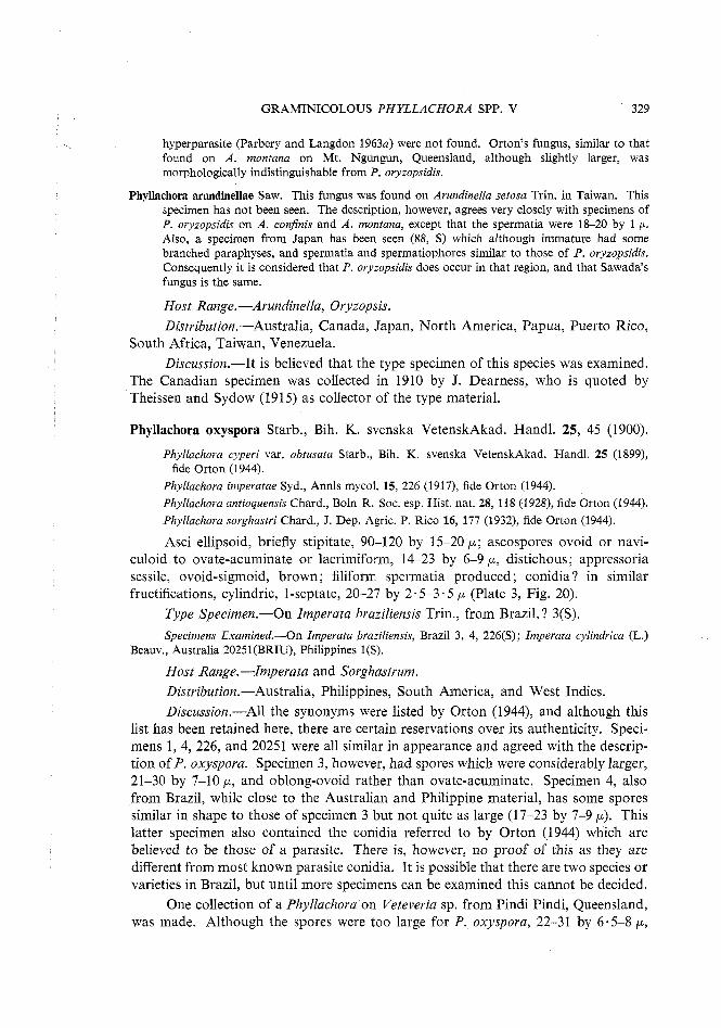
GRAMINICOLOUS PHYLLACHORA SPP. V 329
hyperparasite (Parbery and Langdon 1963a) were not found. Orton's fungus, similar to that found on A. montana on Mt. Ngungun, Queensland, although slightly larger, was morphologically indistinguishable from P. oryzopsidis.
Phyllachora arundinellae Saw. This fungus was found on Arundinella setosa Trin. in Taiwan. This specimen has not been seen. The description, however, agrees very closely with specimens of P. oryzopsidis on A. confinis and A. montana, except that the spermatia were 18-20 by 1 p. Also, a specimen from Japan has been seen (88, S) which although immature had some branched paraphyses, and spermatia and spermatiophores similar to those of P. oryzopsidis. Consequently it is considered that P. oryzopsidis does occur in that region, and that Sawada's fungus is the same.
Host Range.-Arundinella, Oryzopsis. Distribution.-Australia, Canada, Japan, North America, Papua, Puerto Rico,
South Africa, Taiwan, Venezuela. Discussion.-It is believed that the type specimen of this species was examined.
The Canadian specimen was collected in 1910 by J. Dearness, who is quoted by Theissen and Sydow (1915) as collector of the type material.
Phyllachora oxyspora Starb., Bih. K. svenska VetenskAkad. Handl. 25, 45 (1900).
Phyllachora cyperi var. obtusata Starb., Bih. K. svenska VetenskAkad. Handl. 25 (1899), fide Orton (1944).
Phyllachora imperatae Syd., Annls mycol. 15, 226 (1917), fide Orton (1944). Phyllachora antioquensis Chard., Boln R. Soc. esp. Hist. nat. 28, 118 (1928), fide Orton (1944). Phyllachora sorghastri Chard., J . Dep. Agric. P. Rico 16, 177 (1932), fide Orton (1944).
Asci ellipsoid, briefly stipitate, 90-120 by 15-20 p ; ascospores ovoid or navi- culoid to ovate-acuminate or lacrimiform, 14-23 by 6-9 p, distichous; appressoria sessile, ovoid-sigmoid, brown; filiform spermatia produced; conidia? in similar fructifications, cylindric, 1-septate, 20-27 by 2.5-3.5 p (Plate 3, Fig. 20).
Type Specimen.-On Imperata braziliensis Trin., from Brazil,? 3(S).
Specimens Examined.-On Zmpevata braziliensis, Brazil 3, 4, 226(S); Imperata cylindrica (L.) Beauv., Australia 20251 (BRIU), Philippines 1 (S).
Host Range.-Imperata and Sorghastrum. Distribution.-Australia, Philippines, South America, and West Indies. Discussion.-All the synonyms were listed by Orton (1944), and although this
list has been retained here, there are certain reservations over its authenticity. Speci- mens 1, 4, 226, and 20251 were all similar in appearance and agreed with the descrip- tion of P. oxyspora. Specimen 3, however, had spores which were considerably larger, 21-30 by 7-10 p, and oblong-ovoid rather than ovate-acuminate. Specimen 4, also from Brazil, while close to the Australian and Philippine material, has some spores similar in shape to those of specimen 3 but not quite as large (17-23 by 7-9 p). This latter specimen also contained the conidia referred to by Orton (1944) which are believed to be those of a parasite. There is, however, no proof of this as they are different from most known parasite conidia. It is possible that there are two species or varieties in Brazil, but until more specimens can be examined this cannot be decided.
One collection of a Phyllachora on Veteveria sp. from Pindi Pindi, Queensland, was made. Although the spores were too large for P. oxyspora, 22-31 by 6.5-8 p,
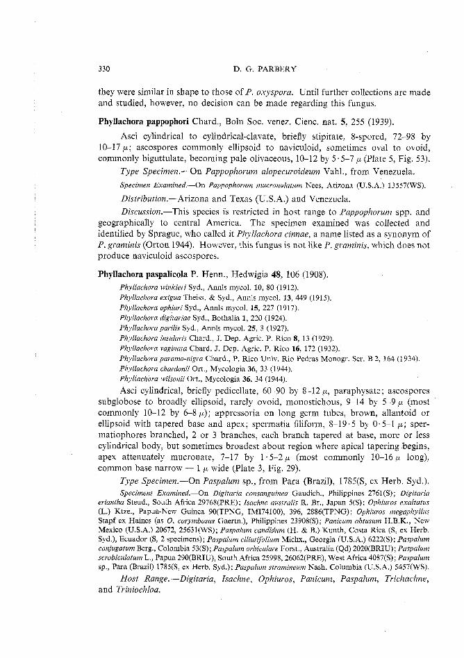
330 D. G. PARBERY
they were similar in shape to those of P. oxyspora. Until further collections are made and studied, however, no decision can be made regarding this fungus.
Phyllachora pappophori Chard., Boln Soc. venez. Cienc. nat. 5, 255 (1939).
Asci cylindrical to cylindrical-clavate, briefly stipitate, 8-spored, 72-98 by 10-17 p ; ascospores commonly ellipsoid to naviculoid, sometimes oval to ovoid, commonly biguttulate, becoming pale olivaceous, 10-12 by 5.5-7 p (Plate 5, Fig. 53).
Type Specimen.-On Pappophorum alopecuroideum Vahl., from Venezuela.
Specimen Examined.-On Pappophorum mucronulatum Nees, Arizona (U.S.A.) 13557(WS).
Distribution.-Arizona and Texas (U.S.A.) and Venezuela. Discussion.-This species is restricted in host range to Pappophorum spp. and
geographically to central America. The specimen examined was collected and identified by Sprague, who called it Phyllachora cinnae, a name listed as a synonym of P. graminis (Orton 1944). However, this fungus is not like P. graminis, which does not produce naviculoid ascospores.
Phyllachora paspalicola P. Henn., Hedwigia 48, 106 (1908). Phyllachora winkleri Syd., Annls mycol. 10, 80 (1912). Phyllachora exigua Theiss. & Syd., Annls mycol. 13, 449 (1915). Phyllachora ophiuri Syd., Annls mycol. 15, 227 (1917). Phyllachora digitariae Syd., Bothalia 1, 220 (1924). Phyllachora parilis Syd., Annls mycol. 25, 3 (1927). Phyllachora insularis Chard., J. Dep. Agric. P. Rico 8, 13 (1929). Phyllachora vaginata Chard. J . Dep. Agric. P. Rico 16, 172 (1932). Phyllachora paramo-nigm Chard., P. Rico Univ. Rio Pedras Monogr. Ser. B 2, 164 (1934). Phyllachora chardonii Ort., Mycologia 36, 33 (1944). Phyllachora wilsonii Ort., Mycologia 36, 34 (1944).
Asci cylindrical, briefly pedicellate, 60-90 by 8-12 p, paraphysate; ascospores subglobose to broadly ellipsoid, rarely ovoid, monostichous, 9-14 by 5-9 p (most commonly 10-12 by 6-8 p); appressoria on long germ tubes, brown, allantoid or ellipsoid with tapered base and apex; spermatia filiform, 8-19.5 by 0.5-1 p ; sper- matiophores branched, 2 or 3 branches, each branch tapered at base, more or less cylindrical body, but sometimes broadest about region where apical tapering begins, apex attenuately mucronate, 7-17 by 1 .5-2 p (most commonly 10-16 p long), common base narrow - 1 p wide (Plate 3, Fig. 29).
Type Specimen.-On Paspalum sp., from Para (Brazil), 1785(S, ex Herb. Syd.). Specimens Examined.-On Digitaria consanguinea Gaudich., Philippines 2761(S); Digitaria
eriantha Steud., South Africa 29768(PRE); Isachne australis R. Br., Japan 5(S); Ophiuros exaltatus (L.) Ktze., Papua-New Guinea 90(TPNG, IM174100), 396, 2886(TPNG); Ophiuros megaphyllus Stapf ex Haines (as 0. corymbosus Gaertn.), Philippines 23908(S); Panicum obtusum H.B.K., New Mexico (U.S.A.) 20672, 25631(WS); Paspalurn candidum (H. & B.) Kunth, Costa Rica (S, ex Herb. Syd.), Ecuador (S, 2 specimens); Paspalum ciliatifolium Michx., Georgia (U.S.A.) 6222(S); Paspalum conjugatum Berg., Colombia 53(S); Paspalum orbiculare Forst., Australia (Qd) 2020(BRIU); Paspalurn scrobiculatum L., Papua 290(BRIU), South Africa 25998,26062(PRE), West Africa 4087(S); Paspalum sp., Para (Brazil) 1785(S, ex Herb. Syd.); Paspalum stramineum Nash, Columbia (U.S.A.) 5457(WS).
Host Range.-Digitaria, Isachne, Ophiuros, Panicum, Paspalum, Trichachne, and Triniochloa.
GRAMINICOLOUS PHYLLACHORA SPP. V 331
Distribution.-Australia, central and South America, Japan, New Guinea, Papua, Philippines, South Africa, West Africa, West Indies.
Synonymy P. winkleri. The type specimen of this species (4087, S) was found on Paspalurn scrobiculatum in
West Africa. Although the general morphology, which is the same as that of P. paspalicola, was described correctly, the spore size given exceeds that of any spores found during the present study. Instead of being 14-17 by 9-12 p, ascospores were 9-12 by 6.5-9 p, which is the same as for P. paspalicola. Ascus size was also incorrect in the original description.
P. exigua. This fungus was found on Isachne australis in Japan (type: 5, S). Ascospores and appressoria in the type material were the same as for P. paspalicola.
P. ophiuri. The specimen examined (23908, S) was the type specimen for this species. The ascospores were not as large as described by Sydow (1917), but were 9-10.5 by 6 p. They were similar in shape to spores of P. paspalicola.
P. digitariae. This fungus was found in South Africa on D. smutsii Stent. Although type material has not been seen, two other specimens, one identified by Sydow (2761, S), the other (29768, PRE) by Doidge (1942), have been examined. In each the ascospores were similar to those of P. paspalicola.
P. parilis. Prior to the present study, this was believed to be the valid species name (Parbery 1962, 1963a, 1963b; Parbery and Langdon 1963~). The type specimen on Paspalurn candidum from Costa Rica (S) was in poor condition. Ascospores only were found, and these were not as big as spores in other specimens identified as P, parilis, although similar in shape to ascospores of Phyllachora paspalicola. Two other specimens on Paspalum candidum from nearby Ecuador had ascospores of similar size and shape to Phyllachova paspalicola and some smaller ones which were the same size as the few spores found in the type material. The spermatial and spermatiophore characters of the latter specimens were the same as those for P. paspalicola.
P. insularis. No specimen of this fungus has been examined. The description, however, is very close to that of P. paspalicola. This species was found on Tvichachne insulavis (L.) Nees (as Volatia insularis Chase) from Puerto Rico. Orton (1944) later ascribed a conidial state to this fungus. He described the conidia as occurring in similar fructifications to the asci, lunate, hyaline, 1-celled, and 26-29 by 3-3.6 p. Chardon (1929) did not see these spores, so that it is likely that Orton found spores of a parasite, possibly attacking the spermatial state, which often happens (Parbery and Langdon 1963b).
P. vaginata. Orton listed this fungus as a synonym of P. paspalicola. The fungus was found on Paspalum vaginatum in the Dominican Republic. Since the description of P. vaginata is similar to that of P. paspalicola, which occurs in this area, Orton's view is accepted.
P. paramo-nigra. The type specimen of this species was found on Triniochloa stipoides (H.B.K.) Hitch. in Venezuela. Asci were cylindric, 68-88 by 9-11 p, short, pedicellate. Ascospores were elliptical with blunt ends, 9.5-13 by 5.5-7 p. No specimen of this fungus has been seen, but the description of P. paramo-nigra is very similar to that of P. paspalicola.
P. chardonii. No specimen has been seen of this fungus, which was found on Panicum germinatum Forssk. in Puerto Rico. It is described in similar terms to Phyllachova paspalicola, except that the ascospores are slightly longer, and the asci are sometimes larger. Its distribution is also similar to that of P. paspalicola, so there is strong evidence that they are the same species.
P. wilsonii. The type material of this species, found on Paspalum setaceum Michx. from New Jersey, U.S.A., has not been seen. Another specimen (5457, WS) on the type host identified by Orton (1944) as Phyllachova wilsonii, has been examined, and is the same as P. paspalicola, while agreeing in ascal and spermatial detail with Orton's description (Orton 1944). The conidial state described by Orton was not present, and it is believed that the conidia described were those of a hyperparasite, not of the Phyllachova (Parbery and Langdon 19636). The spermatiophores found in this specimen were the same as in P, paspalicola.

332 D. G. PARBERY
Discussion.-The ascospore shape and spermatiophore morphology each distinguish this species from most others and together from all others. P. maydis and P. sphaerosperma have fairly similar ascospores, but the former has multibranched spermatiophores and the latter simple ones.
The name P. paspali-virgati should perhaps be listed as a synonym of P. paspalicola. Orton (1944) gives it as a synonym of P. guianensis, which is the same as P. punctum. Chardon (1929), however, when describing P. paspali-virgati stated that it differed from P. insularis only in stromatal characters (gross colony characters, i.e. outline of clypeus) and this has been shown as a false criterion of species (Parbery and Langdon 1964). This problem cannot be solved, however, until the type material of P. paspali-virgati can be found and re-examined.
Another name which should possibly be listed as a synonym is P. kwantungensis Petr. This fungus was found in China on Paspalum repens L. Although generally broader than ascospores of Phyllachora paspalicola, the spores of P. kwantungensis are similar in shape. Petrak (1955) compared them to the ascospores of P. sphaero- sperma (known only on Cenchrus spp.) but decided that the fungus was a new species because the spores were wider and more subglobose than those of P. sphaerosperma. When the opportunity becomes available, P. kwantungensis should be compared with P. paspalicola.
This species has a very similar geographic distribution to P. setariaecola, with which it occurs in common on two genera, Digitaria and Panicum. The two Phyllachora species are morphologically distinct.
Chardon (1929, 1934) refers to collections of a Phyllachora which he calls P. paspalicola, as having citriform ascospores. It is evident, however, that he is referring to another and probably an undescribed species, because the type material examined during this study lacked that kind of spore. Neither Hennings (1908) nor Orton (1944) found citriform ascospores in P. paspalicola. Chardon's reports (1929, 1934) are of interest, however, as no other graminicolous Phyllachora is known which has citriform spores.
Phyllachora pennisetina Syd., Annls mycol. 20, 64 (1922).
Phyllachova penniseti-japonici Saw., Rep. Govt Res. Inst. Dep. Agric. Formosa No. 87, 19 (1044).
Asci cylindrical-clavate, stipitate, 8-spored, copiously paraphysate, 55-100 by 12-20 p ; ascospores monostichous or distichous, subglobose to globose, 9-13 by 7-10 p.
Type Specimen.-On Pennisetum alopecuroides (L.) Spreng., from China. Although specimens of neither Phyllachora pennisetina nor P. penniseti-japonici
have been seen, there is no doubt of their being the one species. The descriptions given by Sydow (1922) and Sawada (1944) are identical in essential detail, and each specimen was found on a species of Pennisetum in adjacent areas. Phyllachora penniseti-japonica was found on Pennisetum japonicum Trin. in Taiwan.
It has not been possible to compare this species with others with near-spherical ascospores, because of a lack of material. It is possible that P. kwantungensis is a

GRAMINICOLOUS PHYLLACHORA SPP. V 333
synonym of P. pennisetina, for it was also found in China on Paspalum, and is described in terms identical with it. Both, however, may be synonyms of P. paspalicola.
Phyllachora perotidis Doidge, Bothalia 4(2), 439 (1942).
Asci 8-spored, cylindrical, 60-70 by 10-1 1 5 p; paraphyses numerous, fibrous, hyaline; ascospores ellipsoid, 10-12.5 by 5-7 p ; filiformsperrnatia present.
Type Specimen.-On Perotis indica (L.) Ktze., from Pretoria (South Africa). Discussion.-Unfortunately no specimen of this fungus has been examined. It
is unusual, however, for any species to have solely ellipsoid spores, and many species possess ellipsoid spores as well as others. When an opportunity arises, therefore, this species should be compared with others, such as P. arthraxonis and other small-spored species which produce ellipsoid spores.
Phyllachora phalaridis Ort., Mycologia 36, 39 (1944).
Asci cylindrical, 70-80 by 7.5-9 p ; ascospores broadly ellipsoid, 7.5-10 by 4-5 p.
Type Specimen.-On Phalaris arundinacea L., from Massachusetts (U.S.A.). Phyllachora phalaridis is known only from the type locality. No specimen
was examined.
Phyllachora phyllostachydis Hara, Bot. Mag., Tokyo 27, 248 (1913).
Asci cylindrical, apices rounded, briefly stipitate, 120-180 by 9-1 1 p ; paraphyses filiform, simple, continuous, 150-200 by 1-1.5 p ; ascospores monostichous, oblong, oblong-fusoid, ovate-oblong, or fusoid (see below), ends more or less rounded, hyaline, guttulate, 16 5-19.5 by 6-7 5 p.
Type Specimen.-On Phyllostachys bambusoides Sieb. & Zucc., from Japan. A specimen of this species has not been seen. The illustrations of Hino and
Katumoto (1961), however, suggest that this is a distinct species of Phyllachora and certainly one different to any other on the bamboos. Their (Hino and Katumoto 1961) description of spore shape, however, does not conform to the current views, according to which the spores would be described as ellipsoid, naviculoid, or ovoid.
Phyllachora platyelliptica sp. nov. Asci cylindrici, breviter stipitati, 82-120 x 10-16 p (paraphyses adsunt);
ascosporae usitate uniseriatae, aliquando biseriatae, plerumque anguste et leviter ellipsoideae usque ad ovatae, raro ovales, aliquae sporae vel planoconvexae (semi- ellipsoideae) vel leviter curvatae, saepe ovales vice rotundae (in sectione transversali), 14-21 x 5-7 p.
Typus.-In folio vivo Themedae australis (R.Br.) Stapf, Dandenong Mountains, Victoria, Australia, 2801 (MELU).
Colonies black, 1.5-2 by 0.5-1 mm, scattered, usually discrete, raised on the dorsal surface of host leaf not so on ventral surface, develop in living tissue only; each colony consists of dorsal and ventral clypei with 3 to several perithecia between them, leaf twice as thick through colony; upper clypei thicker (in dorsal epidermis) 12-32 p thick (epidermal cell 11-32 p deep), lower clypei (in ventral epidermis) only 12-16 p thick (epidermal cells 8-14 p), at first clypei of a single layer of hyphae inside
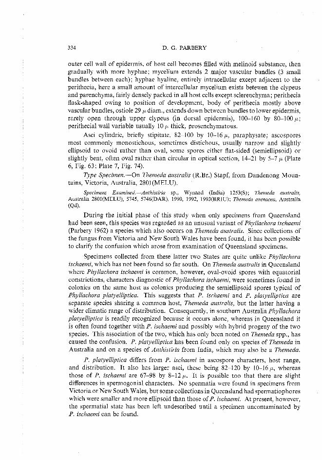
334 D. G. PARBERY
outer cell wall of epidermis, of host cell becomes filled with melinoid substance, then gradually with more hyphae; mycelium extends 2 major vascular bundles (3 small bundles between each); hyphae hyaline, entirely intracellular except adjacent to the perithecia, here a small amount of intercellular mycelium exists betewen the clypeus and parenchyma, fairly densely packed in all host cells except sclerenchyma; perithecia flask-shaped owing to position of development, body of perithecia mostly above vascular bundles, ostiole 29 p diam., extends down between bundles to lower epidermis, rarely open through upper clypeus (in dorsal epidermis), 100-160 by 80-100 p ; perithecial wall variable usually 10 p thick, prosenchymatous.
Asci cylindric, briefly stipitate, 82-100 by 10-16 p, paraphysate; ascospores most commonly monostichous, sometimes distichous, usually narrow and slightly ellipsoid to ovoid rather than oval, some spores either flat-sided (semiellipsoid) or slightly bent, often oval rather than circular in optical section, 14-21 by 5-7 p (Plate 6, Fig. 63; Plate 7, Fig. 74).
Type Specimen.-On Themeda australis (R.Br.) Stapf, from Dandenong Moun- tains, Victoria, Australia, 2801(MELU).
Specimens Examined.-Anthistiria sp., Wynaad (India) 1253(S); Themeda australis, Australia 2801(MELU), 5745, 5746(DAR), 1990, 1992, 1993(BRIU); Themeda avenacea, Australia (Qd).
During the initial phase of this study when only specimens from Queensland had been seen, this species was regarded as an unusual variant of Phyllachora ischaemi (Parbery 1962) a species which also occurs on Themeda australis. Since collections of the fungus from Victoria and New South Wales have been found, it has been possible to clarify the confusion which arose from examination of Queensland specimens.
Specimens collected from these latter two States are quite unlike Phyllachora ischaemi, which has not been found so far south. On Themeda australis in Queensland where Phyllachora ischaemi is common, however, oval-ovoid spores with equatorial constrictions, characters diagnostic of Phyllachora ischaemi, were sometimes found in colonies on the same host as colonies producing the semiellipsoid spores typical of Phyllachora platyelliptica. This suggests that P. ischaemi and P. platyelliptica are separate species sharing a common host, Themeda australis, but the latter having a wider climatic range of distribution. Consequently, in southern Australia Phyllachora platyelliptica is readily recognized because it occurs alone, whereas in Queensland it is often found together with P. ischaemi and possibly with hybrid progeny of the two species. This association of the two, which has only been noted on Themeda spp., has caused the confusion. P. platyelliptica has been found only on species of Themeda in Australia and on a species of Anthistiria from India, which may also be a Themeda.
P. platyelliptica differs from P. ischaemi in ascospore characters, host range, and distribution. It also has larger asci, these being 82-120 by 10-16 p, whereas those of P. ischaemi are 67-98 by 8-12 p. It is possible too that there are slight differences in spermogonial characters. No spermatia were found in specimens from Victoria or New South Wales, but some collections in Queensland had spermatiophores which were smaller and more ellipsoid than those of P. ischaemi. At present, however, the spermatial state has been left undescribed until a specimen uncontaminated by P. ischaemi can be found.
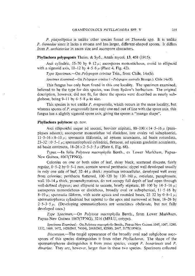
GRAMINICOLOUS PHYLLACHORA SPP. V 335
P. platyelliptica is unlike 'other species found on Themeda spp. It is unlike P. themedae since it lacks a stroma and has larger, different-shaped spores. It differs from P, anthisteriae in ascus size and ascospore characters.
Phyllachora polypogonis Theiss. & Syd., Annls mycol. 13, 458 (1915).
Asci cylindric, 55-70 by 9-12 p ; ascospores monostichous, ovoid to ellipsoid with a sigmoid axis, 10-12 by 4.5-6 p (Plate 4, Fig. 42).
Type Specimen.-On Polypogon crinitus Trin., from Chile, 14a(S). Specimen Examined.-On Polypogon cvinitus (=Polypogon austvalis Brongn.), Chile 14a(S).
This fungus has only been found in this one locality. The specimen examined, believed to be the type for this species, was from Sydow's herbarium. The original description, however, did not fit, for there the spores were described as nearly sub- globose, being 9-1 1 by 6.5-8 p in size.
This species is not unlike P. eragrostidis, which occurs in the same locality, but whereas spores of P. eragrostidis have only one end out of line with the spore axis, this fungus has a slightly sigmoid spore axis, giving the spores a "mango shape".
Phyllachora polytocae sp. nov.
Asci ellipsoidei usque ad saccati, breviter stipitati, 88-100 x 14-5-16 p (para- physes adsunt); ascosporae monostichae vel distichae, late ovales vel subsphaerici, 11 5-16 x 8-10 p; spermatia filiformia, ad apicem acuminata, ad basin rotundata, 23-32 x 0.5-1 p ; spermatiophorii cylindrici, flexuosa, ad apicem gradatim acuminata, ad basin contracta, 18-26 x 2.5-3.5 p (Plate 6, Fig. 68).
Typus.-In folia Polytoca macrophylla Benth., ex Lower Markham, Papua- New Guinea, 1067(TPNG).
Colonies on one or both sides of leaf, shiny black, scattered discrete, fairly regular, 0.5-2 by 0.5-1 mm, contain several perithecia; clypei well developed usually in only one side of leaf, 32-44 p thick; mycelium intracellular, developed well away from colonies; perithecia flattened, 100-120 by 120-160 p, ostiolate, paraphysate, wall 10-14 p thick, prosenchymatous, do not occupy full depth of leaf open through well-defined clypeus; asci ellipsoid to saccate, briefly stipitate, 88-100 by 14.5-16 p ; ascospores monostichous or distichous, broadly oval or subspherical, 11.5-16 by 8-10 p ; spermaiia filifom, with acute apices and rounded bases, 23-32 by 0.8-1 p ; spermatiophores cylindrical but tapered to the apex and narrowed at base, 18-26 by 2.5-3 5 p. (Developing spermatiophores are sometimes obclavate, but not fully developed ones.)
Type Specimen.-On Polytoca macrophylla Benth., from Lower Markham, Papua-New Guinea 1067(TPNG), 3216 (MELU, cotype).
Specimens Examined.-On Polytoca macrophylla Benth., Papua-New Guinea 1045, 1067, 1290, 1332, 1469, 1472, 1626(IMI, 74104), 2863(IMI, 82508), 2997, 3175(TPNG).
Discussion.-The turgid appearance of the broadly oval and subglobose asco- spores of this species distinguishes it from other Phyllachoras. The shape of the spermatiophores distinguishes it from most species, except P. bonariensis and P. dimeriae. They are, however, larger than in these two species. Specimens collected
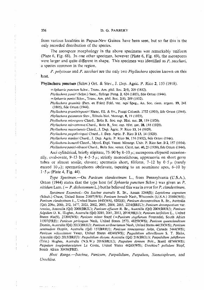
336 D. G. PARBERY
from various localities in Papua-New Guinea have been seen, but so far this is the only recorded distribution of the species.
The ascospore morphology in the above specimens was remarkably uniform (Plate 6, Fig. 68). In one other specimen, however (Plate 6, Fig. 69), the ascospores were larger and quite different in shape. This specimen was identified as P. sacchari, a species common in the region.
P. polytocae and P. sacchari are the only two Phyllachora species known on this host.
Phyllachora punctum (Schw.) Ort. & Stev., J. Dep. Agric. P. Rico 2, 153 (1918).
=Sphaeriapunctum Schw., Trans. Am. phil. Soc. 2(4), 209 (1832). Phyllachora panici (Schw.) Sacc., Sylloge Fung. 2, 624 (1883), fide Orton (1944). ~ S p h a e r i a panici Schw., Trans. Am. phil. Soc. 2(4), 209 (1832). Phyllachora graminis (Pers. ex Fries) Fckl. var, tupi Speg., An. Soc. cient. argent. 19, 241
(1885), fide Orton (1944). Phyllachora graminis-panici Shear, Ell. & Ev., Fungi Columb. 1752 (1903), fide Orton (1944). Phyllachora guianensis Stev., Illinois biol. Monogr. 8, 19 (1923). Phyllachova microspora Chard., Boln R. Soc. esp. Hist. nat. 28, 119 (1928). Phyllachora microstroma Chard., Boln R. Soc. esp. Hist. nat. 28, 118 (1928).
I Phyllachora macorisensis Chard., J . Dep. Agric. P. Rico 13, 14 (1929). Phyllachorapaspali-virgati Chard., J. Dep. Agric. P. Rico B 13, 14 (1929). Phyllachora stanleyi Chard., J . Dep. Agric. P. Rico 16, 174 (1932), fide Orton (1944). Phyllachora leonardi Chard., Mycol. Expl. Venez. Monogr. Univ. P. Rico Ser. B 2, 157 (1934). Phyllachorapanici-olivacei Chard., Boln Soc. venez. Cient. nat. 40,21(1939), fide Orton (1944).
Asci cylindrical, briefly stipitate, 75-90 by 8-10 p ; ascospores ellipsoid occasion- ally, oval-ovate, 9-1 3 by 4-5.5 p ; strictly monostichous, appressoria on short germ tubes or almost sessile, clavate; spermatia short, filiform, 7-12 by 0.5 p (rarely exceed 10 p); spermatiophores obclavate, tapering to an acuminate apex, 6-10 by 1 5 p (Plate 4, Fig. 44).
Type Specimen.-On Panicum clandestinum L., from Pennsylvania (U.S.A.). Orton (1944) states that the type host (of Sphaeria punctum Schw.) was given as P. nitidum Lam. (= P. dichotomum L.) but he believed this was in error for P. clandestinum.
Specimens Examined.-On Isachne australis R. Br., Assam 1248(S); Leptoloma cognatum (Schult.) Chase, United States 21697(WS); Panicum boreale Nash, Wisconsin (U.S.A.) 25866(WS); Panicum clandestinum L., United States 5443(WS), 6202(S); Panicum decompositum R. Br., Australia (Qd) 208a, 2086, 252, i477, 2021, 2002, 2003, 2004, 2005, 2234(BRIU); Panicum decompositum var. tenuius, Australia (Qd) 2008(BRIU); Panicum effusum R. Br., Australia (Qd) 2009(BRIU); Panicum fulgidum D. K. Hughes, Australia (Qd) 2010, 201 1, 2012, 2014(BRIU); Panicum latifolium L., United States 90a(S), 21208(WS); Panicum minus Stapf (=Panicurn stapfianum Fourcade), South Africa 11307(PRE); Panicum pevlongum Nash, United States 2573, 48259(WS); Panicum queenslandicum Domin, Australia (Qd) 2013(BRIU); Panicum scribnerianum Nash, United States 44139(WS); Panicum seminudum Domin, Australia (Qd) 157(BRIU); Panicum tennesseense Ashe, Canada 5444(WS); Panicum wilcoxianum Vasey, United States 48340(WS); Paspalidium albovillosum S. T. Blake, Australia (Qd) 2015(BRIU); Paspalidium distans, Australia (Qd) 216(BRIU); Paspalidium jubiforum (Trin.) Hughes, Australia (N.S.W.) 2016(BRIU); Paspalum densum Poir., Brazil 48749(WS); Paspalum longipedunculatum Le Conte, United States 46263(WS); Urochloa? pullulans Stapf, South Africa 30436(PRE).
Host Range.-Isaclzne, Panicum, Paspalidium, Paspalum, Stenotaphrum, and Urochloa.
GRAMINICOLOUS PHYLLACHORA SPP. V 337
Distribution.-Australia, Brazil, Canada, India, South Africa, United States.
Synonymy Sphaeria punctum, S. panici, Phyllachora panici, P. graminis var. tupi, P. graminis-panici, P. stanleyi,
and P. panici-olivacei were listed as synonyms of P. punctum by Orton (1944) and have been accepted here since the original descriptions are close to that of P. puncium. Orton also included P, oplismeni as a synonym. The latter name is, however, a synonym of P, bonariensis.
P. guianensis. This fungus was described from a specimen on Paspalum virgatum L. from British Guiana. Orton (1944) examined specimens on P. virgatum from Puerto Rico, Cuba, and Jamaica and ascribed a conidial state to the species. No such conidia were found during the present investigation and it is believed that the conidia Orton found were not part of the Phyllachora he examined. The various specimens (48749,46263 WS) identified as P. guianensis were the same as P. punctum in all respects, including spermatial characters.
P. microspora, P. paspali-virgati, and P. leonardi were all given as synonyms of P. guianenesis by Orton (1944) so are now listed as synonyms of P. punctum since the original descriptions of these fungi agree with P. punctum. P. paspali-virgati, however, is doubtfully placed here. The reason for this doubt is discussed in relation to P. paspalicola.
P. microstroma. This fungus was found on Panicum laxum Swartz in Colombia and was named by Chardon (1928). It was later listed by Orton (1944) as a synonym of Phyllachora congruens. A number of specimens identified as P, congruens have been examined. Most of these were P, punctum. The type material of P. congruens, however, is quite different and has been listed as a synonym of P. bonariensis. P. microstroma has been listed as a synonym of P. punctum because it is described in similar terms, and because it is believed that Orton made his decision only after comparing P. microstroma with some American specimens of P. punctum, which he believed were P. congruem
P. macorisensis. Chardon named this species from a specimen found in Santo Domingo on a species of Stenotaphrum. It is described in terms which do not distinguish it from P. punctum. Orton (1944) examined a Phyllachora on S. secundatunz (Walt.) Kuntze which he regarded as the type, and redescribed the species in terms more applicable to P. bonariensis than to P. punctum. He gave the ascospore size as 10-15 by 5-6.5 p. Consequently, until the type of this species can be located and examined, there will be some doubt about the validity of this listing.
Discussion.-This species is a fairly stable one, especially on species of Panicum and Paspalum. On species of Paspalidum, however, although the ascospores were rarely any larger than the size given above, the spermatia were often up to 15 p long, which is unusual for this species. In other respects, however, the specimens on Paspalidum spp. were too like P. punctum to be considered different. Another curious phenomenon associated with the species on specimens found in Australia is that no spermatia were found on any Panicum species. The ascospores were very similar, so that the identification is not doubted. In the Americas, where P. punctum is wide- spread, the spermatial state is more commonly found than not.
In 1944, Orton described a conidial as well as a spermatial state for P. punctum. Later, however (Orton 1956), when he conducted one of the most complete studies so far carried out into the life cycle of a graminicolous species, he concluded that the conidia were not part of the Phyllachora.
This species is distinguished from P. bonariensis by having a strictly mono- stichous ascospore arrangement with generally smaller and more dumpy ascospores which are sometimes ovoid or oval, by having spermatia generally less than 15 p and usually less than 12 p, and by having smaller, more rounded spermatiophores. If the
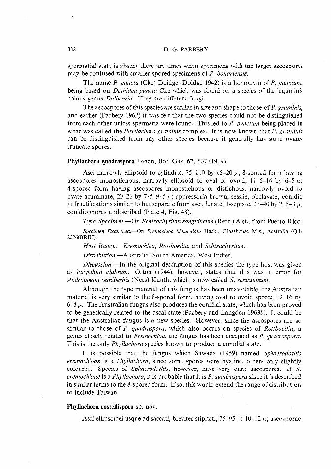
338 D. G. PARBERY
spermatial state is absent there are times when specimens with the larger ascospores may be confused with smaller-spored specimens of P. bonariensis.
The name P. puncta (Cke) Doidge (Doidge 1942) is a homonym of P. punctum, being based on Dothidea puncta Cke which was found on a species of the legumini- colous genus Dalbergia. They are different fungi.
The ascospores of this species are similar in size and shape to those of P. graminis, and earlier (Parbery 1962) it was felt that the two species could not be distinguished from each other unless spermatia were found. This led to P. punctum being-placed in what was called the Phyllachora graminis complex. It is now known that P. graminis can be distinguished from any other species because it generally has some ovate- truncate spores.
Phyllachora quadraspora Tehon, Bot. Gaz. 67, 507 (1919).
Asci narrowly ellipsoid to cylindric, 75-110 by 15-20 p; %spored form having ascospores monostichous, narrowly ellipsoid to oval or ovoid, 11-5-16 by 6-8 p ; 4-spored form having ascospores monostichous or distichous, narrowly ovoid to ovate-acuminate, 20-26 by 7.5-9.5 p; appressoria brown, sessile, obclavate; conidia in fructifications similar to but separate from asci, lunate, l-septate, 23-40 by 2.5-3 p, conidiophores undescribed (Plate 4, Fig. 48).
Type Specimen.-On Schizachyrium sanguineum (Retz.) Alst., from Puerto Ricn.
Specimen Examined.-On Eremochloa bimaculafa Hack., Glasshouse Mts., Australia (Qd) 2026(BRIU).
Host Range.-Eremochloa, Rottboellia, and Schizachyrium. Distribution.-Australia, South America, West Indies. Discussion.-In the original description of this species the type host was given
as Paspalum glabrum. Orton (1944), however, states that this was in error for Andropogon semiberbis (Nees) Kunth, which is now called S. sanguineum.
Although the type material of this fungus has been unavailable, the Australian material is very similar to the %spored form, having oval to ovoid spores, 12-16 by 6-8 p. The Australian fungus also produces the conidial state, which has been proved to be genetically related to the ascal state (Parbery and Langdon 19633). It could be that the Australian fungus is a new species. However, since the ascospores are so similar to those of P, quadrasporn, which also occnrs on species of 1Pottboellia, a genus closely related to Eremochloa, the fungus has been accepted as P. quadraspora. This is the only Phyllachora species known to produce a conidial state.
It is possible that the fungus which Sawada (1959) named Sphaerodothis eremochloae is a Phyllachora, since some spores were hyaline, others only slightly coloured. Species of Sphaerodothis, however, have very dark ascospores. If S. eremochloae is a Phyllachora, it is probable that it is P. quadraspora since it is described in similar terms to the %spored form. If so, this would extend the range of distribution to include Taiwan.
Phyllachora rostellispora sp. nov.
Asci ellipsoidei usque ad saccati, breviter stipitati, 75-95 x 10-12 p; ascosporae
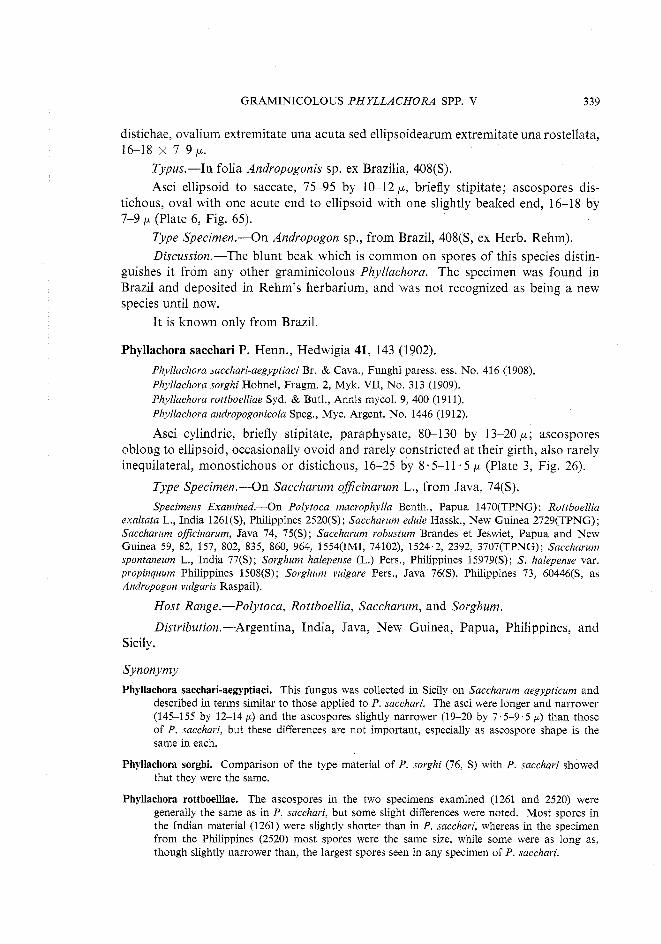
GRAMINICOLOUS PHYLLACHORA SPP. V 339
distichae, ovalium extremitate una acuta sed ellipsoidearum extremitate unarostellata, 16-18 x 7-9 p.
Typus.-In folia Andropogonis sp. ex Brazilia, 408(S). Asci ellipsoid to saccate, 75-95 by 10-12 p, briefly stipitate; ascospores dis-
tichous, oval with one acute end to ellipsoid with one slightly beaked end, 16-18 by 7-9 p (Plate 6, Fig. 65).
Type Specimen.-On Andropogon sp., from Brazil, 408(S, ex Herb. Rehm). Discussion.-The blunt beak which is common on spores of this species distin-
guishes it from any other graminicolous Phyllachora. The specimen was found in Brazil and deposited in Rehm's herbarium, and was not recognized as being a new species until now.
It is known only from Brazil.
Phyllachora sacchari P. Henn., Hedwigia 41, 143 (1902).
Phyllachova sacchari-aegyptiaci Br. & Cava., Funghi paress. ess. No. 416 (1908). Phyllachora sorghi Hohnel, Fragm. 2, Myk. VII, No. 313 (1909). Phyllachora rottboelliae Syd. & Butl., Annls mycol. 9, 400 (1911). Phyllachova andvopogonicola Speg., Myc. Argent. No. 1446 (1912).
Asci cylindric, briefly stipitate, paraphysate, 80-130 by 13-20 p ; ascospores oblong to ellipsoid, occasionally ovoid and rarely constricted at their girth, also rarely inequilateral, monostichous or distichous, 16-25 by 8.5-1 1.5 p (Plate 3, Fig. 26).
Type Specimen.-On Saccharum oficinarum L., from Java, 74(S).
Specimens Examined.-On Polytoca macrophylla Benth., Papua 1470(TPNG): Rottboellia exaltata L., India 1261(S), Philippines 2520(S): Saccharum edule Hassk., New Guinea 2729(TPNG); Saccharum officinarum, Java 74, 75(S); Saccharum robustum Brandes et Jeswiet, Papua and New Guinea 59, 82, 157, 802, 835, 860, 964, 1554(IMI, 74102), 1524.2, 2392, 3707(TPNG); Saccharum spontaneum L., India 77(S); Sorghum halepense (L.) Pers., Philippines 15979(S); S . halepense var. propinquum Philippines 1508(S); Sorghum vulgave Pers., Java 76(S), Philippines 73, 60446(S, as Andropogon vulgaris Raspail).
Host Range.-Polytoca, Rottboellia, Saccharum, and Sorghum.
Distribution.-Argentina, India, Java, New Guinea, Papua, Philippines, and Sicily.
Synonymy
Phyllachora sacchari-aegyptiaci. This fungus was collected in Sicily on Saccharum aegypticum and described in terms similar to those applied to P. sacchari. The asci were longer and narrower (145-155 by 12-14 p) and the ascospores slightly narrower (19-20 by 7.5-9.5 p) than those of P, sacchari, but these differences are not important, especially as ascospore shape is the same in each.
Phyllachora sorghi. Comparison of the type material of P. sorghi (76, S) with P. sacchari showed that they were the same.
Phyllachora rottboelliae. The ascospores in the two specimens examined (1261 and 2520) were generally the same as in P. sacchari, but some slight differences were noted. Most spores in the Indian material (1261) were slightly shorter than in P. sacchari, whereas in the specimen from the Philippines (2520) most spores were the same size, while some were as long as, though slightly narrower than, the largest spores seen in any specimen of P. sacchari.
340 D. G. PARBERY
Phyllachora andropogonicola. This fungus was found in Argentina on Andropogon saccharatus Raspail (Sorghum vulgare var, saccharaturn (L.) Boerl.). Its description closely fits that of P. sacchari.
Discussion.-The distribution of this species is probably much wider than indi- cated above, since various annual reports of Departments of Agriculture in countries where sugar cane or sorghums are grown list P. sacchari or one of its synonyms as parasites of these crops.
The ascospores of P. sacchari are similar in shape and size to those of P. andropogonis Karst. & Har., but lack the marked constriction in the spores which is common in the latter species. If it could be shown that ascospore constriction is not constant in P. andropogonis, then it would mean that P. sacchari would become a synonym of that species.
Another closely allied species is P. sacchari-spontanei, which has fairly similar ascospores to P. sacchari, though narrower, and produces spermatia, which have not been found in P. sacchari. Should a broader-spored form of P. sacchari-spontanei be found, or a specimen of P. sacchari with an identical spermatial state to that of P. sacchari-spontanei, then these two species, which share the common host Saccharum spontaneum, should also be regarded as synonyms. At present, however, there is no evidence to suggest that they are the same. The observation that they both occur in Papua-New Guinea but still maintain distinctly different ascospore shapes, even though size does vary, supports the decision made here.
Phyllachora sacchari-spontanei Syd., Philipp. J. Sci. No. 4(4), 279 (1913).
Asci saccate, paraphysate, briefly stipitate, 75-100 by 18-22 p; ascospores oblong with one rounded end and one bluntly or somewhat attenuately acuminate end, 17.5-25 by 6.5-8 p ; spermogonia bearing filiform spermatia, 16-22 by 0.5-1 p ; spermatiophores single, almost cylindrical but taper to the apex, somewhat flexuous, 16-17 by 1 .5-2 p (Plate 4, Fig. 34).
Type Specimen.-On Saccharum spontaneum L., from the Philippines.
Specimen Examined.-On Saccharurn spontaneurn, India 1269(S), Papua-New Guinea 757, 769.2, 1734, 1991(TPNG).
Discussion.-Theissen and Sydow (1915) give the distribution of this species as India and the Philippines. The specimen examined had been identified as P. cyperi Rehm var. donacis Rehm & Sacc.", but agreed in all details with the description of P. sacchari-spontanei (Sydow 1913). Although some ascospores of this species are similar in shape to those of P. sacchari, they are generally smaller and much narrower and have one acuminate end. The presence of a spermatial state, also described by Sydow, distinguishes this species from P. sacchari, for which no spermatial state has been found. Collections of this species from Papua-New Guinea had identical spermatial states to the Indian material but the ascospores frequently were more attenuate at the acuminate end. They were also larger, being 21-33 by 7-9 p. Their shape, however, was distinct from that of P. sacchari, which was also common in that region. The grass Saccharum spontaneum is host to both species in India.
* P. cyperi var. donacis is a synonym of Apiospova montagnei Sacc. (von Arx 1952), which is a saprophyte on various grasses.
GRAMINICOLOUS PHYLLACHORA SPP. V 341
Phyllachora scanica Starb., in Rehm, Hedwigia 34, 162 (1895).
Asci cylindric, briefly stipitate, 75-100 by 12 p ; ascospores broadly ovoid to ellipsoid, 11-14 by 6-7 p; monostichous or distichous; paraphyses somewhat branched (Plate 2, Fig. 15).
Type Specimen.-On grass leaves (probably Poa sp.), Sweden, 90(S).
Specimen Examined.-On grass leaves, Sweden 89, 90, 1125(S, ex Rehm).
Discussion.-Rehm (1895) apparently published this name and description of the fungus, which had previously been named and described informally by Starbach. Rehm states, however, that it is rather like P. cynodontis, which is true, except for larger asci and branched paraphyses. Unfortunately no spermatial state was found in this specimen (believed to be the type) as this would have shown if it were in fact P. cynodontis. The fungus is not unlike P. helvetica either, and in fact there is doubt about its validity.
Theissen and Sydow (1915), who had access to this same specimen, stated that they believed the grass was a species of Poa.
Rehm described the fungus as having granulated ascospores with a thin slime layer. Neither of these characters was noted during the present examination, nor were they referred to by Theissen and Sydow (1915).
During the present investigation great difficulty has been experienced in locating and often obtaining loans of some specimens. This has been particularly true of many specimens of Phyllachora on bamboos. The P. shiraiana complex is little more than a list of fungi which are very probably synonyms of P. shiraiana, which is listed here with one known synonym.
Since specimens of the fungi called P. shiraiana and P. arundinaria are the only ones seen and compared, all the other fungi grouped in this complex have been recognized solely from descriptions, which because most are available only in the Japanese language are here repeated in part.
Phyllachora shiraiana Syd., Hedwigia 37, 208 (1898).
Phyllachora arundinariae Ort., Mycologia 36, 48 (1944).
Asci cylindrical to ellipsoid, briefly stipitate, 75-100 by 8-10 p; paraphysate, ascospores monostichous, ellipsoid or naviculoid with broadly rounded ends (almost oval) to ellipsoid or naviculoid with fairly acute ends (almost fusiform), 15-22 by 5 5-8 p (Plate 3, Fig. 19).
Type Specimen.-On Sasa japonica (Sieb. & Zucc.) Makino (as Arundinaria japonica Sieb. & Zucc.), from Tokyo (Japan), 10A(S).
Specimens Examined.-On Arundinaria simoni Riv. var. chino, Japan 59A, 95,96(S); Arundinaria tecta (Walt.) Muhl., Darien, Georgia (U.S.A.) 6170(S); Bambusa japonica Hort. ex Gart., Japan 94(S); bamboo leaves, Argentina 5643(S), Venezuela 257(S); Sasa japonica, Japan 10A(S); Schizostachyum acutiflorum Munro, Philippines 265(S).
Host Range.-Arundinaria, Bambusa, Sasa, Schizostachyum.
342 D. G. PARBERY
Distribution.-Argentina, India, Japan, Philippines, United States (Georgia), Venezuela.
The observations of the morphology of the type specimen (10A) agreed with the description of P. shiraiana (Sydow 1898). The ascospores were almost oval to ellipsoid, 17-1 8 . 5 by 5.5-6.5 p, and some were biguttulate or multiguttulate while others were not at all guttulate. Spore sizes noted in other specimens varied a little from that in the type, and the composite spore size is given in the description.
Two specimens 257 and 5643(S) which were examined had been identified as P. bonariensis, but were undoubtedly P. shiraiana s. str. The reason for the mistaken idea that B. bonariensis occurs on bamboos seems to have arisen because the type host of P, bonariensis was called Panicum bambusoides Dew. ex Ham. This host is now called Lasiacis divaricata (L.) Hitch. and its Phyllachora is a different species from P. shiraiana.
Spermatia were not found in any specimen, which suggests either that they are rare or that the report by Hino and Katumato (1955) of finding spermatia like conidia in a specimen of P. shiraiana on Pleioblastus simoni Nakai was incorrect. This finding (loc. cit.) is unusual in that graminicolous species rarely produce conidia (only one case is known) so that if these so-called conidia are really part of the Phyllachora (Parbery and Langdon 19630) it is most likely a new species. The finding is also unusual if the spores are spermatia, because no Phyllachora has previously been found which has spermatia wider than 1 p whereas these spores were 2-3 p wide and 48.3-114 p long, which is once again much longer than found in any other species. Similar spores, believed to be those of a hyperparasite, were found associated with the type specimen of P. indocalami from Japan.
Synonymy and Probable Synonyms The following species are either very similar to or are synonyms of P. shiraiana.
Phyllachora megastroma Pat., Bull. Soc. mycol. Fr. 30, 345 (1914). This fungus was found on an unidentified bamboo in the Congo. Ascospores were ellipsoid, ends broadly rounded, guttulate, 15-18 by 6 p. This name is almost certainly a synonym of P. shiraiana.
Phyllachora permutata Pet., Annls mycol. 25, 268 (1927). Telimena arundinariae Doidge, Bothalia 1(2), 69 (1922). The name T. arundinariae was given to a fungus found on Arundinaria tessellata Munro in South Africa. Petrak (1927), who re-examined the type specimen of Doidge's species, reported that it was a typical Phyllachora and renamed it P. permutata. This fungus has ascospores which are monostichous, ellipsoid or slightly ovate, straight or slightly curved, 16-25 by 7.5-9 p. Having spores slightly bent and broader than 8 p does not necessarily exclude this fungus from P. shiraiana. This is discussed further in relation to P. pachinensis.
Phyllachora pachinensis Saw., Rep. Govt Res. Inst. Dep. Agric. Formosa No. 85, 27 (1943). This fungus was found on Bambusapachinensis Hay. from Taiwan. Sawada gave the following description: "asci tubular, apex rounded, base portion narrow, 146-183 by 9-14 p; paraphyses thread-like, scarce; ascospores elongate or globose elongate 18-26 by 6-8 p". It is difficult to know just what the spore shape was, as the description does not translate readily. The fungus seems intermediate between P. shiraiana and P, perrnutata. The size of the larger ascospores of P. pachinensis and P. permutata is of little significance if the shape is similar to the spores of P. shiraiana. Their size is similar to that found in a specimen considered as
GRAMINICOLOUS PHYLLACHORA SPP. V 343
P. shiraiana by Theissen and Sydow (1915). The specimen (1271, S) came from Assam and had spores similar in shape to P. shiraiana but 21-23 by 8-9 p. Another specimen from the Philippines was the same as 1271, according to Theissen and Sydow (1915), but the specimen was not available during the present study.
P. arundinaria. This name is a synonym of P. shiraiana. The type specimen (6170, S) has been compared with that of P. shiraiana and there is no difference between them. Ascospores were ellipsoid to naviculoid, 16.5-21 .5 by 5-6 p.
Phyllachora chimonobambusae Hino & Kat., Bull. Fac. Agric. Yamaguchi Univ. No. 6, 45 (1955). This fungus was found in Japan on Chimonobambusa marmorea Makino. The description given is: "ascospores monostichous, oblong, ends more or less rounded or obtuse, guttulate, 15.5-23 by 6.5-8 p". The illustrations accompanying this description, however, show ascospores which are slightly ellipsoid or naviculoid, so that they are very like spores of P. shiraiana.
Phyllachora lelebae Saw., Spec. Publs Coll. Agric. natn. Taiwan Univ. No. 8, 57 (1959). The type host of this species, Leleba beisitiku Odashima, also came from Japan. The description given is: "asci cylindrical, apex rounded, attenuated to base, short-stalked, 146-200 by 8-11 p, paraphysate; ascospores ellipsoid-fusoid, rounded each end, 22-24 by 5.5-7 p". The illustrations of the spores show they are similar to spores of P, shiraiana. They are similar to the illustrations of P, shiraiana given by Hino and Katumoto (1955), so that if their fungus is a new species, this fungus, P. lelebae, may be part of it.
Phyllachora sasae Hino & Kat., Bull. Fac. Agric. Yamuguchi Univ. No. 12, 157 (1961). This is the name of yet another bambusicolous Phyllachora from Japan. It occurred on Sasa kurilensis Makino & Shibata, and was described as follows: "asci cylindrical or cylindrical clavate, apex rounded, briefly stipitate, 128-176 by 12.5-14 p; ascospores monostichous or irregularly distichous, oblong or oblong-fusoid, ends rounded, 21-26 by 8-10.5 p". The illustrations suggest that the description would be more accurate if the asci were described as "cylindrical to cylindrical-saccate" and if the spores were given as "oblong to ellipsoid with rounded ends". Once again here is a fungus which clearly belongs in this group, but until specimens can be obtained and studied, no further decisions regarding relationships can be made.
Phyllachora spartinae Ort., Mycologia 36, 44 (1944).
Asci cylindrical to narrowly ellipsoid, 15-20 by 90-1 10 p ; ascospores broadly ovoid to broadly ellipsoid, often nearly spherical, 15-19 by 9-12 p, monostichous.
Type Specimen.-On Spartinia alternzjlora Lois, from Georgia (U.S.A.). Distribution.-East coast of United States, Maryland to Florida. No specimen of this species has been seen.
Phyllachora sphaerosperma Wint., Hedwigia 23, 170 (1884).
Phyllachora sphaerospora Pat., Bull. Soc. mycol. Fr. 3, 126 (1887), fide Orton (1944). Phyllachora cenchricola Speg., Myc. Argent. IV, No. 705 in An. Mus. nac. Hist. nat. B. Aires
12, 245 (1909), fide Orton (1944). Sphaerodothis sphaerosperma (Wint.) Stev. & Moore., Ill. Biol. Monogr. 11, 43 (1927), fide
Orton (1944).
Asci cylindric, briefly stipitate, 65-100 by 10-13 p ; ascospores nearly spherical, 8-11 by 7-9 p ; spermatia filiform, tapered to both ends, more abruptly to base, 18-23 by 0 .5 p; spermatiophores almost cylindric, tapering slightly to the apex which is bluntly acuminate to rounded, 9.5-21 by 1 5-2 p (Plate 2, Fig. 12).
Type Specimen.-On Cenchrus echinatus L., from Brazil, 3062(S).
344 D. G. PARBERY
Specimens Examined.-On Cenchrus echinatus, Brazil 103, 494, 3062(3), Dominican Republic (as Cenchrus viridis Spreng.) 63764, 65(S).
The various synonyms of this species were listed by Orton (1944). The genus Sphaerodothis is one which contains Phyllachora-like fungi with dark ascospores. Some parasites can produce discoloration of ascospores, and others produce dark spores themselves, which could be confused with those of Phyllachora. Evidently Orton found no dark ascospores. Neither did Garces (1944), who examined the fungus on Cenchrus brownii Roem. & Schult. and Cenchrus equinatus from Colombia. None were found during the present study. Consequently Orton's synonyms have been accepted.
Orton (1944) attributed to this species a conidial state, having fusoid to lunate conidia, 1-3 septate and 24-34 by 3-4 p occurring in similar fructifications. No evidence of this was found during the present examination and it is strongly suspected that Orton is referring to a Stagonospora-like hyperparasite which may have attacked colonies at the spermatial stage of development (Parbery and Langdon 1963a). The spermatial state had not previously been described for this species.
There is a contradiction in the literature as to the type locality of the species. In their monograph Theissen and Sydow (1915) stated it as California, U.S.A. This was a mistake caused by the place of origin being Sao Francisco, Brazil, not San Francisco, California.
This species has been found only on species of Cenchrus in the southern United States, South America, and the West Indies.
Phyllachora sporoboli Pat., Bull. Soc. mycol. Fr. 19, 258 (1903).
Phyllachora morganae Doidge, Bothalia 4, 438 (1942).
Asci clavate tapering to the base, 100 by 20 p; ascospores monostichous or distichous, generally narrowly and slightly ovoid, occasional spores ellipsoid to oval and inequilateral, 14-23 by 7-lop; spermatia 8-23 by 0.5 p; spermatiophores cylindric, gently to moderately tapered to the apex, not (or only slightly) narrower at the base, 12 by 1-1.5 ,LL (Plate 3, Fig. 22; Plate 5, Fig. 62d).
Type Specimen.-On Sporobolus pungens Kunth, from Algeria, 38(S). Specimens Examined.-On Sporobolus indicus R . Br., Gold Coast (Africa) 34535(IMI);
Sporoboluspungens Kunth, Algeria 38(S); Sporoboluspyvamidalis Beauv., South Africa 29833(PRE); Sporobolus robustus Kunth, Gold Coast (Africa) 34554(IMI); Sporobo!us virginicu (L.) Kunth, Australia, from Meningie 8245(ADW).
Host Range.-Sporobolus spp. Distribution.-Algeria, Australia, Gold Coast (Africa), and South Africa. Discussion.-P. morganae was described by Doidge (1942) in terms which
distinguished it from P. sporoboli. The holotype of Doidge's fungus (29833) has been re-examined, however, and it has been found that the ascospores are larger, 14-21 by 7-10 p instead of 15-17.5 by 7-10 p, and more variable in shape than originally described, the spores being the same as those of P. sporoboli. Therefore P. morganae is a synonym of P. sporoboli.
The original description of P. sporoboli included small spermatia, but no spermatiophores. A specimen on S. pungens, probably the type from Algeria, con-
GRAMINICOLOUS PHYLLACHORA SPP. V 345
tained spermatia nearer in size to those found by Doidge (1942) as well as sper- matiophores. The spermatial state was not found in all specimens.
Three other Phyllachora species occur on species of Sporobolus; these are P. afra, P. vulgata, and P, sylvatica. Of these, P. sporoboli is likely to be confused with only P. sylvatica, which, however, has narrower ascospores and different spermatiophores.
Phyllachora stenospora (B. & Br.) Sacc., Sylloge Fung. 2, 602 (1883).
~Dothidea stenospora B. & Br., J . Linn. Soc. (Bot.) 14, 134 (1875).
Asci cylindric, briefly stipitate, 52-68 by 8-10 p; ascospores monostichous, ellipsoid, often with a slightly sigmoid axis giving a lacriform shape, 7.5-10 by 3.5-4 p. Spermatia produced but undescribed.
Type Specimen.-On Cyrtococcum trigonum (Retz.) A. Camus (as Panicum trigonum Retz.), from Perideniya, Ceylon, 57(S).
Specimens Examined.-On Cyvtococcum?patens (L.) A. Camus (as Panicum patens) Philippines 266(S); Cyvtococcum trigonum, Perideniya (Ceylon) 57(S).
The type specimen of this species has been seen, but no colonies of Phyllachora were left. Another specimen identified by Sydow as P. stenospora has been examined, but although ascospores of a similar shape to those described by Saccardo (1883) and Theissen and Sydow (1915) were found they were not as long. Theissen and Sydow (1915) point out that specimens of Phyllachora from Java, identified by Raci- borski as P. stenospora, were in fact another species which they described and named P. raciborski (= P. bonariensis).
Theissen and Sydow (1915) found a conidial state -"spores which were I-celled, hyaline, 16-18 by 2 p" - associated with P. stenospora. This investigation has not confirmed their observation, which must remain in doubt (Parbery and Langdon 1963b).
The host range of P. stenospora is of interest. It is one of the two species of Phyllachora known on species of Cyrtococcum. Both hosts were originally described as Panicum species, so that both specimens must have been identified solely on morphological criteria, which strengthens the belief that the specimen examined was the same as the type material. This view does not appear to be weakened by the possibility of C. patens being Brachiaria ramosa (L.) Stapf. The reason for the doubt about the identity of the host in specimen 266 arises because the name was given no authority. According to Bor (1960) four different authors applied the name Panicum patens to four different grasses. Three of these names now apply to different species of Cyrtococcum, the other to Brachiaria ramosa.
I t is possible that Phyllachora stenospora has a host range which includes species of both Brachiaria and Cyrtococcum, but it seems more likely, because of the distribution of the grasses in question, that it is confined to species of the latter genus. It has been found only in Ceylon and in the Philippines.
Only four positive records of Phyllachora on species of Brachiaria are known. Three were identified by Doidge (1942) as P. sanguinolenta (P. bonariensis) but unfortunately the other fungus was not described. This latter specimen was reported
346 D. G. PARBERY
by Hansford (1941) and was found in Uganda. A fifth, but somewhat doubtful record is P. stenostoma. This species is probably a Phyllachora but the description of the spores indicates that the specimen was parasitized by a hyperparasite, and description of the Phyllachora was not made.
Phyllachora sylvatica Sacc. & Speg., Michelia 1, 410 (1878). Phyllachora tracyi Ell. & Ev., J. Mycol. 63 (1888). = Dothidella tracyi (Ell. & Ev.) Sacc., Sylloge Fung. 9, 1039 (1 891). =Endodothella tracyi (Ell. & Ev.) Theiss. & Syd., Annls mycol. 13, 583 (1915). Hornostegia diplocarpa Ell. & Ev., Bull. Torrey Bot. Club 24, 135 (1897). Phyllachora diglocarpa Ell. & Ev., Bull. Torrey Bot. Club 24, 292 (1897). Phyllachora nuttalliana Fairm., Millspa Nutt. Publs. Field Mus. nat. Hist. 5, 345 (1923). Phyllachora cunningharnii Syd., Annls mycol. 22, 301 (1924). Phyllachora distichlis Stevens, Annls mycol. 29, 104 (1931). Phyllachora sporobolica Pet. & Cif., Annls mycol. 30, 250 (1932). Phyllachoraphari-latifolia Chard., Boln Soc. venez. Cienc. nat. 5, 255 (1939). Phyllachora pammelii Ort., Mycologia 36, 44 (1944).
Asci cylindrical to narrowly ellipsoid, 75-100 by 10-15 p, briefly stipitate; ascospores variable in size and shape, most commonly narrowly oval, ovoid, or ellipsoid and often slightly curved to reniform, sometimes semiellipsoid, 12-22 by 4.5-0.8 p ; appressoria oval, brown, and sessile; spermatia filiform, 14-21 by 0 - 8 p ; spermatiophore single or bibranched, each branch bottle-shaped and 10-12.5 by 1.5-3 p (Plate 1, Fig. 2).
Type Specimen.-On Festuca duriuscula L. (=Festuca ovina L.), from northern Italy.
Specimens Examined.-On Distichlis maritima Raf. (= Distichlis spicata), United States 5418, 22867(WS); Distichlis spicata (L.) Greene, United States 5419, 30475,40810(WS), 670,4745, 7781(S); Festuca occidentalis Hook., 5446, 24298(WS); Festuca ovina L., Germany 19347(WS), Sweden 18224(WS), 30527, 33921(IMI); Festuca ovina var. duriuscula (L.) Koch, Germany 131 1, 4750(S); Festuca durnentorum L., France 3333(S); Festuca idahoensis Elmer., United States 21709, 30474(WS); Festuca rubra L., United States 5445(WS); Sporobolus caroli Mez., Australia 2233(BRIU); Sporobolus cryptandrus (Torr.) A. Gray, United States 5451(WS); Sporobolus elongatus R. Br., ~us t ra l i a (Qd) (BRIU).
Synonymy
P. tracyi, P. diplocarpa, D. tracyi, and H. tracyi are all names listed by Theissen and Sydow (1915) as synonyms of Endodothella tracyi, Endodothella being a genus they erected to include Phyllachora-like fungi with 2-celled ascospores. Specimens of P. diplocarpa and specimens identified as P. tracyi (on herbarium sheet No. 22867, WS) have been examined and found to have one-celled ascospores. Consequently it is believed that E. tracyi is an invalid name because the species was based on an incorrect observation, so that the original Phyllachora tracyi would be correct. The various specimens of P, diplocarpa, however, were the same as P. sylvatica, so that P. diplocarpa has become a synonym of it. The specimens of P. tracyi were variable, one being the same as P. sylvatica and the other similar to P. dactylidis. Since P. diplocarpa and at least one specimen of P. tracyi are undoubtedly P, sylvatica, the above five names have been listed as synonyms of P. sylvatica. Until the type material of P. tracyi can be located and examined, however, there must be some doubt about its affinity. Since all these fungi were regarded as the same by Thiessen and Sydow (1915) and because P. diplocarpa is undoubtedly a synonym of P. sylvatica, it is most unlikely that P. tracyi is not also. The confusion which has existed over P. dactylidis and P. sylvatica has most likely been responsible for a specimen of P. dactylidis being incorrectly identified as P. tracyi.
GRAMINICOLOUS PHYLLACHORA SPP. V 347
P. nuttalliana. Was listed as a synonym of P. diplocarpa by Orton (1944) and is described in similar terms to P. sylvatica.
P. cunninghamii. This name was given to a specimen of Phyllachora found on Festuca elatior L. in New Zealand. The description of the ascospores agrees closely with those of P. sylvatica.
P. distichlis. This name was applied to a fungus found on Distichlis thallassica E. Desv. from Peru. The description of the fungus does not separate it from P. sylvatica, which has been found on species of Distichlis in central and North America.
P. sporobolica. This fungus was collected in the Dominican Republic on Spovobolus argutus (Nees) Kunth and is described in terms very similar to P. sylvatica, which has also been collected in America (Colorado) on S. cryptandrus.
P. phari-latifolia. The description of this fungus is very similar to the broader-spored forms of P. sylvatica. It was found on Pharus latifolius L. in Venezuela.
P. pammelii. This fungus was collected in Colorado on Distichlis stricta, a common host of P. sylvatica (identified as P. diplocarpa). The ascospores of this particular fungus are smaller than usual (10-14 by 4.5-5.5 cL) though still within the size range for P. sylvatica. The smaller spores may not have been fully developed.
Host Range.-Distichlis, Festica, Poa, Pharus, and Sporobolus. This is the first record of P. sylvatica on a species of Poa. It was found in New Zealand, where the fungus had previously been found on Festuca elatior. The ascospores were ovoid to ellipsoid, often being inequilateral, 14-17 by 6-7 p. The inclusion of Pharus is doubtful, because it is quite unrelated to the other genera. P. phari-latifolia may be a species in its own right, but if so it is morphologically very near to P. sylvatica.
Another possible host of P. sylvatica is Thellungia sp. An unidentified specimen of Phyllachora was collected on a Thellungia species in Queensland, where P. sylvatica has also been found on species of Sporobolus, a genus related to Thellungia. Although Phyllachora sporoboli also occurs in Australia it has been found only in southern localities, and this indicates that P. sporoboli is unlikely to be the Phyllachora on Thellungia.
Distribution.-Australia, Britain, central and North America, Dominican Republic, France, Germany, Italy, New Zealand, Peru, and Sweden.
Discussion.-This species is very widely distributed, occurriag through Europe, America, and Australasia. The lack of records of P. sylvatica between these regions is probably due to a lack of collecting in Asia. In the past, it has often been confused with P. dactylidis, which occurs on similar hosts, so that many older records may be incorrect, such as those of P. sylvatica on Dactylis species.
The spermatial state of P. sylvatica is rare, having been found only on species of Sporobolus. Grove (1935) found an imperfect fungus associated with P. sylvatica which he named Placosphaeria sylvatica and described as follows: "spores fusoid, often bent, acute at one end or both, septate, 14-1 5 by 2-2.5 p; borne on sporophores 5-6 p long". During the present investigation no fungus of this kind was found and it is suggested that Groves's fungus was a hyperparasite, possibly a species of Stagonospora, Hendersonia, or Davisiella (Parbery and Langdon 1963~).
348 D. G. PARBERY
Phyllachora tehonis (Tehon) Trott., Sylloge Fung. 24, 580 (1919). Phyllachora ischaemi Tehon, Bot. Gaz. 67, 507 (1919).
Asci cylindric, briefly stipitate, 105-150 by 10-12 p ; ascospores monostichous, quite spherical, 8 p diameter.
Type Specimen.-On Ischaemum latifolium Kunth, from Puerto Rico.
Discussion.-This species has been found only from the type locality. No specimen of P. tehonis has been examined. The illustration by Tehon (1919), however, shows that not all ascospores were globose, but that some were broadly ellipsoid. This suggests that P. tehonis should be compared with P. luteo-maculata, which often produces broadly ellipsoid ascospores and which has been found on Ischaemum latifolium from Venezuela.
The authority for the name of the host of P. tehonis was not given by Tehon (1919), which could cause confusion, since there are two grasses to which the name Ischaemum latifolium has been applied. Ischaemum latifolium Kunth was applied to a grass found in tropical America and is the valid name. Ischaemum latifolium Miq. was applied to a grass found in China and is a synonym of Rottboellia latifolia Steud. Consequently there seems to be no doubt that the host of P. tehonis is Ischaemum latifolium Kunth, since it was found in South America.
Phyllachora teneriffae Pet., Sydowia 2, 233 (1948).
Asci numerous, cylindric to cylindric-clavate, apex broadly rounded, .base narrowing to a short thick stalk, 8-spored, 75-85 by 10-12p; ascospores mono- stichous, ovoid to ellipsoid; ends well rounded, narrowed slightly at the lower end (in the ascus) or not at all, spore axis usually straight, occasionally unequilateral, 12-20 by 7.5-11 p, paraphyses numerous, filiform, and break down*.
Type Specimen.-On Festuca angustini Lindringer, from the Canary Islands.
Discussion.-No specimen of this fungus has been available for study. It appears from the description, however, that it is a separate species. Petrak (1948) distinguishes it from P. graminis on stromatal, perithecial, and ascospore characters; while the first two criteria are invalid (Parbery and Langdon 1964), the ascospore shape and size do certainly separate the two species. Petrak also compares this species to P. sylvatica, which he claims has much narrower spores than his fungus as well as being a different shape. Some collections of P. sylvatica, however, appear quite similar to Petrak's fungus, particularly specimens found on Distichlis spp. Although this fungus is fairly close to P. sylvatica, it does produce some spores which are considerably broader than those of P. sylvatica, as well as being more equilateral and rounded in shape.
This species has only been reported from the type locality.
* It is assumed that by "break down" Petrak (1948) meant that the paraphyses are abundant in young perithecia, but that they are absent by the time ascospores are fully developed. It is not known whether this behaviour pattern is common among Phyllachora species. It has not been observed in any of the 14 species collected in Australia and examined fresh from the field.
GRAMINICOLOUS PHYLLACHORA SPP. V 349
Phyllachora tetrasperma Saw., Bull. Govt Forest Exp. Stn Meguro No. 53, 135-94 (1952). Asci cylindrical, base briefly stipitate, apex rounded, 81-100 by 12-15 p;
4-spored; ascospores monostichous, ellipsoid to oblong, but more or less rounded, 27-37 by 10-13 p.
Type Specimen.-On Sasa puniculata (Schmidt.) Makino & Shibata, from Tohoku, Japan.
No specimen of this species has been seen. It is one of a few 4-spored species of Phyllachora, and is known only from the type locality. Although this species has larger spores than P. tetraspora, it may be the same. Comparison of specimens is desirable.
Phyllachora tetraspora Chard., J. Dep. Agric. P. Rico 16, 178 (1932). Asci clavate, 60-85 by 12-14 p, 4-spored; ascospores irregularly arranged,
narrowly ellipsoid, 18-22 by 6-7.5 p, wall rather thick. Type Specimen.-On Bambusa vulgaris Schrad., from the Dominican Republic. This species has been found only on B. vulgaris from South America and the
West Indies (Orton 1944). No specimen was seen during the present investigation. Phyllachora tetrasperma (Sawada 1952) is another 4-spored species occurring in the Bambuseae; it is reported to have larger spores, but a comparison of specimens may show these two species to be the same. Spore shape is similar in P. tetraspora and P. tetrasperma.
Phyllachora tetrasporicola Chard. in Ort., Mycologia 36, 35 (1944). Asci cylindrical, briefly stipitate, 65-85 by 8-10 p; 4-spored; ascospores fusiform
to ovate-acuminate, 15-19 by 6-7 p. Type Specimen.-On Panicum pilosum Swartz, from the Dominican Republic. No specimen of this species has been seen. Orton (1944) reported that P.
tetrasporicola (a name apparently used by Chardon but published by Orton) was known only from the type locality. This is still the case.
It is possible that this 4-spored species is only a variant of some more common 8-spored species, such as P. minutissima, but so far no studies have been made to evaluate such a possibility. One 4-spored species, P. quaduaspora, is known to produce an 8-spored form, so that it is possible that other 4-spored species such as P. tetra- sporicola, P. tetraspora, and P. tetrasperma may behave in a similar way.
Phyllachora themedae S. Ananth., Sydowia 17, 128 (1964). Perithecia embedded in dense stroma which develops beneath the clypeus,
perithecial wall distinct; asci cylindric to clavate-cylindric, briefly stipitate to near sessile, 67-90 by 12-1 5 p ; ascospores monostichous, oval to ovoid, 10-1 3 by 6-7 p (Plate 5, Fig. 57a; Plate 7, Fig. 75).
Type Specimen.-On Themeda tremula Hack., from Bombay (India), 167(MACS Herb.).
Specimen Examined.-On Themeda tremula, India 167(MACS Herb.).
Discussion.-This species is quite unusual among graminicolous Phyllachora species in producing a true stroma apart from the clypeus. The stroma developed
3 50 D. G. PARBERY
beneath the clypeus, and in doing so separated the epidermal (clypeal) layer of the host-fungus tissue away from the palisade mesophyll, which contained intracellular hyphae in the usual way (Parbery 1963~). The stromata were dense and perithecia developed within them. Perithecia were fairly common, so that this does not appear to be an overwintering stage as found in P. lespedezae, a leguminicolous species which produces a non-stromatic summer stage which is the typical Phyllachora colony with abundant perithecia, and a. densely stromatic winter stage with few perithecia at first. According to Miller (1954) the stroma overwinters and perithecia form in the spring. The fact that the material examined contained many immature perithecia, sparsely arranged, suggests that a thorough investigation of the life cycle of this species may prove that it has a similar life cycle to that of P. lespedezae.
The original description of this species (Ananthanarayanan 1964) is rich in morphological detail, but the measurements are questionable. Not all have been checked, but those which have are as follows. In the text Ananthanarayanan gives the ascospores as 21.5-25.8 by 13-15 p. According to his drawing of the spores (scale x283), however, this should be 25-35 by 18-21 p. Similarly ascus width is up to 19 p in the text but 32 p in the drawing. During the present investigation, the asco- spore size was measured as 10-13 by 6-7 p and the width of the asci did not exceed 15 p. Consequently the present description should be regarded as a redescription of this species.
P. themedae is limited to the type locality, and is different from any other species of Phyllachora. Three other species have been recorded on Themeda spp., namely P. ischaemi (as P. anthisteriicola), P. anthisteriae, and P. platyelliptica, none of which are stromatic.
Phyllachora tricholaenae P. Henn., Engl. bot. Jb. 23, 541 (1897).
Asci paraphysate, cylindrical or cylindrical-clavate, briefly stipitate, 67-83 by 12-14 p ; ascospores monostichous or distichous, ellipsoid to slightly naviculoid, broadly rounded at each end, 11-15 by 5-7 p ; appressoria sessile, obclavate-sigmoid type; spermatia hyaline, filiform, 20-36 by 0 - 5-0.7 p; spermatiophores undescribed (Plate 2, Fig. 11).
Type Specimen.-On Rhynchelytrum repens (Willd.) Hubb. (as Tricholaena rosea Nees), from Usambara, West Africa.
Specimen Examined.-On Rhynchelytvum vepens, South Africa 26672(PRE).
This species has been found only on species of Rhynchelytvurn and Tricholaena from the African continent. It is morphologically close to P. bonariensis, except that ascospores are broader and tend to be naviculoid rather than fairly narrowly ellipsoid and the scolecospores are up to twice as long in P. tricholaenae.
Originally, the ascospores of P. tricholaenae were described as ellipsoid, 13 by 7-8 p (Theissen and Sydow 1915). Doidge (1942), however, reports that Sydow compared several South African specimens with the type material and could see no differences. Her description of the species corresponded closely with findings in the present study. Consequently, although the type material has not been examined, the above description is held to be correct.
GRAMINICOLOUS PHYLLACHORA SPP. V 351
Phyllachora tricuspidis Speg., F. Guar. 11, No. 112 in An. Soc. cient. argent. 26 (1888).
Asci paraphysate, cylindric, 85-105 by 12-14 p, ascospores monostichous or distichous, ellipsoid with broadly rounded ends, 17-20 by 6-8 p.
Type Specimen.-On Tricuspis latifolia Griseb. (Triodia sp.), from Guarapi (Brazil).
This species is known only from the type locality. A specimen (99, S) examined and discussed in connection with P. eragrostidis Chard. was named P. tricuspidis, so that if Guarapi, Paraguay (on specimen 99, S) and Guarapi, Brazil, refer to the one place, this specimen was probably the type of P. tricuspidis. Too much doubt surrounds specimen 99, however, to say P. eragrostidis is a synonym of P. tricuspidis, even though it is quite probable.
It is also doubtful whether P. tricuspidis exists as described above, for it seems likely that Spegazzini overestimated the size of his fungus and his mistake has been perpetuated, even though Theissen and Sydow imply that they saw the original (see P. eragrostidis). Because of the doubt about specimen 99, it is preferred to leave this species in the species list instead of the doubtful species list. It is desirable, however, to investigate this species further.
Phyllachora tripsacina Pet. & Cif., Annls mycol. 30, 253 (1932).
Asci long elliptical, 90-1 10 by 15-20 p, with long tapered pedicels; ascospores ellipsoid to fusoid, 16-19 by 6.5-7.5 p, distichous.
Type Specimen.-On Tripsacum dactyloides (L.) L., from the Dominican Republic.
No specimen of this species has been examined during this investigation. It is apparent from the description, however, that it is a separate species. I t is known only on the type host (Orton 1944). The length of the ascus pedicel must be questioned, as this is variable in most species (Parbery and Langdon 1964). Unless the pedicel is at least half as long as the spore-bearing portion of the ascus, it cannot be regarded as long.
Phyllachora urvilliana Speg., Myc. Argent. IV, No. 715 (1909). Phyllachora asseriensis Syd., Annls mycol. 25, 4 (1927). Phyllachora molinae Chard., ap. Chard. & Toro, J. Dep. Agric. P. Rico 14, 252 (1930). Phyllachora tequendanzensis Chard., J . Dep. Agric. P. Rico 14, 257 (1930). Phyllachora isachnes Saw., Spec. Publs Coll. Agric. natn. Taiwan Univ. No. 8, 56 (1959).
Asci cylindric, briefly stipitate, 60-70 by 7-8 p, paraphyses few or finally absent; ascospores distichous, fusiform, 18-20 by 4-7 p.
Type Specimen.-On Panicum urvilleanum Kunth, from Argentina.
Synonymy P. asseriensis. This name was applied to a fungus found in Costa Rica on Paspalum paniculatum L.
The ascospores were described as being oblong to oblong with rounded apices and 14-20 by 5-7 p. Thus the description of P. asseviensis is very similar to that of P. urvilliana.
P. molinae. This fungus occurred on Paspalum paniculatum L. in Colombia and the ascospores were "smooth long ellipsoid (fusiform), 14-17 by 4-5 p". Thus it was on the same host as Phyllachora asseriensis and described in similar terms to P. urvilliana.
352 D. G. PARBERY
P. tequendamensis. Found on grass leaves in Puerto Rico, this fungus was described in terms similar to those used for P. urvilliana: "spores fusoid, acute ends, smooth, hyaline, 14-17 by 6-6.5 p".
P. isachnes. This fungus was found on Isachne debilis Rendle in Taiwan. The ascospores are described as "fusoid, acute at each end, sometimes slightly curved, 14-17 by 3-4 p". The shape and dimensions of the spores are therefore very similar to those of P. urvilliana.
Discussion.-Since specimens of none of the fungi mentioned in this list of names have been seen, it may be preferable to regard them as closely related species in the Phyllachora urvilliana group. It is highly probable, however, that they are all specimens of one species, since the range of sizes and the shape are so even, and they are all from panicoid hosts, mostly from South America.
The host range is Isachne, Panicum, and Paspalum species.
Phyllachora vossiae Syd., Annls mycol. 2, 163 (1904).
Asci paraphysate, cylindrical, briefly stipitate, 55-65 by 9-11 p ; ascospores monostichous, oval to slightly ellipsoid or ovoid (originally described as ellipsoid), 11-13 by 5-7 p.
Type Specimen.-On Vossia cuspidata (Roxb.) Griff. (as Vossiaprocera Wall. & Griff.), from Kordofan (Africa), 1875(S).
Specimen Examined.-On Vossia cuspidata, Africa 1875(S).
Discussion.-What is believed to be the type specimen (1875, S) has been examined, and although it was not in good condition, ascospores larger than 9-1 1 by 4.5-5.5 p were found which were different in shape from that given in the original description (Sydow 1904).
This species is known only from the type locality.
Phyllachora vulgata Theiss. & Syd., Annls mycol. 13, 450 (1915).
Clypei amphigenous, scattered, oval to elliptical, generally small, 0.1-1 mm wide by 0.2-2 mm long, dull black; asci cylindrical, containing 8 monostichously arranged ascospores; ascospores mostly oval to ellipsoid, occasionally ovoid or ovate-truncate, 9-13 by 4-5.5 p ; spermatia frequently present, filiform, hyaline, 7-15 by 0 .5 p, although more commonly 10-14 by 0.5 p. Spermatiophores simple with cylindrical body coming to an acuminate apex and tapered sharply at the base, measuring 7-i0 by 1 .5 p ((Plate 4, Fig. 47).
Type Specimen.-On Muehlenbergia silvatica Torr., Kansas (U.S.A.), 2249(S). Specimens Examined.-On Muehlenbergia asperifolia (Nees & Mey.) Parodi, United States
37042, 42359, 42847(WS); Muehlenbergia cuspidata (Torr.) Rybd., United States 25795(WS); Muehlenbergia diffusa Willd. (= Muehlenbergia schreberi), United States 22865(WS), 8(S); Muehlenber- gia foliosa (Roem. & Schult.) Trin., United States 5453, 20873(WS), 856, 3923(S); Muehlenbergia glauca (Nees) Mez., United States 33934(WS); Muehlenbergia glomerata Trin. (=Muehlenbergia vacemosa), Canada 5456, 48205,48787(WS), United States 640(S); Muehlenbergia longiligula Hitch., United States 20117(WS); Muehlenbergia mexicana (L.) Trin., United States 5452(WS), 594(S); Muehlenbergia racemosa (Michx.) B.S.P., United States 5450,20703(WS), 500,2443(S); Muehlenbergia richardisonis Rybd. (=Muehlenbergia squarrosa), United States 6876(S); Muehlenbergia schrebevi Gmel., United States 216(S); Muehlenbergia silvatica Torr., United States 5454(WS), 2249(S), Uruguay 80500(S); Muehlenbergia squarrosa (Trin.) Rybd., United States 20087, 20104, 20762(WS); Muehlen- bergia subiculifera, United States (S); Muehlenbergia tenuiflora (Willd.) B.S.P., United States 36885,
GRAMINICOLOUS PHYLLACHORA SPP. V 353
48273(WS); Muehlenbergia torreyi (Kunth) Hitch., United States 20649(WS); Muehlenbergia sp., Mexico 42937(WS); Muehlenbergia sp., New Jersey (U.S.A.) 2771(S); Sporobolus asperifolius Nees & Mey. (=Muehlenbergia asperifolia), United States 22866(WS); Sporobolus brevifolius (Nutt.) Scribn. (WS).
Discussion.-There is some doubt about the validity of this name. Ellis (1881) was the first to collect a Phyllachora on a species of Muehlenbergia (from New Jersey) which he named Dothidea muehlenbergiae. Two years later Saccardo (1883) renamed Ellis's fungus Phyllachora muehlenbergiae, but gave a description very different from that of P. vulgata. Ellis's type material has not been seen, but a specimen also on a Muehlenbergia species collected at the type locality by Ellis in 1882 (2771, S) has been examined and this was the same as P. vulgata. It is considered that P. muehlen- bergiae (Ellis) Sacc. is an invalid name since one specimen identified as Dothidea muehlenbergiae by Ellis does not fit P. muehlenbergiae but is P. vulgata. Consequently P. muehlenbergiae is listed as a doubtful species, and P. vulgata has been retained.
This species is very close to P. graminis except that a greater proportion of the ascospores tend to be ellipsoid rather than oval or ovoid. This is the only other species beside P. graminis which has some ovate-truncate spores. The main difference, however, is in the spermatia, which are fairly uniformly 10-14 p long, and in the spermatiophores, which are tapered only in the upper half to third of their length, the rest being cylindric except for the base. This usually narrows at the point of connection with the ground tissue of the spermagonium wall.
Host Range.-Blepharoneuron, Muehlenbergia, and Sporobolus. These three genera are all members of the tribe Agrostideae. Species of Muehlenbergia are by far the most common hosts, although several Sporobolus species are recorded as hosts too. Blepharoneuron tricholopsis (Torr.) Nash is the only species of that genus recorded.
Distribution.-Southern Canada, United States, Mexico, and Uruguay.
DOUBTFUL SPECIES
The following is a list of species for which either no valid description exists and for which no type specimen has been found, or for which no decision has been made, owing to insufficient data being available.
Phyllachora acutispora Speg., F. Guar. nonn. p. 39, Sylloge Fung. 9, 373 (1891). This species was found on grass leaves in Brazil. It is described in terms similar to Phyllachora
sylvatica, which is common on both hosts in the Festucae and which has been found in the Dominican Republic. No specimen of P. acutispora has been examined, so that it has not been possible to confirm its identity.
Phyllachora aristidae (Schw.) Sacc., Sylloge Fung. 2, 623 (1883). It has not been possible to find a specimen of this fungus or any other Phyllachora on
Aristida. The ascospores are described as oblong, 10 by 2 p. The fungus was originally found on A. insularis in North America, and named Dothidea aristidae. This is the only record of a Phyllachora on a host in the tribe Stipeae (Meredith 1955) or Aristidae (Hubbard 1940). This ob- servation indicates that if this fungus were a Phyllachora, it would probably be a separate species.
Phyllachora bromi Fckl., Symb. Myc. 217 (1869). The validity of this species is in doubt and will remain so until the type specimen is found
and examined.
354 D. G. PARBERY
In repeating the description of this species, Saccardo (1883) stated it had globose to ovoid spores 12-14 by 7-8 p, and that it occurred on Brachypodium, Bromus, and Dactylis. Winter (1887), however, gave P. bromi as a synonym of P. graminis. Later Theissen and Sydow (1915) disagreed with Winter's decision. They appear to have done this because of host differences and because of differences in morphological features between the two. Unfortunately they did not compare the ascospores of the two fungi. They do, however, describe P. bromi in similar terms to those used by Saccardo (1883).
Later still Orion (1944) listed P. bromi as a synonym of P. graminis. There is no way of knowing if Orton saw authentic material since the only hosts he mentions were from North America. In the present study all Phyllachora specimens examined on Bromus species from North America were P. graminis. Most other specimens seen on Bromus hosts were also, but there were two notable exceptions. Specimen 41(S) on Bromus sp. from Italy was distinctly different from P. graminis and similar to the description of P, bromi. Similarly specimen 313(S) on Bromus unioloides from Uruguay was different from P. graminis and near P. bromi. Unfortunately neither of these specimens was an authentic specimen. These finds, however, deepen the suspicion that P. bromi is the authentic name for P. dactylidis, the species to which specimens 41 and 313(S) were assigned.
Phyllachora bromi Fckl. var. lloydia (Crovan) Gonz., Fragosa, Boln R. Soc. esp. Hist. nat. 28, 2358 (1927).
No specimen of this fungus has been available for study, and unfortunately the existing descriptions of it (Saccardo 1882; Theissen and Sydow 1915; Gonzales 1927) are too poor to be very helpful. It was found on Holcus mollis L, in Celanova, Mexico. The only other species recorded on a species of Holcus was P. holci-fulvi, which is also of doubtful authenticity. There are two points of slight similarity. Both have oblong ascospores and whereas P, bromi var. lloydia has granulate spores (Saccardo 1882) P. holci-fulvi is described as having papillate ascospores. The meaning of this is unknown. P. bromi var. lloydia has been recorded on Dactylis, Holcus, and Phragmites (Saccardo 1882; Theissen and Sydow 1915; Gonzales 1927).
Phyllachora culmicola (Schw.) Sacc., Sylloge Fung. 2, 622 (1883). This fungus was found on Andropogon avenacea in North America by von Schweinitz, who
named it Dothidea culmicola. No complete description was made by either von Schweinitz or Saccardo and no specimen has been found. If it were a true Phyllachora, it is quite likely that it would be either P. luteo-maculata or P. americana, both of which have been found on Andropogon species in North America.
Phyllachora delicatula (Schw.) Sacc., Sylloge Fung. 2,623 (1883). This fungus, also found in North America on cereals and grasses, was once again called
Dothidea, but no full description or specimen has been found.
Phyllachora digitaricola Doidge, Bothalia 4(2), 433 (1942). The description of this species, which was found on Digitaria brazzae Stapf in South Africa,
is similar to Phyllachora bonariensis, a species common on other grasses in South Africa and found on a species of Digitaria in the Congo. A paratype specimen of P. digitaricola (32130, PRE) has been examined. The spore size was similar to the range of sizes given by Doidge (1942), but the shape varied from ellipsoid or naviculoid to slightly citriform, mango- shaped (more or less ellipsoid with a sigmoid axis), or flat-sided. The presence of many sessile, obclavate appressoria (similar to those of P. bonariensis) suggests that the specimen was wet when collected since a large number of ascospores had been discharged and many had germinated. Wetting could have caused swelling and the consequent production of abnormal shapes in some spores. Therefore it is suggested that P. digitaricola is a synonym of P. bonariensis, but because the only available specimen is apparently damaged no definite conclusion can be reached.
P. digitaricola seems to be quite different from P, digitariae (=P. paspalicola), which has smaller oval to subglobose ascospores.
GRAMINICOLOUS PHYLLACHORA SPP. V
Phyllachora eriochloae Speg., Myc. Argent. IV, No. 708 (1909). This fungus was found on Eriochloa procera (Retz.) C. E. Hubbard (as Eriochloa annulata
(Fluegge) Kunth) in Argentina. It is described in terms which suggest that it is close to P. acuminata. In their monograph, Theissen and Sydow (1915) repeat the original description, but state that they did not see the type specimen. Instead, they examined a fungus collected in Columbia on Eriochloa punctata (L.) Nam. (3a, S), which they named a new variety as Phyllachora eriochloae var. columbiensis (Fig. 55). The specimen they saw had slightly shorter and much narrower ascospores than P, eriochloae and the description is closer to P. bonariensis than to P. acuminata.
During the present study only one specimen (3a, S) of Phyllachora on Eriochloa was seen. The few spores which were found were unlike any known species, and quite unlike the description of P. eriochloae. However, until other specimens can be found and examined, the taxonomy of these fungi will be doubtful (Plate 5, Fig. 55).
Phyllachora gangraena (Fr.) Fckl., Symb. Myc. 207 (1869). The description of this fungus (Saccardo 1883) does not refer to a Phyllachora since the
ascospores are described as 2-septate and yellow. These may have belonged to a hyperparasite of a Phyllachora. The hosts Poa nemoralis and P. pratensis are sometimes parasitized by Phyllachora graminis, P. scanica, or P. sylvatica, but no other Phyllachora has been recorded on a species of Isolepidis.
Phyllachora graminicola Saw., Rep. Govt Res. Inst. Dep. Agric. Formosa No. 87, 15 (1944). This species was found on leaves of an unidentified grass in Taiwan. Ascospores were
monostichous, oblong with rounded ends, 9-12 by 5.5-6 p, and full of papillae. In some respects this fungus resembles P. panici-proliferi (see below), the main difference being that the asci and spores of P. graminicola are slightly smaller. Nothing is known about the papillae, which have not been found in any species during the present study yet have been noted by Sawada (1944, 1959) in three species. It is possible that these fungi are not Phyllachora species at all, but instead are fungi with warted and possibly immature spores.
Phyllachora graminis (Pers. ex Fries) Fckl. var. beckerae-polystachyae Sacc., F. Etiop. 11, No. 84 in Pirotta, Ann. Bot. (1917), also Sylloge Fung. 24, 377.
No specimen of this fungus has been located. The fungus was found in Ethiopia on Beckera polystachyia Fr. and was described as having paraphysate asci, 80-90 by 8-9.5 p, containing ellipsoid, obliquely monostichous ascospores 9-11 by 6-7 p. This description is similar to that given by Doidge (1942) for P. loudetae, which occurs on a grass in the genus Loudeta which is related to Beckera. P. loudetae, however, probably has broader spores.
Phyllachora holci-fulvi Saw., Rep. Govt Inst. Dep. Agric. Formosa No. 87, 16 (1944). This species was found on Holcus fulvus Br. var. genuina Honda in Taiwan. It is another
of Sawada's species with spores full of papilla-like structures. Apart from the papillae, the ascospores were oblong, rounded both ends, 12-20 by 6-8 p. Consequently if this f~mgus is a genuine Phyllachora, it is different from P. graminis, which occurs on species of Holcus, since P. holci-fulvi has much larger spores. It is, however, similar to P. helvetica. Until a specimen of P. holci-fulvi can be examined, no decision about the identity of this species can be made.
Phyllachora idahoensis Pet., Sydowia 11, 342 (1957). This species is described in terms similar to P, teneriffae except that it is described as having
granular protoplasm in the ascospores. P. idahoensis was found on a festucoid grass, but the genus was not identified. No specimen has been seen. The significance of the granular protoplasm is not known. Sawada (1944) refers to papillae in P. holci-fulvi and P. panici- prolverae, which may be similar to the granular protoplasm of P. idahoensis.
Phyllachora kwantungensis Pet., Sydowia 9, 534 (1955). This species was found on Paspalurn repens Bergius, from Kwangtung (China), and was
described as follows: asci cylindrical, broadly rounded apex, brief attenuated stipe, 75-85 by 10-13 p ; ascospores monostichous or distichous, globose or broadly ellipsoid, 10-12 by 9-10 p.
356 D. G. PARBERY
It is known only from the type locality. Petrak (1955) compared P. kwantungensis with P, sphaevospevma, but claimed that they were different species because the ascospores of the latter did not exceed 9 p in width. Although the ascospores of P. kwantungensis are larger than those of P. sphaevospeuma, this need not mean that they are different species, especially as ascospore shape is so similar. Until the appressorium shape is known or a spermatial state found for P. kwantu?zgensis, however, no further comparisons can be made since no other characters can be compared, and the two fungi should be regarded as different species.
P. kwantungensis is also possibly a synonym of P. pennisetina, which also comes from China. The descriptions of the two are very similar and the hosts are related. Since so little material has been seen from this part of the world, and because of the marked differences between spermatial states of other spherical-spored species, the two names have not been put into synonymy.
A third species very near P. kwantungensis is P. paspalicola, which has subglobose to broadly ellipsoid spores, but these are never broader than 8.5 p. P. paspalicola has a distinctive spermatial state, so that here again it is not possible to make comparisons until a specimen of P. kwantungensis is examined.
Because of the confusion over the affinities of P. kwantungensis, it has been left as doubtful.
Phyllachora loudetae Doidge, Bothalia 4(2), 428 (1942). One specimen of this fungus (29756, PRE) has been seen. It was not the type specimen,
but had been identified by Doidge as P. loudetae. Parbery (1962) gave this name as a synonym of P. ovyzopsidis since some ascospores are similar to those of that species, and the spermatial state is the same. The ascospore shape, however, is unusual for P. ovyzopsidis, so that until further studies can be made, it is preferred to leave this species on the doubtful list.
Phyllachora melicicola Speg., Myc. Argent. IV, No. 710 (1909). The ascospores of this species were described as obtuse, rounded and 14-16 by 8 p. This
description is vague, so that since a specimen has not been found, the species is considered doubtful. Spore size excludes it from P. graminis, which has been found on Melica species. P. melicae is a synonym of P. graminis.
Phyllachora monanthochloes Pet., Sydowia 9, 542 (1955). This fungus was found on Monanthochloe littoralis Engleman, from the Gulf of California
and is described thus: asci numerous, clavate or cylindric-clavate, with a broadly rounded or subtruncated apex, base more or less attenuate with a brief stipe, 8-spored, 65-85 by 15-20 p ;
ascospores distichous or less often monostichous, short cylindrical or narrowly ellipsoid, rarely oblong ovoid, usually broadly rounded, not or hardly tapered, straight, rarely inequilateral, hyaline, 15-20 by 7-9.5 p ; paraphyses numerous, broadly filiform, tapered truncate.
It is a very doubtful species. Petrak (1955) states that he had difficulty in finding sufficiently mature ascospores in order to describe it, so that there is no guarantee that this is an accurate description of the mature fungus. If it is, then the fungus is intermediate between P, sylvatica and P. teneriffae, each of which occur on species of Festuca, a genus in the same tribe as Monanthochloe. Consequently until more specimens of this fungus have been collected and examined, the species must be regarded as doubtful.
Phyllachora muehlenbergiae (Ell.) Sacc., Sylloge Fung. 2, 604 (1883). This name was given to a fungus found on rotted culms of Muehlenbergia sp, from New
Jersey (U.S.A.). It was based on the name Dothidea muehlenbevgiae Ell. (1881). The ascospores were oblong-ellipsoid, 20 by 6.5-7.5 p, with a median constriction.
During the present study two specimens on Muehlenbevgia, collected from New Jersey by Ellis, were examined. One had been named Dothidea muehlenbevgiae by Ellis, but it was not the type material since it was collected in 1882. The fungi examined were caulicolous and collected on apparently dead stems, but the spores were very similar to those of Phyllachoun
GRAMINICOLOUS PHYLLACHORA SPP. V 357
vulgata. Consequently it is doubtful whether P. muehlenbergiae is correctly described. It is likely that this is the valid name for P. vulgata, but since the type specimen of P. muehlenbergiae has been lost, no decision has been possible. P, vulgata is usually folicolous.
Phyllachora panici-proliferi Saw., Rep. Govt Res. Inst. Dep. Agric. Formosa No. 87, 18 (1944).
Sawada (1944) found this fungus on Panicum proliferum Lamk., in Taiwan. He described it as: asci clavate, top end rounded, bottom end flattened (probably briefly stipitate with a broadened flat base), 78-89 by 13-18 p ; ascospores monostichous or distichous, oblong to broadly oblong, papillate, 11-13 by 8 p.
This species has not been available for examination. The translation of the description leaves three doubts which cannot be dispelled until the specimen is made available for examination. Sawada described the asci as "hook-like" or "pendulum-like", which has been translated as clavate. The ascospore shape is dubious, since Sawada described most spores as oblong, even when his illustrations show they are not. Finally, the description of ascospores with papillae is puzzling since no Phyllachora specimen has been examined during this study in which ascospores have been found which could be described in this way. Yet, Sawada (1947, 1959) claims to have found three species with papillate ascospores. He states that the ascospores of P. graminicola and P. holci-fulvi were full of papilla-like structures. Until some of his specimens can be located and obtained, it is not possible to evaluate the significance of this. It could be that the effect of oil droplets in the spores, or some breakdown product in old spores, may account for this.
The size of the ascospores, 11-13 by 8 p, suggests that they could belong to either P. bonariensis or P. paspalicola, but for the present it can only be suggested that this is a doubtful species.
Phyllachora setariae Sacc., Sylloge Fung. 2, 623 (1883).
The ascospores of this species, which were described as oblong-cylindric and 10 by 3 p, are unlike those of P. bonariensis and P. minutissima, which both occur on Setaria species. The ascospores are suspiciously like conidia of Coniothyrium occulata Syd. No specimen of Phyllachora setaviae, which was found on Setaria glauca in northern Italy, has been seen.
Phyllachora stenostoma Ell. & Trac., Jour. Myc. 76 (1890).
The description of this Phyllachora indicates that the species is invalid. The ascospores are described as "distichous, oblong, 1-septate, constricted at the septum, yellow-brown, 12-15 by 3-3.5 p". This sounds very much like the hyperparasite, Cryptodidymosphaeria clandestina Syd., which is common on Phyllachora species found on panicoid hosts, especially P. bonariensis. This fungus was found on Brachiaria brizantha (Hochst, ex A. Rich.) Stapf (as Panicum bvizanthemi Hochst. ex A. Rich.) somewhere in Africa. One other collection of a Phyllachora on a species of Brachiaria in Africa (Uganda) has been recorded by Hansford (1941). Unfortunately Hansford's fungus was not described and the specimen has not been seen. Doidge (1942) found P. sanguinolenta (P. bonariensis) on two Brachiaria species in South Africa. The only other possible record of a Phyllachora on a Brachiaria species is discussed in relation to Phyllachora stenospora, which has been found in Ceylon and the Philippines.
The evidence suggests that P. stenostoma was based on a hyperparasite of an unidentified Phyllachora. It is possible that the Phyllachora was P. bonariensis.
Phyllachora tritici-gracilis (Cast.) Sacc., Sylloge Fung. 2, 604 (1883).
This name was applied to a fungus found in France in 1849 on Triticum gvacilis. The description is incomplete, and what there is of it is dubious. Asci are described as cylindrical- clavate and 27 by 3 p. This seems unlikely, since no Phyllachora species has been found to have such narrow or short asci. This would mean that the ascospores would have to be very small indeed. No measurements were given for the ascospores, which were described as subglobose. This was possibly a poor description or a description of an immature specimen of P. graminis, which occurs on some Triticum species.
358 D. G. PARBERY
ADDENDUM
The publication of the following species was too late for its inclusion in the main text of this monograph.
Phyllachora ehrhartae Marasas, Bothalia 9(1), 207-8 (1966).
Asci cylindrical, rounded at the apex, attenuated below to a short, straight or geniculate pedicel, paraphysate, 8-spored, 64-80 by 9.5 p ; paraphyses numerous, filiform, branched, hyaline, 1-2 p diameter. Ascospores subglobose to broadly oval, hyaline, 6.5-13 by 5.0-6.5 p.
Type Specimen.-On Ehrharta erecta Lam., Mariepskob, Transvaal, South Africa (42707, PRE).
Specimen Examined.-On Ehrhauta evecta, South Africa, collector's No. 263*(PRE).
Discussion.-This species has been found only in South Africa on the type host. Although exhibiting some similarity to other Phyllachora species it is sufficiently different to be regarded as a new species. Ascospores of P. pennisetina are broader than those of this species and are always globose or subglobose. The ascospores of P. paspalicola are very similar to those of P. ehrhartae. That species has, however, a distinctive spermatial state which is commonly found. Although a few spermatia were found in specimen 263 they were rare and much more like those of P. vulgata, which are generally shorter than spermatia of P, paspalicola. Consequently, in the absence of more knowledge about the spermatial state of P. ehrhartae the two species P. ehrhartae and P. paspalicola will be regarded as different. The ascospores of P. ammophilae are similar in shape to those of P. ehrhartae but are sometimes up to 15 by 9 p. Also the asci of P. ammophilae are much larger than those of P. ehrhartae.
The author wishes to thank the following people for their help in the preparation of the monograph: Professor J. S. Turner, Department of Botany, University of Melbourne, for providing the facilities for this work and for suggestions regarding the preparation of this manuscript; Dr. R. F. N. Langdon, Department of Botany, University of Queensland, for his encouragement and advice and for many con- structive suggestions during the preparation of the manuscript for this paper; Mr. J. H. Willis, National Herbarium, Victoria, for advice on taxonomic rules and help with the preparation of the Latin diagnoses of the new species; Dr. Suzanne L. Duigan and Mr. E. J. Sonenberg, Department of Botany, University of Melbourne, for their willing help in solving problems associated with the taxonomy of some of the grasses; Dr. H. J. Swart, Department of Botany, University of Melbourne, for help in trans- lations of German into English and for preparing drawings of Phyllachora platy- elliptica and P. themedae; Mr. Sze Yong Shane Zee, School of Botany, University of Melbourne, for translating from Japanese into English all the descriptions of gram- inicoIous Phyllachora species made by Sawada in 1943 and 1944; Mrs. Enid Hallister, Librarian of the Department of Botany, for helping to locate many difficult references;
* Specimen 263 was sent to the author, who examined and returned it. It is probable that specimen 263, in which a few spermatia were found, was later made the type of P. ehuhartae.
GRAMINICOLOUS PHYLLACHORA SPP. V 359
Mrs. Helen Ford, Miss Eileen O'Leary, and Miss Dorothy Burnham for willing technical assistance, and Miss Carolyn Plowman for typing the manuscript.
The author also wishes to thank the directors of the following herbaria, who have generously lent him many specimens without which the present monograph could not have been prepared :
Naturhistoriska Riksmuseet, Botaniska Avdelningen [Botanical Department, Swedish Museum of Natural History] (S), Stockholm. Dr. S. Ahlner, the curator, kindly sent the entire remaining graminicolous Phyllachora collection from Sydow's Herbarium, which amounted to several hundred specimens.
Commonwealth Mycological Institute (IMI), Kew, Surrey, England. Department of Botany, Washington State University (WS), Pullman, Washing-
ton, U S A . National Fungus Collections, Crops Research Division, Plant Industry Station,
United States Department of Agriculture, and Smithsonian Institute co- operating (BPI), Beltsville, U.S.A.
Botanical Research Institute National Herbarium (PRE), Pretoria, South Africa.
Herbarium of the University of Queensland (BRIU), Brisbane, Queensland, Australia.
Herbarium, Department of Botany, University of Melbourne (MELU). Waite Agricultural Research Institute (ADW), Adelaide, South Australia. New South Wales Department of Agriculture, Division of Science Services
(DAR), Rydalmere, N.S.W., Australia. Department of Agriculture, Stock and Fisheries (TPNG), Konedobu, Papua,
Territory of Papua and New Guinea. National Science Museum (TNS), Tokyo, Japan.
Individual mycologists who sent specimens were: Dr. S. Ananthanarayanan, Research Scholar, University Grants Commission,
New Delhi, India, whose specimens are kept in the Herbarium of the Maha- rashtra Association for the Cultivation of Science, Poona, India (MACS Herb.).
Dr. G. C. M. Latch, Plant Disease Division, Department of Scientific and Industrial Research, Palmerston North, New Zealand (NZ).
Thanks are also due to Dr. G. C. Ainsworth, Director, Commonwealth Mycological Institute, Kew, England, for assistance in obtaining difficult references and herbarium specimens, and for his advice regarding the citing of synonyms.
Finally, the author wishes to thank Dr. J. A. von Arx, Director, Centraalbureau voor Schimmelcultures, Baarn, Netherlands, for his spontaneous interest in this work and for advice regarding the valid generic name for the scolecosporic spermatial states of Phyllachora species.
VI. REFERENCES
The references included in this bibliography are those which are not given in the list of species names immediately preceding the reference. Consequently, if a reference does not appear in this list a search

360 D. G. PARBERY
should be made of the references quoted at the beginning of the formal description of the species following which the reference is cited.
AINSWORTH, G. C., and BISBY, G. R. (1961)-"Dictionary of Fungi." 5th Ed. (Commonw. mycol. Inst. : Kew.)
ALLEN, P. J. (1942).-Am. J. Bot. 29, 425-35. ANANTHARAYANAN, S. (1964).-Sydowia 17, 126-31. VON ARX, J. A. (1952).-Bev. schweiz. bot. Ges. 62, 340-62. VON ARX, J. A. (1958).-Acta bot. need 7, 503-18. VON ARX, J. A., and MULLER, E. (1954).-Beitr. KryptogFlora Schweiz 11(1), 1-434. BOR, N. L. (1960).-"The Grasses of Burma, Ceylon, India, and Pakistan." (Pergamon Press:
Oxford.) CHARDON, C. E. (1929)-J. Dep. Agric. P. Rico 13, 5-17. CRARDON, C. E., MILLER, 3. A., ~ i i d MULLER, A. S . (194~).-Mycolo&ia 32, 172-204. CHARDON, C. E., and TORO, R. A. (1934).-Mycological explorations of Venezuela. Monogr.
Univ. P. Rico phys. biol. Sci. No. 2, 1-349. DOIDGE, ETHEL M. (1942).-Bothalia 4(2), 421-63. GARCES, 0. (Carlos Garces Orejuela) (1944).-Mycologia 36, 429-59. GROVE, W. B. (1935)-"British Stem and Leaf Fungi." Vol. 1, 1st Ed. (Cambridge Univ. Press.) GROVE, W. B. (1937).-"British Stem and Leaf Fungi." Vol. 2. 1st Ed. (Cambridge Univ. Press.) HANSFORD, G. (1941).-Proc. Linn. SOC. Lond. 153, 41. HINO, I., and KATUMOTO, K. (1955).-Bull. Fac. Agric. Yamaguchi Univ. No. 6, 29-68. HINO, I., and KATUMOTO, K. (1961).-Bull. Fac. Agric. Yamaguchi Univ. No. 12, 151-62. VON HOHNEL, F. (1910).-Sbev. Akad. Wiss. Wien 119, 630. HUBBARD, C. E. (1940).-"The Grasses of Mauritius and Rodrigues." (Waterlow: London.) JACQUES-FELIX, H. (1959)-Proc. 9th Int. Bot. Congr., Montreal, 1959. Vol. 2, pp. 178-9. KARBUSH, S. S. (1927).-Revue Path. vdg. Ent. agric. Fv. 19(2), 40-2. LANGDON, R. F. N. (1954).-Pap. Dep. Bot. Univ. Qd 3(5), 42-4. LOPEZ-Rosa, J. H., and SHERWOOD, R. T. (1966).-Phytopathology 56, 11 36-42. LUTTRELL, E. S. (1951).-Univ. Mo. Stud. 24(3), 59-94. MEREDITH, D. (1955).-"The Grasses and Pastures of South Africa." (Cape Town.) MILLER, J. H. (1949)-Mycologia 41, 99-127. MILLER, J. H. (1951).-Am. J. Bot. 38, 830-4. MILLER, J. H. (1954).-Am. J. Bot. 41, 825-8. MULLER, E., and VON ARX, J. A. (1962).-Beitr. KryptogFlora Schweiz. 11(2), 1-922. ORTON, C. R. (1924).-Mycologia 16,49-95. ORTON, C. R. (1944).-Mycologia 36, 18-53. ORTON, C. R. (1956)-Phytopathology 46, 4414. PARBERY, D. G. (1962).-M.Agr.Sc. Thesis, Univ. of Queensland. PARBERY, D. G. (1963a).-Aust. J. Bot. 11, 131-40. PARBERY, D. G. (1963b).-Aust. J. Bot. 11, 117-30. PARBERY, D. G., and LANGDON, R. F. N. (1963a).-Aust. J. Bot. 11, 141-51. PARBERY, D. G., and LANGDON, R. F. N. (1963b).-Aust. J. Sci. 25, 469-70. PARBERY, D. G., and LANGDON, R. F. N. (1964).-Aust. J. Bot. 12, 265-81. PETRAK, F. (1924).-Annls mycol. 22, 1-10. PETRAK, F. (1941).-Annls mycol. 39, 253-5. PETRAK, F. (1948).-Sydowia 2, 231-8. PETRAK, F. (1955).-Sydowia 9, 542-6. PETRAK, F. (1 956).-Sydowia 10, 300-1. PILGER, R. (1954).-Bot. Jb. 76, 281-384. PRAT, H. (1960).-Bull. Soc. bot. Fr. 107, 32-79. SACCARDO, P. A. (1882).-Sylloge Fung. 1, 540. SACCARDO, P. A. (1883).-Sylloge Fung. 2, 602-23. SACCARDO, P. A. (1891).-Sylloge Fung. 9, 1026-9. SACCARDO, P. A. (1903).-Annls mycol. 1, 27.

GRAMINICOLOUS PHYLLACHORA SPP. V
SACCARDO, P. A. (1926).-Sylloge Fung. 24, 1333. SAWADA, K. (1943).-Rep. Govt Res. Inst. Dep. Agric. Formosa No. 85, 25-8. SAWADA, K. (1944).-Rep. Govt Res. Inst. Dep. Agric. Formosa No. 87, 11-19. SAWADA, K. (1952)-Bull. Govt Forest Exp. Stn Meguro No. 53, 135-94. SAWADA, K. (1959).-Spec. Publs Coll. Agric. natn. Taiwan Univ. No. 8, 55-7. SESHADRI, V. S. (1965).-Sydowia 19, 123-34. SPEGAZZINI, C. (1888)-An. Soc. cient. argent. 26, 38. STEVENS, F. L. (1920).-Bot. Gaz. 70, 399-402. THEISSEN, F., and SYDOW, H. (1915).--Annls mycol. 13, 431-63. THROWER, L. B. (1965)-Phytopath. 2. 52, 269-94. TILAK, S. T. (1959).--Sydowia 13, 34-6. TILAK, S. T . (1960).-J. Indian bot. Soc. 39, 195-7. WINTER, G. (1887).-Die Pilze. 11. In Rabenhorst's "Kryptogamen Flora von Deutschland,
Oesterreich und der Schweiz". (Verlag von Eduard Kummer: Leipzig.)
The small numbers accompanying each group of drawings are the herbarium numbers of the specimens from which the spores and other structures were obtained.
All drawings are at magnification x 700 unless otherwise stated.
Fig.
Fig.
Fig.
Fig.
Fig.
Fig.
Fig.
Fig.
1 .-Phyllachoua graminis: ascospores, spermatia, and spermatophores.
2.-Phyllachoua sylvatica: ascospores.
3.-Phyllachoua fallax: ascospores.
4.-Phyllachoua helvetica: ascospores.
5.-Phyllachoua anduopogonis: ascospores.
6.-Phyllachoua pogonotheri: ascospores.
7.-Phyllachoua bonauiensis: unnumbered groups of spores are from type specimens of: a, P. vanderstii; b, P. uaciborskii; c, P. oplismeni; d, P. seuiata; e, P , sanguinolenta: f, P. sanguinolenta var. microspoua.
8.-Phyllachoua ischaemi: ascospores and spermatiophore.
Fig. 9.-Phyllachoua cynodontis: ascospores, appressoria (29), spermatia, and spermatiophores.
Fig. 10.-Phyllachoua stenospoua: ascospores.
Fig. 11 .-Phyllachoua tricholaenae : ascospores and appressorium.
Fig. 12.-Phyllachoua sphaerospeuma: ascospores, spermatiophores, and spermatia.
Fig. 13.-Phyllachora dactylidis: ascospores, spermatia, and spermatiophores.
Fig. 14.-Phyllachora gracilis: ascospores, spermatiophores?, and spermatia?
Fig. 15.-Phyllachora scanica : ascospores.
Fig. 16.-Phyllachora chusqueae: a, conidia and conidiophores of a hyperparasite; b, ascospores of P. chusqueae.
Fig. 17.-Phyllachora coicis: ascospores.
Fig. 18.-Phyllachora minutissima: unnumbered groups of spores are from type specimens of: a, P. penniseti; b, P. bokensis; c, P. cornispora.
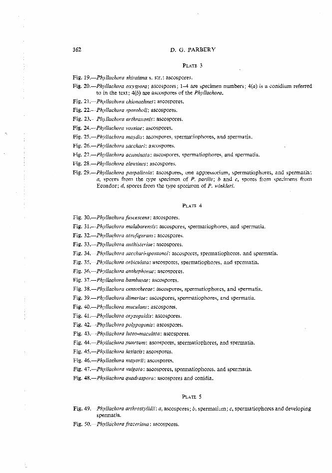
3 62 D. G. PARBERY
Fig. 19.-Phyllachora shiraiana s, str. : ascospores.
Fig. 20.-Phyllachora oxyspora: ascospores; 1-4 are specimen numbers; 4(a) is a conidium referred to in the text; 4(b) are ascospores of the Phyllachora.
Fig. 21 .-Phyllachora chionachnes: ascospores.
Fig. 22.-Phyllachora sporoboli: ascospores.
Fig. 23.-Phyllachora arthraxonis: ascospores.
Fig. 24.-Phyllachora vossiae: ascospores.
Fig. 25.-Phyllachova maydis: ascospores, spermatiophores, and spermatia.
Fig. 26.-Phyllachora sacchari: ascospores.
Fig. 27.-Phyllachora acuminata: ascospores, spermatiophores, and spermatia.
Fig. 28.-Phyllachora eleusines: ascospores.
Fig. 29.-Phyllachora paspalicola: ascospores, one appressorium, sperrnatiophores, and spermatia: a, spores from the type specimen of P. parilis; b and c, spores from specimens from Ecuador; d, spores from the type specimen of P. winkleri.
Fig. 30.-Phyllachora fuscescens: ascospores.
Fig. 31.-Phyllachora malabarensis: ascospores, spermatiophores, and spermatia.
Fig. 32.-Phyllachora atrojigurans: ascospores.
Fig. 33.-Phyllachora anthisteriae: ascospores.
Fig. 34.-Phyllachora sacchari-spontanei: ascospores, spermatiophores, and spermatia.
Fig. 35.-Phyllachora orbiculata: ascospores, spermatiophores, and spermatia.
Fig. 36.-Phyllachora anthephorae: ascospores.
Fig. 37.-Phyllachora bambusae: ascospores.
Fig. 38.-Phyllachora centothecae: ascospores, spermatiophores, and spermatia.
Fig. 39.-Phyllachora dimeriae: ascospores, spermatiophores, and spermatia.
Fig. 40.-Phyllachora maculans: ascospores.
Fig. 41 .-Phyllachora oryzopsidis: ascospores.
Fig. 42.-Phyllachora polypogonis: ascospores.
Fig. 43.-Phyllachora luteo-maculata: ascospores.
Fig. 44.-Phyllachorapunctum: ascospores, spermatiophores, and spermatia.
Fig. 45.-Phyllachora lasiacis: ascospores.
Fig, 46.-Phyllachora mayorii: ascospores.
Fig. 47.-Phyllachora vulgata: ascospores, spermatiophores, and spermatia.
Fig. 48.-Phyllachora quadraspova: ascospores and conidia.
Fig, 49.-Phyllachora arthrostylidii: a, ascospores ; b, spermatium; c, spermatiophores and developing spermatia.
Fig. 50.-Phyllachora frazeriana: ascospores.
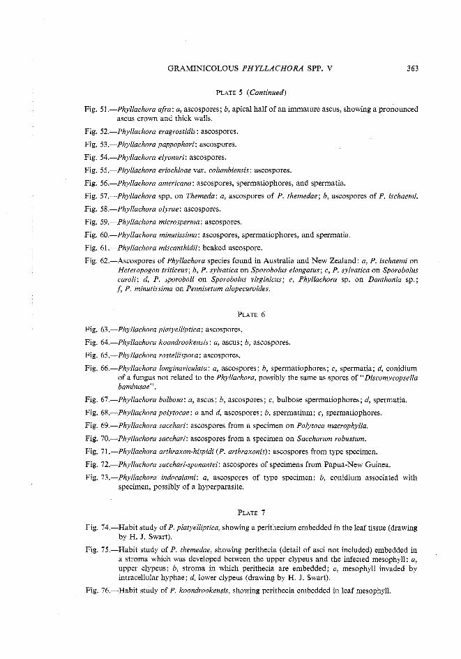
GRAMINICOLOUS P H Y L L A C H O M SPP. V 363
PLATE 5 (Continued)
Fig. 51.-Phyllachora afra: a, ascospores; b, apical half of an immature ascus, showing a pronounced ascus crown and thick walls.
Fig. 52.-Phyllachora eragrostidis: ascospores.
Fig. 53.-Phyllachora pappophori: ascospores.
Fig. 54.-Phyllachora elyonuri: ascospores.
Fig. 55.-Phyllachora eriochloae var. columbiensis: ascospores.
Fig. 56.-Phyllachora americana: ascospores, spermatiophores, and spermatia.
Fig. 57.-Phyllachora spp, on Themeda: a, ascospores of P. themedae; b, ascospores of P. ischaemi.
Fig. 58.-Phyllachora olyrae: ascospores.
Fig. 59.-Phyllachora microsperma: ascospores.
Fig. 60.-Phyllachora minutissima: ascospores, spermatiophores, and spermatia.
Fig. 61 .-Phyllachora miscanthidii: beaked ascospore.
Fig. 62.-Ascospores of Phyllachora species found in Australia and New Zealand: a, P. ischaemi on Heteropogon triticeus; b, P. sylvatica on Sporobolus elongatus; c, P. sylvatica on Sporobolus caroli; d, P. sporoboli on Sporobolus virginicus; e, Phyllachora sp. on Danthonia sp.; J; P. minutissima on Pennisetum alopecuvoides.
Fig. 63.-Phyllachora platyelliptica: ascospores.
Fig. 64.-Phyllachora koondrookensis: a, ascus; b, ascospores.
Fig. 65.-Phyllachora rostellispora: ascospores.
Fig. 66.-Phyllachora longinaviculata: a, ascospores; b, spermatiophores; c, spermatia; d, conidium of a fungus not related to the Phyllachora, possibly the same as spores of "Discomycopsella bambusae".
Fig. 67.-Phyllachora bulbosa: a, ascus; b, ascospores; c, bulbose spermatiophores; d, spermatia.
Fig. 68.-Phyllachora polytocae: a and d, ascospores; b, spermatium; c, spermatiophores.
Fig. 69.-Phyllachora sacchari: ascospores from a specimen on Polytoca macrophylla.
Fig. 70.-Phyllachora sacchari: ascospores from a specimen on Saccharum robustum.
Fig. 71.-Phyllachora arthraxon-hispidi (P. arthvaxonis): ascospores from type specimen.
Fig. 72.-Phyllachora sacchari-sponantei: ascospores of specimens from Papua-New Guinea.
Fig. 73.-Phyllachora indocalami: a, ascospores of type specimen; b, conidium associated with specimen, possibly of a hyperparasite.
Fig. 74.-Habit study of P. platyelliptica, showing a perithecium embedded in the leaf tissue (drawing by H. J. Swart).
Fig. 75.-Habit study of P. themedae, showing perithecia (detail of asci not included) embedded in a stroma which was developed between the upper clypeus and the infected mesophyll: a, upper clypeus; b, stroma in which perithecia are embedded; c, mesophyll invaded by intracellular hyphae; d, lower clypeus (drawing by H. J. Swart).
Fig. 76.-Habit study of P. koondrookensis, showing perithecia embedded in leaf mesophyll.
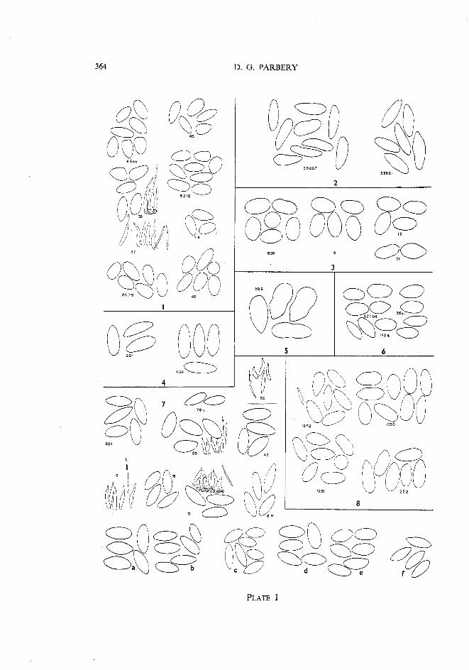
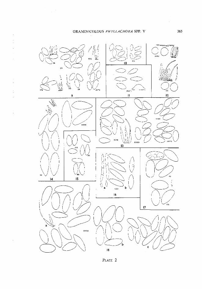

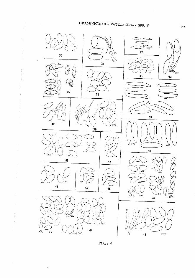
'PE
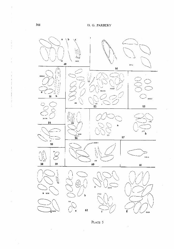
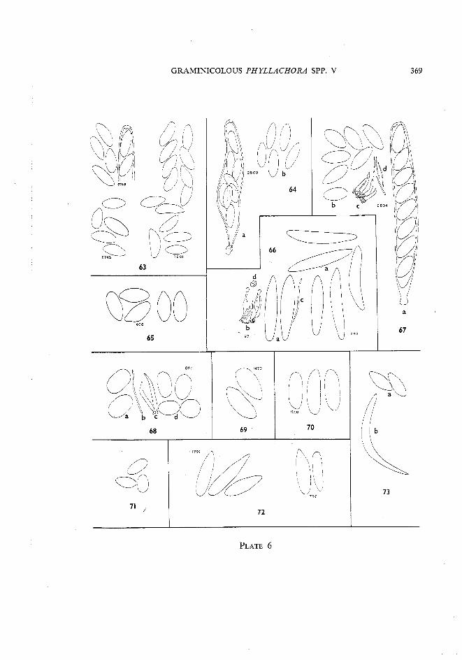

OLE
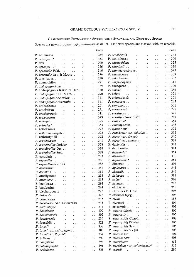
GRAMINICOLOUS PHYLLACHORA SPP . V 371
GRAMINICOLOUS PHYLLACHORA SPECIES. THEIR SYNONYMS. AND DOUBTFUL SPECIES
Species are given in roman type. synonyms in italics . Doubtful species are marked with an asterisk .
P . acuminata . . . . . . P . acutispora* . . . . . . P. afra . . . . . . . . P . agropyri . . . . . . . .
. . . . P agrostidis Fckl . . . . . . . . . . P agrostidis Ort & House
P. americana . . . . . . . . P . ammophilae . . . . . . P . andropogonicola . . . . . .
. . . . . P andropogonis Karst & Har . .. . . P andropogonis Ell & EV
P . andropogonis-aciculatis . . P . andropogonis-micranthi . . P . anthevhorae . . P . anthistiriae . . P . anthistiriicola . . P . antioquensis . . P . apiculata . . . . P . aristidae* . . . . P . arthraxonis . . P . arthraxon-hispidi . . P . arthrostylidii . . P . arundinariae . . P . arundinellae Doidge P . arundinellae Ort . . . P . arundinellae Saw . P . arundinis . . . . P . asperellae . . . . P . asperellae-histricus
. . . . . P asseriensis P . assimilis . . . . P . atrofigurans . . P . atronitens . . . . P. bambusae . . . . P . bambusina . . P . blepharoneuri . . P . bokensis . . . . P . bonariensis . . P . bonariensis var . tonkinensis P . bottomleyae . . . . P . boutelouae . . . . P . boutelonicola . . . . P . brachypodii . . . . P . brevifolia . . . . . . P . bromi* . . . . . . P . bromi var . andropogonis . . P . bromi var . lloydia* . . P . bulbosa . . . . . . P . caespiticia . . . . . .
. . . . . P calamagrostis P . cedralensis . . . .
PAGE . . 289 . . 353 . . 290 . . 286 . . 316 . . 286 . . 290 . . 291 . . 339 . . 310 . . 290 . . 311 . . 311 . . 291 . . 291 . . 311 . . 329 . . 325 . . 353 . . 292 . . 292 . . 292 . . 341 . . 328 . . 328 . . 328 . . 293 . . 286 . . 286 . . 351 . . 311 . . 293 . . 293 . . 294 . . 294 . . 295 . . 325 . . 295 . . 294 . . 311 . . 302 . . 302 . . 286 . . 321 . . 353 . . 309 . . 354 . . 299 . . 294 . . 291 . . 321
. . . . . P cenchricola P . centothecae . . . . P . chaetochloae . . . . P . chardonii . . . . . . P . chiminobambusae . . . . P . chionachnes . . . .
. . . . . P chloridicola P . chrysopogonis . . . .
. . . . . P chusqueae . .
. . . . . P cinnae . . P . coicis . . . . . . P . coloradensis . . . . P . congruens . . . . . . P . coorgiana . . . . . . P . cordobensis . . . . P . cornispora . . . . . . P . cornispora-necrotica . . P . culmicola* . . . . P . cnnninghamii . . . . P. cynodontis . . . . P . cynodontis var . chloridis . . P . cyperi var . donacis P . cyperi var . obtusata
. . . . . P dactylidis P . danthoniae . . P . delicatula* . . P . digitariae . . . . P . digitaricola* . . P . dimeriae . . . . P . diplocarpa . . . . P . distichlis . . . . P . doidgeae . . . . P . dolgei . . . . P . donacina . . . .
. . . . . P ehrhartae P . eleusines P . Henn . P. eleusines Speg . . . P . elymi . . . . P . elyonuri . . . . P . epicampis . . . . P . eragrostidicola . . P. eragrostis . . . . P . eragrostidis Chard . P . eragrostidis Doidge P . eragrostidis Saw . . . P . eragrostidis Viegas
. . . . P erianthi Ort P . erianthi Saw . . . P . eriochloae* . . P . eriochloae var . columbiensis* P . evansii . . . . . .
PAGE . . 343 . . 300 . . 325 . . 330 . . 343 . . 309 . . 302 . . 311 . . 300 . . 286 . . 301 . . 301 . . 295 . . 301 . . 295 . . 325 . . 289 . . 354 . . 346 . . 302 . . 302 . . 340 . . 329 . . 303 . . 304 . . 354 . . 330 . . 354 . . 304 . . 346 . . 346 . . 311 . . 307 . . 293 . . 358 . . 305 . . 308 . . 286 . . 306 . . 307 . . 305 . . 305 . . 308 . . 305 . . 305 . . 308 . . 304 . . 305 . . 355 . . 355 . . 295
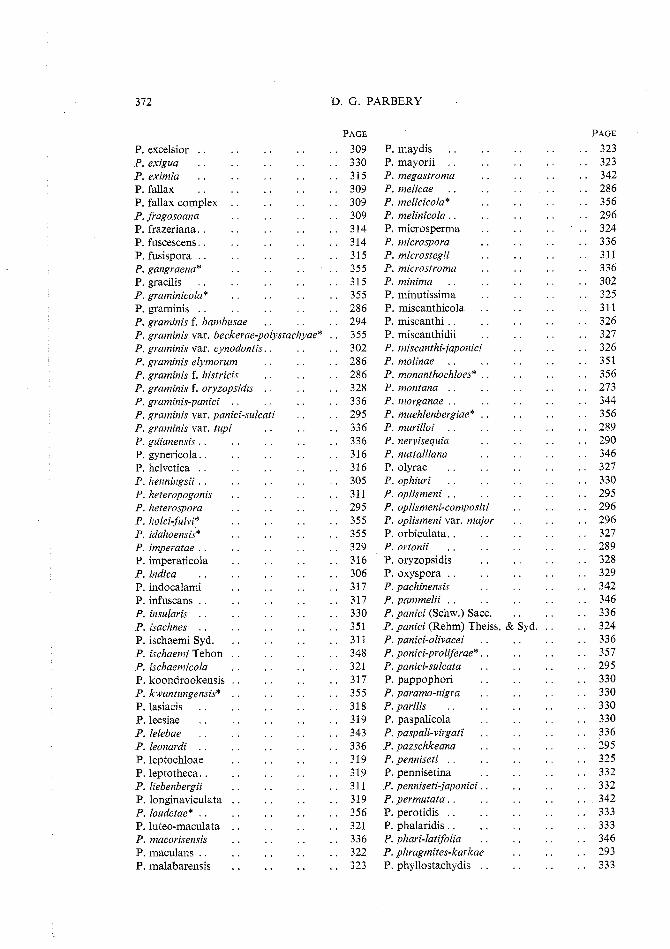
D . G . PARBERY
P . excelsior . . . . P . exigua . . . . P. eximia . . . . P . fallax . . . . P . fallax complex . . P . fvagosoana . . P . frazeriana . . . . P. fuscescens . . . . P . fusispora . . . . P . gangvaena* . . P . gracilis . . . . P . graminicola* . . P . graminis . . . . P . graminis f . bambusae
P . maydis . . P . mayorii . . P . megastvoma P . melicae . . P . melicicola* P . melinicola . . P. microsperma P . microspova P . micvostegii P . microstvoma P . minima . . P . minutissima P . miscanthicola P . miscanthi . .
P . graminis var . beckerae-polystachyae* . . 355 P . miscanthidii . . P . grarninis var . cynodontis . . P . graminis elymovum . . P . graminis f . histricis . . P . graminis f . oryzopsidis . . P . gvaminis-panici . . . . P . grarninis var . panici-sulcati P . graminis var . tupi P . guianensis . . . . P . gynericola . . . . P . helvetica . . . . P . henningsii . . . . P . heteropogonis . . P . hetevospova . . P . holci-fulvi* . . P . idahoensis* . . P . impevatae . . . . P . imperaticola . . P . indica . . . . P . indocalami . . P . infuscans . . . . P . insularis . . . . P . isachnes . . . . P . ischaemi Syd . . . P. ischaemi Tehon . . P . ischaemicola . . P . koondrookensis . . P . kwantungensis* . . P . lasiacis . . . . P . leesiae . . . . P . lelebae . . . . P . leonardi . . . . P . leptochloae . . P . leptotheca . . . . P . liebenbergii . . P . longinaviculata . . P. loudetae* . . . . P . luteo-maculata . . P . macorisensis . . P . maculans . . . . P . malabarensis . .
P . miscanthi-japonici P . molinae . . . . P . monanthochloes* . . P . montana . . . . P . morganae . . . . P . muehlenbergiae* . . P . murilloi . . . . P . nervisequia . . P . nuttalliana . . P . olyrae . . . . P . ophiuri . . . . P . oplismeni . . . . P . oplismeni-compositi P . oplismeni var . major P . orbiculata . . . . P . ovtonii . . . . P . oryzopsidis . . P . oxyspora . . . . P . pachinensis . . P . pammelii . . . . P . panici (Schw.) Sacc .
351 P . panici (Rehm) Theiss . & Syd . . . P . panici-olivacei . . P . panici-proliferue* . . P . panici-sulcata . . P . pappophori . . P . paramo-nigra . . P . parilis . . . . P . paspalicola . . P . paspali-vivgati . . P . pazschkeana . . P . penniseti . . . . P. pennisetina . . P . penniseti-japonici . . P . pevmutata . . . . P . perotidis . . . . P . phalaridis . . . . P . phari-latijolia . . P . phvagmites-karkae P . phyllostachydis . .
PAGE
. . 323
. . 323
. . 342
. . 286
. . 356
. . 296
. . 324
. . 336
. . 311
. . 336
. . 302
. . 325
. . 311
. . 326
. . 327
. . 326
. . 351
. . 356
. . 273
. . 344
. . 356
. . 289
. . 290
. . 346
. . 327
. . 330
. . 295
. . 296
. . 296
. . 327
. . 289
. . 328
. . 329
. . 342
. . 346
. . 336
. . 324
. . 336
. . 357
. . 295
. . 330
. . 330
. . 330
. . 330
. . 336
. . 295
. . 325
. . 332
. . 332
. . 342
. . 333
. . 333
. . 346
. . 293
. . 333
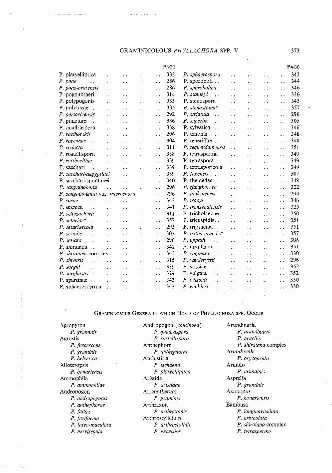
OEE "
OEE "
ZSE "
ZSE "
96Z '
OEE "
TSE "
90E "
LSE "
IS€ "
IS£ "
OSE "
SZE "
9PE "
P6Z . ' ZZE "
6PE "
LO€ "
6PE . ' 6PE "
6PE "
TSE "
8PE "
8PE "
9PE "
SO£ "
962 . ' LSE "
SPE "
9EE "
9PE "
PPE "
£PC "
39Vd
EL£
. . ajua~ou~n8uus .d !aus)uods-psy~3ss !3a~gd&a-!.i~y33~~ 'd .... !lVy33tzS 'd . . auqpoqrlor .d . . s~odsg1alsos 'd . . . . uj3npan .d . . . . aauuanan 'd . . !.2ysAoq!3""'d
. . slodswpsnb 'd
. . ' wnpund 'd
. . s!sua3!no]rod .d

374 D. G. PARBERY
bamboo P. bambusae P. bambusina P. gracilis P. maculans P. malabarensis P. orbiculata P. shiraiana complex P. tetrasperma
Beckera P. graminis var.
beckerae-polystachyae Blepharoneuron
P, blepharoneuri P. muehlenbergiae
Bothriochloa P. ischaemi
Bouteloua P. cynodontis
Brachiaria P. bonariensis P. ? stenospora P. stenostoma Phyllachora sp. (p. 345,357)
Brachyelytrum P. graminis
Brachypodium P. grarninis
Bromus P. bromi P. graminis P. dactylidis
Buchloe P. cynodontis
Calamagrostis P. ammophilae
Capillipedium P. ischaemi
Cenchrus P: spha~m~per.mr?
Centotheca P, centothecae
Chaetochloa P. minutissima
Chionachnes P. fallax
Chiminobambusa P. chiminobambusae
Chloris P. cynodontis P. koondrookensis
Chrysopogon P, fallax P, ischaemi
Cinna P. graminis
Chusquea P. chusqueae
Coix P , coicis
Cymbopogon P. ischaemi
Cynodon P. cynodontis
Cyrtococcum P. bonariensis P. stenospora
Dactylis P. dactylidis
Danthonia P , danthoniae
Dichanthium P. ischaemi
Digitaria P. digitaricola P. bonariensis P. paspalicola
Dimeria P. dimeriae P. elyonuri
Dinochloa P. maculans
Distichlis P. sylvatica
Donax P. atrofigurans
Ehrharta P. ehrhartae
Eleusine P. eleusines P, eragrostidis
Elymus P. graminis
Elyonurus P. dolgei P. elyoneuri
Eragrostis P. eleusines P. eragrostidis
Eremochloa P. quadraspora
Erharta Phyllachora sp.
Erianthus P. dimeriae
Eriochloa P. eriochloae P. eriochloae var.
columbiensis
Festuca P. graminis P. sylvatica P. tenerifae
Gynerium P. gynericola
Heteropogon P. ischaemi
Holcus P. bromi var. lloydia P. holci-fulvi
Hordeum P. graminis
Hyparrhenia P. fallax P. ischaemi
Hystrix P. graminis
Imperata P. imperaticola P. oxyspora
Indocalamus P. indocalami
Isachne P. paspalicola P, urvilliana P, punctum
Ischaemum P. ischaemi P. luteo-maculata P, tehonis
Lasiacis P, bonariensis P. lasiacis
Leersia P. leersiae
Leleba P, lelebae
Leptochloa P. leptochloae
Lcpto!oma P. punctum
Loudedia P. loudetae
Melica P. graminis P. melicicola
Melinis P. bonariensis
Microstegium P. ischaemi P. leptotheca
Miscanthus P. ischaemi P. miscanthii
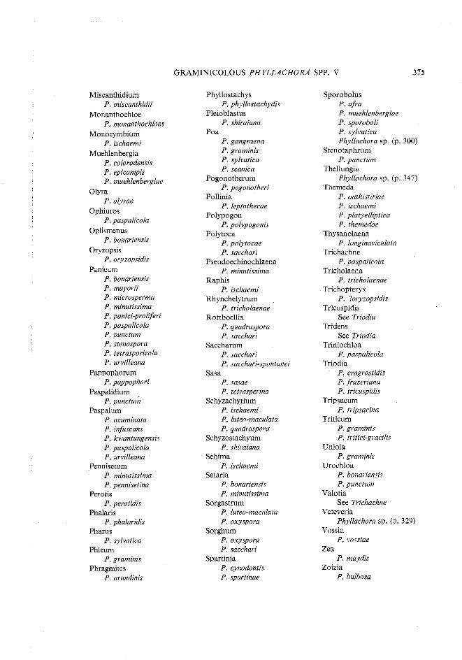
GRAMINICOLOUS PHYLLACHORA SPP. V
Miscanthidium P. rniscanthidii
Monanthochloe P. monanthochloes
Monocymbium P. ischaemi
Muehlenbergia P. coloradensis P. epicampis P. muehlenbergiae
Olyra P. olyrae
Ophiuros P. paspalicola
Oplismenus P. bonariensis
Oryzopsis P. oryzopsidis
Panicum P. bonariensis P. mayorii P. microsperma P. minutissima P. panici-proliferi P. paspalicola P. punctum P. stenospora P. tetrasporicola P. urvilleana
Pappophorum P. pappophori
Paspalidium P. punctum
Paspalum P. acuminata P. infuscans P. kwantungensis P. paspalicola P. urvilleana
Pennisetum P. minutissima P. pennisetina
Perotis P, perotidis
Phalaris P. phalaridis
Pharus P, sylvatica
Phleum P, graminis
Phragmites P, arundinis
Phyllostachys P. phyllostachydis
Pleioblastus P. shiraiana
Poa P. gangraena P. graminis P. sylvatica P. scanica
Pogonotherum P. pogonothevi
Pollinia P. leptothecae
Polypogon P. polypogonis
Polytoca P. polytocae P. sacchari
Pseudoechinochlaena P. minutissima
Raphis P, ischaemi
Rhynchelytrum P. tricholaenae
Rottboellia P. quadraspora P. sacchari
Saccharum P. sacchari P. sacchari-spontanei
Sasa P. sasae P. tetrasperma
Schyzachyrium P. ischaemi P. luteo-maculata P. quadraspora
Schyzostachyum P. shiraiana
Sehima P, ischaemi
Setaria P, bonariensis P, minutissima
Sorgastrum P. luteo-maculata P. oxyspora
Sorghum P. oxyspora P. sacchari
Spartinia P. cynodontis P. spartinae
Sporobolus P. afra P. muehlenbergiae P. sporoboli P. sylvatica Phyllachora sp. (p. 300)
Stenotaphrum P, punctum
Thellungia Phyllachora sp. (p. 347)
Themeda P. anthistiviae P. ischaemi P. platyelliptica P. themedae
Thysanolaena P. longinaviculata
Trichachne P. paspalicola
Tricholaena P. tricholaenae
Trichopteryx P. ?oryzopsidis
Tricuspidis See Triodia
Tridens See Triodia
Triniochloa P. paspalicola
Triodia P. eragrostidis P. frazeriana P. tricuspidis
Tripsacum P, tripsacina
Triticum P. graminis P. tritici-gracilis
Uniola P, graminis
Urochloa P. bonariensis P. punctum
Valotia See Trichachne
Veteveria Phyllachora sp. (p. 329)
Vossia P. vossiae
Zea P, maydis
Zoizia P. bulbosa