Embed Size (px)
Citation preview
Available Online through
www.ijpbs.com (or) www.ijpbsonline.com IJPBS |Volume 2| Issue 3 |JULY-SEPT |2012|15-30
Research Article
Pharmaceutical Sciences
International Journal of Pharmacy and Biological Sciences (e-ISSN: 2230-7605)
Saiprasanna Behera *et al Int J Pharm Bio Sci www.ijpbs.com or www.ijpbsonline.com
Pag
e15
STUDIES ON HEPATOPROTECTIVE ACTIVITY OF HYDROALCOHOLIC LEAF EXTRACT OF PONGAMIA PINNATA AGAINST I/R INDUCED HEPATIC REPERFUSION INJURY
*Saiprasanna Behera1, S. Manohar Babu2, Y. Roja Ramani3, Prasanta Kumar Choudhury1
1*,1
Department of Pharmacology, Royal College of Pharmacy and Health Sciences, Berhampur, Odisha- 760002 2Department of Pharmacology, SIMS College of Pharmacy, Mangaldas Nagar, Guntur- 522001
3Department of Pharmacology, MKCG Medical College, Berhampur, Odisha- 760004
*Corresponding Author Email: [email protected] ; [email protected]
ABSTRACT Pongamia pinnata (Linn) Pierre commonly known as ‘Karanj’ contains various phytoconstituents belonging to alkaloids, glycosides, flavonoids, fixed oils and carbohydrates. Leaves of Pongamia pinnata are digestive, laxative, anthelmintic and are good for diarrhoea, leprosy, dyspepsia and cough. A hot infusion of leaves is good for cleaning ulcers and wounds. This study was designed to determine the possible protective effect of Pongamia pinnata (PP) hydro-alcoholic leaf extract, a plant rich in antioxidant, on hepatic ischemia/reperfusion (I/R) injury. Wistar albino rats were subjected to 45 min of hepatic ischemia, followed by a 60 min reperfusion period. Pongamia pinnata hydro-alcoholic leaf extract were administered in doses of 100, 200 and 400 mg/kg/day, orally for 15 days before I/R injury respectively and repeated before the reperfusion period. Liver samples were taken for histological examination or determination of hepatic malondialdehyde (MDA), super oxide dismutase (SOD) and glutathione (GSH) activity. Serum aspartate aminotransferase (AST), alanine aminotransferase (ALT) alkaline phosphatase (ALP) and bilirubin levels were determined to assess liver functions. Lactate dehydrogenase (LDH) was assayed in serum samples for the evaluation of generalized tissue damage. Ischemia/reperfusion caused a significant decrease in hepatic GSH, and significant increases in MDA levels. Serum AST, ALT, ALP and bilirubin levels, as well as LDH activity levels were also elevated in the I/R group. Treatment with PP hydro-alcoholic leaf extract reversed all these biochemical parameters as well as histological alterations induced by I/R. In all the testing, a significant correlation existed between concentrations of the extract and alteration in the biochemical and histological parameters. In conclusion, PP hydro-alcoholic leaf extract at the dose of 400 mg/kg/day reduced I/R-induced organ injury through its ability to balance the oxidant–antioxidant status.
KEYWORDS Ischemia/reperfusion; Pongamia pinnata (PP) hydro-alcoholic leaf extract; antioxidant
INTRODUCTION
Liver is frequently exposed to IR injury under
different clinical conditions like circulating shock,
intravascular coagulation, liver transplantation and
surgery involving this organ 1, 2. Hepatic ischemia
reperfusion (HIR) injuries cause high morbidity and
consume substantial health care capacities in
patients with primary hepatic injury and systemic
injury 3-5. Oxygen radicals probably mediate some
of the structural and functional alterations
associated with reperfusion of ischaemic liver. A
number of evidences implicate oxygen-derived free
radicals (especially superoxide and hydroxyl
radical) and high-energy oxidants (such as
peroxynitrite) in the IR injury syndrome,
particularly during the reperfusion phase 6. Thus,
agents proposed to be useful in the clinical settings
of Hepatic Ischemia Reperfusion damage include
antioxidants or free radical scavengers 7. The main
characteristic of an antioxidant is its ability to trap
free radicals. Antioxidant compounds like phenolic
Available Online through
www.ijpbs.com (or) www.ijpbsonline.com IJPBS |Volume 2| Issue 3 |JULY-SEPT |2012|15-30
International Journal of Pharmacy and Biological Sciences (e-ISSN: 2230-7605)
Saiprasanna Behera *et al Int J Pharm Bio Sci www.ijpbs.com or www.ijpbsonline.com
Pag
e16
acids, polyphenols and flavonoids scavenge free
radicals such as peroxide, hydroperoxide or lipid
peroxyl and thus inhibit the oxidative mechanisms
that lead to degenerative diseases.
The use of herbal drugs as medicines for the
treatment of a wide range of diseases can be
traced back since ancient time’s i.e during the
Vedic period in India 8. Being the outcome of
therapeutic experiences of generations of
practicing physicians of indigenous systems of
medicine for over hundreds of years medicinal
plants have played a key role in world health 9, 10.
Hence, in spite of the great advances observed in
modern medicine in recent decades, herbal
medicines still make an important contribution to
health care 11. Plants phenolics, in particular
phenolic acid 12-14 tannins 15, 16 and flavonoids 17 are
known to be potent antioxidant and occur in
vegetables, fruits, nuts, seeds, roots, barks and
leaves. In addition to their antioxidant properties,
these compound display a vast variety of
pharmacological activities such as anti-
inflammatory, anti-carcinogenics, antibacterial or
antiviral activities which may explain, at least in
part, its use as alternative or supportive
treatments in various degenerative diseases 18-21.
Pongamia pinnata (Linn) Pierre (synonyms- Indian
beech) commonly known as Karanj belonging to
family Fabaceae. It is also called Pongamia glabra.
It is an indo-Malaysian species, a medium-sized
evergreen tree, and common on alluvial and
coastal situations from India to Fiji, from sea level
to 1200m. Now found in Australia, Florida, Hawaii,
India, Malaysia, Oceania, Philippines and
Seychelles 22-24. The plant affords patulitrin, β -
sitosterol, spicigerine, aminoacids, albumin,
globulin, glutelins, flavonoids such as
furanoflavones, furanoflavanols,
chromenoflavones, furanochalcones 25. The plant
also contains alkaloids, tannins and carbohydrate 26. Phenolics, such as flavonoids, flavonols and
flavones have potent antioxidant capacity and
reports suggest that Pongamia pinnata contains
flavones, pongamones, furanoflavones, pongamol,
pongagalabrone and pongapin, pinnatin and
kanjone which have potent antioxidant activity.
Potent free radical scavenging capacity of
Pongamia pinnata can be attributed to different
flavones present in the extracts. So the plant may
be useful in the management of free radical
mediated diseases. The leaves and stem of the
plant consist of several flavone and chalcone
derivatives such as Pongone, Galbone, Pongalabol,
Pongagallone A and B 27. Punitha and Manohar in
2006 evaluated anti-hyperglycemic and anti-lipid
peroxidative effects of ethanolic extract of
Pongamia pinnata (Linn.) 28. Effects of Pongamia
pinnata on lipid peroxidation products and
antioxidants in hyper-ammonemic rats with
reference to circadian variations were evaluated by
Essa and Subramanian 29. Literature survey of
Pongamia pinnata revealed its potential being like
hepatoprotective and antioxidant 30.However it
remains to be scientifically validated. Hence
present study titled “Studies on Hepatoprotective
Activity of Hydroalcoholic Leaf Extract of Pongamia
Pinnata against I/R Induced Hepatic Reperfusion
Injury” has been conducted.
MATERIALS AND METHODS
METHOD:
The experimental protocols were conducted with
the approval of the Animal Research Committee at
Royal College of Pharmacy and Health Sciences,
Berhampur. Odisha. All animals were maintained in
accordance with the recommendations of the
CPCSEA.
Chemicals:
All the chemicals used were of analytical grade.
ALT, AST, ALP, bilirubin and LDH Kits were obtained
from Crest Biosystems, Bambolim Complex. Goa,
India. All drug solutions were freshly prepared in
saline before each experiment. The extracts were
dissolved in distilled water and administered orally.
Available Online through
www.ijpbs.com (or) www.ijpbsonline.com IJPBS |Volume 2| Issue 3 |JULY-SEPT |2012|15-30
International Journal of Pharmacy and Biological Sciences (e-ISSN: 2230-7605)
Saiprasanna Behera *et al Int J Pharm Bio Sci www.ijpbs.com or www.ijpbsonline.com
Pag
e17
Animals:
Male Wistar albino rats (200–250 g) were obtained
from the animal house of R.C.P.H.S. and were
housed in an air-conditioned room with 12 h light
and dark cycles, with constant temperature (22 ± 2
°C) and relative humidity (65–70%) levels. All
experimental protocols were approved by the
Institutional Animal Ethical Committee (Approval
No-07/IAEC/2011). The rats were anesthetized by
intraperitoneal injection of sodium pentobarbital
(30 mg/kg). All surgical procedures were conducted
with clean but not sterile instruments.
Plant collection:
Leaves of Pongamia pinnata were collected in the
month of December 2011 from its natural habitat
from nearby Mohuda village, Berhampur, Ganjam
district of Odisha. The plant was authenticated
from Department of Botany, Khalikote College,
Berhampur, Odisha. The leaves were cleaned and
dried under the shade to avoid degradation of
volatile oil. The leaves were dried in hot air woven
at 55°C for 3 days and at 40°C for the next 4 days.
Preparation of Plant Extracts:
The dried leaves were coarsely powdered and
extracted with a mixture of methanol: water (7:3,
v/v) by a Soxhlet apparatus at 50°C. The solvent
was completely removed and obtained dried crude
extract which was used for investigation. Further
the extracts were subjected for pharmacological
screening.
EXPERIMENTAL PROTOCOL:
Under anesthesia, a midline laparotomy was made
using minimal dissection. The abdomen was
shaved and a transverse incision was performed.
The bowel loops were covered with saline-soaked
gauze. Total hepatic ischemia was induced for 45
min by clamping the hepatic artery, the portal vein
and the bile duct using a vascular clamp. Then
reperfusion was induced for 60 minutes in rats.
Abdominal incision was closed in layers with 4-0
dexon and 2-0 nylon during reperfusion stage in
order to prevent the loss of body fluid and quantity
of heat.
Animals were divided into seven groups consisting
of six rats each. Pongamia pinnata (PP) hydro-
alcoholic leaf extract was dissolved in water and
administered to the animals. Pongamia pinnata
(PP) hydro-alcoholic leaf extract used in this study
contains Pongagallone A and B
Group-I: - NAIVE-Normal control-rats in this group
did not undergo ischemia or reperfusion and
served as the control group.
Group-II: - SHAM-Sham-operated (animals
subjected to the identical procedure of surgery
without ischemia-reperfusion injury) plus
physiologic saline treatment.
Group-III: - I/R-Animals subjected 45 minutes of
total hepatic ischemia, followed by reperfusion for
60 mins and served as untreated experimental
control.
Group-IV: - PP control- Sham operated plus
Pongamia pinnata control (400 mg/kg body wt.
treatment up to15 days).
Group-V: - PP 100mg/kg + I/R- Hepatic I/R plus
Pongamia pinnata hydro-alcoholic leaf extract 100
mg/kg body wt. treatment up to 15 days
Group-VI: - PP 200mg/kg + I/R- Hepatic I/R plus
Pongamia pinnata hydro-alcoholic leaf extract 200
mg/kg body wt. up to 15 days.
Group-VII: - PP 400mg/kg + I/R- Hepatic I/R plus
Pongamia pinnata hydro-alcoholic leaf extract 400
mg/kg body wt. up to15 days.
None of the animals died during these procedures.
At the end of the reperfusion period, animals were
decapitated and trunk blood samples were
Available Online through
www.ijpbs.com (or) www.ijpbsonline.com IJPBS |Volume 2| Issue 3 |JULY-SEPT |2012|15-30
International Journal of Pharmacy and Biological Sciences (e-ISSN: 2230-7605)
Saiprasanna Behera *et al Int J Pharm Bio Sci www.ijpbs.com or www.ijpbsonline.com
Pag
e18
collected to determine serum alanine
aminotransferase (ALT), aspartate amino
transferase (AST), alkaline phosphatase (ALP),
bilirubin and lactate dehydrogenase (LDH) activity,
the indicators of liver functions and generalized
tissue damage, respectively. The hepatic tissue
samples were stored at −20°C. Afterwards, tissue
malondialdehyde (MDA) levels, an end product of
lipid peroxidation, superoxide dismutase (SOD),
Catalase and glutathione (GSH), key endogenous
antioxidants, were measured in these samples. The
hepatic tissue samples were also placed in
formaldehyde (10%) for histological evaluation.
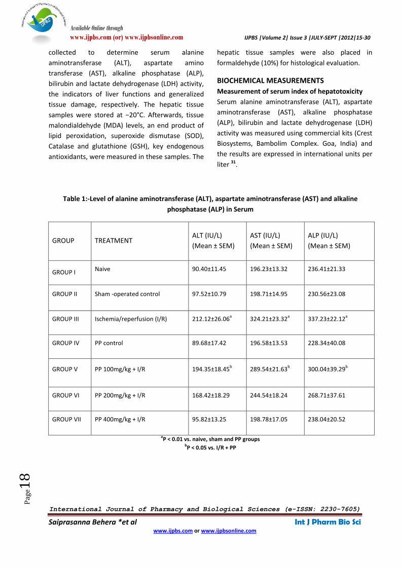
BIOCHEMICAL MEASUREMENTS
Measurement of serum index of hepatotoxicity
Serum alanine aminotransferase (ALT), aspartate
aminotransferase (AST), alkaline phosphatase
(ALP), bilirubin and lactate dehydrogenase (LDH)
activity was measured using commercial kits (Crest
Biosystems, Bambolim Complex. Goa, India) and
the results are expressed in international units per
liter 31.
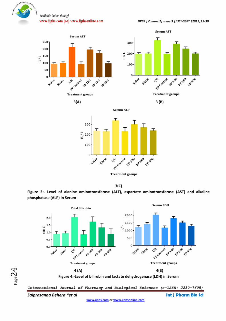
Table 1:-Level of alanine aminotransferase (ALT), aspartate aminotransferase (AST) and alkaline
phosphatase (ALP) in Serum
GROUP TREATMENT ALT (IU/L)
(Mean ± SEM)
AST (IU/L)
(Mean ± SEM)
ALP (IU/L)
(Mean ± SEM)
GROUP I Naive 90.40±11.45 196.23±13.32 236.41±21.33
GROUP II Sham -operated control 97.52±10.79 198.71±14.95 230.56±23.08
GROUP III Ischemia/reperfusion (I/R) 212.12±26.06a 324.21±23.32
a 337.23±22.12
a
GROUP IV PP control 89.68±17.42 196.58±13.53 228.34±40.08
GROUP V PP 100mg/kg + I/R 194.35±18.45b 289.54±21.63
b 300.04±39.29
b
GROUP VI PP 200mg/kg + I/R 168.42±18.29 244.54±18.24 268.71±37.61
GROUP VII PP 400mg/kg + I/R 95.82±13.25 198.78±17.05 238.04±20.52
aP < 0.01 vs. naive, sham and PP groups
bP < 0.05 vs. I/R + PP
Available Online through
www.ijpbs.com (or) www.ijpbsonline.com IJPBS |Volume 2| Issue 3 |JULY-SEPT |2012|15-30
International Journal of Pharmacy and Biological Sciences (e-ISSN: 2230-7605)
Saiprasanna Behera *et al Int J Pharm Bio Sci www.ijpbs.com or www.ijpbsonline.com
Pag
e19
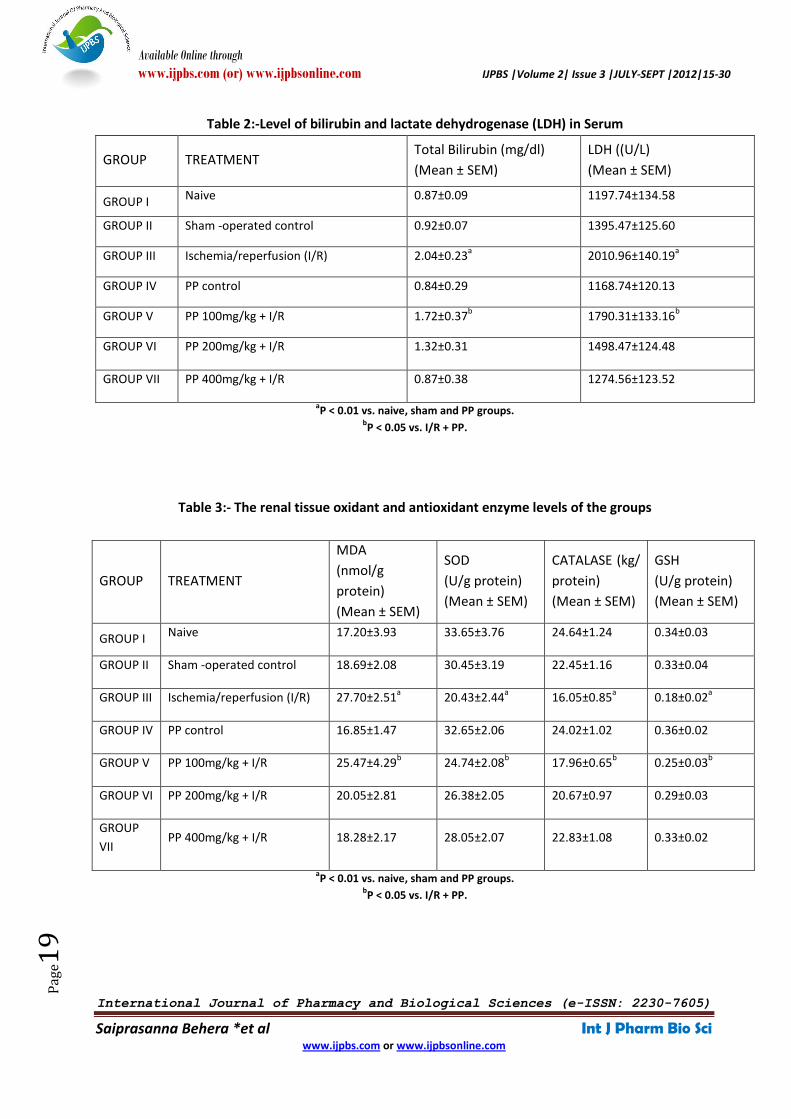
Table 2:-Level of bilirubin and lactate dehydrogenase (LDH) in Serum
GROUP TREATMENT Total Bilirubin (mg/dl)
(Mean ± SEM)
LDH ((U/L)
(Mean ± SEM)
GROUP I Naive 0.87±0.09 1197.74±134.58
GROUP II Sham -operated control 0.92±0.07 1395.47±125.60
GROUP III Ischemia/reperfusion (I/R) 2.04±0.23a 2010.96±140.19
a
GROUP IV PP control 0.84±0.29 1168.74±120.13
GROUP V PP 100mg/kg + I/R 1.72±0.37b 1790.31±133.16
b
GROUP VI PP 200mg/kg + I/R 1.32±0.31 1498.47±124.48
GROUP VII PP 400mg/kg + I/R 0.87±0.38 1274.56±123.52
aP < 0.01 vs. naive, sham and PP groups.
bP < 0.05 vs. I/R + PP.
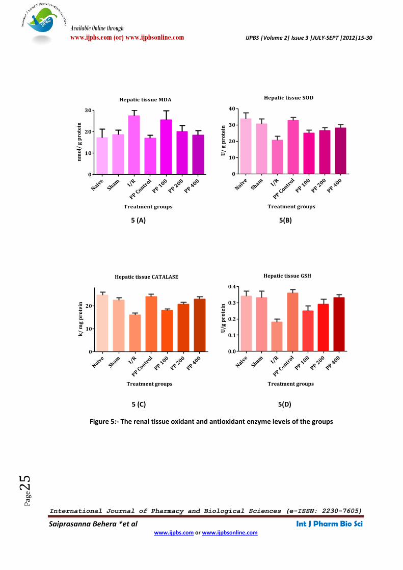
Table 3:- The renal tissue oxidant and antioxidant enzyme levels of the groups
aP < 0.01 vs. naive, sham and PP groups.
bP < 0.05 vs. I/R + PP.
GROUP TREATMENT
MDA
(nmol/g
protein)
(Mean ± SEM)
SOD
(U/g protein)
(Mean ± SEM)
CATALASE (kg/
protein)
(Mean ± SEM)
GSH
(U/g protein)
(Mean ± SEM)
GROUP I Naive 17.20±3.93 33.65±3.76 24.64±1.24 0.34±0.03
GROUP II Sham -operated control 18.69±2.08 30.45±3.19 22.45±1.16 0.33±0.04
GROUP III Ischemia/reperfusion (I/R) 27.70±2.51a 20.43±2.44
a 16.05±0.85
a 0.18±0.02
a
GROUP IV PP control 16.85±1.47 32.65±2.06 24.02±1.02 0.36±0.02
GROUP V PP 100mg/kg + I/R 25.47±4.29b 24.74±2.08
b 17.96±0.65
b 0.25±0.03
b
GROUP VI PP 200mg/kg + I/R 20.05±2.81 26.38±2.05 20.67±0.97 0.29±0.03
GROUP
VII PP 400mg/kg + I/R 18.28±2.17 28.05±2.07 22.83±1.08 0.33±0.02
Available Online through
www.ijpbs.com (or) www.ijpbsonline.com IJPBS |Volume 2| Issue 3 |JULY-SEPT |2012|15-30
International Journal of Pharmacy and Biological Sciences (e-ISSN: 2230-7605)
Saiprasanna Behera *et al Int J Pharm Bio Sci www.ijpbs.com or www.ijpbsonline.com
Pag
e20
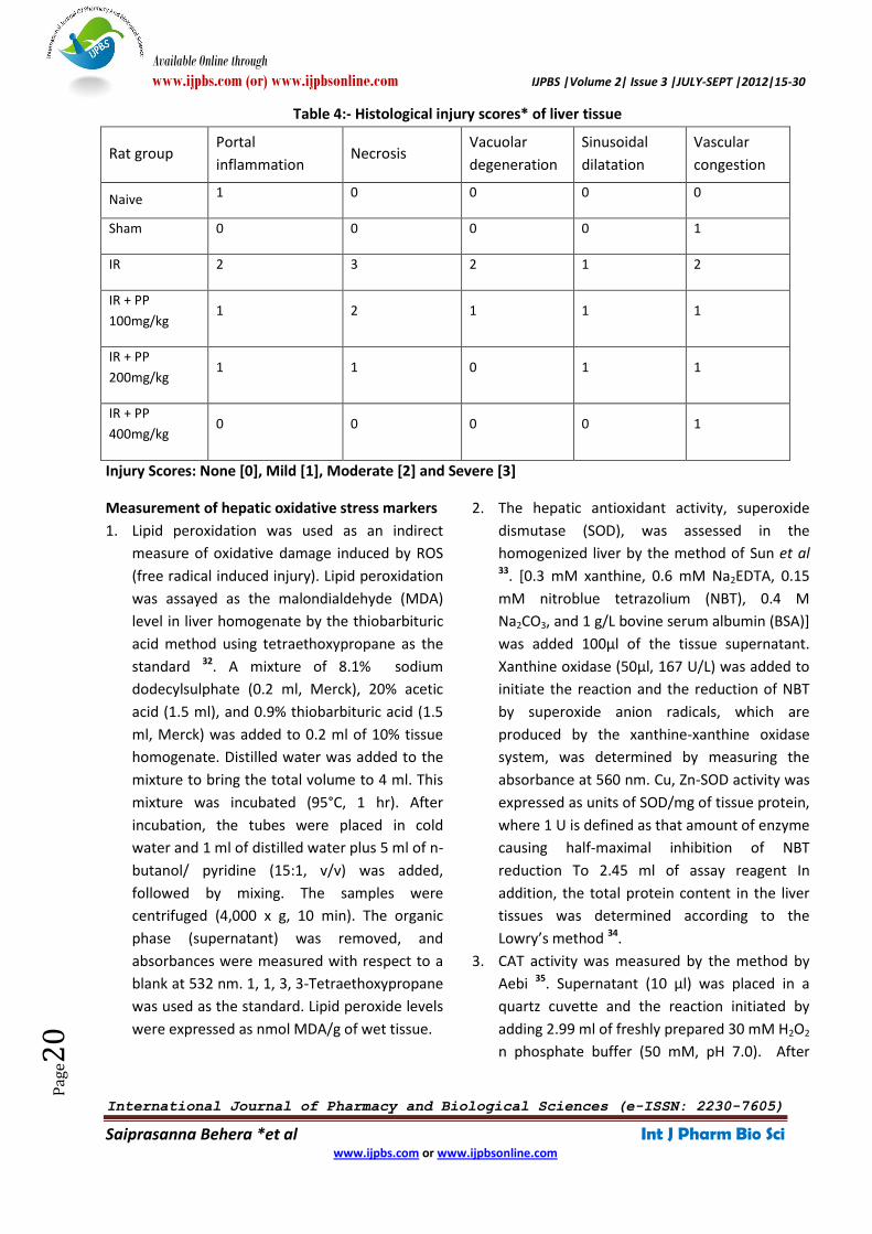
Table 4:- Histological injury scores* of liver tissue
Rat group Portal
inflammation Necrosis
Vacuolar
degeneration
Sinusoidal
dilatation
Vascular
congestion
Naive 1 0 0 0 0
Sham 0 0 0 0 1
IR 2 3 2 1 2
IR + PP
100mg/kg 1 2 1 1 1
IR + PP
200mg/kg 1 1 0 1 1
IR + PP
400mg/kg 0 0 0 0 1
Injury Scores: None [0], Mild [1], Moderate [2] and Severe [3]
Measurement of hepatic oxidative stress markers
1. Lipid peroxidation was used as an indirect
measure of oxidative damage induced by ROS
(free radical induced injury). Lipid peroxidation
was assayed as the malondialdehyde (MDA)
level in liver homogenate by the thiobarbituric
acid method using tetraethoxypropane as the
standard 32. A mixture of 8.1% sodium
dodecylsulphate (0.2 ml, Merck), 20% acetic
acid (1.5 ml), and 0.9% thiobarbituric acid (1.5
ml, Merck) was added to 0.2 ml of 10% tissue
homogenate. Distilled water was added to the
mixture to bring the total volume to 4 ml. This
mixture was incubated (95°C, 1 hr). After
incubation, the tubes were placed in cold
water and 1 ml of distilled water plus 5 ml of n-
butanol/ pyridine (15:1, v/v) was added,
followed by mixing. The samples were
centrifuged (4,000 x g, 10 min). The organic
phase (supernatant) was removed, and
absorbances were measured with respect to a
blank at 532 nm. 1, 1, 3, 3-Tetraethoxypropane
was used as the standard. Lipid peroxide levels
were expressed as nmol MDA/g of wet tissue.
2. The hepatic antioxidant activity, superoxide
dismutase (SOD), was assessed in the
homogenized liver by the method of Sun et al 33. [0.3 mM xanthine, 0.6 mM Na2EDTA, 0.15
mM nitroblue tetrazolium (NBT), 0.4 M
Na2CO3, and 1 g/L bovine serum albumin (BSA)]
was added 100µl of the tissue supernatant.
Xanthine oxidase (50µl, 167 U/L) was added to
initiate the reaction and the reduction of NBT
by superoxide anion radicals, which are
produced by the xanthine-xanthine oxidase
system, was determined by measuring the
absorbance at 560 nm. Cu, Zn-SOD activity was
expressed as units of SOD/mg of tissue protein,
where 1 U is defined as that amount of enzyme
causing half-maximal inhibition of NBT
reduction To 2.45 ml of assay reagent In
addition, the total protein content in the liver
tissues was determined according to the
Lowry’s method 34.
3. CAT activity was measured by the method by
Aebi 35. Supernatant (10 µl) was placed in a
quartz cuvette and the reaction initiated by
adding 2.99 ml of freshly prepared 30 mM H2O2
n phosphate buffer (50 mM, pH 7.0). After
Available Online through
www.ijpbs.com (or) www.ijpbsonline.com IJPBS |Volume 2| Issue 3 |JULY-SEPT |2012|15-30
International Journal of Pharmacy and Biological Sciences (e-ISSN: 2230-7605)
Saiprasanna Behera *et al Int J Pharm Bio Sci www.ijpbs.com or www.ijpbsonline.com
Pag
e21
rapid mixing, the rate of H2O2 decomposition
was determined from absorbance changes at
15 and 30 sec at 240 nm. CAT activity was
expressed as k/mg of tissue protein, where k is
the first order rate constant.
4. GSH in liver tissue was assayed by the method
of Tietze and Anderson 36, 37. Briefly, 100 µl of
tissue supernatant was placed in a 3 ml
cuvette; 750 µl of 10 mM 5-5’-dithio-bis-
2nitrobenzoic acid (DTNB) solution (100 mM
KH2PO4KH plus 5 mM Na2EDTA, pH 7.5 and
GSHRD, 625 U/L) was added and the mixture
was incubated (3 min, room temperature).
Then 150 µl of 1.47 mM β -NADPH was added,
mixed rapidly by inversion, and the rate of 5-
thio-2-nitrobenzoic acid formation
(proportional to the sum of reduced and
oxidized glutathione) was measured
spectrophotometrically for 2 min at 412 nm.
The reference cuvette contained equal
concentrations of DTNB and NADPH, but no
sample; results were expressed as nmol/mg of
wet tissue
Histological procedures
Liver specimens from all groups were rapidly taken
and fixed in Bouin’s solution and processed for
light microscopic study using hematoxylin and
eosin stain 38. For light microscopic investigations,
hepatic tissue specimens were fixed in 10%
formaldehyde, dehydrated in alcohol series,
clearing in toluene and embedding in paraffin.
Paraffin sections (5 μm) were stained with
hematoxylin and eosin (H&E) and examined under
a photomicroscope. All tissue sections were
examined microscopically for the characterization
of histopathological changes by an experienced
histologist in blind fashion (Procedures were
carried out in NIDAN diagnostics, Berhampur.
Odisha. INDIA).
STATISTICAL ANALYSIS
Results are presented as the mean ± SEM. All
statistical analyses were performed using Graph
Pad Prism Software program (version 5) 39. Data
were analyzed using analysis of variance followed
by Bonferroni’s post-test. The Kruskal-Wallis 1-way
analysis of variance by ranks was used to
simultaneously test the pathologic score for the I/R
and I/R ± Pongamia pinnata groups. A P value of <
0.05 was considered statistically significant.
RESULTS AND DISCUSSION
The present study justifies that temporary
blockade of hepatic blood supply yielded structural
and functional alterations in the liver. Hydro-
alcoholic leaf extract of Pongamia pinnata (PP) on
the other hand, reduced the severity of injury.
I/R injury is a complex process and evidence
suggests that oxygen derived free radicals are
involved in the hepatic injury caused by ischemia
and reperfusion 40-43. Thus, therapeutic strategies
are designed so as to reduce free radical induced
damage, either by intervening in the process by
which free radicals are formed or by scavenging
the free radicals that have already been formed.
Different degrees of protection have been acquired
with numerous compounds; however, the
structure-activity relationship, bioavailability and
therapeutic efficacy of these compounds differ
comprehensively. Thus clinical application of these
agents is limited in respect of their
pharmacokinetic and pharmacodynamics
properties.
The present study accentuates three points: (1)
The emergence of free oxygen radicals that arise
after hepatic IR and cause injury which can be
prevented by PP; (2) the increase in AST, ALT, ALP,
Total Bilirubin and LDH →an indication of tissue
damage in hepatic IR injury →is less after PP
treatment; (3) histopathological examination of
liver tissue shows considerably less hepatocyte
injury in rats with PP treatment.
Free oxygen radicals have a marked mediator role
in IR injuries of several organs. Oxygen radicals
probably mediate some of the structural and
functional alterations associated with reperfusion
Available Online through
www.ijpbs.com (or) www.ijpbsonline.com IJPBS |Volume 2| Issue 3 |JULY-SEPT |2012|15-30
International Journal of Pharmacy and Biological Sciences (e-ISSN: 2230-7605)
Saiprasanna Behera *et al Int J Pharm Bio Sci www.ijpbs.com or www.ijpbsonline.com
Pag
e22
of ischaemic liver. Research suggests that
antioxidant molecules may provide protection
from IR injury. Pongamia pinnata, rich in flavonoids
(Pongagallone A and B), is known to be a strong
antioxidant, breaking up free radicals. For this
reason, it is expected to be protective in hepatic IR
injury of rats.
Most procedures of liver ischemia in rats include a
portosystemic shunt (usually using an ex vivo
bypass between a branch of the mesenteric vein
and the jugular vein) with the using of heparin, or
segmental rather than total clamping of the
hepatic blood supply to prevent mesenteric
congestion. Thus partial hepatic ischemia is not a
method from which organ viability or animal
survival can be assessed. Therefore, in this study
surgery was conducted to develop a simple and
reproducible method of total hepatic ischemia in
rat. Another advantage of the availability of such a
simple model is the low cost compared with other
models and the lack of a requirement for
sophisticated expensive materials and can be
instrumental in the research of the mechanisms of
liver ischemia-reperfusion injury in different
pathophysiological conditions.
In IR injury of the liver during the reperfusion
phase, emerging reactive oxygen radicals activate
some mediators and can cause inflammatory
response and tissue damage. The release of liver
enzymes is usually used to assess tissue damage
following ischemia-reperfusion. For this reason
AST, ALT, ALP, Total Bilirubin and LDH activities
may increase 44, 45. The increase of AST, ALT, ALP,
Total Bilirubin and LDH activities in group 3 of our
study supports this finding. In the study, it is shown
that in group 4, 5, 6 and 7, ALT, AST, ALP, Total
Bilirubin and LDH enzymes, markers of liver
parenchymal injury, have decreased levels in
comparison with group 3 (p<0.0001). This finding
supports the protective effect of PP treatment on
IR injury by justifying that the hydro-alcoholic leaf
extract of Pongamia pinnata (PP) have shown very
significant hepatoprotection against I/R-induced
induced hepatotoxicity in wistar albino rats in
reducing serum AST, ALT, ALP, Total Bilirubin and
LDH levels.
This study showed no significant difference
between the biochemical measurements of group
1 and group 2. Hepatic I/R rats (group 3) showed a
significant elevation of serum index of
hepatotoxicity (ALT). ALT, AST and ALP levels were
significantly higher in the I/R group when
compared with those of the control group (p <
0.001). PP treatment reversed these values
significantly. Similarly, in the I/R group, increased
lactate dehydrogenase activity, as an index of
generalized tissue damage, was reversed
significantly by PP treatment (p < 0.01) (Fig.2)
Oxidative stress occurs particularly in reperfusion
after ischemia. Synthesis of proinflammatory
cytokines and cell adhesion molecules is activated,
and the inflammatory response is increased by
oxidative stress. The antioxidant system has an
important role in protection from the damage of
oxidative stress. Lipid peroxide is an intermediate
free radical oxidant that is synthesized during lipid
peroxidation 46, 47. In our study, lipid peroxide levels
were found to be significantly higher in group 3
than in group 1. It was also found that treatment
with PP in group 4, 5, 6 and 7 decreased the lipid
hydroperoxide activity respectively , indicating that
the antioxidant effect of PP had prevented the
emergence of an oxidant agent.In our study, by
measuring Catalase, we obtained information
about hepatic IR injury. Catalase is an antioxidant
enzyme that catalyses the chage from hydrogen
peroxide into water. The concentration of hepatic
reduced glutathione decreases progressively
during ischaemia48 with a corresponding increase
in oxidized glutathione, which is attenuated by
formate, a cell permeable OH scavenger49. It is also
found that treatment with hydro-alcoholic leaf
extract of Pongamia pinnata have brought down
the elevated levels of LPO and also significantly
enhanced the reduced levels of SOD, CAT and GSH
Available Online through
www.ijpbs.com (or) www.ijpbsonline.com IJPBS |Volume 2| Issue 3 |JULY-SEPT |2012|15-30
International Journal of Pharmacy and Biological Sciences (e-ISSN: 2230-7605)
Saiprasanna Behera *et al Int J Pharm Bio Sci www.ijpbs.com or www.ijpbsonline.com
Pag
e23
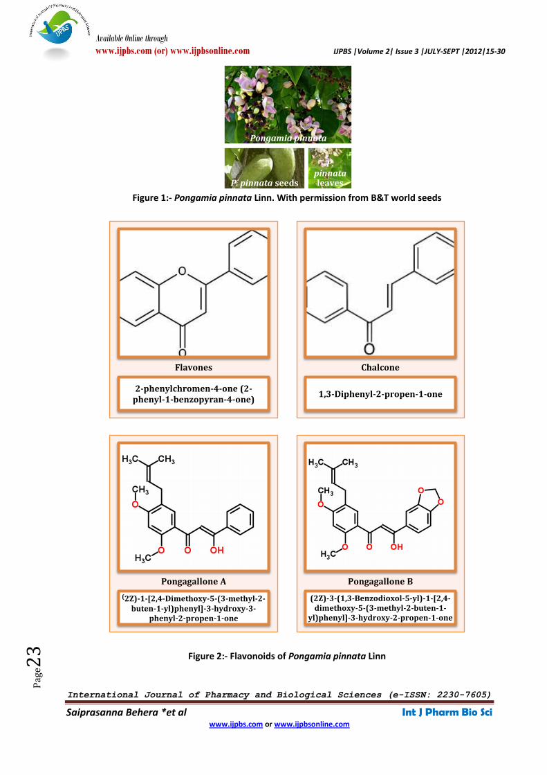
Figure 1:- Pongamia pinnata Linn. With permission from B&T world seeds
Figure 2:- Flavonoids of Pongamia pinnata Linn
Pongamia pinnata
P. pinnata seeds
P. pinnata leaves
2-phenylchromen-4-one (2-phenyl-1-benzopyran-4-one)
Flavones
1,3-Diphenyl-2-propen-1-one
Chalcone
(2Z)-1-[2,4-Dimethoxy-5-(3-methyl-2-buten-1-yl)phenyl]-3-hydroxy-3-
phenyl-2-propen-1-one
Pongagallone A
(2Z)-3-(1,3-Benzodioxol-5-yl)-1-[2,4-dimethoxy-5-(3-methyl-2-buten-1-
yl)phenyl]-3-hydroxy-2-propen-1-one
Pongagallone B
Available Online through
www.ijpbs.com (or) www.ijpbsonline.com IJPBS |Volume 2| Issue 3 |JULY-SEPT |2012|15-30
International Journal of Pharmacy and Biological Sciences (e-ISSN: 2230-7605)
Saiprasanna Behera *et al Int J Pharm Bio Sci www.ijpbs.com or www.ijpbsonline.com
Pag
e24
Serum ALT
Naive
Sham I/R
PP Contr
ol
PP 100
PP 200
PP 400
0
50
100
150
200
250
Treatment groups
IU/
L
Serum AST
Naive
Sham I/R
PP Contr
ol
PP 100
PP 200
PP 400
0
100
200
300
Treatment groups
IU/
L
3(A) 3 (B)
Serum ALP
Naive
Sham I/R
PP Contr
ol
PP 100
PP 200
PP 400
0
100
200
300
Treatment groups
IU/
L
3(C)
Figure 3:- Level of alanine aminotransferase (ALT), aspartate aminotransferase (AST) and alkaline
phosphatase (ALP) in Serum
Total Bilirubin
Naive
Sham I/R
PP Contr
ol
PP 100
PP 200
PP 400
0.0
0.5
1.0
1.5
2.0
Treatment groups
mg
/ d
l
Serum LDH
Naive
Sham I/R
PP Contr
ol
PP 100
PP 200
PP 400
0
500
1000
1500
2000
Treatment groups
U/
L
4 (A) 4(B)
Figure 4:-Level of bilirubin and lactate dehydrogenase (LDH) in Serum
Available Online through
www.ijpbs.com (or) www.ijpbsonline.com IJPBS |Volume 2| Issue 3 |JULY-SEPT |2012|15-30
International Journal of Pharmacy and Biological Sciences (e-ISSN: 2230-7605)
Saiprasanna Behera *et al Int J Pharm Bio Sci www.ijpbs.com or www.ijpbsonline.com
Pag
e25
Hepatic tissue MDA
Naive
Sham I/R
PP Contr
ol
PP 100
PP 200
PP 400
0
10
20
30
Treatment groups
nm
ol/
g p
rote
in
Hepatic tissue SOD
Naive
Sham I/R
PP Contr
ol
PP 100
PP 200
PP 400
0
10
20
30
40
Treatment groups
U/
g p
rote
in
5 (A) 5(B)
Hepatic tissue CATALASE
Naive
Sham I/R
PP Contr
ol
PP 100
PP 200
PP 400
0
10
20
Treatment groups
k/
mg
pro
tein
Hepatic tissue GSH
Naive
Sham I/R
PP Contr
ol
PP 100
PP 200
PP 400
0.0
0.1
0.2
0.3
0.4
Treatment groups
U/g
pro
tein
5 (C) 5(D)
Figure 5:- The renal tissue oxidant and antioxidant enzyme levels of the groups
Available Online through
www.ijpbs.com (or) www.ijpbsonline.com IJPBS |Volume 2| Issue 3 |JULY-SEPT |2012|15-30
International Journal of Pharmacy and Biological Sciences (e-ISSN: 2230-7605)
Saiprasanna Behera *et al Int J Pharm Bio Sci www.ijpbs.com or www.ijpbsonline.com
Pag
e26
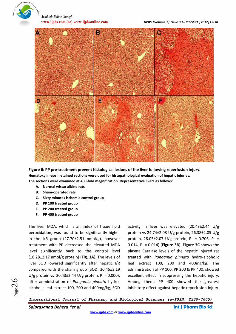
Figure 6: PP pre-treatment prevent histological lesions of the liver following reperfusion injury.
Hematoxylin-eosin-stained sections were used for histopathological evaluation of hepatic injuries.
The sections were examined at 400-fold magnification. Representative livers as follows:
A. Normal wistar albino rats
B. Sham-operated rats
C. Sixty minutes ischemia control group
D. PP 100 treated group
E. PP 200 treated group
F. PP 400 treated group
The liver MDA, which is an index of tissue lipid
peroxidation, was found to be significantly higher
in the I/R group (27.70±2.51 nmol/g), however
treatment with PP decreased the elevated MDA
level significantly back to the control level
(18.28±2.17 nmol/g protein) (Fig. 3A). The levels of
liver SOD lowered significantly after hepatic I/R
compared with the sham group (SOD: 30.45±3.19
U/g protein vs 20.43±2.44 U/g protein, P = 0.000),
after administration of Pongamia pinnata hydro-
alcoholic leaf extract 100, 200 and 400mg/kg, SOD
activity in liver was elevated (20.43±2.44 U/g
protein vs 24.74±2.08 U/g protein, 26.38±2.05 U/g
protein, 28.05±2.07 U/g protein, P = 0.706, P =
0.014, P = 0.014) (Figure 3B). Figure 3C shows the
plasma Catalase levels of the hepatic injured rat
treated with Pongamia pinnata hydro-alcoholic
leaf extract 100, 200 and 400mg/kg. The
administration of PP 100, PP 200 & PP 400, showed
excellent effect in suppressing the hepatic injury.
Among them, PP 400 showed the greatest
inhibitory effect against hepatic reperfusion injury.
Available Online through
www.ijpbs.com (or) www.ijpbsonline.com IJPBS |Volume 2| Issue 3 |JULY-SEPT |2012|15-30
International Journal of Pharmacy and Biological Sciences (e-ISSN: 2230-7605)
Saiprasanna Behera *et al Int J Pharm Bio Sci www.ijpbs.com or www.ijpbsonline.com
Pag
e27
The endogenous antioxidant, GSH, level in the
hepatic tissue was decreased significantly after I/R
(0.18±0.02 U/g protein). On the other hand PP
treatment significantly reversed this I/R-induced
GSH reduction (0.33±0.02 U/g protein) (Figure 3D).
Severe ischemia of organs has metabolic,
functional, and structural consequences and if it
persists too long, it leads to death of the involved
cells. Liver section of hydro-alcoholic leaf extract of
Pongamia pinnata treated animal group clearly
showed normal hepatic cells and central veins
thereby confirming hepatoprotective activity.
Flavonoids bind to subunit of DNA dependent RNA
polymerase I, thus activating the enzyme. As a
result, protein synthesis gets increased leading to
regeneration and production of hepatocytes 50.
After 60 minutes of continuous ischemia, large
confluent areas of tissue lysis with blood
congestion in the sinusoids and leukocyte
infiltrates were observed (Figure 4C). In the liver
treated with PP 100, limited and focal areas of
hepatocyte necrosis were also observed (Figure
4D), whereas the parenchyma was almost normal
after PP 200 and PP 400 treatment respectively
(Figures 4E and 4F).
Light microscopic investigation of the control group
(either given saline or PP) revealed a regular
morphology of liver parenchyma with intact
hepatocytes and sinusoids (Figure 4A). In the I/R
group, severe sinusoidal congestion and
hemorrhage, dilation of central vein,
subendothelial edema and degenerated
hepatocytes with perinuclear vacuolization were
observed (Figure 4C). In the PP treated I/R groups,
histological analysis demonstrated a well-
preserved liver parenchyma. Despite the mild
sinusoidal dilatation and hemorrhage, which were
in localized areas, the usual appearance of the
central vein and hepatocytes was observed in most
areas (Figures 4D, 4E and 4F).
This study had some limitations. Because of the
study protocol, hepatic IR-injured rats were
sacrificed just after reperfusion in order to observe
the effects of PP. If we could have kept the rats
alive, we could also have observed the long-term
effects of PP in hepatic IR injury. We expect that
further studies on the long-term effects of PP will
increase the value of our positive findings. In
addition, it is important to examine the possible
ability of PP to reverse IR-induced damage in liver
tissue and its effects on levels of antioxidant
enzymes
CONCLUSION
Reactive oxygen metabolites are probably
responsible for the tissue injury observed in
hepatic tissues after ischaemia reperfusion. In
conclusion, it was found that the treatment with
hydro-alcoholic leaf extract of Pongamia pinnata
(PP) increased the antioxidant ability and
decreased oxygen free radicals in hepatic IR injury
in rats. Also, evaluation of liver enzymes and
histopathological findings of liver tissue indicated
that PP had beneficial effects on the liver, thus it
can be considered as a preventive treatment agent
in hepatic IR injury. As this study does not contain
information about the long-term results of PP
treatment of hepatic IR injury, further
experimental and clinical studies are needed so as
to re-examine these views.
ACKNOWLEDGEMENTS
Authors acknowledge the immense help received
from the scholars whose articles are cited and
included in references of this manuscript. The
authors are also grateful to authors / editors /
publishers of all those articles, journals and books
from where the literature for this article has been
reviewed and discussed.
REFERENCES 1. De la Monte SM, Arcidi JM, Moore GW, Hutchins GM.
Midzonal necrosis as a pattern of hepatocellular injury
after shock. Gastroenterology, 86:627–31 (1984)
2. Arthur MJP. Reactive oxygen intermediates and liver
injury. Journal of Hepatology, 6:125–31 (1988)
Available Online through
www.ijpbs.com (or) www.ijpbsonline.com IJPBS |Volume 2| Issue 3 |JULY-SEPT |2012|15-30
International Journal of Pharmacy and Biological Sciences (e-ISSN: 2230-7605)
Saiprasanna Behera *et al Int J Pharm Bio Sci www.ijpbs.com or www.ijpbsonline.com
Pag
e28
3. Agnes S, Avolio AW, Magalini SC, Foco M, Castagneto M.
Should retransplantation still be considered for primary
non function after liver transplantation? Transpl. Int,
5(1):170, (1992)
4. Kimura F, Miyazaki M, Suwa T, Sugiura T, Shinoda T, Itoh
H, Nakagawa K, Ambiru S, Shimizu H, Yoshitome H.
Evaluation of total hepatic vascular exclusion and pringle
maneuver in liver resection. Hepatogastroenterology, 49
(43): 225, (2002)
5. Henrion J, Schapira M, Luwaert R, Colin L, Delannoy A,
Heller FR. Hypoxic hepatitis: clinical and hemodynamic
study in 142 consecutive cases. Medicine (Baltimore), 82
(6):392, (2003)
6. Salvatore Cuzzocrea, Dennis P. Riley, Achille P. Caputi,
and Daniela Salvemini. Antioxidant Therapy: A New
Pharmacological Approach in Shock, Inflammation, and
Ischemia/Reperfusion Injury. Pharmacol Rev, 53:135-159,
(2001)
7. Zweier JL, Flaherty JT, Weisfeldt ML. Direct measurement
of free radical generation following reperfusion of
ischemic myocardium. Proc Natl Acad Sci USA, 84: 1404–
07, (1987)
8. Dhiman RK, Chawla YK. Herbal medicines for liver
diseases. Digestive Diseases and Sciences, 50 (10):1807–
1812, (2005)
9. Calixto JB. Efficacy, safety, quality control, marketing and
regulatory guidelines for herbal medicines
(phytotherapeutic agents). Braz J Med Biol Res 2000;
33(2): 179-189.
10. Kamboj VP: Herbal medicine. Current Science, 78(1):35-
39, (2000)
11. De Smet, P.A.G.M., The role of plant-derived drugs and
herbal medicines in healthcare. Drugs, 54: 801-840,
(1997)
12. T.C. Dinis, C.L. Santosa and L.M. Alemida. The apoprotein
is the preferential target for deoxynitirte induced LDL
damage protection by phenolic acids. Free Radical Res, 36
(2): 531-43, (2002)
13. T. Ogiwara, K. Satoh, T. Negoro and S. Fujisawa. Inhibition
of NO production by activated macrophages by phenol
carboxylic acid monomers, polymers, and radical
scavenging activity. Anticancer res, 23 (2B): 1317 (2003)
14. L. Znag, G. Cosma, H. Gardner and V. Vallyathan. Effect of
Chlorogenic acid on hydroxyl radical. Mol. Cell Biochem,
247 (12): 205-10 (2003)
15. C.C. Lin, Y.F. Hsu and T.C Lin. Antioxidant and free radical
scavenging effects of tannins of Terminalia catappa Linn.
Anticancer Res, 21 (2A): 237-43 (2001)
16. T. Okuda. Systematics and health effects of chemically
distinct tannins in medicinal plants. Phytochemistry, 66:
2012-31 (2005)
17. M. Faure, E. Lissi, R. Torres and L.A. Videla. Antioxidant
activities of Lignans and Flavonoids. Phytochemistry, 29
(12): 3773-75 (1990)
18. B. Halliwell. Free radicals, antioxidants and human
disease; curiosity, cause or consequence? Lancet, 344
(8924): 721-24 (1994)
19. P.I. Hyun, H.S. Kun, W.C. Heyun and S.K. Sam. Anti-
inflammatory plants flavonoids and cellular action
mechanism. J. Pharmacol. Sci, 96: 229-45 (2004)
20. L.A. Mitsel, H. Teliekpalli, E. Mcgheee and D.M. Shankel.
Natural antimutagenic agents. Mutation Research, 350
(1): 14463 (1996)
21. R.W. Owen, A. Giacosa, W.E. Hull, R. Haubner and H.
Bartsch. The antioxidant/anticancer potential of phenolic
compounds isolated from olive oil. European Journal of
Cancer, 36 (10): 1235-47 (2000)
22. Chopra RM, Nayer SL, Indian Medicinal Plants (a
compendium of 500 species), 1st edition, Madras, Orient
Longman Ltd; 339-342. (1995)
23. Nadkarni K.M, Indian Materia Medica, 1st edition
Mumbai, Popular Prakashan; 1001-1004. (2000)
24. Rangari VD, Phytochemistry and Pharmacognosy, 1st
edition, part 2nd, Nashik; Career Publication; 259-61.
(2002)
25. Tanaka T, Linuma M, Fujii Y, Yuki K, Mizuno M. Flavanoids
in root, bark of Pongamia pinnnata. Phytochemistry, 31:
993-98, (1992)
26. Yin H, Si Zhang, Wu J, Nan H, Lijuan L, Yang J, et al.
Pongaflavanol: a Prenylated Flavonoid from Pongamia
pinnata with a Modified Ring A. Molecules 2006; 11: 786-
91.
27. Shameel S., Usmanghani K. and Ali M.S., Chemical
constituents from seeds of Pongamia pinnata (L.) Pierre.
Pak J Pharm Sci, 9, 1120, (1996)
28. Punitha R. and Manohar S., Antihyperglycemic and anti-
lipid-peroxidative effects of Pongamia pinnata (Linn.)
Pierre flowers in alloxan induced diabetic rats. J
Ethnopharmacol, 105, 39–46, (2006)
29. Essa M.M., Subramanian P., Suthakar G., Manivasagam T.
and Dakshayani K.B., Protective influence of Pongamia
pinnata (Karanja) on blood ammonia and urea levels in
ammonium chloride induced hyperammonemia. J Appl
Biomed, 3, 133-8, (2005)
30. Chopde VV, Tankar AN, Pande VV, Tekade AR, Gowekar
NM, Bhandari SR,and Khandake, International Journal of
Green Pharmacy, 72-75, (2008)
31. Reitman S, Frankel S. A colorimetric method for
determine ng serum glutamic oxaloacetic and glutamic
pyruvic transminases. Am J Clin Pathol, 28:56–63, (1957)
32. Ohkawa H, Ohishi N, Yagi K. Assay for lipid peroxides in
animal tissues by thiobarbituric acid reaction. Anal
Biochem, 95: 351–358, (1979)
Available Online through
www.ijpbs.com (or) www.ijpbsonline.com IJPBS |Volume 2| Issue 3 |JULY-SEPT |2012|15-30
International Journal of Pharmacy and Biological Sciences (e-ISSN: 2230-7605)
Saiprasanna Behera *et al Int J Pharm Bio Sci www.ijpbs.com or www.ijpbsonline.com
Pag
e29
33. Sun Y, Oberley LW, Li Y. A simple method for clinical assay
of superoxide dismutase. Clin Chem, 34:497-500, (1988)
34. Lowry OH, Rosebrough NJ, Farr AL, Randall RJ. Protein
measurement with the Folin phenol reagent. J Biol Chem,
193:265–275, (1951)
35. Aebi H. Catalase in vitro. Methods Enzymol, 105: 121-126,
(1984)
36. Tietze F. Enzymic method for quantitative determination
of nanogram amounts of total and oxidized glutathione:
applications to mammalian blood and other tissues. Anal
Biochem, 27: 502-522, (1969)
37. Anderson ME. Determination of glutathione and
glutathione disulfide in biological samples. Meth Enzymol,
113: 548-555, (1985)
38. Bancroft JD, Gamble M. Theory and practice of
histological techniques. 6th ed. China: Churchill
Livingstone; 2008
39. Harvey J, Paige SM. The Instat Guide to choosing and
interpreting statistical tests: A manual for Graph pad
Instat, Version 3. San Diego, CA USA. 1998
40. Sener G, Tosun O, Sehirli A O, Kaçmaz A, Arbak S, Ersoy Y,
Ayanoglu Dulger G. Melatonin and N-acetylcysteine has
beneficial effects during hepatic ischemia and
reperfusion. Life Sci, 72 (24): 2707–2718, (2003)
41. Sener G, Sehirli O, Ipçi Y, Ercan F, Sirvanci S, Gedik N,
Yeğen BC. Aqueous garlic extract alleviates ischemia-
reperfusion-induced oxidative hepatic injury in rats. J
Pharm Pharmacol, 57: 145–150, (2005)
42. Ofluoglu E, Kerem M, Pasaoglu H, Turkozkan N, Seven I,
Bedirli A, Utku Yilmaz T. Delayed energy protection of
ischemic pre-conditioning on hepatic ischemia/
reperfusion injury in rats. Eur Surg Res, 38: 114–121,
(2006)
43. Zhu WH, Leng XS, Zhu JY. Effect of Shenfu injection on
ischemia-reperfusion injury of rat liver graft.
Hepatobiliary Pancreat Dis Int, 5: 205–209. (2006)
44. Yildiz F, Coban S, Terzi A, Ates M, Aksoy N, Cakir H, et al.
Nigella sativa relieves the deleterious effects of ischemia
reperfusion injury on liver. World J Gastroenterol,
14:5204-9, (2008)
45. Chouker A, Martignoni A, Schauer RJ, Dugas M,
Schachtner T, Kaufmann I, et al. Alpha-gluthathione S-
transferase as an early marker of hepatic
ischemia/reperfusion injury after liver resection. World J
Surg, 29:528-34, (2005)
46. Altan N, Dincel AS, Koca C. Diabetes mellitus and
oxidative stress. Turk J Biochem, 3:51-6, (2006)
47. Arab K, Steghens JP. Plasma lipid hydroperoxides
measurement by an automated xylenol orange method.
Anal Biochem, 325:158-3, (2004)
48. Marubayashi S. Dohi K, Yamada K, Kawasaki T. Changes in
the levels of endogenous coenzyme Q homologs, alpha-
tocopherol, and glutathione in rat liver after hepatic
ischemia and reperfusion, and the effect of pretreatment
with coenzyme Q10. Biochem Biophys Acta, 797:1-9,
(1984)
49. Siems W, Mielke B, Muller M, Heumann C, Rader L,
Gerber G. Status of glutathione in the rat liver. Enhanced
formation of oxygen radicals at low oxygen tension.
Biomed Biochim Acta, 42:1079-89. (1983)
50. Murray, MT. Quercetin: Nature’s antihistamine. Better
Nutrition (1998)
Available Online through
www.ijpbs.com (or) www.ijpbsonline.com IJPBS |Volume 2| Issue 3 |JULY-SEPT |2012|15-30
International Journal of Pharmacy and Biological Sciences (e-ISSN: 2230-7605)
Saiprasanna Behera *et al Int J Pharm Bio Sci www.ijpbs.com or www.ijpbsonline.com
Pag
e30
*Corresponding Author: Mrs. Saiprasanna Behera Lecturer in Pharmacy Department of Pharmacology Royal College of Pharmacy and Health Sciences Berhampur. Odisha. INDIA-760002