Embed Size (px)
Citation preview
STUDIES ON IMMUNIZATION AGAINST PLAGUE
VI. GROWTH OF PASTEURELLA PESTIS AND THE PRODUCTION OF THE ENVELOPE ANDOTER SOLUBLE ANTIGENS IN A CASEIN HYDROLYZATE MINERAL GLUCOSE MEDIUM1
ELLIS ENGLESBERG AND JUDITH BENTOFF LEVY
George Williams Hooper Foundation, University of California, San Francisco, California
Received for publication October 5, 1953
The immunizing capacity of various plagueantigenic preparations for mice, monkeys, andman depends to a large extent on their Fraction Icontent (Baker et al., 1952; Chen et al., 1952).Fraction I is the designation given to that water-soluble antigenic portion of the plague bacillusthat is soluble at 25 per cent saturated ammoniumsulfate and precipitated at 33 per cent saturation.It appears to consist of two immunologicallyidentical, but chemically different, fractions, IAand IB. Fraction IA is a protein-carbohydratecomplex; Fraction IB is entirely protein (Bakeret al., 1952).
Since the present preparations for immunizingman against plague were found to be low inFraction I and since present methods of preparingFraction I are complicated and costly, involvinggrowth of a virulent strain of Pasteurella pesti8on a complex protein agar medium and subse-quent extraction and fractionation (Baker et al.,1947, 1952), simplified methods of growing P.pesti8 and of producing a preparation of highFraction I content were sought.Evidence also to be presented indicates that
Fraction I is the envelope substance of P. pestiadescribed by Rowland (1914) and SchQtze(1932a). Since this is in disagreement with thefindings of Amies (1951) to the effect that theone important antigen of Pasteurella pestis is acapsular substance which requires vigorousphysical or chemical procedures to remove itfrom the cells, several pertinent experiments ofhis were repeated.
MATERIALS AND METHODS
Organism. PasteureUa pestis, strain A1122(avirulent), originally isolated by Jawetz and
I Sponsored by the Commission on Immuniza-tion of the Armed Forces Epidemiological Boardand supported (in part) by the Office of theSurgeon General, Department of the Army, andthe University of California.
Meyer (1943), was employed for the most partin this study. This organism was selected becauseof its high soluble antigen2 content when grownon a complex protein medium and its avirulenceof long standing. An avirulent strain was chosenmainly on the basis of data that suggested thatantigenically the major difference betweenvirulent and avirulent strains of P. pestis is thequantity of Fraction I produced, not a so-called"virulence antigen" (Englesberg and Levy,1953). Another consideration, of course, was thecomparative siplicity of working with anavirulent strain rather than a virulent one andthe possibility it presented for large-scale produc-tion.
Stock cultures of this strain were maintainedinitially on blood agar slants and then on caseinhydrolyzate mineral glucose slants.Medium. Since 37 C is the approximate opti-
mum temperature for producing Fraction I(Chen et al., 1952), a medium was sought thatwould support growth of P. pestis at this tempera-ture. For growth in a mineral glucose medium at30 C, P. pestis, strain A1122, requires phenyl-alanine, valine, isoleucine, methionine, and thio-sulfate (Englesberg, 1952). Such a medium failedto support growth of this organism at 37 C. Ac-cording to HilLs and Spurr (1952), P. pestis ismuch more demanding in its nutritional require-ments when grown at 37 C than at 30 C, anddifferent strains can be grown at 37 C in mixturesof from 8 to 15 different amino acids in some caseswith (and in others without) the addition of biotinand pantothenate. They found hemin to be re-quired for growth from small inocula at both 30and 37 C, but there are now indications that this
2 Soluble antigen refers to the antigen materialof the plague bacillus extracted by saline at pH 7or in the casein hydrolyzate mineral glucosemedium during growth and forming a precipitatewith an antiplague serum (Lederle serum).
438
on May 30, 2018 by guest
http://jb.asm.org/
Dow
nloaded from
STUDIES ON IMMUNIZATION AGAINST PLAGUE
requirement is an artifact of medium production(Englesberg, 1954).
Since there was the possibility that the entireculture could be used as the antigen complex oras a source of the antigen, it was desirable toemploy a medium free of protein or of highmolecular weight polypeptides. This wouldinsure against the presence of any foreign sensi-tizing or toxic substances and would simplify anyisolation procedure that might eventually beemployed. On the basis of these considerations,attempts were made to grow P. pestis in a caseinhydrolyzate mineral glucose medium (CHMG).The composition of the initial or starting
medium was casamino acid (Difco), 1 per cent(casein hydrolyzate); glucose, 0.2 per cent;NH4C1, 0.1 per cent; CaCl2, 103 per cent; FeCl,(6H20), 2.5 X 10⁴ per cent; MgSO4(7H,O),0.05 per cent; and Na2S203, 0.01 per cent.KH2PO4 and Na2HPO4, pH 7, were added toyield a m/20 solution.The casein hydrolyzate was autoclaved with
distilled water, and the other components wereadded as sterile stock solutions. Sodium hy-droxide was added subsequent to autoclavingto neutralize the casein hydrolyzate in all buta few preliminary experiments. Glucose wassterilized by filtering through sintered glass orautoclaving at 15 lb for 15 min as a 20 per centsolution. A concentrated solution of sodium thio-sulfate was sterilized by filtration, and theremaining inorganic compounds were autoclavedin concentrated solutions at 15 lb for 20 minutes.Three different lots of casino acids were
used. With the lot used in the early experiments,obtained during World War II, a 3 per centsolution yielded optimum results. With morerecently obtained lots of this material, use of a4 per cent solution resulted in the production of alarger quan1ity of antigen than was obtainablewith the older lot. Because of this difference,several experiments perforned with the old lotin which the casein hydrolyzate content wascrucial were repeated with the new lot. It wouldbe repetitious to give the results obtained withboth preparations, so in the case of these dupli-cate experiments only those results obtainedwith the presently obtainable casamino acidwill be reported.
In the development of the optimum culturalconditions for growth and antigen production,the scale of the experimentation was important.
Since the purpose was to provide sufficientmaterial for extensive animal and immuno-chemical investigation and at the same time topave the way for large-scale pilot operation, itwas decided to employ a method for exploringthe optimum conditions for antigen productionthat could be used directly without further modi-fication to provide large amounts of plague cul-ture. This was provided for by employing avariable speed and eccentric rotory type shakingmachine that would hold, for example, eighty 1liter, wide mouth Erlenmeyer flasks. The speedand amplitude of rotation were varied and areindicated in each experiment. Duplicate 1 literwide mouth Erlenmeyer flasks, each containing250 ml of medium, were used, and results recordedare based on an average of determinations fromboth flasks.pH. The pH of the cultures was determined
at intervals by means of pH indicators.Inoculum. The growth of an 18 to 24 hr casein
hydrolyzate mineral glucose (3 per cent caseinhydrolyzate) slant was washed off with 2.5 mlof sterile distilled water and subsequently dilutedto 7.5 ml. Hormone agar slants were used inearly experiments. One ml of this diluted sus-pension was used to inoculate each 250 ml ofliquid medium. This provided an inoculum ofabout 5 X 10⁶ viable cells per ml of medium.
Determination of the amount of growth. Totalcell yields were measured at 24 hr intervals bythe per cent light transmittance determined witha Coleman spectrophotometer at 650 m,u. Theblank employed consisted of the uninoculatedliquid medium or a particular dilution thereof.A calibration curve for this instrument wasestablished with a 3 day old slant culture ofP. pestis suspended in saline. The total count ofthis suspension was determined with a Petroff-Hauser chamber. Microscopic observation ofgram stained preparations was employed todetect contaminants.
Determination of the amount of soluble antigenproduced. A modified semiquantitative turbidi-metric precipitin test (Baker et al., 1952) wasperformed on cell-free supernatants and in somecases on the saline washings of cells and salineextraots from acetone dried cells. Rabbit anti-Fraction I serum (Baker et al., 1952) or LederleAntiplague Immune Serum Globulin (Rabbit)prepared with living P. pestis, strain A1122,
4391954]
on May 30, 2018 by guest
http://jb.asm.org/
Dow
nloaded from
ELLIS ENGLESBERG AND JUDITH B. LEVY
was employed in this test to measure the solubleantigens.
In determining the quantity of antigen inthe supernatants, 24 hr samples of the culturewere centrifuged at 20,000 rpm for 20 min in aSpinco high speed centrifuge. The supernatantwas collected and diluted serially. To one ml oftwo or more dilutions of the supernatant, one mlof a previously standardized serum was added.After the tubes had stood at room temperaturefor two hr or in the refrigerator overnight, oneml of saline was added, and the turbidity of thesolution was read in a Klett-Summerson colorim-eter using a blue filter. The amount of antigenper ml was calculated by means of a standardcurve for the particular serum, which relatesturbidity to quantity of antigen.The one lot of antiplague serum (Lederle)
employed was standardized arbitrarily withFraction IA. One unit of soluble antigen is de-fined as the amount yielding the same turbiditywith antiplague serum as one mg of Fraction IA.Anti-Faction I serum was standardized with
Fraction IA prepared according to the methodof Baker and his associates (1952).
In early experiments the procedure was toneutralize the supernatants prior to dilution andaddition of the serum. This procedure was foundto be unnecessary since turbidity was not af-fected by the pH of the medium.
Trichloracetic acid (TCA) precipitable protein.To determine whether there was any correlationbetween total protein and soluble antigen in thesupernatant (the latter determined with Lederleserum), to 5 or 10 ml of cell-free supernatantplaced in a small centrifuge cup was added 1 or2 ml of 60 per cent trichloracetic acid, respec-tively. The spension was stored overnight inthe refrigerator, and the precipitate was spundown at 20,000 rpm for 20 minutes. The super-natant liquid was poured off, and the tube wasdrained dry. The precipitate was washed into atared watch glas and dried to constant weight.Each determination was performed in duplicate.Blanks were prepared from casein hydrolyzatemedium by the addition of trichloracetic acid,following the procedure described above.
Toxicity. The varying of cultural conditionsfor optimum growth and antigen productionafforded an excellent opportunity for studyingthe factors related to toxin production in P.pest". Portions of the cell-free supernatant
medium, recovered at various intervals duringthe growth of P. pestis for the precipitin test,were used also to test for the presence of toxin.Five-tenths of a ml of various dilutions of thesupernatant was inoculated intravenously intoeach of a group of Swiss mice. LDw was deter-mined by the procedure of Reed and Muench(1938).The "capsular" antigen of Amies. Amies'
(1951) procedure for demonstrating the capsuleof P. pestis, the dissolution of the capsule in thepresence of potassium thiocyanate under con-trolled conditions, and the absence of the en-velope in agar grown cells was duplicated. P.pestis, strain Tjiwidej, 1948, a strain similarto that employed by Amies, was used. The cellswere scraped off the surface of agar plates andsuspended in distilled water. The suspensionwas divided into two equal portions. To one wereadded potaium thiocyanate and potassiumhydroxide to maintain a pH of 7.8, and to theother, only potassium hydroxide. Suspensionswere incubated at 37 C under constant agitation.Precipitin tests described using Lederle anti-plague serum and anti-Fraction I serum were per-formed immediately before the addition of po-tassium thiocyanate or potassium hydroxide andduring the extraction procedure on cell-freesupernatants prepared from samples of the cellsuspension by centrifugation.
RESULTS
Growth and antigen distribution in S per centcasein hydrolyzate mineral glucose medium. Thegrowth and amount of antigen soluble in themedium and remaining attached to the cellsduring 8 days of cultivation in a 3 per cent caseinhydrolyzate (old lot) mineral glucose mediumare shown in figure 1. The cultures were sampledat 24 hr intervals during incubation, and cellcounts were determined directly on these samplesor on 1:5 or 1 :10 dilutions thereof, as was foundnecessary. Smears were made for microscopicexamination, and pH of the cultures was deter-mined. Ten ml of the samples were pipetted theninto centrifuge cups and spun at 20,000 rpm for20 minutes, and the supernatant fluids wereasyed for antigen content. (The above pro-cedure was followed routinely in most of theexperiments to be described.) The recovered cellswere resuspended in 5 ml of saline, placed in therefrigerator overnight, centrifuged, and the
AAn [voL. 67
on May 30, 2018 by guest
http://jb.asm.org/
Dow
nloaded from
STUDIES ON IMMUNIZATION AGAINST PLAGUE
DAYS INCUBATION
Figure 1. Growth of Pasteurella pestis, strainA1122, and distribution of soluble antigens in a 3per cent casein hydrolyzate mineral glucosemedium. Aeration: 95 rpm, 1 cm radius eccentric.
saline extract was collected and assayed for anti-gen content. The washed cells were rewashed thenwith small amounts of distilled water into glasscentrifuge cups, dried by two successive washingswith acetone at -60 C, and placed in a vacuum
desiccator over sulfuric acid. The dried powderwas suspended then with 5 ml of saline and theinsoluble material spun down. The supernatantwas collected and assayed for antigen content.Maximum growth occurred on the third day
of incubation and was followed by slow and thenby rapid lysis. Antigen was released into themedium during this time, reaching a maximum
on the sxth day of incubation. About 82 percent of the antigen that could be removed fromthe cells was released into the supernatant bythe sixth day; only an additional 4 per cent was
removed by saline washings of the cells, and 14per cent by a further acetone drying and salineextraction. A fairly constant amount of solubleantigen remained attached to the cells duringthe 8 day period, while the amount of antigenin the supernatant was large and increasedprogressively. Soluble antigen was produced intwo distinct stages: the initial stage between 0
and 3 days of incubation, prior to cell lysis, andthe secondary stage, which appears to dependon the lytic process. In experiments in whichboth anti-Fraction I serum and Lederle anti-plague serum were used (figure 7) in assaying theantigen content of the supernatant fluid, it wasdemonstrated that these two stages of antigenproduction actuaUly are related to the productionof at least two different antigens. The first stage
Figure B. Growth of Pasteurella pestis, strainA1122, and soluble antigen and toxin productionin a 3 per cent casein hydrolyzate mineral glucosemedium. Aeration: 175 rpm, 1.5 cm radius ec-centric.
is characterized by the production of Fraction Iwhich reaches a peak at maximum growth andremains fairly constant during the cell lysis thatfollows. In the secondary or lytic stage, one ormore antigens other than Fraction I, probablysomatic in orign, are released.The fact that the release of Fraction I into the
medium is separated by a significant timeinterval from the release of other antigens in-dicated that it would be possible to determinethe optimum conditions for the production ofboth these antigenic components of P. pestissimultaneously.The ease with which plague antigens are re-
leased into the medium during the growth cycleindicated the plausibility of exploring theoptimum conditions for soluble antigen produc-tion through assays on the supernatant fluid withantiplague serum. Although at 3 days of incuba-tion (figure 1) only 50 per cent of the total solubleantigen (Fraction I) was found in the supernatant,while the remainder was attached to the cell,with the development of optimum culturalconditions the increased yield of Fraction I pro-duced was found solely in the supernatant fluid(see Discussion). Since Fraction I and little elsethat yields a precipitate with this type of serumare released before cell lysis, Lederle serum canbe employed at the point of maximum growthas a means of estimating Fraction I productionand subsequently as a measure of total somaticantigen production.
Aeration. Employing the same medium as inthe preceding experiment and increasing theaeration of the medium by raising the speed of
1954] 441
on May 30, 2018 by guest
http://jb.asm.org/
Dow
nloaded from
ELLIS ENGLESBERG AND JUDITH B. LEVY
4II)
3
aI.2-
1-
O TCA PROTEINA MAXIMUM CELLCOUNT
* CELL COUNT6th DAY
* 5OLUBLEANTIGEN
o FP.ACTION I
/A/ ,?
PERCENT CASEIN HYDROLYSATE
Figure S. The relationship between the concen-tration of casein hydrolyzate and growth ofPasteurella pe8tis, strain A1122, soluble antigenand protein production. Medium: casein hy-drolyzate concentration as indicated and theingredients of the starting medium. Aeration:175 rpm, 1.5 cm radius eccentric. Numbers oncurve indicate the day of incubation at whichmaximum growth was reached. Shaded area indi-cates the amount of cell lysis.
rotation to 175 rpm and the eccentric to 1.5 cmin radius increased both the rate of growth andthe antigen production as well as the total yieldsof antigen (figure 2). Cells lysed at the third day,and soluble antigen production reached a maxi-mum at the sixth day. About 0.4 mg of Fraction Iwas in solution before lysis set in. Further in-crease in speed of rotation or radius of the ec-centric or reducing volume of medium from 250to 100 ml did not further increase growth orantigen production.
Toxin production. The toxicity of the cell-freesupernatant medium was tested during the varia-tion of concentrations of casein hydrolyzate(old lot), glucose, phosphate buffer, pH, and iron.In all cases the same general pattem was ob-served. A typical example of the relation ofgrowth and toxin and antigen production isshown in figure 2. The toxicity of the supernatantmedium increased during the active growth, thestationary phase, and during cell lysis. Totaltoxin production was always directly correlatedwith the amount of cell growth. The supernatantis detoxified by adding 0.1 to 0.2 per cent forma-Ein. Further substantiation that the toxicity ofthe supernatant is not the result of simpleintermediary metabolic products of P. pestis wasshown by the toxicity of dialyzed supernatantand by the recovery of the toxin by (NH4)2S04
-2.22.2 t I4
tt § *~~~~~~~~~
CELL CUNT Gth DY
-1.4b1~~~~Iq~~ 0 SOLUBLE ANTIGEN
I ~~~~~~R
-0.G .a
0.2 M a 0.0125 0.025 0.05 0.1 Q;M PHOSPHATC BUPFER
~I 8
a0.&4
;t2.0 l
I
1.00
Figure 4. Relationship between concentrationof phosphate buffer and growth of Pasteurellapestis, strain A1122, and soluble antigen produc-tion. Medium: 4 per cent casein hydrolyzate andthe ingredients of the starting medium. Aeration:175 rpm, 1.5 cm radius eccentric. Numbers oncurve indicate the day of incubation at whichmaximum growth was reached. Shaded area in-dicates the amount of cell lysis.
precipitation and subsequent dialysis (Engles-berg and Levy, 1953).
Variation in the concentration of casein hy-drolyzate' (figure 8). Growth, soluble antigen, andFraction I production were maximal with 4 percent casein hydrolyzate. Maxmum growth (3.92X 10⁹ cells per ml) occurred on the fourth dayof incubation and was followed by lysis of ap-proximately 1.4 X 10⁹ cells per ml. The maximumamount of soluble antigen (2.1 units per ml ofsupernatant) was produced on the sixth day ofincubation, while the maximum amount of Frac-tion I (1.3 mg per ml) was found at 3 days' in-cubation and declined slightly thereafter. Totalplague protein in solution 's correlated directlywith the amount of total soluble antigen.
Variation in the concentration of phosphatebuffer (figure 4). Although changing the concen-tration of phosphate buffer from 0.0125 M to0.2 M did not significantly affect the total cellyield, growth lagged appreciably with concentra-tions over 0.05 M and below 0.025 M. At concen-trations below 0.025 m, acid production, indi-cated by the low pH, was probably the cause ofthe delay in growth. With the higher concentra-tions (0.1 and 0.2 M), cells did not lyse by thesixth day; lysis was massi-- in flasks containing0.05 M phosphate. The fact that pH varied
aThe casamino acid (Difco) (casein hy-drolyzate) used in this and in the followingexperiments is material currently available.
442 (VOL. 67
'a
on May 30, 2018 by guest
http://jb.asm.org/
Dow
nloaded from
STUDIES ON IMMUNIZATION AGAINST PLAGUE
Z[ 0.2
0 0.1 0.2 0.3 0.4 0.5PERCENT GLUCOSE
Figure 5. Relationship between concentrationof glucose and growth of Pasteurella pestis,strain A1122, and antigen production. Medium:4 per cent casein hydrolyzate and the ingredientsof the starting medium. Numbers on curve indi-cate the day of incubation at which maximum
growth was reached. Shaded area indicates theamount of cell lysis.
insignificantly during growth with these concen-
trations eliminates pH as the factor responsiblefor lysis. The optimum phosphate concentrationfor Fraction I and soluble antigen production is0.05 M (as in the starting medium).
Variation in the concentration of glucose (figure5). Growth and antigen production were maximal
when the 4 per cent casein hydrolyzate mineralglucose starting medium contained 0.2 per centglucose. Higher concentrations inhibited growthdue to excessive acid production. Althoughmedia without glucose showed little growth ofP. peetia during the first 24 hr (not shown on thegraph), in comparison to those containing from0.05 to 0.2 per cent glucose, the total cell cropwas higher in the medium containing no glucosethan in that containing 0.05 or 0.1 per cent. Thismay be the result of selection of a mutant capableof utilizing the casein hydrolyzate as primarycarbon source in the medium lacking glucoseand inhibition of such selection due to growth ofthe wild type in the presence of a low concentra-tion of glucose (R% and Schneider, 1948).
Variation in pH. Duplicate flasks of caseinhydrolyzate were autoclaved in distilled waterand subsequently brought up to pH 6.5, 6.8, 7.0,7.2, and 7.8 with sterile sodium hydroxide.
0 FPRACTION I
33.00
s.5 7.0 7.5 8.0pH
Figure 6. Relationship between pH and growthof Pasteurella pestis, strain A1122, and antigenproduction. Medium: 4 per cent casein hydrolyzateand the ingredients of the starting medium.Aeration: 175 rpm, 1.5 cm radius eccentric.Shaded area indicates the amount of cell lysis.
pH0.
/CELL COUNT *%
2O 52 a 4 S 6DAYS INCUBATION
Figure 7. Growth of Pasteurella pestis, strainA1122, and antigen production in 4 per centcasein hydrolyzate medium and the ingredientsof the starting medium. Initial pHs 6.8 and 7.0.Aeration: 175 rpm, 1.5 cm radius eccentric.
Phosphate buffer at these pHs and the other in-gredients of the starting medium were added tothe respective flasks. The yield of cells and oftotal soluble antigen was maximal at pH 7(figures 6 and 7). Although the amount ofFraction I was slightly larger when the initialpH of the medium was buffered at pH 6.8 thanat 7.0, the difference does not appear to besignificant (figure 7). This was verified further byseveral repetitions of this experiment with me-
1954] AA3
on May 30, 2018 by guest
http://jb.asm.org/
Dow
nloaded from
ELLIS ENGLESBERG AND JUDITH B. LEVY
dium buffered at these two levels. Although withan initial pH of 7.8 growth was only slightlyless than at pH 7 and the amount of lysis wasgreater, much less Fraction I and total solubleantigen were produced. Whether this is the re-sult of denaturation of the plague antigen dueto excessive alkalinity or to the failure of theorganism to produce these antigens under thesecircumstances has not been determined. There-fore it appears that besides cell yield and tem-perature of incubation (Chen et al., 1952), theinitial pH of the medium is an important factorgoverning antigen production in P. pestis.
Variation in minerals and simplification of themedium. So far it has been demonstrated that a4 per cent casein hydrolyzate, 0.2 per cent glucosemedium, buffered at pH 7.0 with M/20 phosphatebuffer yields maximal growth and soluble antigenproduction. The 4 per cent casamino acid solu-tion contains, per ml, approximately 0.053micromoles of magnesium, 0.025 micromoles ofcalcium, and 0.004 micromoles of iron. (This-information was obtained from Dr. H. W.Schoenlein of Difco Laboratories.) In preparingthe starting medium, the mineral content wasincreased by adding two micromoles of magne-sium, 0.092 micromoles of calcium, and 0.0092micromoles of iron per ml of medium. In addi-tion, 18.8 micromoles of NHit and 0.8 micro-moles of sulfur (as S20;) per ml were added, theformer to spare the casein hydrolyzate and thelatter as a source of cystine (Englesberg, 1952).To determine whether the addition of these min-
erals was essential, each was omitted in turn. Theresults indicated that the addition of mag-nesium is essential for growth of P. pesti and forantigen production. The omission of ammoniumchloride appeared to enhance growth but notbecause of its effect on the pH. The addition ofiron, calcium, or S2O; salts appeared to have littleeffect.
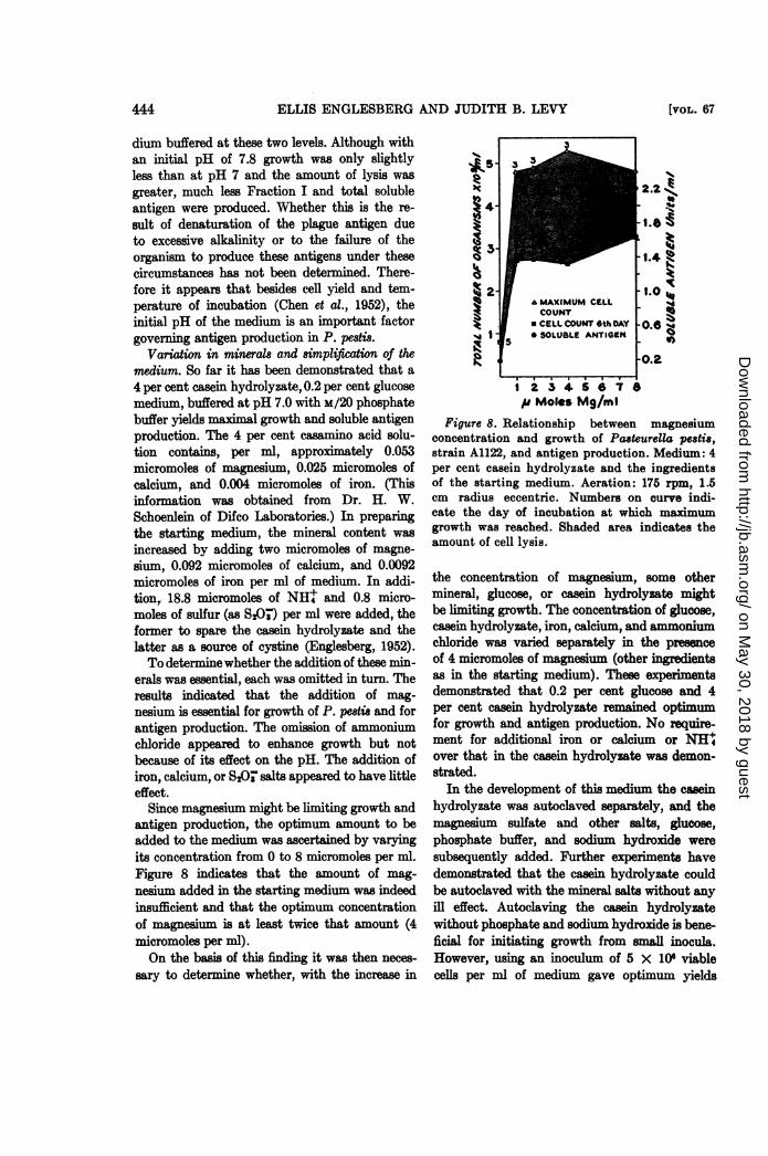
Since magnesium might be limiting growth andantigen production, the optimum amount to beadded to the medium was ascertained by varyingits concentration from 0 to 8 micromoles per ml.Figure 8 indicates that the amount of mag-nesium added in the starting medium was indeedisufficient and that the optimum concentrationof magnesium is at least twice that amount (4micromoles per ml).On the basis of this finding it was then neces-
sary to determine whether, with the increase in
1 2 3 4 5 6 7 ep Moles Mg/mi
Figure 8. Relationship between magnesiumconcentration and growth of Pasteurella pestis,strain A1122, and antigen production. Medium: 4per cent casein hydrolyzate and the ingredientsof the starting medium. Aeration: 175 rpm, 1.5cm radius eccentric. Numbers on curve indi-cate the day of incubation at which maximumgrowth was reached. Shaded area indicates theamount of cell lysis.
the concentration of magnesium, some othermineral, glucose, or casein hydrolyzate mightbe limiting growth. The concentration of glucose,casein hydrolyzate, iron, calcium, and aoniumchloride was varied separately in the presenceof 4 micromoles of magnesium (other ingredientsas in the starting medium). These experimentsdemonstrated that 0.2 per cent glucose and 4per cent casein hydrolyzate remained optimumfor growth and antigen production. No require-ment for additional iron or calcium or NH'over that in the casein hydrolyzate was demon-strated.
In the development of this medium the caseinhydrolyzate was autoclaved separately, and themagnesium sulfate and other salts, glucose,phosphate buffer, and sodium hydroxide weresubsequently added. Further experiments havedemonstrated that the casein hydrolyzate couldbe autoclaved with the mineral salts without anyill effect. Autoclaving the casein hydrolyzatewithout phosphate and sodium hydroxide is bene-ficial for initiating growth from small inocula.However, using an inoculum of 5 X 10⁶ viablecelLs per ml of medium gave optimum yields
AAA [voL. 67
on May 30, 2018 by guest
http://jb.asm.org/
Dow
nloaded from
STUDIES ON IMMUNIZATION AGAINST PLAGUE
TABLE 1Comparison of the Fraction I and total soluble
antigen content of plague vaccines
VACCDMS TOA SBOWBLZ IRACION I
nits/nmi mg/mCutter Laboratoryt 0.44 0.28Haffkine Institutet 0.57 0.68C0MG supernatant 1 0.52CHMG supernatant 2 1.08CHMG supernatant 3 1.14 1.36CEMG supernatant 4 2.50 1.24
* Cutter Laboratory Plague Vaccine is a for-malin killed suspension of virulent plague bacilligrown at 37 C on hormone agar.
Haffkine Institute Plague Vaccine is a formalinkilled casein hydrolyzate culture of virulentplague bacilli probably grown at 28 C.
Casein hydrolyzate mineral glucose (C01MG)supernatants 1 and 2 are from 6 day old CHMGcultures of Pasteurella pestis, strain A1122 (aviru-lent), grown under suboptimum conditions.
Casein hydrolyzate mineral glucose super-natant 3 is from a 3 day old C0EMG (optimum me-dium and conditions) culture of Pasteurella pestis,strain A1122 (avirulent).
Casein hydrolyzate mineral glucose super-natant 4 is a supernatant of a 6 day old culturefrom which supernatant 3 was obtained.
t Cells were spun down and precipitin testswere performed on the supernatant fluid.
when all ingredients (except glucose) were auto-claved together with the casemin hydrolyzate.
Comparison of the antigen coten of variousplague vaccines with that of the casein hydrolyzatemineral glucose supermatants. Four differentcasein hydrolyzate mineral glucose supernatantswere compared with the Cutter LaboratoriesPlague Vaccine and with the Hafiine InstitutePlague Vaccine (table 1). Supernatants 1 and 2were harvested from cultures of P. pestis, strainA1122, grown in suboptimum media. Super-natants 3 and 4 were taken from the sameculture grown under optimum conditions. Super-natant 3 was harvested after incubation for 72hr; supernatant 4 was harvested after 144 hr.Casein hydrolyzate mineral glucose supernatants3 and 4 contained about twice as much Fraction Ias the Haffkine vaccine and about 5 times asmuch as the Cutter vaccine. Supernatant 4 hadabout twice the amount of total soluble antigenthat supernatant 3 had. This represents an in-
crease in somatic antigen content as a result ofcell lysis.
DISCUSSION
The medium finally arrived at for optimumgrowth and soluble antigen production is simplerand showed more than a 10-fold increase inFraction I and somatic antigen production and a4- to 5-fold increase in total cell yield, as com-pared with the starting medium (table 2).Growth of P. pestis, strain A1122, reaches a
maximsum (4 to 5 X 10' organisms per ml), underthe optimum conditions developed, on the thirdday of incubation, and this is followed by massivecell lysis which ceases by the sixth day. At leastthree different components of the plague bacillusare released into the medium during this growthcycle: Fraction I, somatic antigen(s), and toxin.Fraction I is released continuously during thelogarithmic phase of growth, and a maxmumconcentration of about 1.0 to 1.4 mg per ml ofsupernatant fluid is produced coincident withthe mimum yield of celLs. Little, if any, addi-tional Fraction I is released during the lyticperiod. To produce Fraction I in solution rela-tively free of somatic antigens, a 3 day old cultureof P. pestis, grown under the conditions de-scribed, appears optimum. The somatic antigensare released during the cell lysis which occursfrom the fourth to the sixth day. Yields of somaticantigen can be determined by differences inamounts of total soluble antigen and Fraction Ias estimated by turbidimetric precipitin tests,using Lederle and anti-Fraction I serums, re-spectively, and are only approximations. Al-though Lederle serum may form a precipitatewith plague toxin, the amount of toxin-anti-toxin precipitate formed is insignificant (Engles-berg and Levy, 1953). Thus, the precipitin testdescribed measures antigenic material otherthan plague toxin in the supernatant. Smallamounts of toxin, determined by in vivo test,are released during both cell growth and lysis,indicating that the plague toxin cannot be classi-fied satisfactorily as an endotoxin in the oldsense.Growth and antigen production have varied
somewhat in experiments undertaken at differenttimes under presumably the same conditions.For this reason, in developing the optimtummedium, the best medium arrived at in one seriesof experiments was always employed during the
1954] 445
on May 30, 2018 by guest
http://jb.asm.org/
Dow
nloaded from
446 ELLIS ENGLESBERG AND JUDITH B. LEVY
TABLE 2
[VOL. 67
Compari8on of the starting and optimum casein hydrolyzate mineral glucose media
INGRIDIENTS STARTING MDIUM OPTIUM MEDIUM
Casein hydrolyzate (casamino acid (Difco)).... 1.0% 4%Glucose ................................... 0.2% 0.2%/MgSO4.7H20 .................................. 0.05% 0.1%FeCla.6H20.................................... 2.5 X 10⁻⁴%CaCl2......................................... 1.0 X 10⁻³%NH4Cl .................................... 0.1%Na2S2O3 .................................... 0.01%KH2PO4-Na2HPO4 ............................ pH 7, m/20 pH 7, x/20Aeration................................... 95 rpm 175 rpm
1 cm radius (eccentric) 1.5 cm radius (eccentric)
yDu
Total cell count ............................... 1 X 10=/ml 4-5 X 10/mlFraction I................................... Less than 0.1 mg/ml 1.0-1.4 mg/mlOther antigens (somatic) ......... ............. Less than 0.1 unit/ml Approx 1 unit/ml
Sodium hydroxide is added to neutralize the casein hydrolyzate prior to the addition of the phos-phate buffer.
next series so as to establish a reliable base linewith which to judge the results. This variationmay be attributable to fluctuations in tempera-ture of incubation (37 :1X 1 C) (Hills and Spurr,1952).Early experiments (figure 1) indicated that
on the third day of incubation about 50 per centof the total soluble antigens extractable from P.pestis had been released into the supernatantfluid; the remainder was removed from the cellsby saline extraction and subsequent acetonedrying and extraction. A comparison of thedistribution of Fraction I in a culture grown underthe optimum conditions described, however, re-veals that 86 per cent of the Fraction I producedis in the supernatant fluid, an additional one percent is removed by a saline extraction of the cells,and the remainder is removed by saline extractionof acetone dried cells. The amount of antigenremaining attached to the cells appears to befairly constant. Thus, the increase in yield ofFraction I by P. pestie, strain A1122, by growthunder optimum conditions is indicated only inthe supernatant fluid.
Observations on the ease with which Fraction Iis released into the medium during the activegrowth of P. pedsi appear similar to thos ofDochez and Avery (1917) with regard to therelease into the culture medium of the solubleantigen of the pneumococcus and give further
support to the identity of Fraction I as a mucoidsubstance surrounding the plague bacillus. Thismucoid substance, termed an envelope, isdistinguishable from a capsule in that it appearsin the form of an exudate embedding a row oforganisms in a continuous mass of slime. It isirregular in outline and density and is not sharplydemarcated from the surrounding medim(Rowland, 1914; Schiitze, 1932a; Bhatnagar,1940).Evidence from a number of sources indicates
that Fraction I is this envelope substance of P.peetis, or at least that it comprises a good portionof it. Both Fraction I and the envelope are pro-duced in large amounts at 37 C and in smalleramounts at lower temperatures (Schtltze, 1932b;Bhatnagar, 1940; Chen et al., 1952). All virulentand protective avirulent strains of P. pcsXpossess large envelopes and produce large quanti-ties of Fraction I, while all nonprotective aviru-lent strains lack the envelope or possess a verydiminished one and produce little Fraction I(Schtftze, 1932a; Bhatnagar, 1940; Meyer,1950; Englesberg and Levy, 1953). Protectionof laboratory anim agalinst plague is relatedto the amount of antienvelope and anti-FractionI present in the serum (Schtitze, 1932b; Chen etal., 1952). Antiserum prepared with rabbits ormonkeys with Fraction I specifically alters theenvelope of virulent plague bacilli. An immune
on May 30, 2018 by guest
http://jb.asm.org/
Dow
nloaded from
STUDIES ON IMMUNIZATION AGAINST PLAGUE
serum deprived of Fraction I antibody by specificadsorption fails to form aggregates on the enve-lope (Meyer, 1950).Sokhey (1940) and recently Amies (1951)
have attempted to demonstrate the eistence ofa true capsule and the lack of any mucoid en-velope in P. pestis. Observations indicated thatvigorous physical or chemical procedures werenecesary to loosen the capsule and that thecapsular material was highly antigenic (Amies,1951). These findings of Amies appear to be inconflict with the evidence cited above as to thepresence of Fraction I as the easily soluble en-velope substance surrounding P. pestis.To test the possibility that because of its
great solubility the envelope substance (FractionI) had been overlooked by Amies (1951), anexperiment of his (designed to demonstrate thepresence of the capsule, its dissolution withpotassium thiocyanate, and the absence of anenvelope, using agar grown cells) was repeated.This experiment demonstrated that 96 per centof the Fraction I and 89 per cent of the totalextractable soluble antigens were in solution inthe distilled water immediately before the "ex-traction" process was begun. Small additionalamounts of Fraction I and other antigens wereliberated during the "extraction" procedure. Fourper cent additional Fraction I was liberated bothwith cells incubated in H20 at pH 7.8 and withthose incubated in potassium thiocyanate. Elevenper cent additional total soluble antigen wasliberated from the cells suspended in HgO (pH7.8), while only an additional 8 per cent ofsoluble antigen was released from those cellssuspended in potassium thiocyanate. Resultswere similar with cell suspensions in saline atpH 7.8.
India ink preparations of the suspensions be-fore the extraction procedure did show a definitehalo around the bacilli, and this diminished onincubation. However, a similar preparation,made by emulsifying a portion of the stringygrowth of P. pestis directly from the agar surfacewith a small amount of India ink, revealed nodistinct capsules but masses of slime in differentirregular pattems encompassing many bacilli.
Evidence, therefore, points to the extence ofa highly soluble gelatinous envelope surroundingthe cells of P. pestis. This has been confirmedrecently by electron microscopy (Crocker et al.,1954). The envelope dissolves very readily,
leaving behind a small amount of a materialwhich microscopically appears similar to thetypical bacterial capsule. The latter substancegoes into solution with difficulty, perhaps as aresult of its close proximity to the bacterialcell wall.The question arises as to whether there is any
distinction between the easily soluble gelatinousmaterial (Fraction I or the envelope substance)and the "capsular" substance remaining at-tached to the cell. It seems at first glance thatthe comparison of the Amiesantigenand FractionI might lead to an answer to this question.Webster (1953) and Landy (1953) have just re-cently found purified Amies antigen to be identi-cal to Fraction IA by various physical, chemical,and immunological tests, thereby substantiatingour conclusions. Judging from the work of Bakerand his colleagues (1947, 1952), Fraction IBis probably present also in the crude Amies prepa-ration. However, as has been demonstrated,since Fraction I goes into solution so readily, itno doubt would contaminate the smaller quanti-ties of the more difficult to extract "capsular"material. In fact, it is likely that the antigenisolated by the potassium thiocyanate extractionmethod may not be related at all to the capsulewhich is subsequently dissolved. The increasedyield of Fraction I with extraction seems toindicate that the "capsular" material of Amiesmay indeed be additional Fraction I held some-what tenaciously to the cell wall. However, sinceantigens other than Fraction I are extracted atthe same time, one cannot be sure.
In conclusion, therefore, since the high proteincontent of Fraction I makes it unlikely that itcould have originated as a modification of thecell wall (the criterion set down for a true capsuleby Etinger-Tulczynska, 1933) and on morpho-logical grounds as well, being irregular in shapeand density and having no real boundary (Khene-berger-Nobel, 1948; Knaysi, 1951), Fraction Imust be considered as the gelatinous envelope orslime layer of P. pestis. On a morphological basisP. pestis appears to posse a capsule as well.The question of whether or not the plaguebacillus has a capsule in the sense defined byEtinger-Tulezynska (1933), and therefore differingchemically from the envelope substance, willhave to await further investigation.The Haffkine Institute, Bombay, India, has
been producing a plague vaccine using a formalin
44U719541
on May 30, 2018 by guest
http://jb.asm.org/
Dow
nloaded from
ELLIS ENGLESBERG AND JUDITH B. LEVY
killed casein hydrolyzate culture of a virulentstrain of P. pestis (Sokhey et al., 1950). An analy-sis of one sample of this vaccine indicates thatit contained 0.64 mg of Fraction I per ml ofvaccine supernatant, about half as much ascontained in the casein hydrolyzate mineralglucose supernatant described in this report.Mouse immunization experiments to be reportedin a subsequent paper (Englesberg et al., 1954)demonstrated that the greateramount of FractionI in the casein hydrolyzate mineral glucose super-natant over that contained in the Haffkinevaccine is reflected by greater protection of miceagainst plague by the casein hydrolyzate mineralglucose supernatant. Seal and Mukherji (1950)have suggested several modifications of themediun employed in producing the Haffkinevaccine. These include the addition of phosphatebuffer, calcium, magnesium, iron, and liverextract to the medium. Whether these modifica-tions were employed in producing the vaccineanalyzed in this study could not be determined.However, an indication of the comparativeantigen yield in the modified medium suggestedby Seal and Mukherji (1950) and that achievedin the casein hydrolyzate mineral glucose mediummight be ascertained by comparing the cellyields in both media. Seal and Mukherji (1950),culturing at 28 C (temperature optimum forgrowth and suboptimum for Fraction I produc-tion), reported a maxmum of 4.5 X 10⁸ viablecells per ml of medium at 48 hr incubation. Usingthe medium described in this paper at 37 C(optimum for Fraction I production and sub-optimum for growth), more than 3 times thatnumber of viable cells was produced. The rela-tively poor growth reported by these investigatorsmay be the result of insufficient aeration and alimiting carbon source.
ACKNOWLEDGMENTS
We would like to express our thanks to MissL. Foster and Mrs. F. Scott for the mousetoxicity determinations, to Dr. T. H. Chen forpreparations of Fraction IA, and to Mr. R. Muirfor supplying anti-Fraction I serum.
A casein hydrolyzate mineral glucose mediumand cultural conditions have been developed foroptimum growth of PasteureUa petis, strainA1122 (avirulent), for the production of Fraction
I and somatic antigen. Maximum growth of P.pestis (4 to 5 x 10' organi per ml) occurs onthe third day of incubation and is followed bymassive cell lysis which ceases by the sixth day.At least three different components of the plaguebacillus are released into the medilm duringthe growth cycle: Fraction I, somatic antigen(s),and toxin. Fraction I is released continuouslyduring the logarithmic phase of growth, and amaximum concentration of. about 1.0 to 1.4mg per ml of supernatant fluid is produced bythe third day. The somatic antigens are releasedduring the lytic phase, reaching a maximum atthe conclusion of cell lysis. Toxin is releasedboth during cell growth and during lysis. Theantigen content of the casein hydrolyzate mineralglucose supernatant is shown to be higher thanthat in other vaccines tested. Evidence pointsto the existence of Fraction I as the highly solublegelatinous envelope surrounding the cells of P.pedsi. The relationship between this substance,the Amies antigen, and the capsule of P. pe8tsis discused.
REFERENCES
AMIES, C. R. 1951 The envelope substance ofPasteurella pe8tis. Brit. J. Exptl. Pathol.,32, 259-273.
BAKER, E. E., SOMMER, H., FOSTER, L. E., MEY]R,E., AND MEYER, K. F. 1947 Antigenicstructure of Pasteurella pestis and the isola-tion of a crystalline antigen. Proc. Soc.Exptl. Biol. Med., 64, 139-141.
BAKER, E. E., SOMMER, H., FOSTER, L. E., MEYER,E., AND MEYER, K. F. 1952 Studies onimmunization against plague. I. The isola-tion and characterization of the solubleantigen of Pasteurella pestis. J. Immunol.,68, 131-145.
BHATNAGAR, S. S. 1940 Bacteriological studieson Pasteurella peatis and Pasteurella pseudo-tuberculosis. I. The morphology, the growthand the dissociation of Pasteurella pestis.Indian J. Med. Research, 28, 1-15.
CHEN, T. H., QUAN, S. F., AND MEYER, K. F.1952 Studies on immunization againstplague. II. The complement-fixation test.J. Immunol., 68, 147-158.
CROCKER, T. T., CHEN, T. H., AND MEYER, K. F.1954 Unpublished data.
DOCHEZ, A. R., AND AVERY, 0. T. 1917 Theelaboration of specific soluble substance bypneumococcus during growth. J. Exptl.Med., 26, 477-493.
448 [VOL. 67
on May 30, 2018 by guest
http://jb.asm.org/
Dow
nloaded from
STUDIES ON IMMUNIZATION AGAINST PLAGUE
ENGLESBERG, E. 1952 Irreversibility ofmethionine synthesis from cysteine in Pas-teurella pestis. J. Bact., 63, 675-680.
ENGLESBERG, E. 1954 Unpublished data.ENGLESBERG, E., AND LEVY, J. B. 1953 Un-
published data.ENGLESBERG, E., LEVY, J. B., FOSTER, L. E., AND
MEYER, K. F. 1954 Unpublished data.ETINGER-TuLCZYNSKA, R. 1933 Bakterienkap-
seln und Quellungsreaktion. Z. Hyg. In-fektionskrankh., 114, 769-789.
HILLS, G. M., AND SPURR, E. D. 1952 The effectof temperature on the nutritional require-ments of Pasteurella pestis. J. Gen. Micro-biol., 6, 64-73.
JAWETZ, E., AND MEYER, K. F. 1943 Avirulentstrains of Pasteurella pestis. J. InfectiousDiseases, 73, 124-143.
KLIENEBERGER-NOBEL, E. 1948 Capsules andmucoid envelopes of bacteria. J. Hyg., 48,345-348.
KNAYSI, G. 1951 Elements of bacterial cytology.2nd Edition. Comstock Publishing Co.,Ithaca, N. Y.
LANDY, M. 1953 Reported at the 7th annualmeeting of the Commission of Immunization,Armed Forces Epidemiological Board.
MEYER, K. F. 1950 Immunity in plague: Acritical consideration of some recent studies.J. Irnmunol., 64, 139-163.
REED, L. J., AND MUENCH, H. 1938 A simple
method of estimating fifty per cent endpoints.Am. J. Hyg., 27, 493-497.
ROWLAND, S. 1914 The morphology of theplague bacillus. J. Hyg., Plague Suppl. IIIto vol. 13, 418-422.
RYAN, F. J., AND SCHNEIDER, L. K. 1948 Theconsequences of mutation during the growthof biochemical mutants of Escherichia coli.I. The pattern of adaptation. J. Bact., 56,699-708.
SCHtTZE, H. 1932a Studies in B. pestis antigens:I. The antigens and immunity reactions ofB. pestis. Brit. J. Exptl. Pathol., 13, 284-288.
SCHtrTZE, H. 1932b Studies in B. pestis antigens:III. The prophylactic value of the envelopeand somatic antigens of B. pestis. Brit. J.Exptl. Pathol., 13, 293-298.
SEAL, S. C., AND MUKHERJI, S. P. 1950 Hy-drolysate of casein as a fluid medium for thegrowth of Pasteurella pestis. Ann. Biochem.and Exptl. Med. (India), 10, 80-97.
SOKHEY, S. S. 1940 The capsule of the plaguebacillus. J. Pathol. Bacteriol., 51, 97-103.
SOKHEY, S. S., HABBU, M. K., AND BHARUCHA,K. H. 1950 Hydrolysate of casein for thepreparation of plague and cholera vaccines.Bull. World Health Organ., 3, 25-31.
WEBSTER, M. E. 1953 Reported at the 7thannual meeting of the Commission on Im-munization, Armed Forces EpidemiologicalBoard.
19541 449
on May 30, 2018 by guest
http://jb.asm.org/
Dow
nloaded from