Embed Size (px)
Citation preview
The Pennsylvania State University
The Graduate School
Department of Plant Pathology and Environmental Microbiology
STUDIES ON THE MOLECULAR IDENTIFICATION,
BIOLOGICAL CHARACTERISTICS AND INDOOR
CULTIVATION OF MORCHELLA SPP.
A Thesis in
Plant Pathology
by
Siyi Ge
© 2019 Siyi Ge
Submitted in Partial Fulfillment
of the Requirements
for the Degree of
Master of Science
August 2019

The thesis of Siyi Ge was reviewed and approved* by the following:
John A. Pecchia
Associate Professor of Plant Pathology and Environmental Microbiology
Thesis Advisor
David M. Beyer
Professor of Plant Pathology and Environmental Microbiology
Gretchen A. Kuldau
Associate Professor of Plant Pathology and Environmental Microbiology
Michael A. Fidanza
Professor of Plant and Soil Sciences
Carolee T. Bull
Professor of Plant Pathology and Environmental Microbiology
Head of the Department of Plant Pathology and Environmental Microbiology
*Signatures are on file in the Graduate School
iii
ABSTRACT
True morel (Morchella) is a genus of edible fungi in the order Pezizales, Ascomycota, well
known for its desirable flavor. However, most morels are collected in the wild and the short fruiting
season makes it one of the most prized edible fungi in the world. Recently, morels have been cultivated in
China to meet the growing demand of edible morels. The purpose of my thesis is to study the biological
characteristics of Morchella and provide information to improve on the success of future Morchella
cultivation. The Morchella cultural collection in the Spawn Lab at the Pennsylvania State University is
reidentified. The isolates in the collection were identified decades ago based on morphology and older
species concepts. As a result, many species seem to be misidentified. Here, the Morchella isolates were
reidentified based on molecular phylogenetic analysis of the nuc rDNA internal transcribed spacer (ITS1-
5.8D-ITS4) sequences. Based on the cultivation history, geographic origin and characteristic recorded
fruiting patterns, four Morchella isolates (WC 833 M. rufobrunnea, China 1 M. importuna, WC 374 M.
americana and WC 766 M. exuberans) were selected to study the growth characteristics and used for
indoor cultivation cropping trials. Mycelial growth is the first stage in the life cycle of Morchella, and
Morchella fruit bodies develop from pseudosclerotia, which are masses of mycelia acting as a resting
stage. Satisfactory mycelial growth and pseodosclerotia production are both thought to be required for
successful morel cultivation. The effects of pH, light and temperature on mycelial growth and
pseudosclerotia formation were described for four selected Morchella isolates. PH and light had a
significant effect on mycelial growth for three of the Morchella isolates, with no affect observed for M.
exuberans. Light significantly affected the pseudosclerotia formation for M. rubobrunnea and M.
importuna, while pH had no effect. With the exception of M. exuberans, which obtained the most
mycelial growth at 30C, mycelial growth and pseudosclerotia formation were maximized at 20C. M.
americana and M. exuberans did not produce any pseudosclerotium under any of the tested conditions.
Conditions resulting in both the most mycelial growth and pseudosclerotia production in vitro were pH6,
darkness (0 lux), and 20 to 25C for M. rufobrunnea, pH5 to 6, darkness (0 lux), and 20 to 25C for M.

iv
importuna. The condition preference for mycelial growth of M. americana was found to be pH5 to 7,
darkness (0 lux) to low light intensity (400 lux), and 20C. Mycelia of M. exuberans achieved the fastest
mycelial growth in media with a pH value of 5, 25 to 30C, and light intensity did not influence its
mycelial growth. In addition, two indoor morel cultivation experiments were conducted in the Mushroom
Research Center (MRC). The results indicated that the outdoor morel cultivation techniques, currently
being followed in China, are feasible for indoor cultivation, even though many questions remain. Mature
fruit bodies of M. rufobrunnea were successfully obtained from two substrates: soil + straw and soil +
sawdust in experiment 1 but not in experiment 2, even though many primordia were formed in experiment
2. M. rufobrunnea and M. importuna appear to be the most promising candidates of morel cultivation
based on the results from both biological growth studies and the indoor cultivation experiments. These
studies can contribute knowledge to the field of Morchella cultivation by providing information on
environmental factors that contribute to desirable mycelial growth and pseudosclerotia formation, which
are both thought to be required for successful morel cultivation. These results will certainly contribute to
our understanding of the required growing parameters needed for successful Morchella indoor cultivation.
Key Words: Morchella, morel cultivation, environmental factors, identification
v
TABLE OF CONTENTS
LIST OF FIGURES ................................................................................................................. VIII
LIST OF TABLES ....................................................................................................................... X
ACKNOWLEDGEMENT ........................................................................................................ XII
CHAPTER 1 LITERATURE REVIEW .................................................................................... 1
Economic importance of morels ............................................................................................................ 1
Taxonomy of Morchella .......................................................................................................................... 1
Global distribution pattern of Morchella ............................................................................................. 3
Trophic mode of Morchella .................................................................................................................... 4
Life cycle of Morchella............................................................................................................................ 5
Life cycle .............................................................................................................................................. 5
Pseudosclerotia of Morchella ............................................................................................................... 8
Factors that affect pseudosclerotia production ................................................................................... 10
Mitospore ........................................................................................................................................... 13
Etiology of Morchella ............................................................................................................................ 14
Carbon source ..................................................................................................................................... 14
Nitrogen sources ................................................................................................................................. 15
Mineral salts ....................................................................................................................................... 15
Temperature ....................................................................................................................................... 16
Humidity ............................................................................................................................................ 16
pH ....................................................................................................................................................... 17
Light ................................................................................................................................................... 17
Oxygen and carbon dioxide ................................................................................................................ 17
Morel cultivation ................................................................................................................................... 18
History and progress of morel cultivation .......................................................................................... 18
Morel cultivation in China ................................................................................................................. 21
Issues and perspectives ......................................................................................................................... 27
Spawn quality ..................................................................................................................................... 27
Reduction of soil nutrients ................................................................................................................. 28
Discussion .......................................................................................................................................... 28
CHAPTER 2 IDENTIFICATION OF MORCHELLA CULTURAL COLLECTION ....... 32
Abstract ................................................................................................................................................. 32
Introduction .......................................................................................................................................... 32
Material and Methods .......................................................................................................................... 33
Morchella isolates .............................................................................................................................. 33
vi
Culture preparation and DNA extraction ........................................................................................... 33
Polymerase chain reaction (PCR) and agarose electrophoresis ......................................................... 34
Data collection and analysis ............................................................................................................... 35
Results and Discussion ......................................................................................................................... 35
CHAPTER 3 STUDIES OF BIOLOGICAL CHARACTERISTICS OF FOUR
MORCHELLA ISOLATES ........................................................................................................ 47
Abstract ................................................................................................................................................. 47
Introduction .......................................................................................................................................... 48
Material and Methods .......................................................................................................................... 49
Morchella isolates .............................................................................................................................. 49
Effect of pH on the mycelial growth and pseudosclerotia formation ................................................. 50
Effect of light on the mycelial growth and pseudosclerotia formation .............................................. 51
Effect of temperature on the mycelial growth and pseudosclerotia formation................................... 51
Morphological characteristics of pseudosclerotia development ......................................................... 52
Data analysis ...................................................................................................................................... 52
Results and Discussion ......................................................................................................................... 53
Effect of pH on mycelial growth and pseudosclerotia formation ....................................................... 53
Species comparisons .......................................................................................................................... 58
Effect of light intensity on mycelial growth and pseudosclerotia formation ..................................... 61
Effect of temperature on mycelial growth and pseudosclerotia formation ........................................ 64
Morphological characteristics of pseudosclerotia development ......................................................... 69
Conclusion ............................................................................................................................................. 71
CHAPTER 4 INDOOR CULTIVATION OF FOUR MORCHELLA ISOLATES –
CROPPING EXPERIMENT ..................................................................................................... 73
Abstract ................................................................................................................................................. 73
Introduction .......................................................................................................................................... 74
Experiment 1 ......................................................................................................................................... 75
Material and Methods .......................................................................................................................... 75
Morchella isolates .............................................................................................................................. 75
Substrate and nutrient bags preparation ............................................................................................. 75
Spawn production ............................................................................................................................... 76
Morchella indoor cultivation experiment set up ................................................................................ 76
Experiment 2 ......................................................................................................................................... 78
Material and Methods .......................................................................................................................... 78
Morchella isolates .............................................................................................................................. 78
Substrate and nutrient bags preparation ............................................................................................. 79
Spawn production ............................................................................................................................... 79
Morchella indoor cultivation experiment set up ................................................................................ 80
vii
Air humidity and temperature, and soil moisture and temperature measurement .............................. 82
Mycelial growth and conidia growth in soil and nutrient bags rating ................................................ 83
Result and Discussion ........................................................................................................................... 84
Experiment 1 ...................................................................................................................................... 84
Experiment 2 ...................................................................................................................................... 88
What caused the difference between Experiment 1 and 2? ................................................................ 90
Conclusion ............................................................................................................................................. 93
CHAPTER 5 CONCLUSION AND FUTURE WORK .......................................................... 95
APPENDIX A RECORDS OF MORCHELLA INDOOR CULTIVATION FROM
CHAPTER 4 ................................................................................................................................ 99
APPENDIX B THE ITS SEQUENCES DATA FROM CHAPTER 2 ................................ 124
APPENDIX C RAW DATA OF THE MYCELIAL GROWTH AND
PSEUDOSCLEROTIA FORMATION FROM CHAPTER 3 .............................................. 140
LITERATURE CITED ............................................................................................................ 156
viii
LIST OF FIGURES
Figure 1-1. Cultivated area of morels in China by province from 2011 to 2016 ........................................ 22
Figure 1-2. The distribution of morel cultivation in China in 2016 ............................................................ 22
Figure 1-3. Morel cultivation flow chart ..................................................................................................... 22
Figure 2-1. PCR amplification of ITS1-5.8s-ITS4 region of Morchella isolates ....................................... 36
Figure 3-1. Effect of pH on mycelium radial growth of four Morchella isolates ....................................... 57
Figure 3-2. Effect of pH on pseudosclerotia formation of (a) M. rufobrunnea, (b) M. importuna, (c) M.
americana and (d) M. exuberans ................................................................................................................ 60
Figure 3-3. Effect of light intensity on mycelium radial growth of (a) M. rufobrunnea, (b) M. importuna,
(c) M. americana and (d) M. exuberans. ..................................................................................................... 63
Figure 3-3. Effect of light intensity on pseudosclerotia formation of (a) M. rufobrunnea, (b) M.
importuna, (c) M. americana and (d) M. Exuberans .................................................................................. 64
Figure 3-4. Effect of temperature on mycelium radial growth of (a) M. rufobrunnea, (b) M. importuna, (c)
M. americana and (d) M. exuberans ........................................................................................................... 67
Figure 3-5. Effect of temperature on pseudosclerotia formation of (a) M. rufobrunnea, (b) M. importuna,
(c) M. americana and (d) M. exuberans ...................................................................................................... 68
Figure 4-1. Ranking standard for conidia growth in soil. ........................................................................... 83
Figure 4-2. Mycelial growth in nutrient bags. ............................................................................................ 83
Figure 4-3. Fruit body initiation in M. rufobrunnea. .................................................................................. 86
Figure 4-4. Spawn production. .................................................................................................................... 87
A. The grain mixture at 6 dpi. ..................................................................................................................... 87
B. The spawn in the flasks were ready to use at 35 dpi. ............................................................................. 87

ix
Figure 4-5. Microscopic image of asexual spores produced on the surface of the substrate. ..................... 87
Figure 4-6. a. The surface of the substrate after removing nutrients bags. Note the white layer of conidia
(red arrow), pseudosclerotia formation on the nutrient bags attaching site (yellow arrow) and yellow
contamination on the soil surface (black arrow); b. Green contamination on the surface after removing
nutrient bags.; c. Heavy watering on the tubs after removing nutrient bags; d. The mixture of sawdust and
corn used in nutrient bags; e. Nutrient bags filled with sawdust and corn mixture; f. Green contamination
in nutrient bags. ........................................................................................................................................... 88
x
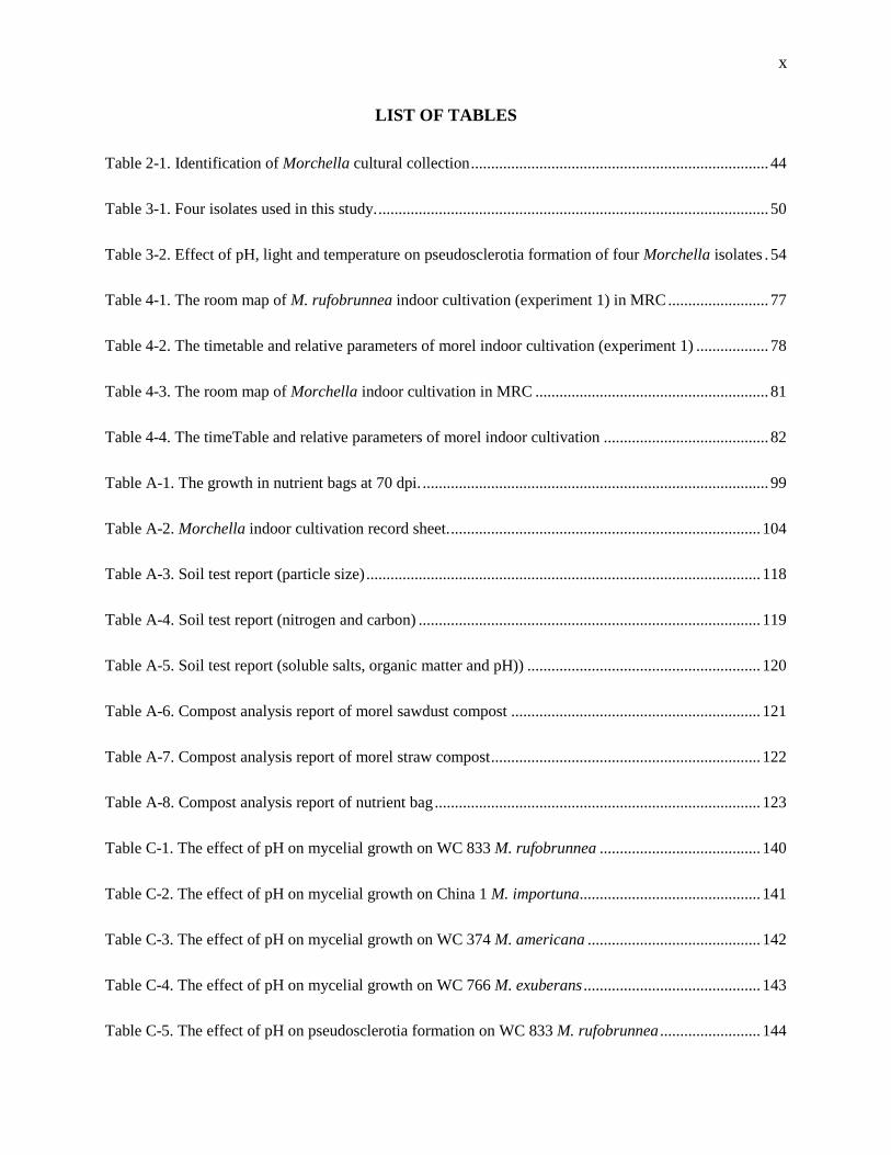
LIST OF TABLES
Table 2-1. Identification of Morchella cultural collection .......................................................................... 44
Table 3-1. Four isolates used in this study. ................................................................................................. 50
Table 3-2. Effect of pH, light and temperature on pseudosclerotia formation of four Morchella isolates . 54
Table 4-1. The room map of M. rufobrunnea indoor cultivation (experiment 1) in MRC ......................... 77
Table 4-2. The timetable and relative parameters of morel indoor cultivation (experiment 1) .................. 78
Table 4-3. The room map of Morchella indoor cultivation in MRC .......................................................... 81
Table 4-4. The timeTable and relative parameters of morel indoor cultivation ......................................... 82
Table A-1. The growth in nutrient bags at 70 dpi. ...................................................................................... 99
Table A-2. Morchella indoor cultivation record sheet. ............................................................................. 104
Table A-3. Soil test report (particle size) .................................................................................................. 118
Table A-4. Soil test report (nitrogen and carbon) ..................................................................................... 119
Table A-5. Soil test report (soluble salts, organic matter and pH)) .......................................................... 120
Table A-6. Compost analysis report of morel sawdust compost .............................................................. 121
Table A-7. Compost analysis report of morel straw compost ................................................................... 122
Table A-8. Compost analysis report of nutrient bag ................................................................................. 123
Table C-1. The effect of pH on mycelial growth on WC 833 M. rufobrunnea ........................................ 140
Table C-2. The effect of pH on mycelial growth on China 1 M. importuna ............................................. 141
Table C-3. The effect of pH on mycelial growth on WC 374 M. americana ........................................... 142
Table C-4. The effect of pH on mycelial growth on WC 766 M. exuberans ............................................ 143
Table C-5. The effect of pH on pseudosclerotia formation on WC 833 M. rufobrunnea ......................... 144
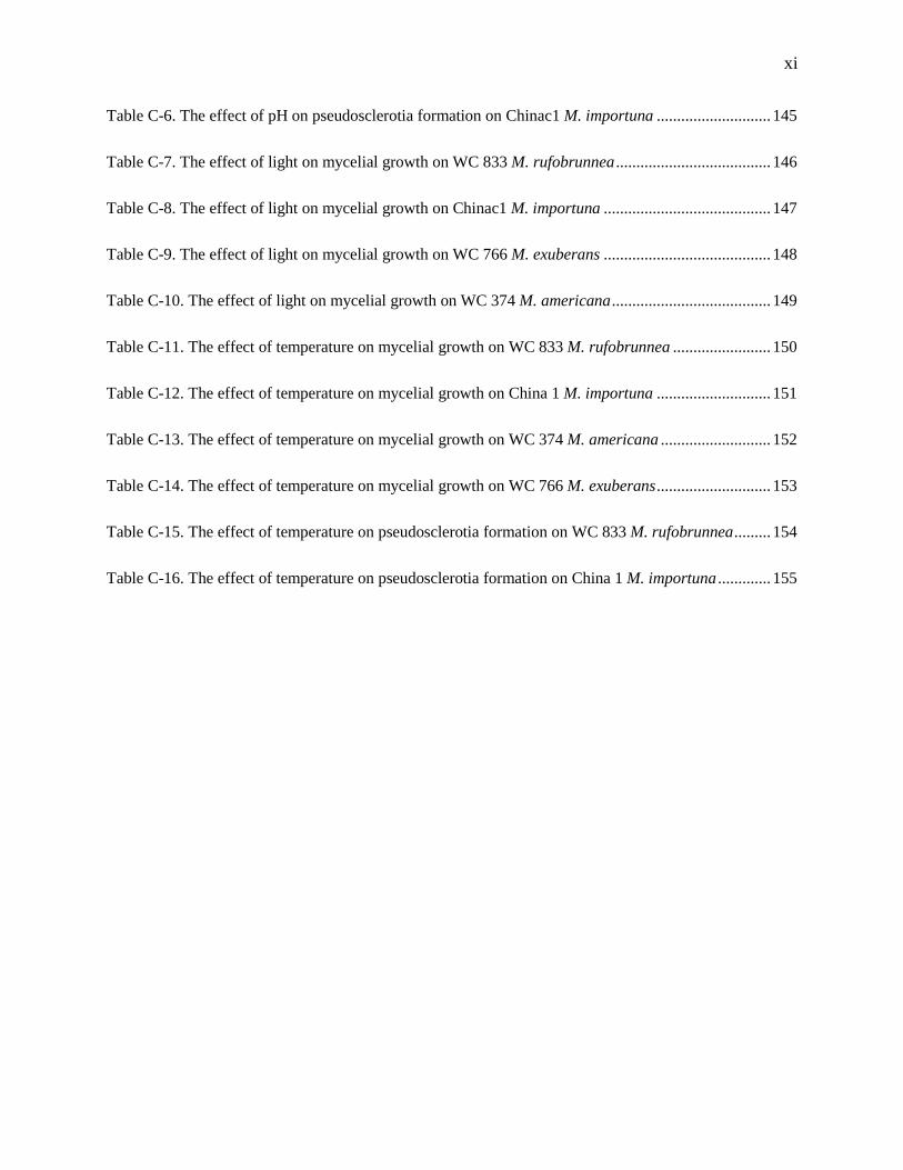
xi
Table C-6. The effect of pH on pseudosclerotia formation on Chinac1 M. importuna ............................ 145
Table C-7. The effect of light on mycelial growth on WC 833 M. rufobrunnea ...................................... 146
Table C-8. The effect of light on mycelial growth on Chinac1 M. importuna ......................................... 147
Table C-9. The effect of light on mycelial growth on WC 766 M. exuberans ......................................... 148
Table C-10. The effect of light on mycelial growth on WC 374 M. americana ....................................... 149
Table C-11. The effect of temperature on mycelial growth on WC 833 M. rufobrunnea ........................ 150
Table C-12. The effect of temperature on mycelial growth on China 1 M. importuna ............................ 151
Table C-13. The effect of temperature on mycelial growth on WC 374 M. americana ........................... 152
Table C-14. The effect of temperature on mycelial growth on WC 766 M. exuberans ............................ 153
Table C-15. The effect of temperature on pseudosclerotia formation on WC 833 M. rufobrunnea ......... 154
Table C-16. The effect of temperature on pseudosclerotia formation on China 1 M. importuna ............. 155

xii
ACKNOWLEDGEMENT
I would first like to express my deepest gratitude to my advisor Dr. John A. Pecchia, who guided
me professionally and shared enthusiasm with me through my master education at Penn State. Without
his help and encouragement, this thesis would not been possible.
I would also like to thank my committee members, Dr. Gretchen Kuldau, Dr. David Beyer and
Dr. Michael Fidanza. They gave me a lot of constructive advices on my proposal and thesis writing.
Without their passionate participation and professional guidance, my work could not have been done.
Lots of thanks to Edward Kaiser, who helped me a lot with the culture preparation. He was so
kind and generous to me when I was working in the Spawn Lab. I owe a deep sense of gratitude to Dr.
Fabrício Vieira, who taught me hand to hand to get DNA sequence from all my Morchella cultures. I
would like to thank Vija Wilkinson. She was so kind to me and taught me how to make spawn in the lab.
I also express my heartiest gratitude and thanks to all the staff in the Mushroom Research Center,
all the faculty, graduate students and staff in the Plant Pathology and Environmental Microbiology
Department at Penn State. I had a great time here and I will miss you all.
Finally, I must express my gratitude to my wonderful parents, Mr. Zhen Ge and Mrs. Jianping
Ma, for providing me continuously encouragement and unconditional support all the time. Special thanks
to my dear husband, Zhisheng Hu, who has always been there for me. Many thanks to my dearest twin
sister, Siyuan Ge, who has been my best friend for 26 years. Last but not least, special thanks to my cat
Enigma for bringing me so much fun in the past three years.
Thanks for all your encouragement!

1
Chapter 1
Literature Review
Economic importance of morels
True morels (Morchella spp.) are prized, edible fungi, belonging to Ascomycota, Pezizomycetes,
Pezizales, Morchellaceae, and Morchella. Due to their short fruiting season, favorable flavor and value in
the food and medical industry, mycologists have been attempting to cultivate morels for decades, and it
has been proven very difficult. However, recently in China, morel cultivation has expanded and the
annual export of dried morels reached 900,000 kg in 2015, averaging $160 US dollars per kilogram (Du
et al. 2015). Large quantities of morels have been harvested in China, India, Pakistan, Turkey, and North
America (Pilz et al. 2007), though the majority are through the collection of wild mushrooms and not
through cultivation.
Taxonomy of Morchella
To date, there are 334 names identified under Morchella, including species, subspecies and
varieties, reported in the latest database of the Index Fungorum
(http://www.indexfungorum.org/names/Names.asp). Due to the high diversity and variation in
morphology under different environmental and ecological conditions and during different developmental
stages, the use of homonyms and synonyms in Morchella spp. very often exists, which has caused
confusion and difficulties in identifying Morchella spp. (Du et al. 2012b). Before the development of
molecular techniques, Morchella spp. were initially divided into three groups: black morels, yellow
morels and semi-free capped morels. In 1998, Guzmán and Tapia (1998) proposed a fourth group, named
the blushing species, in the genus Morchella.
2
The application and development of DNA sequencing techniques and phylogenetic analysis has
made great progress in fungal identification. Sequencing of ITS loci consisting of the 5.8S rRNA gene
along with flanking ITS regions, LSU, SSU, RNA polymerase, and RAPD-PCR analysis have been used
in the identification of Morchella. Wipf et al., 1996, Wipf et al.,1999 sequenced the ITS locus with the
5.8S rRNA gene of a morel in the Elata Clade (M. conica) and a yellow morel in the Esculenta Clade (M.
esculenta) to elucidate the discrepancy between two morel groups. The length of the ITS locus was
confirmed to be 740 - 760 bp for the Elata Clade and 1150 - 1220 bp for the Esculenta Clade. It should be
noted that the name M. conica was not correct and has been applied to different Morchella species-
Morchella deliciosa, Morchella purpurascens, Morchella tridentina, and Morchella vulgaris (Richard et
al. 2015). ITS and RAPD-PCR analysis was used by Pilz et al. (2004) to study the phylogenesis of
Morchella located in northeastern Oregon.
O’ Donnell et al. (2011) used genealogical concordance phylogenetic species recognition
(GCPSR) to investigate the species limits within Morchella. In their study, three lineages within
Morchella were identified: a basal monotypic lineage represented by M. rufobrunnea, the Elata Clade
(black morels) and the Esculenta clade (yellow morels). Even though it was stated in this study that 177
specimens representing the global genetic diversity were finally selected, samples from eastern Asia were
underrepresented (Du et al. 2015). The phylogenetic diversity of Morchella was studied for 361
specimens, collected from 21 provinces in China between 2003 to 2011, by analyzing ITS rDNA
sequences (Du et al. 2012a). Forty Esculenta Clade (yellow morels), 30 Elata Clade (black morels) and 1
in the rufobrunnea Clade were selected.
M. anatolica belongs to the rufobrunnea Clade, along with M .rufobrunnea (Taşkın et al. 2012).
Three additional Morchella species were identified and named as Mel-35 , Mel-36 (Elliott et al. 2014) and
Mel-37 (Pildain et al. 2014) in the Elata Clade and Kuo (2012) described 19 phylogenetic Morchella
species in North America. It is worth noting that 6 species described by Kuo (2012) and Clowez (2010)
3
were reported to be synonyms by Richard et al. (2015). For example, M. conica was determined to be
illegitimate and was a synonym of M. purpurascens. Four previous unnamed Morchella species were
described as M. mediterraneensis (Mel-27), M. fekeensis (Mel-28), M. magnispora (Mel-29), and M.
conifericola (Mel-32) by Taşkın et al. (2016).
Even though ITS rDNA sequencing has been used in many identification studies on Morchella,
ITS rDNA sequencing can only identify approximately 70% of the known species (Du et al. 2012b).. The
error rate is especially high when identifying morels in the species-rich Elata Subclade and two closely
related species in the Esculenta Clade. In addition, more than 66% of the named Morchella sequences
were misidentified in GenBank and the application of synonyms exacerbates the problem (Du et al.
2012b). In order to ascertain the identities and achieve reliable results, additional gene sequences along
with dedicated accessible references are needed.
Global distribution pattern of Morchella
Sixty-five phylogenetic distinct species have been identified in Morchella with 34 species found
in East Asia or China, of which 20 species are endemic. Of 27 species present in Europe, and of 21
species present in North America, 12 and 14 are endemic respectively (Du et al. 2015). Many Morchella
species are found in Turkey with roughly twice as many species found there compared with other regions
in Europe (Taşkın et al. 2012). Mel-10 (M. importuna), Mel-19, Mel-2 (M. tridentina) are found in
Western North America, Eastern North America, Europe and Asia. Mel-7 (M. eximia), which is found in
Western North America, Eastern North America, Europe, Asia, Oceania and South America, is the most
widely distributed among all the disjunct species (Du et al. 2015).
4
Trophic mode of Morchella
Successful cultivation suggests that at least M. importuna, M. sextelata, and M. eximia are
saprophytic. M. tomentosa, M. sextelata, M. eximia and Mel-8 were reported as obligate fire-adapted
species. M. exuberans and M. importuna are considered facultative fire adaptive species (Pilz et al. 2007).
Fruiting bodies of some species in the “black morel” clade can be found commonly after fire (Kuo et al.
2012). Stable isotopes have indicated that some Morchella species are saprotrophic (Hobbie et al. 2001).
Hobbie et al. (2016) accessed the nutritional strategy of post-fire morels using isotope techniques (δ13C
and δ15N) and radiocarbon analysis (Δ14 C). They determined the isotopes values for ectomycorrhizal or
saprotrophic fungi by comparing the stable isotopes and radiocarbon in Morchella with values from
different fungi found in the literature. High 15N enrichment in Morchella indicated that the primary
nitrogen source for Morchella was not recently burned litter. Similar 13C and 15N enrichments to
saprotrophic Plicaria suggested that Morchella assimilated carbon and nitrogen the same as Plicaria,
which indicated saprotrophic nutrition in Morchella spp.. Unfortunately, the authors failed to provide the
species names.
Buscot and Roux (1987) reported that M. rotunda were able to penetrate into root tissues,
indicating a plant parasitic or symbiotic relationship. An ectomycorrhizae association was described
between Picea abies (Norway spruce) and M. rotund, M. esculenta, and M. elata by Buscot and Kottke
(1990) and Buscot (1992a, b, c). With M. rotunda, a compact mycelial sheath was formed around the
roots, which may serve as nutrient source for fructification, and the formation of ectomycorrhizal
mycelium acting as a survival structure for morels in forest ecosystems (Buscot and Kottke 1990).
Subterranean pseudosclerotia of M. elata ensheathed mature roots of Picea abies (Buscot and Kottke
1990). Buscot (1992a) found that M. elata only formed an ectomycorrhizae by replacing a previous
ectomycorrhizae formed by other fungi. Seven ectomycorrhizal types were identified from 155
5
ectomycorrhizal root tips of Picea abies by Buscot (1994). Additionally, an endobacterium was found
growing within the Hartig net of this ectomycorrhiza (Buscot 1994), though the exact role the
endobacterium plays is unknown. However, Dahlstrom et al. (2000) found that Morchella isolates
formed similar structures as the ones described by Buscot (1992a, c) without the help of bacteria or the
presence of previously colonized fungi.
Dahlstrom et al. (2000) reported morels could form an ectomycorrhiza sheath with Pinaceae, but
failed to indicate the Morchella species. In addition, they pointed out that additional microbes were not
required for the formation of ectomycorrizas of morels. Stark et al. (2009) suggested the association
between morels and orchids by analyzing the root-extracted DNA. Q. Li et al. (2013) speculated that
morels with black pilei were considered saprobes, while those with yellow pilei were mycorrhizas. Miller
(2005) was issued a patent (US 6907691) for morel cultivation using the mycorrhizal association between
Morchella mycelium and a tree seedling, though Miller also did not indicate the species in his patent.
Baynes et al. (2012) reported that M. eximia could infect cheatgrass roots but it was not clear if M. eximia
was acting as a pathogen, saprophyte or symbiont in this relationship.
The trophic strategies of Morchella remains to be studied and discussed. Based on the existing
data and information, it is likely that Morchella spp. include saprophytic, mycorrhizal and facultative
mycorrhizal species.
Life cycle of Morchella
Life cycle
The first life cycle of Morchella spp. was proposed by Volk and Leonard (1990). This life cycle
is general and does not confine to any specific Morchella spp. In this life cycle, the primary mycelium,
6
secondary mycelium, the formation of pseudosclerotia, germination of pseudosclerotia and the
development of a fruiting body is identified.
Pilz et al. (2007) proposed a life cycle of true morels. Their life cycle is based on Volk and
Leonard's life cycle, but it includes the cellular stages, showing the nuclei status in the hyphae or
mycelium. What is worth mentioning is that this life cycle also includes some ecological conditions under
which this life cycle takes place, such as soil necromass remaining after fires and live tree
ectomycorrhizae associations.
Alvarado-Castillo et al. (2014) proposed a more complicated and integrated life cycle of
Morchella spp.. This life cycle integrates all of the previous models and experimental observations and
known research related to this genus. This life cycle starts with the fruiting body, ascocarp or ascoma.
Each ascus produces eight ascospores which are released for dispersion. Under certain environmental
conditions, such as drought and low temperatures, ascospores can grow and germinate to form hyphae.
These hyphae can continue to grow and form primary mycelium. There are two subsequent pathways that
the primary mycelium may develop; one is that primary mycelium may produce conidia and the other is
that the primary mycelium can continue growing, intertwining, and develop into compact masses, which
can then give rise to pseudosclerotia formation. Generally, the primary mycelium fuse with other
mycelium from the same or another ascocarp through anastomosis and secondary mycelium is then
produced. Even though Volk and Leonard (1990) suggested that anastomosis of two compatible primary
mycelia occurs with and nuclei pairing in secondary mycelia, other haploid nuclei coexisting in the same
septum instead of pairing to form dikaryons often exists. The haploid hyphae might fuse with existing
heterokaryotic mycelia to simply contribute its own haploid nuclei, which seems to increase gene
diversity and may be a strategy to adapt to environmental changes (Kaul 1997) In addition, it seems that
mycelia of some Morchella isolates (species not indicated) do not anastomose (Volk and Leonard 1990).
There are on average 10 to 15 nuclei in each septum in the secondary mycelium, but numbers can reach
7
up to 65 (Volk and Leonard 1990). The secondary mycelium can also grow repeatedly and intertwine to
form pseudosclerotia. During this process, the secondary mycelium may form chlamydospores (Alvarado-
Castillo et al. 2012), which are thick-walled and function as resting fungal cells. Under certain
environmental stimuli, such as drought, change of temperature, and exposure to fire and flooding, the
pseudosclerotia germinate and give rise to the carpogenic mycelium, which will then develop into a
fruiting body, the ascocarp. However, it is not clear whether the pseudosclerotia produced from the
primary mycelium can develop into fruiting bodies directly, though its’ haploid nature indicates that is
unlikely to occur (Pilz et al. 2007). It is also unknown whether the secondary mycelium is able to develop
into a fruiting body or not.
W. Liu et al. (2017a) proposed a life cycle of M. importuna, the most widely cultivated Morchella
species in China. There is no intrinsic difference between this life cycle and the one Alvarado-Castillo
proposed. Under favorable conditions, the ascospore of M. importuna germinates and forms the primary
mycelium.
The nuclear behavior during meiosis and ascosporogenesis on M. importuna was studied using
confocal laser scanning microscopy (P. He et al. 2017). The study suggested that a total of six nuclear
divisions typically took place during ascosporogenesis. First, two meiotic divisions and subsequently one
mitotic division gives rise to eight nuclei. The nucleus in each ascospore then undergoes three mitotic
divisions and finally six to eight nuclei are formed in each ascospore. Two idiomorphs, MAT1-1 and
MAT1-2 were identified on M. importuna and population genetic investigations suggest heterothallic
characteristics of three Morchella isolates, M. importuna, M. sextelata and Mel-20 (Chai et al. 2017). Du
et al. (2017) noted that 14 Morchella species in the Elata Clade are all heterothallic. W. Liu et al. (2018b)
investigated two monospores with a different mating type isolated from M. importuna. Their results
indicated independent mating type structures in the two monospores, so they suggested that M. importuna
might be a heterothallic fungus.
8
Pseudosclerotia of Morchella
Concept of sclerotia
Fungi can use a variety of strategies to survive under unfavorable conditions (Blackwell 2011).
Many fungi are able to produce durable structures to facilitate dispersion or survival (Stajich et al. 2009),
while some form a kind of multicellular structure, which is a dense aggregation of fungal tissue called
sclerotia (Willetts 1971). Generally, sclerotia consist of a peripheral rind, which is a layer of
pseudoparenchymatous, melanized cells, and the rind encases a broad medulla. However, sclerotia
produced by Morchella are actually not true sclerotia in the classic sense, such as those produced by
Sclerofinia sclerotiorum, which are characterized by the medulla and rind, they are considered
pseudosclerotia (Volk and Leonard 1990), even though the name sclerotia was used in many studies.
Role of pseudosclerotia in the life cycle of Morchella
Based on Volk and Leonard’s theory, pseudosclerotia formation is an indispensable period in the
life cycle of Morchella spp. and plays an essential role in the successful development of fruiting bodies of
Morchella spp.. It is thought that pseudosclerotia can be produced from both primary mycelium and
secondary mycelium. However, Volk and Leonard suggested that only secondary mycelium is able to
give rise to pseudosclerotia (Volk and Leonard 1990), but additional evidence is needed. Formation of
pseudosclerotia is a response to unfavorable environment conditions and also a strategy that fungi use for
nutrient storage, which suggests they are the products of a long-term biological evolution (Xiong et al.
2015).
9
In 1982, Ower was successful in the cultivation of M. rufobrunnea for the first time in history. He
used a jar method to produce pseudosclerotia and then stimulated the germination of pseudosclerotia to
promote fruiting body development. Three patents were issued based on this method, and many
mycologists have since been attempting to study the mechanism of pseudosclerotia development.
However, pseudosclerotia are not always formed under experimental conditions, and the ability and
requirements of pseudosclerotia development varies greatly depending on the species (C. Ding et al.
2008).
Furthermore, types of pseudosclerotia may differ as well. Buscot (1993) studied mycelial
differentiation of M. esculenta in pure culture. Based on morphogenetic and additional characteristics,
two types of pseudosclerotia were identified from freshly germinated ascospores: early, encrusting
pseudosclerotia (EES), and late, isolated pseudosclerotia (LIS), which were initiated by growth
interruption and aging of the culture respectively. It is likely that EES were associated with imperfectly
developed fruiting bodies or some kind of sexual structures, while LIS functioned as storage and resting
structures. On the basis of the interaction of Morchella with bacteria, two types of pseudosclerotia were
produced: pseudosclerotium type 1 (ST1) and pseudosclerotium type 2 (ST2). ST1 initiated first and
aggregated near the inoculum. ST2 formed after ST1. On the contrary to ST1, ST2 were dispersed on the
medium, matured faster and became pigmented (Stott and Mohammed 2004). Stott and Mohammed
(2004) found that M. hortensis and M. rigida only produced pseudosclerotia on the nutrient-rich medium
when inoculated on the nutrient poor medium beforehand. An isolate of M. esculenta, originating from
Greece, also only produced pseudosclerotia on nutrient-rich media after being grown on nutrient-poor
media first (Philippoussis and Zervakis 2000). However, one Tasmanian isolate was able to produce
pseudosclerotia on both nutrient-poor and rich media no matter whether it was inoculated on nutrient poor
medium or not. Philippoussis and Zervakis agreed with Buscot (1993) and Faris et al. (1996) that a
nutrient-poor environment is essential for pseudosclerotia production. But Singh and Vema’s (2000)
research indicated that nutrient-poor conditions are not necessary for the development of pseudosclerotia.
10
In their study, six media were used to cultivate Morchella spp.. Malt Extract Agar was found to be the
best culture medium to get the maximum radial growth and the only one that supported pseudosclerotia
development in all of the test isolates.
Recently, P. He et al. (2018) first highlighted the involvement of autophagy and apoptosis and
lipid accumulation in pseudosclerotia morphogenesis of Morchella importuna (Pseudosclerotia initial
[SI], pseudosclerotia development [SD], pseudosclerotial maturation [SM]). They compared the structural
features of the undifferentiated mycelial stage and three main pseudosclerotial stages by using
transmission electron microscopy. The characteristics of autophagy was observed during the SI phase and
apoptotic characteristics were found in some cells during the SD phase. Moreover, they found lipid was
the energy-rich substance in both hyphae and pseudosclerotia of M. importuna. Pseudosclerotia had a
significantly higher content of lipid than that in hyphae, which is consistent with the hypothesis that
pseudosclerotia serve as nutrient storage organs.
Factors that affect pseudosclerotia production
External factor
Amir et al. (1992; 1993; 1995) studied the effect of medium composition and water potential on
pseudosclerotia formation on M. esculenta using the split plate method (two media that differ in nutrient
content on one plate separated by a plastic barrier). Their study emphasized the important role of high
turgor potential, which is required for the growth of hyphae, in the production of a large quantity of
pseudosclerotia and this turgor potential is regulated by metabolizable substrates. Pseudosclerotia
production was measured by dry weight as well as calculating their surface area with the aid of a camera
and computer program. Water, solute and turgor potential were measured as described by Thompson et al.
11
(1985). Their study defined six growth stages of M. esculenta during hyphal growth and pseudosclerotia
formation on a split plate. The first stage included the growth and extension of mycelium on the nutrient-
poor (noble agar/NA) side. Hyphae were weak, characterized by large vacuoles and growth toward the
nutrient-rich (potato dextrose agar(PDA)) side. Stage II began when hyphae reached the PDA side of the
plate, followed by decreasing the size of hyphal vacuoles and declining of the hyphal glucose content. It
was worth noting that the direction of the cytoplasm stream, including nutrient substances, reversed when
the growth of mycelia reached the end of the plate. The cytoplasm moved from old mycelia (PDA side) to
young mycelia (NA side), but no explanation was provided. Carbohydrates, which were assumed to be
mannitol or arabitol, were translocated from the younger mycelium to the older mycelium. The initiation
of pseudosclerotia on the NA side indicated the beginning of stage III. Stage IV was characterized by the
enlarging pseudosclerotia and this enlargement was supported by the rapid transportation of a large
amount of nutrients to the young pseudosclerotia. Color of the mycelium changed from white to yellow-
brown during stage V and stage VI was characterized by the formation of the peripheries of some
pseudosclerotia. They also pointed out that nutrients from mycelium on the PDA side were transported to
the other side and the pseudosclerotia from the NA side served as a nutrient sink. Moreover, their study
indicated turgor pressure gradients in a split plate, which explained the rapid translocation of nutrients to
pseudosclerotia during the morphogenetic process.
Volk (1989) used a jar method (Ower et al. 1986) to study the effect of a variety of conditions on
pseudosclerotia formation. Complex medium worked better and a smaller sized container achieved better
biological efficiency (BE), calculated as the wet weight of pseudosclerotia divided by the dry weight of
the substrate. He also pointed out that illumination was a limiting factor for the development of
pseudosclerotia. In addition, carbon was not a limiting factor while nitrogen was. Zhao et al. (1997)
reported that glucose, mannitol, mannose and diammonium phosphate promoted pseudosclerotia
formation. They also stated that light is not necessary. However, during the process of making spawn,
there was a significant difference between the light side and the dark side of the spawn container if it was
12
not rotated on a regular basis. On the side that was occasionally exposed to light, large amounts of
pseudosclerotia were produced. On the contrary, production of pseudosclerotia on the dark side was poor,
indicating that light plays a role in pseudosclerotia formation (W. Liu et al. 2017a). Volk and Leonard
(1990) reported that low temperatures or nutrient deficiency is required for the formation of
pseudosclerotia of Morchella spp. Wang (1997) reported that between 15 to 20C is the best temperature
for the formation of pseudosclerotia of Morchella spp. Wang also pointed out that the number and size of
pseudosclerotia can differ significantly on different cultural media even for the same isolate of Morchella
spp.. Li et al. (1998) reported that VB1 favors the formation of aerial pseudosclerotia. Kanwal and Reddy
(2011) compared the effects of various nitrogen (N) and carbon (C) sources on pseudosclerotia formation
and development for Morchella elata and Morchella crassipes. They concluded that for the formation of
larger-sized pseudosclerotia of Morchella, ribose, mannitol, and glucose are the best carbon sources and
the best nitrogen source is sodium nitrate. They also studied ligninolytic enzyme production on different
substrates and during pseudosclerotia formation in M. crassipes (Kanwal and Reddy 2014). Besides
carbon and nitrogen, ligninolytic enzyme production in M. crassipes was influenced by ligninolytic
chemical and natural inducers (Kanwal and Reddy 2010). Their study demonstrated that ligninolytic
enzymes, such as laccase, Manganese peroxidase, and lignin peroxidase are induced in lignin-rich
substrates. These enzymes result in lignin degradation in M. crassipes and it is hypothesized that lac
enzymes play an important role in pseudosclerotia formation and maturation.
Intrinsic factor
Chen et al. (2014) analyzed the differences between gene expression of pseudosclerotia-
producing and non- pseudosclerotia-producing single spore isolates from M. conica using RT-PCR.
Thirteen different positive gene fragments which were thought to be involved in pseudosclerotia
formation of M. Conica were identified by comparing the gene expression of two types of isolates to the
13
housekeeping gene 18S rRNA. Some positive fragments were found similar to genes involved in lipid
metabolism, nitrogen metabolism, the OmpA family protein coding gene and keratin-associated protein 5,
6 coding genes. Some positive fragments that could not be found in the NCBI are thought to be the
specific genes controlling pseudosclerotia formation and differentiation.
Q. Liu et al. (2018) studied the effect of reactive oxygen species on pseudosclerotia formation of
M. importuna. In their study, a higher hydrogen peroxide concentration was observed in the mycelial
growth region compared to the pseudosclerotium-forming region. Moreover, pseudosclerotia formation
and gene expression of organisms possess superoxide dismutases (SOD) was correlated to the
concentration of hydrogen peroxide. It was hypothesized that the MARK pathway was involved in
pseudosclerotia formation in M. importuna.
Mitospore
The mitospore stage of Morchella spp. was first reported by Molliard (1904a) in his experiment
on artificial cultivation of Morchella. He identified the white mold on the soil surface as the hyphomycete
Costantinella. Subsequently, Molliard (1904b) demonstrated that low relative humidity is required for the
development of mitospores. However, he failed to observe the germination of mitospores on various
media. Paden (1972) also reported the development of mitospores in M. elata. Ower found massive
conidia blooms during his cultivation experiment on Morchella. According to the life cycle provided by
Volk and Leonard (1990), mitospores can be produced from primary mycelium but no explanation was
given to their role in fruit body formation. Masaphy (2010) closely observed the morphology of
mitospores during cultivation of M. rufobrunnae. W. Liu et al. (2016a) analyzed the morphology and
structure of mitospores on M. importuna. The mitospores of M. importuna are hyaline, spherical, non-
septate, smooth, 3.5 to 5.2μm in diameter. Conidiophores are cambiform, slightly curved like an “s”, and
are (2.1 - 6.2) × (14.1 - 18.5) μm in size. Four to six conidioiphores can be found on the specialized
14
hypha, which is 6.0-8.5um long, hyaline and thinner than the vegetative hypha. Conidiogenous cells
contain between 2-7 nuclei. Most of the mitospores have only one nucleus, though two, three and four
nuclei are occasionally found. Mitosis can be clearly observed in a single mitospore, indicating the
mitospores with two or more nuclei may result from mitospores with a single nucleus. Even though the
ability to produce mitospores has been demonstrated with M. importuna during artificial cultivation, the
specific role of the mitospores is not yet clear. The relationship between the production of Morchella
fruiting bodies and the number of mitospores, and the role of mitospores in the entire life cycle of
Morchella spp. needs further investigation.
Etiology of Morchella
Carbon source
Zhu et al. (2011) noted that the best carbon source for mycelial growth of M. esculenta is soluble
starch based on the growth rate of mycelium, and the size and thickness of the colony. Using the dry
weight of mycelia, the content of intercellular polysaccharides and the yield of polysaccharides as the
indicators, Yang et al. (2007) pointed out that soluble starch as well as sucrose are the best carbon sources
for the growth of various Morchella spp. (the name of specific species was not provided). This contradicts
previous reports that the best carbon source for mycelial growth is glucose for strain LWY-1(Chai et al.
2010) and strain M-yan-5 (Ren et al. 2006). However, the species of these two strains were not indicated.
In production, wheat, sawdust, corn stover, cottonseed hulls and compost have been used as carbon
sources for Morchella spp. Nevertheless, Morchella has limited abilities to degrade lignin, hemicellulose
and pectin (W. Liu et al. 2017a).
15
Nitrogen sources
S. Liu et al. (1998) reported that the best nitrogen sources for the mycelial growth of M. conica
(reported to be an illegitimate name (Richard et al. 2015)) in vitro are aspartic acid, cysteine and sodium
nitrite, while Dong (2004) reported that potassium nitrate and sodium nitrate are the optimal nitrogen
sources for M. angusticeps. Yang et al. (2007) noted that soybean meal, peptone and fish gelatin are the
best nitrogen sources based on dry weight measurements of mycelium, the content of intercellular
polysaccharides and the yield of polysaccharides as the indicators. Zhu et al. (2011) noted that carbamide
is the best nitrogen source followed by potassium nitrate and calcium nitrate for M. escultanta. Xie et al.
(2009) pointed out that the best nitrogen source for five Morchella strains - M. angusticeps, M. conica,
Morchella spp. (50647), Morchella spp. (50648) and M. esculenta are ammonium sulfate, peptone, yeast
extract, sodium nitrate, and sodium nitrate respectively.
The diverse conclusion regarding the best nitrogen source for the growth of Morchella is related
to the diversity of the stains of Morchella and the concentration of nitrogen source. Unfortunately, there is
not adequate, consistent and convincing data indicating the best carbon to nitrogen ratio or the best source
of carbon or nitrogen for optimum growth and development of Morchella (W. Liu et al. 2017a).
Mineral salts
Mineral salts play an important role in maintaining the normal functions of fungi, such as
adjusting the osmotic pressure and the concentration of hydrogen ions. They are also components of
fungal cells that are involved with maintaining the activity of enzymes. Zhu et al. (2011) reported that
MnSO4 (30 mg/L) promotes the growth of M. esculenta, while MgSO4, K2SO4, NaCl and Na2MoO4
neither promote nor inhibit the growth of M. esculenta. However, KH2PO4, FeSO4, ZnSO4, NaSeO3,
CuSO4, CoCl2 and Ni(NO3)2 potentially inhibit mycelial growth of M. esculenta. CoCl2 only slightly
16
suppressed mycelial growth of M. esculenta when its concentration was around 10 mg/L. As the
concentration of CoCl2 increased, the effect of inhibition of mycelial growth also increased. CuSO4 with a
concentration of 70 mg/L and Ni(NO3)2 with a concentration of 90 mg/L completely stops mycelial
growth of M. esculenta.
Temperature
Chen and Guo (2007) reported that 24 to 26C is optimum temperature for M. esculenta mycelial
growth. Mycelia start to die when temperatures exceed 34C. According to the growers’ experiences, low
temperatures facilitate the differentiation of mycelia (personal communication). Growers believe that a
satisfactory yield of mature fruit bodies will be obtained when the air temperature is below 4C for at
least one month before the primodia formation for M. importuna and M. esculenta (personal
communication). It is also believed that temperature fluctuation of more than 10C in a day facilitates
primodia formation for these two species (personal communication). Nevertheless, additional data is
required to determine if a low temperature period is required for initiation of fruit bodies.
Humidity
W. Liu et al. (2017a) discussed the moisture and humidity levels required for general morel
cultivation (mainly M. importuna, M. sextelata, M. septimelata, and M. esculenta). Ideally, during the
incubation period, the moisture of spawn should be between 60 to 65%. If it is too high, air movement is
inhibited. As a result, anaerobic conditions are created and mycelial growth is suppressed. The moisture
of the soil should be between 18 to 28% before spawning, while during the mycelial growth stage, it is
better to drop the moisture to between 15 and 25%. When fruiting bodies start to form, a soil moisture
level of 20 to 28% is required. Air humidity levels between 65 to 80% is optimal during spawn run and
17
mycelial growth stage. During fruit body maturation, 85 to 95% air humidity is needed to avoid damage
to young fruiting bodies from drought.
pH
Similar to many fungi, Morchella spp. are able to grow under a broad pH range. Chen and Guo
(2007) noted that the optimal pH is 6.4 to 8.7 based on the condition of mycelial growth and
pseudosclerotia formation of M. esculenta. Calcium oxide is commonly used to adjust the pH of soil if it’s
too acidic (W. Liu et al. 2017a).
Light
Volk (1989) noted that light inhibits the pseudosclerotial growth of M. crassipes. However, it was
reported that mild light can actually stimulate the formation of pseudosclerotia during spawn production
of M. importuna (W. Liu et al. 2017a). There have been some reports on the effects of light on some other
edible fungi. For instance, it was reported that light treatment increases the production of Flammulina
velutipes (M. Liu and He 1997), and light has an effect on the development and spore production of
Ganoderma (Hao et al. 2011).
Oxygen and carbon dioxide
Adequate oxygen should be provided since Morchella spp. are aerobic fungi. Mycelia of
Morchella spp. are not reported to be sensitive to carbon dioxide levels (W. Liu et al. 2017a). However,
400 to 600 ppm of carbon dioxide has been reported to be the optimal level for the growth of fruit bodies
(W. Liu et al. 2017a).
18
Morel cultivation
History and progress of morel cultivation
Since 1882, when outdoor cultivation of morels was first reported by Roze (1882) in France,
many researchers and mushroom lovers have attempted various methods of cultivating morels. In 1904,
Molliard reported successful cultivation of morels in an apple compost. Ower claimed successful indoor
artificial cultivation of M. rufobrunnae in 1982. Subsequently, Ower applied for three patents (US Patents
4594809, 4757640, and 4866878) for morel cultivation in 1985, 1986 and 1988 respectively. These
studies revealed the optimal temperature, humidity and other essential factors during different stages, as
well as providing a method of transformation from vegetative growth to sexual reproduction. Moreover,
he emphasized the important role of pseudosclerotia in fruit body development in his method (Ower et al.
1985; 1986; Ower 1988). Methodology provided by these patents was adopted by Neogen Corporation,
Domino’s Pizza Inc., The Terry Companies of Wayzata (Terry Farm) and Diversifield Natural Products
Corporation (DNP, currently named Gourmet Mushrooms Inc.). With the help of Mills, the second author
of the patents, Terry Farms cultivated morels indoors and it was stated that 1400 fresh morels could be
produced per week. However, for unknown reasons, this production line was closed around 1999.
Subsequently, Mills worked for DNP and the company started selling fresh morels in 2005. Due to the
reduction of output and bacterial contamination, they had to stop growing morels in 2008, and since, there
have not been any reports of commercial artificial cultivation of morels in the USA.
In 2005, a patent for the cultivation of Morchella was issued for SC Miller. The key process of
his method was inoculating a tree root system with Morchella mycelium. Masaphy (2010) reported a
relatively detailed biotechnology of morel cultivation. She was successful in getting fruiting bodies of M.
rufobrunnae in a soilless system in laboratory-scale experiments. She also used a multi-layer cultivation
19
method proposed by Ower to cultivate pseudosclerotia. These pseudosclerotia then served as the “seeds”,
which were then inoculated into the compost and water treatment was applied to stimulate the
germination of the pseudosclerotia (Ower 1986, Volk 1990). Masaphy (2010) also pointed out that the
primary reasons for the failure of morel cultivation are the deficiency in knowledge about the
development of the ascocarp and difficulties and unknowns regarding species selection.
Chinese scientists have been attempting to unveil the mystery of Morchella since the 1980’s. The
first report of morel cultivation in China was proposed by Ding (1983) in 1983, though he failed to
publish detailed records of the cultivation process. In the same year, Gu (1983) cultivated spores on a
medium and bean-sized pins were observed after 55 days of cultivation. However, no mature fruiting
bodies were obtained. Zhu Douxi, who is honored as the “father of morels in China”, was issued the first
morel cultivation patent in China in 1994 (D. Zhu 1994). Later, four more patents for morel cultivation
were issued in 2001, 2007, 2009 and 2012 (D. Zhu 2001; D. Zhu and He 2007, 2009, 2012), respectively.
The 2001 Patent reduced the amount of organic matter needed in the soil to between 5 to 20% compared
with 90 to 95% requirements stated in the first patent. Excessive organic components were thought to
facilitate bacterial contamination and was more expensive for those considering large-scale commercial
production. The Patent in 2012 was the most detailed one among those five issued to D. Zhu. In this
patent, D. Zhu provided the formulation of a medium for the mother culture, detailed processes of making
spawn, spawning, and management strategies during different stages. According to published papers and
patents of D. Zhu, the techniques he reported belong to what is considered “bionic cultivation”, since the
species of Morchella he used were not identified and managements of factors, such as light, humidity and
temperature, were difficult to manage.
Apart from Miller, researchers in China also attempted to use the ectomycorrhyzal relationship
between Morchella mycelium and tree seedlings (Zhao et al. 2009), but they failed to examine the

20
effectiveness of this ectomycorrhyzal symbiosis. Compared with the cultivation process described by D.
Zhu, the production period is much longer and techniques are more complex.
In the early 21st century, some farmers in the Yunnan province, China, created a special method
for morel cultivation. They placed a piece of poplar wood on a layer of spawn and then added another
layer of spawn on the top of the wood, repeating this sequence until a “pyramid” was obtained. Then they
covered the “pyramid” with soil, simulating a “natural environment”. This method was reported to be
feasible using M. importuna (Zhao at al. 2014), but the large consumption of wood is not environmentally
friendly.
Intercropping patterns between morels and wheat is currently applied in some regions in China,
especially for those who do not count on growing morels to make profits. The most obvious advantage of
this pattern is the additional output of wheat when not cultivating morels. Therefore, this pattern is only
temporary and a way to cut losses to confront with the immature cultivation technology (W. Liu et al.
2017a). However, the yield of mature morel fruit bodies is generally 10 to 30% higher when intercropping
with wheat comparing to greenhouse cultivation (personal communication). But it is not clear whether
this intercropping pattern effects the production of wheat or not.
The most significant and important process discovery in morel cultivation in China may be the
application of exogenous nutrient bags. Strictly speaking, the concept of additional nutrient sources was
first described by Ower et al. in his patent in 1986 (Ower et al. 1986). However, they did not highlight or
compare the importance of the additional nutrient sources in the cultivation of Morchella and this theory
was never used in morel cultivation until D. Zhu’s reports.
The theory of using nutrient bags is that the sexual development of Morchella requires a
comparatively nutrient-poor environment, but the nutrient-poor soil substrate is not able to supply enough
nutrients for the growth of newly formed mycelium. Therefore, additional nutrients are required to
21
support the formation of mycelium. However, the additional nutrients should be removed later in the
process in order to facilitate the sexual reproduction, which will develop into fruiting bodies.
Morel cultivation in China
Current status
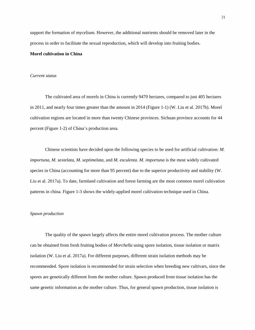
The cultivated area of morels in China is currently 9470 hectares, compared to just 405 hectares
in 2011, and nearly four times greater than the amount in 2014 (Figure 1-1) (W. Liu et al. 2017b). Morel
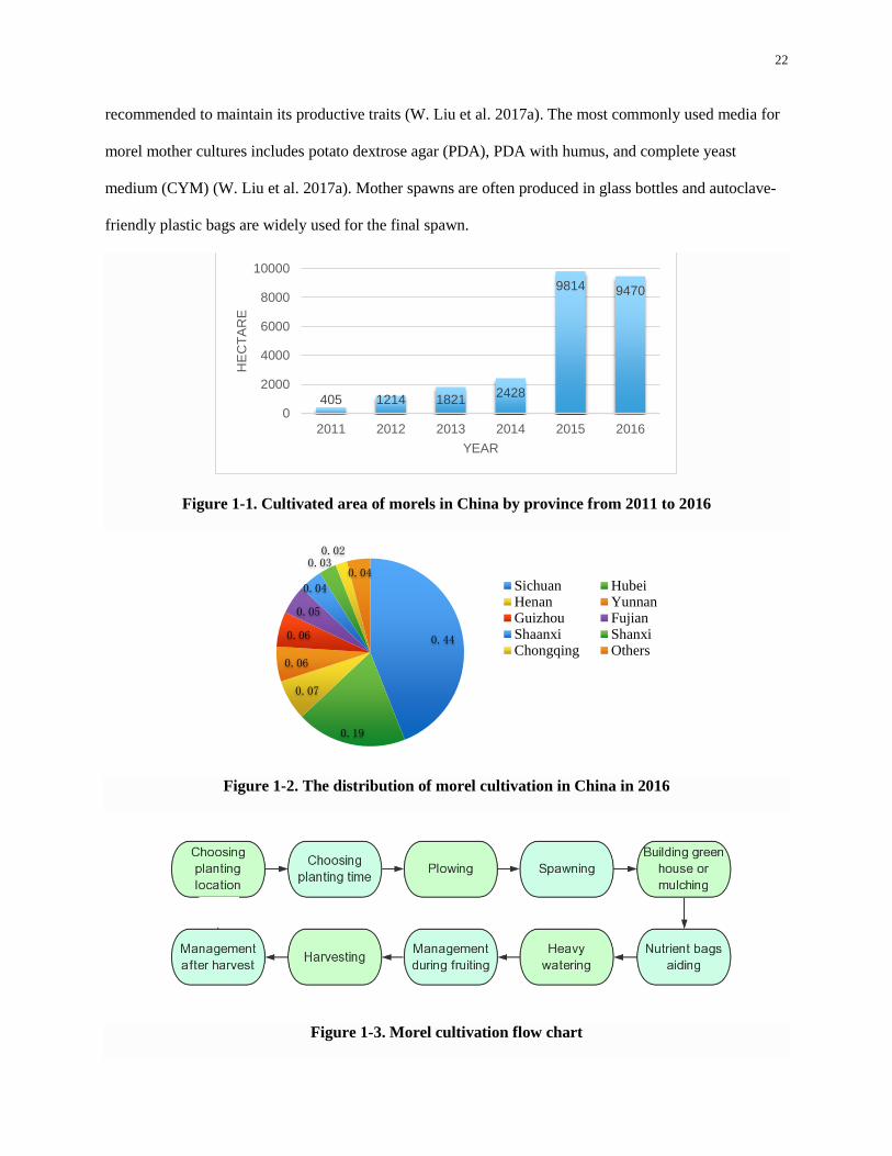
cultivation regions are located in more than twenty Chinese provinces. Sichuan province accounts for 44
percent (Figure 1-2) of China’s production area.
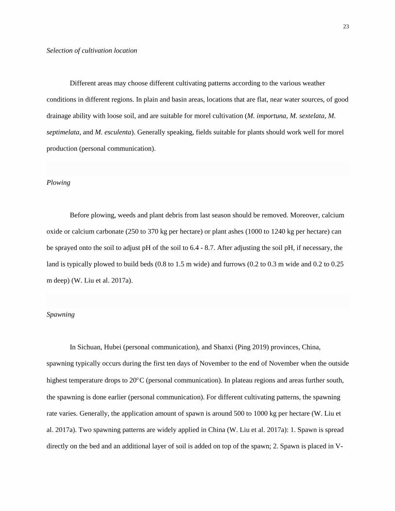
Chinese scientists have decided upon the following species to be used for artificial cultivation: M.
importuna, M. sextelata, M. septimelata, and M. esculenta. M. importuna is the most widely cultivated
species in China (accounting for more than 95 percent) due to the superior productivity and stability (W.
Liu et al. 2017a). To date, farmland cultivation and forest farming are the most common morel cultivation
patterns in china. Figure 1-3 shows the widely-applied morel cultivation technique used in China.
Spawn production
The quality of the spawn largely affects the entire morel cultivation process. The mother culture
can be obtained from fresh fruiting bodies of Morchella using spore isolation, tissue isolation or matrix
isolation (W. Liu et al. 2017a). For different purposes, different strain isolation methods may be
recommended. Spore isolation is recommended for strain selection when breeding new cultivars, since the
spores are genetically different from the mother culture. Spawn produced from tissue isolation has the
same genetic information as the mother culture. Thus, for general spawn production, tissue isolation is
22
recommended to maintain its productive traits (W. Liu et al. 2017a). The most commonly used media for
morel mother cultures includes potato dextrose agar (PDA), PDA with humus, and complete yeast
medium (CYM) (W. Liu et al. 2017a). Mother spawns are often produced in glass bottles and autoclave-
friendly plastic bags are widely used for the final spawn.
Figure 1-1. Cultivated area of morels in China by province from 2011 to 2016
Figure 1-2. The distribution of morel cultivation in China in 2016
Figure 1-3. Morel cultivation flow chart
405 1214 18212428
9814 9470
0
2000
4000
6000
8000
10000
2011 2012 2013 2014 2015 2016
HE
CT
AR
E
YEAR
0.44
0.19
0.07
0.06
0.06
0.05
0.04
0.030.02
0.04Sichuan HubeiHenan YunnanGuizhou FujianShaanxi ShanxiChongqing Others
23
Selection of cultivation location
Different areas may choose different cultivating patterns according to the various weather
conditions in different regions. In plain and basin areas, locations that are flat, near water sources, of good
drainage ability with loose soil, and are suitable for morel cultivation (M. importuna, M. sextelata, M.
septimelata, and M. esculenta). Generally speaking, fields suitable for plants should work well for morel
production (personal communication).
Plowing
Before plowing, weeds and plant debris from last season should be removed. Moreover, calcium
oxide or calcium carbonate (250 to 370 kg per hectare) or plant ashes (1000 to 1240 kg per hectare) can
be sprayed onto the soil to adjust pH of the soil to 6.4 - 8.7. After adjusting the soil pH, if necessary, the
land is typically plowed to build beds (0.8 to 1.5 m wide) and furrows (0.2 to 0.3 m wide and 0.2 to 0.25
m deep) (W. Liu et al. 2017a).
Spawning
In Sichuan, Hubei (personal communication), and Shanxi (Ping 2019) provinces, China,
spawning typically occurs during the first ten days of November to the end of November when the outside
highest temperature drops to 20C (personal communication). In plateau regions and areas further south,
the spawning is done earlier (personal communication). For different cultivating patterns, the spawning
rate varies. Generally, the application amount of spawn is around 500 to 1000 kg per hectare (W. Liu et
al. 2017a). Two spawning patterns are widely applied in China (W. Liu et al. 2017a): 1. Spawn is spread
directly on the bed and an additional layer of soil is added on top of the spawn; 2. Spawn is placed in V-

24
shaped furrows on the beds and then are recovered with soil. On one hand, technically, good
dispensability makes spaying a more effective way to spawn. On the other hand, spawn in the furrows
provides the opportunity for mycelia to grow from nutrient rich side (the site with spawn) to nutrient poor
side (the site with no spawn), which is favorable for pseudosclerotia formation.
Film mulching
The application of mulching films is a recent, innovation in the morel cultivation industry and is
utilized in some regions in China. This process reduces the costs of morel cultivation, which makes the
rapid expansion of morel cultivation more feasible (W. Liu et al. 2017a). Plastic film mulching can be
used both in addition to and in place of the shade cloth housing. Black plastic mulch is most commonly
used in the artificial cultivation of morels in China. It is suggested to choose mulches with a thickness of
0.006 to 0.008 mm (Zhang et al. 2017). Width of mulches is maintained 10 to 20 cm narrower than the
mushroom beds. After spawning, mulches are placed on the beds and fixed by stones or soil with a 50 cm
interval (Zhang et al. 2017). On the basis of the stability of the mulch, openings should be made on the
mulches to allow air circulation (Zhang et al. 2017).
About one day after spawning and mulching, mycelial growth can be seen on the soil (personal
communication). When adding nutrient bags at approximately 7 to 10 days after spawning, the stones or
soil on one side of the plastic should be removed so that the plastic mulch can be moved to one side to
allow for the application of the nutrient bags (personal communication). After application of the bags,
growers then cover the furrows again. W. Liu et al. (2017a) noted that the optimal mulch removing time
is 20 to 25 days after adding nutrient bags. Zhang et al. (2017) suggested that it is better to remove the
plastic mulch 10 to 20 days before fruiting, which depends a lot on personal experience.
25
There are several advantages of using a plastic mulch (Zhang et al. 2017). Moisture preservation
and waterlogging prevention: On one hand, mulches prevent the moisture from evaporation and on the
other hand, raindrops can move along with the mulches into the grooves. Even long periods of rainy days
won’t cause serious damages to the beds. The plastic also provides protection from light and provides
weed control. Strong light will suppress the mycelial growth of morels. Weeds provide habitat for pests
and sabotage the effectiveness of air circulation, contributing to a high-temperatures and a high-moisture
environment, which is favorable for fungal and bacterial infections. Plastic mulch also provides heat
preservation by absorbing heat from the sunlight and warming the soil temperature, which is favorable for
mycelial growth during the winter when temperatures are low. Another possible advantage of the plastic
mulch is that it may reduce conidia growth, even though the necessity of conidia in the life cycle of
Morchella is not clear (W. Liu et al. 2016a), it is proposed that excessive conidia will consume nutrients
making them unavailable for Morchella fruit body initiation. However, it should be noted that the role of
conidia in the morel fruiting body initiation is not clear yet. Mulching is also effective in controlling the
cost. The consumption of spawn can be reduced to 420 bags per hectare (14 × 28 cm plastic bag, 0.6 - 0.7
kg per bag) to obtain the same yield by using mulch instead of growing in shade cloth housing (Zhang et
al. 2017).
Nutrient bags
This is one of the most essential steps throughout cultivating morels. Nutrient bags with openings
are added to the soil with the openings facing the soil surface approximately 7 to 20 days after spawning
(W. Liu et al. 2017a). It is suggested that 4450 to 4950 bags be used, evenly dispersed per hectare (W. Liu
et al. 2017a). Mycelia of Morchella will use the nutrients contained in the bag and grow into the bags.
Nutrients in the bags are absorbed and transferred by mycelia to the soil mycelia, which supports the
fruiting body development. Removal of the bags takes place 20 days before fruiting bodies develop
(Wang et al. 2016). Based on limited information, the composition of the nutrient bags does not appear to

26
be critical. There are many available formulas for nutrient bags listed in Chinese patents (Qin et al. 2015;
Shi 2014).
Heavy watering
Apart from the addition and subsequent removal of supplemental nutrients, the other way to
stimulate fruit body development is through heavy watering (W. Liu et al. 2017a; Ping 2019). After
removing the bags about 20 days before fruiting bodies develop, heavy watering is recommended
between 1 to 3 times onto the beds. Heavy watering or complete submersion is also used to initiate
fruiting with other cultivated mushrooms (ex. Lentinula edodes - Shiitake logs) (Niu 1994).
Management during fruiting
It is worth noting that the fruiting bodies of some Peziza spp. have commonly been observed in
Chinese production systems about one week before the fruiting of Morchella (M. importuna, M.
sextelata, M. septimelata, and M. esculenta) (W. Liu et al. 2017a). Therefore, the presence of these fungal
fruiting bodies is considered a signal for the coming of the fruiting period of Morels.
The pins of morels are fragile and can be damaged easily when temperatures are less than 0C.
One method to minimize frost damage is to add straw or plastic mulch on the beds to keep the soil warm.
When small, immature pins develop into small fruiting bodies, air humidity should be maintained
between 85 to 95% (W. Liu et al. 2017a; Ping 2019) and soil moisture levels between 28 to 35% is
recommended (Zhang et al. 2017). When the fruit bodies grow to 1.5 to 3 cm tall, the air humidity should
be maintained while the soil moisture should be reduced to 18 to 25% (W. Liu et al. 2017a). In the later
maturation period, when fruiting bodies are growing quickly, it is recommended to maintain the
27
temperature between 12 to 16C and increase soil moisture to 20 to 25% (W. Liu et al. 2017a). During the
maturation period, the air humidity should be lowered to 70 to 85% (W. Liu et al. 2017a).
Harvest
When the fruiting body grows to 10 to 15 cm tall with a characteristic appearance of morels, then
growers harvest using a knife and cut the fruit body at the bottom of the stipe. Mature fruit bodies can be
dried at a low temperature and stored for a long period in a dried state (personal communication).
Issues and perspectives
Spawn quality
Quality spawn increases the likelihood of successful mushroom cultivation. Morchella species
display different morphology on different media (Guler and Ozkaya 2009), which is a major issue in the
development of a spawn quality evaluation standard. To date, no morel spawn quality standard is
available and the evaluation mostly depends on farmers’ experience and pseudosclerotia quality (W. Liu
et al. 2017a).
Spawn aging has been one of the major problems in morel cultivation (W. Liu et al. 2017a). The
characteristics of spawn aging include fragile mycelia, slow mycelial growth rate, week anti-competitor
ability and a delayed harvest, all of which will contribute to a reduced morel production. Senescence
(aging process) of M. elata, characterized by slow mycelial growth, premature pigment production and
death of hyphal tips, was observed by successive subculturing (P. He et al. 2015). Microscopy studies
28
revealed the involvement of autophagy, apoptosis and necrosis in senescence of M. elata (P. He et al.
2015). Nevertheless, the molecular mechanism of these behaviors needs further study.
Moreover, a decrease in spawn vigor is often observed in morel spawn production as well
(personal communication). The application of weak spawn characterized by thin, weak mycelia and
prolonged cultivation cycles may be detrimental to morel production directly. Since spawn quality is a
key component of mushroom cultivation, more attention should be given to the issues on spawn quality of
Morchella.
Reduction of soil nutrients
A continuous cropping cycle has demonstrated some problems in morel cultivation. The perpetual
productivity of morels on the same soil tends to decrease yields after two to three years as reported by
farmers. Yield reduction has forced farmers to find new land to cultivate. Unless researchers soon
determine the essential nutrients for morel production, the cost of continuously cultivating new land for
morel production may become prohibitive due to the need for shaded structures being built for outdoor
cultivation.
Discussion
Between 1975 to 1995, researchers from all over the world have done many studies on the
morphology, physiology, ecology, genetics and cultivation of Morchella spp.. However, research on
Morchella have primarily been restricted to taxonomy during the past ten years. Before the development
of DNA-sequencing techniques and molecular phylogenetic analysis, morphological based taxonomy of
Morchella was used and is not as accurate since the morphology of Morchella varies under different
development stages and under different climate and ecological conditions (Volk and Leonard 1989; Kuo
29
et al. 2012; Richard et al. 2015). Lately, molecular analyses based on the Genealogical Concordance
Phylogenetic Species Recognition (GCPSR) were conducted to investigate the phylogeny and
biogeography of Morchella (O ’Donnell et al. 2011; Taşkın et al. 2012; Du et al. 2012a, 2012b; Kuo et al.
2012; Richard et al. 2015).
Understanding the life cycle and reproductive systems are critical for successful morel
cultivation. The general life cycle of Morchella species was studied by a few researchers (Volk and
Leonard 1990; Pilz et al. 2007; Alvarado-Gastillo et al. 2014) and the importance of pseudosclerotia in
the entire life cycle of Morchella was emphasized. It has been proposed that Morchella fruit bodies are
developed from pseudosclerotia produced from primary mycelia or secondary mycelia. However, no
evidence can be found to support this theory. Factors affecting mycelial growth and pseudosclerotia
formation on some Morchella spp. were investigated in vitro by several researchers (Volk and Leonard
1990; Amir et al. 1992, 1993, 1995; Zhao et al. 1997; Li et al. 1998; W. Liu et al. 2017a; Kanwal and
Reddy 2011; Q. Liu et al. 2018). Nevertheless, the results and conclusions have not been consistent due
to the difference strains and experimental designs. For example, light was considered a prohibiting factor
on pseudosclerotia formation of M. esculenta (Volk 1989), while W. Liu et al. (2017a) noted that light
actually promotes pseodosclerotia formation of M. importuna during spawn production. Q. Liu et al.
(2018) concluded that light does not affect pseudosclerotia formation of M. importuna. Thus, it is
necessary to study the biological characteristics of specific Morchella species to provide biological
knowledge required for morel cultivation. Moreover, the exact role of conidia in the life cycle is not clear.
Reproductive systems of Morchella spp. are not completely understood. Several studies indicated that
many species in the Elata Clade are heterothallic (Pagliaccia et al. 2011; W. Liu et al. 2018). Du et al.
(2017) proposed a mixed mating system of Morchella - heterothallism and haploid fruiting which forms
sterile fruit bodies. However, more studies on the determination of mating systems of other Morchella
spp, such as M. rufobrunnea, are needed to facilitate the strain selection for morel cultivation.

30
The trophic mode of Morchella has not been settled yet. The successful cultivation of M.
rufobrunnea, M. importuna, M. sextelata, M. septimelata, and M. esculenta suggests that these species are
saprophytic. However, an ectomycorrhizal association between M. rotunda and Pinaceae plants was also
reported (Buscot and Kottke 1987, 1990). It seems that the trophic mode of Morchella varies according to
the species. Some Morchella spp. may be only saprophytic, while some are mycorrhizal. It was also
hypothesized that Morchella are facultative mycorrhizal species (Du et al. 2015), but no evidence was
provided. For morel cultivation, it is critical to select feasible species that are able to obtain their nutrients
saprophytically using the current cultivation procedures. Obligate mycorrhizal Morchella species are
definitely not good options for morel cultivation.
A comparative complete outdoor morel cultivation process has developed in China, in which the
addition of nutrient bags is one of the key steps. However, the necessity of adding the nutrient bags and
how the nutrient makeup of the bags affects morel fruit body formation remain to be studied. Thus far,
four Morchella spp. (M. importuna, M. sextelata, M. septimelata, and M. esculenta) have been cultivated
commercially in China and M. importuna accounts for more than 95% of the morel cultivating area (W.
Liu et al. 2017a). Compared to outdoor cultivation, which is largely restricted by the natural conditions,
such as nutrients in the soil and weather conditions, indoor cultivation seems to be a better option to
cultivate morels commercially on a year-round basis. However, whether the outdoor morel cultivation
pattern is feasible for indoor cultivation is unknown. Even though there have been some studies on indoor
morel cultivation (Zhu and He, 2007; Y. Liu et al. 2013), the feasibility of these techniques remains
skeptical. There is an urgent need to develop an indoor morel cultivation procedure. Additional research
is needed to determine the parameters (temperature, light condition, CO2 level etc.) of indoor cultivation
for specific Morchella spp.. In addition, strain selection is critical in both indoor and outdoor cultivation
of morels. Morchella spp. are diverse (Richard et al. 2015) and it is the foundation to select the most
suitable species for indoor and outdoor cultivation.

31
In this thesis, I correctly identified the Morchella isolates in the Penn State University culture
collection using molecular phylogenetic analysis and studied the biological characteristics of four
Morchella isolates (WC 833 M. rufobrunnea, China 1 M. importuna, WC 374 M. americana and WC 766
M. exuberans) in vitro with an emphasize on mycelial growth and pseudosclerotia formation aiming to
provide information to improve on the success of future Morchella cultivation. Indoor morel cultivation
research was also conducted in order to find a feasible procedure and select the potential Morchella
isolates for commercial morel cultivation.
32
Chapter 2
Identification of Morchella Cultural Collection
Abstract
True morels (Morchella) are one of the most highly-prized edible fungi. Due to the high vaiability
of apothecium color and shape of morels and the insufficient understanding of phenotypically informative
morphological characteristics within the genus Morchella, morphological species recognition (MSR) has
causing much confusion. The isolates in the Morchella cultural collection in the Spawn Lab at Penn State
were identified decades ago based on morphology. As a result, many species seem to be misidentified.
Here I proposed a revision of the Penn State Morchella cultural collection based on molecular
phylogenetic analysis of the nuclear rDNA region encompassing the internal transcribed spacers 1 and 4,
along with the 5.8S rDNA (ITS). Thirty-three isolates were identified in this study and the identifications
of three of the isolates were confirmed by O'Donnell et al. (2011). Finally, the identifications of 36
isolates were determined, and fifteen, twenty, and one isolate in the updated Morchella cultural collection
belong to the Esculenta Clade, the Elata Clade and the rufobrunnea Clade respectively. Based on the new
identification of the Morchella cultural collection, four Morchella isolates were selected for further study.
Introduction
Morchella have been identified into three main clades, the Esculenta Clade, the Elata Clade and
the rufobrunnea Clade (O'Donnell et al. 2011). These clades are comprised of at least 27, 36, and 2
phylogenetically distinct species respectively (O'Donnell et al. 2011; Du et al. 2012a; Votic et al. 2014).
To date, there are 334 names identified under Morchella, including species, subspecies and varieties,
reported in the latest database of the Index Fungorum
33
(http://www.indexfungorum.org/names/Names.asp). Due to the high diversity and variation in
morphology under different environmental and ecological conditions and during different developmental
stages, the use of homonyms and synonyms in Morchella spp. very often exists, which has caused
confusion and difficulties in identifying Morchella spp. For instance, one species was identified as
Morchella conica, which was determined to be illegitimate and was a synonym of M. purpurascens
(Richard et al. 2015). Thirteen species from the Penn State collection were identified as M. esculenta,
which was reported as the most commom Esculenta clade morel in North America (O'Donnell et al.
2011). However, a recent study indicated that M. esculenta is a European species and not present in North
America (Richard et al. 2015). Thus, the identifications of numerous M. esculenta in the collection may
be incorrect. Moreover, subsequent research relies on the correct identification of Morchella species. The
objective of this research was to use the internal transcribed spacer (ITS) nuc rDNA (ITS1-5.8S-ITS4) to
accurately identify the Morcella cultures in the Penn State University cultural collection.
Material and Methods
Morchella isolates
Thirty-eight Morchella isolates from the Penn State University Spawn Lab culture collection (Table
2-1).
Culture preparation and DNA extraction
Thirty-eight Morchella isolates were removed from liquid nitrogen storage to be recovered and
inoculated onto the middle of 100 mm x15 mm Potato Dextrose Yeast Agar (PDYA) (Potato Dextrose
Agar (Difco™ 39 g/L) + Yeast Extract (Difco™ 1.5 g/L)) plates at room temperature. When the mycelial
34
growth reached the edge of the plate, the edge of the colonies were cut using a 5 mm diameter cork-borer.
Two plugs of mycelia from each isolate were transferred into 50 ml Potato Dextrose Broth (PDB) Media
(Difco™ 24 g/L) in 250 ml flasks and incubated at room temperature. When the diameter of a colony in
the PDB was approximately 2 cm, the samples were stored in a 4℃ refrigerator to inhibit growth of the
faster growing cultures, until all of the isolates obtained a similar level of growth. Cultures were grown in
the PDB for 3 to 7 days at room temperature depending on growth rates of the different isolates. Mycelia
were then removed from the flask using a sterilized inoculation needle and placed in an Eppendorf tube
(1.8 - 2.0 ml) at room temperature. Sterilized needles were used to squeeze out the liquid from the
mycelia in the Eppendorf tube and the liquid was carefully poured out. Mycelia were then lyophilized for
48 hours. Freeze-dry samples were stored at -20℃ until they were used for DNA extraction.
DNA extraction was done following the instructions of the AllPrep® Fungal DNA/RNA/Protein
Kit from MoBio (now Qiagen). After the DNA extraction, DNA samples were stored at -20℃ until used
for further analyses.
Polymerase chain reaction (PCR) and agarose electrophoresis
PCR was used to amplify the ITS segments of DNA in the samples. The internal transcribed
spacer and 5.8s rDNA (ITS) with ITS-1 (forward primer: TCCGTAGGTGAACCTGCGG)/ITS-4 (reverse
primer: TCCTCCGCTTATTGATATGC) were used for all 36 samples (two isolates were dead) in this
study. .For each PCR reaction, 1 μl of the template DNA was used in a 25 μl reaction mixture containing
0.5 μl of dNTP mix (10 mmol), 0.5 μl of each primer, 5 μl of 10 × PCR reaction buffer, 0.125μl Taq DNA
polymerase (New England Biolabs, MA, USA) and 17.375 μl of sterile distilled water. The PCR program
used included an initial 30 min hot start at 94 C and 30 cycles of 20 s of denaturation at 94 C, 30 s of
annealing at 60 C, and 60 s of elongation at 68C, followed by an additional 5 min extension at 68 C.
35
Subsequently, PCR products and a 1 kb DNA ladder (New England Biolabs, MA, USA) were
separated on a 1% agarose gel stained by SYBR Green I Nucleic Acid Gel Stain (New England Biolabs,
MA, USA) and observed under UV illumination. This step was to determine whether the PCR was
successful, whether the resulting product was the correct size and whether the concentration of the
resulting product was suitable for cycle sequencing.
Data collection and analysis
DNA samples were sent to the Nucleic Acid Facility at Penn State. The alignment and edition of
the partial DNA sequence data was conducted with Geneious 10.2.3 (Biomatters Ltd). For each isolate,
the ITS1 forward sequence and the ITS4 reserve sequence were aligned and poor quality bases were
trimmed off the ends of the aligned sequence. Each aligned DNA sequence chromatogram file was
viewed and analyzed visually to ensure that all ambiguous bases were correctly called and miscalled
bases were edited manually in the software. The edited sequences were compared within Genbank and
Morchella MLST (www.westerdijkinstitute.nl/Morchella/) to identify the species or isolates.
Results and Discussion
. WC 328 and WC 743 were not able to be recovered on either PDYA or malt extract media. The
sequence data of WC 198 and WC 757 were too poor to analyze. In this study, PCR results showed that
the primer sets ITS1 and ITS4 amplified an approximately 700 bp to 1500 bp fragment from 32 isolates
(Figure 2-1). Three Morchella clades, the Elata Clade, the Esculenta Clade and the rufobrunnea Clade
were included in this Morchella collection (Figure 2-1).. Most fragments amplified by the primer set ITS1
and ITS4 were around 1500 bp in the Esculenta Clade, while the sizes were generally smaller in the Elata
Clade and the rufobrunnea Clade. The old ID only matched with the new ID in 8 isolates (Table 2-1).
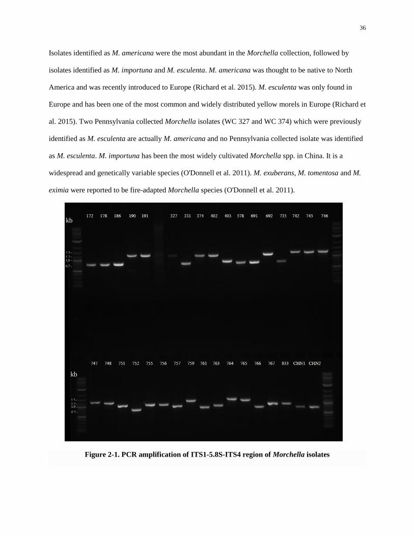
36
Isolates identified as M. americana were the most abundant in the Morchella collection, followed by
isolates identified as M. importuna and M. esculenta. M. americana was thought to be native to North
America and was recently introduced to Europe (Richard et al. 2015). M. esculenta was only found in
Europe and has been one of the most common and widely distributed yellow morels in Europe (Richard et
al. 2015). Two Pennsylvania collected Morchella isolates (WC 327 and WC 374) which were previously
identified as M. esculenta are actually M. americana and no Pennsylvania collected isolate was identified
as M. esculenta. M. importuna has been the most widely cultivated Morchella spp. in China. It is a
widespread and genetically variable species (O'Donnell et al. 2011). M. exuberans, M. tomentosa and M.
eximia were reported to be fire-adapted Morchella species (O'Donnell et al. 2011).
Figure 2-1. PCR amplification of ITS1-5.8S-ITS4 region of Morchella isolates
0.7 -
1,0 - 1,2 -
1,5 -
kb
1,5 -
1,2 -
1,5 1,0 -
1,5
0.7 -
1,5
kb
37
The first and last lane represent the 1 kb New England Biolabs DNA ladder. The numbers above the bands
indicate the PCR products were amplified from the corresponding DNA of Morchella isolates. For example, number
172 refers to Morchella isolate WC 172. CHN1, CHN2 refers to isolate China 1, isolate China 2 respectively.
List of Morchella isolates in the cultural collection belonging to the Esculenta Clade (O'Donnell et al.
2011) based on the results in this study.
WC 190 (NRRL # 20872) M. prava: ITS sequence data was 100% identical (1071/1071 bp) to
M. prava (GenBank accession JQ723099, Dewsbury et al. 2013). The same isolate was identified as M.
prava by O’Donnell et al. 2011. The culture was collected in West Kettle River, British Columbia,
Canada.
WC 191 M. prava: ITS sequence data was 100% identical (1071/1071 bp) to M. prava (GenBank
accession JQ723099, Dewsbury et al. 2013). The culture was collected in West Kettle River, British
Columbia, Canada. It was a duplicated of WC 190.
WC 327 M. americana: ITS sequence data was 100% identical (1030/1030 bp) to M.
esculentoides (GenBank accession JQ723067; Du et al. 2012). The name M. esculentoides was corrected
to M. americana by Richard et al. (2015). The culture was collected in Julian, PA, USA.
WC 374 M. americana: ITS sequence data was 99.8% identical (1038/1040 bp) to M. americana
(GenBank accession KM587949; Richard et al. 2015). The culture was collected in Fisherman’s Paradise,
PA, USA.
38
WC 402 M. americana: ITS sequence data was 100% identical (1030/1030 bp) to M.
esculentoides (GenBank accession JQ723067; Du et al. 2012). And the name M. esculentoides was
corrected to M. americana by Richard et al. (2015).
WC 692 (NRRL # 20877) M. americana: ITS sequence data was 99.8% identical (1040/1042
bp) to M. americana (GenBank accession KM587975; Richard et al. 2015). The same isolate was
identified as M. americana by O'Donnell et al. (2011). The culture was collected in UT, USA.
WC742 (NRRL # 20853) M. esculenta: ITS sequence data of WC742 had 99.8% identity
(1131/1133 bp) to M. esculenta (GenBank accession KM588023; Richard et al. 2015). According to
updated information from USDA, NRRL 20853 was identified as M. esculenta (Link) Persoon, and the
same as CBS 275.88 in CBS catalog. It was collected in Czechoslovakia in 1993.
WC 745 (NRRL # 20884 or 66866) Mel-12: ITS sequence data was 100% identical (1112/1112
bp) to Mel-12 (GenBank accession JQ723079.1; Du et al. 2012). The same isolate was identified as Mel-
1by O'Donnell et al. (2011). The culture was collected in Japan.
WC 746 (NRRL # 20885 or 66865) Mes-9: ITS sequence data was 99.9% identical (1141/1142
bp) to Mes-9 (GenBank accession KM485936.1; Chen et al. 2014, unpublished) and was 100% identical
(1112/1112 bp) to Mes-9 (GenBank accession JQ723094; Du et al. 2012). The same isolate was identified
as Mes-9 by O'Donnell et al. (2011).The culture was collected in Japan.
WC 747 (NRRL # 20886) Mes-9: ITS sequence data was 99.9% identical (1141/1142 bp) to
Mes-9 (GenBank accession KM485936.1; Chen et al. 2014, unpublished) and was100% identical
(1126/1126 bp) to Mes-9 (GenBank accession JQ723094; Du et al. 2012). The same isolate was identified
as Mes-9 by O'Donnell et al. (2011).
39
WC 748 (NRRL # 20914 or 66862) M. ulmaria: ITS sequence data was 100% identical
(1058/1058 bp) to M. ulmaria (GenBank accession JQ723091; Du et al. 2012). The identification of Du
was also confirmed by Richard et al. (2015).
WC 759 (NRRL # 22335) M. americana: ITS sequence data was 99.8% identical (1031/1034
bp) to M. americana (GenBank accession KM587975; Richard et al. 2015).
WC 765 (NRRL # 22476 or 66864) M. americana: ITS sequence data was 99.8% identical
(1032/1034 bp) to M. americana (GenBank accession KM587975; Richard et al. 2015). The same isolate
was identified as M. americana by O'Donnell et al. (2011).
WC 764 (NRRL # 22475) M. esculenta: ITS sequence data was 99.8% identical (1130/1131 bp)
to M. esculenta (GenBank accession KM588023; Richard et al. 2015).
List of Morchella isolates in the cultural collection belonging to the rufobrunnea Clade (O'Donnell et
al. 2011) based on the results in this study.
WC 833 M. rufobrunnea: ITS sequence data had 99.9% identity (842/843 bp) to M. rufobrunnea
(GenBank accession KM588017, Richard et al. 2015).
List of Morchella isolates in the cultural collection belonging to the Elata Clade (O'Donnell et al.
2011) based on the results in this study.
WC 172 M. importuna: ITS sequence data was 100% identical 672/672 bp) to M. importuna
(GenBank accession KM204675; Carris et al. unpublished).
40
WC 178 M. importuna: ITS sequence data was 100% identical (672/672 bp) to M. importuna
(GenBank accession KM204675; Carris et al. unpublished).
WC 186 (NRRL # 20869) M. brunnea: ITS sequence data had 99.6% identity (711/714 bp) to
M. brunnea (GenBank accession FJ594059, Sulej et al. unpublished) and 100% identity (672/672 bp) to
M. brunnea (GenBank accession KM204675, Carris et al. unpublished). The same isolate was identified
as M. brunnea by O'Donnell et al. (2011). The culture was collected in West Kettle River, British
Columbia, Canada.
WC 331 (NRRL # 20871) M. angusticeps: ITS sequence data was 100% identical (681/681 bp)
to M. angusticeps (GenBank accession GU551410; O'Donnell et al. 2011). The same isolate was
identified as M. angusticeps by O'Donnell et al. (2011). The culture was collected in State College, PA,
USA.
WC 403 (NRRL # 20826) M. punctipes: ITS sequence data was 99.6% identical (825/825 bp) to
M. tomentosa (GenBank accession KM588008; Richard et al. 2015). The same isolate was identified as
M. punctipes by O'Donnell et al. (2011).
WC578 M. eohespera: ITS sequence data was 100% identical (705/705 bp) to M. eohespera
(GenBank accession KT819369, O'Donnell et al. 2015).
WC 691 M. importuna: ITS sequence data was 99.8% identical (718/720 bp) to M. importuna
(GenBank accession MG121861.1, Li et al. 2017).
WC735 (NRRL # 25407) M. angusticeps: ITS sequence data was 100% identical (681/681 bp)
to M. angusticeps (GenBank accession JN085151; O'Donnell et al. unpublished) and 99.8% identicle to
41
M. angusticeps (GenBank accession GU551410; O'Donnell et al. unpublished). The same isolate was
identified as M. angusticeps by O'Donnell et al. (2011). The culture was collected in Kent, OH, USA.
WC 751 (NRRL # 22207) M. tridentina: ITS sequence data was 99.7% identical (878/881 bp) to
M. tridentina (GenBank accession KM587964; Richard et al. 2015). The same isolate was identified as
M. tridentina by O'Donnell et al. (2011).
WC 752 (NRRL # 22208) Mel-8: ITS sequence data was 99.8% identical (650/651 bp) to Mel-8
(GenBank accession MH127532, Rockefeller et al. et al. 2018). The same isolate was identified as Mel-8
by O'Donnell et al. (2011).
WC 755 (NRRL # 20851) M. tridentina: ITS sequence data was 100% identical (886/886 bp) to
M. tridentina (GenBank accession KT875083; Garibay-Orijel et al., unpublished) and 99.6% identical
(892/896 bp) to M. tridentina (GenBank accession KM587964, Richard et al. 2015). The same isolate was
identified as M. tridentina by O'Donnell et al. (2011).The culture was collected in BC, Canada.
WC 756 (NRRL # 22314) M. tomentosa: ITS sequence data was 99.8% identical (823/825 bp)
to M. tomentosa (GenBank accession KM587989; Richard et al. 2015).
WC761 (NRRL # 22447) Mel-23: ITS sequence data had 100% identity (713/713 bp) to Mel-23
(GenBank accession JQ618526.1, Du et al. 2012). The same isolate was identified as Mel-23 by
O'Donnell et al. (2011). This culture was collected in Finland.
WC 763 (NRRL # 20862) M. Punctipes: ITS sequence data was 100% identical (726/726 bp) to
M. Punctipes (GenBank accession JQ723026; Du et al. 2012). ITS sequence data had 99.6% identity
(764/767 bp) to M. Punctipes (GenBank accession KM588008, Richard et al. 2015).
42
WC 766 (NRRL # 22216) M. exuberans: ITS sequence data had 100% identity (718/718 bp) to
M. exuberans (GenBank accession KM587918, Richard et al. 2015). The same isolate was identified as
M. exuberans by O'Donnell et al. (2011).
WC 767 (NRRL # 22218) M. tomentosa; ITS sequence data was 100% identical (825/825 bp) to
M. tomentosa (GenBank accession KM587989; Richard et al. 2015). The same isolate was identified as
M. tomentosa by O'Donnell et al. (2011).
China 1 M. importuna: ITS sequence data was 100% identical (710/710 bp) to M. importuna
(GenBank accession JQ618780; Du et al. 2012). The culture was collected in China.
China 2 M. importuna: ITS sequence data was 99.7% identical (707/709 bp) to M. importuna
(GenBank accession JQ618776; Du et al. 2012). The culture was collected in China.
In summary, 33 Morchella isolates were identified based on molecular phylogenetic analysis of
ITS region from the original 38 isolates recovered from the Penn State University Morchella cultural
collection. WC 328 was dead and China 3 was contaminated, so no new identification was made for these
two isolates. WC 198 and WC 757 were identified as M. brunnea and M. popliphila by O'Donnell et al.
(2011). WC 743 was identified as M. semilibera by O'Donnell (unpublished). These three identifications
were adopted in this study. Finally, the identifications of 36 Morchella isolates were determined. Fifteen,
twenty and one isolate in the updated Morchella cultural collection belong to the Esculenta Clade, the
Elata Clade and the rufobrunnea Clade respectively (O'Donnell et al. 2011). The origination area of these
isolates include: Asia, Europe and North America. Results of this study should help inform Morchella
isolates selection for further research. For morel cultivation. Saprophytic Morchella species with high

43
potential for commercial cultivation are the ones that should be focused on. Based on this updated list of
Morchella cultural collection (Table 2-1), four Morchella isolates were selected for the subsequent
biological characteristic studies and morel indoor cropping experiment: WC 833 M. rufobrunnea, China 1
M. importuna, WC 374 M. americana and WC 766 M. exuberans. WC 833 M. rufobrunnea originated
from a commercial isolate cultivated in North America. A successful fruit body initiation of M.
rufobrunnea in laboratory-scale experiments was reported in 2010 (Masaphy 2010). The mature fruit
bodies of M. rufobrunnea have been successfully initiated in the Mushroom Research Center (MRC) at
Penn State. China 1 was originated from China and M. importuna is the most widely cultivated Morchella
species in China (W. Liu et al. 2017a). M. americana is native to North America and was thought to be
recently introduced to Europe (Richard et al. 2015). WC 374 was collected in PA, USA, so it might be a
potential Morchella isolate that can be cultivated outdoors in Pennsylvania. M. exuberans was reported to
be fire-associated species (Richard et al. 2015). Previous cultivation of M. rufobrunnea (Ower 1982) and
M. importuna (Du et al. 2015; W. Liu et al. 2017a) indicates these two species are saprophytic. M.
americana is often found under ashes dead and dying elm trees, but occasionally it can be found
associated with conifers (information provided by Midwest American Mycological Information
(MAMI)), which indicates this species is mostly likely saprophytic but can be mycorrhizal. M. exuberans
often fruits on conifer burning sites (Miller et al. 2017) suggesting that M. exuberans might be
mychorrhizal, but it fruit once the tree is killed. Lastly, other phylogenetic informative loci are needed in
order to better understand the important facets of morel cultivation, such as nutrient mode and mating
systems of different Morchella species.
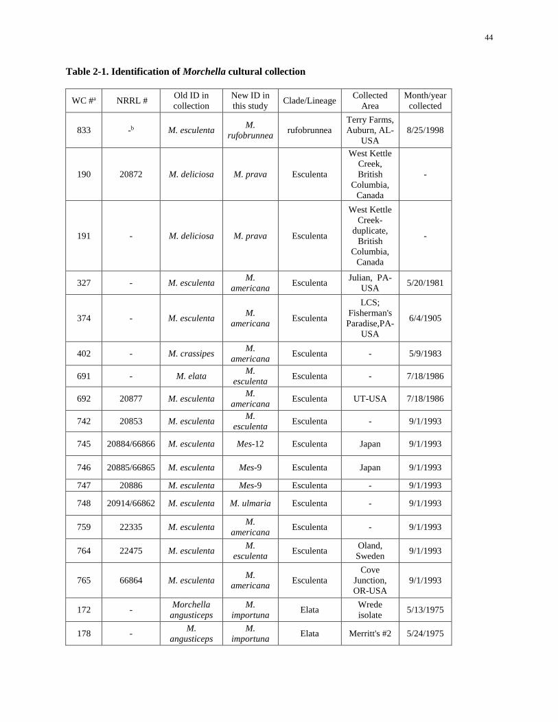
44
Table 2-1. Identification of Morchella cultural collection
WC #a NRRL # Old ID in
collection
New ID in
this study Clade/Lineage
Collected
Area
Month/year
collected
833 -b M. esculenta M.
rufobrunnea rufobrunnea
Terry Farms,
Auburn, AL-
USA
8/25/1998
190 20872 M. deliciosa M. prava Esculenta
West Kettle
Creek,
British
Columbia,
Canada
-
191 - M. deliciosa M. prava Esculenta
West Kettle
Creek-
duplicate,
British
Columbia,
Canada
-
327 - M. esculenta M.
americana Esculenta
Julian, PA-
USA 5/20/1981
374 - M. esculenta M.
americana Esculenta
LCS;
Fisherman's
Paradise,PA-
USA
6/4/1905
402 - M. crassipes M.
americana Esculenta - 5/9/1983
691 - M. elata M.
esculenta Esculenta - 7/18/1986
692 20877 M. esculenta M.
americana Esculenta UT-USA 7/18/1986
742 20853 M. esculenta M.
esculenta Esculenta - 9/1/1993
745 20884/66866 M. esculenta Mes-12 Esculenta Japan 9/1/1993
746 20885/66865 M. esculenta Mes-9 Esculenta Japan 9/1/1993
747 20886 M. esculenta Mes-9 Esculenta - 9/1/1993
748 20914/66862 M. esculenta M. ulmaria Esculenta - 9/1/1993
759 22335 M. esculenta M.
americana Esculenta - 9/1/1993
764 22475 M. esculenta M.
esculenta Esculenta
Oland,
Sweden 9/1/1993
765 66864 M. esculenta M.
americana Esculenta
Cove
Junction,
OR-USA
9/1/1993
172 - Morchella
angusticeps
M.
importuna Elata
Wrede
isolate 5/13/1975
178 - M.
angusticeps
M.
importuna Elata Merritt's #2 5/24/1975
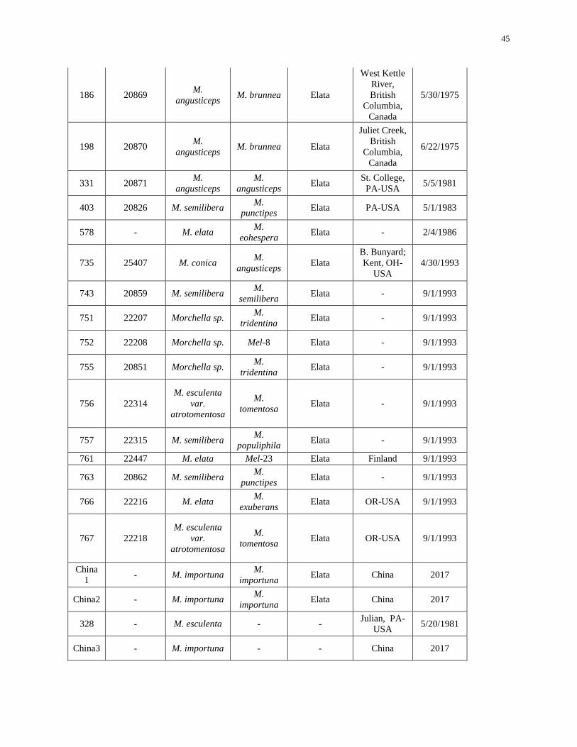
45
186 20869 M.
angusticeps M. brunnea Elata
West Kettle
River,
British
Columbia,
Canada
5/30/1975
198 20870 M.
angusticeps M. brunnea Elata
Juliet Creek,
British
Columbia,
Canada
6/22/1975
331 20871 M.
angusticeps
M.
angusticeps Elata
St. College,
PA-USA 5/5/1981
403 20826 M. semilibera M.
punctipes Elata PA-USA 5/1/1983
578 - M. elata M.
eohespera Elata - 2/4/1986
735 25407 M. conica M.
angusticeps Elata
B. Bunyard;
Kent, OH-
USA
4/30/1993
743 20859 M. semilibera M.
semilibera Elata - 9/1/1993
751 22207 Morchella sp. M.
tridentina Elata - 9/1/1993
752 22208 Morchella sp. Mel-8 Elata - 9/1/1993
755 20851 Morchella sp. M.
tridentina Elata - 9/1/1993
756 22314
M. esculenta
var.
atrotomentosa
M.
tomentosa Elata - 9/1/1993
757 22315 M. semilibera M.
populiphila Elata - 9/1/1993
761 22447 M. elata Mel-23 Elata Finland 9/1/1993
763 20862 M. semilibera M.
punctipes Elata - 9/1/1993
766 22216 M. elata M.
exuberans Elata OR-USA 9/1/1993
767 22218
M. esculenta
var.
atrotomentosa
M.
tomentosa Elata OR-USA 9/1/1993
China
1 - M. importuna
M.
importuna Elata China 2017
China2 - M. importuna M.
importuna Elata China 2017
328 - M. esculenta - - Julian, PA-
USA 5/20/1981
China3 - M. importuna - - China 2017

46
a. The reference number of Morchella isolates from the Penn State University Spawn Lab culture
collection.
b. (-): Not known.
47
Chapter 3
Studies of Biological Characteristics of Four Morchella isolates
Abstract
Morels are one of the most prized edible fungi and morel cultivation has been catching
researchers’ interests. Mycelial growth is the first stage in the morel life cycle and it is believed that
pseudosclerotia formation is one of the most important phases during the morel life cycle. Good mycelial
growth and satisfactory pseudosclerotia formation are thought to be required for successful morel
cultivation. However, how environmental factors effect mycelial growth and pseudosclerotia formation of
many Morchella species is unknown. Even though there have been some reports accessing the effects of
environmental parameters on mycelial growth or pseudosclerotia formation of Morchella, there is little
known about the specific requirements due to the ambiguity of species in many of these studies. In this
study, the effects of pH, light intensity and temperature on mycelial growth and pseudosclerotia formation
of four Morchella isolates selected from Penn State Morchella cultural collection (Morchella
rufobrunne, M. importuna, M. americana and M. exuberans) were investigated. Conditions resulting in
both the fastest mycelial growth and most pseudosclerotia production in vitro were pH 6, darkness (0
lux), and 20 to 25C for M. rufobrunnea, pH 5 to 6, darkness (0 lux), and 20 to 25C for M. importuna.
An Initial pH of the medium from 5 to 8 did not significantly affect pseudosclerotia formation of M.
rufobrunnea and M. importuna. M. americana, and M. exuberans did not produce any pseudosclerotium
under any of the test conditions in this study. The optimum environmental conditions for mycelial growth
of M. americana was found to be pH 5 to 7, darkness (0 lux) to low light intensity (400 lux), and 20C.
Mycelia of M. exuberans achieved the fastest mycelial growth in media with a pH value of 5, 25 to 30C,
and light intensity had no influence on mycelial growth. This work will promote a better understanding of
48
the effect of environmental factors on mycelial growth and pseudosclerotia formation of Morchella,
which are necessary for the improvement of morel cultivation.
Introduction
Morchella species are highly prized, edible fungi belonging to Ascomycota, Pezizomycetes,
Pezizales, Morchellaceae, and Morchella. Due to their short fruiting season, favorable flavor and value in
the food and medical industry, mycologists have been attempting to cultivate morels for decades, and it
has proven very difficult. Ower et al. (1986) claimed successful indoor cultivation of M. rufobrunnae in
1982 and subsequently applied for three patents (US Patents 4594809, 4757640, and 4866878) for morel
cultivation. These studies revealed the optimal temperature, humidity and other essential factors during
different stages, as well as leading to detailed studies on the life cycle of morels. Mycelial growth is the
first stage in the life cycle of Morchella species. Moreover, the essential role of establishment of
pseudosclerotium was emphasized and has become the central focal point in morel fruiting. Many fungi
are able to produce durable structures, such as pseudosclerotium to facilitate dispersion or survival. The
pseudosclerotium of morels is a compact structure composed of large cells with thick walls which allows
the fungus to survive under unfavorable environmental conditions (Volk and Leonard 1990). Formation
of pseudosclerotium of morels is also a strategy the fungus uses for nutrient storage (Buscot, 1989). Amir
et al. (1992, 1993, 1995) studied the effect of medium composition and water potential on pseudosclerotia
formation on M. esculenta using the split plate method. Volk and Leonard (1989) used a jar method to
study the effect of a variety of conditions on pseudosclerotia formation. They pointed out that complex
medium worked better for pseudosclerotia formation and a smaller sized container achieved better
biological efficiency. He also stated that illumination was a limiting factor for the development of
pseudosclerotia. However, during the process of making spawn, there is a significant difference between
the light side and the dark side of the spawn container if it is not rotated on a regular basis (W. Liu et al.
2017a). On the side that was occasionally exposed to light, large amounts of pseudosclerotia were
49
produced. On the contrary, production of pseudosclerotia on the dark side was poor, indicating that light
plays a role in pseudosclerotia formation.
Good mycelia growth and satisfactory pseudosclerotia formation is required for successful morel
cultivation. The first step to successfully cultivate morels is to be able to culture them under conditions
that maximum their mycelial growth and pseudosclerotia formation. Environmental factors may have an
effect on the mycelial growth and sclerotia formation of Morchella. Even though there have been some
reports accessing the effects of environmental parameters on the mycelial growth or pseudosclerotia
formation on Morchella (S. poplarLi et al. 1998; Zhao et al. 1998; X. Liu et al. 2004; Kalyoncu et al.
2009), many of them did not provide the name of the species. Besides, the identification of Morchella in
previous studies may be inaccurate due to the use of homonyms and synonyms (Richard et al. 2015) and
poorly annotated sequences in GenBank (Du et al. 2012b). Therefore, previous studies provide little
reference value for culture and morel cultivation. The aim of this study was to quantify the effect of pH,
light intensity and temperature on the mycelial growth and pseudosclerotia formation of four Morchella
isolates: Morchella importuna, M. rufobrunnea, M. americana and M. exuberans. Here we hypothesized
that: 1) For each Morchella isolate, there is a pH range and a temperature range that it can grow, but there
is an optimal pH and temperature that results in the fastest mycelial growth and most pseudosclerotia
production. 2) The effect of light on Morchella varies by species and 3) Not all tested Morchella isolates
can produce pesudosclerotia under tested conditions.
Material and Methods
Morchella isolates
Four Morchella isolates; Morchella importuna, M. rufobrunnea, M. americana and M. exuberans
were selected from the Penn State culture collection to be utilized for this study.
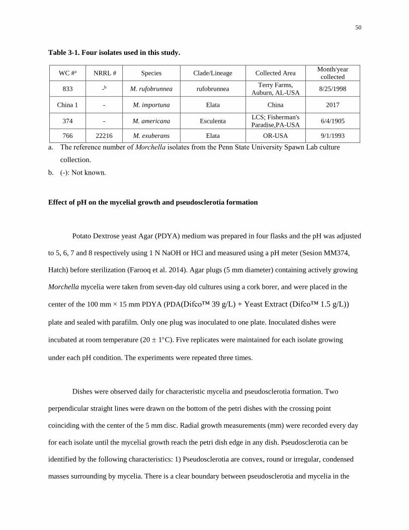
50
Table 3-1. Four isolates used in this study.
WC #a NRRL # Species Clade/Lineage Collected Area Month/year
collected
833 -b M. rufobrunnea rufobrunnea Terry Farms,
Auburn, AL-USA 8/25/1998
China 1 - M. importuna Elata China 2017
374 - M. americana Esculenta LCS; Fisherman's
Paradise,PA-USA 6/4/1905
766 22216 M. exuberans Elata OR-USA 9/1/1993
a. The reference number of Morchella isolates from the Penn State University Spawn Lab culture
collection.
b. (-): Not known.
Effect of pH on the mycelial growth and pseudosclerotia formation
Potato Dextrose yeast Agar (PDYA) medium was prepared in four flasks and the pH was adjusted
to 5, 6, 7 and 8 respectively using 1 N NaOH or HCl and measured using a pH meter (Sesion MM374,
Hatch) before sterilization (Farooq et al. 2014). Agar plugs (5 mm diameter) containing actively growing
Morchella mycelia were taken from seven-day old cultures using a cork borer, and were placed in the
center of the 100 mm × 15 mm PDYA (PDA(Difco™ 39 g/L) + Yeast Extract (Difco™ 1.5 g/L))
plate and sealed with parafilm. Only one plug was inoculated to one plate. Inoculated dishes were
incubated at room temperature (20 ± 1C). Five replicates were maintained for each isolate growing
under each pH condition. The experiments were repeated three times.
Dishes were observed daily for characteristic mycelia and pseudosclerotia formation. Two
perpendicular straight lines were drawn on the bottom of the petri dishes with the crossing point
coinciding with the center of the 5 mm disc. Radial growth measurements (mm) were recorded every day
for each isolate until the mycelial growth reach the petri dish edge in any dish. Pseudosclerotia can be
identified by the following characteristics: 1) Pseudosclerotia are convex, round or irregular, condensed
masses surrounding by mycelia. There is a clear boundary between pseudosclerotia and mycelia in the
51
plate. 2) Pseudosclerotia usually form near the inoculation cite and the plate edges. 3) Pseudosclerotia are
white at first and then turn yellow to brown. 4) The sites of the agar where pseudosclerotia form are
usually brown pigmented. Mycelial color, colony shape, distribution of mycelia and, if present, the color,
shape, area and distribution of pseudosclerotia in each dish was recorded for 20 days post inoculation
(dpi). The area of pseudosclerotia in each dish was measured using Image J software
(https://imagej.nih.gov/ij/), an open source image processing system.
Effect of light on the mycelial growth and pseudosclerotia formation
PDYA medium was prepared in flasks and the pH of the media was adjusted to 6 by using 1 N
NaOH and measured with a pH meter (Sesion MM374, Hatch) before sterilization. The culture
preparation was made following the previously described method. The effect of light on mycelial growth
and pseudosclerotia formation of four Morchella isolates were studied by growing the fungal cultures
under the following light conditions: 0 lux, 400 lux continuous light and 2000 lux continuous light
provided by LED lights (Lightning Ever®, 12V, 6000K). Light intensity was measured by a Dr. Meter®
digital light meter. All plates were incubated at 25C. The temperature of the chambers was monitored
using a thermometer and recorded every 2 days to confirm that the light source did not increase the
temperature in the chambers. Five replicates were maintained for each isolate growing under each light
condition. The experiments were repeated three times. The measurement of mycelial growth and
pseudosclerotia formation was made following the previously described method.
Effect of temperature on the mycelial growth and pseudosclerotia formation
PDYA medium was prepared in flasks and the pH of the media was adjusted to 6 by using 1 N
NaOH. The culture preparation was made following the previously described method. The effect of
52
temperature on mycelial growth and pseudosclerotia formation was studied by growing the fungal
cultures at 20C, 25C and 30C in incubators without light. The temperatures of the chambers were
checked using a hand-held thermometer and recorded every 2 days. The experiments were repeated three
times. The measurement of mycelial growth and pseudosclerotia formation was made following the
previously described method.
Morphological characteristics of pseudosclerotia development
Agar plugs (about 5 mm diameter) containing actively growing mycelia of M. rufobrunnea or M.
importuna (these were the only 2 species that formed pseudosclerotia under the conditions being tested)
were transferred from seven-day old cultures using a cork borer and were placed in the center of the Petri
dishes containing PDYA media with initial pH of 6. Colonies of M. rufobrunnea and M. importuna were
observed using a digital microscope (Celestron 44308, US) (× 40). The pseudosclerotia were taken out
from the media with a sterilized hood and chopped into small strips on a slide, covered with a glass
coverslip and observed with a Nikon Eclipse E400 microscope (Nikon, Minatoku, Tokyo, Japan) (× 400
and × 1000) to characterize the pseudosclerotia.
Data analysis
The experimental design for the growth characteristics were completely randomized factorial
experiments (four isolates and four pH media, four isolates and three light conditions, four isolates and
three temperature), with 15 replicates (5 plates × 3 replicates) per treatment. The non-parametric
analysis of variance (Kruskal-Wallis test, p<0.05) was carried out on the data for mycelial radial growth
and pseudosclerotia formation. A multiple comparison test (Pairwise Wilcox test, p<0.05) was carried out
on data using R software (R.app GUI 1.70).
53
Results and Discussion
Effect of pH on mycelial growth and pseudosclerotia formation
In this study, all Morchella species grew over the pH range of 5 to 8. For M. rufobrunnea, the
complete colonization of at least one dish was observed at 4 dpi. At 4 dpi, significant differences were
only observed between treatments pH5 and pH6 for mycelium radial growth (p<0.05) and the fastest
growth was observed on pH6 (Figure 3-1Aa). There was no significant difference in pseudosclerotia area
among all the pH levels tested (Table 3-2) (p>0.05).
M. importuna generally colonized the entire dish in 3 to 4 days. PDYA medium with an initial pH
of 5 and 6 resulted in the fastest mycelial growth among all the pH levels tested. A pH of 7 and 8
suppressed the mycelial growth of M. importuna compared to a pH of 5 or 6. At 4 dpi, the growth
differences between pH5, pH6 and pH7, pH8 were statistically significant (p<0.0001) (Figure 3-1Ab).
The colony morphological characteristics for the different pH levels were similar (Figure 3-2b). There
was no significant difference in pseudosclerotia area among all the pH levels tested (Table 3-2) (p>0.05).
M. americana grew well on PDYA with a pH of 5, 6 or 7, but it grew significantly slower at pH 8
(p<0.0001) (Figure 3-1Ac). The colony morphological characteristics were not different at any of the pH
levels tested (Figure 3-2c). Mycelia was first white to light yellow and turned to orange around 3 dpi. No
pseudosclerotium was observed at 20 dpi (Table 3-2).
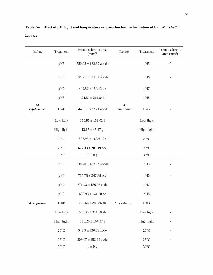
54
Table 3-2. Effect of pH, light and temperature on pseudosclerotia formation of four Morchella
isolates
Isolate Treatment Pseudosclerotia area
(mm2)a Isolate Treatment
Pseudosclerotia
area (mm2)
M.
rufobrunnea
pH5 550.05 ± 183.97 abcde
M.
americana
pH5 -b
pH6 651.91 ± 385.87 abcde pH6 -
pH7 442.52 ± 150.13 de pH7 -
pH8 424.64 ± 212.84 e pH8 -
Dark 544.61 ± 232.21 abcde Dark -
Low light 160.95 ± 153.02 f Low light -
High light 13.15 ± 45.47 g High light -
20C 508.93 ± 167.6 bde 20C -
25C 627.38 ± 206.19 bde 25C -
30C 0 ± 0 g 30C -
M. importuna
pH5 538.98 ± 162.34 abcde
M. exuberans
pH5 -
pH6 715.78 ± 247.38 acd pH6 -
pH7 671.93 ± 186.03 acde pH7 -
pH8 626.93 ± 144.50 ac pH8 -
Dark 727.66 ± 288.86 ab Dark -
Low light 690.38 ± 314.58 ab Low light -
High light 113.26 ± 164.37 f High light -
20C 543.5 ± 220.83 abde 20C -
25C 509.67 ± 192.45 abde 25C -
30C 0 ± 0 g 30C -

55
a. Data shown are the means of 15 replicates from three independent experiments ± SD. Values followed by
the same letter are not significantly different according by the Pairwise Wilcox test (p = 0.05).
Pseudosclerotia area was measured at 20 dpi.
b. (-): no pseudosclerotia produced
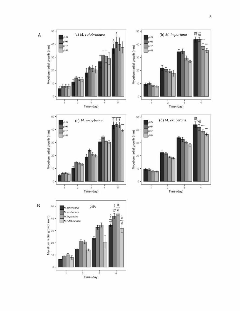
56
A
B
(a) M. rufobrunnea (b) M. importuna
(c) M. americana (d) M. exuberans
pH6

57
Figure 3-1. Effect of pH on mycelium radial growth of four Morchella isolates
A. Effect of pH on mycelium radial growth of (a) M. rufobrunnea, (b) M. importuna, (c) M. americana and (d) M.
exuberans. Data shown are the means of 15 replicates from three independent experiments ± standard deviation
(SD). Statistical analysis was conducted on the data of the last measuring day for each isolate. Bars with “*”,
“**”, “***” at the top without numbers represent values that are significantly different from all other pH values
with a p value less than 0.05, 0.001, 0.0001 respectively. Bars with “*”, “**”, “***” at the top with numbers
represent values that are only significantly different from pH values listed below the asterisk(s) with a p value
less than 0.05, 0.001, 0.0001 respectively. Numbers “5”, “6”, “7” and “8” represent “pH5”, “pH6”, “pH7”, and
“pH8” respectively.
B. Mycelium radial growth of M. rufobrunnea, M. importuna, M. americana and M. exuberans at pH6. Data
shown are the means of 15 replicates from three independent experiments ± standard deviation (SD). Bars with
“*”, “***” at the top with letter(s) represent values that are only significantly different from species listed below
the asterisk(s) with a p value less than 0.05, 0.0001 respectively. Letters “r”, “I”, “a”, and “e” represent “M.
rufobrunnea”, “M. importuna”, “M. americana” and “M. exuberans” respectively.
58
M. exuberans generally completely colonized the dish at 4 dpi. Significant differences were
observed among growth rates for all pH conditions at 4 dpi (pH5 and pH6, p<0.001; others, P<0.0001).
The optimal pH for mycelial growth was pH 5 and the growth decreased as the pH of the medium
increased. Mycelia was white and aggregated, condensed at the edge of growth, but was relatively sparse
in the center. Aged growth rings were formed as the development continued. The mycelia under pH 5 and
6 appeared more condensed than mycelia under pH 7 and 8 (Figure 3-2d). No pseudosclerotium was
observed at 20 dpi (Table 3-2).
Species comparisons
At 4 dpi, all four isolates were significantly different from each other when grown on medium
with pH 6 in terms of mycelial growth. M. importuna had the fastest growth rate, followed by M.
exuberans, M. americana and M. rufobrunnea (Figure 3-1B). There was a significant difference between
M. importuna and M. exuberans in terms of mycelial growth at 4 dpi (P<0.05). Mycelial radial growth of
M. importuna and M. exuberans was significant faster than M. americana and M. rufobrunnea
(P<0.0001).
X. Liu et al. (2004) reported the optimal pH for mycelial growth and pseudosclerotia formation
for two Morchella spp. (species names were not indicated in the paper) was between 5 and 7, indicating
the tested Morchella isolates favors slightly acid to neutral environment. J. He et al. (2004) concluded
that the optimal pH for mycelial growth of five Morchella isolates (species names were not indicated in
the paper) was 6.5. However, Chen and Guo (2015) reported that M. esculenta grew well at pH values
between 6.4 to 8.7, which was neutral to slightly alkaline, and the optimal pH was 7.7. Q. Liu et al.
(2018) reported mycelia of M. importuna grew well at pH values between 4 to 9, and pseudosclerotia
weight was not significant different for pH 5, 6, 7, and 8, which is consistent with our results. It seems
that most Morchella species can grow at pH values between 5 to 8. However, the optimal pH differs
59
according to specific Morchella isolates. Chen and Guo (2007) pointed out that wild morels are likely to
be found in the location where there is a lot of limestone and plant ashes. Nevertheless, many morels are
collected from the soil with a pH value less than 7.0 in North America (Information provided by The
Ohio Mushroom Society), implying that a neutral to alkaline environment is not necessary for the growth
of some Morchella species. Our results demonstrate that all tested Morchella isolates could grow between
pH 5 to 8 and the optimal pH for mycelial growth was pH 6, 5 to 6, 5 to 7, and 5 for M. rufobrunnea, M.
importuna, M. americana, and M. exuberans respectively, suggesting these four tested Morchella isolates
prefer slightly acidity to neutral media. However, pseudosclerotia growth was not significantly different
for pH values from 5 to 8 for M. rufobrunnea and M. importuna. This may be because even though
mycelial growth was slower at higher pH values, mycelia were still able to colonize the entire plate 1 to 3
days later and nutrient in the media was depleted triggering the formation of pseudosclerotia The timing
of sclerotia formation was delayed, but the total area of sclertoia were not affected. For morel cultivation,
the optimal soil pH differs depending on cultivated isolates. Slightly acidity to neutral soil seems to be a
good option for all four tested Morchella isolates in this study. Even though our results indicated that the
optimal pH for the mycelial growth of M.exuberans was 5, M. exuberans still grew well at pH 6, 7 and
even 8. If multiple Morchella isolates in this study are considered for future cultivation, it will be
convenient to adjust the soil pH to 6 to 7. pH can strongly influencenutrient availability (Abubakar et al.
2013). However, it is more complicated in soil since soil pH may affect the biotic factors, such as biomass
composition of fungi and bacteria (Rousk et al. 2009), the effect of which on Morchella mycelial growth
is unknown. Thus, the results obtained in vitro may not accurately predict similar results in morel
cultivation. More studies are needed to investigate the effect of pH on mycelial growth of Morchella in
soil.
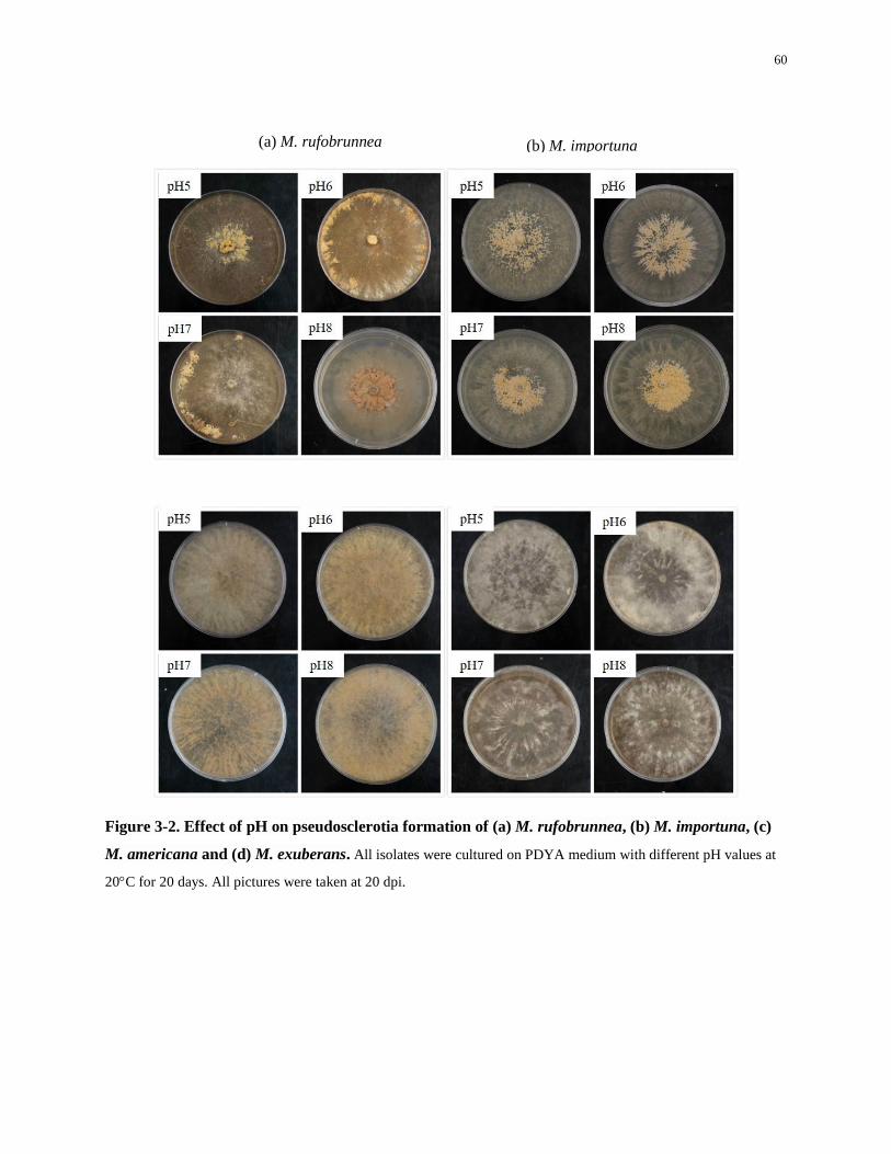
60
Figure 3-2. Effect of pH on pseudosclerotia formation of (a) M. rufobrunnea, (b) M. importuna, (c)
M. americana and (d) M. exuberans. All isolates were cultured on PDYA medium with different pH values at
20C for 20 days. All pictures were taken at 20 dpi.
(a) M. rufobrunnea (b) M. importuna
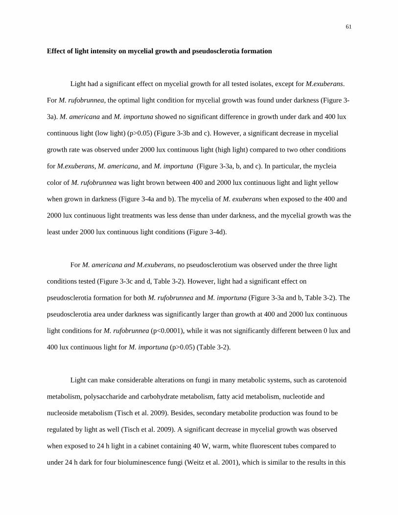
61
Effect of light intensity on mycelial growth and pseudosclerotia formation
Light had a significant effect on mycelial growth for all tested isolates, except for M.exuberans.
For M. rufobrunnea, the optimal light condition for mycelial growth was found under darkness (Figure 3-
3a). M. americana and M. importuna showed no significant difference in growth under dark and 400 lux
continuous light (low light) (p>0.05) (Figure 3-3b and c). However, a significant decrease in mycelial
growth rate was observed under 2000 lux continuous light (high light) compared to two other conditions
for M.exuberans, M. americana, and M. importuna (Figure 3-3a, b, and c). In particular, the mycleia
color of M. rufobrunnea was light brown between 400 and 2000 lux continuous light and light yellow
when grown in darkness (Figure 3-4a and b). The mycelia of M. exuberans when exposed to the 400 and
2000 lux continuous light treatments was less dense than under darkness, and the mycelial growth was the
least under 2000 lux continuous light conditions (Figure 3-4d).
For M. americana and M.exuberans, no pseudosclerotium was observed under the three light
conditions tested (Figure 3-3c and d, Table 3-2). However, light had a significant effect on
pseudosclerotia formation for both M. rufobrunnea and M. importuna (Figure 3-3a and b, Table 3-2). The
pseudosclerotia area under darkness was significantly larger than growth at 400 and 2000 lux continuous
light conditions for M. rufobrunnea (p<0.0001), while it was not significantly different between 0 lux and
400 lux continuous light for M. importuna (p>0.05) (Table 3-2).
Light can make considerable alterations on fungi in many metabolic systems, such as carotenoid
metabolism, polysaccharide and carbohydrate metabolism, fatty acid metabolism, nucleotide and
nucleoside metabolism (Tisch et al. 2009). Besides, secondary metabolite production was found to be
regulated by light as well (Tisch et al. 2009). A significant decrease in mycelial growth was observed
when exposed to 24 h light in a cabinet containing 40 W, warm, white fluorescent tubes compared to
under 24 h dark for four bioluminescence fungi (Weitz et al. 2001), which is similar to the results in this
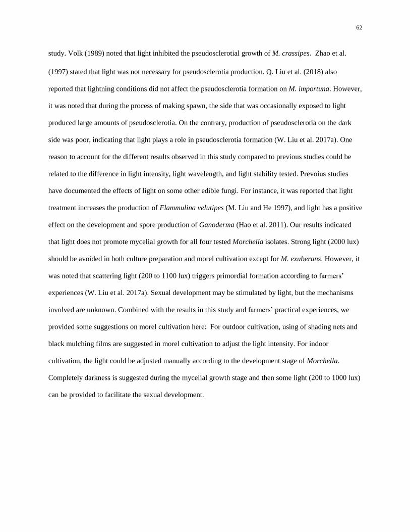
62
study. Volk (1989) noted that light inhibited the pseudosclerotial growth of M. crassipes. Zhao et al.
(1997) stated that light was not necessary for pseudosclerotia production. Q. Liu et al. (2018) also
reported that lightning conditions did not affect the pseudosclerotia formation on M. importuna. However,
it was noted that during the process of making spawn, the side that was occasionally exposed to light
produced large amounts of pseudosclerotia. On the contrary, production of pseudosclerotia on the dark
side was poor, indicating that light plays a role in pseudosclerotia formation (W. Liu et al. 2017a). One
reason to account for the different results observed in this study compared to previous studies could be
related to the difference in light intensity, light wavelength, and light stability tested. Prevoius studies
have documented the effects of light on some other edible fungi. For instance, it was reported that light
treatment increases the production of Flammulina velutipes (M. Liu and He 1997), and light has a positive
effect on the development and spore production of Ganoderma (Hao et al. 2011). Our results indicated
that light does not promote mycelial growth for all four tested Morchella isolates. Strong light (2000 lux)
should be avoided in both culture preparation and morel cultivation except for M. exuberans. However, it
was noted that scattering light (200 to 1100 lux) triggers primordial formation according to farmers’
experiences (W. Liu et al. 2017a). Sexual development may be stimulated by light, but the mechanisms
involved are unknown. Combined with the results in this study and farmers’ practical experiences, we
provided some suggestions on morel cultivation here: For outdoor cultivation, using of shading nets and
black mulching films are suggested in morel cultivation to adjust the light intensity. For indoor
cultivation, the light could be adjusted manually according to the development stage of Morchella.
Completely darkness is suggested during the mycelial growth stage and then some light (200 to 1000 lux)
can be provided to facilitate the sexual development.
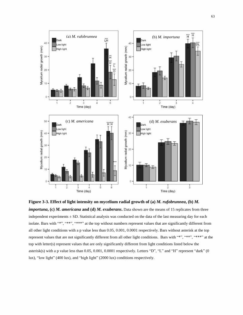
63
Figure 3-3. Effect of light intensity on mycelium radial growth of (a) M. rufobrunnea, (b) M.
importuna, (c) M. americana and (d) M. exuberans. Data shown are the means of 15 replicates from three
independent experiments ± SD. Statistical analysis was conducted on the data of the last measuring day for each
isolate. Bars with “*”, “**”, “***” at the top without numbers represent values that are significantly different from
all other light conditions with a p value less than 0.05, 0.001, 0.0001 respectively. Bars without asterisk at the top
represent values that are not significantly different from all other light conditions. Bars with “*”, “**”, “***” at the
top with letter(s) represent values that are only significantly different from light conditions listed below the
asterisk(s) with a p value less than 0.05, 0.001, 0.0001 respectively. Letters “D”, “L” and “H” represent “dark” (0
lux), “low light” (400 lux), and “high light” (2000 lux) conditions respectively.
(d) M. exuberans
(b) M. importuna
(c) M. americana
(a) M. rufobrunnea
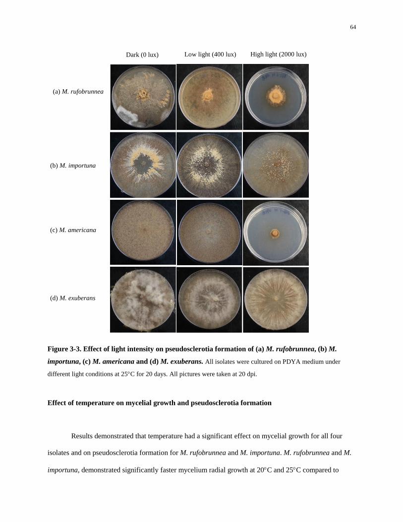
64
Figure 3-3. Effect of light intensity on pseudosclerotia formation of (a) M. rufobrunnea, (b) M.
importuna, (c) M. americana and (d) M. exuberans. All isolates were cultured on PDYA medium under
different light conditions at 25C for 20 days. All pictures were taken at 20 dpi.
Effect of temperature on mycelial growth and pseudosclerotia formation
Results demonstrated that temperature had a significant effect on mycelial growth for all four
isolates and on pseudosclerotia formation for M. rufobrunnea and M. importuna. M. rufobrunnea and M.
importuna, demonstrated significantly faster mycelium radial growth at 20C and 25C compared to
Dark (0 lux) Low light (400 lux)
(a) M. rufobrunnea
High light (2000 lux)
(b) M. importuna
(c) M. americana
(d) M. exuberans
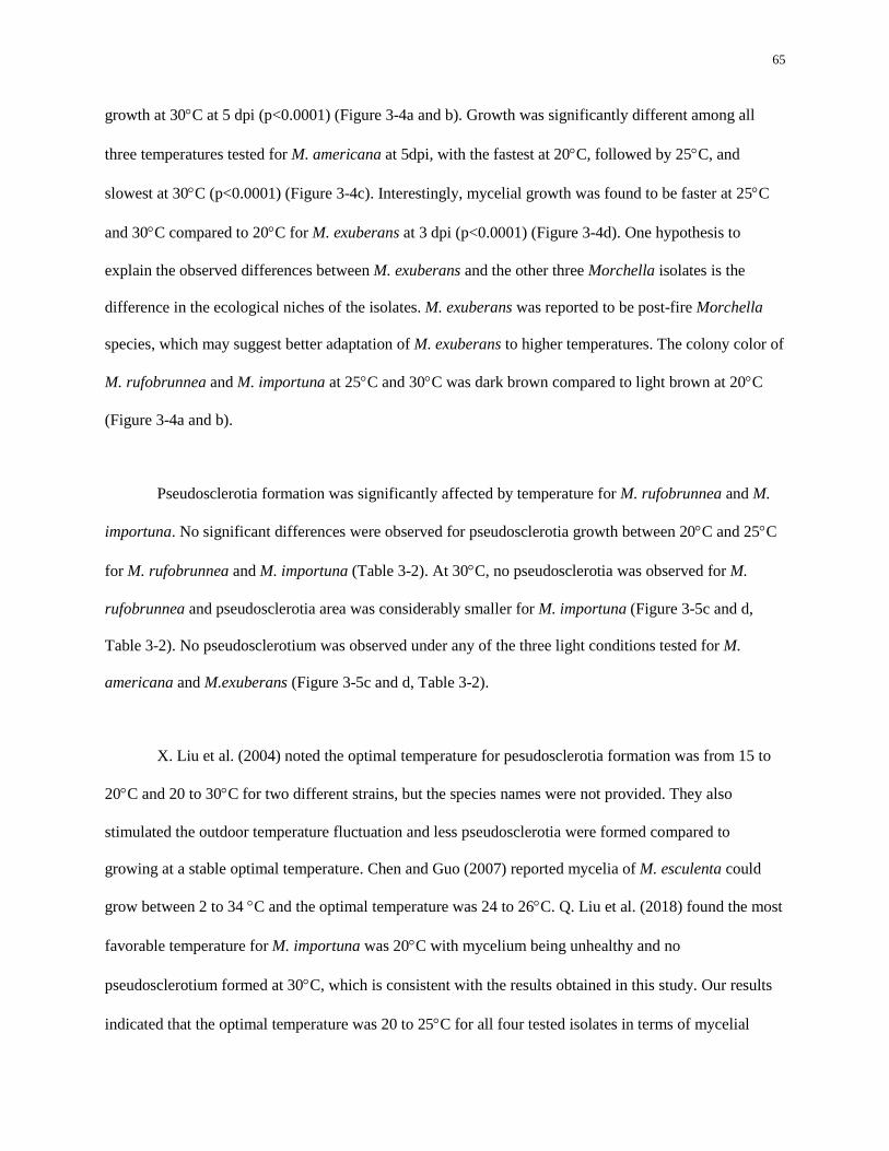
65
growth at 30C at 5 dpi (p<0.0001) (Figure 3-4a and b). Growth was significantly different among all
three temperatures tested for M. americana at 5dpi, with the fastest at 20C, followed by 25C, and
slowest at 30C (p<0.0001) (Figure 3-4c). Interestingly, mycelial growth was found to be faster at 25C
and 30C compared to 20C for M. exuberans at 3 dpi (p<0.0001) (Figure 3-4d). One hypothesis to
explain the observed differences between M. exuberans and the other three Morchella isolates is the
difference in the ecological niches of the isolates. M. exuberans was reported to be post-fire Morchella
species, which may suggest better adaptation of M. exuberans to higher temperatures. The colony color of
M. rufobrunnea and M. importuna at 25C and 30C was dark brown compared to light brown at 20C
(Figure 3-4a and b).
Pseudosclerotia formation was significantly affected by temperature for M. rufobrunnea and M.
importuna. No significant differences were observed for pseudosclerotia growth between 20C and 25C
for M. rufobrunnea and M. importuna (Table 3-2). At 30C, no pseudosclerotia was observed for M.
rufobrunnea and pseudosclerotia area was considerably smaller for M. importuna (Figure 3-5c and d,
Table 3-2). No pseudosclerotium was observed under any of the three light conditions tested for M.
americana and M.exuberans (Figure 3-5c and d, Table 3-2).
X. Liu et al. (2004) noted the optimal temperature for pesudosclerotia formation was from 15 to
20C and 20 to 30C for two different strains, but the species names were not provided. They also
stimulated the outdoor temperature fluctuation and less pseudosclerotia were formed compared to
growing at a stable optimal temperature. Chen and Guo (2007) reported mycelia of M. esculenta could
grow between 2 to 34 C and the optimal temperature was 24 to 26C. Q. Liu et al. (2018) found the most
favorable temperature for M. importuna was 20C with mycelium being unhealthy and no
pseudosclerotium formed at 30C, which is consistent with the results obtained in this study. Our results
indicated that the optimal temperature was 20 to 25C for all four tested isolates in terms of mycelial
66
growth except for M. exuberans, which grew better between 25 to 30C. Even though the optimal
temperature for mycelial growth of those previously mentioned Morchella species was generally between
20 to 26C, in morel production, the optimal spawning temperature is when the maximum temperature is
20C when other microorganisms in the soil are in a state of semi-dormancy (W. Liu et al. 2017a).
According to farmers’ experiences, a satisfactory yield of mature fruit bodies will be more likely be
obtained when the air temperature is below 4C for at least one month before the primordial formation
period and the temperature difference is at least 10C during the day, thereby facilitating primordial
formations in morel cultivation (personal communication). Nevertheless, additional data is required to
determine if a low temperature period is required for fruit body initiation. For culture and spawn
preparation, conditions with optimal temperatures was recommended by growers. For outdoor cultivation,
growers suggest spawning when the air temperature starts to drop and the highest air temperature is below
20C. For indoor cultivation, air temperature can be adjusted manually to below 20C. However, more
studies are needed to investigate how air temperature fluctuation affects mycelial growth, pseudosclerotia
formation and primordial formation both in lab and in the field.
67
Figure 3-4. Effect of temperature on mycelium radial growth of (a) M. rufobrunnea, (b) M.
importuna, (c) M. americana and (d) M. exuberans. Data shown are the means of 15 replicates from three
independent experiments ± SD. Statistical analysis was conducted on the data of the last measuring day for each
isolate. Bars with “*”, “**”, “***” at the top without numbers represent values that are significantly different from
all other temperature treatments with a p value less than 0.05, 0.001, 0.0001 respectively. Bars with “*”, “**”, “***”
at the top with letter(s) represent values that are only significantly different from temperature treatments listed below
the asterisk(s) with a p value less than 0.05, 0.001, 0.0001 respectively. Numbers “1”, “2” and “3” represent “20C”,
“25C”, and “30C” treatments respectively.
(a) M. rufobrunnea (b) M. importuna
(c) M. americana (d) M. exuberans
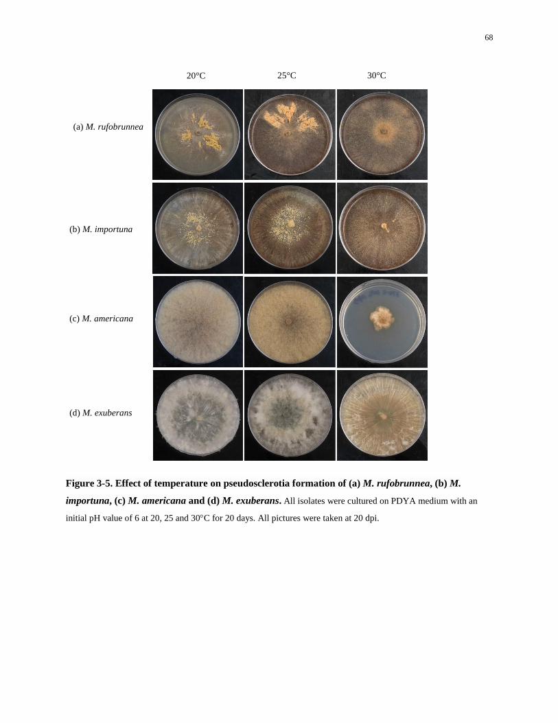
68
Figure 3-5. Effect of temperature on pseudosclerotia formation of (a) M. rufobrunnea, (b) M.
importuna, (c) M. americana and (d) M. exuberans. All isolates were cultured on PDYA medium with an
initial pH value of 6 at 20, 25 and 30C for 20 days. All pictures were taken at 20 dpi.
20°C 25°C
(a) M. rufobrunnea
30°C
(b) M. importuna
(c) M. americana
(d) M. exuberans
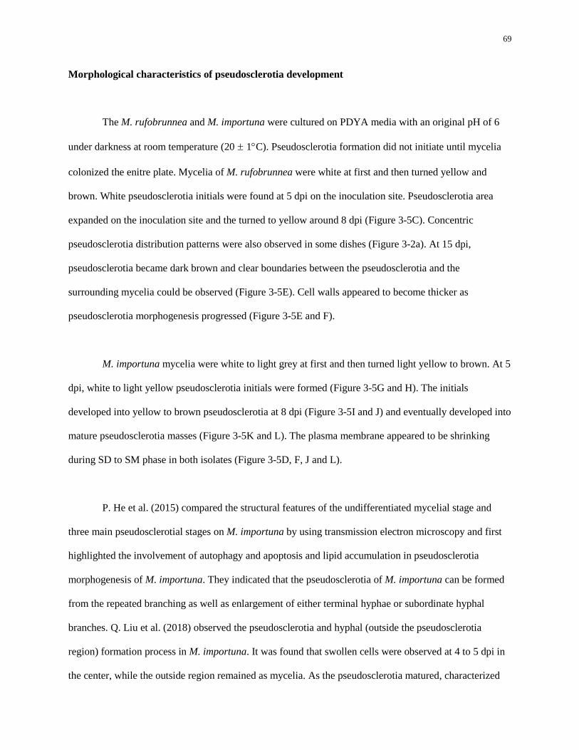
69
Morphological characteristics of pseudosclerotia development
The M. rufobrunnea and M. importuna were cultured on PDYA media with an original pH of 6
under darkness at room temperature (20 ± 1C). Pseudosclerotia formation did not initiate until mycelia
colonized the enitre plate. Mycelia of M. rufobrunnea were white at first and then turned yellow and
brown. White pseudosclerotia initials were found at 5 dpi on the inoculation site. Pseudosclerotia area
expanded on the inoculation site and the turned to yellow around 8 dpi (Figure 3-5C). Concentric
pseudosclerotia distribution patterns were also observed in some dishes (Figure 3-2a). At 15 dpi,
pseudosclerotia became dark brown and clear boundaries between the pseudosclerotia and the
surrounding mycelia could be observed (Figure 3-5E). Cell walls appeared to become thicker as
pseudosclerotia morphogenesis progressed (Figure 3-5E and F).
M. importuna mycelia were white to light grey at first and then turned light yellow to brown. At 5
dpi, white to light yellow pseudosclerotia initials were formed (Figure 3-5G and H). The initials
developed into yellow to brown pseudosclerotia at 8 dpi (Figure 3-5I and J) and eventually developed into
mature pseudosclerotia masses (Figure 3-5K and L). The plasma membrane appeared to be shrinking
during SD to SM phase in both isolates (Figure 3-5D, F, J and L).
P. He et al. (2015) compared the structural features of the undifferentiated mycelial stage and
three main pseudosclerotial stages on M. importuna by using transmission electron microscopy and first
highlighted the involvement of autophagy and apoptosis and lipid accumulation in pseudosclerotia
morphogenesis of M. importuna. They indicated that the pseudosclerotia of M. importuna can be formed
from the repeated branching as well as enlargement of either terminal hyphae or subordinate hyphal
branches. Q. Liu et al. (2018) observed the pseudosclerotia and hyphal (outside the pseudosclerotia
region) formation process in M. importuna. It was found that swollen cells were observed at 4 to 5 dpi in
the center, while the outside region remained as mycelia. As the pseudosclerotia matured, characterized
70
by cell aggregation and dark brown pigmentation, senescent and aging cells were found outside the
pseudosclerotia region. In this study, M. rufobrunnea and M. importuna displayed similar patterns.
Pseudosclerotia initials usually developed at the center of the plate and then expanded cells aggregated
with a white color then turned yellow to brown. For M. importuna, small white to yellow aggregates were
formed first and aggregated to a larger pseudosclerotium in the center later. However, for M.
rufobrunnea, pseudosclerotia were generally formed in the center most of the time, but occasionally some
also formed near the plate edges. The detailed pseudosclerotia mophogenesis of M. rufobrunnea and how
environmental conditions effect pseudosclerotia morphogenesis of both M. importuna and M.
rufobrunnea require further study.
c.
5 dpi 8 dpi
(a) M. rufobrunnea
15 dpi
(b) M. importuna
A C E
B D F
G
H
I
J
K
L

71
Figure 3-5. Partial enlarged detail of pseudosclerotia (row 1 and 3) and the microscopic
characteristic of pseudosclerotia (row 2 and 4) of (a) M. rufobrunnea and (b) M. importuna at 5 dpi,
8 dpi, and 15 dpi. At 5 dpi, mycelia were interwoven (Figure 3-5A, B, G, H, arrow). At 8 dpi, the
mycelial masses turned yellow (Figure 3-5C and I, arrow) and cell walls were thickened (Figure 3-5D and
J, arrow). Orange to brown pseudosclerotia with obvious boundaries with surrounding mycelia (Figure 3-
5E and K, arrow) were at 15 dpi. Note the thickened well wall and shrunken plasma membrane (Figure 3-
5 F and L, arrow). Scale bars = 20 μm.
Conclusion
This study suggested that environmental factors, including pH, light and temperature, play an
important role in mycelial growth and pseudosclerotia formation on M. rufobrunnea, M. importuna, M.
americana, and M. exuberans. All four isolates tested in this study were able to grow in the initial media
with a pH of 5 to 8. Slightly acidic to neutral initial agar media is most favorable for mycelial growth,
while alkaline conditions of the medium suppressed the mycelial growth. However, initial pH of the
medium did not significantly affect pseudosclerotia formation of M. rufobrunnea and M. importuna. M.
americana, and M. exuberans did not produce any pseudosclerotium under any of the test conditions in
this study. Light intensity had different effects on different Morchella isolates. High light intensity (2000
lux) significantly suppressed mycelial growth, except for M. exuberans, and differences in light intensity
also affected pseudosclerotia production for M. rufobrunnea and M. importuna. Darkness (0 lux) and low
light intensity (400 lux) were found to be the most favorable conditions for mycelial growth for M.
rufobrunnea, but pseudosclerotia formation was significantly suppressed under low light conditions (400
lux) compared to darkness (0 lux). M. importuna grew equally well and formed pseudosclerotia with no
significant difference under low light intensity (400 lux) and darkness (0 lux). The temperature preference
for mycelial growth of M. rufobrunnea, M. importuna. M. americana, and M. exuberans was found to be
20 to 25C, 20 to 25C, 25C, and 25 to 30C respectively. It is worth noting that M. exuberans grew well

72
at 30C, at which the mycelial growth of three other Morchella isolates was highly suppressed. One
possible explanation could be due to the special ecological niches of M. exuberans, which was reported to
be a post-fire Morchella species. Moreover, the pseudosclerotia morphogenesis of M. rufobrunnea and M.
importuna was investigated and light microscopic observations represented complex pseudosclerotia
formation patterns. Good mycelia growth and satisfactory pseudosclerotia formation is required for
successful morel cultivation. The results of this research will provide some guidance for future culture
preparation, spawn production and indoor and outdoor morel cultivation.
73
Chapter 4
Indoor cultivation of four Morchella isolates – Cropping Experiment
Abstract
Morels (Morchella) are commercially important edible mushrooms well known for their desirable
flavor and medicinal value. The outdoor cultivation of Morchella species has been achieved in China in
recent years. Even though Morchella species are diverse in the wild, species that can be cultivated are
limited. In this study, we discuss two morel indoor croppping experiments at the Mushroom Research
Center (MRC) at The Pennylvania State University. In experiment 1, we selected five substrates and two
nutrient bags for use in indoor M. rufobrunnea cultivation. M. rufobrunnea was the first reported
cultivated Morchella species in North America. In experiment 2, we selected four Morchella species:
Morchella rufobrunnea, M. importuna, M. americana and M. exuberans, based on previous research and
relevant information for indoor cultivation experiments. Nutrient sources are essential for mycelial
growth and fruiting body development. We wanted to test available and effective carbon sources that
contribute to more efficient fruiting body development and higher yields, and compare results between
different treatments and between different species. These results can provide data that will be used in the
design of future research. In experiment 2, two substrate carbon sources (sawdust and straw) were used as
soil amendments for use in indoor Morchella cultivation. Difference in fruiting body initiation and yield
were assessed based on the two different substrate carbon treatments and for four different Morchella
species in an indoor cultivation system at the MRC. Mature fruit bodies were successfully initiated in
experiment 1 but not in experiment 2, even though many primordia were formed on one Morchella isolate
in experiment 2. Factors that may contribute to the different results in two experiments are discussed.
74
Introduction
Morels (Morchella) are well known for their savory flavor and multiple bioactive compounds.
Morels are often collected in the wild and their fruiting season is very short (Pilz. et al. 2007). Thus, the
cultivation of morels has been attracting many mushroom enthusiasts’ attention. The first morel fruit body
initiation was reported by Ower (1982). He studied morel indoor cultivation on M. rufobrunnea and
applied for several patents. However, due to the lack of basic biological knowledge of morels and some
unknown reasons, indoor commercial production of morels was not successful (Ower et al. 1986, 1988,
1989; Pilz. et al. 2007). Recently, outdoor cultivation of morels has been developing rapidly in China.
The application of exogenous nutrient bags has made a great promotion on commercialized morel outdoor
cultivation (W. Liu et al. 2017a). Thus far, four Morchella spp. (M. importuna, M. sextelata, M.
septimelata, and M. esculenta) have been cultivated commercially in China and M. importuna accounts
for more than 95% of the morel cultivating area (W. Liu et al. 2017a). Nevertheless, morel cultivation has
been proven to be a challenging task due to the lack of knowledge on basic biology about morels, such as
their complex life cycle, strain polarity and ploidy, mating systems, trophic modes and aging, which has
caused notable economic losses (W. Liu et al. 2018). Compared to outdoor cultivation, which is largely
restricted by natural conditions, such as nutrient content of the soil and weather conditions, indoor
cultivation seems to be a better option to consistently cultivate morels commercially. Even though there
have been studies on morel indoor cultivation since Ower (D. Zhu and He, 2007; Y. Liu et al. 2013), the
feasibility of these indoor cultivation techniques is skeptical. There is an urgent need to develop a
successful morel indoor cultivation process. Whether the outdoor morel cultivation process is feasible for
indoor cultivation is unknown. Even though Morchella species are diverse in the wild, species that can be
cultivated may be limited based on nutritional requirements. Nutrient sources are essential for mycelial
growth and fruiting body development. In this study, two morel indoor cultivation experiments were
75
conducted in the MRC. In experiment 1, five substrates and two nutrient bags were selected for use in
indoor M. rufobrunnea cultivation. In experiment 2, four Morchella species: Morchella rufobrunnea, M.
importuna, M. americana and M. exuberans, two substrates and one nutrient bag were selected based on
previous research and relevant information for indoor cultivation experiments. Both experiments
stimulated the curently used ourdoor cultivation techniques. The effect of substrates with different carbon
sources and nutrient bags with different sources on conidia production, primordial formation and mature
fruit body initiation of differernt Morchella species were examined in two experiments. Factors that may
have contributed to the different results observed in the two experiments are also discussed.
Experiment 1
Material and Methods
Morchella isolates
WC 833 M. rufobrunnea obtained from the Penn State Mushroom Spawn Lab was utilized for
this study.
Substrate and nutrient bags preparation
Substrates with five different treatments were used in this study: (1) soil + sawdust (wet, pH
buffered with limestone) (8% by weight); (2) soil + wheat straw (chopped and wet); (3) soil + wheat
straw (chopped and wet) more straws; (4) soil + fresh mushroom compost (8% by weight); (5) soil + aged
mushroom compost (8% by weight). Soil pH was adjusted to 7 with limes and then was mixed with
straws or wood shavings. Each tub was filled to 50 lb. Each treatment consisted of six replicates. There
were totally 30 tubs (1 isolates × 5 treatment × 6 replicates).
76
Two different nutrients bag formulas were used for each isolate in this study: (1) wheat and
sawdust at a ratio of 3:2 (m/m); (2) corn and sawdust at a ratio of 3:2 (m/m). The moisture was adjusted to
40% using tap water. Each plastic autoclavable bag (12 cm × 24 cm × 5 cm) was filled with 350 g of the
mixture and sealed with a twist tie. The bags were autoclaved for 35 minutes at 121C before using them.
Two nutrient bags with the same formula were used for each tub. For each substrate treatment, six
replicates were divided to two group corresponding to nutrient bags with two formulas.
Spawn production
Morchella mycelia grown on PDYA medium were used to inoculate rye grains. Two plugs of
agar containing active growing Morchella mycelia were inoculated onto sterilized rye grains in a 250 ml
flask incubated at room temperature for 7 days. Rye grains were then transferred from the 250 ml flasks
to sterilized rye grain spawn in 1000 ml flasks and incubated at room temperature for 35 days. Flasks
were shaken every two days. Spawn was ready to use after the Morchella mycelia completely colonized
the grains.
Rye grains used for spawn production were produced using the following formulas: For 250 ml
flasks, 50 ml beaker level full of rye grain, 1/2 tsp. Calcium Carbonate, powder (lime), 1/4 tsp. Calcium
Sulfate (gypsum) and 60 ml warm water were mixed together and then autoclaved for 35 minutes at
121C. For 1000 ml flasks, 250 ml beaker level full of rye grain, 2 tsp. Calcium Carbonate, powder
(lime), 1 tsp. Calcium Sulfate (gypsum) and 220 ml warm water were mixed together and then autoclaved
for 35 minutes at 121C.
Morchella indoor cultivation experiment set up

77
Thirty tubs were randomly assigned to four three-level shelves in one room in MRC (Table 4-1).
For each tub, 77g spawn were mixed with the soil substrate. A sterilized knife was used to cut a 3 to 4 cm
opening on the bags and two sterilized nutrient bags were placed on the soil surface for each tab with the
opening attached to the soil surface at 45 days post inoculation (dpi). The tubs were watered heavily at 55
dpi using tap water and then nutrient bags were removed at 90 dpi. The used bags were steamed to kill the
microbes before disposal.
The procedure of morel indoor cultivation and relative parameters are listed in Table 4-2. When
the size of fruit body stops increasing, the stem of fruit body was cut and harvested. Each fruit body was
measured for length and weight. Harvested fruit bodies were dried at room temperature.
Table 4-1. The room map of M. rufobrunnea indoor cultivation (experiment 1) in MRC
Higher level Middle level Lower level
Asile
Door
Lower level Middle level Higher level
2a Wb
30c
1 W
18
1 C
17
1 C
16
4 C
5
1 W
15
1 C
14
5 C
13
4 C
8
1 W
12
5 C
11
5 C
10
3 W
25
3 C
26
2 C
17
5 W
9
4 C
8
4 W
7
3 W
22
3 C
23
2 C
24
5 W
6
4 C
5
4 W
4
3 W
19
3 C
20
2 C
21
5 W
3
4 C
2
4 W
1
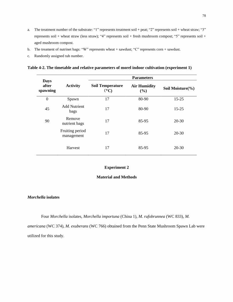
78
a. The treatment number of the substrate: “1” represents treatment soil + peat; “2” represents soil + wheat straw; “3”
represents soil + wheat straw (less straw); “4” represents soil + fresh mushroom compost; “5” represents soil +
aged mushroom compost.
b. The treament of nutrinet bags: “W” represents wheat + sawdust; “C” represents corn + sawdust.
c. Randomly assigned tub number.
Table 4-2. The timetable and relative parameters of morel indoor cultivation (experiment 1)
Days
after
spawning
Activity
Parameters
Soil Temperature
(C)
Air Humidity
(%) Soil Moisture(%)
0 Spawn 17 80-90 15-25
45 Add Nutrient
bags 17 80-90 15-25
90 Remove
nutrient bags 17 85-95 20-30
Fruiting period
management 17 85-95 20-30
Harvest 17 85-95 20-30
Experiment 2
Material and Methods
Morchella isolates
Four Morchella isolates, Morchella importuna (China 1), M. rufobrunnea (WC 833), M.
americana (WC 374), M. exuberans (WC 766) obtained from the Penn State Mushroom Spawn Lab were
utilized for this study.
79
Substrate and nutrient bags preparation
Two soil substrates with different carbon sources, oak wood shavings (sawdust) and straw, were
used for all isolates. Soil was supplemented with chopped wheat straw (4% by dry weight) or
supplemented with oak wood shavings (8% by weight). Soil pH was adjusted to 7 with crushed limestone
(calcium carbonate) and then the soil was mixed with straws or wood shavings and 35 lb of each mix was
filled into a tub to a 4 inch layer of perlite (to allow for soil drainage and raise the soil level to near the
surface of the tubs). Each treatment consisted of six replicates with a total of 48 tubs (4 isolates × 2
carbon sources × 6 replicates). The soil and substrate samples were sent to the Penn State University
Agricultural Analytical Services Laboratory to analyze for chemical composition (Table A-3, A-4, A-5,
A-6, A-7).
Wheat grain and oak sawdust were mixed together at a ratio of wheat to sawdust of 3:2 (m/m).
The moisture was adjusted to 40% using tap water. Each plastic autoclavable bag (12 cm × 24 cm × 5
cm) was filled with 350 g of the wheat/sawdust mixture and sealed with a twist tie. The bags were
autoclaved for 35 minutes at 121C before using them. Two nutrient bags were used for each tub. The
nutrient bag sample was sent to the Agricultural Analytical Services Laboratory to analyze for chemical
composition (Table A-8).
Spawn production
Morchella mycelia grown on PDYA medium were used to inoculate rye grains. Two plugs of
agar containing active growing Morchella mycelia were inoculated onto sterilized rye grains in a 250 ml
flask incubated at room temperature for 7 days. Rye grains were then transferred from the 250 ml flasks
to sterilized rye grain spawn in 1000 ml flasks and incubated at room temperature for 35 days. Flasks
80
were shaken every two days. Spawn were ready to use after the Morchella mycelia completely colonized
the grains.
Rye grains used for spawn production were produced using the following recipes: For 250 ml
flasks, 50 ml beaker level full of rye grain, 1/2 tsp. Calcium Carbonate, powder (lime), 1/4 tsp. Calcium
Sulfate (gypsum) and 60 ml warm water were mixed together and then autoclaved for 35 minutes at
121C. For 1000 ml flasks, 250 ml beaker level full of rye grain, 2 tsp. Calcium Carbonate, powder
(lime), 1 tsp. Calcium Sulfate (gypsum) and 220 ml warm water were mixed together and then autoclaved
for 35 minutes at 121C.
Morchella indoor cultivation experiment set up
Fourty-eight tubs were randomly assigned to four three-level shelves in one room in MRC
(Table 4-3). For each tub, 77g spawn were mixed with the soil substrate. Two sterilized nutrient bags
were placed on the soil surface for each tab at 15 days post inoculation (dpi). A sterilized knife was used
to cut a 3 to 4 cm opening on the bags and two sterilized nutrient bags were placed on the soil surface for
each tab with the opening attached to the soil surface at 15 dpi. The tubs were watered heavily at 55 dpi
using tap water and then nutrient bags were removed at 65 dpi. The used bags were steamed sterilized
before disposal.
The procedure of morel indoor cultivation and relative environmental growth parameters are
listed in Table 4-4. When the size of the fruiting bodies stopped increasing, the stem of fruiting bodies
will be cut and harvested. Each fruit body will be measured for length and weight.
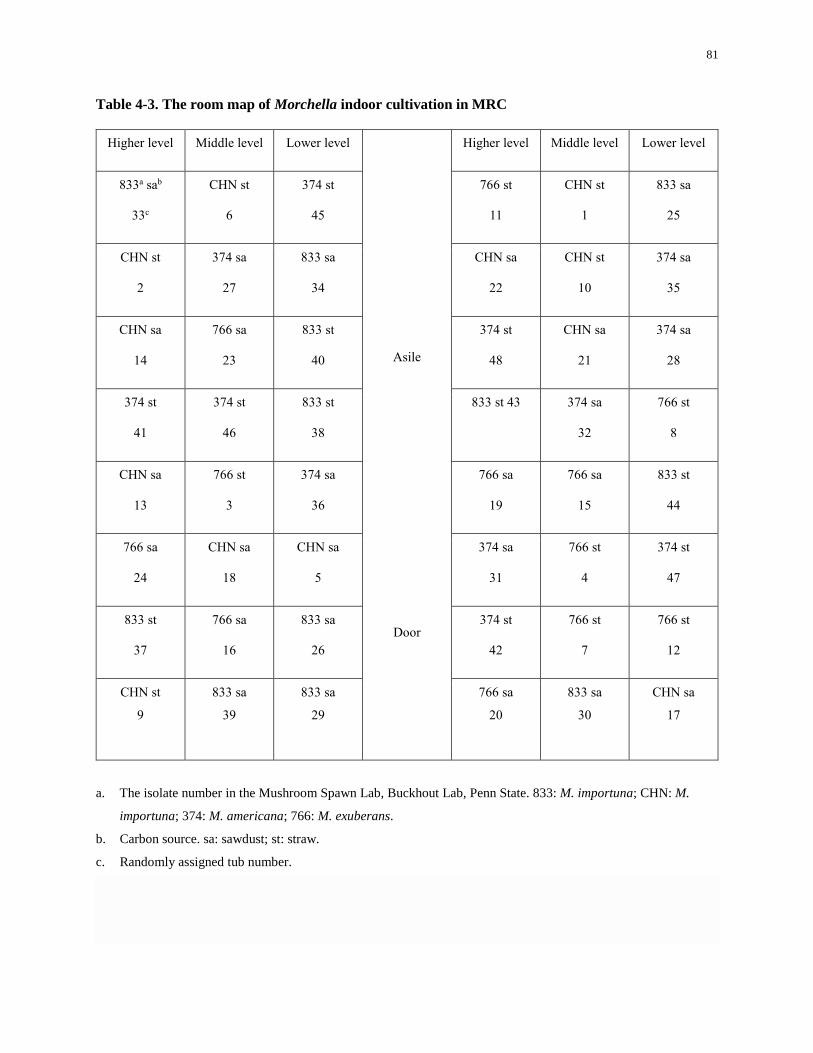
81
Table 4-3. The room map of Morchella indoor cultivation in MRC
Higher level Middle level Lower level
Asile
Door
Higher level Middle level Lower level
833a sab
33c
CHN st
6
374 st
45
766 st
11
CHN st
1
833 sa
25
CHN st
2
374 sa
27
833 sa
34
CHN sa
22
CHN st
10
374 sa
35
CHN sa
14
766 sa
23
833 st
40
374 st
48
CHN sa
21
374 sa
28
374 st
41
374 st
46
833 st
38
833 st 43 374 sa
32
766 st
8
CHN sa
13
766 st
3
374 sa
36
766 sa
19
766 sa
15
833 st
44
766 sa
24
CHN sa
18
CHN sa
5
374 sa
31
766 st
4
374 st
47
833 st
37
766 sa
16
833 sa
26
374 st
42
766 st
7
766 st
12
CHN st
9
833 sa
39
833 sa
29
766 sa
20
833 sa
30
CHN sa
17
a. The isolate number in the Mushroom Spawn Lab, Buckhout Lab, Penn State. 833: M. importuna; CHN: M.
importuna; 374: M. americana; 766: M. exuberans.
b. Carbon source. sa: sawdust; st: straw.
c. Randomly assigned tub number.
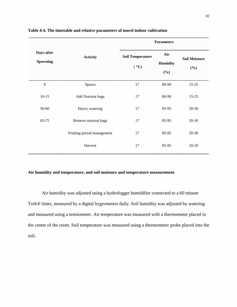
82
Table 4-4. The timetable and relative parameters of morel indoor cultivation
Days after
Spawning Activity
Parameters
Soil Temperature
(C)
Air
Humidity
(%)
Soil Moisture
(%)
0 Spawn 17 80-90 15-25
10-15 Add Nutrient bags 17 80-90 15-25
50-60 Heavy watering 17 85-95 20-30
65-75 Remove nutrient bags 17 85-95 20-30
Fruiting period management 17 85-95 20-30
Harvest 17 85-95 20-30
Air humidity and temperature, and soil moisture and temperature measurement
Air humidity was adjusted using a hydrofogger humidifier connected to a 60 minute
Tork® timer, measured by a digital hygrometers daily. Soil humidity was adjusted by watering
and measured using a tensiometer. Air temperature was measured with a thermometer placed in
the center of the room. Soil temperature was measured using a thermometer probe placed into the
soil.
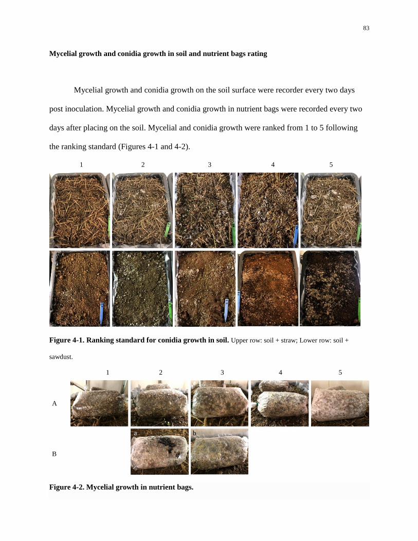
83
Mycelial growth and conidia growth in soil and nutrient bags rating
Mycelial growth and conidia growth on the soil surface were recorder every two days
post inoculation. Mycelial growth and conidia growth in nutrient bags were recorded every two
days after placing on the soil. Mycelial and conidia growth were ranked from 1 to 5 following
the ranking standard (Figures 4-1 and 4-2).
1 2 3 4 5
Figure 4-1. Ranking standard for conidia growth in soil. Upper row: soil + straw; Lower row: soil +
sawdust.
1 2 3 4 5
Figure 4-2. Mycelial growth in nutrient bags.
A
B
a b
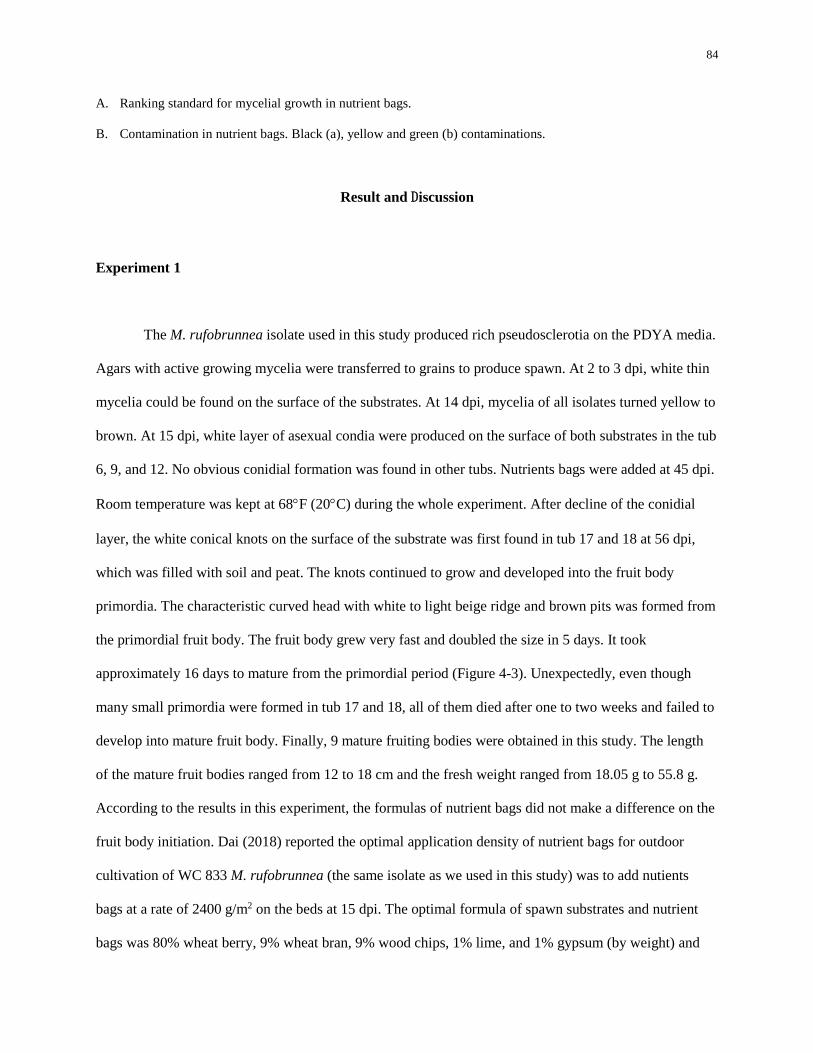
84
A. Ranking standard for mycelial growth in nutrient bags.
B. Contamination in nutrient bags. Black (a), yellow and green (b) contaminations.
Result and Discussion
Experiment 1
The M. rufobrunnea isolate used in this study produced rich pseudosclerotia on the PDYA media.
Agars with active growing mycelia were transferred to grains to produce spawn. At 2 to 3 dpi, white thin
mycelia could be found on the surface of the substrates. At 14 dpi, mycelia of all isolates turned yellow to
brown. At 15 dpi, white layer of asexual condia were produced on the surface of both substrates in the tub
6, 9, and 12. No obvious conidial formation was found in other tubs. Nutrients bags were added at 45 dpi.
Room temperature was kept at 68F (20C) during the whole experiment. After decline of the conidial
layer, the white conical knots on the surface of the substrate was first found in tub 17 and 18 at 56 dpi,
which was filled with soil and peat. The knots continued to grow and developed into the fruit body
primordia. The characteristic curved head with white to light beige ridge and brown pits was formed from
the primordial fruit body. The fruit body grew very fast and doubled the size in 5 days. It took
approximately 16 days to mature from the primordial period (Figure 4-3). Unexpectedly, even though
many small primordia were formed in tub 17 and 18, all of them died after one to two weeks and failed to
develop into mature fruit body. Finally, 9 mature fruiting bodies were obtained in this study. The length
of the mature fruit bodies ranged from 12 to 18 cm and the fresh weight ranged from 18.05 g to 55.8 g.
According to the results in this experiment, the formulas of nutrient bags did not make a difference on the
fruit body initiation. Dai (2018) reported the optimal application density of nutrient bags for outdoor
cultivation of WC 833 M. rufobrunnea (the same isolate as we used in this study) was to add nutients
bags at a rate of 2400 g/m2 on the beds at 15 dpi. The optimal formula of spawn substrates and nutrient
bags was 80% wheat berry, 9% wheat bran, 9% wood chips, 1% lime, and 1% gypsum (by weight) and

85
the moisture was adjusted to 60% (by weight). Wheat and corn worked equally well in terms of sclerotia
production in nutirient bags. However, the moisture in the bags was easily lost because the density of the
filled bags was too low (too much air space). Moreover, Morchella cound not consume the nutrients in
the bags very fast, so more contamination was found in the bags filled with corn. Such problems were not
observed in our study, possibly because the growing room used in our experiment was cleaner than the
greenhouse where Dai conducted his study. In morel production, growers generally use what is available
and cheap when preparing nutrient bags (personal communication). Morchella fruit bodies were
successfully initiated under substrate treatment 1 (soil + sawdust), 2 and 3 (soil + wheat straw). Three,
five and two were harvested from treatment 1, 2 and 3 respectively. No mature fruit body was harvested
from treatment 4 and 5. The color of the fruit bodies varied from light beige to dark brown due to
different environmental conditions, such as light intensity, humidity and temperature. Even though the
variation in the environmental factors were controlled to minimize varibaility, due to the location of the
humidifier and the position of the tubs on the shelves, there was some difference in the light condition,
soil moisture and temperature. The tubs located at the lower level of the shelves were more likely to be
flooded, while those placed at the higher level were drier. Tub 10, 7 and 4 were became saturated due to
excessive free water from the humidifier. As a result, no primordial or mature fruit bodies were formed in
these tubs. Tubs 15, 18, 19 and 22 were drier compared to other tubs and no fruit bodies were formed
from these tubs as well.
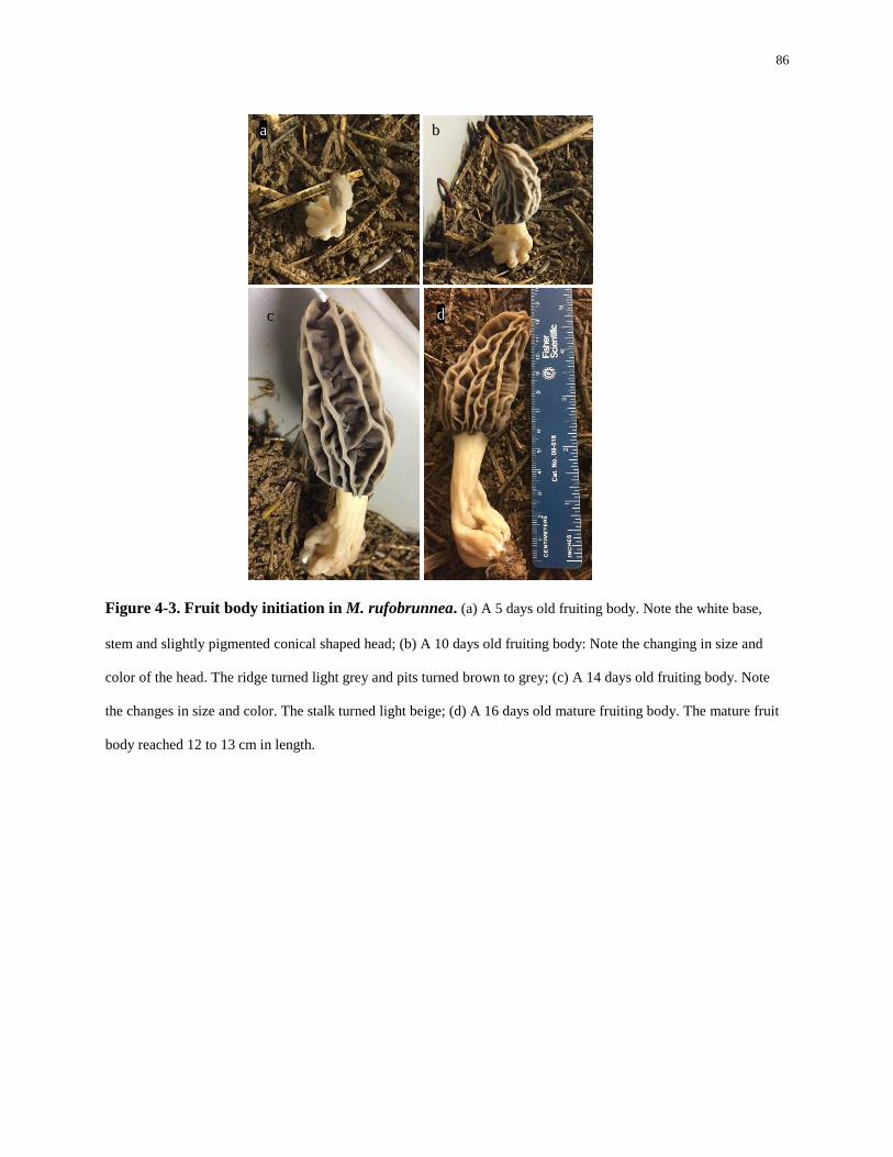
86
Figure 4-3. Fruit body initiation in M. rufobrunnea. (a) A 5 days old fruiting body. Note the white base,
stem and slightly pigmented conical shaped head; (b) A 10 days old fruiting body: Note the changing in size and
color of the head. The ridge turned light grey and pits turned brown to grey; (c) A 14 days old fruiting body. Note
the changes in size and color. The stalk turned light beige; (d) A 16 days old mature fruiting body. The mature fruit
body reached 12 to 13 cm in length.
a b
d c
c
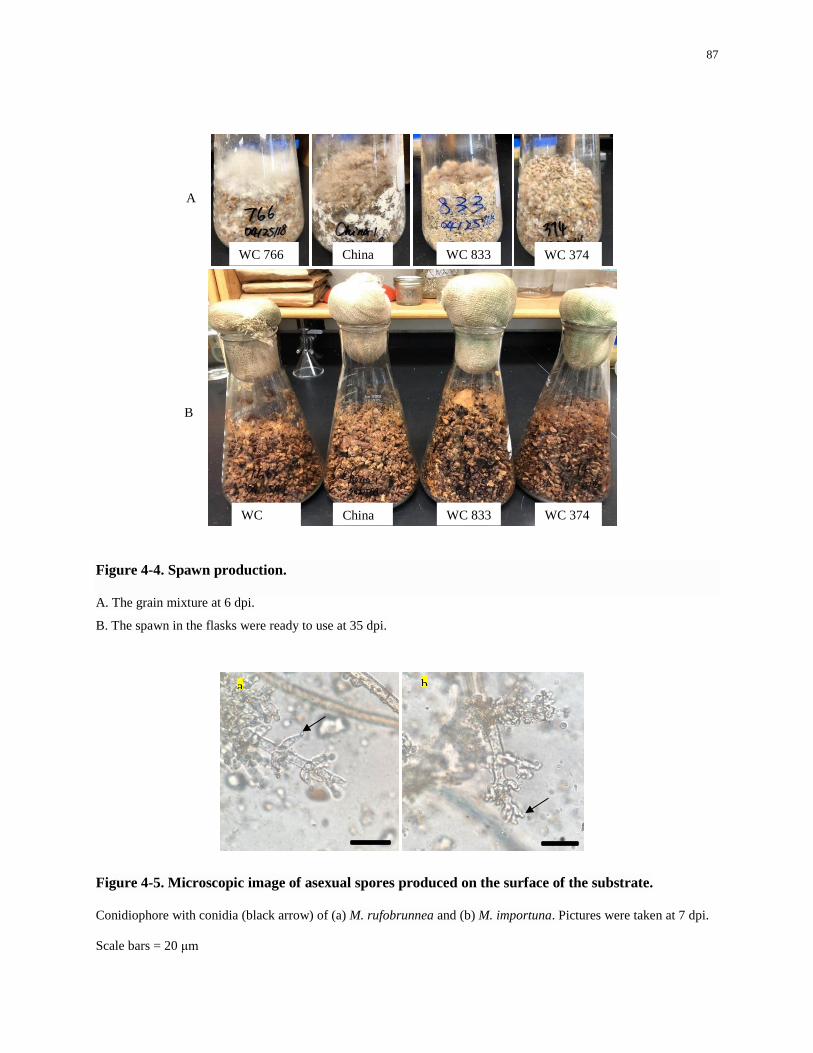
87
Figure 4-4. Spawn production.
A. The grain mixture at 6 dpi.
B. The spawn in the flasks were ready to use at 35 dpi.
Figure 4-5. Microscopic image of asexual spores produced on the surface of the substrate.
Conidiophore with conidia (black arrow) of (a) M. rufobrunnea and (b) M. importuna. Pictures were taken at 7 dpi.
Scale bars = 20 μm
China
16
WC833
6
WC374
6
A
B
WC 766 China
1 WC 833 WC 374
WC
7666 China
1 WC 833 WC 374
a b
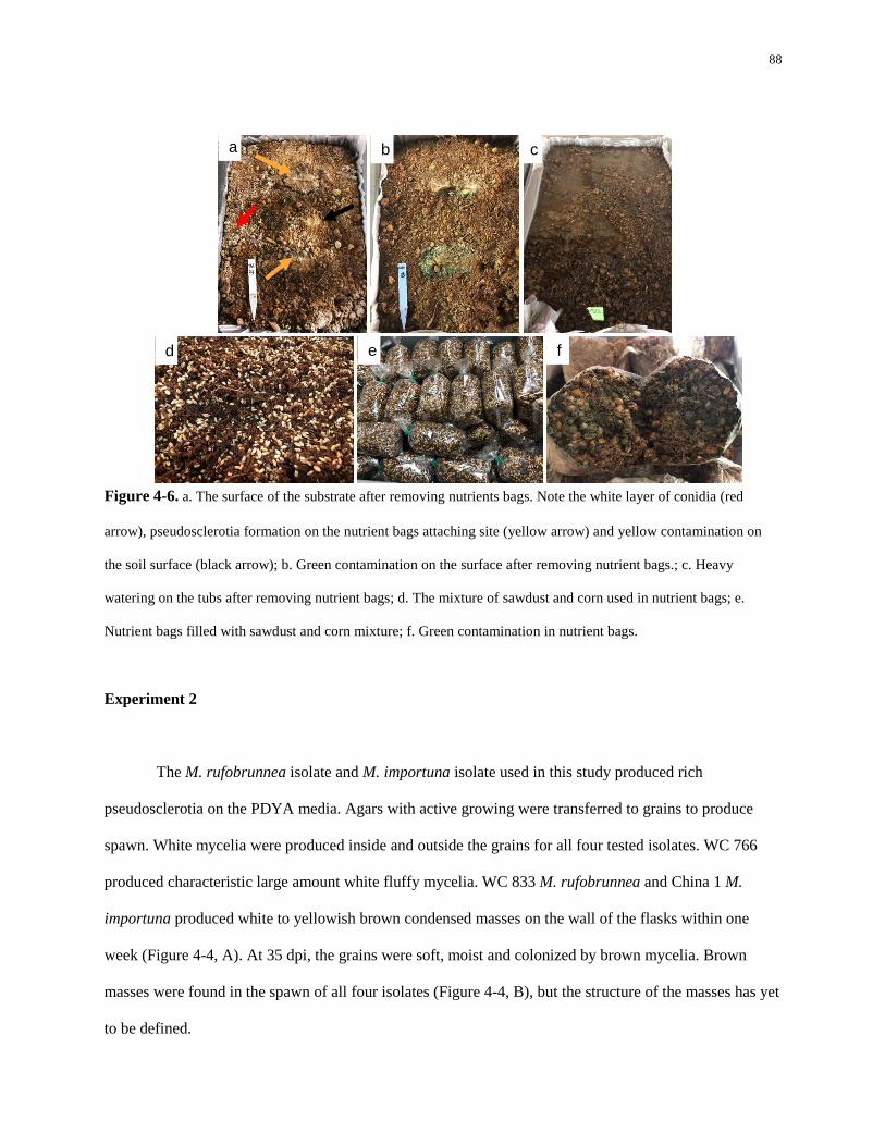
88
Figure 4-6. a. The surface of the substrate after removing nutrients bags. Note the white layer of conidia (red
arrow), pseudosclerotia formation on the nutrient bags attaching site (yellow arrow) and yellow contamination on
the soil surface (black arrow); b. Green contamination on the surface after removing nutrient bags.; c. Heavy
watering on the tubs after removing nutrient bags; d. The mixture of sawdust and corn used in nutrient bags; e.
Nutrient bags filled with sawdust and corn mixture; f. Green contamination in nutrient bags.
Experiment 2
The M. rufobrunnea isolate and M. importuna isolate used in this study produced rich
pseudosclerotia on the PDYA media. Agars with active growing were transferred to grains to produce
spawn. White mycelia were produced inside and outside the grains for all four tested isolates. WC 766
produced characteristic large amount white fluffy mycelia. WC 833 M. rufobrunnea and China 1 M.
importuna produced white to yellowish brown condensed masses on the wall of the flasks within one
week (Figure 4-4, A). At 35 dpi, the grains were soft, moist and colonized by brown mycelia. Brown
masses were found in the spawn of all four isolates (Figure 4-4, B), but the structure of the masses has yet
to be defined.
a b c
d e f
89
At 2 to 3 dpi, white thin mycelia could be found on the surface of the substrates, which was
especially obvious on the straw substrates. At 14 dpi, mycelia of all isolates turned yellow to brown.
Nutrients bags were added at 20 dpi. At 15 dpi, white layer of asexual condia were produced on the
surface of both substrates for M. rufobrunnea and M. importuna. The conidia were produced by
vegetative mycelium, which may be produced from pseudosclerotia. The amount of conidia and covering
area of condia on the surface of the substrates were increased for the first month, while at about 35 to 40
dpi, the amount of observable conidia on the surface of the substrates started to decrease. At 48 dpi, only
a small amount of conidia could be observed on the surface of the substrates. For M. exuberans, conidia
were found on the surface of the straw substrate at 42 dpi in only one tub. However, condia were
observed in all replicates for the sawdust substrate at 30 dpi. It is worth mentioning that no conidia were
found in vitro experiments for these four tested isolates in this study. The exact role and the importance of
conidia in the Morchella cultivation is not clear yet. There is a hypothesis that excessive conidia will
consume nutrients making them unavailable for Morchella fruit body initiation. Microscopy observation
showed that conidia were produced on phialides located on conidiophores (Figure 4-5).
In this study, pseudosclerotia were not observed on the surface except the area attached to the
nutrient bags. However, whether pseudosclerotia were formed inside the substrates was unknown. One
reason is that I did not observe into the substrates in order to keep the substrate intact. The other reason is
it was difficult to distinguish the mycelia and pseudosclerotia from the substrates under dark conditions.
For M. rufobrunnea, small white initial knots were formed around 90 dpi. The knots continued to
grow and developed into primordial stems. Then the stalk and the head with pre-apothecia was formed.
Up to 200 small observable knots were observed at 130 dpi, but more than 50% were died at 140 dpi.
Some white knots disappeared without any noticing changes. However, those with a bigger size (2 to 3
mm in height) turned dark brown and died eventually. No primordial fruit body successfully developed
into mature fruit body. For M. importuna, white masses were found on the surface, however no pins and
90
primordial stage fruiting body was found in this study. For M. exuberans and M. americana, no obvious
formation of pseudosclerotia and primordial fruit body was found.
Similar to experiment 1, some tubs located at the higher level were drier than others. But
according to the conidial growth rating, soil moisture did not affect the production of conidia on M.
rufobrunnea and M. importuna.
What caused the difference between Experiment 1 and 2?
Even though the basic techniques and procedures used in two experiments were similar, the
results were different: Morchella fruit body formation and maturation was successfully initiated in
Experiment 1, while not a single mature fruit body was obtained in experiment 2. Here several possible
factors that may have influenced the differences in fruiting body maturation are discussed:
1. Temperature. The room temperature in experiment 1 was 20C (68F) during the whole procedure,
while in experiment 2, the temperature was 19.4C (67F) for the first two days and maintained 14.4
to 15C (58 to 59F) for the most time. In order to trigger the fruit body initiation, the temperature
was downregulated to 12.2C (54F) at 50 dpi and upregulated to 20C (68F) at 67 dpi, but this
adjustment did not make noticeable effect on the fruit boy initiation. According to the growers’
experiences, low temperatures facilitate the differentiation of mycelia (personal communication).
However, in this study, lower temperature did not contribute to desirable mycelial differentiation in
experiment 2 when compared to experiment 1. The effect of temperature may be covered up by other
factors. Additional data is required to verify the effect of temperature on fruit body initiation on
Morchella indoor cultivation.
91
2. Spawn quality. Even though the spawn for both experiments were produced following the same
protocol, the quality of the spawn may be different. Aging of the cultures was a potential problem in
spawn production. We observed weak mycelial growth and diminished pseudosclerotia production on
some aged Morchella cultures, especially for M. rufobrunnea and M. americana. Morchella species
represent different morphology on different media, which is a major issue in the development of a
spawn quality evaluation standard. Due to the fact that there is no available standard for the
evaluation of morel spawn quality, the evaluation mostly based on personal experience.
Pseudosclerotia production is considered an essential factor in spawn quality evaluation. However,
the pseudosclerotia production in the spawn from two experiment is difficult to compare.
Unfortunately, I did not take photos of the spawn used in experiment 1, so it is impossible to simply
compare the pseudosclerotia production via pictures. Moreover, whether a desirable amount of
pseudosclerotia guarantee a good quality of spawn is unclear.
3. Contamination and flies. Contamination of nutrient bags was appeared in both experiments, but
more frequent and severer contamination was observed in experiment 2. Contamination were
observed in both nutrient bags and on soil surface. The nutrient in the nutrient bags were consumed
by the contaminators, which resulted in the shortage of nutrient for Morchella. Mycelial growth was
suppressed in the contaminated area, characterized by lighter color and dryer texture of the substrate.
Weak mycelial growth might be a reason for the failure of fruit body initiation. The contaminations
were probably originated from the soil or raw materials. Moreover, more flies were found in
experiment 2. The eggs may survive in the soil and then developed into flies under warm and moist
environment.
4. Conidial formation. Conidia were formed in all M. rufobrunnea inoculated tubs in experiment 2,
while conidial formation was only observed in 3 tubs in experiment 1. Conidia formation is very
common in Morchella outdoor cultivation (personal communication). As fast as 5 dpi, a white layer
92
of conidia (2-3 mm thick) could be seen on the soil surface. Environmental conditions, such as light
intensity, air humidity, soil humidity and oxygen level, are the key factors that affect the conidial
formation. However, how these factors that may affect the conidial formation needs further study.
According to farmers’ personal experience, complete darkness suppresses the formation of conidia
and high oxygen level and lower temperature stimulates the conidial formation. Dai (2018) reported
there was a weak, but not significant negative correlation, between conidial production and numbers
of primordia, numbers of mature fruit bodies and yield of M. rofubrunnea. Unfortunately, it is not
clear that what is the light intensity threshold of conidial formation is, and no available data and
analysis indicated how air and soil humidity influenced the conidial formation. Nevertheless, conidial
production varies largely on Morchella species. For instance, the conidial production of M. sextelata
is generally more abundant than conidial production of M. importuna in the outdoor cultivation. In
experiment 2, M. americana did not produce any conidium in either substrates. In contrast, M.
rufobrunnea, M. importuna and M. exuberans produced conidia in both substrates. In experiment 1,
all fruit bodies were collected from the tubs without conidial formation. It seems that conidial
formation may not be necessary for Morchella fruit body initiation. The relationship between conidial
production and yield of morels needs further investigation.
5. Heavy watering. Heavy watering is commonly employed to stimulate the fruit body initiation in
Morchella outdoor cultivation. All the tubs were watered heavily at 65 dpi as soon as the nutrient
bags were removed in experiment 2, but this treatment was not applied to experiment 1. Due to the
complicated interacting factors, it is difficult to conclude the effectiveness of heavy watering on
Morchella fruit body initiation. Moreover, heavy watering could be a reason that caused more
contamination in experiment 2.
93
6. The timing of adding nutrient bags. Nutrients bags were added to the surface of substrate at 45 dpi
and at 20 dpi in experiment 1 and 2 respectively. It is proposed that nutrient bags provide additional
nutrients to facilitate mycelial growth and pseudosclerotia formation, which supports the fruit body
development. In the outdoor cultivation, nutrient bags with openings are added to the soil with the
openings facing the soil surface approximately 7 to 20 days after spawning (W. Liu et al. 2017a).
The timing of adding nutrient bags may have an effect on Morchella cultivation. Dai (2018) found
that the timing of adding nutrient bags (5, 10, and 15 dpi) only effected the fruiting time of M.
rufobrunnea. The earlier nutrient bags were added, the earlier fruit bodies were formed. However, the
yield was not affected by the timing of adding nutrient bags. . Two possible disadvantages may result
from adding nutrients bags too early: 1) Morchella mycelia may not have completely colonized the
surface of the substrate, which can result in the delay of mycelial growth into the nutrient bags. Other
competitive microbses, such as Trichoderma, can take advantage of this to colonize the nutrient bags
due to a lack of competition; 2) Untimely mycelial growth toward nutrient bags may result in
inadequate extension of mycelial growth into the soil, which may reduce the fruit body initiation area.
This could possibly explain why more contaminated bags were found in experiment 2. Adding
nutrient bags too late may delay the nutrient supplies to mycelial growth.
Conclusion
In experiment 1, our results found that the morel outdoor cultivation process was feasible indoors,
even though many questions remained. The formula of the nutrient bags was found to have no difference
based on the results of fruit body initiation in this study. Many primordia were formed, while only 9
mature fruit bodies of M. rufobrunnea were obtained from three tested substrates, and subsequently two
substrates (soil + sawdust and soil + wheat straw) were selected for future research. In experiment 2, no
mature fruit bodies were formed, however, up to 200 primordia were found on M. rufobrunnea in each
tub on both tested substrates. The observed differences between the two experiments might be related to

94
the difference in temperature, spawn quality, contamination of soil, or the timing of adding nutrient bags.
More attention should be focused on investigating the effects of environmental growth conditions on
primordia maturation to determine why the small fruit bodies never matured in the 2nd experiment and
why only a few matured in the first.
95
Chapter 5
Conclusion and Future Work
The objective of this thesis was to shed light on the biological characteristics of Morchella and
eventually lead to the successful cultivation of Morchella. The first hypothesis was that many isolates in
the Morchella cultural collection in the Spawn Lab at Penn State were misidentified based on older
species concept and outdated techniques. The ITS sequences of the isolates of Morchella in the collection
were determined and molecular identifications were made comparing these sequences to curated
databases. In consideration of the cultivation history, geographical origin and characteristic recorded
fruiting patterns, four Morchella isolates (WC 833 M. rufobrunnea, China 1 M. importuna, WC 374 M.
americana and WC 766 M. exuberans) were selected for subsequent studies.
Environmental factors such as pH, light intensity and temperature, may have an effect on the
mycelial growth and pseudosclerotia formation of Morchella. We hypothesized that: 1) For each of the
Morchella isolates, there is a pH range and a temperature range that it can grow, but there is an optimal
pH and temperature that results in the fastest mycelial growth and most pseudosclerotia production. 2)
The effect of light on Morchella varies based on light intensity and Morchella species. 3) Not all tested
Morchella isolates can produce pesudosclerotia under tested conditions. The hypothesises were tested by
culturing four Morchella isolates on PDYA media at four pH levels (5, 6, 7, and 8), under three light
intensity conditions (complete darkness (0 lux), low intensity (400 lux), and high intensity (2000 lux)),
and under three temperature conditions (20C, 25C, and 30C). For M. rufobrunnea, the optimal pH,
light condition and temperature resulting in a high level of mycelial growth was at a pH of 6, darkness (0
lux), and at a temperature of 20 and 25C. For M. importuna, the optimal pH, light condition and
temperature resulting in a high level of mycelial growth was at a pH of 5 to 6, under darkness (0 lux) and
at a low light intensity (400 lux), and at a temperature of 20 to 25C. For M. exuberans, the optimal pH
96
and temperature was at a pH of 5, and a temperature of 25 to 30C. Light did not have a significant effect
on the mycelial growth of M. exuberans. For M. americana, the optimal pH, light condition and
temperature for mycelial growth was at a pH of 5 to 7, under darkness (0 lux) and low light intensity (400
lux), and at a temperature of 20C. M. exuberans and M. americana did not produce pseudosclerotium
under any test conditions. The media pH did not affect pseudosclerotia formation for either M.
rufobrunnea or M. importuna. Mycelial growth is the first stage in the life cycle of Morchella and
pseudosclerotia are essential in the entire Morchella life cycle (Volk and Leonard 1990). Based on the
results in this study, M. rufobrunnea and M. importuna demonstrated satisfactory mycelial growth and
pseudosclerotia production, so these two isolates are suggested for future cultivation trials. Since high
temperatures (30C) significantly suppressed mycelial growth and pseudosclerotia formation of M.
rufobrunnea and M. importuna, it is suggested that the highest temperature to cultivate these two isolates
outdoors should be less than 30C. It is also suggested to move up the the spawn date in the southern area
where the temperature is generally higher to avoid the unfavorable temperatures during the mycelial
growth phases when growing Morchella outdoors. Light is a suppressing factor of the pseudosclerotia
formation of M. rufobrunnea and M. importuna. The application of mulching films or shade cloths can
not only benefit the outdoor cultivation by reserving moisture, but also by protecting Morchella mycelia
from light. The determination of these optimal environmental conditions resulting in desirable mycelial
growth and pseudosclerotia formation will provide guidance on the Morchella culture preparation, spawn
production and outdoor cultivation.
For the future direction, the following experiments can be pursued:
Study 1

97
Objective: To test heat and low temperature treatment on pseudosclerotia formation on
Morchella exuberans.
M. exuberans did not produce any pseudosclerotium under tested conditions. However, it was
reported to be a post-fire specie. Heat treatment mimicking wildfires may help triggering
pseudosclerotium formation. Interestingly, I found white to orange pseudosclerotia on two old M.
exuberans cultures growing on PDYA media. These two cultures grew at room temperature (~20C for
the first month and were then stored at 4C for more than two months. However, pseudosclerotium was
not observed in the cultures stored at room temperature. Thus, low temperature treatment may be a
potential factor that triggering the pseudosclerotium formation for this species. The experiment can be
designed as followed: Three groups of M. exuberans will be cultured on PDYA plates at 25C for 4 to 5
days until the plates are fully colonized. Three cultures will be grown at 4C, 25 C and 40 C for 1 day
respectively, and then all the plates will be cultured at 25C again. Temperature and culturing duration at
each temperature may need some adjustments. Plates will be observed every day to determine if any
pseudosclerotium form and if any differences are observed based on the different temperature exposure.
This study will improve our understanding of the factors that lead to pseudosclerotium development.
Study 2
Objective: To test the effects of environmental factors on Morchella spawn production.
The quality of spawn is largely determined by the amount of mycelia and pseudosclerotia
formation. Even though the effects of pH, light and temperature on mycelial growth and pseudosclerotia
production in vitro were investigated for four Morchella isolates, it is not really known how these factors
affect the spawn quality and subsequent fruiting potential. Therefore, it is necessary to test how
environmental factors affect the amount the mycelia and pseudosclerotia in the spawn and relate how
98
these factors ultimately affect fruit body production. However, how to effectively and scientifically
measure the mycelial growth and pseudosclerotia production in the spawn is a question that needs to be
investigated.
Study 3
Objective: To study the effect of successive subculturing on the morphology, mycelial
growth and pseudosclerotia formation of four Morchella isolates.
The aging of spawn cultures and repeated subculturing are concerning problems in morel
cultivation. The use of older spawn may result in reduce yield and profits to farmers depending on how
spawn quality impacts fruiting. Microscopy studies revealed the involvement of autophagy, apoptosis and
necrosis with the senescence of M. elata (P. He et al. 2015). Nevertheless, the molecular mechanism of
these behaviors need further study. Delayed mycelial growth, thin mycelia, and the reduction of
pseudosclerotia production has been observed in WC 833 and WC 374 after successive subculturing. This
study would aim to determine the threshold of subculturing and culture age on Morchella spp. viability. It
will also be interesting to study if subculturing an isolate in particular subtrates can revive its vigor.
Study 4
Objective: To select species for indoor and outdoor cultivation with high potential for
commercial adoption.
Though Morchella species are diverse in the wild, species that can be cultivated are limited. In the
second cropping trial, I tested four Morchella isolates for indoor cultivation, unfortunately no mature
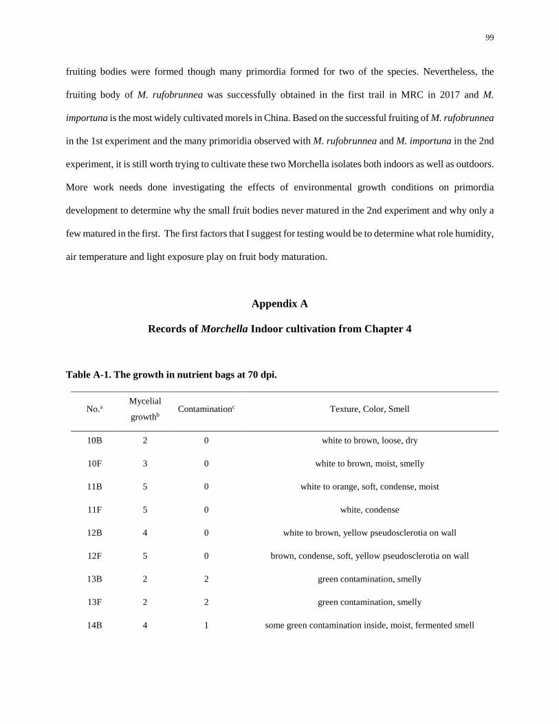
99
fruiting bodies were formed though many primordia formed for two of the species. Nevertheless, the
fruiting body of M. rufobrunnea was successfully obtained in the first trail in MRC in 2017 and M.
importuna is the most widely cultivated morels in China. Based on the successful fruiting of M. rufobrunnea
in the 1st experiment and the many primoridia observed with M. rufobrunnea and M. importuna in the 2nd
experiment, it is still worth trying to cultivate these two Morchella isolates both indoors as well as outdoors.
More work needs done investigating the effects of environmental growth conditions on primordia
development to determine why the small fruit bodies never matured in the 2nd experiment and why only a
few matured in the first. The first factors that I suggest for testing would be to determine what role humidity,
air temperature and light exposure play on fruit body maturation.
Appendix A
Records of Morchella Indoor cultivation from Chapter 4
Table A-1. The growth in nutrient bags at 70 dpi.
No.a Mycelial
growthb Contaminationc Texture, Color, Smell
10B 2 0 white to brown, loose, dry
10F 3 0 white to brown, moist, smelly
11B 5 0 white to orange, soft, condense, moist
11F 5 0 white, condense
12B 4 0 white to brown, yellow pseudosclerotia on wall
12F 5 0 brown, condense, soft, yellow pseudosclerotia on wall
13B 2 2 green contamination, smelly
13F 2 2 green contamination, smelly
14B 4 1 some green contamination inside, moist, fermented smell
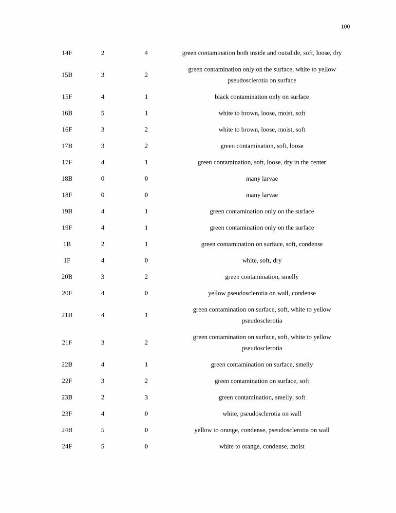
100
14F 2 4 green contamination both inside and outsdide, soft, loose, dry
15B 3 2 green contamination only on the surface, white to yellow
pseudosclerotia on surface
15F 4 1 black contamination only on surface
16B 5 1 white to brown, loose, moist, soft
16F 3 2 white to brown, loose, moist, soft
17B 3 2 green contamination, soft, loose
17F 4 1 green contamination, soft, loose, dry in the center
18B 0 0 many larvae
18F 0 0 many larvae
19B 4 1 green contamination only on the surface
19F 4 1 green contamination only on the surface
1B 2 1 green contamination on surface, soft, condense
1F 4 0 white, soft, dry
20B 3 2 green contamination, smelly
20F 4 0 yellow pseudosclerotia on wall, condense
21B 4 1 green contamination on surface, soft, white to yellow
pseudosclerotia
21F 3 2 green contamination on surface, soft, white to yellow
pseudosclerotia
22B 4 1 green contamination on surface, smelly
22F 3 2 green contamination on surface, soft
23B 2 3 green contamination, smelly, soft
23F 4 0 white, pseudosclerotia on wall
24B 5 0 yellow to orange, condense, pseudosclerotia on wall
24F 5 0 white to orange, condense, moist
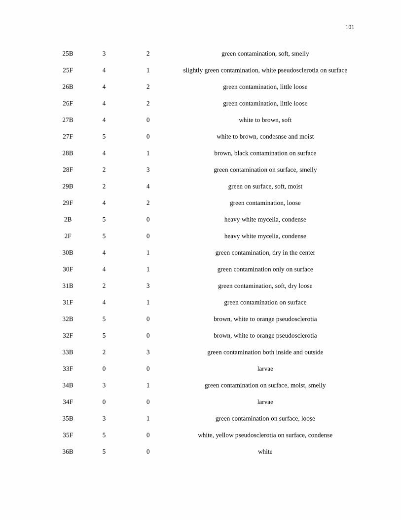
101
25B 3 2 green contamination, soft, smelly
25F 4 1 slightly green contamination, white pseudosclerotia on surface
26B 4 2 green contamination, little loose
26F 4 2 green contamination, little loose
27B 4 0 white to brown, soft
27F 5 0 white to brown, condesnse and moist
28B 4 1 brown, black contamination on surface
28F 2 3 green contamination on surface, smelly
29B 2 4 green on surface, soft, moist
29F 4 2 green contamination, loose
2B 5 0 heavy white mycelia, condense
2F 5 0 heavy white mycelia, condense
30B 4 1 green contamination, dry in the center
30F 4 1 green contamination only on surface
31B 2 3 green contamination, soft, dry loose
31F 4 1 green contamination on surface
32B 5 0 brown, white to orange pseudosclerotia
32F 5 0 brown, white to orange pseudosclerotia
33B 2 3 green contamination both inside and outside
33F 0 0 larvae
34B 3 1 green contamination on surface, moist, smelly
34F 0 0 larvae
35B 3 1 green contamination on surface, loose
35F 5 0 white, yellow pseudosclerotia on surface, condense
36B 5 0 white
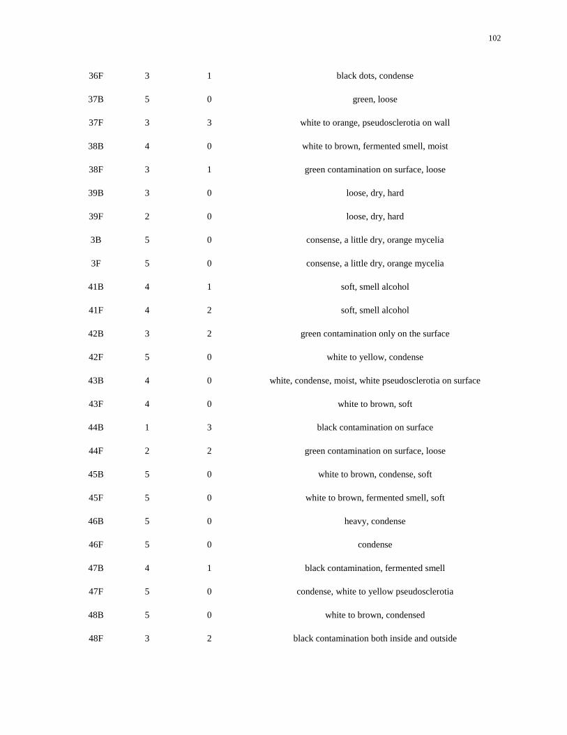
102
36F 3 1 black dots, condense
37B 5 0 green, loose
37F 3 3 white to orange, pseudosclerotia on wall
38B 4 0 white to brown, fermented smell, moist
38F 3 1 green contamination on surface, loose
39B 3 0 loose, dry, hard
39F 2 0 loose, dry, hard
3B 5 0 consense, a little dry, orange mycelia
3F 5 0 consense, a little dry, orange mycelia
41B 4 1 soft, smell alcohol
41F 4 2 soft, smell alcohol
42B 3 2 green contamination only on the surface
42F 5 0 white to yellow, condense
43B 4 0 white, condense, moist, white pseudosclerotia on surface
43F 4 0 white to brown, soft
44B 1 3 black contamination on surface
44F 2 2 green contamination on surface, loose
45B 5 0 white to brown, condense, soft
45F 5 0 white to brown, fermented smell, soft
46B 5 0 heavy, condense
46F 5 0 condense
47B 4 1 black contamination, fermented smell
47F 5 0 condense, white to yellow pseudosclerotia
48B 5 0 white to brown, condensed
48F 3 2 black contamination both inside and outside
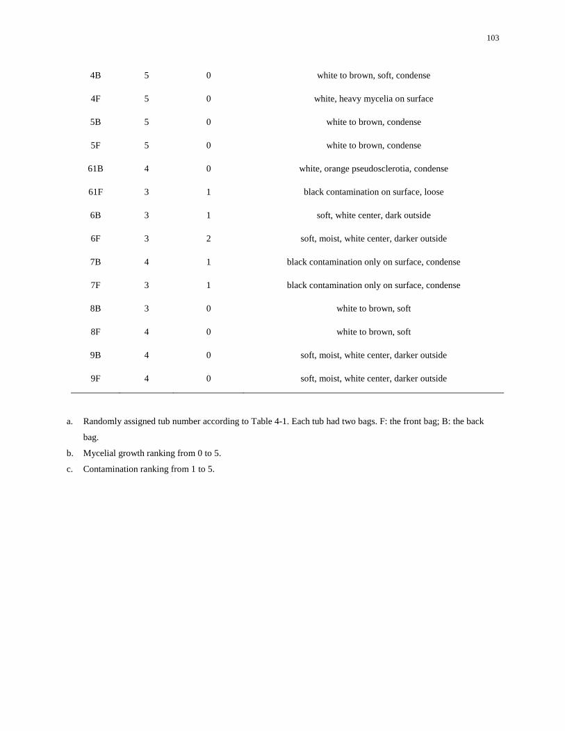
103
4B 5 0 white to brown, soft, condense
4F 5 0 white, heavy mycelia on surface
5B 5 0 white to brown, condense
5F 5 0 white to brown, condense
61B 4 0 white, orange pseudosclerotia, condense
61F 3 1 black contamination on surface, loose
6B 3 1 soft, white center, dark outside
6F 3 2 soft, moist, white center, darker outside
7B 4 1 black contamination only on surface, condense
7F 3 1 black contamination only on surface, condense
8B 3 0 white to brown, soft
8F 4 0 white to brown, soft
9B 4 0 soft, moist, white center, darker outside
9F 4 0 soft, moist, white center, darker outside
a. Randomly assigned tub number according to Table 4-1. Each tub had two bags. F: the front bag; B: the back
bag.
b. Mycelial growth ranking from 0 to 5.
c. Contamination ranking from 1 to 5.
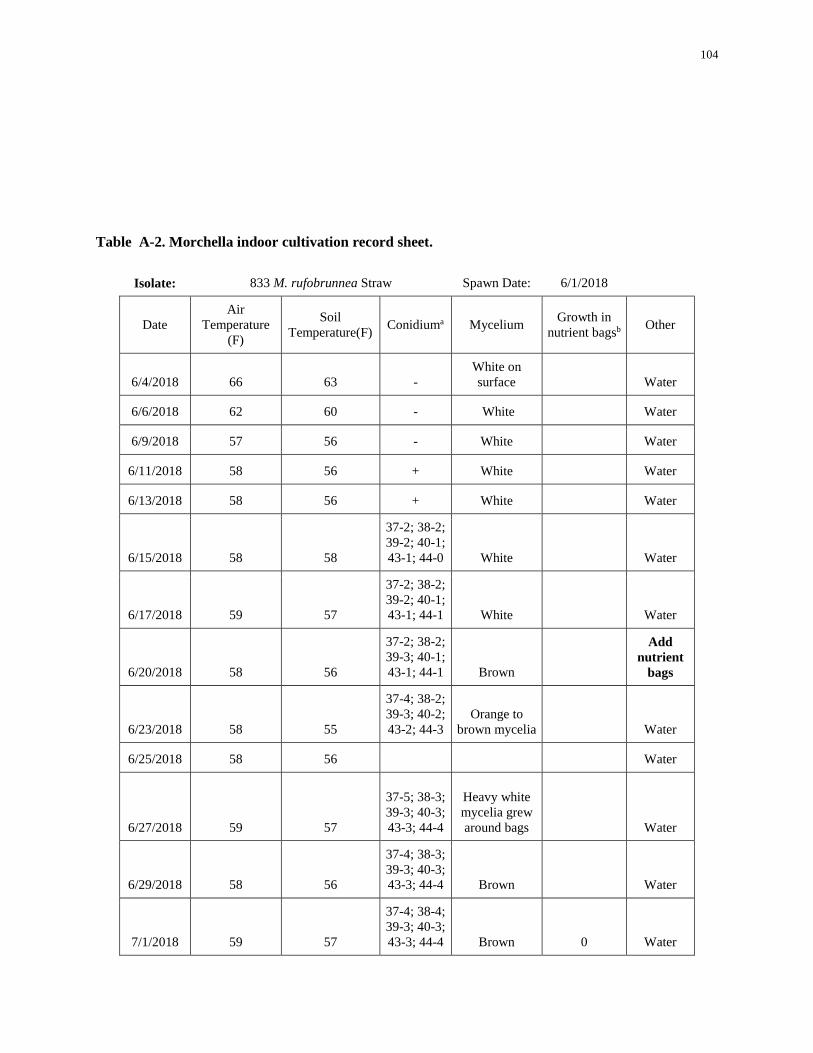
104
Table A-2. Morchella indoor cultivation record sheet.
Isolate: 833 M. rufobrunnea Straw Spawn Date: 6/1/2018
Date Air
Temperature
(F)
Soil
Temperature(F) Conidiuma Mycelium
Growth in
nutrient bagsb Other
6/4/2018 66 63 - White on
surface Water
6/6/2018 62 60 - White Water
6/9/2018 57 56 - White Water
6/11/2018 58 56 + White Water
6/13/2018 58 56 + White Water
6/15/2018 58 58
37-2; 38-2;
39-2; 40-1;
43-1; 44-0 White Water
6/17/2018 59 57
37-2; 38-2;
39-2; 40-1;
43-1; 44-1 White Water
6/20/2018 58 56
37-2; 38-2;
39-3; 40-1;
43-1; 44-1 Brown
Add
nutrient
bags
6/23/2018 58 55
37-4; 38-2;
39-3; 40-2;
43-2; 44-3 Orange to
brown mycelia Water
6/25/2018 58 56 Water
6/27/2018 59 57
37-5; 38-3;
39-3; 40-3;
43-3; 44-4
Heavy white
mycelia grew
around bags Water
6/29/2018 58 56
37-4; 38-3;
39-3; 40-3;
43-3; 44-4 Brown Water
7/1/2018 59 57
37-4; 38-4;
39-3; 40-3;
43-3; 44-4 Brown 0 Water
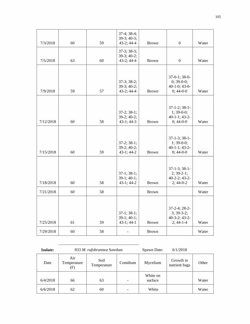
105
7/3/2018 60 59
37-4; 38-4;
39-3; 40-3;
43-2; 44-4 Brown 0 Water
7/5/2018 63 60
37-3; 38-3;
39-3; 40-2;
43-2; 44-4 Brown 0 Water
7/9/2018 59 57
37-3; 38-2;
39-3; 40-2;
43-2; 44-4 Brown
37-0-1; 38-0-
0; 39-0-0;
40-1-0; 43-0-
0; 44-0-0 Water
7/12/2018 60 58
37-2; 38-1;
39-2; 40-2;
43-1; 44-3 Brown
37-1-2; 38-1-
1; 39-0-0;
40-1-1; 43-2-
0; 44-0-0 Water
7/15/2018 60 59
37-2; 38-1;
39-2; 40-2;
43-1; 44-2 Brown
37-1-3; 38-1-
1; 39-0-0;
40-1-1; 43-2-
0; 44-0-0 Water
7/18/2018 60 58
37-1; 38-1;
39-1; 40-1;
43-1; 44-2 Brown
37-1-3; 38-1-
2; 39-2-1;
40-2-2; 43-2-
2; 44-0-2 Water
7/21/2018 60 58 Brown Water
7/25/2018 61 59
37-1; 38-1;
39-1; 40-1;
43-1; 44-1 Brown
37-2-4; 28-2-
3; 39-3-2;
40-3-2; 43-2-
2; 44-1-4 Water
7/29/2018 60 58 - Brown Water
Isolate: 833 M. rufobrunnea Sawdust Spawn Date: 6/1/2018
Date Air
Temperature
(F)
Soil
Temperature Conidium Mycelium
Growth in
nutrient bags Other
6/4/2018 66 63 - White on
surface Water
6/6/2018 62 60 - White Water
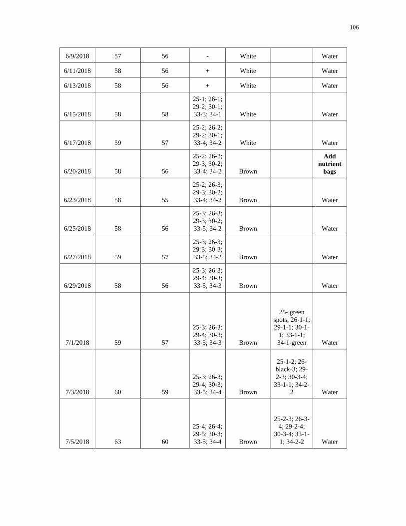
106
6/9/2018 57 56 - White Water
6/11/2018 58 56 + White Water
6/13/2018 58 56 + White Water
6/15/2018 58 58
25-1; 26-1;
29-2; 30-1;
33-3; 34-1 White Water
6/17/2018 59 57
25-2; 26-2;
29-2; 30-1;
33-4; 34-2 White Water
6/20/2018 58 56
25-2; 26-2;
29-3; 30-2;
33-4; 34-2 Brown
Add
nutrient
bags
6/23/2018 58 55
25-2; 26-3;
29-3; 30-2;
33-4; 34-2 Brown Water
6/25/2018 58 56
25-3; 26-3;
29-3; 30-2;
33-5; 34-2 Brown Water
6/27/2018 59 57
25-3; 26-3;
29-3; 30-3;
33-5; 34-2 Brown Water
6/29/2018 58 56
25-3; 26-3;
29-4; 30-3;
33-5; 34-3 Brown Water
7/1/2018 59 57
25-3; 26-3;
29-4; 30-3;
33-5; 34-3 Brown
25- green
spots; 26-1-1;
29-1-1; 30-1-
1; 33-1-1;
34-1-green Water
7/3/2018 60 59
25-3; 26-3;
29-4; 30-3;
33-5; 34-4 Brown
25-1-2; 26-
black-3; 29-
2-3; 30-3-4;
33-1-1; 34-2-
2 Water
7/5/2018 63 60
25-4; 26-4;
29-5; 30-3;
33-5; 34-4 Brown
25-2-3; 26-3-
4; 29-2-4;
30-3-4; 33-1-
1; 34-2-2 Water
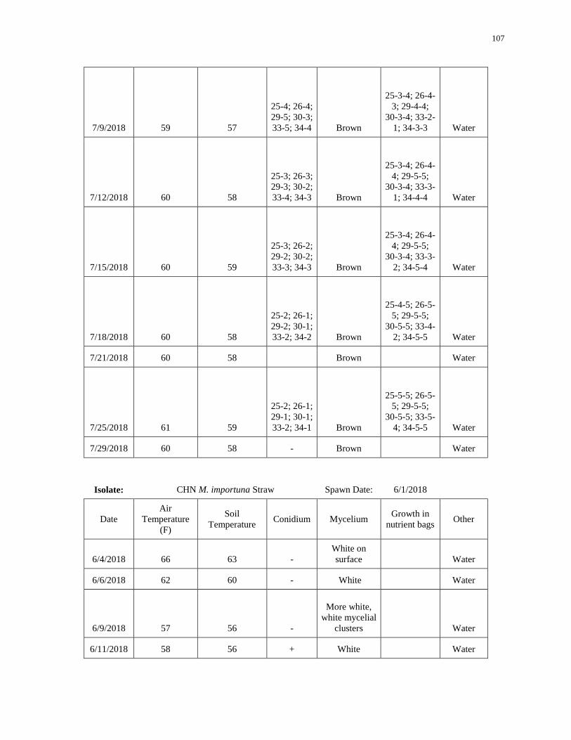
107
7/9/2018 59 57
25-4; 26-4;
29-5; 30-3;
33-5; 34-4 Brown
25-3-4; 26-4-
3; 29-4-4;
30-3-4; 33-2-
1; 34-3-3 Water
7/12/2018 60 58
25-3; 26-3;
29-3; 30-2;
33-4; 34-3 Brown
25-3-4; 26-4-
4; 29-5-5;
30-3-4; 33-3-
1; 34-4-4 Water
7/15/2018 60 59
25-3; 26-2;
29-2; 30-2;
33-3; 34-3 Brown
25-3-4; 26-4-
4; 29-5-5;
30-3-4; 33-3-
2; 34-5-4 Water
7/18/2018 60 58
25-2; 26-1;
29-2; 30-1;
33-2; 34-2 Brown
25-4-5; 26-5-
5; 29-5-5;
30-5-5; 33-4-
2; 34-5-5 Water
7/21/2018 60 58 Brown Water
7/25/2018 61 59
25-2; 26-1;
29-1; 30-1;
33-2; 34-1 Brown
25-5-5; 26-5-
5; 29-5-5;
30-5-5; 33-5-
4; 34-5-5 Water
7/29/2018 60 58 - Brown Water
Isolate: CHN M. importuna Straw Spawn Date: 6/1/2018
Date Air
Temperature
(F)
Soil
Temperature Conidium Mycelium
Growth in
nutrient bags Other
6/4/2018 66 63 - White on
surface Water
6/6/2018 62 60 - White Water
6/9/2018 57 56 -
More white,
white mycelial
clusters Water
6/11/2018 58 56 + White Water
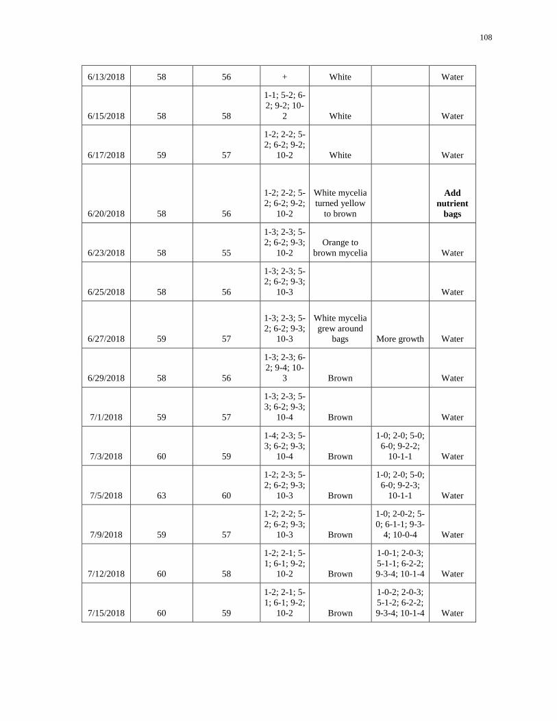
108
6/13/2018 58 56 + White Water
6/15/2018 58 58
1-1; 5-2; 6-
2; 9-2; 10-
2 White Water
6/17/2018 59 57
1-2; 2-2; 5-
2; 6-2; 9-2;
10-2 White Water
6/20/2018 58 56
1-2; 2-2; 5-
2; 6-2; 9-2;
10-2
White mycelia
turned yellow
to brown
Add
nutrient
bags
6/23/2018 58 55
1-3; 2-3; 5-
2; 6-2; 9-3;
10-2 Orange to
brown mycelia Water
6/25/2018 58 56
1-3; 2-3; 5-
2; 6-2; 9-3;
10-3 Water
6/27/2018 59 57
1-3; 2-3; 5-
2; 6-2; 9-3;
10-3
White mycelia
grew around
bags More growth Water
6/29/2018 58 56
1-3; 2-3; 6-
2; 9-4; 10-
3 Brown Water
7/1/2018 59 57
1-3; 2-3; 5-
3; 6-2; 9-3;
10-4 Brown Water
7/3/2018 60 59
1-4; 2-3; 5-
3; 6-2; 9-3;
10-4 Brown
1-0; 2-0; 5-0;
6-0; 9-2-2;
10-1-1 Water
7/5/2018 63 60
1-2; 2-3; 5-
2; 6-2; 9-3;
10-3 Brown
1-0; 2-0; 5-0;
6-0; 9-2-3;
10-1-1 Water
7/9/2018 59 57
1-2; 2-2; 5-
2; 6-2; 9-3;
10-3 Brown
1-0; 2-0-2; 5-
0; 6-1-1; 9-3-
4; 10-0-4 Water
7/12/2018 60 58
1-2; 2-1; 5-
1; 6-1; 9-2;
10-2 Brown
1-0-1; 2-0-3;
5-1-1; 6-2-2;
9-3-4; 10-1-4 Water
7/15/2018 60 59
1-2; 2-1; 5-
1; 6-1; 9-2;
10-2 Brown
1-0-2; 2-0-3;
5-1-2; 6-2-2;
9-3-4; 10-1-4 Water
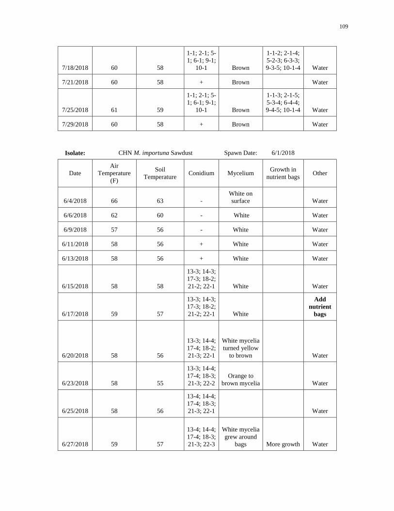
109
7/18/2018 60 58
1-1; 2-1; 5-
1; 6-1; 9-1;
10-1 Brown
1-1-2; 2-1-4;
5-2-3; 6-3-3;
9-3-5; 10-1-4 Water
7/21/2018 60 58 + Brown Water
7/25/2018 61 59
1-1; 2-1; 5-
1; 6-1; 9-1;
10-1 Brown
1-1-3; 2-1-5;
5-3-4; 6-4-4;
9-4-5; 10-1-4 Water
7/29/2018 60 58 + Brown Water
Isolate: CHN M. importuna Sawdust Spawn Date: 6/1/2018
Date Air
Temperature
(F)
Soil
Temperature Conidium Mycelium
Growth in
nutrient bags Other
6/4/2018 66 63 - White on
surface Water
6/6/2018 62 60 - White Water
6/9/2018 57 56 - White Water
6/11/2018 58 56 + White Water
6/13/2018 58 56 + White Water
6/15/2018 58 58
13-3; 14-3;
17-3; 18-2;
21-2; 22-1 White Water
6/17/2018 59 57
13-3; 14-3;
17-3; 18-2;
21-2; 22-1 White
Add
nutrient
bags
6/20/2018 58 56
13-3; 14-4;
17-4; 18-2;
21-3; 22-1
White mycelia
turned yellow
to brown Water
6/23/2018 58 55
13-3; 14-4;
17-4; 18-3;
21-3; 22-2 Orange to
brown mycelia Water
6/25/2018 58 56
13-4; 14-4;
17-4; 18-3;
21-3; 22-1 Water
6/27/2018 59 57
13-4; 14-4;
17-4; 18-3;
21-3; 22-3
White mycelia
grew around
bags More growth Water
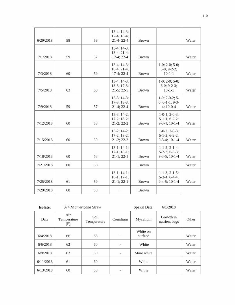
110
6/29/2018 58 56
13-4; 14-3;
17-4; 18-4;
21-4- 22-4 Brown Water
7/1/2018 59 57
13-4; 14-3;
18-4; 21-4;
17-4; 22-4 Brown Water
7/3/2018 60 59
13-4; 14-3;
18-4; 21-4;
17-4; 22-4 Brown
1-0; 2-0; 5-0;
6-0; 9-2-2;
10-1-1 Water
7/5/2018 63 60
13-4; 14-3;
18-3; 17-3;
21-5; 22-5 Brown
1-0; 2-0; 5-0;
6-0; 9-2-3;
10-1-1 Water
7/9/2018 59 57
13-3; 14-3;
17-3; 18-3;
21-4; 22-4 Brown
1-0; 2-0-2; 5-
0; 6-1-1; 9-3-
4; 10-0-4 Water
7/12/2018 60 58
13-3; 14-2;
17-2; 18-2;
21-2; 22-2 Brown
1-0-1; 2-0-3;
5-1-1; 6-2-2;
9-3-4; 10-1-4 Water
7/15/2018 60 59
13-2; 14-2;
17-2; 18-2;
21-2; 22-2 Brown
1-0-2; 2-0-3;
5-1-2; 6-2-2;
9-3-4; 10-1-4 Water
7/18/2018 60 58
13-1; 14-1;
17-1; 18-1;
21-1; 22-1 Brown
1-1-2; 2-1-4;
5-2-3; 6-3-3;
9-3-5; 10-1-4 Water
7/21/2018 60 58 Brown Water
7/25/2018 61 59
13-1; 14-1;
18-1; 17-1;
21-1; 22-1 Brown
1-1-3; 2-1-5;
5-3-4; 6-4-4;
9-4-5; 10-1-4 Water
7/29/2018 60 58 + Brown
Isolate: 374 M.americana Straw Spawn Date: 6/1/2018
Date Air
Temperature
(F)
Soil
Temperature Conidium Mycelium
Growth in
nutrient bags Other
6/4/2018 66 63 - White on
surface Water
6/6/2018 62 60 - White Water
6/9/2018 62 60 - More white Water
6/11/2018 61 60 - White Water
6/13/2018 60 58 - White Water
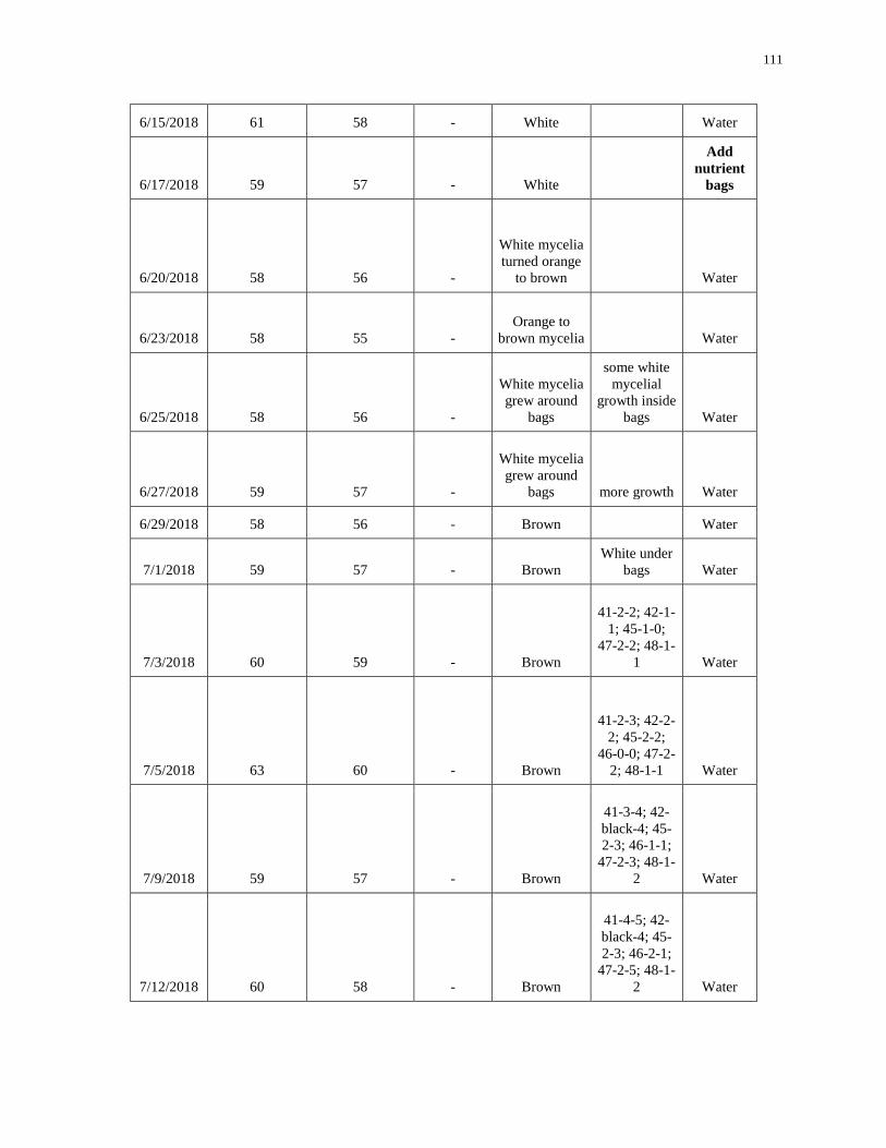
111
6/15/2018 61 58 - White Water
6/17/2018 59 57 - White
Add
nutrient
bags
6/20/2018 58 56 -
White mycelia
turned orange
to brown Water
6/23/2018 58 55 - Orange to
brown mycelia Water
6/25/2018 58 56 -
White mycelia
grew around
bags
some white
mycelial
growth inside
bags Water
6/27/2018 59 57 -
White mycelia
grew around
bags more growth Water
6/29/2018 58 56 - Brown Water
7/1/2018 59 57 - Brown White under
bags Water
7/3/2018 60 59 - Brown
41-2-2; 42-1-
1; 45-1-0;
47-2-2; 48-1-
1 Water
7/5/2018 63 60 - Brown
41-2-3; 42-2-
2; 45-2-2;
46-0-0; 47-2-
2; 48-1-1 Water
7/9/2018 59 57 - Brown
41-3-4; 42-
black-4; 45-
2-3; 46-1-1;
47-2-3; 48-1-
2 Water
7/12/2018 60 58 - Brown
41-4-5; 42-
black-4; 45-
2-3; 46-2-1;
47-2-5; 48-1-
2 Water
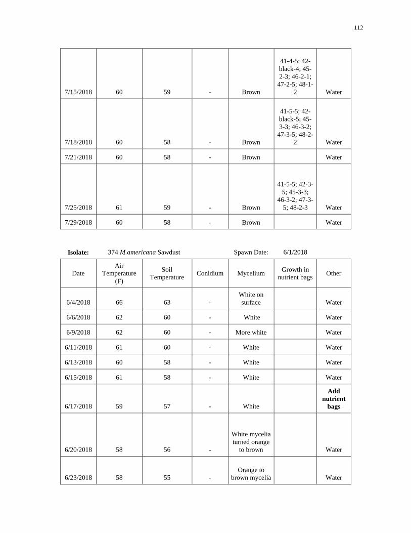
112
7/15/2018 60 59 - Brown
41-4-5; 42-
black-4; 45-
2-3; 46-2-1;
47-2-5; 48-1-
2 Water
7/18/2018 60 58 - Brown
41-5-5; 42-
black-5; 45-
3-3; 46-3-2;
47-3-5; 48-2-
2 Water
7/21/2018 60 58 - Brown Water
7/25/2018 61 59 - Brown
41-5-5; 42-3-
5; 45-3-3;
46-3-2; 47-3-
5; 48-2-3 Water
7/29/2018 60 58 - Brown Water
Isolate: 374 M.americana Sawdust Spawn Date: 6/1/2018
Date Air
Temperature
(F)
Soil
Temperature Conidium Mycelium
Growth in
nutrient bags Other
6/4/2018 66 63 - White on
surface Water
6/6/2018 62 60 - White Water
6/9/2018 62 60 - More white Water
6/11/2018 61 60 - White Water
6/13/2018 60 58 - White Water
6/15/2018 61 58 - White Water
6/17/2018 59 57 - White
Add
nutrient
bags
6/20/2018 58 56 -
White mycelia
turned orange
to brown Water
6/23/2018 58 55 - Orange to
brown mycelia Water
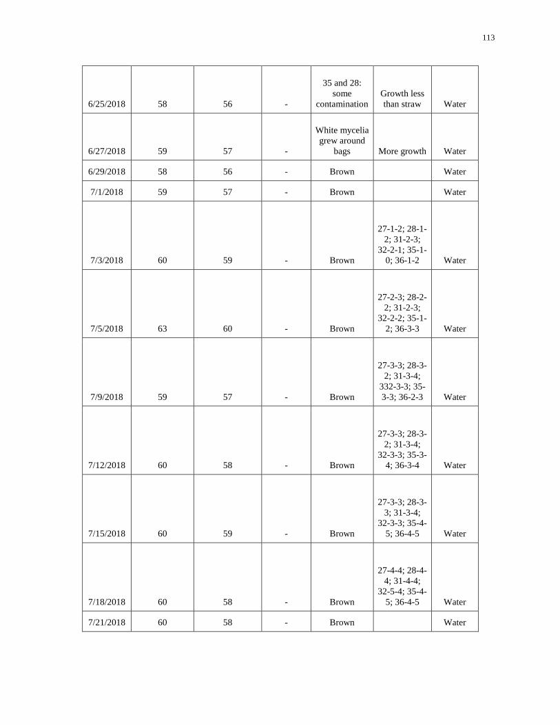
113
6/25/2018 58 56 -
35 and 28:
some
contamination Growth less
than straw Water
6/27/2018 59 57 -
White mycelia
grew around
bags More growth Water
6/29/2018 58 56 - Brown Water
7/1/2018 59 57 - Brown Water
7/3/2018 60 59 - Brown
27-1-2; 28-1-
2; 31-2-3;
32-2-1; 35-1-
0; 36-1-2 Water
7/5/2018 63 60 - Brown
27-2-3; 28-2-
2; 31-2-3;
32-2-2; 35-1-
2; 36-3-3 Water
7/9/2018 59 57 - Brown
27-3-3; 28-3-
2; 31-3-4;
332-3-3; 35-
3-3; 36-2-3 Water
7/12/2018 60 58 - Brown
27-3-3; 28-3-
2; 31-3-4;
32-3-3; 35-3-
4; 36-3-4 Water
7/15/2018 60 59 - Brown
27-3-3; 28-3-
3; 31-3-4;
32-3-3; 35-4-
5; 36-4-5 Water
7/18/2018 60 58 - Brown
27-4-4; 28-4-
4; 31-4-4;
32-5-4; 35-4-
5; 36-4-5 Water
7/21/2018 60 58 - Brown Water
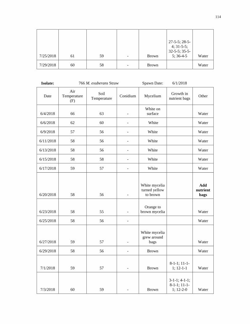
114
7/25/2018 61 59 - Brown
27-5-5; 28-5-
4; 31-5-5;
32-5-5; 35-5-
5; 36-4-5 Water
7/29/2018 60 58 - Brown Water
Isolate: 766 M. exuberans Straw Spawn Date: 6/1/2018
Date Air
Temperature
(F)
Soil
Temperature Conidium Mycelium
Growth in
nutrient bags Other
6/4/2018 66 63 - White on
surface Water
6/6/2018 62 60 - White Water
6/9/2018 57 56 - White Water
6/11/2018 58 56 - White Water
6/13/2018 58 56 - White Water
6/15/2018 58 58 - White Water
6/17/2018 59 57 - White Water
6/20/2018 58 56 -
White mycelia
turned yellow
to brown
Add
nutrient
bags
6/23/2018 58 55 - Orange to
brown mycelia Water
6/25/2018 58 56 - Water
6/27/2018 59 57 -
White mycelia
grew around
bags Water
6/29/2018 58 56 - Brown Water
7/1/2018 59 57 - Brown 8-1-1; 11-1-
1; 12-1-1 Water
7/3/2018 60 59 - Brown
3-1-1; 4-1-1;
8-1-1; 11-1-
1; 12-2-0 Water
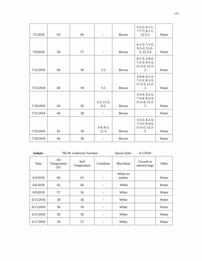
115
7/5/2018 63 60 - Brown
3-2-2; 4-1-1;
7-7-7; 8-1-1;
12-2-2 Water
7/9/2018 59 57 - Brown
4-1-2; 7-1-2;
8-1-2; 11-2-
3; 12-2-4 Water
7/12/2018 60 58 3-3 Brown
4-1-3; 3-4-4;
7-2-3; 8-2-3;
11-3-3; 12-2-
3 Water
7/15/2018 60 59 3-3 Brown
3-4-4; 4-1-3;
7-2-3; 8-3-3;
11-3-3; 12-2-
3 Water
7/18/2018 60 58 3-3; 11-2;
8-2 Brown
3-5-4; 4-2-3;
7-3-4; 8-3-3;
11-5-4; 12-2-
5 Water
7/21/2018 60 58 - Brown Water
7/25/2018 61 59 3-4; 8-2;
11-3 Brown
3-5-5; 4-2-3;
7-3-5; 8-4-4;
11-5-5; 12-2-
5 Water
7/29/2018 60 58 - Brown Water
Isolate: 766 M. exuberans Sawdust Spawn Date: 6/1/2018
Date Air
Temperature
(F)
Soil
Temperature Conidium Mycelium
Growth in
nutrient bags Other
6/4/2018 66 63 - White on
surface Water
6/6/2018 62 60 - White Water
6/9/2018 57 56 - White Water
6/11/2018 58 56 - White Water
6/13/2018 58 56 - White Water
6/15/2018 58 58 - White Water
6/17/2018 59 57 - White Water
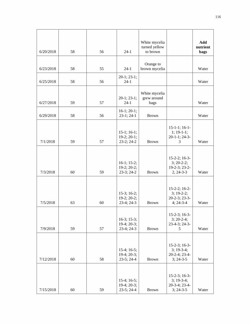
116
6/20/2018 58 56 24-1
White mycelia
turned yellow
to brown
Add
nutrient
bags
6/23/2018 58 55 24-1 Orange to
brown mycelia Water
6/25/2018 58 56 20-1; 23-1;
24-1 Water
6/27/2018 59 57 20-1; 23-1;
24-1
White mycelia
grew around
bags Water
6/29/2018 58 56 16-1; 20-1;
23-1; 24-1 Brown Water
7/1/2018 59 57
15-1; 16-1;
19-2; 20-1;
23-2; 24-2 Brown
15-1-1; 16-1-
1; 19-1-1;
20-1-1; 24-3-
3 Water
7/3/2018 60 59
16-1; 15-2;
19-2; 20-2;
23-3; 24-2 Brown
15-2-2; 16-3-
3; 20-2-2;
19-2-3; 23-2-
2, 24-3-3 Water
7/5/2018 63 60
15-3; 16-2;
19-2; 20-2;
23-4; 24-3 Brown
15-2-2; 16-2-
3; 19-2-2;
20-2-3; 23-3-
4; 24-3-4 Water
7/9/2018 59 57
16-3; 15-3;
19-4; 20-3;
23-4; 24-3 Brown
15-2-3; 16-3-
3; 20-2-4;
23-4-3; 24-3-
5 Water
7/12/2018 60 58
15-4; 16-5;
19-4; 20-3;
23-5; 24-4 Brown
15-2-3; 16-3-
3; 19-3-4;
20-2-4; 23-4-
3; 24-3-5 Water
7/15/2018 60 59
15-4; 16-5;
19-4; 20-3;
23-5; 24-4 Brown
15-2-3; 16-3-
3; 19-3-4;
20-3-4; 23-4-
3; 24-3-5 Water
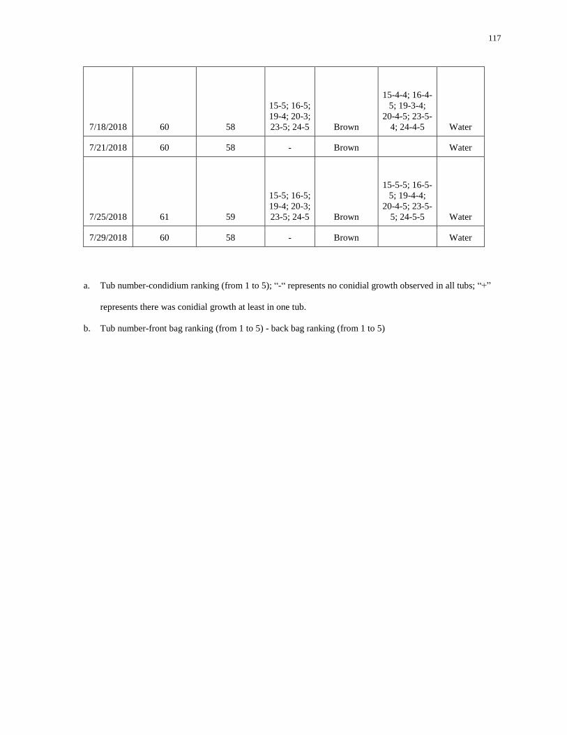
117
7/18/2018 60 58
15-5; 16-5;
19-4; 20-3;
23-5; 24-5 Brown
15-4-4; 16-4-
5; 19-3-4;
20-4-5; 23-5-
4; 24-4-5 Water
7/21/2018 60 58 - Brown Water
7/25/2018 61 59
15-5; 16-5;
19-4; 20-3;
23-5; 24-5 Brown
15-5-5; 16-5-
5; 19-4-4;
20-4-5; 23-5-
5; 24-5-5 Water
7/29/2018 60 58 - Brown Water
a. Tub number-condidium ranking (from 1 to 5); “-“ represents no conidial growth observed in all tubs; “+”
represents there was conidial growth at least in one tub.
b. Tub number-front bag ranking (from 1 to 5) - back bag ranking (from 1 to 5)

118
Table A-3. Soil test report (particle size)
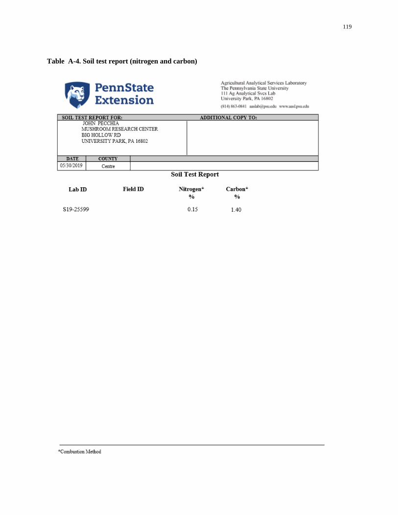
119
Table A-4. Soil test report (nitrogen and carbon)

120
Table A-5. Soil test report (soluble salts, organic matter and pH))

121
Table A-6. Compost analysis report of morel sawdust compost
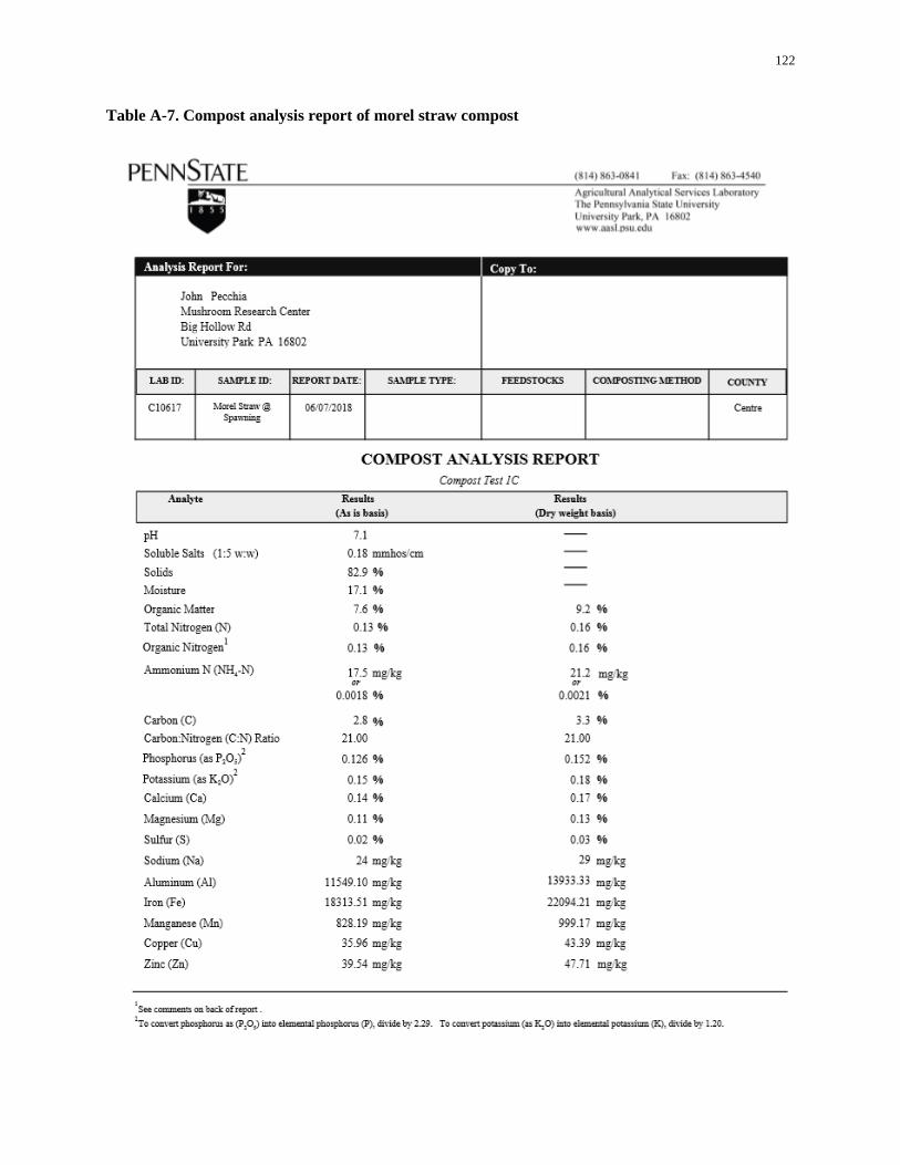
122
Table A-7. Compost analysis report of morel straw compost
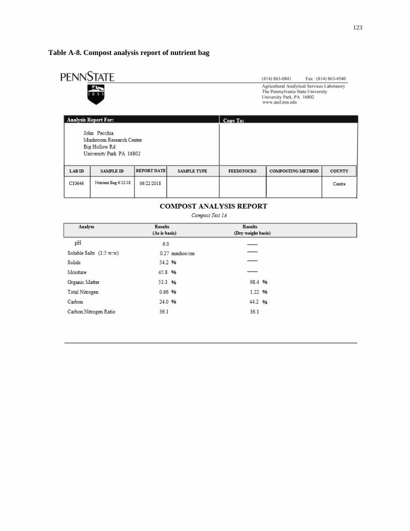
123
Table A-8. Compost analysis report of nutrient bag
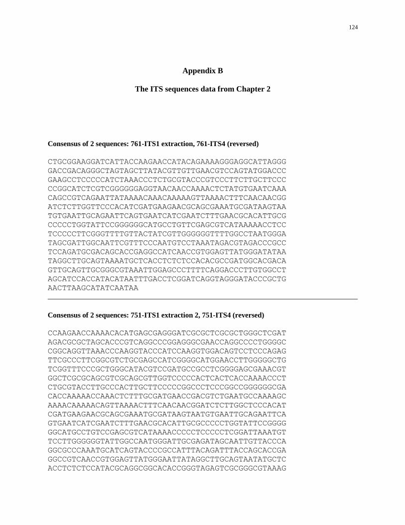
124
Appendix B
The ITS sequences data from Chapter 2
Consensus of 2 sequences: 761-ITS1 extraction, 761-ITS4 (reversed)
CTGCGGAAGGATCATTACCAAGAACCATACAGAAAAGGGAGGCATTAGGG
GACCGACAGGGCTAGTAGCTTATACGTTGTTGAACGTCCAGTATGGACCC
GAAGCCTCCCCCATCTAAACCCTCTGCGTACCCGTCCCTTCTTGCTTCCC
CCGGCATCTCGTCGGGGGGAGGTAACAACCAAAACTCTATGTGAATCAAA
CAGCCGTCAGAATTATAAAACAAACAAAAAGTTAAAACTTTCAACAACGG
ATCTCTTGGTTCCCACATCGATGAAGAACGCAGCGAAATGCGATAAGTAA
TGTGAATTGCAGAATTCAGTGAATCATCGAATCTTTGAACGCACATTGCG
CCCCCTGGTATTCCGGGGGGCATGCCTGTTCGAGCGTCATAAAAACCTCC
TCCCCCTTCGGGTTTTGTTACTATCGTTGGGGGGTTTTGGCCTAATGGGA
TAGCGATTGGCAATTCGTTTCCCAATGTCCTAAATAGACGTAGACCCGCC
TCCAGATGCGACAGCACCGAGGCCATCAACCGTGGAGTTATGGGATATAA
TAGGCTTGCAGTAAAATGCTCACCTCTCTCCACACGCCGATGGCACGACA
GTTGCAGTTGCGGGCGTAAATTGGAGCCCTTTTCAGGACCCTTGTGGCCT
AGCATCCACCATACATAATTTGACCTCGGATCAGGTAGGGATACCCGCTG
AACTTAAGCATATCAATAA
Consensus of 2 sequences: 751-ITS1 extraction 2, 751-ITS4 (reversed)
CCAAGAACCAAAACACATGAGCGAGGGATCGCGCTCGCGCTGGGCTCGAT
AGACGCGCTAGCACCCGTCAGGCCCGGAGGGCGAACCAGGCCCCTGGGGC
CGGCAGGTTAAACCCAAGGTACCCATCCAAGGTGGACAGTCCTCCCAGAG
TTCGCCCTTCGGCGTCTGCGAGCCATCGGGGCATGGAACCTTGGGGGCTG
TCGGTTTCCCGCTGGGCATACGTCCGATGCCGCCTCGGGGAGCGAAACGT
GGCTCGCGCAGCGTCGCAGCGTTGGTCCCCCACTCACTCACCAAAACCCT
CTGCGTACCTTGCCCACTTGCTTCCCCCGGCCCTCCCGGCCGGGGGGCGA
CACCAAAAACCAAACTCTTTGCGATGAACCGACGTCTGAATGCCAAAAGC
AAAACAAAAACAGTTAAAACTTTCAACAACGGATCTCTTGGCTCCCACAT
CGATGAAGAACGCAGCGAAATGCGATAAGTAATGTGAATTGCAGAATTCA
GTGAATCATCGAATCTTTGAACGCACATTGCGCCCCCTGGTATTCCGGGG
GGCATGCCTGTCCGAGCGTCATAAAACCCCCTCCCCCTCGGATTAAATGT
TCCTTGGGGGGTATTGGCCAATGGGATTGCGAGATAGCAATTGTTACCCA
GGCGCCCAAATGCATCAGTACCCCGCCATTTACAGATTTACCAGCACCGA
GGCCGTCAACCGTGGAGTTATGGGAATTATAGGCTTGCAGTAATATGCTC
ACCTCTCTCCATACGCAGGCGGCACACCGGGTAGAGTCGCGGGCGTAAAG
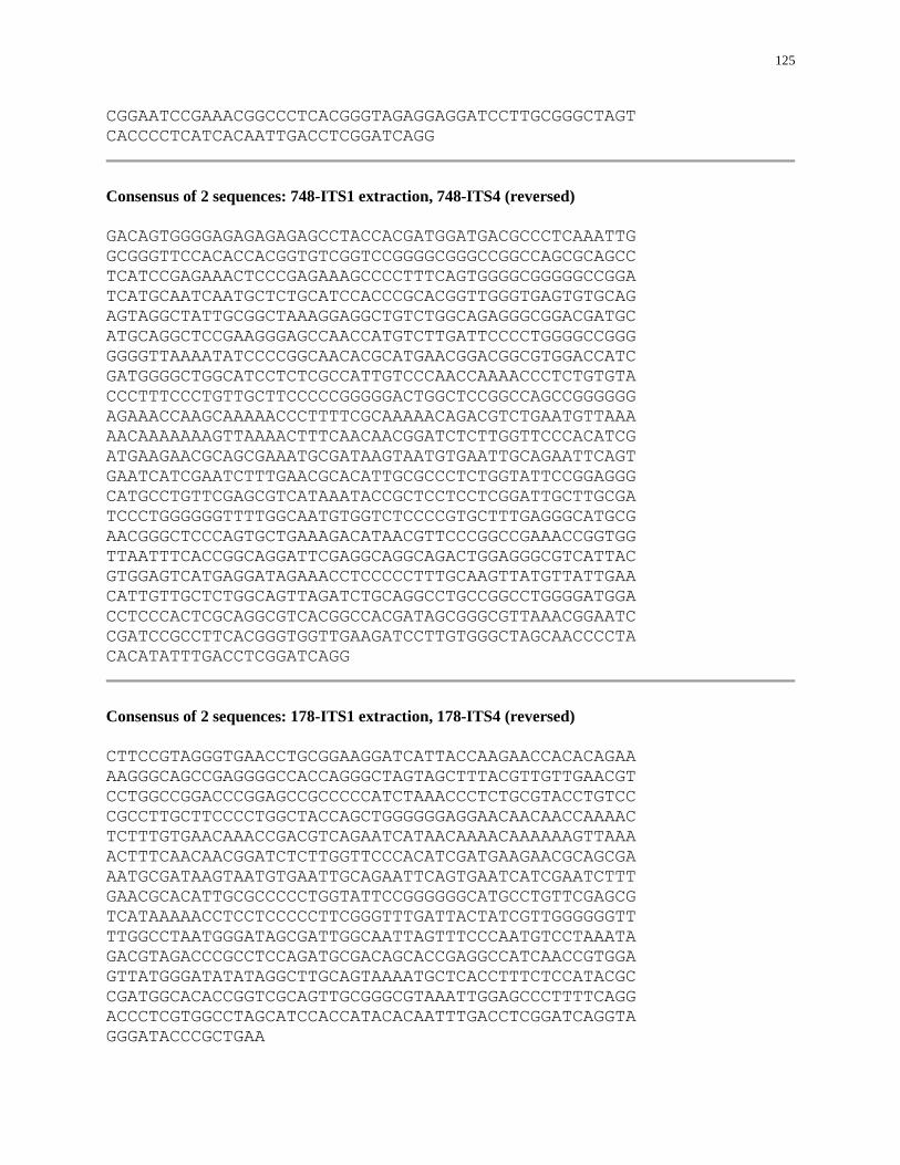
125
CGGAATCCGAAACGGCCCTCACGGGTAGAGGAGGATCCTTGCGGGCTAGT
CACCCCTCATCACAATTGACCTCGGATCAGG
Consensus of 2 sequences: 748-ITS1 extraction, 748-ITS4 (reversed)
GACAGTGGGGAGAGAGAGAGCCTACCACGATGGATGACGCCCTCAAATTG
GCGGGTTCCACACCACGGTGTCGGTCCGGGGCGGGCCGGCCAGCGCAGCC
TCATCCGAGAAACTCCCGAGAAAGCCCCTTTCAGTGGGGCGGGGGCCGGA
TCATGCAATCAATGCTCTGCATCCACCCGCACGGTTGGGTGAGTGTGCAG
AGTAGGCTATTGCGGCTAAAGGAGGCTGTCTGGCAGAGGGCGGACGATGC
ATGCAGGCTCCGAAGGGAGCCAACCATGTCTTGATTCCCCTGGGGCCGGG
GGGGTTAAAATATCCCCGGCAACACGCATGAACGGACGGCGTGGACCATC
GATGGGGCTGGCATCCTCTCGCCATTGTCCCAACCAAAACCCTCTGTGTA
CCCTTTCCCTGTTGCTTCCCCCGGGGGACTGGCTCCGGCCAGCCGGGGGG
AGAAACCAAGCAAAAACCCTTTTCGCAAAAACAGACGTCTGAATGTTAAA
AACAAAAAAAGTTAAAACTTTCAACAACGGATCTCTTGGTTCCCACATCG
ATGAAGAACGCAGCGAAATGCGATAAGTAATGTGAATTGCAGAATTCAGT
GAATCATCGAATCTTTGAACGCACATTGCGCCCTCTGGTATTCCGGAGGG
CATGCCTGTTCGAGCGTCATAAATACCGCTCCTCCTCGGATTGCTTGCGA
TCCCTGGGGGGTTTTGGCAATGTGGTCTCCCCGTGCTTTGAGGGCATGCG
AACGGGCTCCCAGTGCTGAAAGACATAACGTTCCCGGCCGAAACCGGTGG
TTAATTTCACCGGCAGGATTCGAGGCAGGCAGACTGGAGGGCGTCATTAC
GTGGAGTCATGAGGATAGAAACCTCCCCCTTTGCAAGTTATGTTATTGAA
CATTGTTGCTCTGGCAGTTAGATCTGCAGGCCTGCCGGCCTGGGGATGGA
CCTCCCACTCGCAGGCGTCACGGCCACGATAGCGGGCGTTAAACGGAATC
CGATCCGCCTTCACGGGTGGTTGAAGATCCTTGTGGGCTAGCAACCCCTA
CACATATTTGACCTCGGATCAGG
Consensus of 2 sequences: 178-ITS1 extraction, 178-ITS4 (reversed)
CTTCCGTAGGGTGAACCTGCGGAAGGATCATTACCAAGAACCACACAGAA
AAGGGCAGCCGAGGGGCCACCAGGGCTAGTAGCTTTACGTTGTTGAACGT
CCTGGCCGGACCCGGAGCCGCCCCCATCTAAACCCTCTGCGTACCTGTCC
CGCCTTGCTTCCCCTGGCTACCAGCTGGGGGGAGGAACAACAACCAAAAC
TCTTTGTGAACAAACCGACGTCAGAATCATAACAAAACAAAAAAGTTAAA
ACTTTCAACAACGGATCTCTTGGTTCCCACATCGATGAAGAACGCAGCGA
AATGCGATAAGTAATGTGAATTGCAGAATTCAGTGAATCATCGAATCTTT
GAACGCACATTGCGCCCCCTGGTATTCCGGGGGGCATGCCTGTTCGAGCG
TCATAAAAACCTCCTCCCCCTTCGGGTTTGATTACTATCGTTGGGGGGTT
TTGGCCTAATGGGATAGCGATTGGCAATTAGTTTCCCAATGTCCTAAATA
GACGTAGACCCGCCTCCAGATGCGACAGCACCGAGGCCATCAACCGTGGA
GTTATGGGATATATAGGCTTGCAGTAAAATGCTCACCTTTCTCCATACGC
CGATGGCACACCGGTCGCAGTTGCGGGCGTAAATTGGAGCCCTTTTCAGG
ACCCTCGTGGCCTAGCATCCACCATACACAATTTGACCTCGGATCAGGTA
GGGATACCCGCTGAA
126
Consensus of 2 sequences: 833-ITS1 extraction, 833-ITS4 (reversed)
GCGGAAGGATCATTACAAAAATCCAACCAAAAAGAGGCGAGGACCGCGGC
CGCCTGGTCTCCCCGGTTAAGAGGCACCCACCCTCAGCCCCCATCTGCGC
GCGGCGGGGGATGCGGGGCCTCCCGTACTGGCCGGGCCGCGGGCCCCTCC
GTCTTCACACAAACCCTCTGTGTACCTTGTCCCCATTGCTTCCCTCGGAA
CGAGAGCTTCTCCGGGCTGGTCCGGGTGGCGGCGCCCCGTGGAACGGGCG
GCGCAACCCGATTGGCCACGCCTGGCTCGCTCCTCGCCCGGGGGGAGGAA
AACCCAAACAAACCCTTTGCGCGAAAAACGACGTCTGAACAGAAAAGAAA
ACAAACTAAAAGTTAAAACTTTCAACAACGGATCTCTTGGTTCCCACATC
GATGAAGAACGCAGCGAAATGCGATAAGTAATGTGAATTGCAGAATTCAG
TGAATCATCGAATCTTTGAACGCACATTGCGCCCTCTGGTATTCCGGGGG
GCATGCCTGTTCGAGCGTCATAAAAACCTCTCCACCCCGGGTTCATGAGA
TCCGGGGCGGGTATTGGCGGTGCGGATCCCTCCCCCCTATACAACGGGAC
GGAGGCGCCCACCGCTCAAAGCCATGAAGATAACCCAGCCGAAAGCGCCA
GGGCGTCAACCGTGGAGTCATGAGGAACCACGCCCCGCGAGGGGTGCCTC
CCACTCTGGGCGACCGGTGTATGGAAATGCGGGCGTAAAAGCACGGGAAA
TCCGCGGGAACCGGCGCCGTCTCTGGCTGCGGCCGTGGATGATCCCCGTG
GGGGGCTAGATACCCAATCACATTCAAGTTTGACCTCGGATC
Consensus of 2 sequences: 767-ITS1 extraction, 767-ITS4 (reversed)
AACGAAGCAACGGAACGGTGCATCTGCGCCAGTGACGGGCCAGTCCTGGG
GTGCACGGTAAGACGTCCCGGGCGCTGTTTGACGGTGGAAGGGAGCGTTT
CCAGGCACTCTTGTGCTTGGGCGTATTGACCCCCCACCGTCCTTACCTGA
CACGCACCGGGTCGCACAGAGCGGCCAGGCCCCCCCACAGTGCACATGTT
CCCCTTCCGTTGCGGAGTGGAGTCGAACCAAGACCCTCTGCGTACCTTGC
CTTCCTTGCTTCCCCCCGACCACCTGGGCGGGGGGGAGAAAACAAACCAC
AAACTCTTTGCGAATAACCGACGTCAGAAAACAAAACAAAAAAAGTTAAA
ACTTTCAACAACGGATCTCTTGGTTCCCACATCGATGAAGAACGCAGCGA
AATGCGATAAGTAATGTGAATTGCAGAATTCAGTGAATCATCGAATCTTT
GAACGCACATTGCGCCCCCTGGTATTCCGGGGGGCATGCCTGTTCGAGCG
TCACAAACACCTCTCCCCCTCGGGTTTAGCTACCCTTGGGGGGTATTGGC
CAATGGCATTGCGAGATAGCAATTGTTAGCCAGGAGCCCAAATGCATTAG
TTTCCCGCCATTTTTAAGATGCAACAGCACCGAGGGGTATCCAAATCGGG
GTTCTAATGGTTTGAGGGAGGCCCGCTGCGGTAGTACGCTCGTGGCCTTC
TCCCGCCCCACCGCCGGACCCACATTAGTTGCAGTCGCGGGCGTAAAGCG
ACATCCGATCATCCCTCGCGGGGTGTTGACGATCATCGGGGTTAATAGCC
CCTACACAAAGTTTGACCTCGGATC
Consensus of 2 sequences: 766-ITS1 extraction, 766-ITS4 (reversed)
ACCTGCGGAAGGATCATTACCAAGAACCAAACAGAAAAGGGCAGCCGAGG
127
GGCCAACAGGGCTAGTAGCTTTACGTTGTTGAACGTCCTGGAACGGACCC
GGAGCCGCCCCCATCTAAACCCTCTGTGTACCTGTCCCACCTTGCTTCCC
CCGGCTACCCGTCGGGGGGAGGAACAACAACCAAAACTCTTTGTGAACAA
ACAGACGTCAGAATCACAACAACAAAAAAAAGTTAAAACTTTCAACAACG
GATCTCTTGGTTCCCACATCGATGAAGAACGCAGCGAAATGCGATAAGTA
ATGTGAATTGCAGAATTCAGTGAATCATCGAATCTTTGAACGCACATTGC
GCCCCCTGGTATTCCGGGGGGCATGCCTGTTCGAGCGTCATAAAAACCTC
CTCCCCCTTCGGGTTTGATTACTATCGTTGGGGGGTTTTGGCCTAATGGG
ATAGCGATTGGCAATCAGTTTCCCAATGTCCTAAATAGACGTAGACCCGC
CTCCAGATGCGACAGCACCGAGGCCATCAACCGTGGAGTTATGGGATATA
TAGGCTTGCAGTAAAATGCTCACCTTTCTCCATACGCCGATGGCACACCG
GTCGCAGTTGCGGGCGTAAATTGGAGCCCTTTTCAGGACCCTCGTGGCCT
AGCATCCACCTCTCACAATTTGACCTCGGATCAGGTAGGGATACCCGCTG
AACTTAAGCATATCAATA
Consensus of 2 sequences: 765-ITS1 extraction, 765-ITS4 (reversed)
AAGACAGTGGGGAGAGAGGCACCAGCGATGGAGGACGCCGCCACATTGGC
GGGTTCCGTACCCCAGTGTTGGTCCAGGGCGGGCCAGCTGCGACGGTCTC
ATCCGCATTCCCTGCGTTTTGGCAGCAAGCCCCGAATCGGGGTGGGAACG
GATCGTGCAATCAATGCTCTGCAGCCATCCATACGGGTTGGGTGAGCGCG
CAGAGTAGACTATTGCTGCGCTAAAGGAGACCCGTCGCGGCAGGTGGCGG
ACGGCACATGCAGGACTGATCCCAACCATGTGTTGATTCCCCTGGGGCCG
GGGTTGTAGAATACCCCGGCGACTCGCAGACGGCGTGTACCCTCGTTTGG
CCCCCTCTCGCCATTGTCCCAACCAAAACCCTCTGTGTACCCCTTCCCTG
TTGCTTCCCCCGGGGCACTGGCTCCGGCCAGCCGGGGGGAGAAACCAAGC
AAAAACCCTTTTTCGCAAAAACAGACGTCTGAACATCAAAAACAAAAAAG
TTAAAACTTTCAACAACGGATCTCTTGGTTCCCACATCGATGAAGAACGC
AGCGAAATGCGATAAGTAATGTGAATTGCAGAATTCAGTGAATCATCGAA
TCTTTGAACGCACATTGCGCCCTCTGGTATTCCGGAGGGCATGCCTGTTC
GAGCGTCATAAATACCCCTCCTCCTCGGATTGCTTTTGCGGTCCCTGGGG
GGTTCTGGCAATGTGGATTCCCCGTGCTTTGAGGGCATGCGGACGGGCTC
CCAGTGCTGAAAGACATAATGTTCCCGGCCGAAACCGGTGATTAATTTCA
CTGGCAGGATTCGTGGCAGGTAGACTGAGGGCGTCAACCGTGGAGTCATG
AGGGTAGAAACCTCCCCCTTTGCAAGTAACATTGCTCTGGCAGTTAGATC
TGCAGGCCCGACGGTCTGGGGATGGACCTCCCACTCGCAGGCGTCACGGC
CACGATAGCGGGCGTTAAACGGAATCCGATCCGCCTTCACGGGTGGTTGA
AGATCCTTGTGGGCTAGCAACCCCTAAACATATT
Consensus of 2 sequences: 764-ITS1 extraction, 764-ITS4 (reversed)
GACAGTGGCGGTCGAGAGAGAGTCAGCTACGATGGGGCGCGCCGGCAACC
AGACCGCTGGCACACTGGTGGCGGGTACTGTCACCCGCACTTTGTTGCAA
CGTCCAGGGCGAGAGCCTTAGCCGGGTGTGGCCCCATCCGTTAAGCTAGA
CAATTCCATGCTGCCTGCTGCGCTCCCTCCCCACGCCCTGGGCAGCAGCT
128
CCCGCAATCGGGGGTGGAAGCCGGATCAAGGAATCAATGCTCTGCATACA
TTCATTCAATTGAATGTGGTGCAGAGTAGGTTATTTCTGCTGAAGGGGGA
TCCCCCGGCAGAGGTGGACGGCACATGCAGGCTCCGAACGACGGAGGCCA
ACCATGTCGCTGAGTCCCCTGGGGCTAGGGGCGGTAAAAGCCCCTAGCCA
CTCCCCGCCGACGGCGTTGACCCCATCGTTCTGGCACCCTCTCGCCACTG
TCCCAACCAAAACCCTCTGTGTACCCTTCCCTGTTGCTTCCCCCGGGCAA
CTGGCTCCGGCCAGCCGGGGGGAGAAACCAAGCAAAAACCCTTTTCGCAA
AACAGACGTCTGAACGCAAAAAACAAAACAAAAAAAAGTTAAAACTTTCA
ACAACGGATCTCTTGGTTCCCACATCGATGAAGAACGCAGCGAAATGCGA
TAAGTAATGTGAATTGCAGAATTCAGTGAATCATCGAATCTTTGAACGCA
CATTGCGCCCTCTGGTATTCCGGAGGGCATGCCTGTTCGAGCGTCATAAA
TACCGCTCCCCCTCGGATTGCTTGCGGTCCCTGGGGGGTTCTGGCAATGT
GGATTCCCCGTGCTTTGAGGGCATGCGAACGGGCTCCCAGTGCTGAAAGA
CATAATGTTCCCAGCCGAAACCGGTGATTAATTTCATCGGCAGGATTCGT
GGCAGGCAGACTGAGGGCGTCAACCGTGGAGTCATGAGGGTAGAAACCTC
CCCCTTTGCAAGTAACATTGCTCTGGCAGTTAGATCTGCAGGCCTGCCGG
TCTGGGGATGGACCTCCCACTCGCAGGCGTCACGGCCACGATAGCGGGCG
TTAAACGGAATCCGATCCGCCCTCACGGGTGGTTGAAGATCCTTGTGGGC
TAGCAACCCCTAAACACATTTTGACCTCGGA
Consensus of 2 sequences: 763-ITS1 extraction, 763-ITS4 (reversed)
GGAAGGATCATTACAAAAAAAACGTTCGGACGGGGGGCACAAGTGGTGGG
AAGCCAAGGCGATGCTAGCTGGACGCCGTGTGGCCTGTGGTTGCCGGGGG
TTAAACGTTCACACCCCCCCTCGCGCAGCACACATCAGTGGCCGAACCCA
GGCAAAGCACCGGCCCCCATCGACTGCCCCCAGTCCCAAAACAAAAACCC
TCTGCGTACTCTCCCCTCCTTGCTTCCCCCGGCACACGTCGGGGGGAGGA
ACACAAACCAAAACTCTTAGCGTGTAACCGCCGTCAGAAATACAGATACA
AAAGAAAAGTTAAAACTTTCAACAACGGATCTCTTGGTTCCCACATCGAT
GAAGAACGCAGCGAAATGCGATAAGTAATGTGAATTGCAGAATTCAGTGA
ATCATCGAATCTTTGAACGCACATTGCGCCCCCTGGTACTCCGGGGGGCA
TGCCTGTTCGAGCGTCATAAAACCATCTCCCCCAAGCGTGTTCTACGCTT
GGGGGGTCTTGGCCTGCGGGAAAGCGACTGGCAATCCGCTCCCGAGAGCC
CAAATACAGGAGACCCGCCATCTGATGCGACAGCACCGAGGCCCTCAACC
GTGGAGTCATGGGACTTTTGGATAGCAGTAATATGCTCACCTTTCTTCAC
ACGCCGTTGGCACACCAGTCGCAGTTGCGGGCGTAATATGGAGTCCCCCA
GGGGGGGACCCTTGTGGATTAGCACCCACCTCACGAACTTTGACCTCGGA
TCAGGTAGGGATACCCGCTGAACTTAAGCATATCAAT
Consensus of 2 sequences: 759-ITS1 extraction 2, 759-ITS4 (reversed)
AAAGACAGTGGGGAGAGAGGCACCAGCGATGGAGGACGCCGCCACATTGG
CGGGTTCCGTACCCCAGTGTTGGTCCAGGGCGGGCCAGCTGCGACGGTCT
CATCCGCATTCCCTGCGTTTTGGCAGCAAGCCCCGAATCGGGGTGGGAAC
GGATCGTGCAATCAATGCTCTGCAGCCATCCATACGGGTTGGGTGAGCGC
129
GCAGAGTAGACTATTGCTGCGCTAAAGGAGACCCGTCGCGGCAGGTGGCG
GACGGCACATGCAGGACTGATCCCAACCATGTGTTGATTCCCCTGGGGCC
GGGGTTGTAGAATACCCCGGCGACTCGCAGACGGCGTGTACCCTCGTTTG
GCCCCCTCTCGCCATTGTCCCAACCAAAACCCTCTGTGTACCCCTTCCCT
GTTGCTTCCCCCGGGGCACTGGCTCCGGCCAGCCGGGGGGAGAAACCAAG
CAAAAACCCTTTTTCGCAAAAACAGACGTCTGAACATCAAAAACAAAAAA
GTTAAAACTTTCAACAACGGATCTCTTGGTTCCCACATCGATGAAGAACG
CAGCGAAATGCGATAAGTAATGTGAATTGCAGAATTCAGTGAATCATCGA
ATCTTTGAACGCACATTGCGCCCTCTGGTATTCCGGAGGGCATGCCTGTT
CGAGCGTCATAAATACCCCTCCTCCTCGGATTGCTTTTGCGGTCCCTGGG
GGGGTTCTGGCAATGTGGATTCCCCGTGCTTTGAGGGCATGCGGACGGGC
TCCCAGTGCTGAAAGACATAATGTTCCCGGCCGAAACCGGTGATTAATTT
CACTGGCAGGATTCGTGGCAGGTAGACTGAGGGCGTCAACCGTGGAGTCA
TGAGGGTAGAAACCTCCCCCTTTGCAAGTAACATTGCTCTGGCAGTTAGA
TCTGCAGGCCCGACGGTCTGGGGATGGACCTCCCACTCGCAGGCGTCACG
GCCACGATAGCGGGCGTTAAACGGAATCCGATCCGCCTTCACGGGTGGTT
GAAGATCCTTGTGGGCTAGCAACCCCTAAACATA
Consensus of 2 sequences: 756-ITS1 extraction, 756-ITS4 (reversed)
ACAAACGAAGCAACGGAACGGTGCATCTGCGCCAGTGACGGGCCAGTCCT
GGGGTGCACGGTAAGACGTCCCGGGCGCTGTTTGACGGTGGAAGGGAGCG
TTTCCAGGCACTCTTGTGCTTGGGCGTATTGACCCCCCACCGTCCTTACC
TGACACGCACCGGGTCGCACAGAGCGGCCAGGCCCCCCCACAGTGCACAT
GTTCCCCTTCCGTTGCGGAGTGGAGTCGAACCAAGACCCTCTGCGTACCT
TGCCTTCCTTGCTTCCCCCCGACCGCCTGGGCGGGGGGGAGAAAACAAAC
CACAAACTCTTTGCGAATAACCGACGTCAGAAAACAAAACAAAAAAAGTT
AAAACTTTCAACAACGGATCTCTTGGTTCCCACATCGATGAAGAACGCAG
CGAAATGCGATAAGTAATGTGAATTGCAGAATTCAGTGAATCATCGAATC
TTTGAACGCACATTGCGCCCCCTGGTATTCCGGGGGGCATGCCTGTTCGA
GCGTCACAAACACCTCTCCCCCTCGGGTTTAGCTACCCTTGGGGGGTATT
GGCCAATGGCATTGCGAGATAGCAATTGTTAGCCAGGAGCCCAAATGCAT
TAGTTTCCCGCCATTTTTAAGATGCAACAGCACCGAGGGGTATCCAAATC
GGGGTTCTAATGGTTTGAGGGAGGCCCGCTGCGGTAGTACGCTCGTGGCC
TTCTCCCGCCCCACCGCCGGACCCACATTAGTTGCAGTCGCGGGCGTAAA
GCGACATCCGATCATCCCTCGCGGGGTGTTGACGATCATCGGGGTTAATA
GCCCCTACACAAAGTTGACCTCGGAT
Consensus of 2 sequences: 755-ITS1 extraction 2, 755-ITS4 (reversed)
GCGGAAGGATCATTACCAAGAACCAAAACACATGAGCGAGGGATCGCGCT
CGCGCTGGGCTCGATAGACGCGCTAGCACCCGTCAGGCCCGGAGGGCGAA
CCAGGCCCCTGGGGCCGGCAGGTTAAACCCAAGGTACCCATCCAAGGTGG
ACAGTCCTCCCAGAGTTCGCCCTTCGGCGTCTGCGAGCCATCGGGGCATG
GAACCTTGGGGGCTGTCGGTTTCCCGCTGGGCATACGTCCGATGCCGCCT
130
CGGGGAGCGAAACGTGGCTCGCGCAGCGTCGCAGCGTTGGTCCCCCACTC
ACTCACCAAAACCCTCTGCGTACCTTGCCCACTTGCTTCCCCCGGCCCTC
CCGGCCGGGGGGCGACACCAAAAACCAAACTCTTTGCGATGAACCGACGT
CTGAATGCCAAAAGCAAAACAAAAACAGTTAAAACTTTCAACAACGGATC
TCTTGGCTCCCACATCGATGAAGAACGCAGCGAAATGCGATAAGTAATGT
GAATTGCAGAATTCAGTGAATCATCGAATCTTTGAACGCACATTGCGCCC
CCTGGTATTCCGGGGGGCATGCCTGTCCGAGCGTCATAAAACCCCCTCCC
CCTCGGATTAAATGTTCCTTGGGGGGTATTGGCCAATGGGATTGCGAGAT
AGCAATTGTTACCCAGGCGCCCAAATGCATCAGTACCCCGCCATTTACAG
ATTTACCAGCACCGAGGCCGTCAACCGTGGAGTTATGGGAATTATAGGCT
TGCAGTAATATGCTCACCTCTCTCCATACGCAGGCGGCACACCGGGTAGA
GTCGCGGGCGTAAAGCGGAATCCGAAACGGCCCTCACGGGTAGAGGAGGA
TCCTTGCGGGCTAGTCACCCCTCATCACAATTTGACCTCGGATCAG
Consensus of 2 sequences: 752-ITS1 extraction, 752-ITS4 (reversed)
TGCGGAAGGATCATTACCAAGAACCACACAGAAAAGGGCAGCCGAGGGGC
CACCAGGGCTAGTAGCTTTACGTTGTTGAACGTCCTGGAACGGACCCGGA
GCCGCCCCCATCTAAACCCTCTGCGTACCTGTCCCACCTTGCTTCCCCCG
GCTTACCCGCTGGGGGGAGGAACAACAACCAAAACTCTTTGTGAAGAAAC
AGACGTCAGAATCATAACAAAAAAAAGTTAAAACTTTCAACAACGGATCT
CTTGGTTCCCACATCGATGAAGAACGCAGCGAAATGCGATAAGTAATGTG
AATTGCAGAATTCAGTGAATCATCGAATCTTTGAACGCACATTGCGCCCT
CTGGTATTCCGGGGGGCATGCCTGTTCGAGCGTCATAAAAACCTCCTCCC
CCTTCGGGTTTGATTACTATCGTCGGGGGGTTTTGGCCTAATGGGAAAGC
GATTGGCAATTGTCTTCCCAATGTCCTAAATACACTTAGACCCGCCTCCA
GATGCGACAGCACCGAGGCCATCAACCGTGGAGTTATGGGACACCGTTCT
CCATACGCCGATGGCAAACCGGTCGCAGTTGCGGGCGTAAATTGGAGTCC
TCTTCAGGACCCTCGTGGCCTAGCATCCACCATACATAATTNGACCTCGG
ATCAGGTAGGGATACCCGCTGAACTTAAGCATATCAATAAGC
Consensus of 2 sequences: 747-ITS1 extraction, 747-ITS4 (reversed)
ACAGTGGCGGTCGAGAGAGAGTCAGCTATACACGATGGGGCGCGCCGGCA
AACCAGACCGCTGGCACACTGGTGGCGGGTACTGTCACCCGCGCCGTTTG
TCGCAACGTCCAGGGCGAGAGCCTTAGCCGGGTGTGGCCCCATCCGTTAT
GCTAGCCAATTCCATGCTGCCTGCTGCGCTCCCTCCCCACGCCCTGGGCA
GCAGCTCCCGCAATCGGGGGTGGAAGCCGGATCAAGGAATCAATGCTCTG
CACCCATTCAGTCAATTGAATGTGGTGCAGTTTAGGTTATTTCTGCTGAA
GGGGGATCCCCCGGCAGAGGTGGACGGCACATGCAGGCTCCGAACGACGG
AAGCCAACCATGATGCTGAGTCCCCTGGGGCTAGGGGTGGTAAAAGCCCC
TAGCCACTCCCCACCGACGGCGTTGACCCCATCGTTCTGGCACCCTCTCG
CCACTGTCCCAACCAAAACCCTCTGTGTACCCTTCCCTGTTGCTTCCCCC
GGGAAACTGGCTCCGGCCAGCCGGGGGGAGAAACCAAGCAAAAACCCTTT
TCGCAAAACAGACGTCTGAACGCAAAAAACAAAACAAAAAAAAGTTAAAA
131
CTTTCAACAACGGATCTCTTGGTTCCCACATCGATGAAGAACGCAGCGAA
ATGCGATAAGTAATGTGAATTGCAGAATTCAGTGAATCATCGAATCTTTG
AACGCACATTGCGCCCTCTGGTATTCCGGAGGGCATGCCTGTTCGAGCGT
CATAAATACCGCTCCCCCTCGGATTGCTTGCGGTCCCTGGGGGGTTCTGG
CAATGTGGATTCCCCGTGCTTTGAGGGCATGCGAACGGGCTCCCAGTGCT
GAAAGACATAATGTTCCCAGCCGAAACCGGTGATTAATTTCATCGGCAGG
ATTCGTGGCAGGCAGACTGAGGGCGTCAACCGTGGAGTCATGAGGGTAGA
AACCTCCCCCTTTGCAAGTAACATTGCTCTGGCAGTTAGATCTGCAGGCC
TGCCGGTCTGGGGATGGACCTCCCACTCGCAGGCGTCACGGCCACGATAG
CGGGCGTTAAACGGAATCCGATCCGCCCTCACGGGTGGTTGAAGATCCTT
GTGGGCTAGCAACCCCTAAACACATTTGACCTCGGATCAGG
Consensus of 2 sequences: 746-ITS1 extraction, 746-ITS4 (reversed)
AAGACAGTGGCGGTCGAGAGAGAGTCAGCTATACACGATGGGGCGCGCCG
GCAAACCAGACCGCTGGCACACTGGTGGCGGGTACTGTCACCCGCGCCGT
TTGTCGCAACGTCCAGGGCGAGAGCCTTAGCCGGGTGTGGCCCCATCCGT
TATGCTAGCCAATTCCATGCTGCCTGCTGCGCTCCCTCCCCACGCCCTGG
GCAGCAGCTCCCGCAATCGGGGGTGGAAGCCGGATCAAGGAATCAATGCT
CTGCACCCATTCAGTCAATTGAATGTGGTGCAGTTTAGGTTATTTCTGCT
GAAGGGGGATCCCCCGGCAGAGGTGGACGGCACATGCAGGCTCCGAACGA
CGGAAGCCAACCATGATGCTGAGTCCCCTGGGGCTAGGGGTGGTAAAAGC
CCCTAGCCACTCCCCACCGACGGCGTTGACCCCATCGTTCTGGCACCCTC
TCGCCACTGTCCCAACCAAAACCCTCTGTGTACCCTTCCCTGTTGCTTCC
CCCGGGAAACTGGCTCCGGCCAGCCGGGGGGAGAAACCAAGCAAAAACCC
TTTTCGCAAAACAGACGTCTGAACGCAAAAAACAAAACAAAAAAAAGTTA
AAACTTTCAACAACGGATCTCTTGGTTCCCACATCGATGAAGAACGCAGC
GAAATGCGATAAGTAATGTGAATTGCAGAATTCAGTGAATCATCGAATCT
TTGAACGCACATTGCGCCCTCTGGTATTCCGGAGGGCATGCCTGTTCGAG
CGTCATAAATACCGCTCCCCCTCGGATTGCTTGCGGTCCCTGGGGGGTTC
TGGCAATGTGGATTCCCCGTGCTTTGAGGGCATGCGAACGGGCTCCCAGT
GCTGAAAGACATAATGTTCCCAGCCGAAACCGGTGATTAATTTCATCGGC
AGGATTCGTGGCAGGCAGACTGAGGGCGTCAACCGTGGAGTCATGAGGGT
AGAAACCTCCCCCTTTGCAAGTAACATTGCTCTGGCAGTTAGATCTGCAG
GCCTGCCGGTCTGGGGATGGACCTCCCACTCGCAGGCGTCACGGCCACGA
TAGCGGGCGTTAAACGGAATCCGATCCGCCCTCACGGGTGGTTGAAGATC
CTTGTGGGCTAGCAACCCCTAAACACATTTGACCTCGGATC
Consensus of 2 sequences: 745-ITS1 extraction, 745-ITS4 (reversed)
ATGACAGTGGAGAGAGAGAGAGCCAACTGCGGTGGGGGACGCCGTCACTT
CGGCGGGTTCCGTGTACCACTGTGTTGGTCCAGGGCGGGCCTGCCAGGGA
GACCTCATCCGTGTAATCAAATCCTGCTGACCCTAATGCGCTCCCTCCCC
ACGCCTTAGGCAGCAACCCCCCGCATTGGGGGGTGGAGCCGGATCTTAGC
AATCAATGCTCCGCATCCATCCACACAGTTGGGTGCCGGTGCGGAGTAGA
132
CTATTGCAGCTAAAGGAGGATTCCCTGGCAGGTGGCGGACAACACATGCC
CGGCTCCTTACGCCTTCCGCTCGGTTGGATCCGTCTGCGTAGCCGCTCAT
TGCTGTTGATTCCCCTGGGGCTGGAGGTGTAACGACCTCCAGCGACCCCC
ATCGACGGCGTGGAAACCCATCGTTTGGCACCCTCTCGCCATTGCCCCAA
CCAAAACCCTCTGTGTACCCTTCCCTGTTGCTTCCCCCGGGCACCTGGCT
CCGGCCAGTCGGGGGGAGAAACCAAGCAAAAACCCTTTTCGCAAAACAGA
CGTCTGAATGTAAAAAAACAAAACAAAAAGTTAAAACTTTCAACAACGGA
TCTCTTGGTTCCCACATCGATGAAGAACGCAGCGAAATGCGATAAGTAAT
GTGAATTGCAGAATTCAGTGAATCATCGAATCTTTGAACGCACATTGCGC
CCTCTGGTATTCCGGAGGGCATGCCTGTTCGAGCGTCATAAATACCGCTC
CCCCTCGGATTGCTTGCGGTCCCTGGGGGGTTCTGGCAATGTGGATTCCC
CGTGCTTTGAGGGCATGCGAACGGGCTCCCAGTGCTGAAAGACATAATGT
TCCCAGCCGAAACCGGTGATTAATTTCATCGGCAGGATTCGTGGCAGGCA
GACTGAGGGCGTCAACCGTGGAGTCATGAGGATAGAAACCTCCCCCTTTG
CAAGTAACATTGCTCTGGCAGTTAGATCTGCAGGCCTGCCGGTCTGGGGA
TGGACCTCCCACTCGCAGGCGTCACGGCCACGATAGCGGGCGTTAAACGG
AATCCGATCCGCCTTCATTGGTGGTTGAAGATCCTTGTGGGCTAGCAACC
CCTAAACACATTT
Consensus of 2 sequences: 742-ITS1 extraction, 742-ITS4 (reversed)
GACAGTGGCGGTCGAGAGAGAGTCAGCTACGATGGGGCGCGCCGGCAACC
AGACCGCTGGCACACTGGTGGCGGGTACTGTCACCCGCACTTTGTTGCAA
CGTCCAGGGCGAGAGCCTTAGCCGGGTGTGGCCCCATCCGTTAAGCTAGA
CAATTCCATGCTGCCTGCTGCGCTCCCTCCCCACGCCCTGGGCAGCAGCT
CCCGCAATCGGGGGTGGAAGCCGGATCAAGGAATCAATGCTCTGCATACA
TTCATTCAATTGAATGTGGTGCAGAGTAGGTTATTTCTGCTGAAGGGGGA
TCCCCCGGCAGAGGTGGACGGCACATGCAGGCTCCGAACGACGGAAGCCA
ACCATGTCGCTGAGTCCCCTGGGGCTAGGGGCGGTAAAAGCCCCTAGCCA
CTCCCCGCCGACGGCGTTGACCCCATCGTTCTGGCACCCTCTCGCCACTG
TCCCAACCAAAACCCTCTGTGTACCCTTCCCTGTTGCTTCCCCCGGGCAA
CTGGCTCCGGCCAGCCGGGGGGAGAAACCAAGCAAAAACCCTTTTCGCAA
AACAGACGTCTGAACGCAAAAAACAAAACAAAAAAAAGTTAAAACTTTCA
ACAACGGATCTCTTGGTTCCCACATCGATGAAGAACGCAGCGAAATGCGA
TAAGTAATGTGAATTGCAGAATTCAGTGAATCATCGAATCTTTGAACGCA
CATTGCGCCCTCTGGTATTCCGGAGGGCATGCCTGTTCGAGCGTCATAAA
TACTGCTCCCCCTCGGATTGCTTGCGGTCCCTGGGGGGTTCTGGCAATGT
GGATTCCCCGTGCTTTGAGGGCATGCGAACGGGCTCCCAGTGCTGAAAGA
CATAATGTTCCCAGCCGAAACCGGTGATTAATTTCATCGGCAGGATTCGT
GGCAGGCAGACTGAGGGCGTCAACCGTGGAGTCATGAGGGTAGAAACCTC
CCCCTTTGCAAGTAACATTGCTCTGGCAGTTAGATCTGCAGGCCTGCCGG
TCTGGGGATGGACCTCCCACTCGCAGGCGTCACGGCCACGATAGCGGGCG
TTAAACGGAATCCGATCCGCCCTCACGGGTGGTTGAAGATCCTTGTGGGC
TAGCAACCCCTAAACACATTTGACCTCGGATC
133
Consensus of 2 sequences: 735-ITS1 extraction, 735-ITS4 (reversed)
GGGGGCGAAGGGGCCGACAGGATTAGTAGCTTATACGTTGTTGAACGTCC
TGGCTGGACCCGGAGCCTCCCCCATCTAAACCCTCTGCGTACCTGTCCCT
TCTTGCTTCCCCCGGCATCTCTCGTCGGGGGGAGGGAACAACCAAAACTC
TTTGTGAATCAAACAGCCGTCAGAATTACGAAACAAACAAAAAGTTAAAA
CTTTCAACAACGGATCTCTTGGTTCCCACATCGATGAAGAACGCAGCGAA
ATGCGATAAGTAATGTGAATTGCAGAATTCAGTGAATCATCGAATCTTTG
AACGCACATTGCGCCCCCTGGTATTCCGGGGGGCATGCCTGTTCGAGCGT
CATAAAAACCTCCTCCCCCTTCGGGTTTTGTTACTATCGTTGGGGGGTTT
TGGCCTAATGGGATAGCGTTTGGCAATTCGTTTCCCAATGTCCTAAATAA
ACGTAGACCCGCCTCCAGATGCGACAGCACTGAGGCCATCAACCGTGGAG
TTATGGGATATATAGGCTTGCAGTAAAATGCTCACCTCTCTTCACACGCA
GATGGCACGACAGTTGCAGTTGCGGGCGTAAATTGGAGCCCTTTTCAGGA
CCCTCGTGGCCTAGCATCCACCATACATAATTTGACCTCGGATCAGGTAG
GGATACCCGCTGAACTTAAGCATATCAATAA
Consensus of 2 sequences: 692-ITS1 extraction, 692-ITS4 (reversed)
ACAGTGGGGAGAGAGGCACCAGCGATGGAGGACGCCGCCACATTGGCGGG
TTCCGTACCCCAGTGTTGGTCCAGGGCGGGCCAGCTGCGACGGTCTCATC
CGCATTCCCTGCGTTTTGGCAGCAAGCCCCGAATCGGGGTGGGAACGGAT
CGTGCAATCAATGCTCTGCAGCCATCCATACGGGTTGGGTGGGCGTGCAG
AGTAGACTATTGCTGCGCTAAAGGAGACCCGTCGCGGCAGGTGGCGGACG
GCACATGCAGGACTGATCCCAACCATGTGTTGATTCCCCTGGGGCCGGGG
TTGTAGAATACCCCGGCGACTCGCAGACGGCGTGTACCCTCGTTTGGCCC
CCTCTCGCCATTGTCCCAACCAAAACCCTCTGTGTACCCCTTCCCTGTTG
CTTCCCCCGGGGCACTGGCTCCGGCCAGCCGGGGGGAGAAACCAAGCAAA
AACCCTTTTTCGCAAAAACAGACGTCTGAACATCAAAAACAAAAAAGTTA
AAACTTTCAACAACGGATCTCTTGGTTCCCACATCGATGAAGAACGCAGC
GAAATGCGATAAGTAATGTGAATTGCAGAATTCAGTGAATCATCGAATCT
TTGAACGCACATTGCGCCCTCTGGTATTCCGGAGGGCATGCCTGTTCGAG
CGTCATAAATACCCCTCCCCCTCGGATTGCTTTTGCGGTCCCTGGGGGGT
TCTGGCAATGTGGATTCCCCGTGCTTTGAGGGCATGCGGACGGGCTCCCA
GTGCTGAAAGACATAATGTTCCCGGCCGAAACCGGCGATTAATTTCACTG
GCAGGATTCGTGGCAGGTAGACTGAGGGCGTCAACCGTGGAGTCATGAGG
GTAGAAACCTCCCCCTTTGCAAGTAACATTGCTCTGGCAGTTAGATCTGC
AGGCCCGACGGTCTGGGGATGGACCTCCCACTCGCAGGCGTCACGGCCAC
GATAGCGGGCGTTAAACGGAATCCGATCCGCCTTCACGGGTGGTTGAAGA
TCCTTGTGGGCTAGCAACCCCTAAACATATTTGACCTCGGAT
Consensus of 2 sequences: 691-ITS1 extraction, 691-ITS4 (reversed)
GACCTGCGGAAGGATCATTACCAAGAACCACACAGAAAAGGGCAGCCGAG
GGGCCACCAGGGCTAGTAGCTTTACGTTGTTGAACGTCCTGGCCGGACCC
134
GGAGCCGCCCCCATCTAAACCCTCTGCGTACCTGTCCCGCCTTGCTTCCC
CCGGCTACCCGCTGGGGGGAGGAACAACAACCAAAACTCTTTGTGAACAA
GCCGACGTCAGAATCATAACAAAACAAAAAAGTTAAAACTTTCAACAACG
GATCTCTTGGTTCCCACATCGATGAAGAACGCAGCGAAATGCGATAAGTA
ATGTGAATTGCAGAATTCAGTGAATCATCGAATCTTTGAACGCACATTGC
GCCCCCTGGTATTCCGGGGGGCATGCCTGTTCGAGCGTCATAAAAACCTC
CTCCCCCATCGGGTTTGATTACTATCGTTGGGGGGTTTTGGCCTAATGGG
ATAGCGATTGGCAATTAGTTTCCCAATGTCCTAAATAGACGTAGACCCGC
CTCCAGATGCGACAGCACCGAGGCCATCAACCGTGGAGTTATGGGATATA
TAGGCTTGCAGTAAAATGCTCACCTTTCTCCATACGCCGATGGCACACCG
GTCGCAGTTGCGGGCGTAAATTGGAGCCCTTTTCAGGACCCTCGTGGCCT
AGCATCCACCATACACAATTTGACCTCGGATCAGGTAGGGATACCCGCTG
AACTTAAGCATATCAATA
Consensus of 2 sequences: 578-ITS1 extraction, 578-ITS4 (reversed)
ATCATTACCAAGAACCACACAGAAAAGGGAGGCAAAGGGGCCTACAGGGC
TAGTAGCTTATACGTTGTTGAACGTCCTGCCTGGACCCGGAGCCGCCCCC
ATCTAAACCCTCTGCGTACCTGTCCCTTCTTGCTTCCCCCGGCATCTCGT
CGGGGGGAGGTAACAACCAAAACTCTCTGTGAATCAAACAGCCGTCAGAA
TTATAAAACAAACAAAAGTTAAAACTTTCAACAACGGATCTCTTGGTTCC
CACATCGATGAAGAACGCAGCGAAATGCGATAAGTAATGTGAATTGCAGA
ATTCAGTGAATCATCGAATCTTTGAACGCACATTGCGCCCCCTGGTATTC
CGGGGGGCATGCCTGTTCGAGCGTCATAAAAACCTCCTCCCCCTTCGGGT
TTTGTTACTATCGTTGGGGGGTTTTGGCCTAATGGGATAGCGATTGGCAA
TTCGTTTCCCAATGTCCTAAATAGACGTAGACCCGCCTCCAGATGCGACA
GCACCGAGGCCATCAACCGTGGAGTTATGGGATATAATAGGCTTGCAGTA
AAATGCTCACCTCTCTCCACACGCCGATGGCACGACAGTTGCAGTTGCGG
GCGTAAATTGGAGCCCTTTTCAGGACCCTTGTGGCCTAGCATCCACCATA
CATAATTTGACCTCGGATCAGGTAGGGATACCCGCTGAACTTAAGCATAT
CAATA
Consensus of 2 sequences: 403-ITS1 extraction, 403-ITS4 (reversed)
TGCGGAAGGATCATTACAAAAAAAACGTTCGGACGGGGGGCACAAGTGGT
GGGAAGCCAAGGCGATGCTAGCTGGACGCCGTGTGGCCTGTGGTTGCCGG
GGGTTAAACGTTCACACCCCCCCTCGCGCAGCACACATCAGTGGCCGAAC
CCAGGCAAAGCACCGGCCCCCATCGACTGCCCCCAGTCCCAAAACAAAAA
CCCTCTGCGTACTCTCCCCTCCTTGCTTCCCCCGGCACACGTCGGGGGGA
GGAACACAAACCAAAACTCTTAGCGTGTAACCGCCGTCAGAAATACAGAT
ACAAAAGAAAAGTTAAAACTTTCAACAACGGATCTCTTGGTTCCCACATC
GATGAAGAACGCAGCGAAATGCGATAAGTAATGTGAATTGCAGAATTCAG
TGAATCATCGAATCTTTGAACGCACATTGCGCCCCCTGGTACTCCGGGGG
GCATGCCTGTTCGAGCGTCATAAAACCATCTCCCCCAAGCGTGTTCTACG
CTTGGGGGGTCTTGGCCTGCGGGAAAGCGACTGGCAATCCGCTCCCGAGA

135
GCCCAAATACAGGAGACCCGCCATCTGATGCGACAGCACCGAGGCCCTCA
ACCGTGGAGTCATGGGACTTTTGGATAGCAGTAATATGCTCACCTTTCTT
CACACGCCGTTGGCACACCAGTCGCAGTTGCGGGCGTAATATGGAGTCCC
CCAGGGGGGGACCCTTGTGGATTAGCACCCACCTCACGAACTTTGACCTC
GGATCAGGTAGGGATACCCGCTGAACTTAAGCATATCAATAA
Consensus of 2 sequences: 402-ITS1 extraction, 402-ITS4 (reversed)
AAGACAGTGGGGAGAGAGGCACCAGCGATGGAGGACGCCGCCACATTGGC
GGGTTCCGTACCCCAGTGTTGGTCCAGGGCGGGCCAGCTGCGACGGTCTC
ATCCGCAGTCCCTGCGTTTTGGCAGCAAGCCCCGAATCGGGGTGGGAGCG
GATCGTGCAATCAATGCTCTGCAGCCACCCATACGGGTTGGGTGAGCGCG
CAGAGTAGACTATTGCTGCGCTAAAGGAGACCCGTCGCGGCAGGTGGCGG
ACGGCACATGCAGGACTGATCCCAACCATGTGTTGATTCCCCTGGGGCCG
GGGTTGTAGAATACCCCGGCGACTCGCAGACGGCGTGTACCCTCGTCTGG
CCCCCTCTCGCCATTGTCCCAACCAAAACCCTCTGCGTACCCCTTCCCTG
TTGCTTCCCCCGGGGCACTGGCTCCGGCCAGCCGGGGGGAGAAACCAAGC
AAAAACCCTTTTTCGCAAAAACAGACGTCTGAACATCAAAAACAAAAAAG
TTAAAACTTTCAACAACGGATCTCTTGGTTCCCACATCGATGAAGAACGC
AGCGAAATGCGATAAGTAATGTGAATTGCAGAATTCAGTGAATCATCGAA
TCTTTGAACGCACATTGCGCCCTCTGGTATTCCGGAGGGCATGCCTGTTC
GAGCGTCATAAATACCCCTCCCCCTCGGATTGCTTTTGCGGTCCCTGGGG
GGTTCTGGCAATGTGGATTCCCCGTGCTTTGAGGGCATGCGGACGGGCTC
CCAGTGCTGAAAGACATAATGTTCCCGGCCGAAACCGGTGATTAGTTTCA
CTGGCAGGATTCGTGGCAGGTAGACTGAGGGCGTCAACCGTGGAGTCATG
AGGGTAGAAACCTCCCCCTTTGCAAGTAACATTGCTCTGGCAGTTAGATC
TGCAGGCCCGACGGTCTGGGGATGGACCTCCCACTCGCAGGCGTCACGGC
CACGATAGCGGGCGTTAAACGGAATCCGATCCGCCTTCACGGGTGGTTGA
AGATCCTTGTGGGCTAGCAACCCCTAAACATAT
Consensus of 2 sequences: 374-ITS1 extraction, 374-ITS4 (reversed)
AAGACAGTGGGGAGAGAGGCACCAGCGATGGAGGACGCCGCCACATTGGC
GGGTTCCGTACCCCAGTGTTGGTCCAGGGCGGGCCAGCTGCGACGGTCTC
ATCCGCAGTCCCTGCGTTTTGGCAGCAAGCCCCGAATCGGGGTGGGAGCG
GATCGTGCAATCAATGCTCTGCAGCCACCCATACGGGTTGGGTGAGCGCG
CAGAGTAGACTATTGCTGCGCTAAAGGAGACCCGTCGCGGCAGGTGGCGG
ACGGCACATGCAGGACTGATCCCAACCATGTGTTGATTCCCCTGGGGCCG
GGGTTGTAGAATACCCCGGCGACTCGCAGACGGCGTGTACCCTCGTCTGG
CCCCCTCTCGCCATTGTCCCAACCAAAACCCTCTGCGTACCCCTTCCCTG
TTGCTTCCCCCGGGGCACTGGCTCCGGCCAGCCGGGGGGAGAAACCAAGC
AAAAACCCTTTTTCGCAAAAACAGACGTCTGAACATGAAAAACAAAAAAG
TTAAAACTTTCAACAACGGATCTCTTGGTTCCCACATCGATGAAGAACGC
AGCGAAATGCGATAAGTAATGTGAATTGCAGAATTCAGTGAATCATCGAA
TCTTTGAACGCACATTGCGCCCTCTGGTATTCCGGAGGGCATGCCTGTTC
136
GAGCGTCATAAATACCCCTCCCCCTCGGATTGCTTTTGCGGTCCCTGGGG
GGTTCTGGCAATGTGGATTCCCCGTGCTTTGAGGGCATGCGGACGGGCTC
CCAGTGCTGAAAGACATAATGTTCCCGGCCGAAACCGGTGATTAGTTTCA
CTGGCAGGATTCGTGGCAGGTAGACTGAGGGCGTCAACCGTGGAGTCATG
AGGGTAGAAACCTCCCCCTTTGCAAGTAACATTGCTCTGGCAGTTAGATC
TGCAGGCCCGACGGTCTGGGGATGGACCTCCCACTCGCAGGCGTCACGGC
CACGATAGCGGGCGTTAAACGGAATCCGATCCGCCTTCACGGGTGGTTGA
AGATCCTTGTGGGCTAGCAACCCCTAAACATATTTGACCT
Consensus of 2 sequences: 331-ITS1 extraction, 331-ITS4 (reversed)
GGGGGCGAAGGGGCCGACAGGATTAGTAGCTTATACGTTGTTGAACGTCC
TGGCTGGACCCGGAGCCTCCCCCATCTAAACCCTCTGCGTACCTGTCCCT
TCTTGCTTCCCCCGGCATCTCTCGTCGGGGGGAGGGAACAACCAAAACTC
TTTGTGAATCAAACAGCCGTCAGAATTACAAAACAAACAAAAAGTTAAAA
CTTTCAACAACGGATCTCTTGGTTCCCACATCGATGAAGAACGCAGCGAA
ATGCGATAAGTAATGTGAATTGCAGAATTCAGTGAATCATCGAATCTTTG
AACGCACATTGCGCCCCCTGGTATTCCGGGGGGCATGCCTGTTCGAGCGT
CATAAAAACCTCCTCCCCCTTCGGGTTTTGTTACTATCGTTGGGGGGTTT
TGGCCTAATGGGATAGCGTTTGGCAATTCGTTTCCCAATGTCCTAAATAA
ACGTAGACCCGCCTCCAGATGCGACAGCACTGAGGCCATCAACCGTGGAG
TTATGGGATATATAGGCTTGCAGTAAAATGCTCACCTCTCTTCACACGCA
GATGGCACGACAGTTGCAGTTGCGGGCGTAAATTGGAGCCCTTTTCAGGA
CCCTCGTGGCCTAGCATCCACCATACATAATTTGACCTCGGATCAGGTAG
GGATACCCGCTGAACTTAAGCATATCAATAA
Consensus of 2 sequences: 327-ITS1 extraction, 327-ITS4 (reversed)
ACAGTGGGGAGAGAGGCACCAGCGATGGAGGACGCCGCCACATTGGCGGG
TTCCGTACCCCAGTGTTGGTCCAGGGCGGGCCAGCTGCGACGGTCTCATC
CGCAGTCCCTGCGTTTTGGCAGCAAGCCCCGAATCGGGGTGGGAGCGGAT
CGTGCAATCAATGCTCTGCAGCCACCCATACGGGTTGGGTGAGCGCGCAG
AGTAGACTATTGCTGCGCTAAAGGAGACCCGTCGCGGCAGGTGGCGGACG
GCACATGCAGGACTGATCCCAACCATGTGTTGATTCCCCTGGGGCCGGGG
TTGTAGAATACCCCGGCGACTCGCAGACGGCGTGTACCCTCGTCTGGCCC
CCTCTCGCCATTGTCCCAACCAAAACCCTCTGCGTACCCCTTCCCTGTTG
CTTCCCCCGGGGCACTGGCTCCGGCCAGCCGGGGGGAGAAACCAAGCAAA
AACCCTTTTTCGCAAAAACAGACGTCTGAACATCAAAAACAAAAAAGTTA
AAACTTTCAACAACGGATCTCTTGGTTCCCACATCGATGAAGAACGCAGC
GAAATGCGATAAGTAATGTGAATTGCAGAATTCAGTGAATCATCGAATCT
TTGAACGCACATTGCGCCCTCTGGTATTCCGGAGGGCATGCCTGTTCGAG
CGTCATAAATACCCCTCCCCCTCGGATTGCTTTTGCGGTCCCTGGGGGGT
TCTGGCAATGTGGATTCCCCGTGCTTTGAGGGCATGCGGACGGGCTCCCA
GTGCTGAAAGACATAATGTTCCCGGCCGAAACCGGTGATTAGTTTCACTG
GCAGGATTCGTGGCAGGTAGACTGAGGGCGTCAACCGTGGAGTCATGAGG
137
GTAGAAACCTCCCCCTTTGCAAGTAACATTGCTCTGGCAGTTAGATCTGC
AGGCCCGACGGTCTGGGGATGGACCTCCCACTCGCAGGCGTCACGGCCAC
GATAGCGGGCGTTAAACGGAATCCGATCCGCCTTCACGGGTGGTTGAAGA
TCCTTGTGGGCTAGCAACCCCTAAACATAT
Consensus of 2 sequences: 191-ITS1 extraction, 191-ITS4 (reversed)
AGACAGTGGGGGGAGAGAGAGTGCGTAACCACTTGTGGGGGCCTGCCGTA
CCGTGACTGGCGTGTTCCGGCGCGGCACCTAGGGCGTCCACGGTTCAGGG
CGGGCCAGCCAGGGAACCACATCCGAGTAGTCAAATCCACGCTGCCTGGC
CTGACATCCAGGCAGTACAAGGAGCCGGATCGAGCATTAAACGCTCTGCA
CCCATCCCACACAGTTGGGTGAAGGTGCAGAGCGGATTTCTGTAGCTAAA
GGAGGCACCCCTGGAAAGCGGCGGAGGATGCAAGCAGGGCTGAAACAAAC
CCCTACCACTGCGTCGATTCCCCTGTGGCTGTGGGTGTAAAGACCCACAG
CGACTCGCACCGCGGCAGGACACCACTCTTAGGTTTAGCACGCTCTCGCC
ACTGTCCCAATCAAAACCCTCTGTGTACCCTTCCCAGTTGCTTCCCCCGG
GGAACTGGCTCTGGCCAGCCGGGGGGAGAAACCAAGCAAAAACCCTTTTT
TCGCAAAACAGACGTCCGAATTTAAAAACAAAACAAAAAGTTAAAACTTT
CAACAACGGATCTCTTGGTTCCCACATCGATGAAGAACGCAGCGAAATGC
GATAAGTAATGTGAATTGCAGAATTCAGTGAATCATCGAATCTTTGAACG
CACATTGCGCCCTCTGGTATTCCGGAGGGCATGCCTGTTCGAGCGTCATA
AATACCGCTCCCCCTCGGATTGCTTGCGATCCCTGGGGGGTTCTGGCAAT
GTGGTCTCCCCGTGCTTTGAGGGCATGCGAACGGGCTCCCAGTGCTGAAA
GACATAACGTTCCCGGCCGAAACCGGTGATTAATTTCACTGGCAGGATTC
GTGGCAGGCAGACTGAGGGCGTCAACCGTGGAGTCATGAGGATAGAAACC
TCCCCCTTTGCAAGTAACATTGCTCTGGCAGTTAGATCTGCAGGCCTGCC
GGTCTGGGGATGGACCTCCCACTCGCAGGCGTCACGGCCACGATAGCGGG
CGTTAAACGGAATCCGATCCGCCTTCACTGGTGGTTGAAGATCCTTGTGG
GCTAGCAACCCCTAAACATATTTGACCTCGGAT
Consensus of 2 sequences: 190-ITS1 extraction, 190-ITS4 (reversed)
GACAGTGGGGGGAGAGAGAGTGCGTAACCACTTGTGGGGGCCTGCCGTAC
CGTGACTGGCGTGTTCCGGCGCGGCACCTAGGGCGTCCACGGTTCAGGGC
GGGCCAGCCAGGGAACCACATCCGAGTAGTCAAATCCACGCTGCCTGGCC
TGACATCCAGGCAGTACAAGGAGCCGGATCGAGCATTAAACGCTCTGCAC
CCATCCCACACAGTTGGGTGAAGGTGCAGAGCGGATTTCTGTAGCTAAAG
GAGGCACCCCTGGAAAGCGGCGGAGGATGCAAGCAGGGCTGAAACAAACC
CCTACCACTGCGTCGATTCCCCTGTGGCTGTGGGTGTAAAGACCCACAGC
GACTCGCACCGCGGCAGGACACCACTCTTAGGTTTAGCACGCTCTCGCCA
CTGTCCCAATCAAAACCCTCTGTGTACCCTTCCCAGTTGCTTCCCCCGGG
GAACTGGCTCTGGCCAGCCGGGGGGAGAAACCAAGCAAAAACCCTTTTTT
CGCAAAACAGACGTCCGAATTTAAAAACAAAACAAAAAGTTAAAACTTTC
AACAACGGATCTCTTGGTTCCCACATCGATGAAGAACGCAGCGAAATGCG
ATAAGTAATGTGAATTGCAGAATTCAGTGAATCATCGAATCTTTGAACGC
138
ACATTGCGCCCTCTGGTATTCCGGAGGGCATGCCTGTTCGAGCGTCATAA
ATACCGCTCCCCCTCGGATTGCTTGCGATCCCTGGGGGGTTCTGGCAATG
TGGTCTCCCCGTGCTTTGAGGGCATGCGAACGGGCTCCCAGTGCTGAAAG
ACATAACGTTCCCGGCCGAAACCGGTGATTAATTTCACTGGCAGGATTCG
TGGCAGGCAGACTGAGGGCGTCAACCGTGGAGTCATGAGGATAGAAACCT
CCCCCTTTGCAAGTAACATTGCTCTGGCAGTTAGATCTGCAGGCCTGCCG
GTCTGGGGATGGACCTCCCACTCGCAGGCGTCACGGCCACGATAGCGGGC
GTTAAACGGAATCCGATCCGCCTTCACTGGTGGTTGAAGATCCTTGTGGG
CTAGCAACCCCTAAACATATTTGACCTCGGATC
Consensus of 2 sequences: 186-ITS1 extraction, 186-ITS4 (reversed)
TGCGGAAGGATCATTACCAAGAACCATACAGAAAAGGGAGGCATTAGGGG
GCCGACCAGGGCTAGTAGCTTTACGTTGTTGAACGTCCAGTATGGACCCG
AAGCCTCCCCCATCTAAACCCTCTGCGTACCTGTCCCTTCTTGCTTCCCC
CGGCATCTCGTCGGGGGGAGGTAACAACCAAAACTCTCTGTGAATCAAAC
AGCCGTCAGAATTATAAAACAAACAAAAAGTTAAAACTTTCAACAACGGA
TCTCTTGGTTCCCACATCGATGAAGAACGCAGCGAAATGCGATAAGTAAT
GTGAATTGCAGAATTCAGTGAATCATCGAATCTTTGAACGCACATTGCGC
CCCCTGGTATTCCGGGGGGCATGCCTGTTCGAGCGTCATAAAAACCTCCT
CCCCCTTCGGGTTTTGTTACTATCGTTGGGGGGTTTTGGCCTAATGGGAT
AGCGATTGGCAATTCGTTTCCCAATGTCCTAAATAGACGTAGACCCGCCT
CCAGATGCGACAGCACCGAGGCCATCAACCGTGGAGTTATGGGATATAAT
AGGCTTGCAGTAAAATGCTCACCTCTCTCCACACGCCGATGGCACGACAG
TTGCAGTTGCGGGCGTAAATTGGAGCCCTTTTCAGGACCCTCGTGGCCTA
GCATCCACCATACATAATTTGACCTCGGATCAGGTAGGGATACCCGCTGA
ACTTAAGCATATCAATAA
Consensus of 2 sequences: 172-ITS1 extraction, 172-ITS4 (reversed)
CCGTAGGGTGAACCTGCGGAAGGATCATTACCAAGAACCACACAGAAAAG
GGCAGCCGAGGGGCCACCAGGGCTAGTAGCTTTACGTTGTTGAACGTCCT
GGCCGGACCCGGAGCCGCCCCCATCTAAACCCTCTGCGTACCTGTCCCGC
CTTGCTTCCCCTGGCTACCAGCTGGGGGGAGGAACAACAACCAAAACTCT
TTGTGAACAAACCGACGTCAGAATCATAACAAAACAAAAAAGTTAAAACT
TTCAACAACGGATCTCTTGGTTCCCACATCGATGAAGAACGCAGCGAAAT
GCGATAAGTAATGTGAATTGCAGAATTCAGTGAATCATCGAATCTTTGAA
CGCACATTGCGCCCCCTGGTATTCCGGGGGGCATGCCTGTTCGAGCGTCA
TAAAAACCTCCTCCCCCTTCGGGTTTGATTACTATCGTTGGGGGGTTTTG
GCCTAATGGGATAGCGATTGGCAATTAGTTTCCCAATGTCCTAAATAGAC
GTAGACCCGCCTCCAGATGCGACAGCACCGAGGCCATCAACCGTGGAGTT
ATGGGATATATAGGCTTGCAGTAAAATGCTCACCTTTCTCCATACGCCGA
TGGCACACCGGTCGCAGTTGCGGGCGTAAATTGGAGCCCTTTTCAGGACC
CTCGTGGCCTAGCATCCACCATACACAA
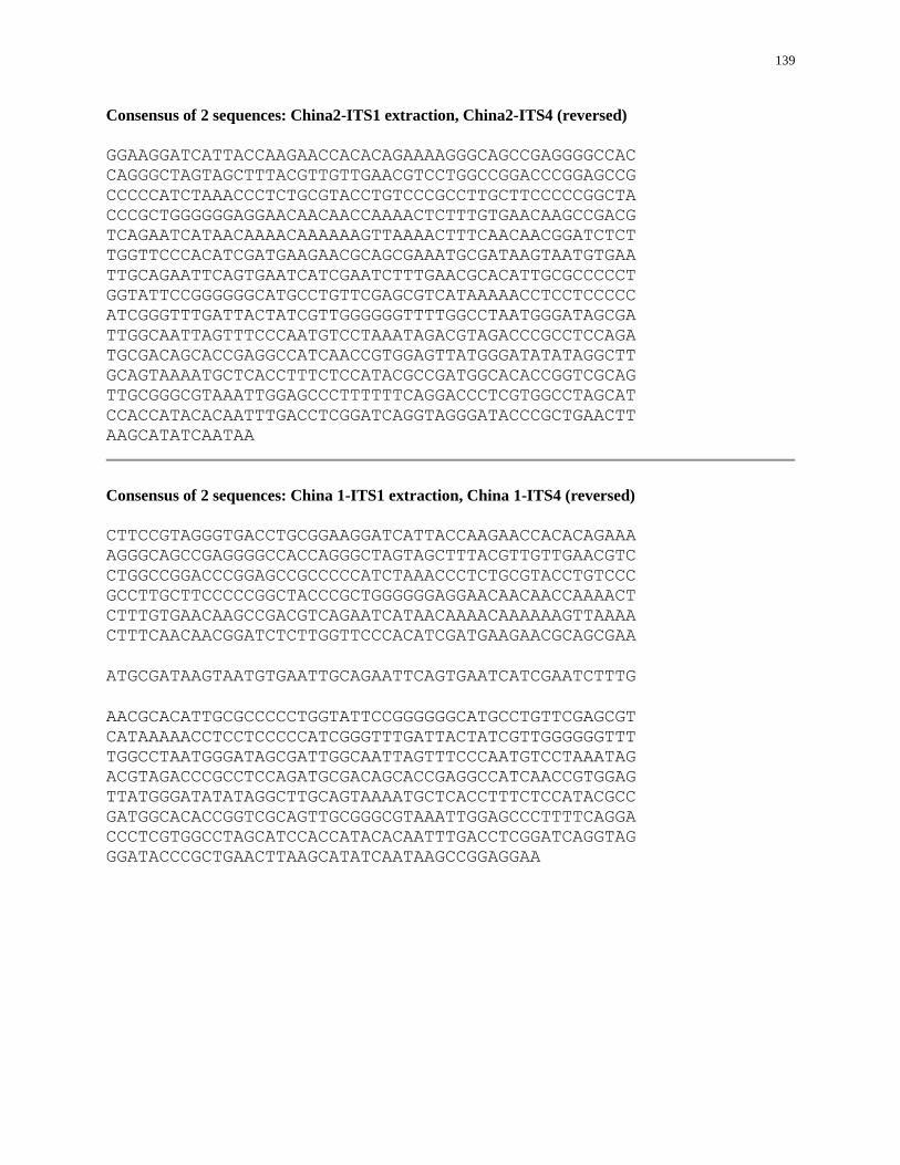
139
Consensus of 2 sequences: China2-ITS1 extraction, China2-ITS4 (reversed)
GGAAGGATCATTACCAAGAACCACACAGAAAAGGGCAGCCGAGGGGCCAC
CAGGGCTAGTAGCTTTACGTTGTTGAACGTCCTGGCCGGACCCGGAGCCG
CCCCCATCTAAACCCTCTGCGTACCTGTCCCGCCTTGCTTCCCCCGGCTA
CCCGCTGGGGGGAGGAACAACAACCAAAACTCTTTGTGAACAAGCCGACG
TCAGAATCATAACAAAACAAAAAAGTTAAAACTTTCAACAACGGATCTCT
TGGTTCCCACATCGATGAAGAACGCAGCGAAATGCGATAAGTAATGTGAA
TTGCAGAATTCAGTGAATCATCGAATCTTTGAACGCACATTGCGCCCCCT
GGTATTCCGGGGGGCATGCCTGTTCGAGCGTCATAAAAACCTCCTCCCCC
ATCGGGTTTGATTACTATCGTTGGGGGGTTTTGGCCTAATGGGATAGCGA
TTGGCAATTAGTTTCCCAATGTCCTAAATAGACGTAGACCCGCCTCCAGA
TGCGACAGCACCGAGGCCATCAACCGTGGAGTTATGGGATATATAGGCTT
GCAGTAAAATGCTCACCTTTCTCCATACGCCGATGGCACACCGGTCGCAG
TTGCGGGCGTAAATTGGAGCCCTTTTTTCAGGACCCTCGTGGCCTAGCAT
CCACCATACACAATTTGACCTCGGATCAGGTAGGGATACCCGCTGAACTT
AAGCATATCAATAA
Consensus of 2 sequences: China 1-ITS1 extraction, China 1-ITS4 (reversed)
CTTCCGTAGGGTGACCTGCGGAAGGATCATTACCAAGAACCACACAGAAA
AGGGCAGCCGAGGGGCCACCAGGGCTAGTAGCTTTACGTTGTTGAACGTC
CTGGCCGGACCCGGAGCCGCCCCCATCTAAACCCTCTGCGTACCTGTCCC
GCCTTGCTTCCCCCGGCTACCCGCTGGGGGGAGGAACAACAACCAAAACT
CTTTGTGAACAAGCCGACGTCAGAATCATAACAAAACAAAAAAGTTAAAA
CTTTCAACAACGGATCTCTTGGTTCCCACATCGATGAAGAACGCAGCGAA
ATGCGATAAGTAATGTGAATTGCAGAATTCAGTGAATCATCGAATCTTTG
AACGCACATTGCGCCCCCTGGTATTCCGGGGGGCATGCCTGTTCGAGCGT
CATAAAAACCTCCTCCCCCATCGGGTTTGATTACTATCGTTGGGGGGTTT
TGGCCTAATGGGATAGCGATTGGCAATTAGTTTCCCAATGTCCTAAATAG
ACGTAGACCCGCCTCCAGATGCGACAGCACCGAGGCCATCAACCGTGGAG
TTATGGGATATATAGGCTTGCAGTAAAATGCTCACCTTTCTCCATACGCC
GATGGCACACCGGTCGCAGTTGCGGGCGTAAATTGGAGCCCTTTTCAGGA
CCCTCGTGGCCTAGCATCCACCATACACAATTTGACCTCGGATCAGGTAG
GGATACCCGCTGAACTTAAGCATATCAATAAGCCGGAGGAA
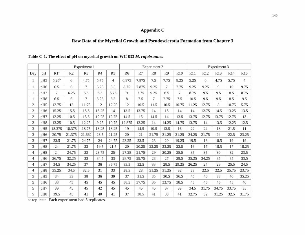
140
Appendix C
Raw Data of the Mycelial Growth and Pseudosclerotia Formation from Chapter 3
Table C-1. The effect of pH on mycelial growth on WC 833 M. rufobrunnea
Experiment 1 Experiment 2 Experiment 3
Day pH R1a R2 R3 R4 R5 R6 R7 R8 R9 R10 R11 R12 R13 R14 R15
1 pH5 5.25b 6 4.75 5.75 4 6.875 7.875 7.5 7.75 8.25 5.25 6 4.75 5.75 4
1 pH6 6.5 6 7 6.25 5.5 8.75 7.875 9.25 7 7.75 9.25 9.25 9 10 9.75
1 pH7 7 6.25 6.5 6.5 6.75 9 7.75 9.25 6.5 7 8.75 9.5 9.5 8.5 8.75
1 pH8 6.5 6 7 5.25 6.5 8 7.5 7 7.75 7.5 10.5 9.5 9.5 8.5 9.5
2 pH5 12.75 13 11.75 12 12.25 12 10.5 11.5 10.5 10.75 11.25 12.75 8 10.75 5.75
2 pH6 15.25 15.5 15.5 15.25 14 13.5 13.75 14 15 14 14 12.75 14.5 13.25 13.5
2 pH7 12.25 10.5 13.5 12.25 12.75 14.5 15 14.5 14 13.5 13.75 12.75 13.75 12.75 13
2 pH8 13.25 10.5 12.25 9.25 10.75 12.875 13.25 14 14.25 14.75 13.75 14 13.5 12.25 12.5
3 pH5 18.375 18.375 18.75 18.25 18.25 19 14.5 19.5 13.5 16 22 24 18 21.5 11
3 pH6 20.75 21.375 21.662 23.5 21.25 20 21 21.75 21.25 21.25 24.25 21.75 24 22.5 23.25
3 pH7 23.5 21.75 24.75 24 24.75 23.25 23.5 23 20 19.25 19.5 18 18.5 19 19
3 pH8 24 21.75 23 19.5 21.5 20 20.25 22.25 23.25 22.5 16 17 18.5 17 18.25
4 pH5 24 24.75 23 23.75 25 27.25 21.75 29 20.25 25.5 35 35 30 32 23.5
4 pH6 26.75 32.25 33 34.5 33 28.75 29.75 28 27 29.5 35.25 34.25 35 35 33.5
4 pH7 34.5 34.25 37 36 36.75 33.5 32.5 33 28.5 29.25 26.25 24 26 25.5 24.5
4 pH8 35.25 34.5 32.5 31 33 28.5 28 31.25 31.25 32 23 22.5 22.5 25.75 23.75
5 pH5 34 33 38 36 39 37 31.5 35 30.5 36.5 45 40 38 40 35.25
5 pH6 38 45 45 45 45 38.5 37.75 35 33.75 38.5 45 45 45 45 40
5 pH7 39 45 45 42 45 45 45 45 37 39 34.5 31.75 34.75 33.75 35
5 pH8 39.5 45 41 40 41 37 38.5 41 38 41 32.75 32 31.25 32.5 31.75
a: replicate. Each experiment had 5 replicates.
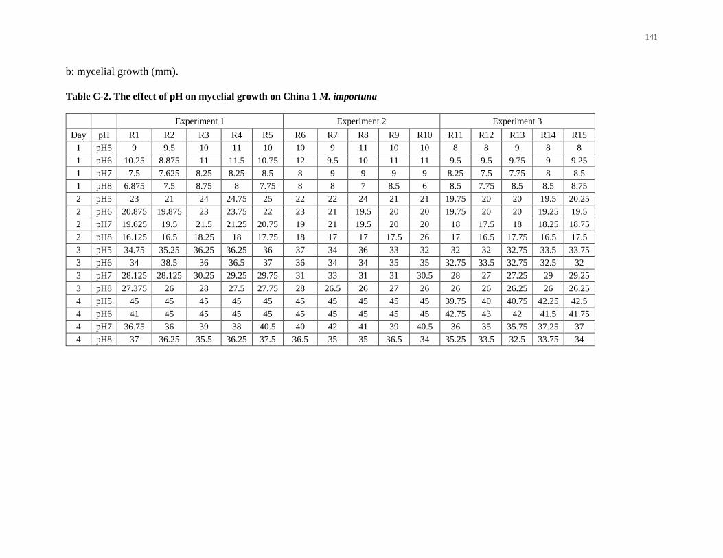
141
b: mycelial growth (mm).
Table C-2. The effect of pH on mycelial growth on China 1 M. importuna
Experiment 1 Experiment 2 Experiment 3
Day pH R1 R2 R3 R4 R5 R6 R7 R8 R9 R10 R11 R12 R13 R14 R15
1 pH5 9 9.5 10 11 10 10 9 11 10 10 8 8 9 8 8
1 pH6 10.25 8.875 11 11.5 10.75 12 9.5 10 11 11 9.5 9.5 9.75 9 9.25
1 pH7 7.5 7.625 8.25 8.25 8.5 8 9 9 9 9 8.25 7.5 7.75 8 8.5
1 pH8 6.875 7.5 8.75 8 7.75 8 8 7 8.5 6 8.5 7.75 8.5 8.5 8.75
2 pH5 23 21 24 24.75 25 22 22 24 21 21 19.75 20 20 19.5 20.25
2 pH6 20.875 19.875 23 23.75 22 23 21 19.5 20 20 19.75 20 20 19.25 19.5
2 pH7 19.625 19.5 21.5 21.25 20.75 19 21 19.5 20 20 18 17.5 18 18.25 18.75
2 pH8 16.125 16.5 18.25 18 17.75 18 17 17 17.5 26 17 16.5 17.75 16.5 17.5
3 pH5 34.75 35.25 36.25 36.25 36 37 34 36 33 32 32 32 32.75 33.5 33.75
3 pH6 34 38.5 36 36.5 37 36 34 34 35 35 32.75 33.5 32.75 32.5 32
3 pH7 28.125 28.125 30.25 29.25 29.75 31 33 31 31 30.5 28 27 27.25 29 29.25
3 pH8 27.375 26 28 27.5 27.75 28 26.5 26 27 26 26 26 26.25 26 26.25
4 pH5 45 45 45 45 45 45 45 45 45 45 39.75 40 40.75 42.25 42.5
4 pH6 41 45 45 45 45 45 45 45 45 45 42.75 43 42 41.5 41.75
4 pH7 36.75 36 39 38 40.5 40 42 41 39 40.5 36 35 35.75 37.25 37
4 pH8 37 36.25 35.5 36.25 37.5 36.5 35 35 36.5 34 35.25 33.5 32.5 33.75 34
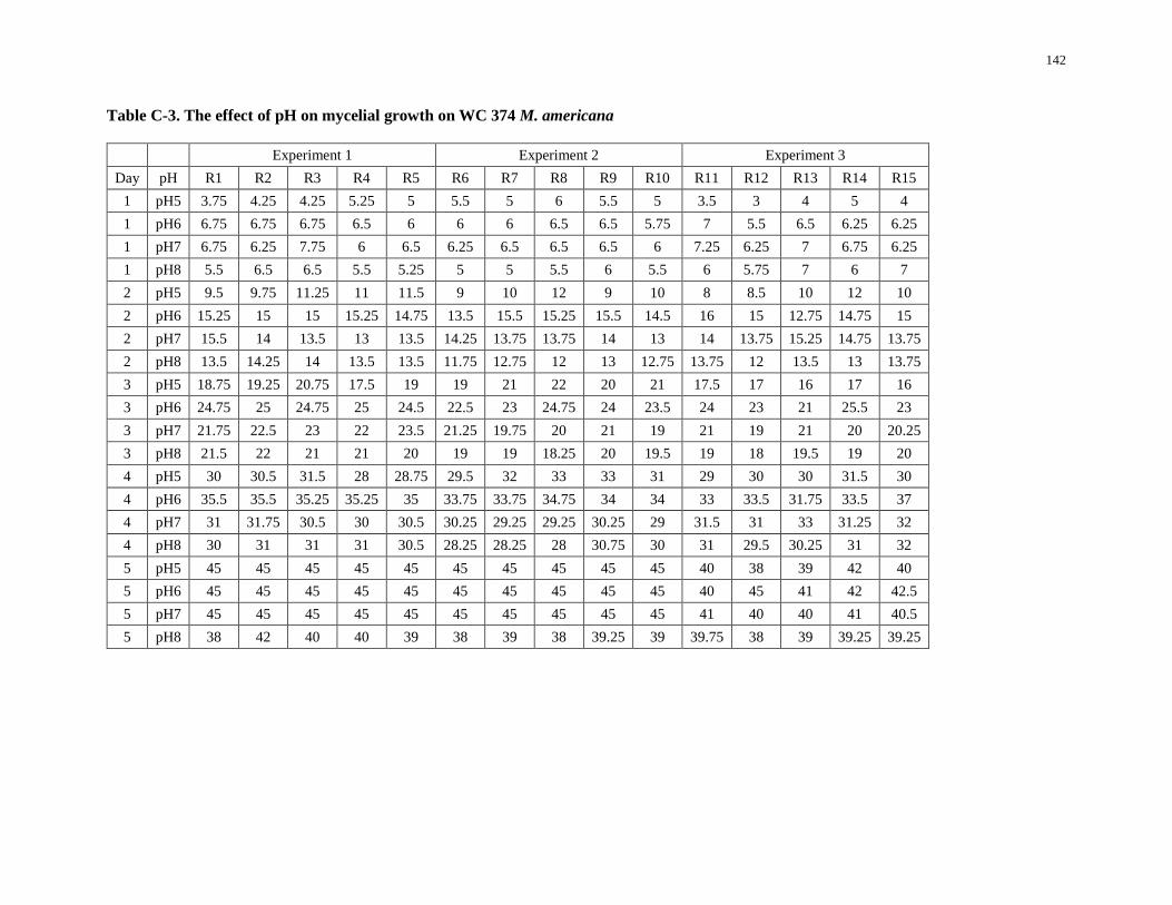
142
Table C-3. The effect of pH on mycelial growth on WC 374 M. americana
Experiment 1 Experiment 2 Experiment 3
Day pH R1 R2 R3 R4 R5 R6 R7 R8 R9 R10 R11 R12 R13 R14 R15
1 pH5 3.75 4.25 4.25 5.25 5 5.5 5 6 5.5 5 3.5 3 4 5 4
1 pH6 6.75 6.75 6.75 6.5 6 6 6 6.5 6.5 5.75 7 5.5 6.5 6.25 6.25
1 pH7 6.75 6.25 7.75 6 6.5 6.25 6.5 6.5 6.5 6 7.25 6.25 7 6.75 6.25
1 pH8 5.5 6.5 6.5 5.5 5.25 5 5 5.5 6 5.5 6 5.75 7 6 7
2 pH5 9.5 9.75 11.25 11 11.5 9 10 12 9 10 8 8.5 10 12 10
2 pH6 15.25 15 15 15.25 14.75 13.5 15.5 15.25 15.5 14.5 16 15 12.75 14.75 15
2 pH7 15.5 14 13.5 13 13.5 14.25 13.75 13.75 14 13 14 13.75 15.25 14.75 13.75
2 pH8 13.5 14.25 14 13.5 13.5 11.75 12.75 12 13 12.75 13.75 12 13.5 13 13.75
3 pH5 18.75 19.25 20.75 17.5 19 19 21 22 20 21 17.5 17 16 17 16
3 pH6 24.75 25 24.75 25 24.5 22.5 23 24.75 24 23.5 24 23 21 25.5 23
3 pH7 21.75 22.5 23 22 23.5 21.25 19.75 20 21 19 21 19 21 20 20.25
3 pH8 21.5 22 21 21 20 19 19 18.25 20 19.5 19 18 19.5 19 20
4 pH5 30 30.5 31.5 28 28.75 29.5 32 33 33 31 29 30 30 31.5 30
4 pH6 35.5 35.5 35.25 35.25 35 33.75 33.75 34.75 34 34 33 33.5 31.75 33.5 37
4 pH7 31 31.75 30.5 30 30.5 30.25 29.25 29.25 30.25 29 31.5 31 33 31.25 32
4 pH8 30 31 31 31 30.5 28.25 28.25 28 30.75 30 31 29.5 30.25 31 32
5 pH5 45 45 45 45 45 45 45 45 45 45 40 38 39 42 40
5 pH6 45 45 45 45 45 45 45 45 45 45 40 45 41 42 42.5
5 pH7 45 45 45 45 45 45 45 45 45 45 41 40 40 41 40.5
5 pH8 38 42 40 40 39 38 39 38 39.25 39 39.75 38 39 39.25 39.25
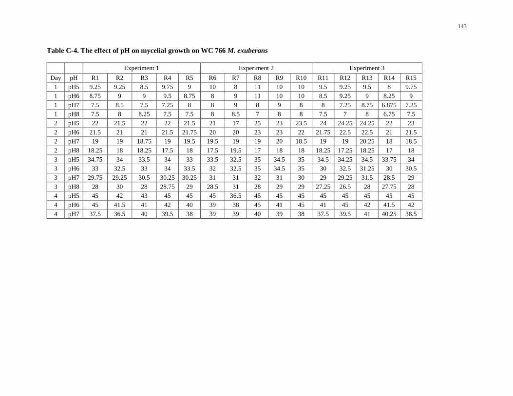
143
Table C-4. The effect of pH on mycelial growth on WC 766 M. exuberans
Experiment 1 Experiment 2 Experiment 3
Day pH R1 R2 R3 R4 R5 R6 R7 R8 R9 R10 R11 R12 R13 R14 R15
1 pH5 9.25 9.25 8.5 9.75 9 10 8 11 10 10 9.5 9.25 9.5 8 9.75
1 pH6 8.75 9 9 9.5 8.75 8 9 11 10 10 8.5 9.25 9 8.25 9
1 pH7 7.5 8.5 7.5 7.25 8 8 9 8 9 8 8 7.25 8.75 6.875 7.25
1 pH8 7.5 8 8.25 7.5 7.5 8 8.5 7 8 8 7.5 7 8 6.75 7.5
2 pH5 22 21.5 22 22 21.5 21 17 25 23 23.5 24 24.25 24.25 22 23
2 pH6 21.5 21 21 21.5 21.75 20 20 23 23 22 21.75 22.5 22.5 21 21.5
2 pH7 19 19 18.75 19 19.5 19.5 19 19 20 18.5 19 19 20.25 18 18.5
2 pH8 18.25 18 18.25 17.5 18 17.5 19.5 17 18 18 18.25 17.25 18.25 17 18
3 pH5 34.75 34 33.5 34 33 33.5 32.5 35 34.5 35 34.5 34.25 34.5 33.75 34
3 pH6 33 32.5 33 34 33.5 32 32.5 35 34.5 35 30 32.5 31.25 30 30.5
3 pH7 29.75 29.25 30.5 30.25 30.25 31 31 32 31 30 29 29.25 31.5 28.5 29
3 pH8 28 30 28 28.75 29 28.5 31 28 29 29 27.25 26.5 28 27.75 28
4 pH5 45 42 43 45 45 45 36.5 45 45 45 45 45 45 45 45
4 pH6 45 41.5 41 42 40 39 38 45 41 45 41 45 42 41.5 42
4 pH7 37.5 36.5 40 39.5 38 39 39 40 39 38 37.5 39.5 41 40.25 38.5
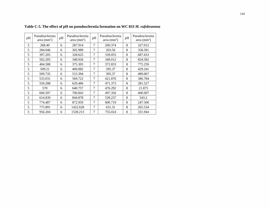
144
Table C-5. The effect of pH on pseudosclerotia formation on WC 833 M. rufobrunnea
pH Pseudosclerotia
area (mm2) pH
Pseudosclerotia
area (mm2) pH
Pseudosclerotia
area (mm2) pH
Pseudosclerotia
area (mm2)
5 268.49 6 287.914 7 200.374 8 327.012
5 284.046 6 302.989 7 203.56 8 358.391
5 397.205 6 328.625 7 339.055 8 687.633
5 502.205 6 348.926 7 349.012 8 824.582
5 404.588 6 375.305 7 372.833 8 775.259
5 509.21 6 400.082 7 395.37 8 429.241
5 509.735 6 515.394 7 395.37 8 489.067
5 533.031 6 569.722 7 421.876 8 386.784
5 550.288 6 629.486 7 471.373 8 281.527
5 570 6 640.757 7 476.292 8 21.875
5 600.597 6 700.843 7 497.356 8 400.007
5 614.839 6 844.878 7 528.237 8 543.2
5 774.487 6 872.959 7 600.719 8 247.506
5 775.891 6 1422.628 7 631.31 8 265.534
5 956.204 6 1538.213 7 755.024 8 331.944
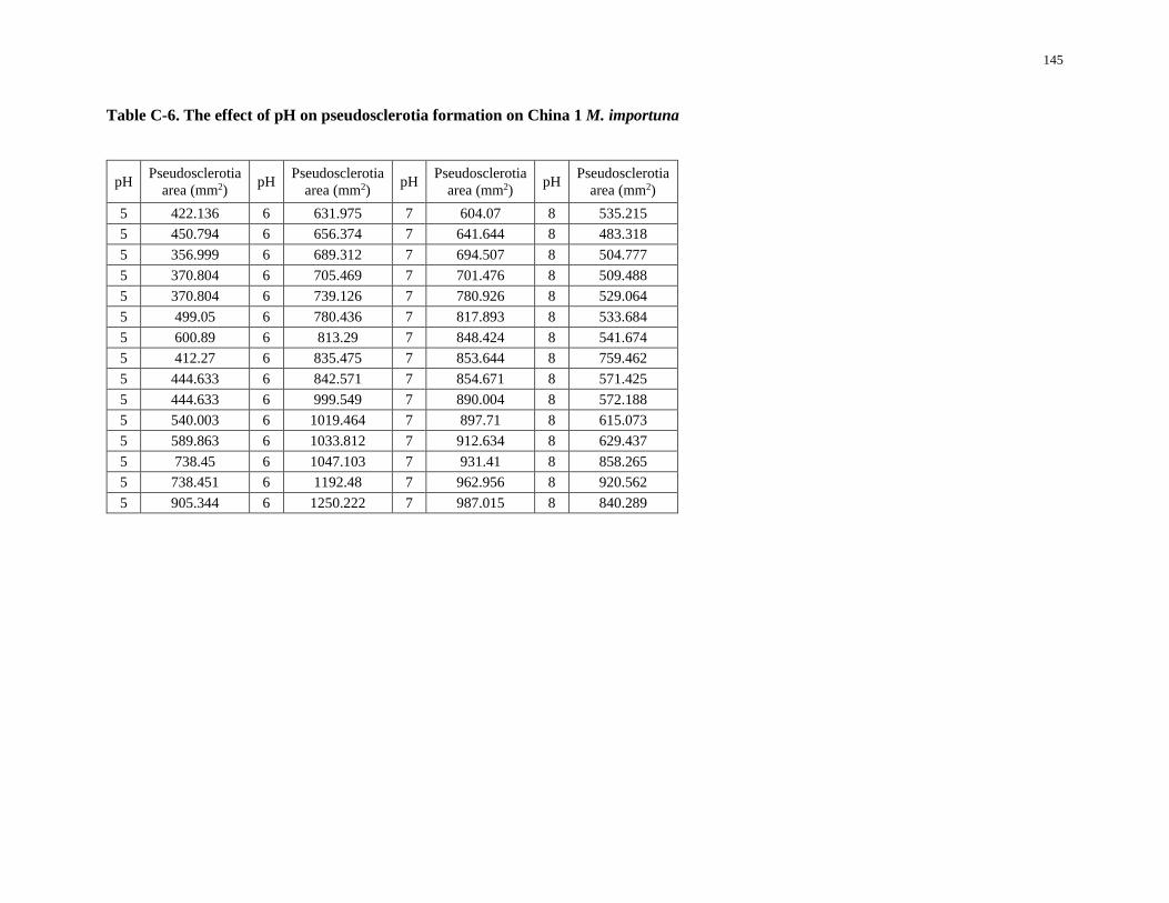
145
Table C-6. The effect of pH on pseudosclerotia formation on China 1 M. importuna
pH Pseudosclerotia
area (mm2) pH
Pseudosclerotia
area (mm2) pH
Pseudosclerotia
area (mm2) pH
Pseudosclerotia
area (mm2)
5 422.136 6 631.975 7 604.07 8 535.215
5 450.794 6 656.374 7 641.644 8 483.318
5 356.999 6 689.312 7 694.507 8 504.777
5 370.804 6 705.469 7 701.476 8 509.488
5 370.804 6 739.126 7 780.926 8 529.064
5 499.05 6 780.436 7 817.893 8 533.684
5 600.89 6 813.29 7 848.424 8 541.674
5 412.27 6 835.475 7 853.644 8 759.462
5 444.633 6 842.571 7 854.671 8 571.425
5 444.633 6 999.549 7 890.004 8 572.188
5 540.003 6 1019.464 7 897.71 8 615.073
5 589.863 6 1033.812 7 912.634 8 629.437
5 738.45 6 1047.103 7 931.41 8 858.265
5 738.451 6 1192.48 7 962.956 8 920.562
5 905.344 6 1250.222 7 987.015 8 840.289
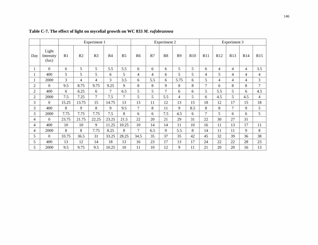
146
Table C-7. The effect of light on mycelial growth on WC 833 M. rufobrunnea
Experiment 1 Experiment 2 Experiment 3
Day
Light
Intensity
(lux)
R1 R2 R3 R4 R5 R6 R7 R8 R9 R10 R11 R12 R13 R14 R15
1 0 6 5 5 5.5 5.5 6 6 6 5 5 6 4 4 4 3.5
1 400 5 5 5 6 5 4 4 6 5 5 4 5 4 4 4
1 2000 3 4 4 3 3.5 6 5.5 6 5.75 6 5 4 4 4 3
2 0 9.5 8.75 9.75 9.25 9 8 8 9 8 8 7 6 8 8 7
2 400 6 6.25 6 7 6.5 5 5 7 6 6 5 5.5 5 6 4.5
2 2000 7.5 7.25 7 7.5 7 5 5 5.5 4 5 6 4.5 5 4.5 4
3 0 15.25 13.75 15 14.75 13 13 11 12 13 15 18 12 17 15 18
3 400 8 9 8 9 9.5 7 8 11 9 8.5 8 8 7 9 5
3 2000 7.75 7.75 7.75 7.5 8 6 6 7.5 4.5 6 7 5 6 6 5
4 0 23.75 21.75 22.25 23.25 21.5 22 20 21 29 31 22 30 27 31
4 400 10 10 9 11.25 10.25 10 14 14 11 10 16 11 13 17 11
4 2000 8 8 7.75 8.25 8 7 6.5 9 5.5 8 14 11 11 9 8
5 0 33.75 36.5 31 33.25 28.25 34.5 35 37 35 42 45 32 39 36 38
5 400 13 12 14 18 13 16 23 17 13 17 24 22 22 28 23
5 2000 9.5 9.75 9.5 10.25 10 11 10 12 9 11 21 20 20 16 13
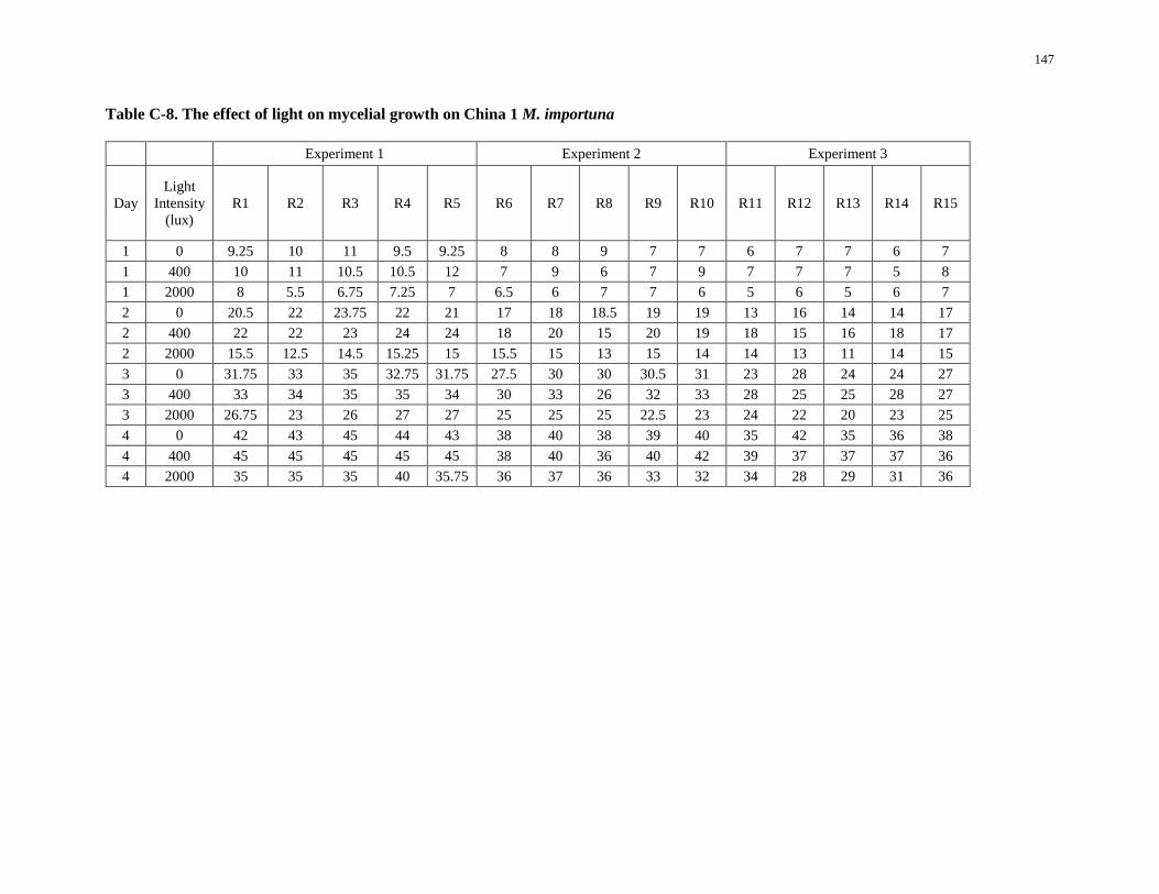
147
Table C-8. The effect of light on mycelial growth on China 1 M. importuna
Experiment 1 Experiment 2 Experiment 3
Day
Light
Intensity
(lux)
R1 R2 R3 R4 R5 R6 R7 R8 R9 R10 R11 R12 R13 R14 R15
1 0 9.25 10 11 9.5 9.25 8 8 9 7 7 6 7 7 6 7
1 400 10 11 10.5 10.5 12 7 9 6 7 9 7 7 7 5 8
1 2000 8 5.5 6.75 7.25 7 6.5 6 7 7 6 5 6 5 6 7
2 0 20.5 22 23.75 22 21 17 18 18.5 19 19 13 16 14 14 17
2 400 22 22 23 24 24 18 20 15 20 19 18 15 16 18 17
2 2000 15.5 12.5 14.5 15.25 15 15.5 15 13 15 14 14 13 11 14 15
3 0 31.75 33 35 32.75 31.75 27.5 30 30 30.5 31 23 28 24 24 27
3 400 33 34 35 35 34 30 33 26 32 33 28 25 25 28 27
3 2000 26.75 23 26 27 27 25 25 25 22.5 23 24 22 20 23 25
4 0 42 43 45 44 43 38 40 38 39 40 35 42 35 36 38
4 400 45 45 45 45 45 38 40 36 40 42 39 37 37 37 36
4 2000 35 35 35 40 35.75 36 37 36 33 32 34 28 29 31 36
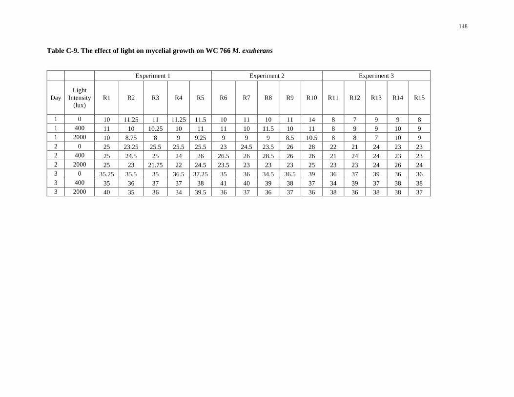
148
Table C-9. The effect of light on mycelial growth on WC 766 M. exuberans
Experiment 1 Experiment 2 Experiment 3
Day
Light
Intensity
(lux)
R1 R2 R3 R4 R5 R6 R7 R8 R9 R10 R11 R12 R13 R14 R15
1 0 10 11.25 11 11.25 11.5 10 11 10 11 14 8 7 9 9 8
1 400 11 10 10.25 10 11 11 10 11.5 10 11 8 9 9 10 9
1 2000 10 8.75 8 9 9.25 9 9 9 8.5 10.5 8 8 7 10 9
2 0 25 23.25 25.5 25.5 25.5 23 24.5 23.5 26 28 22 21 24 23 23
2 400 25 24.5 25 24 26 26.5 26 28.5 26 26 21 24 24 23 23
2 2000 25 23 21.75 22 24.5 23.5 23 23 23 25 23 23 24 26 24
3 0 35.25 35.5 35 36.5 37.25 35 36 34.5 36.5 39 36 37 39 36 36
3 400 35 36 37 37 38 41 40 39 38 37 34 39 37 38 38
3 2000 40 35 36 34 39.5 36 37 36 37 36 38 36 38 38 37
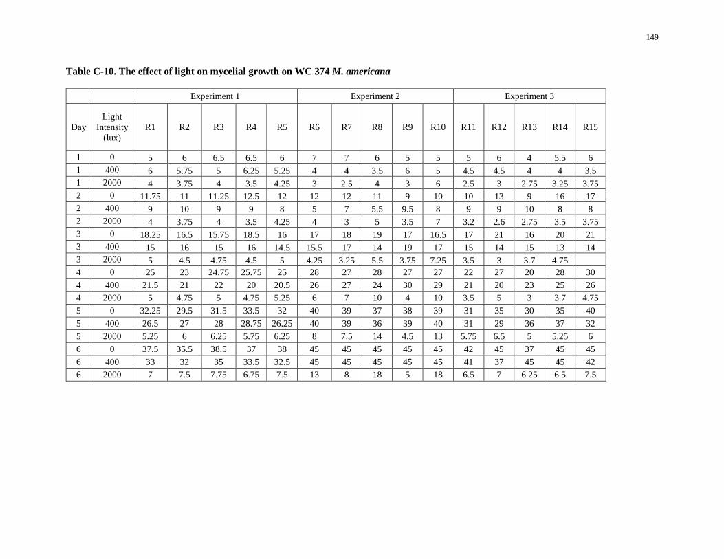
149
Table C-10. The effect of light on mycelial growth on WC 374 M. americana
Experiment 1 Experiment 2 Experiment 3
Day
Light
Intensity
(lux)
R1 R2 R3 R4 R5 R6 R7 R8 R9 R10 R11 R12 R13 R14 R15
1 0 5 6 6.5 6.5 6 7 7 6 5 5 5 6 4 5.5 6
1 400 6 5.75 5 6.25 5.25 4 4 3.5 6 5 4.5 4.5 4 4 3.5
1 2000 4 3.75 4 3.5 4.25 3 2.5 4 3 6 2.5 3 2.75 3.25 3.75
2 0 11.75 11 11.25 12.5 12 12 12 11 9 10 10 13 9 16 17
2 400 9 10 9 9 8 5 7 5.5 9.5 8 9 9 10 8 8
2 2000 4 3.75 4 3.5 4.25 4 3 5 3.5 7 3.2 2.6 2.75 3.5 3.75
3 0 18.25 16.5 15.75 18.5 16 17 18 19 17 16.5 17 21 16 20 21
3 400 15 16 15 16 14.5 15.5 17 14 19 17 15 14 15 13 14
3 2000 5 4.5 4.75 4.5 5 4.25 3.25 5.5 3.75 7.25 3.5 3 3.7 4.75
4 0 25 23 24.75 25.75 25 28 27 28 27 27 22 27 20 28 30
4 400 21.5 21 22 20 20.5 26 27 24 30 29 21 20 23 25 26
4 2000 5 4.75 5 4.75 5.25 6 7 10 4 10 3.5 5 3 3.7 4.75
5 0 32.25 29.5 31.5 33.5 32 40 39 37 38 39 31 35 30 35 40
5 400 26.5 27 28 28.75 26.25 40 39 36 39 40 31 29 36 37 32
5 2000 5.25 6 6.25 5.75 6.25 8 7.5 14 4.5 13 5.75 6.5 5 5.25 6
6 0 37.5 35.5 38.5 37 38 45 45 45 45 45 42 45 37 45 45
6 400 33 32 35 33.5 32.5 45 45 45 45 45 41 37 45 45 42
6 2000 7 7.5 7.75 6.75 7.5 13 8 18 5 18 6.5 7 6.25 6.5 7.5
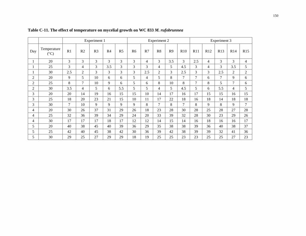
150
Table C-11. The effect of temperature on mycelial growth on WC 833 M. rufobrunnea
Experiment 1 Experiment 2 Experiment 3
Day Temperature
(C) R1 R2 R3 R4 R5 R6 R7 R8 R9 R10 R11 R12 R13 R14 R15
1 20 3 3 3 3 3 3 4 3 3.5 3 2.5 4 3 3 4
1 25 3 4 3 3.5 3 3 3 4 5 4.5 3 4 3 3.5 5
1 30 2.5 2 3 3 3 3 2.5 2 3 2.5 3 3 2.5 2 2
2 20 9 5 10 6 6 5 4 5 8 7 7 6 7 9 6
2 25 8 7 10 9 6 5 6 8 10 8 7 8 5 7 6
2 30 3.5 4 5 6 5.5 5 5 4 5 4.5 5 6 5.5 4 5
3 20 20 14 19 16 15 15 10 14 17 16 17 15 15 16 15
3 25 18 20 23 21 15 10 11 17 22 18 16 18 14 18 18
3 30 7 10 9 9 9 9 8 7 8 7 8 9 8 9 7
4 20 30 26 37 31 29 26 18 23 28 30 28 25 28 27 28
4 25 32 36 39 34 29 24 20 33 39 32 28 30 23 29 26
4 30 17 17 17 18 17 12 12 14 15 14 16 18 16 16 17
5 20 40 38 45 40 39 36 29 35 38 38 39 36 40 38 37
5 25 42 40 45 38 42 30 36 39 42 38 39 39 32 41 36
5 30 29 25 27 29 29 18 19 25 25 23 23 25 25 27 23
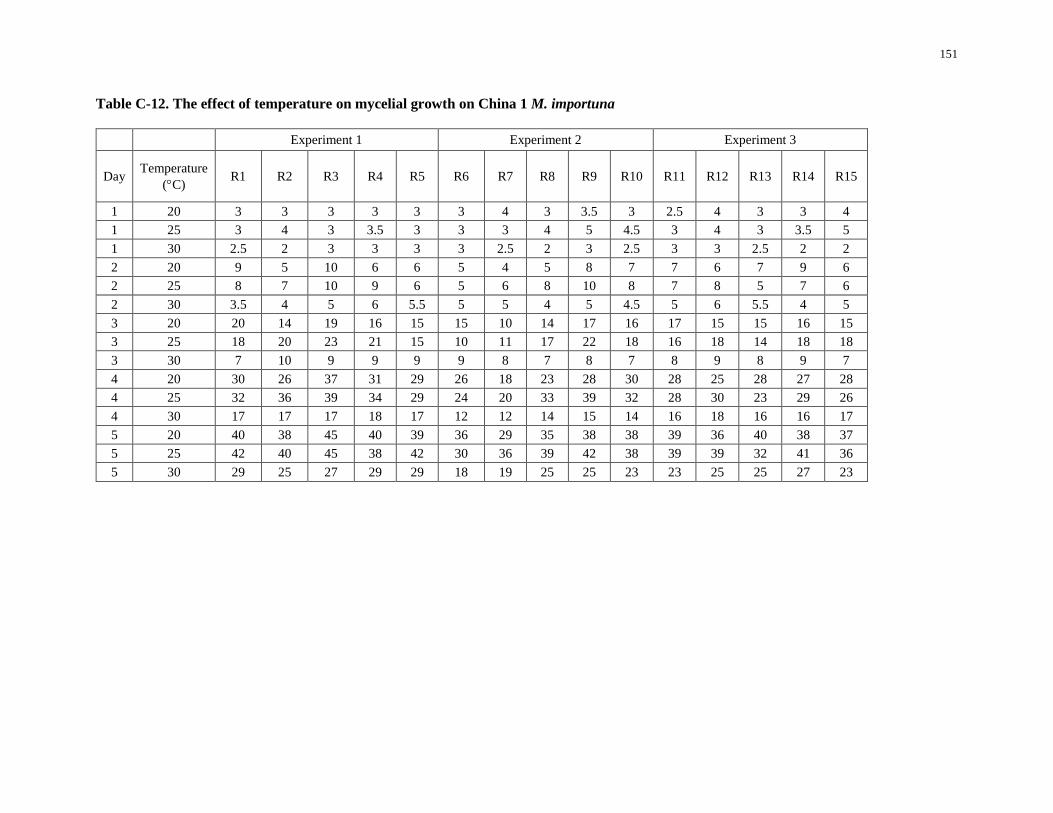
151
Table C-12. The effect of temperature on mycelial growth on China 1 M. importuna
Experiment 1 Experiment 2 Experiment 3
Day Temperature
(C) R1 R2 R3 R4 R5 R6 R7 R8 R9 R10 R11 R12 R13 R14 R15
1 20 3 3 3 3 3 3 4 3 3.5 3 2.5 4 3 3 4
1 25 3 4 3 3.5 3 3 3 4 5 4.5 3 4 3 3.5 5
1 30 2.5 2 3 3 3 3 2.5 2 3 2.5 3 3 2.5 2 2
2 20 9 5 10 6 6 5 4 5 8 7 7 6 7 9 6
2 25 8 7 10 9 6 5 6 8 10 8 7 8 5 7 6
2 30 3.5 4 5 6 5.5 5 5 4 5 4.5 5 6 5.5 4 5
3 20 20 14 19 16 15 15 10 14 17 16 17 15 15 16 15
3 25 18 20 23 21 15 10 11 17 22 18 16 18 14 18 18
3 30 7 10 9 9 9 9 8 7 8 7 8 9 8 9 7
4 20 30 26 37 31 29 26 18 23 28 30 28 25 28 27 28
4 25 32 36 39 34 29 24 20 33 39 32 28 30 23 29 26
4 30 17 17 17 18 17 12 12 14 15 14 16 18 16 16 17
5 20 40 38 45 40 39 36 29 35 38 38 39 36 40 38 37
5 25 42 40 45 38 42 30 36 39 42 38 39 39 32 41 36
5 30 29 25 27 29 29 18 19 25 25 23 23 25 25 27 23
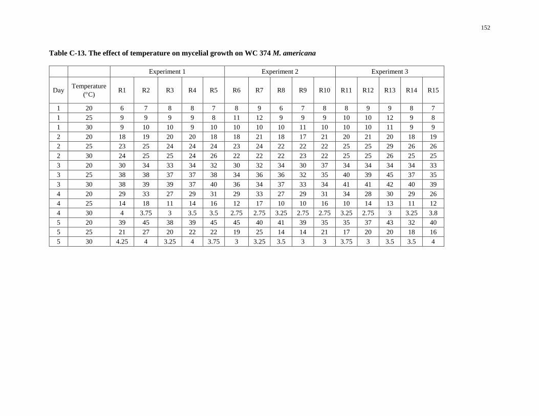
152
Table C-13. The effect of temperature on mycelial growth on WC 374 M. americana
Experiment 1 Experiment 2 Experiment 3
Day Temperature
(C) R1 R2 R3 R4 R5 R6 R7 R8 R9 R10 R11 R12 R13 R14 R15
1 20 6 7 8 8 7 8 9 6 7 8 8 9 9 8 7
1 25 9 9 9 9 8 11 12 9 9 9 10 10 12 9 8
1 30 9 10 10 9 10 10 10 10 11 10 10 10 11 9 9
2 20 18 19 20 20 18 18 21 18 17 21 20 21 20 18 19
2 25 23 25 24 24 24 23 24 22 22 22 25 25 29 26 26
2 30 24 25 25 24 26 22 22 22 23 22 25 25 26 25 25
3 20 30 34 33 34 32 30 32 34 30 37 34 34 34 34 33
3 25 38 38 37 37 38 34 36 36 32 35 40 39 45 37 35
3 30 38 39 39 37 40 36 34 37 33 34 41 41 42 40 39
4 20 29 33 27 29 31 29 33 27 29 31 34 28 30 29 26
4 25 14 18 11 14 16 12 17 10 10 16 10 14 13 11 12
4 30 4 3.75 3 3.5 3.5 2.75 2.75 3.25 2.75 2.75 3.25 2.75 3 3.25 3.8
5 20 39 45 38 39 45 45 40 41 39 35 35 37 43 32 40
5 25 21 27 20 22 22 19 25 14 14 21 17 20 20 18 16
5 30 4.25 4 3.25 4 3.75 3 3.25 3.5 3 3 3.75 3 3.5 3.5 4
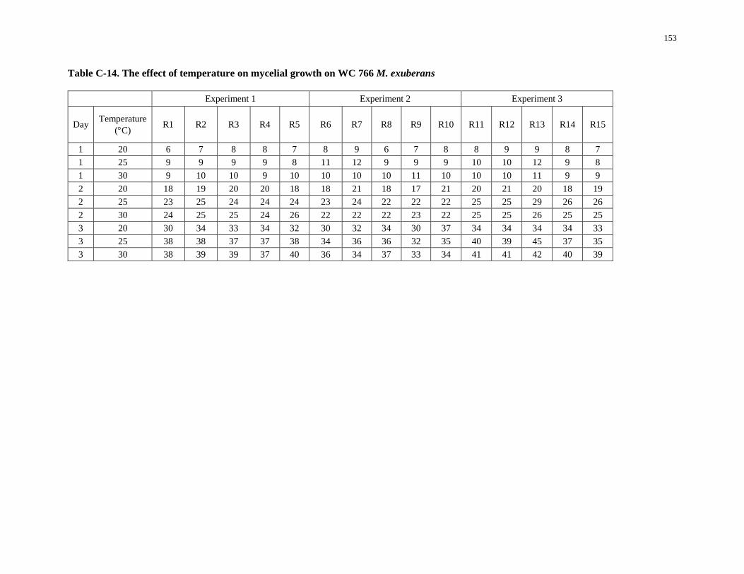
153
Table C-14. The effect of temperature on mycelial growth on WC 766 M. exuberans
Experiment 1 Experiment 2 Experiment 3
Day Temperature
(C) R1 R2 R3 R4 R5 R6 R7 R8 R9 R10 R11 R12 R13 R14 R15
1 20 6 7 8 8 7 8 9 6 7 8 8 9 9 8 7
1 25 9 9 9 9 8 11 12 9 9 9 10 10 12 9 8
1 30 9 10 10 9 10 10 10 10 11 10 10 10 11 9 9
2 20 18 19 20 20 18 18 21 18 17 21 20 21 20 18 19
2 25 23 25 24 24 24 23 24 22 22 22 25 25 29 26 26
2 30 24 25 25 24 26 22 22 22 23 22 25 25 26 25 25
3 20 30 34 33 34 32 30 32 34 30 37 34 34 34 34 33
3 25 38 38 37 37 38 34 36 36 32 35 40 39 45 37 35
3 30 38 39 39 37 40 36 34 37 33 34 41 41 42 40 39
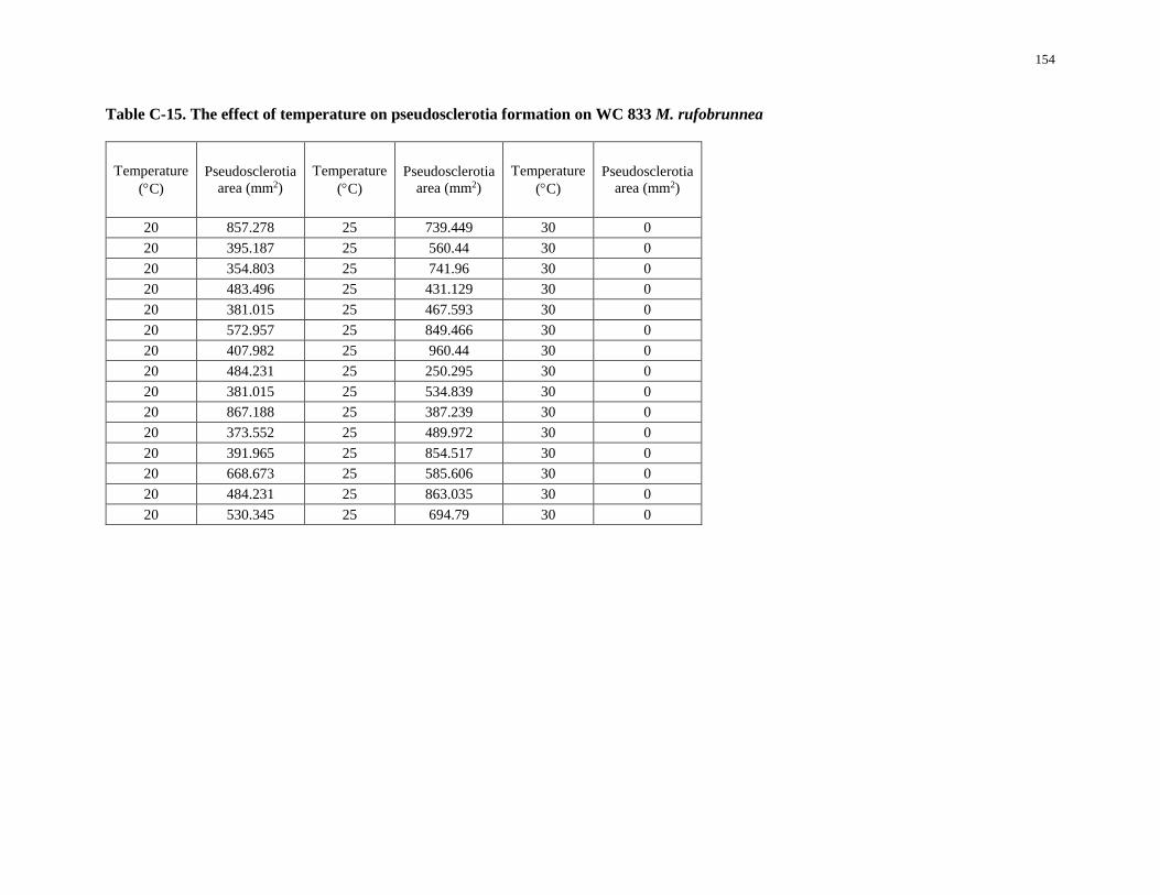
154
Table C-15. The effect of temperature on pseudosclerotia formation on WC 833 M. rufobrunnea
Temperature
(C)
Pseudosclerotia
area (mm2)
Temperature
(C)
Pseudosclerotia
area (mm2)
Temperature
(C)
Pseudosclerotia
area (mm2)
20 857.278 25 739.449 30 0
20 395.187 25 560.44 30 0
20 354.803 25 741.96 30 0
20 483.496 25 431.129 30 0
20 381.015 25 467.593 30 0
20 572.957 25 849.466 30 0
20 407.982 25 960.44 30 0
20 484.231 25 250.295 30 0
20 381.015 25 534.839 30 0
20 867.188 25 387.239 30 0
20 373.552 25 489.972 30 0
20 391.965 25 854.517 30 0
20 668.673 25 585.606 30 0
20 484.231 25 863.035 30 0
20 530.345 25 694.79 30 0
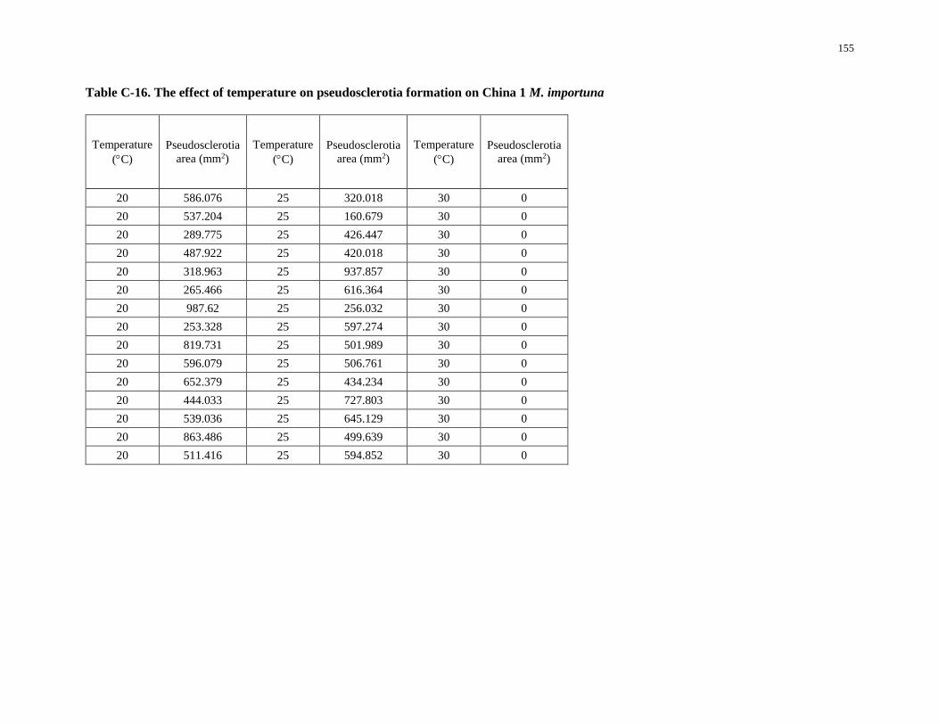
155
Table C-16. The effect of temperature on pseudosclerotia formation on China 1 M. importuna
Temperature
(C)
Pseudosclerotia
area (mm2)
Temperature
(C)
Pseudosclerotia
area (mm2)
Temperature
(C)
Pseudosclerotia
area (mm2)
20 586.076 25 320.018 30 0
20 537.204 25 160.679 30 0
20 289.775 25 426.447 30 0
20 487.922 25 420.018 30 0
20 318.963 25 937.857 30 0
20 265.466 25 616.364 30 0
20 987.62 25 256.032 30 0
20 253.328 25 597.274 30 0
20 819.731 25 501.989 30 0
20 596.079 25 506.761 30 0
20 652.379 25 434.234 30 0
20 444.033 25 727.803 30 0
20 539.036 25 645.129 30 0
20 863.486 25 499.639 30 0
20 511.416 25 594.852 30 0
156
LITERATURE CITED
Alvarado-Castillo, G. , Mata, G. , Pérez-Vázquez, A. , Martfnez-carrera, D. , Nava, ME. , Gallardo-
López, F. , and Osorio-Acosta, F. . Formación de esclerocios de Morchella esculenta y M. conica in
vitro. Revista Mexicana de Micologia, 35:3S-41, 2012.
Alvarado-Castillo, G. , Mata, G. , and Sangabriel-Conde, W. . Understanding the life cycle of morels
(Morchella spp.). Revista Mexicana de Micología, 40, 2014.
Amir, R. , Levanon, D. , Hadar, Y. , and Chet, I. . Formation of sclerotia by Morchella esculenta:
relationship between media composition and turgor potential in the mycelium. Mycological
Research, 96(11):943–948, 1992.
Amir, R. , Levanon, D. , Hadar, Y. , and Chet, I. . Morphology and physiology of Morchella esculenta
during sclerotial formation. Mycological Research, 97(6):683–689, 1993.
Amir, R. , Levanon, D. , Hadar, Y. , and Chet, I. . Factors affecting translocation and sclerotial formation
in Morchella esculenta. Experimental Mycology, 19(1):61–70, 1995.
Amir, R. , Steudle, E. ,Levanon, D. ,Hadar, Y. ,and Chet, I. . Turgor changes in Morchella esculenta
during translocation and sclerotial formation. Experimental Mycology, 19(2):129–136, 1995.
Baynes, M. , Newcombe, G. , Dixon, L. , Castlebury, L. , and O’Donnell, K. . A novel plant fungal
mutualism associated with fire. Fungal biology, 116(1):133– 144, 2012.
Blackwell, M. . The fungi: 1, 2, 3 5.1 million species? American Journal of Botany, 98(3):426–438, 2011.
Buscot, F. . Mycorrhizal succession and morel biology. In: Read, D.J.; Lewis, D.H.; Fitter, A.H. [and
others], eds. Mycorrhizas in ecosystems. Wallingford, Oxon, United Kingdom: CAB International:
220–224, 1992a.
Buscot, F. . Strategies écologiques et biologiques des morilles. (Ecological and biological strategies of
morels) (In French, English abstract). Cryptogamie Mycologie. 13: 171–179, 1992b.
Buscot, F. . Synthesis of two types of association between Morchella esculenta and Picea abies under
controlled culture conditions. Journal of Plant Physiology. 141: 12–17, 1992c.
157
Buscot, F. . Mycelial differentiation of Morchella esculenta in pure culture. Mycological Research,
97(2):136–140, 1993.
Buscot, F. . Ectomycorrhizal types and endobacteria associated with ectomycorrhizas of Morchella elata
(fr.) boudier with Picea abies (l.) karst. Mycorrhiza, 4(5):223–232, 1994.
Buscot, F. and Roux, J. . Association between living roots and ascocarps of Morchella rotunda.
Transactions of the British Mycological Society, 89(2):249– 252, 1987.
Buscot, F. and Kottke, I. . The association of Morchella rotunda (pers.) boudier with roots of Picea abies
(l.) karst. New Phytologist, 116(3):425–430, 1990.
Carris, LM. , Peever, TL. , and McCotter, SW. . Mitospore stages of Disciotis, Gyromitra and Morchella
in the inland pacific northwest USA. Mycologia, 107(4):729–44, 2015.
Chai, H. , Chen, L. , Chen, W. , Zhao, Q. , Zhang, X. , Su, K. , and Zhao, Y. . Characterization of mating-
type idiomorphs suggests that Morchella importuna, Mel-20 and M. .sextelata are heterothallic.
Mycological Progress, 16(7):743–752, 2017.
Chai, L. , Li, L. , Ji, B. , and Sun, C. . Biological characteristics of Morchella lwy-1(Chinese). Edible
Fungi, (1):12–14, 2010.
Chen, L. , Chai, H. , Chen, W. , Huang, X., and Zhao, Y. . Gene expressing difference in sclerotial
formation of Morchella conica. Indian J Microbiol, 54(3):274–83, 2014.
Chen, Y. and Guo, Y. . The effects of temperature and pH on the mycelial growth of Morchella spp.
(Chinese). Sunan Sci-Tech. Devel, 8:28–29, 2007.
Clowez, P. . Lesmorilles: Une nouvelle approche mondiale du genre Morchella. Bulletin trimestriel de la
Société mycologique de France, 126(3-4):199–376, 2010.
Dahlstrom, JL. , Smith, JE. , and Weber, NS. . Mycorrhiza-like interaction by Morchella with species of
the Pinaceae in pure culture synthesis. Mycorrhiza, 9(5):279–285, 2000.
Dai, J.. Nutritional supply and conversion research of morel (Chinese). Master Thesis, 2018.
Ding, C. , Cui, J. , Liu, L. , and Fan, L. . Research status on sclerotia and nutritional type of Morchella
spp. (Chinese). Edible Fungi, (2):1–3, 2008.
158
Ding, W. . Spawn isolation and artificial cultivation of morels (Chinese).Chinese Edible Fungi, (3):14–
16, 1983.
Dong, X. . Researches on biological characteristics of Morchella angusticeps (Chinese). Master Thesis,
2004.
Du, X. , Zhao, Q. , and Yang, Z. . A review on research advances, issues, and perspectives of morels.
Mycology, 6(2):78–85, 2015.
Du, X. , Zhao, Q. , O’Donnell, K. , Rooney, A. P. , and Yang, Z. L. . Multigene molecular phylogenetics
reveals true morels (Morchella) are especially species rich in China. Fungal Genetics and Biology,
49(6):455–469, 2012a.
Du, X. , Zhao, Q. , Yang, Z. , Hansen, K. , Takin, H. , Büyükalaca, S. , Dewsbury, D. , Moncalvo, J. ,
Douhan, G. W. , and Robert, V. A. R. G. . How well do its rDNA sequences differentiate species of
true morels (Morchella)? Mycologia, 104(6):1351–1368, 2012b.
Du, X. , Zhao, Q. , Xia, E. , Gao, L. , Richard, F. , Yang, Z. . Mixed-reproductive strategies, competitive
mating-type distribution and life cycle of fourteen black morel species. Scientific Reports,
4;7(1):1493, 2017.
Elliott, TF. , Bougher, NL. , O’Donnell, K. , and Trappe, JM. . Morchella australiana sp. nov., an
apparent Australian endemic from New South Wales and Victoria. Mycologia, 106(1):113–118,
2014.
Faris, H. , Broderick, A. , and Nair, NG. . Occurrence and initial observations of Morchella in Australia.
In Mushroom biology and mushroom products: proceedings of the 2nd International Conference,
June 9-12, 1996, University Park, Pennsylvania. University Park, PA: Pennsylvania State
University;[Sl]: World Society for Mushroom Biology and Mushroom Products.
Farooq, MU. , Chioza, A. , Ohga, S. . Vegetative development of Sparassis crispa in various growth
conditions and effect of electric pulse simulation on its fruit body production. Advances in
Microbiology, 4:267-274, 2014.
159
Gu, L. . First exploration on the domestication of black morels (Chinese).Chinese Edible Fungi, (2):8–10,
1983.
Guler, P. and Ozkaya, EG. . Morphological development of Morchella conica mycelium on different agar
media. J Environ Biol, 30:601–604, 2009.
Guzmán, G. and Tapia, F. . The known morels in Mexico, a description of a new blushing species,
Morchella rufobrunnea, and new data on M. guatemalensis. Mycologia, 90(4):705–714, 1998.
Hajjaj, H. , Niederberger, P. , and Duboc, P. . Lovastatin biosynthesis by Aspergillus terreus in a
chemically defined medium. Appl Environ Microbiol, 67(6):2596–602, 2001.
Hao, J. , Chen, X. , and Lan, J. . Effect of light on the growth and antioxidase of Ganoderma (Chinese).
Chinese Medicine, 42(12):2529–2534, 2011.
He, J. , Ma, H. , and Ren, J. . Investigations on the pH range of mycelial growth of morels in the North
Shaanxi (Chinese). Edible Fungi, 6:13-14, 2004.
He, P. , Cai, Y. , Liu, S. , Han, L. , Huang, L. , and Liu, W. . Morphological and ultrastructural
examination of senescence in Morchella elata. Micron, 78:79–84, 2015.
He, P. , Wang, K. , Cai, Y. , Hu, X. , Zheng, Y. , Zhang, J. , and Liu, W. . Involvement of autophagy and
apoptosis and lipid accumulation in sclerotial morphogenesis of Morchella importuna. Micron,
109:34–40, 2018.
He, P. , Wang, K. , Cai, Y. , and Liu, W. . Live cell confocal laser imaging studies on the nuclear behavior
during meiosis and ascosporogenesis in Morchella importuna under artificial cultivation. Micron,
101:108–113, 2017.
Hobbie, EA. , Rice, SF. , Weber, NS. , and Smith, JE. . Isotopic evidence indicates saprotrophy in post-
fire Morchella in Oregon and Alaska. Mycologia, 108(4):638–45, 2016.
Hobbie, EA. , Weber, NS. , and Smith, JE. . Physiology of sclerotia of Sclerotinia sclerotiorum during
growth and maturation. New Phytologist, 150:601– 610, 2001.
Kalyoncu, F. , Oskay, M. , and Kalyoncu, M. . The effects of some environmental parameters on mycelial
growth of six Morchella species. Journal of Pure and Applied Microbiology, 3(2):467-472, 2009.
160
Kanwal, HK. and Reddy, MS. . Influence of sclerotia formation on ligninolytic enzyme production in
Morchella crassipes. J Basic Microbiol, 54 Suppl 1:S63–9, 2014.
Kanwal, HK. and Reddy, MS. . Effect of carbon, nitrogen sources and inducers on ligninolytic enzyme
production by Morchella crassipes. World Journal of Microbiology and Biotechnology, 27(3):687–
691, 2010.
Kanwal, HK. and Reddy, MS. . The effect of carbon and nitrogen sources on the formation of sclerotia in
Morchella spp. Annals of Microbiology, 62(1):165– 168, 2011.
Kaul, TN. . Recent developments in morel biology. In: Rai, R.D.; Dhar, B.L.; Verma, R.N., eds.
Advances in mushroom biology and production. Solan, India: Mycological Society of India: 59–81,
1997.
Kuo, M. , Dewsbury, DR. , O’Donnell, K. , Carter, MC. , Rehner, SA. , Moore, J.D. , Moncalvo, JM. ,
Canfield, SA. , Stephenson, SL. , and Methven, AS. . Taxonomic revision of true morels (Morchella)
in Canada and the United States. Mycologia, 104(5):1159–1177, 2012.
Li, Q. , Ding, C. , and Fan, L. . Trophic manner of morels analyzed by using stable carbon isotopes
(Chinese). Mycosystema, 32:213–223, 2013.
Li, X. , Shang, C. , Shi, A. , and He, Y. . Observations on the characteristics of mycelial growth and
sclerotia formation on morels (Chinese). Journal of Shanxi Agricultural Sciences, 26(2):72-75, 1998.
Liu, M. and He, C. . Effect of light on the fruiting body development of Flammulina velutipes (Chinese).
Chinese Edible Fungi, (6):11–13, 1997.
Liu, Q. , Ma, H. , Zhang, Y. , and Dong, C. . Artificial cultivation of true morels: current state, issues and
perspectives. Crit Rev Biotechnol, 38(2):259-271, 2017.
Liu, Q. , Zhao, Z. , Dong, H. , and Dong, C. . Reactive oxygen species induce sclerotial formation in
Morchella importuna. Applied Microbiology and Biotechnology, 102(18):7997–8009, 2018.
Liu, S. and Liu, W. . Effect of different carbon sources on the growth of Morchella conica (Chinese).
Journal of Xuzhou Normal University, 16(3):62–64, 1998.
161
Liu, W., Cai, Y. , and He, P. . Artifical cultivation of Morchella (Chinese). In Compilation of information
from sceneview of morel cultivation and 2016 annual meeting of edible fungi association of Hubei
Province, 27-36, 2016a.
Liu, W. , Cai, Y. , and He, P. . Morphological and structural analysis of mitospore of Morchella
importuna (Chinese). Researches on Fungi, 2016(03):157–161, 2016b.
Liu, W. , Zhang, Y. , and He, P. . Morel biology and cultivation (Chinese). Jilin Science and Technology
Press, 2017a.
Liu, W. , Zhang, Y. , Cai, Y. , Bian, Y. . The status and trending of morel cultivation in China (Chinese).
Edible and Medicinal Mushrooms, 2:77-83, 2017b.
Liu, W. , Cai, Y. , and Zhang, Y. . The resolution of key technologies for rapid development of Morchella
cultivation in China (Chinese). Edible and Medicinal Mushrooms, 26:142-147, 2018a.
Liu, W. , Chen, L. , Cai, Y. , Zhang, Q. , and Bian, Y. . Opposite polarity monospore genome de novo
sequencing and comparative analysis reveal the possible heterothallic life cycle of Morchella
importuna. International Journal of Molecular Sciences, 19(9):2525, 2018b.
Liu, X. , Chen, F. , Tan, F. , and Lin, Q. . A study of the sclerotium producing conditions of Morchella sp.
(Chinese). J Sichuan For Sci Technol, 25(3):44–47, 2004.
Liu, Y. , Feng, M. , Liu, D. , and Liu, Z. . First exploration on morel cultivation (Chinese). Modern
Gardening, (20):48, 2013.
Masaphy, S. . Biotechnology of morel mushrooms: successful fruiting body formation and development
in a soilless system. Biotechnology Letters, 32(10):1523– 1527, 2010.
Miller, SC. . Cultivation of Morchella. United States patent US 6,907,691, 2005.
Miller, AN. , Raudabaugh, DB. , Iturriaga, T. , Matheny, PB. , Petersen, RH. , Hughes, KW. , Gube, M. ,
Powers, RA. , James, TY. , O’Donnell, K. . First report of the post-fire morel Morchella exuberans
in eastern North America. Mycologia, 109(5):710-714, 2017.
Niu, T. . Four ways to stimulate shiitake fruit initiation (Chinese). Economy and Technologies in
Countryside, 08, 1994.
162
O’Donnell, K. , Rooney, AP. , Mills, GL. , Kuo, M. , Weber, NS. , and Rehner, S.A. . Phylogeny and
historical biogeography of true morels (Morchella) reveals an early cretaceous origin and high
continental endemism and provincialism in the holarctic. Fungal Genet Biol, 48(3):252–65, 2011.
Ower, RD. . Notes on the development of the morel ascocarp. Mycologia, 74:142–144, 1982.
Ower, RD. , Mills, GL. , and Malachowski, JA. , inventors; Neogen Corporation, assignee. Cultivation of
Morchella. United States patent US 4,594,809, 17 June, 1986.
Ower, RD. , Mills, GL. , and Malachowski, JA. , inventors; Neogen Corporation, assignee. For culturing
ascocarps of species of the genus Morchella. United States patent US 4,757,640, 19 July, 1988.
Ower, R.D. , Mills, GL. , and Malachowski, JA. , inventors; Neogen Corporation, assignee. Cultivation of
Morchella. United States patent US 4,866,878, 19 September, 1989.
Paden, J. W. . Imperfect states and the taxonomy of the Pezizales. Persoonia Molecular Phylogeny and
Evolution of Fungi, 6(4):405–414, 1972.
Pagliaccia, D. , Douhan, GW. , Douhan, L. . Development of molecular markers and preliminary
investigation of the population structure and mating system in one lineage of black morel (Morchella
elata) in the Pacific Northwestern USA. Mycologia, 103:969–982, 2011.
Philippoussis, A. and Zervakis, G. . Cultivation of edible mushrooms in Greece: presentation of the
current status and analysis of future trends. Mushroom Science, 15:843–848, 2000.
Pildain, MB. , Visnovsky, SB. , and Barroetavena, C. . Phylogeneticdiversity of true morels (Morchella),
the main edible non-timber product from native Patagonian forests of Argentina. Fungal Biol, 118(9-
10):755–63, 2014.
Pilz, D. , McLain, R. , Alexander, S. , Villarreal-Ruiz, L. , Berch, S. , Wurtz, TL. , Parks, C. G. ,
McFarlane, E. , Baker, B. , and Molina, R. . Ecology and management of morels harvested from the
forests of Western North America. General Technical Report, pnw-gtr-710. Report 0887-4840,
2007.
163
Pilz, D. , Weber, NS. , Carter, MC. , Parks, CG. , and Molina, R. . Productivity and diversity of morel
mushrooms in healthy, burned, and insect-damaged forests of North Eastern Oregon. Forest Ecology
and Management, 198(1):367–386, 2004.
Ping H. . The key notes on morel cultivation in field (Chinese). Edible Fungi, 01, 2019.
Qin, X. , Zhang, G., and Shi, X. . Morchella nutrition formula, nutrition bag, preparation method of
nutrition bag and Morchella culture method (Chinese). In. China. Patent, 2015.
Ren, G. , He, X. , and Lu, M. . Research on the carbon sources required for the mycelial growth on
Morchella M-yan-5(Chinese). Agricultral science of Anhui, 34(13):2947–2948, 2006.
Richard, F. , Bellanger, JM. , Clowez, P. , Hansen, K. , O’Donnell, K. , Urban, A. , Sauve, M. ,
Courtecuisse, R. , and Moreau, P. A. . True morels (Morchella, Pezizales) of Europe and North
America: evolutionary relationships inferred from multilocus data and a unified taxonomy.
Mycologia, 107(2):359–82, 2015.
Rousk, J. , Brookes, PC. , Bååth, E. . Contrasting soil pH effects on fungal and bacterial growth suggest
functional redundancy in carbon mineralization. Appl Environ Microbiol, 75(6):1589-1596, 2009.
Roze, M. E. . Adherence de la base dappareils ascospores de Morchella sur helianthus tuberosus. Bull Soc
Bot Fr, 19:166–167, 1882.
Shi, D. Preparation method of culture material formula and nutrition pack formula for morel (Chinese).
In. China: Chendu Tianlv Fungi Co Ltd, 2014.
Singh, S. K. and Verma, R. N. . Effect of nutrients on mycelial growth and sclerotia formation in
Morchella esculenta, Science and cultivation of edible fungi. Proceedings of the 15th International
Congress on the Science and Cultivation of Edible Fungi, Maastricht, Netherlands, 531–534, 2000.
Stajich, JE. , Berbee, ML. , Blackwell, M. , Hibbett, DS. , James, TY. , Spatafora, JW. , and Taylor, JW. .
The fungi. Current Biology, 19(18):R840–R845, 2009.
Stark, C. , Babik, W. , and Durka, W. . Fungi from the roots of the common terrestrial orchid
Gymnadenia conopsea. Mycological research, 113(9):952–959, 2009.
164
Stott, K. and Mohammed, C. . Specialty mushroom production systems: maitake and morels. RIRDC,
2004.
Takn, H. , Büyükalaca, S. , Hansen, K. , and O’Donnell, K. . Multilocus phylogenetic analysis of true
morels (Morchella) reveals high levels of endemics in turkey relative to other regions of Europe.
Mycologia, 104(2):446–461, 2012.
Tisch, D. and Schmoll, M. . Light regulation of metabolic pathways in fungi. Appl Microbiol Biotechnol,
85(5):1259-77, 2010.
Volk, TJ. and Leonard, TJ. . Physiological and environmental studies of sclerotium formation and
maturation in isolates of Morchella crassipes. Applied and Environmental Microbiology,
55(12):3095–3100, 1989.
Volk, TJ. and Leonard, TJ. . Cytology of the life-cycle of Morchella. Mycological Research, 94(3):399–
406, 1990.
Voitk, A. , Burzynski, M. , O’Donnell, K. , Voitk, M. , Marceau, A. . Mel-36 – preliminary description of
a new morel species. Omphalina, 5:7–10, 2014.
Wang, X. . Exploration on the sclerotia formation on morels (Chinese). Edible Fungi, (6):4–5, 1997.
Wang, Z. , Wang, C. , Cai. L. , Wang, Q. , Li, F. , and Liu, W. . Artificial cultivation techniques of morels
(Chinese). Chinese Edible Fungi, (4):87–91, 2016.
Willetts, H. J. . The survival of fungal sclerotia under adverse environmental conditions. Biological
Reviews, 46(3):387–407, 1971.
Wipf, D. , Fribourg, A. , Munch, JC. , Botton, B. , and Buscot, F. . Diversity of the internal transcribed
spacer of rDNA in morels. Canadian Journal Of Microbiology, 45(9):769–778, 1999.
Wipf, D. , Munch, J.-C. , Botton, B. , and Buscot, F. . DNA polymorphism in morels: complete sequences
of the internal transcribed spacer of genes coding for rRNA in Morchella esculenta (yellow morel)
and Morchella conica (black morel). Applied and Environmental Microbiology, 62(9):3541–3543,
1996.
165
Xie, Z. , He, Z. , Tang, L. , Wang, X. , Zhu, H. , Yang, J. , and Sa, W. . Selection of optimal carbon and
nitrogen sources on five Morchella strains and the strain selection for fermentation (Chinese). Edible
Fungi Journal, 16(1):43–46, 2009.
Xiong, C. , Li, X. , Li, Q. , Liang, Z. , and Zheng, L. . Research on life cycle, artificial cultivation and
efficacy of morels (Chinese). Edible Fungi of China, 34(1):7–12, 2015.
Yang, F. , Wang, X. , Gang, L. , and Zhu, J. . Effect of various carbon and nitrogen sources on the growth
and intercellular polysaccharides of Morchella spp. (Chinese). Food Science, 28(11):396–400, 2007.
Yoon, CS. , Gessner, RV. , Romano, MA. . Population genetics and systematics of the Morchella
esculenta complex. Mycologia, 82:227–235, 1990.
Zhang, Y. , Cai, Y. , and Liu, W. . The mulching techniques in morel cultivation (Chinese). Edible and
Medicinal Mushrooms, 25(2):133–137, 2017.
Zhao, Q. , Xu, Z. , Cheng, Y. , Qi, S. , and Hou, Z. . Bionic cultivation of Morchella conica (Chinese).
Southwest China Journal of Agricultural Sciences, 22(6):1690– 1693, 2009.
Zhao, Y. , Wang, F. , and Wu, Y. . Studies on scelrotia formation on Morchella (Chinese). Chinese Edible
Fungi, 17(1):5–7, 1997.
Zhu, D. , inventor; Cultivation method of morels (Chinese). Chinese Patent CN 1,095,885,A, 1994.
Zhu, D. , inventor; Cultivation method of morels (Chinese). Chinese Patent CN 1,315,129,A, 2001.
Zhu, D. , and He, R. , inventors; Method of morel indoor cultivation (Chinese). Chinese Patent CN 1,010,
533,02A, 2007.
Zhu, D. , inventor; Method of commercial cultivation of wild morels (Chinese). Chinese Patent CN
1,014,386,67A, 2009
Zhu, D. , inventor; A new method of morel cultivation (Chinese). Chinese Patent 1,028,234,29A, 2012.
Zhu, Y. , Du, S. , Che, J. , Chang, X. , Jiang, W. , and Wang, C. . Effect of different carbon sources and
nitrogen sources on the mycelial growth of Morchella spp. (Chinese). Journal of Northwest A&F
University (Natural Science Edition), 39(3):113– 118, 2011.

166
Zhu, Y. . Study on the key factors in the growth of mycelium and sclerotia formation of M. esculenta
(Chinese). Master thesis, 2011.