Embed Size (px)
Citation preview

Studies on the Pathogenesis of an Immune Defectin Multiple Myeloma
TERESAPAGLIERONI and MALCOLMR. MACKENZIE
From the Department of Internal Medicine, Section of Hematology and Oncology, University ofCalifornia at Davis School of Medicine, Davis, California 95616
A B S T RA C T The reduced capacity of patients withmultiple myeloma to respond to antigen challengeis well recognized. Response to antigen involvesantigen recognition, cell proliferation, and synthesisand secretion of antibody. This study examines thissequence of events in peripheral blood lymphocytesfrom untreated and treated patients with myeloma,from individuals with benign monoclonal gammop-athy, and from normal healthy donors. Antigen-binding capacity was assessed by testing the abilityof lymphocytes to bind radio-labeled pneumococcalpolysaccharide, tetanus toxoid, or diptheria toxin.The in vitro proliferative response to these antigensas well as to pokeweed mitogen and streptokinase-streptodomase was evaluated. The secretion of im-munoglobulin in response to pneumococcal polysac-charide, tetanus toxoid, and pokeweed mitogen by2-4 x 10⁶ lymphocytes in 7-day cultures was deter-mined. The effects of coculture of myeloma peripheralblood lymphocytes and normal peripheral bloodlymphocytes on immunoglobulin production andmixed leukocyte reactions were explored. All myelomapatients had normal numbers (3-8/5,000 cells) of anti-gen-binding cells. However, most showed a di-minished antigen-induced blast transformation asmeasured by uptake of [125I ]5-iodo-2'-deoxyuridinein culture. Immunoglobulin production in response tospecific antigen in myeloma lymphocytes was 30-80%less than in normal lymphocytes. Immunoglobulinsynthesis and mixed leuikocyte responses by normalperipheral blood lymphocytes could be suppressed bymyeloma lymphocytes. Mtultiple suppressor popula-tions were present. Thus, the immune defect in mye-loma is beyond the antigen recognition step and in-volves both the proliferation of antigen-sensitive cellsand immunoglobulin production. Further suppressiveeffects are imposed on normal cells, implying defectsin immunoregulation in this disease.
Received for publication 26 April 1976 and in revisedform 4 February 1977.
INTRODUCTION
Multiple myeloma is a disease characterized by malig-nant proliferation of plasma cells resulting in bonemarrow replacement, osteolytic bone lesions, and para-protein production. Despite the increased paraproteinproduction seen in many patients with multiplemyeloma, there is impairment of polyclonal immtuno-globulin synthesis as demonstrated both functionallyby decreased capacity to form specific antibody, andclinically by increased susceptibility to certain bac-terial infections (1). Lack of antigen-binding cells(ABC),' faulty expansion of the antigen-triggered im-munocyte pool, increased catabolism of immunoglob-ulin, presence of "chalones" affecting maturation ofbone marrow-derived (B) lymphocyte immunoglobulinsynthesizing capacity, or presence of suppressor cellsaffecting B-cell maturation could all contribute tothe observed immunoglobulin synthesis impairment(2-5). Data to be presented suggest that in patientswith multiple myeloma, there are sufficient numbers ofABC that circulating lymphocytes have reducedcapacity to undergo transition to immunoglobulin-secreting cells when exposed to pokeweed mitogen andcertain antigens, that a suppressor cell populationcontributes to decreased immunoglobulin production,and that suppressor cell populations for other cell
I Abbreviations used in this paper: ABC, antigen-binding cells; BMG, benign monoclonal gammopathy; BSA,bovine serum albumin; EAC-RFL, lymphocytes formingrosettes with sheep erythrocytes coated with IgM anti-sheep erythrocyte antibody and C3 from human serum;EA-RFC, cells forming rosettes with human type 0 Rh-positive erythrocytes coated with human anti-D immuno-globulin; E-RFL, lymphocytes forming rosettes with sheeperythrocytes; [251I]UdR, [25I ]5-iodo-2'-deoxyuridine; MLC,mixed leukocyte culture; MMC,mitomycin C: PBL, periph-eral blood leukocytes, PBS, phosphate-buffered saline;PHA-M, phytohemagglutinin-M; PP, pneumococcal polysac-charide III; PWM,pokeweed mitogen; SIg, surface immtuno-globulin-bearing cell; SKSD, streptokinase-streptodomase;SRBC, sheep erythrocytes; TT, tetanus toxoid.
The Journal of Clinical Investigation Volume 59 June 1977 -1120-11331120

functions may play a role in this disease. Such ob-servations are significant in relation to approaches totherapy in this disease.
METHODSPatient selection. 30 different healthy volunteer donors
between 22 and 69 yr of age who were receiving nomedication were used as controls. A total of 32 differentpatients with multiple myeloma between 55 and 89 yrof age (9 of these patients were untreated at the time ofstudy) and 15 different patients with benign monoclonalgammopathy were included in these studies. The diagnosisof multiple myeloma was based on at least two of thefollowing findings: (a) discovery of plasma cells constitutingmore than 20% of the total nucleated cellularity of aspiratedbone marrow without other known cause, (b) presence ofa monoclonal serum or urine gammaglobulin peak on elec-trophoresis in combination with a reduction of other serumimmunoglobulins, and (c) presence of bony lesions in theskull, vertebral column, or pelvic bones. Patients with multi-ple myeloma were treated with intermittent dosages of alky-lating agents and corticosteroids, and were sampled 4-6wk after their last drug cycle. All multiple myeloma pa-tients tested had IgA or IgG Mcomponents; none had macro-globulinemia or light-chain disease. Patients were diagnosedas having benign monoclonal gammopathy if their serumgamma globulin peak was less then 3 g/dl and if otherimmunoglobulin levels were within normal limits. There wasno evidence of anemia, renal disease, or skeletal lesions.These patients were asymptomatic, and their serum gammaglobulin levels were stable for at least 3 mo.
Isolation of peripheral blood lymphocytes. Lympho-cytes were separated from fresh heparinized blood (20U preservative-free heparin/ml of blood) obtained by veni-puncture on Ficoll-Hypaque density gradients (PharmaciaFine Chemicals Inc., Piscataway, N.J.) using a modi-fication of the method of Boyum (6). Contaminating erythro-cytes were lysed by resuspending the Ficoll-Hypaqueinterface layer in 0.83% ammonium- chloride for 7 min beforewashing the cells three times in phosphate-buffered saline(PBS, 0.15 M NaCl in 0.01 M Na2PO4, pH 7.2-7.4). Prep-arations were 85-95% viable as determined by trypan bluedye exclusion and were only used if greater than 70% of thelymphocytes present in the original specimen were re-covered. Lymphocyte preparations were depleted of phago-cytic mononuclear cells by incubating 50 ml of blood with250 mg of sterile carbonyl-iron powder (Gallard-SchlesingerChemical Mfg. Corp., Carle Place, N. Y.) at 37°C in 5%CO2 for 30-60 min with gentle continuous agitation. Iron-filled cells were removed by differential centrifugationthrough Ficoll-Hypaque density gradients. This method re-sulted in lymphocyte populations containing less than 1%monocytes as determined by nonspecific esterase staining (7)and latex particle ingestion.
Depletion of lymphocyte subpopulations. Populations en-riched for B cells were obtained by removing sheep eryth-rocyte receptor-bearing lymphocytes (E-RFL) on Ficoll-Hypaque density gradients (8). Several 10 x 75-mm plastictubes, each containing 0.4 ml of a 3-5 x 10⁶ lymphocyte/mlsuspension, were incubated with an equal volume of 0.5%sheep erythrocytes (SRBC) for 5 min at 37°C. Mixtureswere centrifuged at 200 g for 5 min, then incubated at 40Cfor 4 h. The erythrocyte/lymphocyte pellets were gentlydispersed, pooled until a 3-ml volume was obtained, and thenlayered onto a Ficoll-Hypaque mixture consisting of threeparts 9% Ficoll and two parts 35% Hypaque. After centrif-
ugation at 400 g for 40 min, cells were removed fromthe interface layer and again carried through the aboveprocedure. E-RFL were found in the pellet. A lymphocytesubpopulation enriched for B cells remained at the Ficoll-Hypaque mixture interface. Less than 1% E-RFL remainedin the interface layer.
Populations enriched for thymus-derived (T) cells were ob-tained using three different methods. (a) Surface immuno-globulin-positive lymphocytes were removed from Ficoll-Hypaque lymphocyte preparations by passage through nylonfiber columns using methods described by Greaves andBrown (9). Briefly, 300 mg of sterile washed nylon fibers(Nylon fiber LP 1 Leuko-pack, Fenwal Inc., Ashland,Mass.) were packed in 5-ml sterile plastic syringes. Columnswere equilibrated at 37°C with RPMI 1640 supplementedwith 2 mMglutamine, 15%decomplemented-pooled gamma-globulin-free AB serum, 100 U/ml penicillin, and 100 ,ug/mlstreptomycin. Viable lymphocytes (5 x 10⁷ in 1 ml) wereadded to the equilibrated columns. Lymphocytes were elutedwith 10 ml of supplemented RPMI 1640 at a rate of 2 ml/min.Approximately 50%o of the cells applied to the columns wererecovered. Viability was 90% or greater. Eluted cell sub-populations contained less then 2% immunoglobulin-positivelymphocytes as judged by standard immunofluorescencemethods (10) and less than 1% monocytic cells as judgedby nonspecific esterase stains and latex particle ingestion.(b) Cells forming rosettes with human type 0 Rh-positiveerythrocytes coated with human anti-D immunoglobulinwere removed from Ficoll-Hypaque-purified monocyte-depleted lymphocyte preparations by methods previouslydescribed (11). Ficoll-Hypaque-purified monocyte-depletedperipheral blood leukocytes (PBL) (20 x 106 cells) in 1 ml ofRPMI 1640 supplemented as above were mixed with anequal volume of a 1.0% suspension of human type 0 Rh-positive cells sensitized for 30 min at 37°C with humananti-D. Cells were centrifuged at 200g for 5 min and thenwere incubated at room temperature for 30 min. The pelletwas gently resuspended, then layered on a Ficoll-Hypaquegradient prepared as described above. Gradients werecentrifuged for 40 min at 400g. EA-RFC were present inthe pellet. Lymphocytes containing less than 1% EA-RFCremained at the Ficoll-Hypaque mixture interface. (c)Complement receptor-bearing lymphocytes (EAC-RFL) wereremoved from Ficoll-Hypaque-purified monocyte-depletedlymphocyte preparations by methods similar to those de-scribed above. Monocyte-depleted lymphocyte preparations(3-5 x 10⁶ cells/ml) were mixed at 37°C for 30 min with anequal volume of a 0.5% suspension of erythrocytes pre-pared as follows: immunoelectrophoretically pure rabbitanti-sheep erythrocyte IgM was titrated to one half of theminimum agglutinating dose in Hanks' balanced salts solu-tion. Approximately 0.1 ml of packed washed SRBC lessthan 1 wk old was incubated with an equal volume of theappropriate dilution of IgM for 30 min at 37°C and for15 min at 4°C. The antibody-coated cells were washedthree times in normal saline, then incubated with a 1:10dilution of fresh human serum absorbed at 37 and 4°C withwashed SRBC. The antibody- and complement-coated cellswere washed three times in PBS, then resuspended to0.5% in Hanks' balanced salts solution warmed to 37°C.As above, cells were layered on a Ficoll-Hypaque densitygradient and centrifuged at 400 g for 40 min. EAC-RFLwere present in the pellet. The SRBCcould be dissociatedfrom the lymphocytes by treatment with a 1:10 dilution ofgoat anti-human C3 for 30 min. Cells present in the inter-face layer contained less than 2% EAC-RFL. Since mostEAC-RFL from normal patients also bear surface immuno-globulin and would be removed by passage over nylon
Immune Defects in Multiple Myeloma 1121

fiber coluimnis. the EAC-RFL-depletion techlniquie was uisedon peripheral Wlood lymphocyte populations from myelomapatients who had an abnormal population of EAC-RFLwhich (lid not simutltaneouisly bear sturf'ace innmtinoglobtlin( 12).
Antiscr-a. Antisera coated with fluioroscein (rabbit anti-htuman IgG, IgM, IgA, and polyvalent anti-i mmunoglobulin)were puirchased from Meloy Laboratories Inc. (Springfield,V7a.). Conjugated antisera with fluiorescein to protein ratiosbetween two and six were run through Sephadex G-25coluimns (Pharmacia Fine Chemicals Inc.) equiilibrated withPBS before uise. Untused conjutgates were frozen in smallaliquiots at a protein concentrationi of 2 mg/mnI or greaterfor later tuse. In addition, IgG, rabbit anti-humlllan IgG, rabbitanti-himian IgM, and goat anti-rabbit immuinoglobtilin werepurchased from Meloy Laboratories Inc. A crtucial aspect ofthis sttudv was the antisera specificity. Reagents were testedby gel douible difftusion and immtunoelectrophoresis of theantisera against huiman senim tundiluted and diltuted 1:4.Antisera was shown to agglutinate erythrocytes coated withpurified immtunoglobulin of the homologotus class but noother. In blocking experiments, pturified immutnoglobulin of'the homologouis class wotuld inhibit the action of the antisera.To rule ouit the possibility that the antisera were contaminatedwith light chain antibody, antisera were tested before andafter absorption with htuman light chains (Bench Jones kappaand lambda type, Meloy Laboratories, Inc.). No differencesin restults were fouind before and after absorption withhtuman light chains.
Antigens. Partially purified diphtheria toxin, tetanustoxoid (TT), pneinmococcal polysaccharide III (PP), and pturi-fied protein derivative (Parke, Davis & Company, Detroit,Mich.) were obtained. Diptheria toxin, TT or bovine serumalbtumin (BSA) (60 ,g) was labeled with 2.5 mCi 1251tusing 20 ,ug chloramine T at pH 7.3 (13). PP internallylabeled with 14C was a gift. All antigens, both radiolabeledand unlabeled, were dialyzed extensively against saline soltu-tions before use.
Antigen-binding cells. The techniquie f'or determinationof ABC was a modification of the method described byLibuird and McPherson (14). Varying concentrations ofantigen, ranging from 10 to 80 ng, were added to 1 x 10⁶Ficoll-Hypaquie-purified lymphocytes in 1 ml of RPMI 1640supplemented with 15% decomplemented gammaglobtulin-f'ree AB serLm, 100 U/mIl penicillin, 100 ,ug/ml strepto-mycin, and 2 mMgluttaminie (Gibco Diagnostics, Graind IslandBiological Co., Grand Island, N. Y.) in 12 x 75-mm plastic tubes(Falcon Plastics, Division of BioQuest, Oxnard, Calif:). Pre-liminarv experiments showed that 80 ng of'antigen was optimalfor the detection of ABC and resuilts shown are from experi-ments using this antigen concentration. After a 30-min incuba-tion at 4°C, tulbes were centrifiged at 2,000 g for 15 min at4°C. The supernate was decanted, and the cells were washedthree more times in 3-ml volumes of RPMI 1640. Cellpellets containing '25I-labeled antigen were counted directlyin an automatic Searle gammacouinter, model 1185 (SearleAnalytic Inc., Des Plaines, Ill.). Cell pellets containing14C-labeled antigen were first solubilized in Protosol/toluene1:3 (New England Nuclear, Boston, Mass.), then counted ona Beckman LS-100 C beta couinter (Beckman Instruments,Fullerton, Calif.) in 10 ml of Omnifluor (New EnglandNuclear). The amount of radioactivity bound per cell wascorrelated with cell-bouind antigen as determined by auto-radiography (12). For example, the binding of 19,900 countsof a particular batch of' ['25I]TT corresponded to 10 ABC!5,000 lymphocytes as determined by autoradiography. Thenumber of cells binding antigen were then calculated.
Mitogen and atntigen stimulatiotn. Ficoll-Hypa(jpe-puri-fied monocyte-depleted lymphocytes (1 x 10⁵ cells) wereincubated in 1 nml of RPMI 1640 stupplemented with 2 mNIglultamine, 20% decomplemented fetal calf sertum, 100 U/VmIlpenicillin, and 100 Ag/ml streptomycin in tissuie cuiltuire plates(3008 Multiwell tisstue cuilture plates, Falcon Plastics) con-taining 10, 25, or 50 /.l/ctultture phytohemagglhtinin-M(PHA-M, Difco Laboratories, Detroit, Mich), 5, 10, or 20 ,ullcultutre pokeweed mitogen (PWM, Gibco Diagnostics), 80 ngTT, 80 ng streptokinase-streptodornase (SKSD, LederleLaboratories, Pearl River, N.Y.), 80i ng PP, 80 nig purifieclprotein derixvative, or 80 ng BSA for 4 days at 370Cin a 5% C02-hutmiiidified incuibator. Control culttures consistedof lymphocytes incuibated withouit mitogen or antigen. After4 days in cuiltture, ctultuires were pulsed witlh 1.0 ,Cictultture [1251 ]5-iodo-2'-deoxyturidine ([1251 ]UdR), specific ac-tivity 1 Ci/mmol (Amersham/Searle Corp., Arlington Heights,Ill.) for 6 h before harvesting (15). Cells were harvested,waslhed, and theni couinted on an auitomiiatic Searle gamm,.acounter, model 1185, for 1-10 min. Resuilts were expressedas counts per minuite.
Assay system for IgG and IgM prodnced in cuiltture. IgGor IgM produiced by Ficoll-Hypaque-puirified moniocvte-depleted lymphocyte preparations in cuilture was determinedas described previouisly by Waldmann et al. (16). Briefly,2 x 106 Ficoll-Hypaquie-pturified lvmphocytes/ml in 2-mI cuil-thires were incubated at 37°C in a 5% C02-humiiidified in-cubator in RPMI 1640 stupplemented with 2 mMglitamine,20% decomplemented fetal calf seruim, 100 U/ml penicillin,and 100()g/,ml streptomycin in 13 x 100-mmil plastic cuiltuiretubes (Falcon Plastics) in the presence or absence of op)timalamounts of PWM,TT, or PP. After 7 days in cultuire, tubeswere centrifuiged at 2,500 rpm for 10 min. The amount ofIgG or IgM synthesized and secreted into the cuiltture super-nate was determined by a double-antibody radioimmunnoas-say. Specificity of antisera used in these experiments wasdetermined as described in the antisera section above.1 mg of IgG or IgM in 1 ml of 0.05 M phosphate buffer,pH 7.0, was labeled with 2-3 mCi 1251 using a chloramiine-Tmethod. The dilution of anti-IgG or anti-IgM which wouldbind 60% of a suitable amount of labeled IgG or IgMwas determined. Antigen-antibody dilution curves were con-stnicted over an antigen range of 1.0-0.1 gg of labeledantigen and 1:10-1:500 dilution of antisera. The amouint ofgoat anti-rabbit gamma globulin added to precipitate therabbit immunoglobulin was twice the equiivalence amount.In the actual assav, a dilution of cultuire supernate orstandard (0.05 ml) was added to 0.05 ml of anti-IgG oranti-IgM for 3 h at room temperature. Then 0.05 ml of goatanti-rabbit immuinoglobuilin was added to the 10 x 75-mmnlsiliconized glass tubes which were incubated for 15 hat 4°C. After overnight incubation, 1.0 ml of 0.01 M Trisbuffer, pH 7.0-7.4, was added to each tube. Tuibes werecentrifuged at 4-6°C at 2,000g for 20 min. The supernatewas decanted and the pellet was counted. Standards weremade uip in 0.01 M Tris buffer, pH 7.4, with 20% normalrabbit serum. The binding of labeled immunoglobbulin couldbe inhibited by cold specific immunoglobulin up to 90%.When known amounts of IgG or IgM were added to appro-priate cultuire supernates, 90-95% of the added IgG orIgM was detected.
Co-culture experiments. To determine the presence or ab-sence of circulating suppressor cells, a coculture techniquiesimilar to one previously described bv Waldmann et al. (16)was used. Ficoll-Hypaquie-purified, or Ficoll-Hypaque-pturi-fied monocyte-depleted cell popuilations from multiplemyeloma patients, benign monoclonal gammopathy patients,
1122 T. Paglieroni and M. R. MacKenzie

or allogeneic. nrmal patients (4 x 10⁶ cells in 2-ml culturesin RPMI 1610 supplemented as above) were incubatedwith equal concentrations of lymphocytes from normalcontrol patients in the presence of 20 ,ul/culture PWM(thetotal culture volume was 4 ml). Preliminary experimentsshowed that in most cases, 20 .1/culture PWMproducedmaximal stimulation of immunoglobulin production. The ex-pected IgG or IgM production in the absence of suppressoreffects approximated the sum of IgG or IgM producedby each cell population cultured alone (4 x 10⁶ cells/2 ml)in the presence of optimal amounts of PWM. Percentof expected Ig synthesis by cells in coculture = 100 x (Nano-grams of Ig synthesized by 4 x 10⁶ PBL from both subjectsin coculture in the presence of PWM/sum of the nano-grams of Ig synthesized by 4 x 10⁶ PBL from each subjectcultured separately in the presence of PWM). Percent sup-pression of Ig synthesis = 100 - percent of expected Igsynthesis by cells in co-culture.
Mitomycin C treatment of lymphocytes. Ficoll-Hypaque-purified lymphocytes (5-10 x 106 cells) were incubatedwith 10 ml of RPMI 1640 containing 50 ,tg/ml of mito-mycin C (MMC, Sigma Chemical Co., St. Louis, Mo.)for 30 min at 37°C. After the incubation, the cells werewashed three times in PBS.
One-way mixed leukocyte culture. Viable Ficoll-Hy-paque-purified but not monocyte-depleted mononuclear cellpreparations from normal volunteers (1 x 10⁶ cells/0.1 ml)were incubated with an equal number of MMC-treated allo-geneic normal peripheral blood stimulator cells in triplicatecultures in wells of 3008 Multiwell tissuie culture plates.Controls consisted of PBL incubated with autologous MMC-treated PBL. To test if suppressor cells affected the abilityof a cell population to stimulate another unrelated cellpopulation in one-way mixed leukocyte culture, viable MMC-treated PBL from multiple myeloma patients or benign mono-clonal gammopathy patients were added in varying concentra-tions (0.5, 1, 2, and 4 x 10⁶ cells /0.1 ml) to the abovecultures. Cultures were incubated for 6 days, then they werepulsed for 6 h with [125I]UdR (1 MCi/culture), harvested, andwashed. The radioactivity in the harvested cells was deter-mined by counting them in an automatic Searle gammacounter, model 1185 (17). Results were expressed in countsper minute. The percent suppression in the presence ofstimulator cells was calculated as follows: percent expectedcounts = (cpm of one-way mixed leukocyte culture [MLC]in the presence of a "suppressor" cell population/cpm ofone-way MLC in the absence of the suppressor cell popu-lation) x 100. Percent suppression = 100 - percent expectedcounts. If the percent suppression was a negative number,there was no suppression but stimulation instead. Percentsuppression of 50% or greater was considered significantinhibition.
Inhibition of MLC by MLC supernates. Supematantfluid was harvested from MLC of allogeneic normals andleukocytes manifesting suppressor activity. 1 ml of super-natant fluid was mixed with an equal volume of freshmedia and used as the culture media for a standard allo-geneic MLC reaction. A 50% or greater reduction in theexpected cpm was considered significant inhibition.
Statistical analysis. In experiments where individualresults were shown, data were expressed as mean countsper minute/culture±SEM for triplicate determinations. Thestatistical significance of the difference between mean valuesin the various groups was determined by Student's t test.The difference between means were considered signif-icant whenever the P values were less than or equal to0.05.
10 CB6s1NORMALS(14)CMMYELOMAUNTREATED(5)E3 MYELOMATREATED(11)
.0 8MBENIGNMONOCLONAL(6)
Tetanus Diptherio Pneumococol B S AToxoid Toxin P?Iysocchoride
ANTIGENS
FIGUBRE 1 Antigen-binding cells in peripheral blood.Ficoll-Hypaque-purified PBL ( 1 x 10⁶ cells) were incubatedwith the labeled antigens shown above. The amount of labeltaken up by the cells was correlated with the number ofantigen-binding cells present. Results are expressed as themean± 1 SD.
RESULTS
Demonstration of antigen-binding cells in theperipheral blood of patients with multiple myelorna.Fig. 1 shows the results of antigen-bindling cell ex-periments. 14 normal patients tested ranged from 25-69 yr of age. All patients used in the calculationshad been immunized against tetanus and diphtheria.PBL from one patient tested who had not been im-munized against tetanus or diptheria had less thanone antigen-binding cell per 5,000 lymphocytes. Thispatient's results were not included in the normalgroup mean. BSA was used in these experimentsfor baseline purposes. Less than 2 ABC/5000 lympho-cytes were present in most patients tested. Of thosetested, one untreated patient with multiple myelomaand three patients treated for myeloma had hospitaldocumentation of pneumococcal pneumonia. Whenmeans were compared using Student's t test, thenumber of antigen-binding cells in the peripheralblood of normal patients did not differ at the 95%oconfidence level from the number of antigen-bindingcells in the peripheral blood of patients treated formyeloma, those with myeloma who were untreated,or those with benign monoclonal gammopathy.
Stimulation of DNA synthesis in pueripherall bloodlymphocytes of multiple myeloma patients by non-specific mitogens or specific antigens. Resullts in Fig.2A show that the peripheral blood lymphocyte re-sponses to PHA-M or PWMwere normal or increasedin untreated patients with multiple myeloma but thatthe PWMresponse was significantly decreased ( P< 0.01) in patients treated for multiple myeloma.
Immune Defects in Multiple Myeloma 1123

m NORMAL03 MYELOMAUNTREATE
1 MYELOMATREATEDCMBENIGNMONOCLONA
©A,E
I00 NORMAL90 MYELOA
-19 MYELOA8 IS9 BENIGN
i
0
Ea
2-
AUlATmo
Tetonus Streptokinase PneumococcalToxoid Streptodornaos P*socchoride
FIGURE2 (A) Stimulation of PBL with nonspeFicoll-Hypaque-purified PBL (1 x 10⁵ cells) Mwith 10 ,ul/culture PWMor 25 ,A/culture PHWas described in Methods. Tests were perforcate. Results are expressed as the mean±1 SI20 normal, 5 untreated multiple myeloma, 13 trmyeloma, and 7 BMGpatients. (B) Stimulby antigens. Ficoll-Hypaque-purified PBLwere incubated with or without the approof antigen for 4 days as described in Melrepresent the mean±1 SD of data from 20treated multiple myeloma, 11 treated multiple5 BMGpatients. All tests were performed in t
However, dose-response curves for trple myeloma, untreated multiple myelimonoclonal gammopathy, and control pewere different. For example, DNAsynthured by [125I]UdR uptake after stimula,u1/culture PHA-M was similar in all giHowever, stimulation with greater concPHA-M (30-100 jul/culture) caused sigrcreased responses (P < 0.025) in both tretreated multiple myeloma patient Ficpurified monocyte-depleted lymphocytewhen compared to responses in equivacyte populations from normal control pitients with benign monoclonal gammresponse of untreated myeloma patientconcentrations of PWM(2 ,ul/culture) was
greater than responses of normal PBL to low con-ED centrations of PWM. At concentrations of 10 ,ul/kL culture PWM, responses of untreated multiple mye-
loma patient PBL were greater than or equal tonormal PBL responses. At higher concentrationsof PWM, the response curves for untreated multiplemyeloma patients plateaued sooner than did the curvesfor normal control or benign monoclonal gammop-athy patients. Lymphocytes from treated multiplemyeloma patients were stimulated to a lesser degreethan lymphocytes from untreated multiple myelomapatients, benign monoclonal gammopathy patients, ornormal controls at all concentrations of PWMtested.These studies are presented in detail elsewhere (12,
JNTREATED 18). All of these studies were performed usingrINOAED lymphocyte preparations depleted of monocytes by car-NOCLONAL
bonyl-iron ingestion followed by Ficoll-Hypaque puri-fication. If monocytes were not removed, we foundsignificantly different results, i.e. PHA-M responsesin untreated and treated multiple myeloma patientswere slightly but significantly decreased as comparedto normals.
The [125I ]UdR uptake of treated and untreated mye-loma patient PBL in the presence of optimal amountsof SKSD, PP, or TT was slightly but significantlydecreased (P < 0.05, Fig. 2B) when compared to PBLfrom benign monoclonal gammopathy patients or nor-mal controls. PBL from patients with benign mono-
cific mitogens. clonal gammopathy showed normal stimulation by TTeMe for4bdatyd and PP, but decreased stimulation by SKSD. Again,med in tripli- dose response curves for the various patient groupsD of data from varied slightly but not significantly. Results shown are*eated multiple for only one antigen concentration.lation of PBL In vitro immunoglobulin production. The in vitro
priate amount production of IgG and IgM by peripheral bloodthods. Results lymphocytes after PWMor antigen stimulation wasnormal, 5 un- determined in 15 normal individuals. These resultsmyeloma, and were contrasted with the nanograms of IgG pro-triplicate. duced in culture by PBL from untreated and treated
IgA multiple myeloma and IgA benign monoclonal*eated multi- gammopathy patients (Fig. 3A) or with the nano-oma, benign grams of IgM produced in culture by PBL from un-atient groups treated and treated IgG multiple myeloma and IgGesis as meas- benign monoclonal gammopathy patients (Fig. 3B).tion with 25 The production of immunoglobulin by all groups ofroups tested. multiple myeloma patients tested was significantlyentrations of depressed (P <0.05) when compared to immuno-aificantly de- globulin produced in vitro by PBL from normal andated and un- benign monoclonal gammopathy patients in responseoll-Hypaque- to the same mitogens or antigens. Note: these ex-
populations periments were performed using lymphocyte prepara-lent lympho- tions depleted of monocytes by carbonyl iron inges-
itients or pa- tion followed by Ficoll-Hypaque purification.opathy. The Suppression of in vitro ['25I]UdR uptake. Periph-PBL to low eral blood lymphocytes from 10 different normal pa-significantly tients were incubated in the presence of optimal
1124 T. Paglieroni and M. R. MacKenzie

amounts of PWM, TT, and SKSD. In addition, anequal number of MMC-treated lymphocytes (from prep-arations which had not been monocyte depleted)from treated and untreated multiple myeloma patients,or patients with benign monoclonal gammopathy wereadded. Results are shown in Table I. Two out offour untreated multiple myeloma patients tested sup-pressed PWMresponses of normal PBL, and four outof four untreated multiple myeloma patients testedsuppressed normal PBL responses to TT and SKSD.In all four cases, suppression was significantly de-creased in the absence of monocytes. No suppressionwas observed in any of the benign monoclonal gammop-athy patients tested. Normal PBL responses weresuppressed by only two out of seven treated multi-ple myeloma patients tested.
Suppression of in vitro immunoglobulin production.To determine if circulating suppressor cells couldcause the decreased in vitro immunoglobulin produc-tion, coculture experiments were performed. Whenco-culture experiments were done between 15 pairs ofunrelated normal patients, the in vitro IgG or IgMsynthesis suppression never exceeded 28%. PBL from5 untreated multiple myeloma, 15 treated multiple mye-loma, and 10 benign monoclonal gammopathy patientswere cocultuired with PBL from 15 different normalpatients. A summary of the results obtained is shownin Fig. 4. In five untreated multiple myeloma patientstested, the percent suppression of the appropriateimmunoglobtulin measured varied from 38-65% inPWM-stimulated cultures, from 31-75% in TT-stimu-lated cultures, and from 47-82% in PP-stimulated cul-tures depending on which normal patient was used inthe coculture experiment. In 15 treated myeloma pa-tients tested, suppression of the appropriate immuno-globulin synthesis measured varied from 8-61%in PWM-stimulated cultures, from 1-75% in TT-stim-ulated cultures, and from 1-54% in PP-stimulated cul-tures. Only 6 out of 15 different patients treatedfor multiple myeloma who were tested demonstratedgreater than 30% suppression of in vitro synthesisof the particular immunoglobulin tested. When PBLfrom 10 patients with benign monoclonal gammop-athy were cocuiltured with PBL from different nor-mal patients, suppression of the appropriate immuno-globtulin production in the presence of PWM,TT, orPP was less than 15% except in the case of oneIgG kappa benign monoclonal gammopathy (BMG)patient. This patient suppressed normal PBL IgMsynthesis in the presence of PWM, TT, and PP;41, 45, and 47%, respectively. If BMGpatient PBLwere incubated with treated or untreated myelomaPBL shown to stuppress normal PBL in coculture,BMGpatient PBL were also stuppressed.
MMCtreatment of mtultiple myeloma PBL beforethey were used in co-culture experiments only slightly
10 NORMALSES IgA MYELOMAUNTREATEDE IgAMYELOMATREATEDC3 BENIGN MONOCLONAL
'-430
0
PWM Tetanus Pneumococcd UnstimulatedToxoid Polysaccharide
5_ NORMALS
El IgA MYELOMAUNTREATED4 Ml IgAMYELOMATREATED2 E3~~~~BENIGN MONOCLO,'AL
03 4A u0
PWM Tetanus Pneumococcal UnstimulatedToxoid Polysaccharide
FIGURE 3 (A) Quantity of IgG produced and released intothe supemate in 7-day cultures by 4 x 10⁶ antigen- or mitogen-stimulated PBL. PBL from 20 normal, 2 uintreated IgAmtultiple myeloma, 5 treated IgA multiple myeloma, and 4IgA BMGpatients were included in this study. Results rep-resent the mean nanograms± 1 SD of IgG prodtuced andreleased in vitro. Experiments were performed in triplicate.(B) Quantity of IgM produced and released into the supernatein 7-day cultures by 4 x 10⁶ antigen- or mitogen-stimulatedPBL. PBL from 20 normal, 4 untreated IgG multiple myeloma,14 treated IgG multiple myeloma, and 7 IgG BMGpa-tients were included in this study. Results represent the meannanograms±1 SD of IgM produced and released in vitro.Experiments were performed in triplicate.
decreased the suppressive effect observed in theabove co-culture experiments. However, if cells werenot viable, no suppressive effect could be demon-strated. Concanavalin A activation (50 gg/ml) of multi-ple myeloma PBL populations for 48 h before co-culture with normal PBL enhanced the suppressiveeffect from 20-75%. Activation with either PHA-Mor PWMbefore co-culture experiments had no particu-lar enhancing effect. The ratio of myeloma patientPBL to normal PBL had to be 0.5-1 or greater for thesuppressive effect to be observed. PBL from one uin-treated mtultiple myeloma patient tested were able tosuppress responses of PBL from six different normalpatients and four different BMG patients tested.No immunoglobulin synthesis suppression was ob-served when nornal PBL were co-cultured with PBL
Immutne Defects in Multiple Myeloma 1125

TABLE IThe Ef fect of MMC-Treated PBL from Treated and Untreated Multiple Myeloma Patients and BMGPatients on
Mitogenic Responses of Normal Human Peripheral Blood Lymphocytes
['25I]UdR incorporation in response to mitogens
PWM TT SKSDHespondi f, MMNC-treated
cells* cells r-1p1m1 Inhibitioni Cpun Inhibition cpM Inhibition
(7,. % 0,,
+54 ~ 4,116±t2862,313± 156
+43 3,763+ 185+43 ~~1,911±+1634,096+222+8 1,896+236
4,233+269-6 1,008+303
-34 3,166±2264,432±299
-32 3,068±3064,013±511
-34 3,383±4495,066±221
-22 3,879±2284,963±269
_30 3,636±2594,993±263
-1 3,323±2994,469±351
o 4,116±2865,032±4033,763±185
-3 3,936±5314,233±269-8 4,944±396
+60 3,166±2263,962±2993,383±449+66 2,916±256
-2 3,879±2284,062±383
-32 3,636±2594,946±444
-43 3,323±2994,796±306
+44 5,996+2262,226±451
+49 5,854+3722,286+201+54 6,616±189
2,292+161
+76 8,016+2122,203±206
-40 5,456±2157,676±2986,063+303-31 6,998+414
-20 6,696+2866,878±373
-28 6,369+3096,662+2175,966±226
-37 6,906±358
-34 6,062+4017,062+438
-22 5,996±2265,998+1985,854+372
-5 5,898±446
- 17 8,016+2129,019+502
-25 5,456+2151,986+403
+ 14 6,696+2861,962+4416,369+3096,516+409
-36 5,966+2267,997+463-44 6.062±401
8,093+403
* Normal donor PBL (1 x 10⁵ cells) were mixed with 10,ul/culture PWM,80 ng TT, or 80 ng SKSD/culture in the presence or
absence of an equal number of MMC-treated allogeneic cells. PBL (1 x 10⁵ cells) from untreated multiple myeloma, treatedmtultiple myeloma, BMG,or allogeneic normal patients were treated with MMCand added to cultures of normal PBL stimulatedwith PWM,TT, or SKSD.
I Indicates PBL were from uintreated muiltiple myeloma patients. Cultures were incubated 5 days and were then pulsed for 6 hwith 1 Ci/cultuire ['251 ]UdR. Resuilts are expressed as mean couints per minute of triplicate cultuLres +the SE. Percent expectedcouints = (counts in presence of MMC-treated cells/counts in absence of MMC-treated cells) x 100. Percent inhibition = 100- percent expected couints. Negative resuilts implied stimuilation.
1126 T. Paglieroni and M. R. MacKenzie
PBLNI
PBLN2
PBLN3
PBLN4
PBLN5
PBLN6
PBLN7
PBLN8
PBLN9
PBLN10
PBLN1
PBLN2
PBLN4
PBLN5
PBLN7
PBLN8
PBLN9
PBLN10
tPBLMM1
4PBLMM2
tPBLMM3
tPBLMM4
PBLBM(, 1
PBLBMG2
IPBLBMG3
PBLBMG4
PBLBMG12
PBLMM8
PBLMM9
PBLMM10
PBLMMI1
PBLMM12
PBLMM13
PBLMM14
PBLNI
PBLN2
9,863±3334,486±3068,762+3765,013±387
11,596±69210,662±589
13,163±36613,986±3827,986+372
10,698±477
9,959±35613,163±779
8,966±24911,989±456
9,963 ±44312,161+8069,929±442
12,863±769
9,896±7039,969+8769,863±3339,844+2628,762±3769,066±386
13,163+36614,161±+998
7,986+3723,232±336
8,966±2493,066±206
9,963±44310,196±532
9,929±44213,142+7669,896±703
14,116+991
+63
+61
+65
+73
-41
-15
-3
-5
-16
-16
0
- 1
-13
+64
+71
-2
-34
-34

from 10 different patients with chronic lymphocyticleukemia (2 of whomwere untreated).
The suppressor cell. Experiments performed in thepreceding section used Ficoll-Hypaque-purified PBLpreparations from multiple myeloma patients whichwere monocyte depleted. Additional experimentswere performed as above except PBL from mtultiplemyeloma patients were not monocyte depleted. Whenfive different normal PBL were mixed with PWMand Ficoll-Hypaque-purified monocyte-depleted prep-arations from five different untreated multiple my-eloma patients, the observed suppression of eitherIgG or IgM in vitro immunoglobulin synthesis bystimtulated normal PBL was 38, 49, 51, 68, and 69%,respectively (Fig. 4). When the same normal PBLwere mixed with PWMand Ficoll-Hypaque-pturi-fied but not monocyte-depleted preparations from thesame untreated multiple myeloma patients as above,the observed suppression of immunoglobtulin synthesiswas 95, 55, 52, 92, and 75%, respectively. Similarly,when normal PBL were mixed with PWMand mono-cyte-depleted PBL preparations from treated multiplemyeloma patients, 6/15 treated multiple myeloma pa-tients tested suppressed appropriate PWM-stimulatedimmtunoglobulin synthesis by normal PBL greater than30% (Fig. 4). However, when PBL preparations fromthese treated multiple myeloma patients were usedwhich were not monocyte depleted, 8/15 patientsshowed 30% suppression or greater of the expectedPWM-stimulated production of appropriate immuno-
100
_580
oEo0 o 60-
_40-E
o 20
z
0
I-
u
m 7500
a.
o 50z
0
cnUnw
0- 25
U)
at
*IgGOIg M
PWM4TT PP IPWMI TT I PP IPWMI TT PP IPW4 TT PP
II
Ip
NORMALNORMAL
NORMAL NORMAL+
MYELOMA MYELOMAUNTREATED TREATED
0
NORMAL+
B8MG
FIGURE 4 Suppression of mitogen- or antigen-stimulatedimmunoglobulin synthesis of normal PBL bV PBL frommultiple myeloma patients. Normal PBL (4 x 10⁶ cells) fromdifferent normal donors were cutltuired for 7 days in the pres-ence of PWM, TT, or PP and an equial number of Ficoll-Hypaquie-purified monocyte- depleted PBL from 15 allo-
geneic normal, 5 uintreated multiple myeloma, 15 treatedmultiple myeloma, or 10 BMGpatients. The percent sup-pression of expected immunoglobtulin prodtuction was cal-culated as described in Methods.
globulin by normal PBL. These results differ fromresults reported by Broder et al. (5), and the resultssuiggest that a significant stuppressive response is
03 NOTFRACTIONATEDQEAC-RFC DEPLETED
3 EA-RFC DEPLETEDE SIg DEPLETED
T CME DEPLETED
Stimulated with Stimulated with Stimulated withPokeweed Mitogen Pneumococcal Tetanus Toxoid
Polysaccharide
FIGURE 5 Immunoglobulin production by normal PBL co-cultured with myeloma PBLsubpopulations. Normal donor PBL (2 x 10⁶ cells) were stimulated for 7 days with PWM(10 Al/culture) in the presence or absence of an equal number of MMC-treated lymphocytesubpopulation-depleted cells from three IgG-untreated and two IgA-untreated multiple mye-loma patients. The IgM produced by PWM-stimulated normal PBL (from five differentnormals) in the presence or absence of IgG multiple myeloma patient cell subpopulationsand the IgG produced by PWM-stimulated normal PBL in the presence or absence ofIgA multiple myeloma patient cell populations was determined. Percent expected Ig production= (nanograms of Ig produced by PWM-stimulated normal PBL in the presence of myelomaPBL subpopulations/nanograms of Ig produced by PWM-stimulated normnal PBL in the ab-sence of myeloma PBL subpopulations) x 100.
Immune Defects in Multiple Myeloma 1127
I IDI
0 Ig0 C
0 10 0
I. . 01
I 10 01 I~0
I I~0 00 JO
1l 10
l0 10l
,
0 00(

present which is independent of a phagocytic Removal of the EA-RFC reduced the observed sup-monocyte. pression of in vitro immunoglobulin synthesis more
Shown in Fig. 5 are the combined results of five than did the removal of either E-RFL or surfaceseparate experiments using lymphocyte subpopula- immunoglobulin-bearing cells (P < 0.01). In the PWMtion-depletion techniques to determine which cell system, removal of EAC-RFL also reduced the ob-populations had suppressor activity. Experiments served suppression more than did the removal of eitherwere done with monocyte-depleted cell populations. E-RFL or surface immunoglobulin-bearing cells (P
TABLE IIThe Effect of Cell Subpopulations from Multiple Myeloma Patients on Suppression of PWM-Stimulated Immunoglobulin
Synthesis by Normal Peripheral Blood Lymphocytes
PWM-stimtulated immtunoglobtilin synthesisby normal PBL
Responder cellsstimtulated Soturce of MMC-treated stuppressor IgG prodtuced IgM prodtuced
with PWM* suippressor cellt cell fraction depleted ot in cuiltture in ctultture Stuppression
ng %
PBLN1 3,896+223
PBLN1 IgA-untreated Unfractionated 148±59 96myeloma 6 PBL Monocyte depleted 961±192 75
E-RFL depleted 866±101 78EA-RFC depleted 3,050+406 22EAC-RFL depleted 2,526+199 35SIg depleted 768±89 80
PBLN2 2,800±303
PBLN2 IgG-untreated Unfractionated 588±112 80myeloma 7 PBL Monocyte depleted 716±99 75
E-RFL depleted 1,011±206 65EA-RFC depleted 2,324±386 20EAC-RFL depleted 1,731±207 40SIg depleted 809+104 72
PBLN3 3,316+156
PBLN3 IgA-treated Unfractionated 832±50 75myeloma 12 Monocyte depleted 1,499±202 55PBL E-RFL depleted 924±153 72
EA-RFC depleted 2,809+115 15EAC-RFL depleted 2,315+106 30SIg depleted 770±95 77
PBLN4 2,599±389
PBLN4 IgG-treated Unfractionated 1,168±183 55myeloma 13 Monocyte depleted 1,814±203 30PBL E-RFL depleted 1,255±301 52
EA-RFC depleted 2,086±199 20EAC-RFL depleted 1,554±323 40SIg depleted 1,200±112 54
* Normal donor PBL (1 x 10⁶ cells) from four different patients were mixed with 10,u/culture PWMin the presence or absence ofan equal number of MMC-treated myeloma patient PBL subpopulations.I Myeloma patient PBL were fractionated as described in Methods. Cultures were incubated for 7 days, then the amount ofIgG or IgM produced and released into the culture supemates was determined by a double antibody radioimmunoassay.Results = mean nanograms of the appropriate immunoglobulin produced in triplicate PWM-stimulated cultures in thepresence or absence of suppressor cells±SE. Percent expected immunoglobulin production = (nanograms of Ig producedin the presence of MMC-treated PBL subpopulations from myeloma patients/nanograms produced in the absence of theMMC-treated suppressor cells) x 100. Percent suppression = 100 - percent expected immunoglobulin production.
1128 T. Paglieroni and M. R. MacKenzie

TABLE IIIThe Effect of Depletion of Multiple Myeloma Patient Peripheral Blood Cell Subpopulations on PWM-Stimulated
in Vitro Immunoglobulin Synthesis
IgG or IgM prodtuced by PWM-stimtulated cuiltuires
PWM-stimtulated Ficoll-Hypaqtue Monocyte-depleted EA-RFC-depleted E-RFL-depleted EAC-RFL-depletedcutlttures of.... pturified PBL PBL PBL PBL PBL
ng
Untreated multiple 2,063+443 3,036+333 3,596+452 406±75 3,383+441myeloma patients 2,414+406 2,616+444 3,816+309 512+44 3,341+485
1,516+395 1,116+253 2,213+338 411±52 3,031+403
Treated multiple 1,225+406 1,512+298 1,414+212 516±99 1,515+241myeloma patients 1,611+303 1,689+303 1,518+233 919±122 1,618+222
1,413+331 1,799+332 1,613+206 716±85 1,616+2891,412+401 1,518+317 1,818+219 615±77 1,717+ 179
Various subpopulation-depleted PBL preparations (2 x 10⁶ cells/culture) were mixed with 10 ,l/culture PWM.The amountof IgG or IgM produced after 7 days in culture was calculated. Results represent the mean± SEM. Cultures were done in triplicate.
< 0.05). The EA-RFC is a cell left after carbonyl irontreatment. It is nonphagocytic for latex particles andhas no granules staining for nonspecific esterase.
Shown in Table II are experiments involving twountreated multiple myeloma and two treated multiplemyeloma patients not included in the studies repre-sented in Fig. 5. Again, in one of the four patientsshown in the experiment, phagocytic monocytes ap-peared to play a significant role in the suppressiveeffect. In each case studied, removal of EA-RFCand to a lesser extent removal of EAC-RFL diminishedthe suppressive effect. Removal of T cells (E-RFL)from the cell populations did not decrease the sup-pressive effect. Additional experiments were done inwhich normal PBL were mixed with PWMand MMC-treated purified E-RFL, purified EA-RFC, purifiedmonocytes, and purified EAC-RFL from untreatedmultiple myeloma patients. Suppression of PWM-stimulated IgG or IgM production by normal PBLin coculture was 2-4, 33-37, 13-17, and 32-45%in the presence of untreated multiple myeloma pa-tient peripheral blood E-RFL, EA-RFC, purified mono-cytes, and EAC-RFL, respectively.
Multiple myeloma patient peripheral blood lympho-cyte populations depleted of E-RFL, EA-RFC, EAC-RFL, and monocytes were cultured in the pres-ence of PWM, and the amount of IgG or IgMproduced in 7-day cultures was determined. Resultsare shown in Table III. Three untreated multiplemyeloma patients and four treated multiple myelomapatients were tested. In the majority of untreated multi-ple myeloma patients, PWM-stimulated in vitro im-munoglobulin production substantially improvedwhen EA-RFC or EAC-RFL subpopulations were re-moved. However, removal of any of the above cellpopulations did not improve PWM-stimulated in vitro
immunoglobulin production by PBL from treatedmultiple myeloma patients.
Suppressor cells for other functions. In previousstudies (12), it was shown that PBL from both untreatedand treated patients with multiple myeloma had de-creased ability to stimulate allogenic normal PBL ina one-way MLCwhen compared to PBL from normalage-matched patients (P < 0.05). Experiments wereperformed to determine if this defect could be due tosuppressor activity. MMC-treated Ficoll-Hypaque-purified PBL from six different untreated patients withmultiple myeloma and from five different patientstreated for multiple myeloma were each added to one-way MLCbetween PBL from 10 different allogeneicnormal patients. Four out of six of the untreatedmyeloma patients MMC-treated PBL preparationssuppressed the normal allogeneic one-way MLC re-sponse greater than 50% (Table IV). In three out of sixcases, a supernatant factor could be shown to suppressthe normal allogeneic response (results not shown).To determine which lymphocyte subpopulation mightbe involved in the suppressive effect, lymphocytesubpopulation-depleted PBL from multiple myelomapatients were added to one-way MLCbetween PBLfrom four allogeneic normal patients. In three out ofthe four cases, no suppression was observed whenEAC-RFL were depleted, and in one case no sup-pression was observed when E-RFL were depleted(Table V). No suppressive effects could be demon-strated by PBL from any of the four BMGpatientstested, from any of the five treated myeloma patientstested, or from normal PBL tested. It is possible thattherapy destroyed the relevant population in the pa-tients who were treated for multiple myeloma. Ficoll-Hypaque-purified preparations were used in these ex-periments. They were not monocyte depleted before
Immtne Defects in Multiple Myeloma 1129
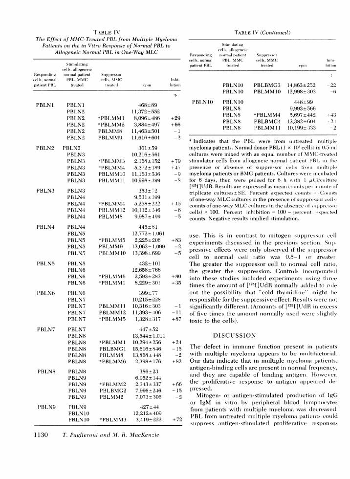
TABLE I V"The Effect of MMC-Treated PBL from Multiple Myelotmia
Patients on the in Vitro Response of Normal PBL toAllogeneic Normal PBL in One-Way MLC
Stinuitilatinigcells, atllogeneic
Responding uioriualil patient Suppressorcells, nionial 1'B L, M)M. C cells, MN\IC InIhi-patient PBL treated treated cp)1n hition
PBLN1 PBLN1PBLN2PBLN2PBLN2PBLN2PBLN2
PBLN2 PBLN2PBLN3PBLN3PBLN3PBLN3PBLN3
PBLN3 PBLN3PBLN4PBLN4PBLN4PBLN4
PBLN4 PBLN4PBLN5PBLN5PBLN5PBLN5
PBLN5 PBLN5PBLN6PBLN6PBLN6
PBLN6 PBLN6PBLN7PBLN7PBLN7PBLN7
PBLN7 PBLN7PBLN8PBLN8PBLN8PBLN8PBLN8
PBLN8 PBLN8PBLN9PBLN9PBLN9PBLN9
PBLN9 PBLN9PBLN1OPBLN110
468+8911,372+552
*PBLiMM 8,096±486*PBLMM2 3,884+497PBLMM8 11,463+501PBLMM19 11,616+601
361+5910,216±561
*PBLM.\1;3 2,168+152BPBL.NM14 5,372+189PBLMM1O 11,163-536PBLMM11 10,998t:399
353 729,531 l399
*PBLU1M4 5,258±922PBLMM12 10, 112+346PBLMM8 9,987±499
445±8 112,772+1,061
*PBLMM5 2,225±+206PBLMM9 13,063+1,099PBLMMI10 13,398±699
432±10112,658+766
*PBLMM6 2,593+283*PBLM\1M 8,229+301
399±7,10,215+228
PBLNMM11 10,316+303PBLMM12 11,393+406*PBLM\15 1,328+317
447+5213,544+1,011
*PBLM\lM1 10,294+256PBLBNIG1 15,616±846PBLXIM8 13,868+448*PBLMM6 2,398+176
386±236,952±144
*PBLMM2 2,343±337PBLBMG2 7,996+246PBLMM2 7,073+306
427±4412,212+409
*PBL\NIM3 3,419+222
+29+66
-1-2
TABLE IV (Continuted)
Stimuiiilatinigcells, allogenieic
Responding niormal patient Stippressorcells, normal PBL, N\IIC cells, MMC Iinhi-patient PBL treatedi treated cpin bition
PBLN10 PBLBMG3 14,863+252 -22PBLN10 PBLMM1O 12,998+(303 -6
PBLN1O PBLN10 448+99PBLN8 9,993±566PBLN8 *PBLMM4 5,697+442 +-43PBLN8 PBLBMG4 12,382+6(04 -2.4PBLN8 PBLMM11 10, 199+;3:33 -2
* Indicates that the PBL were from uniitreated mutiltiplemyeloma patients. Normal donor PBL (1 x 1()5 cells) in 0.5-ilcuiltures were mixed with an eqtial nurnber of 'MXC-treated
+79 stimulator cells from allogeneic normal :atieuit PBL in thet+47 presence or absenee of suppressor cells tizxoui mulntiple
-9 myeloma patients or B.MG patients. Cuiltures were incubated-8 for 6 days, then- were pulsed tor 6 h kw th 1 4.tCi/culture
['251]UdR. Results are expressed as meani couinits per- tititinte oftriplicate cultures- SE. Percent expected coulnts -"Coinitsof one-way MLCcutlttures in the presence of stuppiressor ceils/
645 counts of one-way MLCcuilttures in the absence of wflpressor-6 cells) x 100. Percent inhibition = 100 - percent -xpected
couints. Negative restults implied stimutlation.
uise. This is in contrast to mitogen suppressor cell+83 experiments discutssed in the previolls section. Siup)-
-2 pressive effects were only observed if the siuppressor-cell to normal cell ratio was 0.5-1 or greater.The greater the stuppressor cell to normal cell ratio,the greater the stuppression. Controls incorporated
+80 into these sttudies incltuded experiments uising three+35 times the amount of [1251]UdR normally added to roile
otlt the possibility that "cold thymidine" might beresponsible for the suippressive effect. Resuilts were not
-1 significantly dlifferent. (Amouints of [1251]UdR in excess-l of five times the amouint normally uised were slightly
+87 toxic to the cells).
DISCUSSION+24-24 The defect in immune ftunction present in patients
-2 with mtultiple myeloma appears to be mltltif'actoritl.+82 Our data indicate that in multiple myeloma patients,
antigen-binding cells are present in normal frequtiency,and they are capable of binding antigen. However,
+66 the proliferative response to antigen appeared (de--15 pressed.-2 Mitogen- or antigen-stimtulated prodtuction of IgG
or IgM in vitro by peripheral blood lymphocytesfrom patients with mutltiple myeloma was decreased.
+72 PBL from iiintreated mutltiple myelomn<la patients couildsuippress antigen-stimutlated proliferative responses
1130 T. Paglieroni and .I. R. MacKe72zie

and immunoglobulin production of normal lympho-cytes. Similarly, suppression of normal PBL responses
to allogeneic stimuli in one-way MLC in the pres-
ence of PBL from untreated myeloma patients couldbe demonstrated. Suppression of immunoglobulinproduction in vitro was influenced by the presence
of EA-RFC and phagocytic monocytes, whereas,suppression in a one-way MLC was influenced bythe presence of EACand E-RFL.
These results are compatible with the studies of Zollaet al. (2) and Tanapatchaiyapong and Zolla (3).In a mouse model of myeloma, these authors demon-strated a depressed primary immune response, at bothB- and T-cell levels which affected the maturationof antibody-forming cells. Broder et al. (5) demon-strated impaired synthesis of polyclonal immuno-globulins in multiple myeloma patients that was medi-ated by a phagocytic macrophage. In contrast, mostpatients in our studies demonstrated a significantamount of suppressor activity in the absence of phago-cytic macrophages. In our studies, the suppressor
activity appeared to be mediated in part by an EA-RFCwhich was l)resent after carbonyl-iron treatment ofblood. This suppression appears to be in addition
to that mediated by phagoeytic monocytes. Haywardand Greaves (19) have suggested that some cellsleft behind after carbonyl-iron ingestion have manyproperties of monocytes but do not phagocytose.They suggest that these cells may correspond to andoverlap with a population previously described asnull cell. The EA-RFC described in our studies are
probably of this type.The diminished capacity of myeloma patient PBL
to stimulate in MLC and their ability to inhibitPBL is similar to the suppressor cell effects re-
ported in Hodgkin's disease (20). The absence of thislatter effect in treated myeloma patients suggests thatthe cells responsible for this effect are sensitive toalkvlating agents (12).
Several theories have been proposed to explainthe immunological defects in patients with multiplemyeloma. First, myeloma cells may release RNAmolectules that alter the surface immunoglobulinreceptors on B lymphocytes, thus interfering with hostrecognition of antigen and subsequent antibody forma-tion (21, 22). Second, a feedback inhibition by chalonesmay block the expansion of B lymphocytes in re-
sponse to antigen challenge (3, 4). Third, immunoregu-
TABLE VSuppression of One-Way MLCReactions Between Normal and Allogeneic Normal PBL by Subpopulations of
PBL from Untreated Multiple Myeloma Patients
Suppressor cell depleted populations
Responder Stimulator Suppressor Non- Monocyte EAC-RFL E-RFL SIg EA-RFCcells cells cells fractionated depleted depleted depleted depleted depleted
PBLN1 PBLN1 - 444+99 - -PBLN1 PBLN2 11,898+552
PBLN1 PBLN2 PBLMM2 3,884+208 3,036+388 12,712±999 4,118+289 3,838+289 3,182+449PBLN1 PBLN2 PBLMM3 2,575±281 4,563+289 10,373+789 2,246+178 2,246+177 2,912+238PBLN1 PBLN2 PBLMM5 3,323±303 4,991±441 4,820+244 4,586+123 4,352+331 4,821+328PBLN1 PBLN2 PBLMM6 2,246±344 4,014+228 9,063±443 3,229+176 2,762+289 4,072+299
PBLN2 PBLN2 346±89 - -PBLN2 PBLN3 - 10,898±662
PBLN2 PBLN3 PBLMM3 2,586±203 6,696+356 8,616+266 8,063±334 2,767+202 3,036+222
PBLN3 PBLN3 358±87 - -PBLN3 PBLN4 9,439+422 -
PBLN3 PBLN4 PBLMM5 2,984±233 5,033±389 3,363+244 5,696±206 3,531+198 3,886+255
PBLN4 PBLN4 425±84PBLN4 PBLN2 - 12,238±446 -
PBLN4 PBLN2 PBLMM6 2,448±289 5,191+443 10,164±777 5,999+289 3,363+288 3,562±263
Normal donor PBL (1 x 10⁵ cells) in 0.5-ml cultures were mixed with an equal number of MMC-treated stimulator cellsfrom allogeneic normal patient PBL in the presence or absence of suppressor cell subpopulations from untreated multiplemyeloma patients. Cultures were incubated for 6 days, then were pulsed with 1 uCi/culture [125I]UdR. Results are expressedas mean counts per minute of triplicate cultures±SE.
Immune Defects in Multiple Myeloma 1131
latory macrophages may inhibit immunoglobulin pro-duction (5). Our data demonstrate that there aremultiple levels of immune dysfunction in multiplemyeloma patients, suggesting that a single theory ofpathogenesis is inadequate. In addition, Spitler et al.(23) have presented evidence that decreased levelsof C4 and a defect in polymorphonuclear cell adhe-siveness in addition to depressed antibody formationmay contribute to the development of frequent infec-tions in patients with multiple myeloma.
In the normal immune response, it appears thatsuppressor mechanisms, both humoral (24, 25) andcellular (26), operate simultaneously to maintain adelicate regulatory balance. There is thus the possi-bility that the immunoproliferative disease in multi-ple myeloma is a manifestation of an immune defectin a regulatory compartment and a suppressor functiondefect may be implicated. In other systems, suppressorfunctions have not only been attributed to T and Bcells (16, 27), but to macropages (28) and nylon-adherent nontheta-bearing cells (29) as well.
Unfortunately, demonstration of suppressive effectsin vitro does not dignify their importance in the patho-genesis of the disease in which they are found(30). Suppressor activity has been found after concana-valin A treatment of lymphocytes from normal donors(31). Since plasma cells from multiple myelomapatients have been shown to possess surface antigencapable of eliciting a blastogenic response in autol-ogous PBL (32), and since it has recently been pro-posed that oncofetal antigen expression of tumorsmay activate appropriate suppressor cells and thusinduce one type of immune tolerance towards a tumor(33), one approach to showing the significance of theobserved suppressive effects in multiple myelomapatients would be the demonstration of suppressionof lymphocyte blastogenesis to autologous tumor cellsby cells added to the mixture of reactor and tumorcells.
Patients diagnosed as having BMGdid not showmany of the immune defects discussed in this paper.Such patients have in common with myeloma pa-tients the proliferation of plasma cells and the produc-tion of excessive amounts of immunoglobulin. The ab-sence of many of immune defects seen in myelomagives important insight into the pathogenesis of thedisease. It is probable that some patients now classi-fied as having benign disease may develop multiplemyeloma. Longitudinal studies comparing develop--ment of immune defects with the clinical course willbe of particular interest.
ACKNOWLEDGMENTSThe authors wish to thank Dr. V. Caggiano and all hispatients used in this study for their assistance.
REFERENCES1. Cone, L., and J. M. Uhr. 1964. Immunological defi-
ciency disorders associated with chronic lymphocyticleukemia and multiple myeloma. J. Clin. Invest. 43:2241-2248.
2. Zolla, S., D. Naor, and P. Tanapatchaiyapong. 1974.Cellular basis of immunodepression in mice bearingplasmacytomas.J. Immunol. 112: 2068-2076.
3. Tanapatchaiyapong, P. and S. Zolla. 1974. Humoral im-munosuppressive substance in mice bearing plasmacy-tomas. Science (Wash. D. C.). 186: 748-750.
4. Salmon, S. E. 1974. "Paraneoplastic" syndromes as-sociated with monoclonal lymphocyte and plasma cellproliferation. Ann. N. Y. Acad. Sci. 230: 228-239.
5. Broder, S. R., Humphrey, M. Durm, M. Blackman,B. Meade, C. Goldman, W. Strober, and T. Waldmann.1975. Impaired synthesis of polyclonal (non-paraprctein)immunoglobulins by circulating lymphocytes frompatients with multiple myeloma. Role of suppressorcells. N. Engl. J. Med. 293: 887-892.
6. Boyum, A. 1968. Separation of leukocytes from bloodand bone marrow. Scand. J. Clin. Lab. Invest. 21 (Suppl.97): 77-89.
7. Yam, L. T., C. Y. Li, and W. H. Crosby. 1971. Cyto-chemical identification of monocytes and granulocytes.Am. J. Clin. Pathol. 55: 283-290.
8. Froland, S. S. 1972. Binding of sheep erythrocytes tohuman lymphocytes. A probable marker of T lympho-cytes. Scand. J. Immunol. 1: 269-280.
9. Greaves, M. F., and G. Brown. 1974. Purification ofhuman T and B lymphocytes. J. Immunol. 112: 420-423.
10. Rabellino, E., S. Colon, H. M. Grey, and E. R. Unanue.1971. Immunoglobulins on the surface of lymphocytes. I.Distribution and quantitation.J. Exp. Med. 133: 156-167.
11. Froland, S. S., F. Wisloff, and T. E. Michaelsen. 1974.Human lymphocytes with receptors for IgG. A populationof cells distinct from Tand Blymphocytes. Int. Arch.Allergy Appl. Immunol. 47: 124-138.
12. Paglieroni, T. G. 1975. Multiple myeloma: an immunolog-ical profile in humans. Dissertation, University ofCalifornia, Davis, Calif. 216 pp.
13. Klinman, N. R. and R. B. Taylor. 1969. General methodsfor the study of cells and serum during the immuneresponse: the response to dinitrophenyl in mice. Clin.Exp. Immunol. 4: 473-487.
14. Liburd, E. M., and T. A. McPherson. 1973. A simplifiedtechnique for the evaluation of antigenbinding cells. J.Immunol. Methods. 3: 79-86.
15. Pellegrino, M. A., S. Ferrone, A. Pellegrino, and R. A.Reisfeld. 1973. A rapid microtechnique for in vitrostimulation of human lymphocytes by phytohemaggluti-nin. Clin. Immunol. Immunopathol. 2: 67-73.
16. Waldmann, T. A., M. Durm, S. Broder, M. Blackman, R. M.Blaese, and W. Strober. 1974. Role of suppressor T cells inpathogenesis of common variable hypogammaglobulin-aemia. Lancet. 2: 609-613.
17. Bach, F. H. and N. K. Voynow. 1966. One way stimulationin mixed leukocyte cultures. Science (Wash. D. C.). 153:545-547.
18. MacKenzie, M. R. and T. Paglieroni. 1977. Multiplemyeloma: an immunologic profile. I. Peripheral bloodstudies. J. Immunol. In press.
19. Hayward, A. R., and M. R. Greaves, 1975. Identification ofcells with monocyte markers in panhypogammaglobulin-aemia. Scand. J. Immunol. 4: 563-570.
20. Twomey, J. J., A. H. Laughter, S. Farrow, and C. C.Douglass. 1975. Hodgkin's disease. An immunodepleting
1132 T. Paglieroni and M. R. MacKenzie

and immunosuppressive disorder. J. Clin. Invest. 56:467-475.
21. Giacomoni, D., V. Yakulis, S. R. Wang, A. Cooke, S. Dray,and P. Heller. 1974. In vitro conversion of normal mouselymphocytes by plasmacytoma RNAto express idiotypicspecificities on their surface characteristic of theplasmacytoma immunoglobulin. Cell. Immunol. 11:389-400.
22. Chen, Y., N. Bhoopalam, V. Yakulis, and P. Heller. 1975.Changes in lymphocyte surface immunoglobulins inmyeloma and the effect of an RNAcontaining plasmafactor. Ann. Intern. Med. 83: 625-631.
23. Spitler, L. E., P. Spath, L. Petz, N. Cooper, and H. H.Fudenberg. 1975. Phagocytes and C4 in paraproteinaemia.Br. J. Haematol. 29: 279- 292.
24. Uhr, J. W., and G. Moller. 1968. Regulatory effect ofantibody on the immune response. Adv. Immunol. 8:81-127.
25. Hellstrom, K. E., and I. Hellstrom. 1971. Someaspects ofimmune defense against cancer. I. In vitro studies onanimal tumors. II. In vitro studies on human tumors.Cancer. 28: 1266- 1268.
26. Gershon, R. K. 1975. T cell control of antibody production.In Contemporary Topics in Immunobiology. N. L. Warnerand M. D. Cooper, editors. Plenum Publishing Corpora-tion, New York. 3: 1-40.
27. Gorczynski, R. M. 1974. Immunity to murine sarcomavirus-induced tumors. II. Suppression of T cell-mediatedimmunity by cells from progressor animals. J. Immunol.112: 1826-1838.
28. Kirchner, H., T. M. Chused, R. B. Herberman, H. T.Holden, and D. H. Lavrin. 1974. Evidence of suppressorcell activity in spleens of mice bearing primary tumorsinduced by Maloney Sarcoma Virus. J. Exp. Med. 139:1473-1487.
29. Eggers, A. E., and J. R. Wunderlich. 1975. Suppressor cellsin tumor-bearing mice capable of nonspecific blocking ofin vitro immunization against transplant antigens. J.Immunol. 114: 1554-1556.
30. Siegal, F. P., M. Siegal, and R. A. Good. 1976. Suppressionof B-cell differentiation by leukocytes from hypogam-maglobulinemic patients. J. Clin. Invest. 58: 109- 122.
31. Shou, L., S. A. Schwartz, and R. A. Good. 1976. Suppressorcell activity after Concanavalin A treatment of lympho-cytes from normal donors. J. Ex). Med. 143: 1100- 1110.
32. MacKenzie, M. R. and T. G. Paglieroni. 1977. Plasma cellantigens in human multiple myeloma.J. Lab. Clin. Med.In press.
33. Sinkovics, J. G. 1976. Suppressor cells and humanmalignant disease. Br. Med. J. 2: 1072-1073.
Immutne Defects in Multiple Myeloma 1133