Embed Size (px)
Citation preview
From DEPARTMENT OF PHYSIOLOGY & PHARMACOLOGY
Karolinska Institutet, Stockholm, Sweden
STUDIES ON THE ROLE OF NGF IN
ARTHRITIS-INDUCED PAIN
TRANSMISSION USING GAIT AND WEIGHT
BEARING AS OUTCOME MEASURES
Kristina Ängeby Möller
Stockholm 2019

Cover photo: Rat crossing the glass walkway in the PawPrint setup.
Photo by Odd-Geir Berge.
All previously published papers were reproduced with permission from the publisher.
Published by Karolinska Institutet.
Printed by Eprint AB
© Kristina Ängeby Möller, 2019
ISBN 978-91-7831-565-9

Studies on the role of NGF in arthritis-induced pain
transmission using gait and weight bearing as outcome
measures
THESIS FOR DOCTORAL DEGREE (Ph.D.)
ACADEMIC DISSERTATION
For the degree of Ph.D. at Karolinska Institutet
This thesis will be defended in public at the CMM Lecture Hall, L8:00, Karolinska
University Hospital, Stockholm, Sweden
Friday the 1st November 2019, 9:00
By
Kristina Ängeby Möller
Principal Supervisor:
Professor Camilla Svensson
Karolinska Institutet
Department of Physiology & Pharmacology
Co-supervisors:
Senior lecturer Xiaojun Xu
Karolinska Institutet
Department of Physiology & Pharmacology
Pain Research Director Carina Stenfors
Orion Corporation, Espoo, Finland
Opponent:
Adjunct Professor Antti Pertovaara
University of Helsinki, Finland
Department of Physiology
Examination Board:
Docent Erika Roman
University of Uppsala, Sweden
Department of Pharmaceutical Biosciences
Docent Klas Abelson
University of Copenhagen, Denmark
Section of Research and Education
Professor Katarzyna Starowicz
Maj Institute of Pharmacology, Polish Academy of
Sciences, Poland


To
Amilie, Johan, Anders
The meaning of my life


ABSTRACT
Pain is one of the most common reasons for seeking healthcare, with approximately forty
percent of those suffering from chronic pain having joint pain. Osteoarthritis is the most
common cause of joint pain, but currently there are few treatments available. The search for
new, effective pain treatment has been mostly unsuccessful, in spite of the discovery of
mechanisms that are involved in the transmission of nociceptive signals from the periphery to
the central nervous system where pain is experienced. This work focuses on the evaluation of
rodent joint pain models, the behavioural manifestations of the injuries, and the possibility to
detect treatment effects in these models.
Three models have been evaluated in rats; intra-articular injection of carrageenan, Freund´s
complete adjuvant (CFA), and monoiodoacetate (MIA) into one hind leg. In mice, two
models have been evaluated; intra-articular injection of CFA, and the surgical model of
anterior cruciate ligament transection (ACLT). Carrageenan injection resulted in an acute,
robust inflammation, CFA injection caused a more long-lasting strong joint inflammation,
and MIA injection resulted in an almost complete loss of joint cartilage after a few weeks.
The model more resembling osteoarthritis was the surgical model, ACLT, which gave severe
cartilage degeneration, osteophytes, and pathophysiological changes in synovia and
ligaments.
Gait and weight bearing during locomotion have been tested in all models. The degree of
weight bearing reduction in the affected limb was largest in the CFA- and carrageenan-
induced model, followed by the MIA model and least effect was seen in the ACLT surgical
model. Thus the ACLT model was not possible to use for pharmacological evaluation of
drugs, whereas carrageenan- and CFA-induced monoarthritis resulted in a big enough
difference between animals with monoarthritis and those without, to test drugs commonly
used for pain as well as those under investigation for effects on pain.
Conventional pain relieving drugs such as non-stereoidal anti-inflammatory drugs (NSAIDs)
and opioids were able to normalize effects on weight bearing caused by both the carrageenan-
and the CFA-induced monoarthritis, as were treatments based on inhibiting the NGF-TrkA
pathway; an anti-NGF antibody and two pan-Trk compounds. However, an antagonist of the
TRPV1 receptor lacked effect.
We also investigated mice with a mutation in the R100 NGFß gene (hR100E), in comparison
with mice possessing a human wild-type NGF (hWT), similar but not exactly like the one
found in a hereditary sensory and autonomic neuropathy type V (HSAN V) disorder. This
disorder leads to insensitivity to deep pain in homozygous patients, with sensory and
autonomic functions remaining almost normal. In mice with the hR100E mutation, we found
similar behavioural outcome; normal peripheral sensory functions but less pain-like
behaviour when assessing joint pain with gait and weight bearing.
In summary, this work shows that in order to detect translatable effects on joint pain, models
need to be robust enough, especially for pharmacological testing, but more important, the
methods of testing need to be relevant for the study aim.

LIST OF SCIENTIFIC PAPERS
I. Ängeby-Möller K, Berge OG, Hamers FP.
Using the CatWalk method to assess weight-bearing and pain behaviour
in walking rats with ankle joint monoarthritis induced by carrageenan:
effects of morphine and rofecoxib
J Neurosci Methods. 2008 Sep;174(1):1-9. doi:
10.1016{j.jneumeth.2008.06.017. Epub 2008 Jun 27.
II. Ängeby Möller K, Kinert S, Størkson R, Berge OG.
Gait analysis in rats with single joint inflammation: influence of
experimental factors
PLoS One. 2012;7(10):e46129. doi: 10.137/journal.pone.0046129. Epub 2012
Oct 5.
III. Finn A, Ängeby Möller K, Gustafsson C, Abdelmoaty S, Nordahl G, Ferm
M, Svensson C.
Influence of model and matrix on cytokine profile in rat and human
Rheumatology (Oxford). 2014 Dec;53(12):2297-305. doi:
10.1093/rheumatology/keu281. Epub 2014 Jul 26.
IV. Ängeby Möller K, Berge OG, Finn A, Stenfors C, Svensson CI.
Using gait analysis to assess weight bearing in rats with Freund´s
complete adjuvant-induced monoarthritis to improve predictivity:
Interfering with the cyclooxygenase and nerve growth factor pathways
Eur J Pharmacol. 2015 Jun:756:75-84. doi: 10.1016/j.ejphar.2015.02.050.
Epub 2015 Mar 16.
V. Ängeby Möller K, Svärd H, Souminen A, Immonen J, Holappa J, Stenfors C.
Gait analysis and weight bearing in pre-clinical joint pain research
J Neurosci Methods. 2018 Apr;300:92-102. doi:
10.1016/j.jneumeth.2017.04.011. Epub 2017 Apr 24.
VI. Ängeby Möller K, Klein S, Seeliger F, Finn A, Stenfors C, Svensson CI.
Monosodium iodoacetate-induced monoarthritis develops differently in
knee versus ankle joint in rats
Neurobiol Pain. 2019. doi: https://doi.org/10.1016/j.ynpai.2019.100036.

VII. Ängeby Möller K, Aulin C, Svensson CI.
Modelling reality: gait and weight bearing in two mouse models of
arthritis
Manuscript
VIII. Kato J, Morado Urbina C, Ultenius C, Ängeby Möller K, Villarreal Salcido
J, Sandor K, Svensson CI.
Characterization of the effect of targeted NGF mutation (R100E) on
pain-related behaviour and peripheral sensory innervation
Manuscript
Publications not included in the thesis
I. Schött E, Berge OG, Ängeby-Möller K, Hammarström G, Dalsgaard CJ,
Brodin E.
Weight bearing as an objective measure of arthritic pain in the rat
J Pharmacol Toxicol Methods. 1994 Apr;31(2):79-83. PMID: 8032098.
II. Ängeby Möller K, Johansson B, Berge O-G.
Assessing mechanical allodynia in the rat paw with a new electronic
algometer
J Neurosci Methods. 1998:84:41-47.
III. Karlsson U, Sjödin J, Ängeby Möller K, Johansson S, Wikström L,
Näsström J.
Glutamate-induced currents reveal three functionally distinct NMDA
receptor populations in rat dorsal horn – effects of peripheral nerve
lesion and inflammation
Neuroscience. 2002;112(4):861-8. PMID: 12088745.
IV. Krekels EHJ, Angesjö M, Sjögren I, Ängeby Möller K, Berge O-G, Visser
SAG.
Pharmacokinetic-Pharmacodynamic modeling of the inhibitory effects of
naproxen on the time-course of inflammatory pain, fever, and the ex vivo
synthesis of TXB2 and PGE2 in rats
Pharmaceutical Research. 2011:28(7):1561-1576.
V. Steinz MM, Persson M, Aresh B, Olsson K, Cheng AJ, Ahlstrand E, Lilja M,
Lundberg TR, Rullman E, Ängeby Möller K, Sandor K, Ajeganova S,
Yamada T, Beard N, Karlsson BC, Tavi P, Kenne E, Svensson CI, Rassier
DE, Karlsson R, Friedman R, Gustafsson T, Lanner JT.
Oxidative hotspots on actin promote skeletal muscle weakness in
rheumatoid arthritis
JCI Insight. 2019 Mar 28;5. Pii: 126347. Doi: 10.1172/jci.insight.126347.
VI. Bersellini Farinotti A, Wigerbland G, Nascimento D, Bas, DB, Morado
Urbina C, Nandakumar KS, Sandor K, Xu B, Abdelmoaty S, Hunt MA,
Ängeby Möller K, Baharpoor A, Sinclair J, Jardemark K, Lanner JT,
Khmaladze I, Borm LE, Zhang L, Wermeling F, Cragg M, Lengqvist J,
Chabot-Doré AJ, Diachenko L, Belfer I, Collin M, Kultima K, Heyman B,
Andrade Jimenez JM, Codeluppi S, Holmdahl R, Svensson CI.
Cartilage-binding antibodies induce pain through immune complex-
mediated activation of neurons
J Exp Med. 2019 Aug 5;216(8):1904-1924. doi: 10.1084/jem.20181657.
CONTENTS
1 Introduction ..................................................................................................................... 1
1.1 Background ............................................................................................................ 1
1.2 Osteoarthritis ......................................................................................................... 2
1.2.1 Risks for getting osteoarthritis .................................................................. 2
1.2.2 Osteoarthritis pathology ............................................................................ 3
1.2.3 Current treatments for osteoarthritis ......................................................... 4
1.3 Pain mechanisms ................................................................................................... 5
1.4 Animal models of inflammatory pain and osteoarthritis ...................................... 5
1.4.1 Carrageenan ............................................................................................... 6
1.4.2 FCA ........................................................................................................... 6
1.4.3 MIA ........................................................................................................... 6
1.4.4 Surgical OA models .................................................................................. 7
1.4.5 Joint loading model ................................................................................... 7
1.4.6 Spontaneous models .................................................................................. 7
1.5 Assessment of pain-like behaviour in rodents ...................................................... 8
1.5.1 Sensory testing .......................................................................................... 8
1.5.2 Assessment of non-evoked or spontaneous behaviours ........................... 9
1.6 Pharmacology ...................................................................................................... 11
1.6.1 Drugs inhibiting NGF and its receptor TrkA ......................................... 11
1.6.2 Inhibitor of the TRPV1 receptor ............................................................. 13
1.6.3 Non-steroidal anti-inflammatory drugs .................................................. 13
1.6.4 Opioids .................................................................................................... 13
1.6.5 Paracetamol ............................................................................................. 14
1.6.6 Pregabalin ................................................................................................ 14
1.7 Mutations of NGF ............................................................................................... 14
2 Aim of thesis.................................................................................................................. 15
3 Material and methods .................................................................................................... 17
3.1 Animal models ..................................................................................................... 17
3.1.1 Animals ................................................................................................... 17
3.1.2 Chemically-induced monoarthritis models ............................................ 17
3.1.3 Anterior cruciate ligament transection model ........................................ 18
3.2 Drugs and drug delivery ...................................................................................... 18
3.2.1 Drugs interfering with the NGF-Trk pathway ....................................... 19
3.2.2 Drugs interfering with the ion channel TRPV1 ..................................... 19
3.2.3 Non-steroidal anti-inflammatory drugs .................................................. 19
3.2.4 Opioids .................................................................................................... 19
3.2.5 Other drugs .............................................................................................. 20
3.3 Assessment of pain-like behaviour ..................................................................... 20
3.3.1 Gait and weight bearing during locomotion ........................................... 20
3.3.2 Stationary weight bearing ....................................................................... 21

3.3.3 Static weight bearing ............................................................................... 22
3.3.4 Mechanical sensitivity ............................................................................. 22
3.3.5 Cold sensitivity ........................................................................................ 22
3.3.6 Thermal sensitivity .................................................................................. 22
3.3.7 The formalin test ..................................................................................... 22
3.4 Collection of fluids and tissues ........................................................................... 23
3.4.1 Blood ....................................................................................................... 23
3.4.2 Synovial fluid .......................................................................................... 23
3.4.3 Hind leg joints ......................................................................................... 23
3.4.4 Nervous tissue and skin ........................................................................... 23
3.5 Biochemical biomarker analysis ......................................................................... 24
3.5.1 Colometric assay ..................................................................................... 24
3.5.2 Immunoassay ........................................................................................... 24
3.6 Immunohistochemistry ........................................................................................ 24
3.7 Assessment of inflammation ............................................................................... 25
3.8 Statistical analysis ................................................................................................ 25
4 Results ............................................................................................................................ 27
4.1 Gait and weight bearing as outcome measure of spontaneous pain in
animal models of arthritis .................................................................................... 27
4.1.1 Carrageenan, CFA- and MIA-induced monoarthritis in rats ................. 27
4.1.2 Pro-inflammatory biomarkers in synovial fluid and blood after
CFA- and MIA-induced monoarthritis in rat ......................................... 29
4.1.3 CFA-induced monoarthritis in mice ....................................................... 30
4.1.4 Anterior cruciate ligament transection in mice ...................................... 31
4.2 Arthritis-induced changes in gait and weight bearing are normalized by
conventional pain relieving drugs ....................................................................... 32
4.3 NGF blockage and TrkA antagonists reverse arthritis-induced changes in
weight bearing and gait, but a TRPV1 antagonist failed to do so ...................... 33
4.4 Mice with non-functional NGF are protected from arthritis-induced
changes in gait and weight bearing ..................................................................... 36
5 Discussion and concluding remarks ............................................................................. 38
6 Acknowledgements ....................................................................................................... 43
7 References ..................................................................................................................... 45

LIST OF ABBREVIATIONS
ACLT
CI
COX
CNS
DWB
ECL
FCA/CFA
HBC
HPMC
HSAN
IL
LOQ
MCC/NaCMC
MCP-1
MIA
MIP-1
NGF
NK-1
NSAID
OA
OARSI
anterior cruciate ligament transection
confidence interval
cyclooxygenase
central nervous system
Dynamic Weight Bearing
electro-chemiluminescence
Freund´s Complete Adjuvant
hydroxypropyl-ß-cyclodextrin
hydroxypropylmethylcellulose
hereditary sensory and autonomic neuropathy
interleukin
lower limit of quantification
microcrystalline cellulose/sodiumcarboxymethyl cellulose
monocyte chemoattractant protein-1
monosodium iodoacetate
macrophage inflammatory protein-1
nerve growth factor
neurokinin-1
non-steroid anti-inflammatory drug
osteoarthritis
Osteoarthritis Research Society International
PGE2
PVP
RA
SDS
SEM
TNF
Trk
TRPV1
UV
prostaglandin E2
polyvinylpyrrolydone
rheumatoid arthritis
sodiumlaurylsulphate
standard error of the mean
tumor necrosis factor
tropomyosin receptor kinase
transient receptor potential vanilloid 1
ultraviolet


1
1 INTRODUCTION
1.1 BACKGROUND
Chronic pain is a major health problem affecting 10-20 percent of the population, resulting in
marked reduced quality of life for the individual. Pain is also a substantial socio-economical
problem and the cost for chronic pain in Sweden has been estimated to 87.5 billion SEK in
medical treatment, loss of productivity and disability (SBU 2006). A large-scale survey
undertaken in 15 European countries showed that 40% of the people reporting persistent pain
(> 6 months) had joint pain. OA, followed by RA, is the most common cause of joint pain
(Breivik et al., 2006). In these conditions, disability and pain upon movement are frequently
reported to have a significant impact on the quality of life (Rejeski et al., 1996; van Baar et al.,
1998).
Thus the overall aim of my work has been to elucidate the possibility to use measurement of
weight bearing and gait in rodent models of joint pain, in hopes that it would give results that
are more predictive for the clinical outcome in trials with patients experiencing joint pain. As
my interest and motivation is based on many years of looking at animal behaviour, that is where
my focus lies. So far several new chemical entities have been tested in the clinic based on other
measurements pre-clinically, such as effects on mechanical or thermal sensitivities, and as these
attempts have not proven successful my conviction is that new ways of measurements are
needed. Drug development in the area of chronic pain has hitherto been mostly unsuccessful,
and there are still few available effective treatments for chronic pain conditions. This is in spite
of immense resources being spent on developing new molecular targets and testing them, first
thoroughly in the preclinical setting, and later some selected promising molecules in the clinic.
Several potential new drug targets have been tested in patients, and despite positive outcomes
in preclinical models, not many have been progressed to produce drugs that alleviate pain in
humans. Although the majority of the preclinical targets never reach the stage of testing in
humans, even those that do mostly failed. This is not a problem only for the pain indication but
several of the large pharmaceutical companies, including AstraZeneca, have closed down their
research in the central nervous system (CNS)/pain field and with that all research for new pain
medication. As I have personal experience of working in projects bringing chemical
compounds all the way to clinical testing, I feel deeply that whatever my experience could add
to the possibility of finding new treatment for chronic pain would make my life’s work
worthwhile.
When I started looking at joint pain models more than twenty-five years ago, not many of the
models and methods used today were known. Manual scoring to assess guarding behaviour had
recently been published by Coderre and Wall 1987, and that was what I used in some models
of monoarthritis. However the results were not satisfactory for me as I could see that scores
changed over the 4 minute films that I analysed, and there were no suggestions of how to adjust

2
the scores for these temporal changes. New devices to measure static weight bearing
encouraged me to look also for the possibility to measure as the animal walked, and in
collaboration with Frank Hamers who developed the CatWalk, the analysis algorithms were
improved by adding light intensity as an indirect method to measure weight bearing. By using
this way of assessing the effect of inhibitors of the nerve growth factor (NGF) pathway – which
worked better than any treatment I had hitherto tested – and of the transient receptor potential
cation channel subfamily V member 1 (TRPV1) receptor – which had no effect – my belief in
choosing a model, and even more importantly a mode of measuring effects, that mirrors what
patients complain about grew stronger.
As OA is the most common reason for joint pain, and no good disease modifying medication
exists today, these patients have been chosen for many of the clinical trials. Below I will review
what is known about risks for getting OA, as well as OA pathology and the treatments used for
OA pain today. Then follows a part where possible mechanisms of pain in OA are discussed.
The next part describes animal models of joint pain; the ones I have used and some other
possible models, and different ways of measuring outcome in the animal models. Finally a part
on pharmacology, including possible targets for treatment that I have used in my studies.
1.2 OSTEOARTHRITIS
1.2.1 Risks for getting osteoarthritis
What causes the start of processes in the joint that lead to OA is not yet known, but several
underlying risks for developing OA have been found (Felson et al., 2000; Zhang et al., 2010).
As in many other joint diseases there is a difference of OA prevalence between women and
men; females generally have a higher risk at getting OA and it gets more severe compared to
that in males (Srikanth et al., 2005; Glass et al., 2014). This becomes more obvious as
patients grow older, and age is a high risk in itself (Zhang et al., 2010; Neogi and Zhang
2013; Loeser 2013), and perhaps one of the strongest risk factors for OA (Lawrence et al.,
2008). Furthermore fractures of the meniscus and injuries of the cruciate ligaments
surrounding the knee increase the risk of later getting OA (Englund et al., 2003; von Porat et
al., 2003; Muthuri et al., 2011), but damage to the meniscus and disruption of the anterior
cruciate ligament have also been found in OA patients without previous injuries (Hill et al.,
2005; Englund et al., 2003), and could perhaps be another part of the pathology itself.
Another factor shown to have an impact on the occurrence of OA is body weight, and obesity
adds to the development of OA (Felson et al., 1988; Niu et al., 2009; Blagojevic et al., 2010).
The reason for the increased risk may be the obvious increased load on joints, but there are
also suggestions that the reason is metabolic, as joints that are not loaded such as the hands
and shoulders are more affected in patients with obesity (Oliviera et al., 1999). In addition,
there is a large and complex genetic component to the risk of developing OA, and it has been
reported that the heritability of OA is more than 60%, but differs with respect to the different
joints (Spector and MacGregor 2004).
3
1.2.2 Osteoarthritis pathology
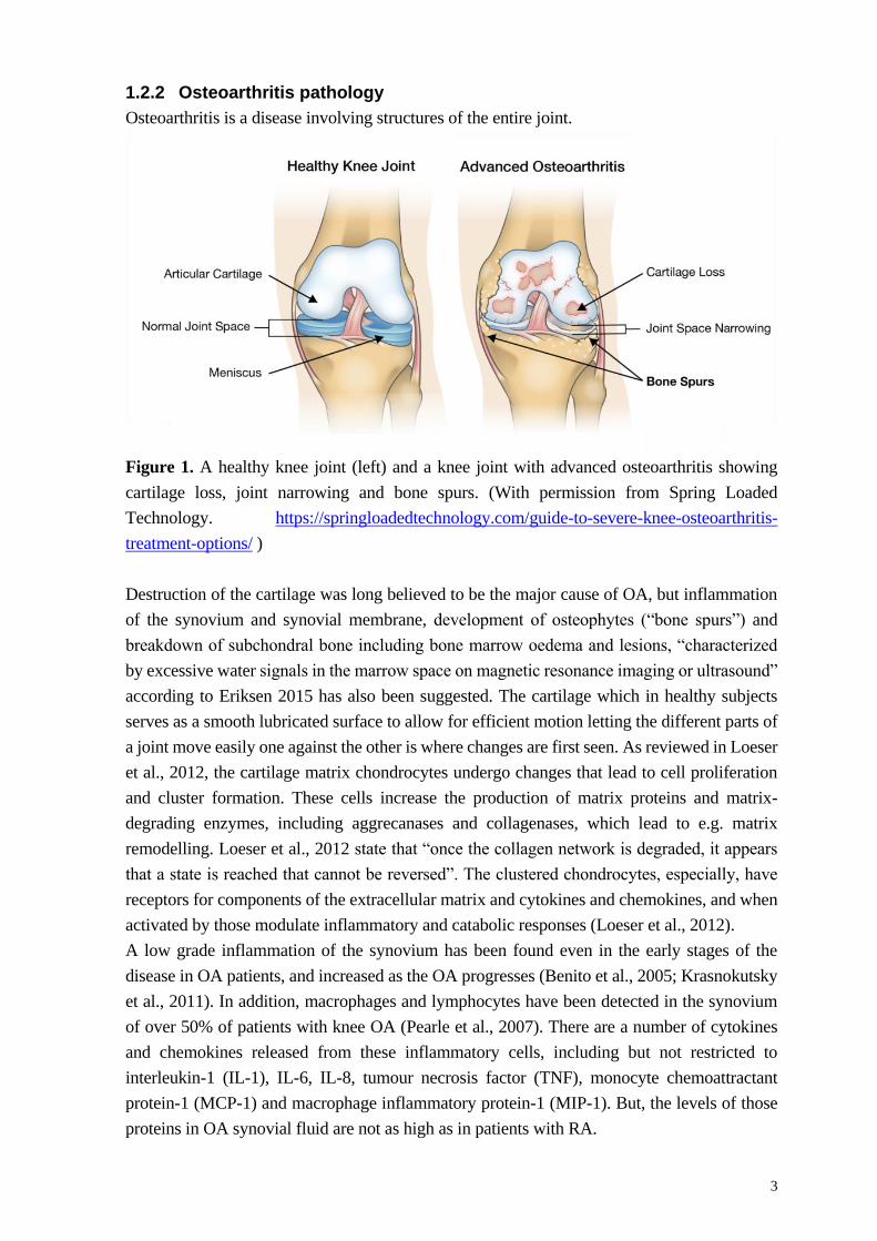
Osteoarthritis is a disease involving structures of the entire joint.
Figure 1. A healthy knee joint (left) and a knee joint with advanced osteoarthritis showing
cartilage loss, joint narrowing and bone spurs. (With permission from Spring Loaded
Technology. https://springloadedtechnology.com/guide-to-severe-knee-osteoarthritis-
treatment-options/ )
Destruction of the cartilage was long believed to be the major cause of OA, but inflammation
of the synovium and synovial membrane, development of osteophytes (“bone spurs”) and
breakdown of subchondral bone including bone marrow oedema and lesions, “characterized
by excessive water signals in the marrow space on magnetic resonance imaging or ultrasound”
according to Eriksen 2015 has also been suggested. The cartilage which in healthy subjects
serves as a smooth lubricated surface to allow for efficient motion letting the different parts of
a joint move easily one against the other is where changes are first seen. As reviewed in Loeser
et al., 2012, the cartilage matrix chondrocytes undergo changes that lead to cell proliferation
and cluster formation. These cells increase the production of matrix proteins and matrix-
degrading enzymes, including aggrecanases and collagenases, which lead to e.g. matrix
remodelling. Loeser et al., 2012 state that “once the collagen network is degraded, it appears
that a state is reached that cannot be reversed”. The clustered chondrocytes, especially, have
receptors for components of the extracellular matrix and cytokines and chemokines, and when
activated by those modulate inflammatory and catabolic responses (Loeser et al., 2012).
A low grade inflammation of the synovium has been found even in the early stages of the
disease in OA patients, and increased as the OA progresses (Benito et al., 2005; Krasnokutsky
et al., 2011). In addition, macrophages and lymphocytes have been detected in the synovium
of over 50% of patients with knee OA (Pearle et al., 2007). There are a number of cytokines
and chemokines released from these inflammatory cells, including but not restricted to
interleukin-1 (IL-1), IL-6, IL-8, tumour necrosis factor (TNF), monocyte chemoattractant
protein-1 (MCP-1) and macrophage inflammatory protein-1 (MIP-1). But, the levels of those
proteins in OA synovial fluid are not as high as in patients with RA.
4
Both mechanical load and the inflammatory process of OA leads to adaptations of the bone,
making it less resistant to damage. Bone remodelling is thought to represent attempts to heal
the bone, and may be the starting point of osteophytes, i.e. bony protrusions at the edge of the
joint. As a response to the injury, including bone marrow lesions, the tissue is activated and
new vascular channels grow into the affected bone to start the regeneration (Loeser et al.,
2012). Along the vessels sensory nerves may grow, that are believed to add to the pain
signalling in OA (Suri at al., 2007).
1.2.3 Current treatments for osteoarthritis
Treatments available today for OA do not stop disease progression, but are aimed to alleviate
the major symptom, pain (Jordan et al., 2003; Zhang et al., 2007; Zhang et al., 2008; Bruyère
et al., 2014). The only “treatment” with proven efficacy is physical exercise, and both aerobic
training and strength exercise give the largest effects in relieving pain and improving function
(Walker-Bone et al., 2000; Bennell and Hinman 2011; Juhl et al., 2014). In addition, for
patients with obesity, body weight reduction is important and will relieve symptoms. However,
both exercise and weight reduction necessitates behavioural changes that are difficult to control
and are challenging for patients as positive results are not immediate, and so the most common
treatment remains pharmacological. This is mainly pain reducing medication such as
paracetamol, non-steroidal anti-inflammatory drugs (NSAIDs) and weak opioids (Felson
2009). But, according to a review by Smith et al., 2016, these treatments are similar in their
efficacy but can only reduce the pain with about 20%. As the condition proceeds and OA
reaches a stage with severe pain, excessive degeneration of the joint and increasing levels of
disability, many go through a total joint replacement. Unfortunately as many as 15%-30% of
those patients still report considerable pain (Hawker 2006; Wylde et al., 2011). As OA is a
chronic condition, pain medication is needed for many years. The long-term pharmacological
treatments recommended today come with considerable complications; gastrointestinal,
cardiovascular and renal adverse side effects for NSAIDs and nausea, constipation and
dizziness for opioids (Avouac et al., 2007; O´Niel et al., 2012; Pelletier et al., 2016). When
considering this, it is obvious that there is a great need for better medication.
Drug development in the area of chronic pain has been insufficient in general, and there are
currently few available effective treatments. Thus, it is critical to increase our understanding
for how chronic pain is regulated in order to identify new biological targets. Several potential
new drug targets have been tested in patients, and despite positive outcomes in preclinical
models, very few have been progressed to produce drugs to alleviate pain in humans. Hence,
it is critical to improve our tools for assessing potential new targets in a way so that they
predict clinical efficacy.

5
1.3 PAIN MECHANISMS
In the joint, the ligaments, fibrous capsule, meniscus, periosteum and synovial layer are
innervated by sensory A, A- and C-fibres and noxious (pain) sensation can be evoked from
these structures (Schaible et al., 2009). However the cartilage is not innervated and it is believed
that no sensation can be emitted from cartilage in healthy subjects. Upon activation of the
sensory nerves, signals are sent to the spinal cord dorsal horn where these primary afferents
couple to second order neurons or interneurons, which project the information towards the
brain. Patients with joint inflammation experience pain during normal movement of the joint
and even gentle pressure, e.g. palpation, often elicits pain. The increase in sensitivity is thought
partly to depend on inflammatory factors released locally in the joint. For example
prostaglandins, bradykinin, growth factors such as NGF, tumour necrosis factor and
interleukins reduce the threshold for activation and increase the responsiveness of the sensory
neuron, giving rise to pain sensations in response to both noxious and non-noxious stimuli
(termed hyperalgesia and allodynia, respectively) (McMahon et al., 2005).
There is mounting evidence that there is a comparable release of similar mediators in the spinal
dorsal horn, where these factors mediate spinal facilitation of pain processing, further
amplifying nociceptive signals conveyed to the brain. Hence, many factors that are involved in
the inflammatory process at the peripheral site may also drive spinal sensitization. It is
important to note that an inflammatory response normally is self-limiting, serving as a
protective mechanism in response to injury or infection. All the same, there are many long-
term or chronic conditions in which inflammation does not resolve, and this may have a
significant impact on the sensory neurons and lead to chronic pain.
Lately many studies have shown that sensitization of the central pain system occurs which
contributes to pain in preclinical models of OA (Abaej et al., 2016; Knazovicky et al., 2016),
and in chronic OA pain patients (Akinci et al., 2016; Moss et al., 2016). Both central
integration of the pain signal and the descending pain pathways may play a part in the
sensitization (Arendt-Nielsen et al., 2015; Sikander et al., 2016). The signs include increased
spread of tenderness in areas not associated with the affected joints, shown e.g. by decreased
thresholds for mechanical sensitivity. Additionally, there are reports that OA with time can
manifest in neuropathic pain states (Dimitroulas et al., 2014; Roubille et al., 2014).
1.4 ANIMAL MODELS OF INFLAMMATORY PAIN AND OSTEOARTHRITIS
In order to study chronic inflammatory joint pain, the choice of animal model is important. The
optimal model would be one that completely mirrors what is found in human patients, but as
even in those there is a range of different structural and sensory changes, this becomes nigh
impossible. Several aspects then form the basis for which model to choose. If the main focus
is to look at structural changes the model needs to be one showing similar modifications, e.g.
in cartilage, synovium and subchondral bone, as those seen in at least a subgroup of patients.

6
As my interest lies in measurement of pain-like behaviour, the models chosen for my work
need to give significant effects in tests aimed at assessing mechanical or thermal sensitivity, or
even more important, in tests attempting to measure spontaneous or non-evoked pain. Several
widely used animal models of arthritis-like pain involve intra-articular ankle or knee injection
of chemical agents such as carrageenan and Freund’s complete adjuvant (FCA/CFA).
1.4.1 Carrageenan
The carrageenan-induced monoarthritis produces a pronounced inflammatory reaction where
granulocytes rapidly infiltrate the site of injection and which gives nociceptive behaviour in
rats that lasts for 24-48 hours. It contains a sulphated mucopolysaccharide derived from
Chondrus crispus or from red Scottish seaweed (Smith and Cook 1953) and a single intra-
articular injection can reduce the proteoglycan content and the rate of proteoglycan synthesis
in the cartilage (Lowther and Gillard 1976; Santer et al., 1983). In 1992, Tonussi and Ferreira
described how carrageenan injected into the rat knee joint caused a concentration-dependent
incapacitation which reached a maximum within 24-48 hours after the injection. In addition, it
caused a pronounced swelling of the joint. As there is a possibility that the swelling itself could
cause sensory activation, dextran was injected intra-articularly into the knee joint in another
group of rats. However dextran, a substance that causes oedema but does not involve
inflammatory mediators or cellular infiltration, failed to induce any incapacitation.
1.4.2 FCA
FCA/CFA is a mixture of mineral oil and inactivated, dried mycobacteria, which produces a
more prolonged stimulation of cell-mediated immune responses leading to a local joint
inflammation that lasts for up to two weeks or more in rats. In 1992 Butler et al. described a
method to induce an inflammation locally in the ankle joint of one hind paw in rats. This new
technique of intra-articular injection led to a restricted monoarthritis, not the generalized
polyarthritis which is a result of injecting FCA subcutaneously (Waksman et al., 1960; Pearson
et al., 1960; Pearson 1963). In contrast to the carrageenan model, injection of FCA to the rat
ankle joint leads to a number of structural changes such as pannus formation, bone remodelling
and cartilage erosion (Brenner et al., 2006; Hashmi et al., 2010).
1.4.3 MIA
A low-grade inflammatory model, first described by Kalbhen DA in 1987: “Chemical model
of osteoarthritis – a pharmacological evaluation” is induced by the intra-articular injection of
monosodium iodoacetate (MIA) that inhibits glyceraldehyde-3-phosphate dehydrogenase and
disrupts the chondrocyte glycolysis leading to ensuing cell death. In the MIA model the major
inflammatory phase resolves within a week, while collagen degradation progressively makes
the cartilage thinner until the underlying bone emerges (Guzman et al., 2003; Fernihough et al.,
2004).
These three rat models are commonly used in the pain research field and have the advantage of
a predictable onset, high success rate and fast disease progression. They are also the models
that form the basis for my work. However, it has been debated whether using longer-lasting

7
animal models would generate changes in the sensory system that more closely mimic changes
in the mechanisms of chronic arthritis-induced pain in humans, and efforts have been made to
find models that more resembles osteoarthritis.
1.4.4 Surgical OA models
One of the most common ways of modelling OA to understand the histological consequences
is by inflicting a surgical injury to the knee joint in different animal species. Several procedures
have been described, such as meniscal tear, partial or total meniscectomy and anterior cruciate
or other ligament transection, and all could be assumed to model aspects of traumatic causes
of OA in humans. By inflicting destabilization of the knee joint in rats or mice, similar
structural changes to those in patients have been described, such as loss of cartilage, occurrence
of osteophytes and subchondral sclerosis (Fernihough et al., 2004; Janusz et al., 2002; Bove et
al., 2006). In addition, several of these models show development of mechanical
hypersensitivity and guarding of the injured limb. When comparing to the slowly evolving
human OA, these models have the advantage of a slower induction (weeks) and a longer lasting
(months) hypersensitivity than those chemically induced. Although differing in the pathology,
surgical models could potentially be used to evaluate new mechanisms and targets for treatment
of joint pain.
1.4.5 Joint loading model
Recently, a mouse model focusing on increased loading of the knee joint was developed and
validated (De Souza et al., 2005; Poulet et al 2011, ter Heegde et al 2019). This model was
induced by putting one hind leg of an anaesthetized mouse in a holder, where the knee joint
was placed into one cup and the ankle joint into another. A small baseline load, 2N, kept the
leg in place, and the load was then increased to 9-11N for 0.05 seconds every 10 seconds. The
peak loading was repeated 40 times in one session, and three sessions per week was performed
for two consecutive weeks. The model results in cartilage lesions and loss of structural integrity
of the lateral femur (Poulet et al 2011), as well as development of mechanical hypersensitivity
and reduced weight bearing of the affected hind limb, lasting for more than six weeks (ter
Heegde et al 2019).
1.4.6 Spontaneous models
Spontaneously occurring knee OA has been observed in several animal species, including
some strains of mice, guinea pigs and hamsters (Walton 1979; Nordling et al., 1992;
Silverstein and Sokoloff 1958; Bendele and Hulman 1988). Even though one may think that
these animals would be the best in which to study OA, it is impossible to know exactly when
symptoms appear in the individual mouse or guinea pig. As a result, planning studies are
difficult and the time to conduct a study can become far longer than recourses allow for. In
addition, the pathogenesis of this OA is not completely understood (Bendele 2001). These
facts make well powered, planned and controlled tests expensive and challenging, if not
almost impossible.

8
1.5 ASSESSMENT OF PAIN-LIKE BEHAVIOUR IN RODENTS
Assessment of pain-like behaviour in rodents remains a challenge, more so in mice than in rats.
Behavioural testing in rodent pain models can be divided into two categories: performer
stimulus-evoked, and non-evoked or spontaneous behavioural responses. The traditional way
to test is by establishing a threshold where the animal reacts with a behaviour that can be
assumed to be a response to a nociceptive signal – an evoked response to mechanical, thermal
or cold stimuli.
1.5.1 Sensory testing
1.5.1.1 Mechanical sensitivity
As far back as 1896 Max von Frey in his dissertation described the use of human and horse
hairs of different stiffness for detecting mechanical sensation, and since then similar artificial
hairs have been named von Frey filaments (von Frey 1896). The traditional material used for
the test filaments is synthetic fibres such as nylon, but as those are sensitive to changing
temperature and humidity (Andrews 1993; Ängeby Möller et al., 1998) new hairs made of glass
fibre, the von Frey Optihairs, have also been introduced (Fruhstorfer et al., 2001). Several
different approaches have been used to establish a threshold: application of one or more
selected filaments for a specified number of times and counting percentage of responses to each
filament; application of filaments with increasing force to find the one where the animal starts
responding; or using the up-down technique first described by Dixon in 1980, and modified by
Chaplan et al. 1994. Whichever method of testing that is chosen, they are all dependent on a
performer and his or her subjective decision of when a withdrawal response is achieved. To
overcome the subjectivity attempts have been made to develop more objective methods, using
an electronic von Frey filament which is applied to the paw with increasing force and that gives
a threshold automatically as the paw is withdrawn (Ängeby Möller et al., 1998; Cairns et al.,
2002). For testing sensitivity to pressure in deeper lying tissues several devices have been
presented, such as that described by Randall and Selitto (Randall and Selitto 1957).
In my work before becoming a PhD student I have tested mechanical sensitivity in rats with
both traditional nylon von Frey filaments (Stoelting) and von Frey Optihair, as well as an
electronic von Frey device developed by Somedic (Ängeby Möller et al., 1998). The up-down
method to calculate a withdrawal threshold was always used, as this method seems to have
become the method of choice for many pain researchers. However the majority of these studies
were performed on rats subjected to models of neuropathic pain. In my present work the more
stable von Frey Optihairs have been used.
1.5.1.2 Thermal sensitivity
Testing sensitivity to heat comes with similar difficulties. Thus directing a light beam towards
the plantar skin of a paw to assess the latency until withdrawal was described by Hargreaves
1988 (Hargreaves et al., 1988), but as the skin can have varying basic temperature which affects
the result it can be difficult to establish what has more impact – the increase in temperature or
the starting skin temperature (Tjølsen et al., 1989). When the baseline skin temperature is high,
9
the latency until withdrawal is short, while it takes considerably longer time until withdrawal
if the skin temperature in the beginning of testing is low (Hole and Tjølsen 1992).
1.5.1.3 Cold sensitivity
Cold sensitivity is often measured with the application of an acetone drop (De la Calle et al.,
2002; Dowdall et al., 2005), but spraying of ethyl chloride has also been reported (Gustafsson
and Sandin 2009). These applications are considered to be pain-like if the animal starts licking
and/or shaking the affected paw. The decrease in skin temperature following application is
much larger for ethyl chloride, but in both cases the amount applied is hard to control as is the
exact site of stimulation.
1.5.2 Assessment of non-evoked or spontaneous behaviours
Still, for models of arthritis it is also possible to assess spontaneous or non-evoked behavioural
responses, which probably more reflect the sense of pain in the animals. This has been the main
focus for my work, as I believe that this could mirror what patients experience in the clinic. I
have specifically tried to find ways of measuring pain-like behaviour of animals in locomotion,
as that is the major complaint in patients with joint pain. Many ways of measuring total
locomotor activity exist, but there are equally many reasons for a decrease in locomotor
activity, making this way of testing specific models difficult.
1.5.2.1 Static weight bearing
When only one hind paw has been subjected to an induction of arthritis, scoring of guarding
behaviour during standing and walking was first described by Coderre and Wall (Coderre and
Wall 1987). Scoring is by its nature subjective and many are those that have come up with
methods trying to make assessment of guarding more objective. By fitting force plates in the
floor of a box the weight load of the two hind paws could be measured (Schött et al., 1994),
and later the Incapacitance tester was introduced where animals stand with the two hind paws
on force plates while the front paws are placed on a ramp (Bove et al., 2003). In both methods,
placing an animal in the box or holder induces stress and needs training before a trustworthy
result can be obtained, and varying placements of the tail or the animal leaning onto the side of
the box can distort the readout.
1.5.2.2 Stationary weight bearing
Recently another device has been introduced, the Advanced Dynamic Weight Bearing (DWB),
where a sensor mat measures the load and a camera fitted on top of the box films the animal
simultaneously making it possible to validate the detection of each paw’s placement (Tetreault
et al., 2011). The animal is allowed to move within a restricted area during a selected period to
give weight bearing data of all four paws both when standing and when rearing.
1.5.2.3 Gait and weight bearing during locomotion
As movement-induced pain is a major complaint in patients with chronic pain from the joints,
exploring behaviour in rodents with and without joint inflammatory pain during locomotion

10
may give added information of mechanisms and treatment. Several ways to do this have been
introduced, but few have yet been extensively used.
Tonussi and Ferreira (Tonussi and Ferreira 1992) invented a setup where rats were fitted with
specially designed metal gaiters wrapped around the paw of interest, and the time of contact
with a mesh floor covering a rotating cylinder was then assessed. With this device they could
establish the pharmacological effects of some analgesic drugs after injection of carrageenan
into one knee joint of rats. By attaching an electrode between the pads of the hind paw the
method was improved and the rats could walk more freely on the cylinder (López-Muñoz et
al., 1993). Still these were setups that were complicated and difficult to manufacture to
become commercially available.
Another approach was introduced – to use video recordings from underneath to assess
walking rats. To make the paw contact areas more visible a frosted plexiglass surface was
suggested, which gave several new parameters of all four paws such as gait stance – the time
from initial contact of a paw with the floor until lifting the paw as next step was started
(Walker et al., 1994). The weight bearing during locomotion turned out to be more difficult to
assess. By adapting a technique described by Betts and Duckworth in 1978 for measuring
human plantar pressure, based on white light entering the long edges of a 6 mm thick glass
where the light was internally reflected between the two surfaces of the glass until e.g. a rat
paw was placed which then scattered the light, K Clarke built a walkway that could study the
paw contact patterns of rats (Clarke 1992). He later combined it with load measurements
underneath two 10x5 cm areas at half-way of the walkway, one for each side of the glass
floor, and showed that the coefficient of correlation between load cell output and output from
the scattered light of a paw in contact with the floor was 0.92 (Clarke 1995). Using this setup,
effects of the MIA model of OA in rats, as well as detailed load and temporal aspects of
mouse locomotion were assessed (Clarke et al., 1997; Clarke and Still 1999; Clarke and Still
2001).
Hamers et al., 2001 introduced a walkway with light entering the long edge of a glass floor,
the CatWalk, where a semi-automated interactive analysis algorithm gave a large number of
parameters assessed in rats with spinal cord injury (step sequence distribution, regularity
index, print area, maximum contact area during stance, base of support, stride length, swing
duration, and duration of tail dragging and abdominal dragging). Later the amount of light
intensity per pixel was added to make it possible also to indirectly assess weight bearing in
rats with a model of neuropathy (Vrinten and Hamers 2003). Similar setups have followed,
manufactured by several companies focusing on devices for animal behavioural research.
Others tried a slightly different technique, such as that of the TreadScan (CleverSys) where
rodents’ paws are filmed from underneath as they walk on a sliding treadmill belt which can
be controlled for the speed of the animal as it is forced to move at the same speed as the belt
(Beare et al., 2009). Temporal and all area-dependent parameters can be assessed, and it’s

11
suggested that changes in weight bearing are indirectly detected as the paw is pressed against
the floor and the colour of the plantar skin becomes lighter when more of the small capillaries
are obstructed.
Still, the most common tests rely on tester-evoked responses such as assessment of mechanical
or thermal hypersensitivity by generating a withdrawal from the source of stimulus. According
to Mogil and Crager 2004, 90% out of 259 articles between 2000 and 2004 in the journal “Pain”
that described animal studies of neuropathic and inflammatory pain in which behavioural
testing was included, exclusively measured changes in threshold or response to evoked stimuli.
A number of drugs, for example neurokinin-1 (NK-1) antagonists and transient receptor
potential vanilloid 1 (TRPV1) antagonists (Svensson et al., 2010) have failed in clinical trials
despite repeatedly showing effect against hypersensitivity states. There is an increasing
suspicion that tester-evoked tests do not reflect the human joint pain problem, and that non-
evoked behaviours such as locomotion, guarding of the effected limb, reduced weight bearing
and gait changes may provide a better correlation to the human pain state.
Hence, it is critical to improve our tools for assessing potential new targets in a way so that
they predict clinical efficacy. From a pain perspective, it is essential to evaluate models that
best mimic the human pain condition, and perhaps even more important, which endpoints to
use when measuring pain behaviour. As movement-induced pain is a problem for arthritis
patients we have investigated if gait and weight bearing can be used as endpoints that better
mimic the pain state in humans, and thus hopefully better predict results in clinical trials.
1.6 PHARMACOLOGY
Pharmacological evaluation was performed in the present work, to elucidate the usefulness of
the chosen models and measurement methods, and include several NSAIDs, paracetamol,
pregabalin and opioids. In addition new chemical entities targeting the NGF pathway – both an
anti-NGF antibody and two pan-Trk inhibitors; inhibitors of tropomyosin receptor kinases
(Trk) A, B and C – and an inhibitor of the TRPV1 receptor have been evaluated. Thus, below
I describe some of the characteristics of these pharmacological agents.
1.6.1 Drugs inhibiting NGF and its receptor TrkA
Several attempts have been made to find suitable pharmacological treatment for chronic pain
using the NGF/TrkA pathway as target. One of the most encouraging is the development by
Pfizer of a monoclonal antibody that inhibits NGF, Tanezumab, which shows efficacy both in
rodent models (Shelton et al., 2005) and in patients with OA (Lane et al., 2010). The reduction
of knee pain in the patients while walking was more pronounced than that being achieved by
commonly used NSAIDs (Schnitzer et al., 2015). NGF is a member of the neurotrophin family
and activates the TrkA with high affinity, and the p75 receptor with low affinity. It is essential
for axonal growth and survival, especially in the neonatal time period, and later in life plays a
major role in pain processing. When tissue is injured or inflamed, NGF is released from mast

12
cells, macrophages, keratinocytes and T-cells (Figure 2; for review see Pezet and McMahon
2006). Of note, when activated, the TrkA receptor forms a complex with NGF and is
internalized and transported to the nucleus where it promotes expression of a number of other
pain-associated receptors, such as CGRP, TRPV1, P2X3 and ion channels neurotransmitters
(Heppenstall and Lewin 2000; Winston et al., 2001; Bonnington and McNaughton 2003; Pezet
and McMahon 2006).
Figure 2. Neurotransmitters, receptors and ion channels that are modulated and up-regulated
by NGF binding to TrkA-positive primary afferent sensory nerve fibres are shown in (A). In
(B), cells that release NGF during injury or inflammation is shown. (Reprinted with permission
from Mantyh et al., 2011)
Injection of NGF into the paw causes hypersensitivity to mechanical stimuli in rats (Lewin and

13
Mendell 1993), as does injection into the masseter muscle in humans, where pain intensity also
increased when chewing and yawning (Svensson et al., 2003). NGF is elevated in synovial
fluid from patients with RA (Halliday et al., 1998), however the effect of NGF blockage on
pain in RA animal models or RA patients has not been extensively studied.
1.6.2 Inhibitor of the TRPV1 receptor
The TRPV1 receptor is activated by heat and acidic environment containing increased levels
of H+ (Caterina et al., 1997). Capsaicin, the ingredient giving food some of its “hot” taste, is
an agonist of TRPV1, and injecting capsaicin into human or animal skin results in an intense
but short-lasting pain (Lamotte et al., 1992; Kinnman and Levine 1995). Activation of the
TrkA receptor by NGF leads to a rapid increase in TRPV1 receptor membrane expression on
primary sensory neurons (Zhang et al., 2005; Stein et al., 2006) and selective antagonism of
the TRPV1 receptor has been suggested as a potential drug target for treatment of joint
inflammation (Keeble et al., 2005). A model of heat pain that is commonly used in clinical
trials is the ultraviolet (UV) burn, and TRPV1 antagonists have been shown to alleviate this
pain. However trials with TRPV1 antagonists in patients with OA failed as no effect on pain
was found (Svensson et al., 2010; Segerdahl et al., 2010).
1.6.3 Non-steroidal anti-inflammatory drugs
One of the mediators produced in inflamed tissue is prostaglandin E2 (PGE2). The origin is
fatty acids derived from cell membrane, and it is synthesized when the tissue is injured by
physical trauma or when cytokines or growth factors such as NGF signal the need for tissue
inflammation and repair. The production of prostaglandins requires several enzymes, one of
which is cyclooxygenase (COX), the enzyme inhibited by NSAIDs. There are two different
variants of COX; COX-1 and COX-2. COX-1 is believed to be constitutive, whereas COX-2
is predominantly produced in inflammation. Production of PGE2 leads to the well-known
symptoms of inflammation – pain, swelling and fever.
The first report of the effects of COX inhibition in humans through use of willow bark was
found in: Stone E. “An account of the success of the bark of the willow in the cure of agues”
(Stone 1763), and reported in Vane and Botting 1998. However willow bark, and other plant
derived decoctions containing COX inhibiting salicylates have been used for thousands of
years, and are known from the ancient Egyptians through the Ebers papyrus, and ancient
Greeks in recommendations by Hippocrates. Still today, COX inhibitors are the most
commonly used pain relieving drugs and are recommended for many conditions that have an
inflammatory component, including joint pain originating from e.g. OA.
1.6.4 Opioids
Inhibition of the opioid receptor has been used to relieve pain for thousands of years as well,
but not until the last century have there been an opportunity to reveal the mechanism behind
the effect. Today we know that opioids preferentially act on the µ opioid receptor subtype, a
G-protein coupled receptor which has effects on ion-channels in the membrane of nervous
tissues, resulting in a reduction of neuronal excitability and transmitter release (for review see
Corbett et al., 2006).
14
1.6.5 Paracetamol
In spite of its widespread use and good effect on pain and fever, but not inflammation, the
mechanism of paracetamol is still not known.
1.6.6 Pregabalin
Although introduced as an anti-epileptic drug, the group of drugs that pregabalin belongs to
also have an effect on pain. The mechanism, believed to be reduction of neurotransmitter
release through binding to the α2δ1 subunit of the voltage-gated calcium channels, leads to less
neural activity (for review see Sills GJ 2006).
In summary, one aim of this project was to test the predictive value of the rat and mouse
models of joint inflammation and surgical OA and the described behavioural endpoints using
the NGF/Trk mechanism as a positive target, TRPV1 as a target without effect and compare
those to effects by medications used in treatment of arthritis today, e.g. NSAIDs, COX-2
selective antagonists, paracetamol and opioids. In addition, we wanted to explore the role of
NGF in arthritis-induced pain and arthritis pathology.
1.7 MUTATIONS OF NGF
Mutations of NGF or the NGF receptor TrkA leads to congenital insensitivity to pain
(Nagasako et al., 2003). The resulting condition is named hereditary sensory and autonomic
neuropathy (HSAN), and one of these mutations was detected in Sweden; the type V (HSAN
V; Minde 2006). Peripheral nerves in homozygous patients show reduced amounts of Aδ and
C fibres. These patients develop severe joint problems, e.g. Charcot joints, but unlike other
types of HSAN, no mental retardation or lack of sweating ability has been found. The exact
mutation was found to be a replacement of an amino-acid in the NGFß gene, such that a basic
arginine was exchanged for a non-polar tryptophan (Einarsdottir et al 2004), which then
produced the NGFR100W protein. Attempts have been made to breed mice with the exact same
mutation, but these were not viable unless the mother received normal NGF from gestation
until end of lactation, and the pups were given NGF from birth. Even with this treatment, the
body weight of these mice pups did not increase after the age of three weeks (Testa et al., 2019).
AstraZeneca bred a mouse carrying a similar mutation, the R100E, where arginine was replaced
by glutamic acid. These mice, who survive until adulthood, were donated to our group at KI
and we were fortunate to be able to test them extensively to evaluate whether these mice show
a phenotype similar to the homozygous patients with HSAN V found in the north of Sweden.

15
2 AIM OF THESIS
The overall aim of the study was to investigate whether behavioural endpoints exist that mimic
the movement-induced ongoing pain state in arthritis patients, to give a better predictive value
for clinical trials. Two specific aims of this thesis were:
1. Examine if alterations in gait and weight bearing can be used as outcome measure of
spontaneous pain in animal models of joint inflammation
1.1. Investigate if and how joint inflammation alters standing weight bearing (static),
weight bearing during voluntary and forced locomotion (dynamic) and gait in animal
models of arthritis
1.2. Examine if arthritis-induced changes in static and dynamic locomotion and gait are
normalized by conventional pain relieving drugs
2. Explore the role of NGF and the ion channel TRPV1 in arthritis-induced spontaneous pain
2.1. Investigate if NGF blockade and TrkA and TRPV1 antagonists reverse arthritis-
induced changes in weight bearing and gait
2.2. Investigate if mice with non-functional NGF develop arthritis pathology and/or if they
are protected from arthritis-induced changes in gait and weight bearing


17
3 MATERIAL AND METHODS
3.1 ANIMAL MODELS
3.1.1 Animals
All animal experiments were performed according to regulations and approved by the Swedish
local ethical committees. Animals were housed three to six per cage in transparent Macrolon®
cages with food and tap water ad libitum and lighting controlled.
Rat strains included Sprague-Dawley (Paper I, II, III and IV), Lewis (Paper III, IV and VI)
and Wistar (Paper V), and all studies with rats were done in males. The behavioural
experiments were a continuous process that took place over several years. Due to parvovirus
infection in the breeding facility at the animal vendor affecting the availability of rat strains,
the studies were undertaken in three different rat strains, and Sprague–Dawley rats from two
different breeding facilities. Although not examined in a systematic fashion, some variance in
duration of ankle joint inflammation in response to the FCA was noted; however the overall
behavioural response was similar.
Wild-type C57BL/6JRj mice (Paper VII and VIII) and C57BL/6 mice, of both genders,
genetically modified by a knock-out of the mNGF, followed by either a knock-in with a wild-
type human NGF or with the hNGF containing the mutation (hR100E) in the coding region of
the NGFß gene, replacing arginine with glutamic acid (Paper VIII) were used.
3.1.2 Chemically-induced monoarthritis models
All joint injections were done while the animals were deeply anaesthetized using 4-5%
isoflurane. The injection was completed in less than one minute, and the animals woke up
within two to three minutes.
3.1.2.1 Carrageenan-induced monoarthritis
Lambda-carrageenan (Sigma-Aldrich, Sigma Chemicals Co. St Louis, MO, USA) was
dissolved in physiological saline. Forty to fifty µl was injected into one hind limb ankle joint
in rats, containing 0.47, 0.94, 1.9, 3.8, 7.5, 15 or 30 mg/ml, for a concentration effect study in
Paper II, and 7.5 mg/ml was chosen for comparison between ankle and knee joint injection
and for pharmacological studies in Paper I and II.
3.1.2.2 Complete Freund´s adjuvant-induced monoarthritis
FCA/CFA was prepared by adding 100 mg of Mycobacterium tuberculosis (Difco
Laboratories) to 12.5 ml of Freund´s incomplete adjuvant and dilute to make the different
concentrations (0.12, 0.25, 0.50, 1.0, 2.0, 4.0, 8.0 mg/ml) injected with 50 µl into one hind limb
ankle joint in the concentration effect study in paper II. CFA (Sigma-Aldrich, Sigma
Chemicals Co. St Louis, MO, USA) containing 1.0 mg/ml Mycobacterium tuberculosis was
chosen for comparison between ankle and knee joint injection (Paper II), and for
pharmacological studies in rats (Paper II, III, IV and V). In mice, CFA (1.0 mg/ml; Sigma-
Aldrich or 10 mg/ml; Chondrex) was injected in volumes of 5 µl or 10 µl into one hind limb

18
ankle joint (Paper VI). In Paper VIII, CFA (10 mg/ml, Chondrex) was injected into one ankle
joint in a volume of 5 µl, and 10 µl was injected into one knee joint.
3.1.2.3 Sodium monoiodoacetate-induced monoarthritis
Rats were injected into one hind limb ankle or knee joint with 25 µl (Paper III) or 50 µl (Paper
VI) solution containing 2 mg sodium monoiodoacetate (MIA; Sigma) dissolved in
physiological saline.
3.1.3 Anterior cruciate ligament transection model
Surgical destabilization of the left knee joint in mice (Paper VII) was established by
transection of the anterior cruciate ligament (ACLT) under deep anaesthesia, and assured by
controlling rotational capacity. Buprenorphine was administered post-surgery as analgesic.
3.2 DRUGS AND DRUG DELIVERY
Table 1. An overview of the drugs used
DRUG PAPER SPECIES ROUTE DOSES
(MG/KG)
DOSES
(µMOL/KG)
INDUCTION
AGENT
MEDI-578 Paper IV Rat i.v. 0.03, 0.1, 0.3,
3.0, 30 mg/kg
CFA
Paper VII Mouse s.c. 0.3, 1.0, 3.0
mg/kg
CFA
AZ6623 Paper IV Rat p.o. 10, 30, 100
µmol/kg
CFA
30, 60, 120
µmol/kg
Carrageenan
AZ7092 Paper IV Rat p.o. 1.0, 3.0, 10
µmol/kg
CFA
2.0, 6.0, 20, 60
µmol/kg
Carrageenan
AZD1386 Paper IV Rat p.o. 100 µmol/kg CFA
NAPROXEN Paper II Rat p.o. 1.0, 10, 100
µmol/kg
Carrageenan
Paper IV Rat p.o. 7.5, 30, 90 µmol/kg CFA
Paper V Rat p.o. 2.5, 7.6, 11.7
mg/kg
CFA
IBUPROFEN Paper II Rat p.o. 30, 100, 300
µmol/kg
Carrageenan
Paper IV Rat p.o. 100, 300 µmol/kg CFA
DICLOFENAC Paper II Rat p.o. 1.0, 3.0, 10
µmol/kg
Carrageenan
ROFECOXIB Paper I Rat p.o. 2.4, 9.4 mg/kg 7.5, 30 µmol/kg Carrageenan
Paper IV Rat p.o. 30, 100, 300
µmol/kg
CFA
VALDECOXIB Paper IV Rat p.o. 10, 30, 100
µmol/kg
CFA
MORPHINE Paper I Rat s.c. 1.2, 4.8 mg/kg 3.75, 15 µmol/kg Carrageenan
OXYCODONE Paper II Rat s.c. 1.0, 3.0, 10
µmol/kg
Carrageenan
PARACETAMOL Paper II Rat p.o. 1000, 2000, 4000
µmol/kg
Carrageenan
PREGABALIN Paper V Rat p.o. 10, 30 mg/kg CFA

19
3.2.1 Drugs interfering with the NGF-Trk pathway
The monoclonal NGF antibody MEDI-578 was provided by AstraZeneca, MedImmune,
dissolved in 50mM sodium acetate and 100 mM sodium chloride and given to rats in a volume
of 1 ml/kg intravenously at 0.03, 0.1, 0.3, 3.0 and 30 mg/kg, (Paper IV). In studies with mice,
MEDI-578 (0.3, 1.0 and 3.0 mg/kg) was injected subcutaneously in a volume of 5 ml/kg (Paper
VII).
The two pan-Trk antagonists AZ6623 (10, 30, 60, 100 and 120 µmol/kg) and AZ7092 (1.0,
2.0, 3.0, 6.0, 10, 20 and 60 µmol/kg) were provided by AstraZeneca, dissolved in 0.5%
hydroxypropylmethylcellulose (HPMC) and 0.1% Tween 80, and administered to rats per os
in a volume of 5 ml/kg (Paper IV).
3.2.2 Drugs interfering with the ion channel TRPV1
AZD1386, a TRPV1 antagonist, was provided by AstraZeneca, suspended in 5%
hydroxypropyl-ß-cyclodextrin (HBC), 0.2% (w/v) sodiumlaurylsulphate (SDS), 1% (w/v)
polyvinylpyrrolydone (PVP), 0.25 sodium citrate in water, and administered to rats per os in a
volume of 5 ml/kg at 100 µmol/kg (Paper IV).
3.2.3 Non-steroidal anti-inflammatory drugs
Naproxen (Sigma-Aldrich) was dissolved in physiological saline and administered to rats per
os in a volume of 2 ml/kg in doses of 1.0, 10 and 100 µmol/kg, (Paper II), in a volume of 5
ml/kg in doses of 7.5, 30 and 90 µmol/kg (Paper IV), and in a volume of 3 ml/kg in doses of
2.5, 7.6 and 22.7 mg/kg (Paper V).
Ibuprofen (synthesized at AstraZeneca) was dissolved in physiological saline and administered
to rats per os in a volume of 5 ml/kg in doses of 30, 100 and 300 µmol/kg (Paper II), and
ibuprofen (Sigma-Aldrich) was dissolved in physiological saline and administered in doses of
100 and 300 µmol/kg (Paper IV).
Diclofenac (Sigma-Aldrich) was dissolved in physiological saline and administered to rats per
os in a volume of 5 ml/kg in doses of 1.0, 10 and 100 µmol/kg, (Paper II).
Rofecoxib (VIOXX, Merck Sharp & Dohme), 2.4 and 9.4 mg/kg (7.5 and 30 µmol/kg) in
suspension and diluted with water, given to rats per os in a volume of 8 ml/kg (Paper I). In
Paper IV rofecoxib (30, 100 and 300 µmol/kg) synthesized at AstraZeneca was suspended in
1% microcrystalline cellulose/sodiumcarboxymethyl cellulose (MCC/NaCMC)+0.6% soy
lecithin (Lipoid S100) in water and given to rats per os in a volume of 5 ml/kg.
Valdecoxib (Kemprotec), 10, 30 and 100 µmol/kg was suspended in 1% MCC/NaCMC+0.6%
soy lecithin (Lipoid S100) in water and given to rats per os in a volume of 5 ml/kg (Paper IV).
3.2.4 Opioids
Morphine (Apoteksbolaget, Göteborg, Sweden), which binds preferentially to the µ opioid
receptor, in doses of 1.2 and 4.8 mg/kg (3.75 and 15 µmol/kg), was dissolved in physiological
saline, injected s.c. in a volume of 1.0 ml/kg (Paper I).
Oxycodone (Sigma-Aldrich), was dissolved in physiological saline and administered to rats
subcutaneously in a volume of 2 ml/kg in doses of 1.0, 3.0 and 10 µmol/kg (Paper II).

20
3.2.5 Other drugs
Paracetamol (Fluka), was dissolved in 0.5 % methyl cellulose and given to rats per os in a
volume of 5 ml/kg in doses of 1000, 2000 and 4000 µmol/kg (Paper II).
Pregabalin (Toronto Research Chemicals Inc.), which binds to the α2ß subunit of voltage-gated
calcium channels to reduce the calcium current after activation (Bauer et al., 2010), was
dissolved in physiological saline to get 10 and 30 mg/kg and given per os in a volume of 3
ml/kg in Paper V.
3.3 ASSESSMENT OF PAIN-LIKE BEHAVIOUR
3.3.1 Gait and weight bearing during locomotion
Assessment of gait and weight bearing parameters were based on letting animals voluntarily
cross an enclosed walkway with a glass floor where light entered the long edge. The projected
light is almost entirely reflected within the glass except when an object, such as a paw, touches
it. The light is then scattered and produces an illuminated image of e.g. the paw. The light
intensity depends on the force of the paw-floor contact and the accumulated light intensity for
all pixel in a paw print gives an indirect measure of the weight bearing. A video-camera
mounted underneath the glass floor to film the animal as it crossed the walkway gives a number
of two-dimensional as well as temporal parameters. In our studies, rats or mice were placed in
a goal-cage containing all their cage-mates at the exit of the walkway, and was brought one at
a time to the entrance of the walkway. They were allowed to habituate to the walkway and
without interference make their own way to the exit in order to find the goal cage and their
cage-mates. At least two training sessions were performed before the study was started.
Looking at the films in a pilot study, manual scoring of the weight bearing during locomotion
in rats was performed using a visual rating scale, based on the prints and the walking pattern
and adapted from the rating scale described by Coderre and Wall (1987) (Paper II). In the
following studies, two different methods for automated analysis of the results were used; the
CatWalk and the PawPrint.
The CatWalk, developed by Frank Hamers and later purchased by Noldus (Noldus Information
Technology, The Netherlands), consisted in its first version (Paper I) of an analysing tool
where the experimenter interactively categorized the points of contact: right/left, fore/hind paw,
nose, abdomen, tail and artefacts. This process was later automated, but retained the possibility
to edit the resulting classifications (Paper V, VII and VIII). Parameters used in our CatWalk
studies include: print area, weight load (mean light intensity), stance phase duration, regularity
index, walking speed, duty cycle, and weight bearing (Table 2). The PawPrint (developed by
AstraZeneca, Sweden) was a custom made computer program for rats only (Paper II, IV and
VI), with an automated analysis of the crossing. The program immediately calculated results
and displayed a picture of the prints in false colours (one colour for each paw) which allowed
the experimenter to make a quality check of the results. Editing of the classifications were not
possible. If results were not acceptable, the rat was allowed to walk again across the walkway
to give at least three paw placements by each non-arthritic paw. Parameters used in our
PawPrint studies include weight bearing and regularity index.
21
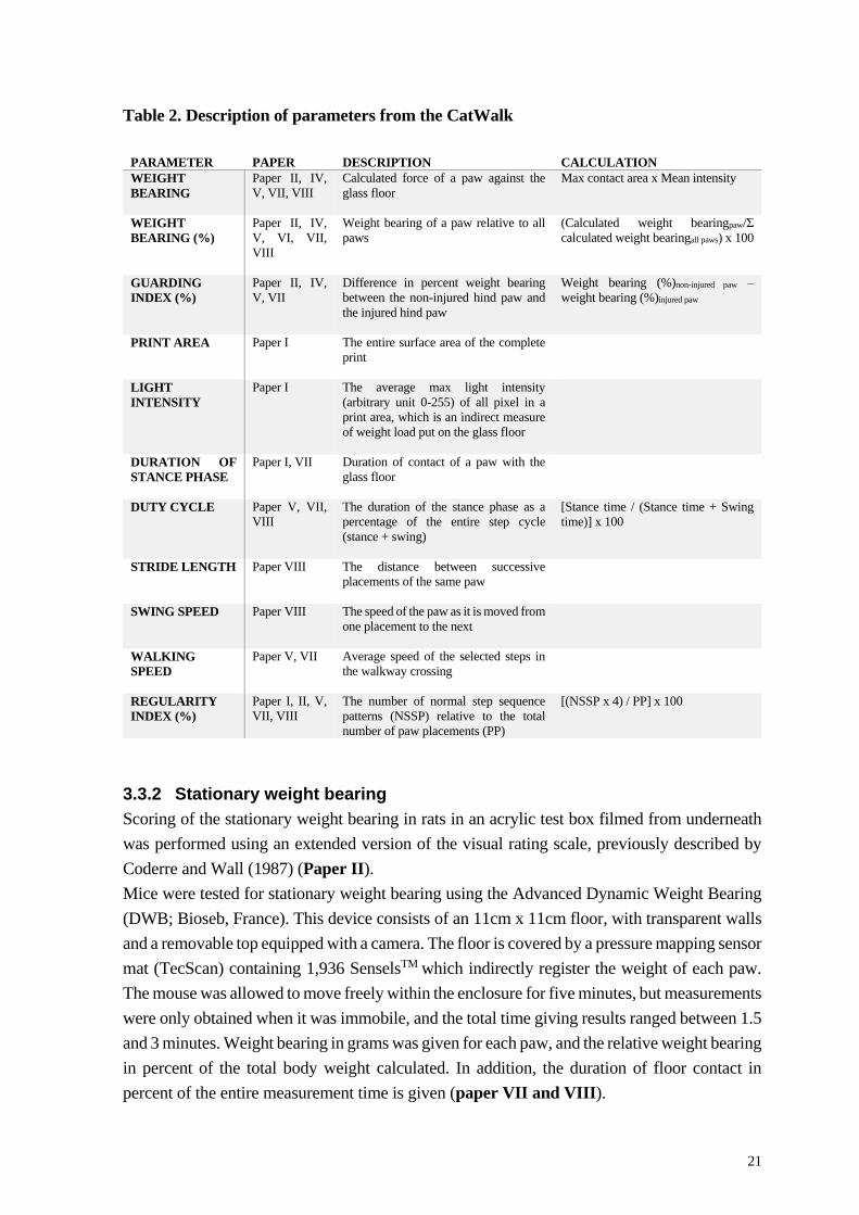
Table 2. Description of parameters from the CatWalk
PARAMETER PAPER DESCRIPTION CALCULATION
WEIGHT
BEARING
Paper II, IV,
V, VII, VIII
Calculated force of a paw against the
glass floor
Max contact area x Mean intensity
WEIGHT
BEARING (%)
Paper II, IV,
V, VI, VII,
VIII
Weight bearing of a paw relative to all
paws
(Calculated weight bearingpaw/Σ
calculated weight bearingall paws) x 100
GUARDING
INDEX (%)
Paper II, IV,
V, VII
Difference in percent weight bearing
between the non-injured hind paw and
the injured hind paw
Weight bearing (%)non-injured paw –
weight bearing (%)injured paw
PRINT AREA
Paper I The entire surface area of the complete
LIGHT
INTENSITY
Paper I The average max light intensity
(arbitrary unit 0-255) of all pixel in a
print area, which is an indirect measure
of weight load put on the glass floor
DURATION OF
STANCE PHASE
Paper I, VII Duration of contact of a paw with the
glass floor
DUTY CYCLE
Paper V, VII,
VIII
The duration of the stance phase as a
percentage of the entire step cycle
(stance + swing)
[Stance time / (Stance time + Swing
time)] x 100
STRIDE LENGTH
Paper VIII The distance between successive
placements of the same paw
SWING SPEED
Paper VIII The speed of the paw as it is moved from
one placement to the next
WALKING
SPEED
Paper V, VII Average speed of the selected steps in
the walkway crossing
REGULARITY
INDEX (%)
Paper I, II, V,
VII, VIII
The number of normal step sequence
patterns (NSSP) relative to the total
number of paw placements (PP)
[(NSSP x 4) / PP] x 100
3.3.2 Stationary weight bearing
Scoring of the stationary weight bearing in rats in an acrylic test box filmed from underneath
was performed using an extended version of the visual rating scale, previously described by
Coderre and Wall (1987) (Paper II).
Mice were tested for stationary weight bearing using the Advanced Dynamic Weight Bearing
(DWB; Bioseb, France). This device consists of an 11cm x 11cm floor, with transparent walls
and a removable top equipped with a camera. The floor is covered by a pressure mapping sensor
mat (TecScan) containing 1,936 SenselsTM which indirectly register the weight of each paw.
The mouse was allowed to move freely within the enclosure for five minutes, but measurements
were only obtained when it was immobile, and the total time giving results ranged between 1.5
and 3 minutes. Weight bearing in grams was given for each paw, and the relative weight bearing
in percent of the total body weight calculated. In addition, the duration of floor contact in
percent of the entire measurement time is given (paper VII and VIII).
22
3.3.3 Static weight bearing
Using the Incapacitance tester (Linton Instrumentation, UK) the static weight bearing of the
two hind paws was assessed in rats. The animals were placed in restrainers with the front paws
on a ramp and their hind paws resting on two separate scales which measured the load of each
paw. The average of five consecutive recordings, each lasting three seconds, were calculated,
and weight bearing is shown as percent of both hind paws (Paper V and VI).
3.3.4 Mechanical sensitivity
Optihair von Frey filaments (MARSTOCK nervtest, Germany) with logarithmically
incremental stiffness were used to assess mechanical sensitivity in both rats and mice (Paper
V and VIII). The animals were placed in individual enclosures on a wire mesh floor. Rats were
habituated to the observation boxes for 10 minutes before testing, while mice needed
habituation for at least 30-40 minutes. Filaments were applied perpendicular to the plantar
surface from underneath the grid floor until slight buckling of the filament. The 50%
withdrawal threshold was calculated using the up/down method (Chaplan et al., 1994, as
adapted from Dixon 1980); in the event of a paw withdrawal a positive response was noted,
and a filament with a lower bending force was subsequently applied; in the case of no paw
withdrawal a filament with a higher bending force was applied.
3.3.5 Cold sensitivity
Acetone was applied from underneath to the plantar surface of the hind paw to assess cold
sensitivity in mice (Paper VIII). After habituation in enclosures on a wire mesh floor,
approximately 10 µl of acetone was applied and the time spent reacting (e.g. licking, biting or
shaking) was measured over 60 seconds. This procedure was repeated three times and the mean
value was calculated.
3.3.6 Thermal sensitivity
Using the Hargreaves´ apparatus, mice were tested for latency to withdrawal from a beam of
radiant heat stimulus directed from underneath onto the plantar surface of the hind paw (Paper
VIII). Before testing, the mice were habituated in Plexiglass enclosures on a glass surface. This
procedure was repeated three times for each hind paw, and the mean value was calculated.
3.3.7 The formalin test
The formalin test in mice consists of recording the behaviour subsequent to dorsal
subcutaneous injection into one hind paw of 10 µl formalin 5% in physiological saline (Paper
VIII). The injection was performed under isoflurane anaesthesia, and the animals were directly
afterwards put into Perspex enclosures (13 cm x 13 cm) resting on a pane of Lexan
(polycarbonate), and filmed from the front with a mirror fitted at an angle below the box to
make it possible to observe in detail what the animal was doing also from underneath. The
amount and time spent flinching or licking/biting the injected paw was recorded in 12 blocks
of 5 minutes for a total of one hour.

23
3.4 COLLECTION OF FLUIDS AND TISSUES
3.4.1 Blood
3.4.1.1 Blood collected for exposure analysis in pharmacological effect studies
Blood was collected via heart puncture into test tubes containing EDTA at termination of
testing from anaesthetized rats included in the behavioural effect studies and centrifuged to get
plasma in Paper II and IV. To get information of the plasma exposure also at the time-points
of behavioural testing, blood was obtained from the tail vein from three satellite animals per
treatment group. Samples were kept on ice until centrifugation at 4°C and supernatants from
all samples were stored at -80°C until analysis. Analysis of drug exposure was performed using
a bioanalytical method that was developed for each drug to determine concentrations in the low
nanomolar range in plasma samples by reversed-phase liquid chromatography and electrospray
tandem mass spectrometry.
3.4.1.2 Blood collected for analysis of inflammatory biomarkers
Blood was collected via heart puncture and divided into two tubes; one without additives to
yield serum, and another containing EDTA to yield plasma (Paper III). To get serum, the
blood was kept in room temperature until clotting (approximately 30 minutes) and then
centrifuged at 4°C and supernatants from all samples were stored at -80°C until analysis.
3.4.2 Synovial fluid
Synovial fluid was collected immediately following sacrifice of the rat by intra-cardiac
injection of pentobarbital (Paper III, IV and VI). The skin was cut transversally above the
ankle or the knee joint and the ligament above the joint punctured by a scalpel. The synovial
fluid was collected by rinsing the joint cavity four subsequent times by a pipette containing 25
µl 0.05M EDTA (Sigma, pH 7.5). Samples were kept on ice until centrifugation at 4°C and
supernatants from all samples were stored at -80°C until biomarker analysis.
3.4.3 Hind leg joints
Ankle or knee joints were taken from rats in Paper VI, and from mice in Paper VII. Joints
were taken immediately after euthanasia, skin and soft tissues carefully removed and the
diaphyses carefully opened proximal and distal of the joint to ensure rapid internal fixation.
The joints were then fixated in 4% phosphate buffered paraformaldehyde (Histolab Products
AB) for at least 24 hours. Decalcification of the mineralized tissue was done in EDTA (0.1
mol/l, pH 7.4; Sigma-Aldrich) for at least two weeks before dehydration and embedding in
paraffin.
3.4.4 Nervous tissue and skin
The plantar skin from hind paws, the sciatic nerves and the L5 dorsal root ganglion were
harvested in Paper VIII, from mice deeply anesthetized with isoflurane and transfused
transcardially with phosphate-buffered saline (PBS) followed by 4% paraformaldehyde. The
tissue was post-fixed and cryoprotected in 20% sucrose in PBS until embedding in paraffin.

24
3.5 BIOCHEMICAL BIOMARKER ANALYSIS
3.5.1 Colometric assay
In Paper III and VI, L(+)-Lactate levels were assessed in synovial fluid from rats. Samples
were diluted up to 1:32 with the provided kit buffer before transferral to a 96-well microplate.
After mixing with an equal volume of the reaction mix provided by the vendor, the samples
were incubated for 30 minutes at room temperature before the microplate was read at 570 nm
on a Spectramax 340 (Molecular Devices). By extending the standard curve and converting the
concentrations to millimoles per litre, the colorimetric assay (Biovision) was slightly modified,
and the lower limit of quantification (LOQ) was 0.02 mmol/l.
3.5.2 Immunoassay
3.5.2.1 NGF
Levels of rat NGF were assessed in synovial fluid in Paper IV. This immunoassay was
developed using electro-chemiluminescence (ECL) technique, where plates were coated with
rat beta-NGF antibody, then blocked with 3% bovine serum albumin for 30 minutes before
washing. Synovial fluid samples were diluted 1:4 in assay diluents and added to the plates to
incubate for 2 hours. Recombinant rat beta-NGF was used as a calibrator. After washing, rat
Beta-NGF biotinylated antibody provided with MSD Sulfo TAG (Meso Scale Discovery) was
dispensed to all wells and the plates were incubated to an additional 1.5 hours. Once again the
plates were washed, followed by adding read buffer to the wells and subsequent analysis on
the SECTOR Imager (SI6000, Meso Scale Discovery). The LOQ was 9.77 pg/ml.
3.5.2.2 Cytokines and chemokines
Levels of MCP-1, MIP-3α, KC/GRO and IL-6 were assessed in synovial fluid, serum and
EDTA plasma from rats in Paper III, and in synovial fluid in Paper VI. Plasma and serum
samples were diluted 1:2, and synovial fluid was diluted 1:4 in assay diluents and added to the
plates. Custom-made immunoassay kits (Meso Scale Discovery), tested and validated for all
matrices following the manufacturer´s instructions were used. Concentrations of IL-1ß and
TNF, assessed in synovial fluid, serum and EDTA plasma from rats in Paper III, were assessed
using commercially available multiplex immunoassay kits. The LOQ, corrected for dilutions,
were 624 pg/ml for MCP-1, 39 pg/ml for MIP-3α, KC/GRO, Il-1ß and TNF, and 79 pg/ml for
IL-6.
3.6 IMMUNOHISTOCHEMISTRY
Joints imbedded in paraffin were cut in 4-6 µm serial sections, mounted on glass slides and
stained with Safranin-O (Paper VI) to detect loss of proteoglycan in the rat cartilage
extracellular matrix, done under light microscopic analysis using a Zeiss Axioplan 2
microscope, or Sag-O-Fast (Paper VII) followed by a semi-quantitative scoring according to
Osteoarthritis Research Society International (OARSI) histological scoring for mice.

25
Nervous tissue cut in 12 µm serial sections were mounted on glass slides and rehydrated with
PBS, permeabilized with 0.2 Triton X in PBS, and incubated with 5% normal goat or donkey
serum in 0.2% Triton X/PBS to block non-specific binding (Paper VIII). The sections were
subsequently incubated overnight at 4° with different primary antibodies:
Primary antibody Vendor Catalog number Dilution
Rat anti-TrkA R&D Systems AF1056 1:100
Rabbit anti-PGP 9.5 Abcam Ab37188 1:1000
Rabbit ant-CGRP Sigma C8198 1:2000
Mouse anti-Tuj-1 Promega G7121 1:2000
To visualize the immunoreactivity, the secondary antibodies were conjugated to Alexa 488
(1:300; Life Technologies), Alexa 555 (1:300; Life Technologies), Cy2 (1:300; Jackson
Laboratory) or Cy3 (1:600; Jackson Laboratory). An inverted fluorescence microscope was
used to acquire the images.
The number of fibres/µm3 and the fibre length/µm3 was assessed in glabrous skin using the
Fibre Nerve Counting Rules (33, 34) and quantification of sciatic nerve fibres labelled with the
antibodies described above was carried out using a custom script; 20 lines at even intervals
throughout the region of interest were drawn perpendicular to the direction of axons and the
pixel intensities along each line were plotted. The intensity matrices were then processed
through a peak-detection algorithm to detect the labelled nerve fibres represented as positive
intensity peaks on individual line plottings. The proportion of CGRP positive fibres in Tuj1
positive nerve fibres was analysed in four to five sections per animal (with a minimum of 30
μm apart) and the average value for each animal was calculated. For quantifying the proportion
of TrkA positive neurons in L5 DRG, the number of TrkA positive neurons was counted and
divided by the number of PGP9.5 (pan-neuronal marker) positive neurons. Four to five sections
per animal (with a minimum of 30 μm apart) were analysed and the average value for each
animal was calculated.
3.7 ASSESSMENT OF INFLAMMATION
The diameters of ankle or knee respectively were assessed in Paper III, VI, VII and VIII,
taking the mean of 3 measurements from a three-button digital calliper (Limit, Alingsås,
Sweden).
3.8 STATISTICAL ANALYSIS
Two-way analysis of variance (ANOVA) was used for behavioural studies where changes over
time were compared. Duncan´s test was used as post-hoc test in Paper I, and Bonferroni´s
multiple comparison test in Paper II, IV, VI, VII and VIII. In Paper V, the non-parametric
Mann-Whitney test for each time point was used instead, as not all time points included all rats
and the variances were not equally distributed.

26
When comparison was made between three or more groups, one-way ANOVA followed by
Dunnett´s multiple comparison was used in Paper II and VII. When analysing the effects of
treatment in the carrageenan-induced monoarthritis in Paper IV, one-way ANOVA was
followed by Bonferroni´s t-test versus the vehicle group, and levels of NGF in synovial fluid
were analysed by the non-parametric Kruskall-Wallis one-way ANOVA followed by Student-
Newman-Keuls method. In Paper V the Kruskall-Wallis test was used followed by Dunn´s
multiple comparison test. Levels of L(+)-Lactate in Paper VI were analysed using Kruskall-
Wallis test with Mann-Whitney´s post hoc test.
Differences between two groups were analysed by student´s t-test (Paper VII and VIII).
However, data in Paper III from the rat plasma and serum groups were log-transformed before
comparison with the t-test, whereas comparison between cytokine levels in human patients and
healthy controls was done using the Mann-Whitney U-test.
In Paper II, the non-parametric Kendall rank correlation coefficient test was used to compare
visual scoring of static versus dynamic paw pressure in rats. Pearson´s correlation coefficient
with the corresponding P-value was used in Paper III and VII.
Data in Paper I, II and VIII are presented as the mean ± standard error of the mean (SEM).
In Paper IV, the behavioural data are presented as the mean ± SEM, and the levels of NGF in
synovial fluid are presented as the mean ± 95% confidence interval (CI). Data in Paper III, V,
VI and VII are presented as the mean ± 95% CI. Results from analysis of biochemical mediator
levels in Paper VI are presented as individual levels and medians. Statistical significance is
shown as *P < .05, **P < .01, ***P < .001. Software for statistical tests were SigmaPlot 11.0
(Paper I, II and IV), GraphPad Prism 6.00, 6.03 or 7 (Paper III, V, VI, VII and VIII), and
SAS/STAT 9.3 (Paper III).

27
4 RESULTS
4.1 GAIT AND WEIGHT BEARING AS OUTCOME MEASURE OF
SPONTANEOUS PAIN IN ANIMAL MODELS OF ARTHRITIS
4.1.1 Carrageenan, CFA- and MIA-induced monoarthritis in rats
Paper I, II, IV, V, VI
The effects of a short-lasting inflammatory model – carrageenan-induced monoarthritis – was
tested to establish its suitability for further studies. The effects on weight bearing while standing
(static) was tested using the Incapacitance tester, where naïve rats put close to 50% of the
weight on each hind paw. Rats with carrageenan intra-articular ankle joint injection (7.5 mg/ml;
375 µg in 50 µl) reduced the weight bearing of the injected paw to values between 31% and
33%, assessed at 3, 5 and 24 hours after injection (Paper II, Fig 10).
Using the CatWalk setup, the effects of carrageenan injected into the ankle joint on gait and
weight bearing during locomotion was assessed. Compared to saline injected animals, the
monoarthritic rats showed significantly reduced print area, mean weight load (light intensity)
and duration of stance for the injected paw at all times tested, and loss of inter-limb
coordination (Paper I, Fig 3).
As the CatWalk was not commercially available at the time, AstraZeneca built a similar
equipment in-house, based on the same principles, and named it PawPrint. The analysis
algorithms in the PawPrint allowed for immediate automated analysis, with results shown as a
picture of the prints in false colours for easy identification (Figure 3), and thus allowed for a
quality check of the results (Paper II, Fig 1).
Figure 3. Picture of prints from a naïve rat and a rat with monoarthritis from the PawPrint analysis.
Prints from the left side are coloured red, and prints from the right side blue. Light colours show front
paws, while dark colours show hind paws.
The parameters included print area and light intensity from all prints. Using accumulated light
intensity from the number of pixel in a print area we calculated weight bearing. The four
median values from prints of the four paws were chosen from a walkway crossing, and the
relative contribution for each paw was calculated as per cent of the sum of weight bearing
values for all paws. The difference in percent weight bearing between the two hind paws was
calculated to show the shift in weight bearing from the inflamed hind paw to the non-inflamed
hind paw, and this new parameter was named guarding index. Using the PawPrint setup a range
of seven concentrations of carrageenan injected into one hind paw ankle joint was tested. The
results showed that the reduction in weight bearing of the injected hind paw was compensated

28
by increased weight bearing of the front paw on the same side as the injected paw, and by the
contralateral hind paw (Paper II, Fig 6). The guarding index, showing values close to zero in
naïve or saline-injected rats, increased significantly and concentration-dependently already
from the low amount of 0.024 µg at 5 hours after injection, and lasted for up to 48 hours after
the high amount of 1500 µg (Paper II, Fig 7). Regularity index, close to 100% in naïve or
saline-injected rats, demonstrate that those animals use all four paws in a coordinated fashion
during locomotion. Carrageenan concentrations above 1.9 mg/ml (0.095 µg) significantly
reduced the regularity index, but these effects were normalized at 24 hours after injection.
Subsequently FCA, in concentrations between 0.12 to 8.0 mg/ml (0.006 to 400 µg in 50 µl),
was used to induce ankle joint inflammation and effects measured with the PawPrint setup.
Similar effects regarding the four paw’s weight bearing as after injection of carrageenan were
found (Paper II, Fig 4), but the increase of guarding index was significant for all concentrations
(even for Freund’s incomplete adjuvant) and lasted for more than 10 days for the high
concentration (Paper II, Fig 5). Regularity index was decreased significantly for all
concentrations at some time point, but again lasted for a shorter time period than did effects on
weight bearing.
Next, MIA in a concentration (2 mg in 50 µl; 2000 µg) that had previously been shown to
induce weight bearing deficits (Combe et al., 2004) was injected into the ankle joint and tested
for effects on weight bearing during locomotion up to 28 days after injection. Surprisingly, a
reduction in weight bearing of the injected paw, compensated by an increase in the contralateral
hind paw, was only seen at one day after induction (Paper VI, Fig 1A). As published results
are all based on the effects of MIA intra-articular injection into the knee joint, this was then
tested and showed significant effects on weight bearing during locomotion, but which started
only at three weeks after induction. In contrast, static weight bearing was significantly reduced
from day 1, not on day 7, but lasted for the rest of the 28 days test period (Paper VI, Fig 2B).
To further elucidate the differences between monoarthritis induced in the ankle joint compared
to the knee joint, both carrageenan (375 µg), FCA (50 µg) and MIA (2000 µg) groups with
knee joint injection were included in the studies using ankle joint injection. The effects of the
joint inflammation on weight bearing during locomotion were more pronounced when injecting
into the ankle joint for all agents (Figure 4), and the responses after intra-articular injection of
carrageenan and FCA were stronger. Only MIA-induced knee monoarthritis in rats developed
a second phase (Paper VI, Fig 2A), at the time when cartilage breakdown has been reported
(Guinkamp et al., 1997; SBU, 2006), and no tendency of a second phase was found after one
single joint injection of carrageenan or FCA.
To compare the usage of the injected paw when standing to that when walking, rats with
monoarthritis induced by injection of Freund´s complete adjuvant were filmed from
underneath, both in a box (length 30cm, width 20cm, and height 18cm) and in a walkway
(length 1m, width 10cm, open top), and later scored manually for guarding/paw pressure with
the rating scale described by Coderre and Wall 1987. Results showed that guarding scores were
always higher as the rats walked compared to when they were standing still (Paper II, Fig 3).

29
Time after injection of carrageenan (Cg)
pre 35h 1 2 3 4 days
Gu
ard
ing
in
dex (
%)
0
10
20
30
40Control
Cg ankle
Cg knee †
†††
††
Time after injection of FCA
pre 5 h 1 2 3 4 5 6 7 8 10 12 14 17 21 days
Gu
ard
ing
in
dex (
%)
0
10
20
30
40Control
FCA ankle
FCA knee
***
**
Time after injection of MIA
pre 1 2 3 4 5 6 7 9 11 13 15 days
Gu
ard
ing
in
dex (
%)
0
10
20
30
40Control (ankle)
MIA ankle
control (knee)
MIA knee
***
************
******
***
******
*
*
**
†††
††††††
†
†
***
*********
******
******
******
***
*********
†
***
*
***
*****
*********
***
***
Figure 4. Injection site of carrageenan (7.5 mg/ml, upper panel), FCA (1.0 ml/ml, middle panel) and
MIA (2 mg, lower panel) plays a role for magnitude of response on guarding index assessed in the
PawPrint. Bonferroni´s test subsequent to 2-way ANOVA: *P < .05, **P < .01, ***P < .001 compared
to the control group at the same time point. Data shown as mean ± SEM, n = 10 per group. (Adapted
from Paper II, Figs 8 and 9)
4.1.2 Pro-inflammatory biomarkers in synovial fluid and blood after CFA- and
MIA-induced monoarthritis in rat
Paper III, IV, VI
To more closely study also the levels of inflammatory factors in the joint, and compare to levels
in the blood, we performed an additional study. Here we used FCA (50 µg) and MIA (2000
µg) to induce monoarthritis, and plasma, serum and synovial fluid was collected from rats at
several time-points and analysed for contents of IL-1β, IL-6, KC/GRO, MCP-1, MIP-3α, TNF
and the indicator of a successful induction of inflammation; L(+)-lactate (Finn and Oerther,
2010). All markers showed elevated levels in synovial fluid from monoarthritic rats compared
to control groups, although with considerably lower magnitude in the MIA injected rats, where
indications of a biphasic pattern appeared (Paper III, Figs 1 and 2). Levels of KC/GRO and
MIP-3α in serum from the FCA model, and IL-6 in serum from the MIA model followed the
pattern of levels found in the synovial fluid.

30
To compare some of the same inflammatory factors in ankle versus knee intra-articular
injection of MIA (2000 µg), synovial fluid was collected at termination of the 28 day
behavioural MIA study. Results confirmed the disparate injection site profiles; thus knee
injection caused elevated levels of MCP-1, MIP-3α, KC/GRO and IL-6 whereas ankle injection
resulted in inflammatory factors at levels similar to those in naïve rats at 28 days after injection
(Paper VI, Fig 4). Additionally, joint pathology showed similar differences; only injection of
MIA into the knee joint caused severe cartilage erosions, exposure of subchondral bone and
osteophyte formation, while ankle joint injection caused very mild changes in a few rats (Paper
VI, Figs 5 and 6).
4.1.3 CFA-induced monoarthritis in mice
Paper VII, VIII
At Karolinska Institutet, the focus for me has shifted to using mice as test species.
Tests with monoarthritic mice assessing stationary weight bearing on the DWB showed that
this setup produced data revealing a robust separation between monoarthritic (CFA; 10 and
100 µg) compared to control animals. Naïve mice exerted significantly more weight on hind
paws (≈80%) than on front paws (≈20%), and hind paws stayed in contact with the floor for
longer. Mice with CFA injected into the ankle joint of one hind paw showed reduced stationary
weight bearing of the injected paw, outlasting the 2-week testing period. A corresponding
increase of weight bearing was seen for the front paw on the injected side, and to some degree
also for the non-injected hind paw. Similarly, the duration of floor contact was reduced for the
injected hind paw and increased for the front paw on the injected side (Paper VII, Fig 2).
Using the CatWalk setup to test mice induced with CFA monoarthritis (10 and 100 µg) for
changes in gait and weight bearing during locomotion, similar features as those found in rats
were found; the weight bearing of the injected paw was drastically reduced, while the other
paws compensated for the loss of weight bearing. However mice compensated by putting more
weight not only on the contralateral hind paw, but also on the contralateral front paw, in contrast
to rats that compensate with the contralateral hind paw and ipsilateral front paw. Moreover, the
high amount CFA (100 µg) caused a prolonged behavioural effect, and no return to normal
usage of the paws was seen over the two week test period (Figure 5).
0 1 2 3 4 5 6 7 1 0 1 4
0
2 0
4 0
6 0
C o n tro l
C F A 1 .0 m g /m l
T im e a f te r in je c tio n (d a y s )
Gu
ard
ing
in
de
x (
%)
C F A 1 0 m g /m l
****
**
****
***
***
***** ***
*** ** ***

31
Figure 5. Guarding index assessed on the CatWalk in mice with injection of FCA (10 and 100 µg) into
one hind paw ankle joint. Bonferroni´s test subsequent to 2-way ANOVA: *P < .05, **P < .01, ***P <
.001 compared to the control group at the same time point. Data shown as mean ± SEM, n = 10 per
group. (Adapted from Paper VII, Fig 3)
In Paper VIII; manuscript, we tested mice with the human wild-type NGF gene, injected with
CFA (10 mg/ml; 5µl or 10µl into the ankle or knee joint respectively) for effects on stationary
(DWB) and dynamic (CatWalk) weight bearing. These mice showed similar response to those
tested for Paper VII; significant reduction of the injected hind leg´s weight bearing (Paper
VIII, Figs 6 and 7; manuscript).
4.1.4 Anterior cruciate ligament transection in mice
Paper VII
During the eight weeks of testing mice with sham surgery for stationary weight bearing on the
DWB, this control group resembled previous data; ≈80% weight bearing was on the hind paws
and ≈20% was on the front paws. The duration of floor contact showed a similar pattern.
After ACLT surgery, the injured paw exerted less body weight for two weeks and then returned
to normal. The weight bearing decrease was compensated by a small increase on the non-
injured hind paw and on the front paw on the side of the surgery. However, the small increase
in weight bearing of the non-injected hind paw remained evident until the end of the study, and
resulted in a significant increase of the stationary guarding index which correlated with the
degree of structural changes in the injured joint (Figure 6). The duration of floor contact was
affected for one week post-surgery only (Paper VII, Fig 1; manuscript).
Figure 6. Guarding index assessed on the DWB in mice with injection of FCA (10 and 100 µg) into
one hind paw ankle joint (upper panel), and in mice with ACLT in one hind leg knee joint (lower panel).
Bonferroni´s test subsequent to 2-way ANOVA: *P < .05, **P < .01, ***P < .001 compared to the
control group at the same time point. Data shown as mean ± SEM, n = 10 per group. (Adapted from
Paper VII, Figs 1 and 2)
p re 1 2 3 4 5 6 7 1 0 1 4 d a y s
0
2 0
4 0
6 0
T im e a fte r in je c tio n o f C F A
Gu
ard
ing
in
de
x (
%)
C F A 2 .0 m g /m l
C o n tro l
C F A 1 0 m g /m l
* * *
* * *
0
2 0
4 0
6 0
T im e a fte r A C L T s u rg e ry
Gu
ard
ing
in
de
x (
%)
A C L
S h a m
* * *
* * *
* * ** * *
* * **
p r e 3 d 1 2 3 4 5 6 7 8 w e e k s

32
4.2 ARTHRITIS-INDUCED CHANGES IN GAIT AND WEIGHT BEARING ARE
NORMALIZED BY CONVENTIONAL PAIN RELIEVING DRUGS
Paper I, II, IV, V
When assessed on the PawPrint, rats with monoarthritis induced by carrageenan or CFA
reached guarding index levels of about 30%, from levels around 0% before induction and in
control rats; naïve or injected by saline into the ankle joint.
In rats with carrageenan induced monoarthritis the drugs were given before testing at a time-
point which produced the highest plasma concentration at testing, as established by the
DMPK department at AstraZeneca. Single administration of the tested compounds produced
dose-related reductions in the response to carrageenan as measured by the guarding index
(Figure 7). Significant effects were seen at doses of 10 (naproxen), 100 (ibuprofen), 1
(diclofenac), 3 (oxycodone) and 2000 (paracetamol) µmol/kg, corresponding to 2.3
(naproxen), 23 (ibuprofen), 0.30 (diclofenac), 1.1 (oxycodone) and 300 (paracetamol) mg/kg.
All compounds produced dose related total plasma exposures (Paper II, Table 2).
Figure 7. Guarding index assessed on the PawPrint in rats with carrageenan monoarthritis (7.5 mg/ml)
of one hind paw ankle joint. Dunnett´s test subsequent to 1-way ANOVA: *P < .05, **P < .01, ***P <
.001 compared to the control group. Data shown as mean ± SEM, n = 8-10 per group. (Adapted from
Paper II, Table 2)
Administration of the non-selective COX inhibitors naproxen and ibuprofen (Paper IV, Fig.
6A–D) also attenuated the increase in guarding index induced by FCA ankle injection. The
highest doses of naproxen (90 mmol/kg) and ibuprofen (300 mmol/ kg) normalized guarding
index on day 3 of treatment compared to the respective vehicle treated groups. The two COX-
2-selective inhibitors valdecoxib and rofecoxib (Paper IV, Fig. 7A–D) showed similar
normalising effects on guarding index (Figure 8). The effects were statistically significant at 3
days after treatment start compared to vehicle treated rats. But the degree of normalization is
C o n tro l 1 .0 1 0 1 0 0
0
1 0
2 0
3 0
4 0
N a p ro x e n
Gu
ard
ing
in
de
x (
%)
* *
* * *
C o n tro l 3 0 1 0 0 3 0 0
0
1 0
2 0
3 0
4 0
Ib u p ro fe n
*
* *
C o n tro l 1 3 1 0
0
1 0
2 0
3 0
4 0
D ic lo fe n a c
D o s e (µ m o l/k g )
* * *
* **
C o n tro l 1 3 1 0
0
1 0
2 0
3 0
4 0
O x y c o d o n e
D o s e (µ m o l/k g )
Gu
ard
ing
in
de
x (
%)
* * * *
C o n tro l 1 0 0 0 2 0 0 0 4 0 0 0
0
1 0
2 0
3 0
4 0
P a ra c e ta m o l
D o s e (µ m o l/k g )
* ** *
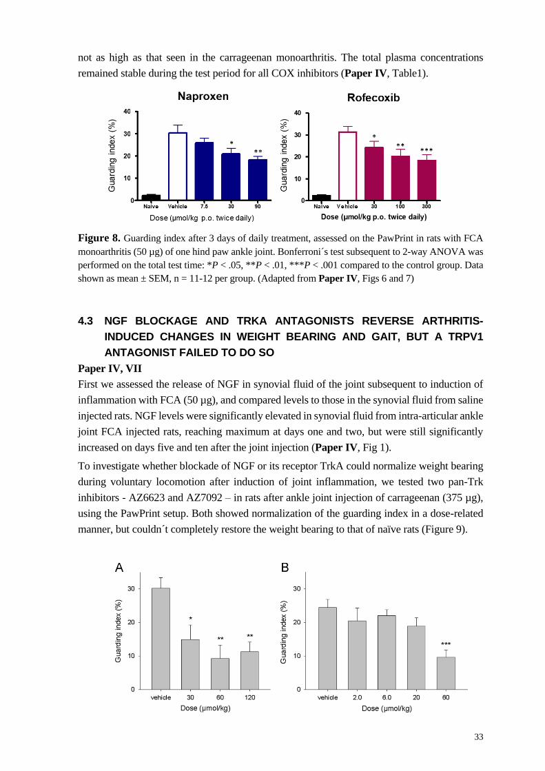
33
not as high as that seen in the carrageenan monoarthritis. The total plasma concentrations
remained stable during the test period for all COX inhibitors (Paper IV, Table1).
Figure 8. Guarding index after 3 days of daily treatment, assessed on the PawPrint in rats with FCA
monoarthritis (50 µg) of one hind paw ankle joint. Bonferroni´s test subsequent to 2-way ANOVA was
performed on the total test time: *P < .05, **P < .01, ***P < .001 compared to the control group. Data
shown as mean ± SEM, n = 11-12 per group. (Adapted from Paper IV, Figs 6 and 7)
4.3 NGF BLOCKAGE AND TRKA ANTAGONISTS REVERSE ARTHRITIS-
INDUCED CHANGES IN WEIGHT BEARING AND GAIT, BUT A TRPV1
ANTAGONIST FAILED TO DO SO
Paper IV, VII
First we assessed the release of NGF in synovial fluid of the joint subsequent to induction of
inflammation with FCA (50 µg), and compared levels to those in the synovial fluid from saline
injected rats. NGF levels were significantly elevated in synovial fluid from intra-articular ankle
joint FCA injected rats, reaching maximum at days one and two, but were still significantly
increased on days five and ten after the joint injection (Paper IV, Fig 1).
To investigate whether blockade of NGF or its receptor TrkA could normalize weight bearing
during voluntary locomotion after induction of joint inflammation, we tested two pan-Trk
inhibitors - AZ6623 and AZ7092 – in rats after ankle joint injection of carrageenan (375 µg),
using the PawPrint setup. Both showed normalization of the guarding index in a dose-related
manner, but couldn´t completely restore the weight bearing to that of naïve rats (Figure 9).

34
Figure 9. Effects of the pan-Trk antagonists AZ6623 and AZ7062 on carrageenan-induced changes in
guarding index are shown in (A) and (B) respectively. AZ6623 (n = 8/group) and AZ7092 (n = 10-
12/group) were given as a single administration orally, 1 h after ankle joint injection of carrageenan,
and testing was performed 2 h later. Data shown as mean ± SEM, and analysed by 1-way ANOVA
followed by Bonferroni´s t-test. *P < .05, **P < .01, ***P < .001 compared to the respective
carrageenan injected vehicle group. (From Paper IV, Fig 4)
Next the pan-Trk inhibitors AZ6623 and AZ7092, and the anti-NGF antibody MEDI-578 were
tested in the PawPrint setup for effects on FCA-induced guarding index during voluntary
locomotion (Figure 10). Here, one of the pan-Trk inhibitors was slightly more efficacious than
the NGF antibody, but all were more efficacious than the previously tested NSAIDs. Thus, the
normalization of guarding index compared to vehicle treated rats reached approximately 60 to
80% (Figs 2 and 3 in paper IV).
Figure 10. Guarding index at 3 days after one single intravenous injection of MEDI-578, assessed on
the PawPrint in rats with FCA monoarthritis (50 µg) of one hind paw ankle joint. Bonferroni´s test
subsequent to 2-way ANOVA was performed on the total test time: *P < .05, **P < .01, ***P < .001
compared to the control group. Data shown as mean ± SEM, n = 11-12 per group. (Adapted from Paper
IV, Fig 2)

35
In contrast, when testing a TRPV1 inhibitor at a high dose, no effect was observed (Figure 11).
Figure 11. Guarding index before and at 3 days after twice daily oral treatment of AZD1386, assessed
on the PawPrint in rats with FCA monoarthritis (50 µg) of one hind paw ankle joint. 2-way ANOVA
was performed. Data shown as mean ± SEM, n = 25 per group. (Adapted from Paper IV, Fig 5A)
In mice, significant effects on stationary guarding index (DWB) was achieved one day after
MEDI578 injection for the two lower doses (0.3 mg/kg and 1.0 mg/kg) and at three days for
all doses. When the same mice were tested in the CatWalkTM, a significant reduction of
guarding index during locomotion was attained only with the high dose (3.0 mg/kg) three days
after injection, and the dose relationship was weak (Figure 12).
Figure 12. Guarding index before and after one single subcutaneous injection of MEDI-578, assessed
on the DWB and CatWalk in mice with CFA monoarthritis (10 µg) of one hind paw ankle joint. The
guarding index of naïve mice before CFA injection is referred to as “no pain”, while “max pain” refers
to results at one day after CFA injection, before drug treatment. Each individual mouse´s difference
between measurements at “max pain” and the corresponding measurement at each time point after
treatment start was used for subsequent analysis. 1-way ANOVA followed by Dunnett´s test versus
isotype control mice was performed. Data shown as mean ± 95% CI, n = 11 per group. (From Paper
VII, Fig 4 A and B)
Willingness to place the injured paw on the floor was increased already one day after treatment
by some doses, and all doses reached significant effects three days after injection, both when
assessed when the mice were stationary and during locomotion (Paper VII, Figs. 4 C and D).
B e fo r e
F C A
Ve h ic le AZD 1 3 8 6
1 0 0 µ m o l/k g
0
1 0
2 0
3 0
4 0
A Z D 1 3 8 6
Gu
ard
ing
in
de
x (
%)
B L C F A + 1 d d ru g + 1 d d ru g + 3 d
Gu
ard
ing
in
de
x
(dif
f fr
om
CF
A+
1d
; m
ea
n ±
95
% C
I)
is o ty p e c o n tro l
M E D I5 7 8 0 .3 m g /k g
M E D I5 7 8 1 .0 m g /k g
M E D I5 7 8 3 .0 m g /k g
n o
p a in
m a x
p a in
****
***
*
B L C F A + 1 d d ru g + 1 d d ru g + 3 d
n o
p a in
m a x
p a in
*
D W B C a tW a lk

36
All data from the drug effect studies with mice were pooled and results for each individual
mouse from the DWB versus results from the CatWalkTM were plotted. Stationary guarding
index (DWB) and guarding index during locomotion (CatWalkTM) correlated strongly, as did
duration of floor contact versus duty cycle (Paper VII, Figs 4 E and F; p<0.0001 for both).
4.4 MICE WITH NON-FUNCTIONAL NGF ARE PROTECTED FROM
ARTHRITIS-INDUCED CHANGES IN GAIT AND WEIGHT BEARING
Paper VIII
We characterized mice with mutations in the gene coding for NGF. Homozygous mice with
the human NGF (hWT mice) or with the human NGF containing a mutation, replacing the
arginine 100 by glutamic acid in the NGFß gene (hR100E mice), were tested with respect to
their response to basic sensory tests and their response to acute and longer lasting models of
inflammatory pain. In addition, nervous tissue from skin, the sciatic nerve and DRG were
analysed. No difference was found between the two with respect to development of the
nociceptive system (Figure 13).
Figure 13. Representative images of PGP9.5(+), NF200(+), CGRP(+) and TrkA(+) nerve fibres in the
glabrous skin of hWT (left) and hR100E (right) mice. Fibre area and length from the contralateral hind
paw skin of hWT (white; n = 7) and hR100E (black; n = 7) mice. To compare levels between groups, t-
test was performed. Data presented as mean ± SEM. (From Paper VIII, Fig 2 E and F)
Neither was any difference between hWT and hR100E mice regarding baseline mechanical,
cold or thermal sensitivity found (Paper VIII, Fig 3), nor in response to acute pain as measured
with the formalin test (Paper VIII, Fig 4). Further, when testing mechanical and thermal
hypersensitivity after ankle joint injection of CFA (5 µl of 10 mg/ml), no significant difference
was seen between the two (Paper VIII, Fig 5 B and C). However, when assessing stationary

37
weight bearing (Incapacitance tester) and gait and weight bearing during locomotion (CatWalk)
after knee joint injection of CFA in those mice, we did see a significant difference (Figure 14).
Figure 14. Stationary (dynamic) weight bearing and weight bearing during locomotion assessed on
the DWB and CatWalk respectively, before and after intra-articular knee joint injection of CFA (100
µg) in hWT and hR100E mice of both genders. 2-way ANOVA followed by Bonferroni´s test versus
baseline values before CFA injection was performed. Data shown as mean ± SEM. The number of
animals were: male; hWT n = 7, hR100E n = 7; female; hWT n = 5, hR100E n = 7 per group. (Adapted
from Paper VIII, Fig 6 C and 7 C)
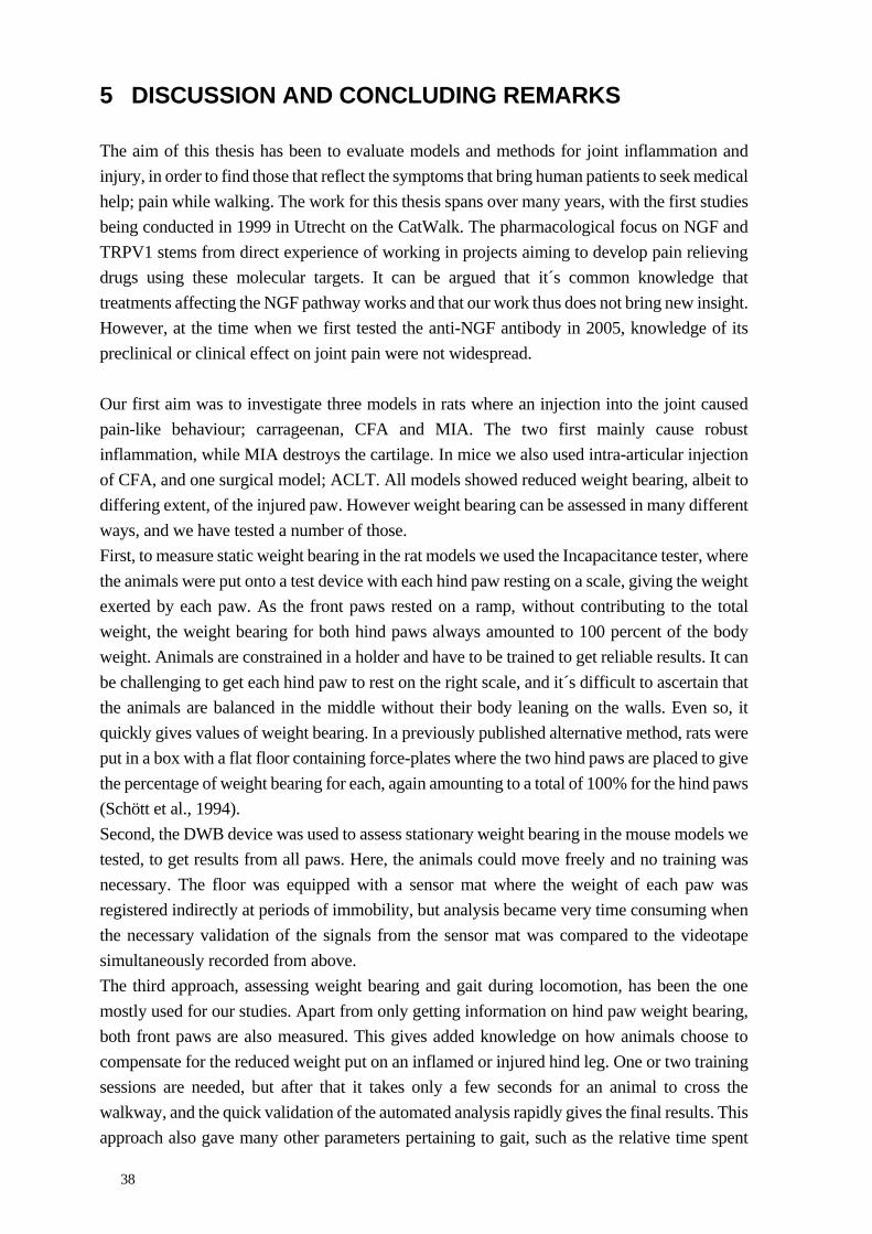
38
5 DISCUSSION AND CONCLUDING REMARKS
The aim of this thesis has been to evaluate models and methods for joint inflammation and
injury, in order to find those that reflect the symptoms that bring human patients to seek medical
help; pain while walking. The work for this thesis spans over many years, with the first studies
being conducted in 1999 in Utrecht on the CatWalk. The pharmacological focus on NGF and
TRPV1 stems from direct experience of working in projects aiming to develop pain relieving
drugs using these molecular targets. It can be argued that it´s common knowledge that
treatments affecting the NGF pathway works and that our work thus does not bring new insight.
However, at the time when we first tested the anti-NGF antibody in 2005, knowledge of its
preclinical or clinical effect on joint pain were not widespread.
Our first aim was to investigate three models in rats where an injection into the joint caused
pain-like behaviour; carrageenan, CFA and MIA. The two first mainly cause robust
inflammation, while MIA destroys the cartilage. In mice we also used intra-articular injection
of CFA, and one surgical model; ACLT. All models showed reduced weight bearing, albeit to
differing extent, of the injured paw. However weight bearing can be assessed in many different
ways, and we have tested a number of those.
First, to measure static weight bearing in the rat models we used the Incapacitance tester, where
the animals were put onto a test device with each hind paw resting on a scale, giving the weight
exerted by each paw. As the front paws rested on a ramp, without contributing to the total
weight, the weight bearing for both hind paws always amounted to 100 percent of the body
weight. Animals are constrained in a holder and have to be trained to get reliable results. It can
be challenging to get each hind paw to rest on the right scale, and it´s difficult to ascertain that
the animals are balanced in the middle without their body leaning on the walls. Even so, it
quickly gives values of weight bearing. In a previously published alternative method, rats were
put in a box with a flat floor containing force-plates where the two hind paws are placed to give
the percentage of weight bearing for each, again amounting to a total of 100% for the hind paws
(Schött et al., 1994).
Second, the DWB device was used to assess stationary weight bearing in the mouse models we
tested, to get results from all paws. Here, the animals could move freely and no training was
necessary. The floor was equipped with a sensor mat where the weight of each paw was
registered indirectly at periods of immobility, but analysis became very time consuming when
the necessary validation of the signals from the sensor mat was compared to the videotape
simultaneously recorded from above.
The third approach, assessing weight bearing and gait during locomotion, has been the one
mostly used for our studies. Apart from only getting information on hind paw weight bearing,
both front paws are also measured. This gives added knowledge on how animals choose to
compensate for the reduced weight put on an inflamed or injured hind leg. One or two training
sessions are needed, but after that it takes only a few seconds for an animal to cross the
walkway, and the quick validation of the automated analysis rapidly gives the final results. This
approach also gave many other parameters pertaining to gait, such as the relative time spent
39
putting weight on the floor by each paw, the interlimb coordination, i.e. the regularity of the
four paw´s placement on the floor while walking, and distance between the paw placements.
Furthermore, we tested an approach where mice were forced to walk within a restrained area
on a transparent floor (TreadScan from CleverSys); a belt that moved and forced the mice
forward to avoid getting pushed from behind or even get squeezed between the bumper at the
end of the walkway and the floor (not reported). Mice induced with CFA monoarthritis became
very stressed by the setup and many refused to move. The basis for calculating stance pressure
(≈ weight bearing) was the colour of the plantar side of the paw, which is calibrated for each
strain of mice. The assumption that the skin colour is changed as it’s placed on the floor and
small vessels get occluded does not seem to hold true.
All three ways of getting data that we decided to continue with; static, stationary and during
locomotion, show that joint inflammation alters weight bearing. For the stationary weight
bearing and that during locomotion we introduced a new calculated result parameter; the
guarding index, which shows the shift of body weight from the injured hind leg to the non-
injured leg. Guarding index is the parameter mainly used in the figures of this thesis. The
models we have used give varying effects, both in terms of magnitude and of duration. Thus,
the two mainly inflammatory models (carrageenan and CFA) showed the largest difference
between those with injection and controls, whereas the MIA model showed an intermediate
difference and the surgical ACLT had difficulties reaching significant difference compared to
controls. Unfortunately, this is opposite to what would be advantageous when studying OA.
Another aim was to test whether conventional pain relieving drugs could normalize the effects
on gait and weight bearing in the models we used. The carrageenan model, which is the model
with shortest duration, provided a large difference in guarding index between animals with
monoarthritis and controls and is therefore suited for pharmacological tests. NSAIDs were able
to completely restore weight bearing, reducing the guarding index to levels below that of
controls, but opioids did not reach a similar efficacy. These drugs also showed effect on gait
parameters such as fraction of total step duration (duty factor) and regularity index. Thus it
seems that this model lends itself primarily to testing drugs that inhibit COX enzymes. In
contrast, the NSAIDs tested in the longer-lasting CFA monoarthritis model were unable to fully
normalize the weight bearing decrease, reaching only around 40% reduction of the guarding
index. The smaller differences between the two hind leg´s weight bearing (guarding index) in
the MIA and the ACLT models made it almost impossible to test pain relieving drugs with the
number of animals per group conventionally used in those studies. It´s unfortunate that models
mimicking the structural changes seen in OA don´t give the robust behavioural changes needed
for pharmacological testing of pain-relieving drugs. Perhaps several models could be used to
include the different aspects of OA pain, in such a way that the CFA-induced monoarthritis,
albeit definitely not a model of OA, could represent pain occurring from the inflammatory
aspect, whereas the MIA model more closely resembles the phase of OA pain when the
cartilage becomes degraded. These two models cannot, however, be used to understand the
underlying mechanisms of progression disease. To more closely look at those aspects the

40
surgical and natural occurring models are better suited. It´s a challenge to find a model that
both mirrors the structural changes in OA, and that makes it possible to test new chemical
targets for pain treatment. The work on the models in this thesis can be summarized in table 3.
Table 3 Comparison between the models used
Model Inflammation OA-like
structural
changes
Magnitude of
weight bearing
changes
Affected by
conventional pain
drugs
Carra-
geenan
+++ +++ +++
CFA
+++ + +++ ++
MIA
+ ++ ++ +
ACLT
+++ +
The next aim of this thesis was to explore the role of NGF and the ion channel TRPV1 in
arthritis-induced spontaneous pain. For this we used pharmacological treatments developed by
AstraZeneca; one anti-NGF antibody and two small molecule inhibitors of the Trk receptors
A, B and C. In the carrageenan monoarthritis model the pan-Trk inhibiting compounds showed
significant effects, but the efficacy was below that of all NSAIDs and opioids tested. However,
in the CFA-induced monoarthritis the anti NGF antibody and the pan-Trk inhibitors were able
to normalize the CFA-induced increase of guarding index with 60-80%, thus showing a
stronger effect than that by NSAIDs in this model. Thus it´s clear that the models engage
different parts of the immunological system, and that the CFA-induced model recruits other
pathways than those entirely dependent on COX.
The genetically modified mice given to us by AstraZeneca, where the arginine at R100 was
replaced by glutamic acid in the NGFß gene, responded normally when testing for baseline
sensitivity and effects of formalin-induced nociception, and showed normal response to tester-
evoked sensory stimuli after induction of CFA-induced monoarthritis. This was in line with the
normal development and differentiation of the nociceptive system found in nervous tissue of
these mice. In contrast, assessment of gait and weight bearing in the NGF R100E mutated mice
showed a significant lack of pain-like response compared to wild-type mice, pointing to the
larger susceptibility by this mutation in deep tissues of the joint.

41
Figure 15. The difference in percentage of TrkA positive nerves in bone compared to that
found in skin. (Reprinted with permission from Mantyh et al., 2011)
One of the possible reasons for this disparity between skin and joint sensitivity could be the
different expression of the TrkA receptor; only about 30% of the sensory nerves in skin are
TrkA positive, while more than 80% of the sensory nerves in bone are TrkA positive (Figure
15; Mantyh et al., 2011; Guedon et al., 2016). The information gained by using gait and
stationary weight bearing or weight bearing during locomotion would not have been discovered
with the traditional tester-evoked assessments.


43
6 ACKNOWLEDGEMENTS
First, I want to give thanks to my supervisor Camilla I Svensson for giving me the opportunity
to continue my research at the Karolinska Institutet and pursue a PhD. I was very sad when
AstraZeneca closed their research facility, the R&D Södertälje, Research Area CNS & Pain
Control, and was eager to somehow continue.
To my co-supervisor Carina Stenfors – what would I do without you? Carina, you have been
the one I´ve turned to when I needed a wise person to give me ideas and inspiration! Thanks
go also to my other co-supervisor Xiaojun Xu. I owe a big thank you to my mentor – Odd-
Geir Berge – who was also my boss at AstraZeneca and shared my interest for pain models
and how to measure results from these.
Special thanks go to Frank Hamers, who generously shared his thoughts and experience about
the CatWalk, and invited me to work with him in Utrecht for the first study. Seldom have I met
such an intelligent and yet humble person! And Rolf Størkson, without you there wouldn´t
have been any PawPrint. Susanne Kinert, the Master of Science student I was given the
opportunity to be supervisor for – what a team we made. Stephanie Klein – without you there
would not be a Paper VI.
Of course, all past and present members of pain research at KI; Ernst Brodin, Eva Schött,
Henrik Dahllöf, Azar Baharpoor, Katalin Sandor, Camilla Ultenius, Nilesh Agalave,
Gustaf Wigerblad, Teresa Fernandez Zafra, Sally Abdelmoaty, Jungo Kato, Alex
Bersellini Farinotti, Resti Rudjito, Alexandra Jurczak, Carlos Morado Urbina, Jaira
Villarreal Salcido, Yuki Nomura, and Vinko Palada. I´m sure I´ve forgotten to add a lot of
you – sorry for that. One I couldn´t forget is Cecilia Aulin – all the hours we spent in the in
vivo lab make precious memories.
Anja Finn! My colleague and friend – I don´t need to say more…
Many of my former colleagues at AZ: Sven-Ove Ögren, thank you for taking me into your
group and believing in what I accomplished! Trevor Archer; so many dances! Anna, Anne-
Berit, Pär, Sofie, Sandra, Maria, Camilla, Julia, Helen, Calle! What a lot of fun we had,
and so many excellent studies we performed together! Thank you!
Inga-Britt Hellberg! You were my personal mentor at Astra Pain Control, and we discussed
everything. We travelled together, both work-related and other. Midsummer, crayfish parties,
pickled herring dinners! And of course Joe. So many happy memories and more to come.

44
A special thanks to my colleagues and friends at Orion in Turku! Jucka, Johanna, Jarmo,
Heta, Ullamari, Mervi, Anni, Tiina, Ari-Pekka. You made my year in Finland one I will
always remember with joy.
Family! Mother Gockan, brothers and sister; Stefan, Gisela, Elisabet and Jakob. We have
memories stretching far back, both good and bad!
To my ex-husband Hasse, a special thanks for supporting my endeavours!
Last but not least, to my beloved children Amilie, Johan and Anders. Who and what would I
be without you??

45
7 REFERENCES
Abaei M, Sagar DR, Stockley EG, Spicer CH, Prior M, Chapman V, et al (2016) Neural correlates
of hyperalgesia in the monosodium iodoacetate model of osteoarthritis pain, Molecular Pain
12:1-12. Doi:10.1177/1744806916642445.
Akinci A, Al Shaker M, Chang MH, Cheung CW, Danilov A, José Dueñas H, et al (2016) Predictive
factors and clinical biomarkers for treatment in patients with chronic pain caused by
osteoarthritis with a central sensitisation component, Int J Clin Pract 70(1):31-44.
Doi:10.1111/ijcp.12749.
Andrews K (1993) The effect of changes in temperature and humidity on the accuracy of von Frey
hairs, J Neurosci Methods 50:91-93. Doi:10.1016/0165-0270(93)90059-z.
Ängeby Möller K, Johansson B, & Berge O-G (1998) Assessing mechanical allodynia in the rat paw
with a new electronic algometer, J Neurosci Methods 84:41-47. Doi:10.1016/s0165-
0270(98)00083-1.
Arendt-Nielsen L, Skou ST, Nielsen TA, & Petersen KK (2015) Altered central sensitization and
pain modulation in the CNS in chronic joint pain, Curr Osteoporos Rep 13:225-234.
Doi:10.1007/s11914-015-0276-x.
Avouac J, Gossec L, & Dougados M (2007) Efficacy and safety of opioids for osteoarthritis: a meta-
analysis of randomized controlled trials, Osteoarthritis Cartilage 15:957-965.
Doi:10.1016/j.joca.2007.02.006.
Bauer CS, Rahman W, Tran-van-Minh A, Lujan R, Dickenson AH, Dolphin AC (2010) The anti-
allodynic alpha(2)delta ligand pregabalin inhibits the trafficking of the calcium channel
alpha(2)delta subunit to presynaptic terminals in vivo, Biochem Soc Trans 38:525-528.
Doi:10.1042/BST0380525.
Beare JE, Morehouse JR, DeVries WH, Enzman GU, Burke DA, Magnuson DSK, et al (2009) Gait
analysis in normal and spinal contused mice using the TreadScan system, J Neurotrauma
26:2045-2056. Doi:10.1089/neu.2009.0914.
Bendele AM, & Hulman JF (1988) Spontaneous cartilage degeneration in guinea pigs, Arthritis
Rheum 31(4):561-565. Doi:10.1002/art.1780310416.
Bendele AM (2001) Animal models of osteoarthritis, J Musculoskel Neuron Interact 1(4):363-376.
PMID:15758487.
Benito MJ, Veale DJ, FitzGerald O, van den Berg WB, & Bresnihan B (2005) Synovial tissue
inflammation in early and late osteoarthritis, Ann Rheum Dis 64:1263-1267.
Doi:10.1136/ard.2004.025270.
Bennell KL, & Hinman RS, (2011) A review of the clinical evidence for exercise in osteoarthritis of
the hip and knee, J Sci Med Sport 14(1):4-9.doi:10.1016/j.jsams.2010.08.002.
Betts RP, & Duckworth TA (1978) A device for measuring plantar pressures under the sole of the
foot, Eng Med 7:223-228.
Blagojevic M, Jinks C, Jeffery A, & Jordan KP (2010) Risk factors for onset of osteoarthritis of the
knee in older adults: a systematic review and meta-analysis, Osteoarthritis Cartilage 18:24-33.
Doi:10.1016/j.joca.2009.08.010.
Bonnington JK & McNaughton PA (2003) Signalling pathways involved in the sensitisation of mouse
nociceptive neurons by nerve growth factor, J Physiol 551(2):433-446.
Doi:10.1113/jphysiol.2003.039990.
Bove SE, Calcaterra MS, Brooker RM, Huber CM, Guzman RE, Juneau PL, et al (2003) Weight
bearing as a measure of disease progression and efficacy of anti-inflammatory compounds in
a model of monosodium iodoacetate-induced osteoarthritis,Osteoarthritis Cartilage 11:821-830.
Bove SE, Laemont KD, Brooker RM, Osbom MN, Sanchez BM, Guzman RE, et al (2006) Surgically
induced osteoarthritis in the rat results in the development of both osteoarthritis-like joint pain
and secondary hyperalgesia, Osteoarthritis Cartilage 14:1041-1048.
Doi:10.10167j.joca.2006.05.001.

46
Breivik H, Collett B, Ventafridda V, Cohen R,& Gallacher D (2006) Survey of chronic pain in Europe:
prevalence, impact on daily life, and treatment, Eur J Pain 10:287-333.
Doi:10.1016/j.ejpain.2005.06.009.
Brenner M, Braun C, Oster M, & Gulko PS (2006) Thermal signature analysis as a novel method
for evaluating inflammatory arthritis activity, Ann Rheum Dis 65:306-311.
Doi:10.1136/ard.2004.035246.
Bruyère O, Cooper C, Pelletier J-P, Branco J, Brandi ML, Guillemin F, et al (2014) An algorithm
recommendation for the management of knee osteoarthritis in Europe and internationally: a
report from a task force of the European Society for Clinical and Economic Aspects of
Osteoporosis and Osteoarthritis (ESCEO), Semin Arthritis Rheum 44:253-263.
Doi:10.1016/j.semiarthrit.2014.05.014.
Butler SH, Godefroy F, Besson JM, Weil-Fugazza J (1992) A limited arthritic model for chronic pain
studies in the rat, Pain 48:73-81. doi:10.1016/0304-3959(92)90133-V.
Cairns BE, Gambarota G, Svensson P, Arendt-Nielsen L, & Berde CB (2002) Glutamate-induced
sensitization of rat masseter muscle fibers, Neuroscience 109(2):389-399. Doi:10.1016/s0306-
4522(01)00489-4.
Caterina MJ, Schumacher MA, Tominaga M, Rosen TA, Levine JD, & Julius D (1997) The capsaicin
receptor: a heat-activated ion channel in the pain pathway, Nature 389(6653):816-824.
Doi:10.1038/39807.
Chaplan SR, Bach FW, Pogrel JW, Chung JM, & Yaksh TL (1994) Quantitative assessment of
tactile allodynia in the rat paw. J Neurosci Methods 53:55-63. PMID: 7990513.
Clarke KA (1992) A technique for the study of spatiotemporal aspects of paw contact patterns,
applied to rats treated with a TRH analogue, Behav Research Methods Instr Comp 24(3):407-
411.
Clarke KA (1995) Differential fore- and hindpaw force transmission in the walking rat, Physiol Behav
58(3):415-419. Doi:10.1016/0031-9384(95)00072-q.
Clarke KA, Heitmeyer SA, Smith AG, & Taiwo YO (1997) Gait analysis in a rat model of
osteoarthrosis, Physiol Behav 62(5):951-954. Doi:10.1016/s0031-9384(97)00022-x.
Clarke KA, & Still J (1999) Gait analysis in the mouse, Physiol Behav 66(5):723-729.
Doi:10.1016/s0031-9384(98)00343-6.
Clarke KA, & Still J (2001) Development and consistency of gait in the mouse, Physiol Behav
73:159-164. Doi:10.1016/s0031-9384(01)00444-9.
Coderre TJ, & Wall PD (1987) Ankle joint urate arthritis (AJUA) in rats: an alternative animal model
of arthritis to that produced by Freund´s adjuvant, Pain 28:379-393. Doi:10.1016/0304-
3959(87)90072-8.
Combe R, Bramwell S, & Field MJ (2004) The monosodium iodoacetate model of osteoarthritis: a
model of chronic nociceptive pain in rats? Neuroscience Letters 370:236-240.
Doi:10.1016/j.neulet.2004.08.023.
Corbett AD, Henderson G, McKnight AT, Paterson SJ (2006) 75 years of opioid research: the
exciting but vain quest for the Holy Grail, Br J Pharmacol 147 Suppl 1:S153-162.
Doi:10.1038/sj.bjp.0706435.
De la Calle JL, Mena MA, González-Escalade JR, & Paíno CL (2002) Intrathecal transplantation of
neuroblastoma cells decreases heat hyperalgesia and cold allodynia in a rat model of
neuropathic pain, Brain Research Bulletin 59(3):205-211. Doi:10.1016/s0361-9230(02)00867-
5.
De Souza RL, Matsuura M, Eckstein F, Rawlingson SCF, Lanyon LE, Pitsillides AA (2005) Non-
invasive axial loading of mouse tibiae increases cortical bone formation and modifies trabecular
organization: a new model to study cortical and cancellous compartments in a single loaded
element, Bone 37:810-818. Doi:10.1016/j.bone.2005.07.022.
Dimitroulas T, Duarte RV, Behura A, Kitas GD, & Raphael JH (2014) Neuropathic pain in
osteoarthritis: a review of pathophysiological mechanisms and implications for treatment, Sem
Arthritis Rheum 44:145-154. doi:10.1016/j.semarthrit.2014.05.011.
Dixon WJ (1980) Efficient analysis of experimental observations. Annu Rev Pharmacol Toxicol
20:441-462. Doi:10.1146/annurev.pa.20.040180.002301.
47
Dowdall T, Robinson I, & Meert TF (2005) Comparison of five different rat models of peripheral
nerve injury, Pharmacol Biochem Behav 80:93-108. Doi:10.1016/j.pbb.2004.10.016.
Einarsdottir E, Carlsson A, Minde J, Toolanen G, Svensson O, Solders G, Holmgren G, Holmberg
D, Holmberg M (2004) A mutation in the nerve growth factor beta gene (NGFB) causes losss
of pain perception, Hum Mol Genet 13(8):799-805. Doi:10.1093/hmg/ddh096.
Englund M, Roos EM, & Lohmander LS (2003) Impact of type of meniscal tear on radiographic and
symptomatic knee osteoarthritis: a sixteen-year followup of meniscectomy with matched
controls, Arthritis Rheum 48(8):2178-2187. Doi:10.1002/art.11088.
Eriksen EF (2015) Treatment of bone marrow lesions (bone marrow edema), Bonekey Rep 4:755.
doi:10.1038/bonekey.2015.124
Felson DT (2009) Develoments in the clinical understanding of osteoarthritis, Arthritis Res Ther
11(1):203. doi:10.1186/ar2531.
Felson DT, Anderson JJ, Naimark A, Walker AM, & Meenan RF (1988) Obesity and knee
osteoarthritis. The Framingham study, Ann Intern Med 109(1):18-24. Doi: 10.7326/0003-4819-
109-1-18.
Felson DT, Lawrence RC, Dieppe PA, Hirsch R, Helmick CG, Jordan JM, et al (2000) Osteoarthritis:
new insights. Part 1: the disease and its risk factors, Ann Intern Med 133(8):635-646.
Doi:10.7326/0003-4819-133-8-200010170-00016.
Fernihough J, Gentry C, Malcangio M, Fox A, Rediske J, Pellas T, et al (2004) Pain related
behaviour in two models of osteoarthritis in the rat knee, Pain 112:83-93.
Doi:10.1016/j.pain.2004.08.004.
Finn A, & Oerther SC (2010) Can L(+)-lactate be used as a marker of experimentally induced
inflammation in rats? Inflamm Res 59:315-321. Doi:10.1007/s00011-009-0107-6.
Fruhstorfer H, Gross W, & Selbman O (2001) von Frey hairs: new materials for a new design, Eur
J Pain 5:341-342. Doi:10.1056/eujp.2001.0250.
Glass N, Segal NA, Sluka KA, Torner JC, Nevitt MC, Felson DT, et al (2014) Examinig sex
differences in knee pain: the multicenter osteoarthritis study, Osteoarthritis Cartilage
22(8):1100-1106. Doi:10.1016/j.joca.2014.06.030.
Guedon JMG, Longo G, Majuta LA, Thomson ML, Fealk MN, Mantyh PW (2016) Dissociation
between the relief of skeletal pain behaviors and skin hypersensitivity in a model of bone cancer
pain, Pain 157(6):1239-1274. Doi:10.1097/j.pain.0000000000000514.
Guingamp C, Gegout-Pottie P, Philippe L, Terlain B, Netter P, & Gillet P (1997) Mono-iodoacetate-
induced experimental osteoarthritis: a dose-response study of loss of mobility, morphology, and
biochemistry, Arthritis Rheum 40(9):1670-1679. Doi:10.1002/art.1780400917.
Gustafsson H, & Sandin J (2009) Oral pregabalin reverses cold allodynia in two distinct models of
peripheral neuropathic pain, Eur J Pharmacol 605:103-108. Doi:10.1016/j.ejphar.2009.01.014.
Guzman RE, Evans MG, Bove S, Morenko B, & Kilgore K (2003) Mono-iodoacetate-induced
histologic changes in subchondral bone and articular cartilage of rat femorotibial joints: an
animal model of osteoarthritis, Toxicol Pathol 31:619-624. Doi:10.1080/01926230390241800.
Halliday DA, Zettler C, Rush RA, Scicchitano R, & McNiel JD (1998) Elevated nerve growth factor
levels in the synovial fluid of patients with inflammatory joint disease, Neurochem Res
23(6):919-922. Doi:10.1023/a:1022475432077.
Hamers FPT, Lankhorst AJ, van Laar TJ, Veldhuis WB, & Gispen WH (2001) , Automated
quantitative gait analysis during overground locomotion in the rat: its application to spinal cord
contusion and transection injuries, J Neurotrauma 18(2):187-201.
Hargreaves K, Dubner R, Brown F, Flores C, & Joris J (1988) A new and sensitive method for
measuring thermal nociception in cutaneous hyperalgesia, Pain 32(1):77-88.
doi:10.1016/0304-3959(88990026-7.
Hashmi JA, Yashpal K, Holdsworth DW, & Henry JL (2010) Sensory and vascular changes in a rat
monoarthritis model: prophylactic and therapeutic effects of meloxicam, Inflam Res 59:667-678.
doi:10.1007/s00011-010-0179-3.
Hawker GA (2006) Who, when, and why total joint replacement surgery? The patient´s perspective,
Curr Opin Rheumatol 18(5):526-530. Doi:10.1097/01.bor.0000240367.62583.51.
Hill CL, Seo GS, Gale D, Totterman S, Gale ME, & Felson DT (2005) Cruciate ligament integrity in
osteoarthritis of the knee, Arthritis Rheum 52(3):794-799. Doi:10.1002/att.20943.

48
Heppenstall PA & Lewin GR (2000) Neurotrophins, nociceptors and pain, Curr Opin Anaesthesiol
13:573-576. PMID:17016360.
Hole K, & Tjølsen A (1992) The tail-flick and formalin tests in rodents: changes in skin temperature
as a confounding factor, Pain 53:247-254. Doi:10.1016/0304-3959(93)90220-j.
Janusz MJ, Bendele AM, Brown KK, Taiwo YO, Hsieh L, & Heitmeyer SA (2002) Induction of
osteoarthritis in the rat by surgical tear of the meniscus: Inhibition of joint damage by a matrix
metalloproteinase inhibitor, Osteoarthritis Cartilage 10:785-791. PMID:12359164.
Jordan KM, Arden NK, Doherty M, Bannwarth B, Bijlsma JWJ, Dieppe P, et al (2003) EULAR
Recommendations 2003: and evidence based approach to the management of knee
osteoarthritis: Report of a Task Force of the Standing Committee for International Clinical
Studies Including Therapeutic Trials (ESCISIT), Ann Rheum Dis 62:1145-1155.
Doi:10.1136/ard.2003.011742.
Juhl C, Christensen R, Roos EM, Zhang W, & Lund H (2014) Impact of exercise type and dose on
pain and diability in knee osteoarthritis: a systematic review and meta-regression analysis of
randomized controlled trials, Arthritis Rheum 66(3):622-636. Doi:10.1002/art.38290.
Kalbhen DA (1987) Chemicla model of osteoarthritis – a pharmacological evaluation, J Rheumatol
14:130-131. PMID:3625668.
Keeble J, Russell F, Curtis B, Starr A, Pinter E, & Brain SD (2005) Involvement of transient receptor
potential vanilloid 1 in the vascular and hyperalgesic components of joint inflammation, Arthritis
Rheum 52(10):3248-3256. Doi:10.1002/art.21297.
Kinnman E, & Levine JD (1995) Involvement of the sympathetic postganglionic neuron in capsaicin-
induced secondary hyperalgesia in the rat, Neuroscience 65(1):283-291. Doi:10.1016/0306-
4522(94)0047-j.
Krasnokutsky S, Belitskaya-Lévy I, Bencardino J, Samuels J, Attur M, Regatte R, et al (2011)
Quantitative magnetic resonance imaging evidence of synovial proliferation is associated with
radiographic severity of knee osteoarthritis, Arthritis Rheum 63(10):2983-2991.
Doi:10.1002/art.30471.
Knazovicky D, Helgeson ES, Case B, Gruen ME, Maixner W, & Lascelles DX (2016) Widespread
somatosensory sensitivity in naturally occuring canine model of osteoarthritis, Pain
157(6):1325-1332. Doi:10.1097/j.pain.0000000000000521.
LaMotte RH, Lundberg LE, & Torebjörk HE (1992) Pain, hyperalgesia and activity in nociceptive C
units in humans after intradermal injection of capsaicin, J Physiol 448:749-764.
Doi:10.1113/jphysiol.1992.sp019068.
Lane NE, Schnitzer TJ, Birbara CA, Mokhtarani M, Shelton DL, Smith MD, & Brown MT (2010)
Tanezumab for the treatment of pain from osteoarthritis of the knee. N Engl J Med 363:1521-
1531. Doi: 10.1056/NEJMoa0901510.
Lawrence RC, Felson DT, Helmick CG, Arnold LM, Choi H, Deyo RA, et al (2008) Estimates of the
prevalence of arthritis and other rheumatic conditions in the United States. Part II, Arthritis
Rheum 58(1):26-35. Doi:10.1002/art.23176.
Lewin GR & Mendell LM (1993) Nerve growth factor and nociception, TINS 16(9):353-359.
Doi:10.1016/0166-2236(93)90092-z.
Loeser RF (2013) Aging processes and the development of osteoarthritis, Curr Opin Rheumatol
25(1):108-113. Doi:10.1097/BOR.0b013e32835a9428.
Loeser RF, Goldring SR, Scanzello CR & Goldring MB (2012) Osteoarthritis: a disease of the joint
as an organ, Arthritis Rheum 64(6):1697-1707. Doi:10.1002/art.34453.
López-Muñoz FJ, Salazar LA, Castañeda-Hernánez G, & Villarreal JE (1993) A new model to
assess analgesic activity: Pain-induced functional impairment in the rat (PIFIR), Drug Dev Res
28:169-175.
Lowther DA, & Gillard GC (1976) Carrageenin-induced arthritis. I. The effect of intraarticular
carrageenin on the chemical composition of articular cartilage, Arthritis Rheum 19(4):769-776.
Doi:10.1002/1529-0131(197607/08)19:4<769::aid-art1780190419˃3.0.co:2-v.
Mantyh PW, Koltzenburg M, Mendell LM, Tive L, Shelton DL (2011) Antagonism of nerve growth
factor-TrkA signaling and the relief of pain, Anesthesiology 115(1):189-204.
Doi:10.1097/ALN.0b013e31821b1ac5.

49
McMahon SB, Cafferty WBJ, & Marchand F (2005) Immune and glial cell factors as pain mediators
and modulators, Exp Neurol 192(2):444-462.doi:10.1016/j.expneurol.2004.11.001.
Minde J, Toolanen G, Andersson T, Nennesmo I, Nilsson Remahl I, Svensson O, Solders G (2004)
Familial insensitivity to pain (HSAN V) and a mutation in the NGFB gene. A neurophysiological
and pathological study. Muscle Nerve 30:752-760. Doi: 10.1002/mus.20172.
Mogil JS, & Crager SE (2004) What should we be measuring in behavioral studies of chronic pain
in animals? Pain 112:12-15. Doi:10.10167j.pain.2004.09.028.
Moss P, Knight E, & Wright A (2016) Subjects with knee osteoarthritis exhibit widespread
hyperalgesia to pressure and cold, PLoS ONE 11(1): doi:10.1371/journal.pone.0147526.
doi:10.1371/journal.pone.0147526.
Muthuri SG, McWilliams DF, Doherty M, & Zhang W (2011) History of knee injuries and knee
osteoarthritis: a meta-analysis of observational studies, Osteoarthritis Cartilage 19:1286-1293.
Doi:10.1016/j.joca.2011.07.015.
Nagasako EM, Oaklander AL, & Dworkin RH (2003) Congenital insensitivity to pain: an update,
Pain 101:213-219. Doi:10.10167s0304-3959(02)00482-7.
Neogi T, & Zhang Y (2013) Epidemiology of osteoarthritis, Rheum Dis Clin North Am 39(1):1-19.
Doi:10.10167j.rdc.2012.10.004.
Niu J, Zhang YQ, Torner J, Nevitt M, Lewis CE, Aliabade P, et al (2009) Is obesity a risk factor for
progressive radiographic knee osteoarthritis? Arthritis Rheum 61(3):329-335.
Doi:10.1002/art.24337.
Nordling C, Karlsson-Parra A, Jansson L, Holmdahl R, & Klareskog L (1992) Characterization of a
spontaneously occuring arthritis in male DBA/1 mice, Arthritis Rheum 35(6):717-722.
Doi:10.1002/art.1780350619.
Oliviera SA, Felson DT, Cirillo PA, Reed JI, & Walker AM (1999) Body weight, body mass index,
and incident symptomatic osteoarthritis of the hand, hip, and knee, Epidemiology 10(2):161-
166. PMID:10069252.
O´Neil CK, Hanlon JT, Marcum ZA (2012) Adverse effects of analgesics commonly used by older
adults with osteoarthritis: focus on non-opioid and opioid snalgesics, Am J Geriatr
Pharmacother 10(6):331-342. Doi:10.1016/j.amjopharm.2012.09.004.
Pearle AD, Scanzello CR, George S, Mandl LA, DiCarlo EF, Peterson M, et al (2007) Elevated high-
sensitivity C-reactive protein levels are associated with local inflammatory findings in patients
with osteoarthritis, Osteoarthritis Cartilage 15:516-523. Doi:10.1016/j.joca.2006.10.010.
Pearson CM, Waksman BH, & Sharp JT (1960) Studies of arthritis and other lesions induced in rats
by injection of mycobacterial adjuvant. J Exp Med 113:485-510. Doi:10.1084/jem.113.3.485.
Pearson CM (1963) Experimental joint disease observations on adjuvant-induced arthrtiis, J
Chronic Dis 16:863-874. Doi:10.1016/0021-9681(63)90136-x.
Pelletier JP, Martel-Pelletier J, Rannou F, Cooper C (2016) Efficacy and safety of oral NSAIDs and
analgesics in the management of osteoarthritis: Evidence form real-life setting trials and
surveys, Sem Arthritis Rheum 45:S22-S27. Doi:10.1016/j.semarthrit.2015.11.009.
Pezet S & McMahon SB (2006) Neurotrophins: mediators and modulators of pain, Annu Rev
Neurosci 29:507-538. Doi:10.1146/annurev.neuro.29.051605.112929.
Poulet B, Hamilton RW, Shefelbine S, Pitsillides AA (2011) Characterizing a novel and adjustable
noninvasive murine joint loading model, Arthritis Rheum 63:137-147. Doi:10.1002/art.27765.
Randall LO, & Selitto JJ (1957) A method for measurement of analgesic activity on inflamed tissue,
Arch Int Pharmacodyn Ther 111(4):409-419. PMID: 13471093.
Rejeski WJ, Brawley LR, & Shumaker SA (1996) Physical activity and health-related quality of life,
Exerc Sport Sci Rev 24:71-108. PMID:8744247.
Roubille C, Raynauld J-P, Abram F, Paiement P, Dorais M, Delorme P, et al (2014) The presence
of meniscal lesions is a strong predictor of neuropathic pain in symptomatic knee osteoarthritis:
a cross-sectional pilot study, Arthritis Res Therapy 16:507. Doi:10.1186/s13075-014-0507-z.
Santer V, Sriratana A, & Lowther DA (1983) Carrageenin-induced arthritis: V. A morphologic study
of the development of inflammation in acute arthritis, Semin Arthritis Rheum 13(2):160-168.
PMID:6673111.
SBU (2006) Methods of treating chronic pain: A systematic review, SBU-rapport nr 177/1.ISBN 91-
85413-08-9; nr 177/2. ISBN 91-85413-09-7. PMID:28876750.

50
Schaible H-G, Richter F, Ebersberger A, Boettger MK, Vanegas H, Natura E, et al (2009) Joint pain,
Exp Brain Res 196:153-162. Doi:10.1007/s00221-009-1782-9.
Schnitzer TJ, Ekman EF, Spierings EL, Greenberg HS, Smith MD, Brown MT, West CR, Verburg
KM (2015) Efficacy and safety of tanezumab monotherapy or combined with non-steroidal anti-
inflammatory drugs in the treatment of knee or hip osteoarthrtis pain, Ann Rheum Dis
74(6):1202-1211. Doi:10.1136/annrheumdis-2013-204905.
Schött E, Berge O-G, Ängeby Möller K, Hammarström G, Dalsgaard C-J, & Brodin E (1994) Weight
bearing as an objective measure of arthritic pain in the rat, JPM 31(2):79-83. PMID:8032098.
Segerdahl M, Quiding H, Enhörning G, Karin A, Carlsson MA, Lindberg A, & Jonzon B (2010) Local
effects of the TRPV1 antagonist AZ12048189 on quantitative sensory testing (QST) in normal
and UV irradiated skin in healthy subjects, Congr Pain Abstr PH410 2010.
Shelton DL, Zeller J, Ho WH, Pons J, & Rosenthal A (2005) Nerve growth factor mediates
hyperalgesia and cachexia in auto-immune arthritis, Pain 116:8-16.
Doi:10.1016/j.pain.2005.03.039.
Sikander S, Kvanner Aasvang E, & Dickenson AH (2016) Scratching the surface: the processing of
pain from deep tissues, Pain Manag 6(2):95-102. doi:10.2217/pmt.15.50.
Sills GJ (2006) The mechanisms of action of gabapentin and pregabalin, Curr Opin Pharmacol
6(1):108-113.doi:10.1016/j.coph.2005.11.003.
Silverstein E, & Sokoloff L (1958) Natural history of degenerative joint disease in small laboratory
animals. 5. Osteoarthritis in guinea pigs, Nat Inst Arthritis Met Dis 82-86.
doi:10.1002/art.1780010112.
Smith DB, & Cook WH (1953) Fractionation of carrageenin, Arch Biochem Biophys 45:232-233.
doi:10.1016/0003-9861(53)90421-4.
Smith SR, Deshpande BR, Collins JE, Katz JN, Losina E (2016) Comparative pain reduction of oral
non-steroidal anti-inflammatory drugs and opioids for knee osteoarthritis: systematic analytic
review, Osteoarthritis Cartilage 24:962-972. Doi:10.1016/j.joca.2016.01.135.
Spector TD, & MacGregor AJ (2004) Risk factors for osteoarthritis: genetics, Osteoarthritis Cartilage
12:S39-S44. PMID:14698640.
Srikanth VK, Fryer JL, Zhai G, Winzenberg TM, Hosmer D, & Jones G (2005) A meta-analysis of
sex differences prevalence, incidence and severity of osteoarthritis, Osteoarthritis Cartilage
13:769-781. Doi:10.1016/j.joca.2005.04.014.
Stein AT, Ufret-Vincenty CA, Hua L, Santana LF, & Gordon SE (2006) Phosphoinositide 3-kinase
binds to TRPV1 and mediates NGF-stimulated TRPV1 trafficking to the plasma membrane, J
Gen Physiol 128(5):509-522. Doi:10.1085/jgp.200609576.
Stone E (1763) Philosophical Transactions of the Royal Society 53:195-200.
Svensson O, Thorne C, Miller F, Björnssom M, Reimfelt A, & Karlsten R (2010) A phase II
randomized controlled trial evaluating the efficacy and safety of the TRPV1 antagonist
AZD1386 in osteoarthritis of the knee, Congr Pain Abstr 2010.
Svensson P, Cairns BE, Wang K, & Arendt-Nielsen L (2003) Injection of nerve growth factor into
human masseter muscle evokes long-lasting mechanical allodynia and hyperalgesia. Pain
104:241-247. Doi: 10.1016/S0304-3959(03)00012-5.
Suri S, Gill SE, Massena de Camin S, Wilson D, McWilliams DF, & Walsh DA (2007) Neurovascular
invasion at the osteochondral junction and in osteophytes in osteoarthritis, Ann Rheum Dis
66:1423-1428. Doi:10.1136/ard.2006.063354.
ter Heegde F, Luiz AP, Santana-Varela S, Chessell IP, Welsh F, Wood JN, Chenu C (2019)
Noninvasive mechanical joint loading as an alternative model for osteoarthritic pain, Arthritis
Rheum 71:1078-1088. Doi: 10.1002/art.40835.
Testa G, Calvello M, Cattaneo A, Capsoni S (2019) Cholinergic striatal neurons are increased in
HSAN V homozygous mice despite reduced NGF bioavailability. Biochem Biophys Res
Commun 509:763-766. Doi: 10.1016/j.bbrc.2018.12.178.
Tetreault P, Dansereau M-A, Doré-Savard L, Beaudet N, & Sarret P (2011) Weight bearing
evaluation in inflammatory, neuropathic and cancer chronic pain in freely moving rats, Physiol
Behav 104:495-502. Doi:10.1016/j.physbeh.2011.05.015.

51
Tjølsen A, Lund A, Berge O-G, & Hole K (1989) An improved method for tail-flick testing with
adjustment for tail-skin temperature, J Neurosci Methods 26:259-265. doi:10.1016/0165-
0270(89)90124-6.
Tonussi CR, & Ferreira SH (1992) Rat knee-joint carrageenin incapacitation test: an objective
screen for central and peripheral analgesics, Pain 48:421-427. doi:10.1016/0304-
3959(92)90095-s.
van Baar ME, Dekker J, Lemmens JA, Oostendorp RA, & Bijlsma JW (1998) Pain and disability in
patients with osteoarthritis of hip or knee: the relationship with articular, kinesiological, and
psychological characteristics, J Rheumatol 25(1):125-133. PMID:9458215.
Vane JR, & Botting RM (1998) Anti-inflammatory drugs and their mechanism of action, Inflamm Res
47 Suppl 2:S78-87. PMID:9831328.
Vrinten DH, & Hamers FPT (2003) ´CatWalk´automated quantitative gait analysis as a novel
method to assess mechanical allodynia in the rat; a comparison with von Frey testing, Pain
102:203-209. doi:10.1016/s0304-3959(02)00382-2.
von Frey M (1896) Haut Abhandl Sachs Ges Wiss 23:175-266.
von Porat A, Roos EM, & Roos H (2003) High prevalence of osteoarthritis 14 years after an anterior
cruciate ligament tear in male soccer players: a study of radiographic and patien relevant
outcomes, Ann Rheum Dis 63:269-273. doi:10.1136/ard.2003.008136.
Waksman BH, Pearson CM, & Sharp JT (1960) Studies of arthritis and other lesions induced in rats
by injection of mycobacterial adjuvant. II. Evidence that the disease is a disseminated
immunologic response to exogenous antigen, J Immunol 85:403-417. PMID: 13782608.
Walker JL, Evans JM, Meade P, Resig P, & Sisken BF (1994) Gait-stance duration as a measure
of injury and recovery in the rat sciatic nerve model, J Neurosci Methods 52:47-52.
doi:10.1016/0165-0270(94)90054-x.
Walker-Bone K, Javaid K, Arden N, Cooper C (2000) Regular review: medical management of
osteoarthritis, BMJ 321(7266):936-940. doi:10.1136/bmj.321.7266.936.
Walton M (1979) Patella displacement and osteoarthrosis of the knee joint in mice, J Pathology
127(4):165-172. doi:10.1002/path.1711270402.
Winston J, Toma H, Shenoy M, & Pasricha PJ (2001) Nerve growth factor regulates VR-1 mRNA
levels in cultures of adult dorsal root ganglion neurons, Pain 89:181-186. doi:10.1016/s0304-
3959(00900370-5.
Wylde V, Jeffery A, Dieppe P, & Gooberman-Hill R (2011) The assessment of persistent pain after
joint replacement, Osteoarthritis Cartilage 20(2):102-105. doi:10.1016/j.joca.2011.11.011.
Zhang W, Moskowitz RW, Nuki G, Abramson S, Altman RD, Arden N, et al (2007) OARSI
recommendations for the management of hip and knee osteoarthritis, part I: critical appraisal of
existing treatment guidelines and systematic review of current research evidence, Osteoarthritis
Cartilage 15:981-1000. Doi:10.1016/j.joca.2007.06.014.
Zhang W, Moskowitz RW, Nuki G, Abramson S, Altman RD, Arden N, et al (2008) OARSI
recommendations for the management of hip and knee osteoarthritis, part II: OARSI evidence-
based, expert consensus guidelines, Osteoarthritis Cartilage 16:137-162.
Doi:10.1016/j.joca.2007.12.013.
Zhang Y, & Jordan JM (2010) Epidemiology of osteoarthritis, Clin Geriatr Med 26(3):355-369.
doi:10.1016/j.cger.2010.03.001.
Zhang X, Huang J, & McNaughton A (2005) NGF rapidly increases membrane expression of
TRPV1 heat-gated ion channels, EMBO J 24:4211-4223. Doi:10.1038/sj.emboj.7600893.