Embed Size (px)
Citation preview
Study Manual for the REST trial
pRotective vEntilation with veno-venouS lung assisT in
respiratory failure
Study Team Contact:
Email: [email protected]
Tel: +44 (028) 90635794

Version 1.0 Authors
Date: 10th Feb 2016 M Gillies, N Barrett, S Finney, J McNamee, D McAuley, K Daly
2
Table of Contents
1. Introduction ...................................................................................................... 4
2. Equipment for ECCO2R system ........................................................................... 4
2.1 Catheter ................................................................................................................. 4
2.2 Cartridge ................................................................................................................ 5
2.3 The Hemolung Controller ........................................................................................ 7
2.4 Theory of Operation ............................................................................................... 7
3. Set-up of the circuit ........................................................................................... 8
4. Insertion of Catheter ......................................................................................... 9
4.1 Personnel ............................................................................................................... 9
4.2 Procedure ............................................................................................................ 10
5. Connecting tubing and Securing Catheter ........................................................ 12
5.1 Connecting Tubing ................................................................................................ 12
5.2 Securing Catheter ................................................................................................. 13
6. Starting ECCO2R Therapy ................................................................................. 14
7. Ventilator and ECCO2R Settings During Trial..................................................... 15
8. Anticoagulation Management ......................................................................... 17
9. Staffing and Maintenance while on Hemolung ................................................. 18
9.1 Medical and Nursing Staff – General Principles ...................................................... 18
9.2 Criteria for Replacement of Hemolung Cartridge ................................................... 19
11. Ending therapy and Decannulation ................................................................ 22
12. Nursing Care while on ECCO2R ....................................................................... 23
12.1 Maintaining a Safe Environment ......................................................................... 23
12.2 Monitoring the Patient/Circuit ............................................................................ 24
12.3 Patient Positioning, Patient Care and Pressure Area Care .................................... 25
13. Complications ............................................................................................... 26
13.1 Low Flow ............................................................................................................ 26
13.2 Bleeding ............................................................................................................. 26
13.3 Decannulation .................................................................................................... 27
13.4 Circuit Air Embolism ........................................................................................... 27
13.5 Membrane gas exchanger failure ........................................................................ 28
14. Contact Numbers .......................................................................................... 29
APPENDIX A - Complete list of anticipated adverse events and their mitigation ... 30
APPENDIX B –Predicted Body Weight Guide ........................................................ 40
APPENDIX C – PEEP Ladder .................................................................................. 41

Version 1.0 Authors
Date: 10th Feb 2016 M Gillies, N Barrett, S Finney, J McNamee, D McAuley, K Daly
3
APPENDIX D – Example Timeline for Initiation of Therapy ................................... 42
Abbreviations AC: Assist Control APRV: Airway Pressure Release Ventilation APTTr: Activated Partial Thromboplastin Time Ratio ARDS: Acute Respiratory Distress Syndrome CO2: Carbon Dioxide CRRT: Continuous Renal Replacement Therapy ECCO2R: Extracorporeal CO2 Removal ECLS: Extracorporeal Life Support ECMO: Extracorporeal Membrane Oxygenation HFOV: High Frequency Oscillatory Ventilation IBW: Ideal Body Weight NMB: Neuromuscular Blockers PPlat: Inspiratory Plateau Pressure PBW: Predicted Body Weight PC: Pressure Control PEEP: Positive End Inspiratory Pressure RR: Respiratory Rate SIMV: Synchronised Intermittent Mandatory
Ventilation TEG: Thromboelastography SVC: Superior Vena Cava UPS: Uninterruptible Power Supply VC: Volume Control Vt: Tidal Volume

Version 1.0 Authors
Date: 10th Feb 2016 M Gillies, N Barrett, S Finney, J McNamee, D McAuley, K Daly
4
1. Introduction This document is intended to provide an overview of providing veno-venous
extracorporeal CO2 removal (ECCO2R) therapy and ventilation management as part
of the REST Trial. The eligibility criteria for the REST trial are described in the study
protocol.
ECCO2R is an unproven therapy in hypoxaemic respiratory failure and its use is
discouraged as a salvage therapy for standard care. The crossover and use of the
device in the non-interventional arm will be considered a protocol violation.
Persistent violations may result in termination of the trial at that site.
2. Equipment for ECCO2R system The ECCO2R system used in the REST trial is the Alung Hemolung-RAS system. This is
a relatively low-flow CO2 removal system operating at blood flow rates of
approximately 350-550 mL/min. The components of this system are the catheter,
the cartridge and the controller:
2.1 Catheter
The Hemolung catheter is a 15.5F reinforced, dual lumen venous catheter, which can
be inserted into the right internal jugular or any femoral vein. It is acceptable to
rewire a pre-placed central line as an insertion technique if the team are confident it
is correctly sited and can be undertaken aseptically. Two catheters of different shape,
dimension and flow rate are available, depending on the approach used (Figure 1.). A
curved 17cm catheter is used for right internal jugular approach and a longer 26cm
straight catheter is used for femoral vein approach. It is vital that the correct
catheter be used for each approach.

Version 1.0 Authors
Date: 10th Feb 2016 M Gillies, N Barrett, S Finney, J McNamee, D McAuley, K Daly
5
Figure 1. Hemolung Catheter
After cannulation, the patient is connected to the Hemolung circuit, which
incorporates the circuit tubing, the Hemolung cartridge and a pump, operated by the
Hemolung controller.
2.2 Cartridge
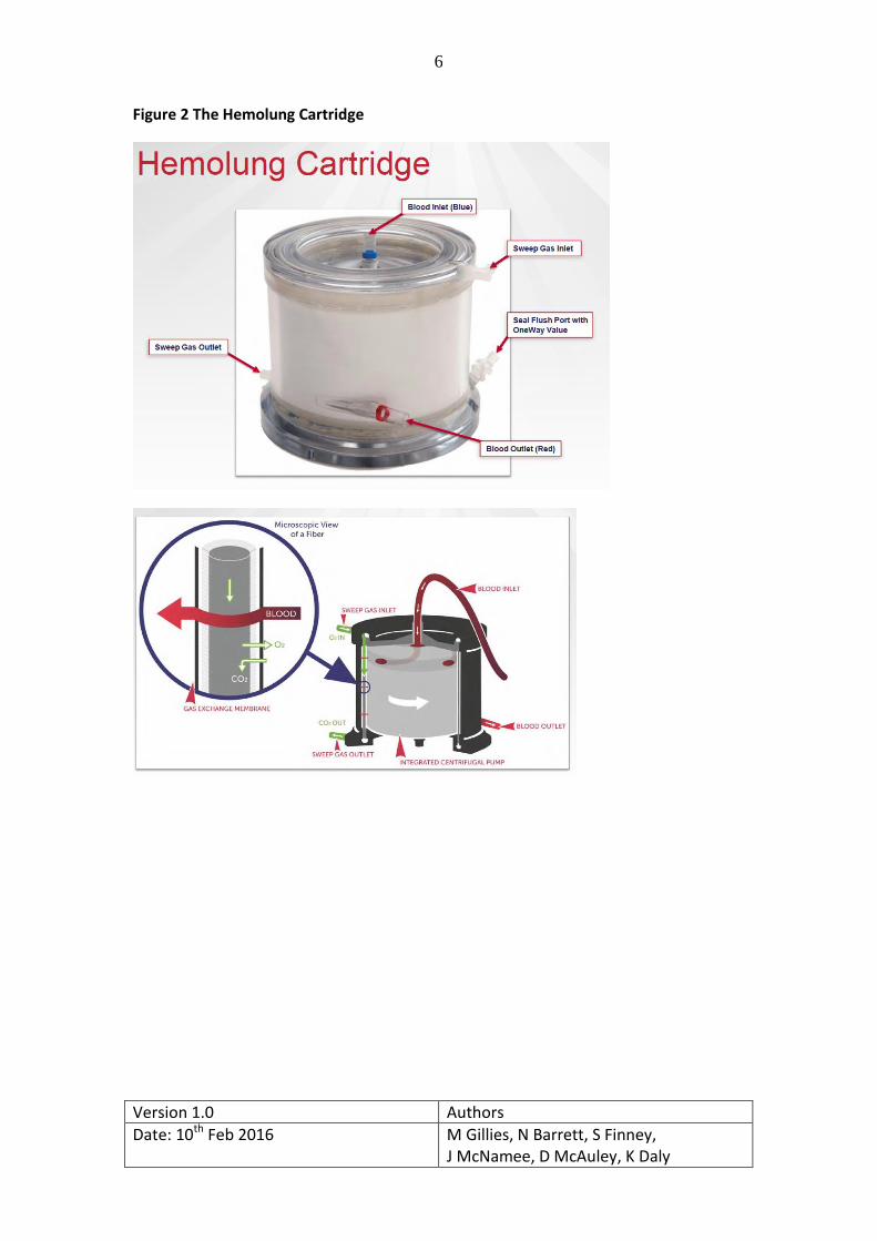
The Hemolung cartridge incorporates a gas exchange membrane with a centrifugal
pump (Figure 2). Blood is pumped from the patient through the cartridge and
“sweep gas” is attached to the gas exchange membrane with tubing at a gas flow
rate of 0-10 L/min. CO2 removal is achieved by running “sweep gas” (which for the
purposes of the trial will be room air) through the centre of the hollow fibres in the
cartridge while blood is circulated around the outside of the fibres (Figure 2). The set
“Sweep Gas Flow Rate” determines the sweep gas flow and the “Blood Flow Rate” is
determined by the pump speed i.e. revolutions per minute (RPM) of the centrifugal
pump. The patients blood enters the cartridge at the top and leaves from the
bottom side of the cartridge.
Version 1.0 Authors
Date: 10th Feb 2016 M Gillies, N Barrett, S Finney, J McNamee, D McAuley, K Daly
6
Figure 2 The Hemolung Cartridge
Version 1.0 Authors
Date: 10th Feb 2016 M Gillies, N Barrett, S Finney, J McNamee, D McAuley, K Daly
7
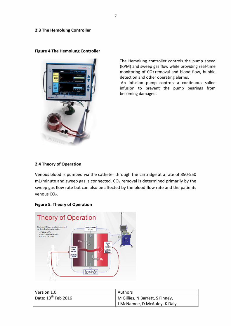
2.3 The Hemolung Controller
Figure 4 The Hemolung Controller
2.4 Theory of Operation
Venous blood is pumped via the catheter through the cartridge at a rate of 350-550
mL/minute and sweep gas is connected. CO2 removal is determined primarily by the
sweep gas flow rate but can also be affected by the blood flow rate and the patients
venous CO2.
Figure 5. Theory of Operation
The Hemolung controller controls the pump speed (RPM) and sweep gas flow while providing real-time monitoring of CO2 removal and blood flow, bubble detection and other operating alarms. An infusion pump controls a continuous saline infusion to prevent the pump bearings from becoming damaged.

Version 1.0 Authors
Date: 10th Feb 2016 M Gillies, N Barrett, S Finney, J McNamee, D McAuley, K Daly
8
Figure 6. Bubble detector and flow sensor
3. Set-up of the circuit If randomised to ECCO2R, initial ventilator settings may be any mode not excluded in
the study protocol. Initial recommended ventilator settings while you apply ECCO2R
are as according to the ARDSNetwork ARMA trial. Target tidal volumes will be ≤ 6
mL/kg PBW, plateau pressure ≤ 30cmH2O with PEEP level determined from the
PEEP/FiO2 table from the ARMA study (Appendix B and C). ECCO2R should ideally be
initiated within 8 hours from randomisation. It is advisable to use neuromuscular
blocking drugs if not already doing so to allow insertion of catheter, application of
ECCO2R and initiation of lower tidal volume ventilation.
Priming the Hemolung Circuit
Before cannulation, the Hemolung should be primed for use.
Personnel should refer to Chapter 4 of the Hemolung RAS Training Workbook for
priming and setup and have watched the Hemolung training videos “Setup and
Priming” and “Starting and Managing Therapy”.
Prime the Hemolung circuit in accordance with manufacturers instructions. A
laminated quick reference guide will be attached to the console.

Version 1.0 Authors
Date: 10th Feb 2016 M Gillies, N Barrett, S Finney, J McNamee, D McAuley, K Daly
9
Important points to remember when priming the circuit:
The priming solution should be heparinised saline (1u heparin / mL 0.9%NaCl)
When replacing the soda lime column, make sure to reuse the endcaps.
The recirculation bag must hang below the heparinised saline bag.
Maintain sterility of the blood tubing with the sheath until catheter
connection.
Apply a small amount of petroleum jelly to the part of the tubing that sits in
the blood flow sensor. DO NOT place petroleum jelly on the bubble detector
section (Figure 6).
Make sure connections are secure and that there are no leaks or air in the
circuit. If air bubbles are found, guide them into the recirculation bag.
Select ROOM AIR as the sweep gas source.
4. Insertion of Catheter 4.1 Personnel
An appropriately trained clinician (normally a consultant or senior ICU trainee) with
the required competencies will insert the Hemolung catheter and this should be
performed as a two-person procedure. Both clinicians must be familiar with the
components of the Hemolung Catheter Insertion and Hemolung Setup. Personnel
should refer to Chapter 4 Part 2 of the Hemolung RAS Training Workbook for
catheter insertion and have watched the Hemolung training video “Hemolung
Catheter Insertion”.
The Hemolung cartridge and circuit must be set up and primed before catheter
insertion to ensure rapid connection and avoid potential thrombus formation. Insert
the Hemolung catheter in accordance with manufacturers instructions. A laminated
quick reference guide will be attached to the console.

Version 1.0 Authors
Date: 10th Feb 2016 M Gillies, N Barrett, S Finney, J McNamee, D McAuley, K Daly
10
4.2 Procedure
It is recommended that all equipment for cannulation is stored in preparation for
insertion in a designated box, tray or trolley.
It is mandatory that ultrasound is used to guide insertion of the catheter. These
patients will normally be fully sedated and often receiving neuromuscular blocking
drugs.
Patient preparation
Site for catheter insertion chosen (Right Internal Jugular or Femoral
Veins)
Preliminary ultrasound scan for vessel patency and anatomical difficulty
Excessive hair removal
Selection of appropriate Hemolung catheter (femoral or jugular)
Cardiovascular and respiratory stability with intra-arterial BP
monitoring
Cannulation procedure
Full asepsis i.e. gloves, gown, mask, hat, large bed drape with skin
decontamination as per local policy. If using alcohol based solutions
allow to fully dry and avoid contact with the circuit as it can degrade
the circuit.
Real-time ultrasound guided Seldinger technique
Approach vessel at shallow angle to ensure straight path for the
guidewire
Once the guidewire is in place its position should be confirmed with
ultrasound
Give 80 units/kg of intravenous heparin bolus (rounded to nearest 50
units) after guidewire insertion BEFORE dilatation.
Dilate skin and soft tissues with tapered dilator. Shallow angle will
minimise risk of guidewire kinking or vessel trauma. Remove dilator and
leave guidewire in place.
Insert catheter and aspirate both lumens, demonstrating the free flow
of blood
Measure venous blood gas and ensure both lumens are flushed with
20mL heparinised saline (10 units/mL) after sampling (“rapid flush”
technique).
Chest X-ray is not necessary before ECCO2R initiation but can be
considered if there are concerns regarding catheter position or

Version 1.0 Authors
Date: 10th Feb 2016 M Gillies, N Barrett, S Finney, J McNamee, D McAuley, K Daly
11
complication. Be aware that delays to initiating therapy may be
associated with line thrombus.
After catheter placement, withdraw the guidewire from the stylet.
Remove the stylet from the catheter by unscrewing it from the priming
adaptor and withdrawing. Close both slide clamps.
Check catheter patency and remove any air by releasing each slide
clamp in turn and aspirating and flushing. Blood should aspirate easily
through both lumens. If either lumen exhibits excessive resistance to
blood aspiration, rotate or reposition the catheter to obtain adequate
blood flow. Each lumen should be flushed with 20 mL of heparinised
saline before re-clamping.

Version 1.0 Authors
Date: 10th Feb 2016 M Gillies, N Barrett, S Finney, J McNamee, D McAuley, K Daly
12
5. Connecting tubing and Securing Catheter 5.1 Connecting Tubing
After catheter insertion you are ready to connect the circuit to the patient. Catheter
should be connected to the Hemolung circuit and blood flow commenced as quickly
as possible following insertion (see below). If a delay occurs in establishing
extracorporeal blood flow then the catheter lumens should be flushed continuously
to prevent clotting. Personnel should refer to Chapter 4 Part 3 connecting tubing to
catheter in the Hemolung Training Workbook and have watched the video
“Hemolung Catheter Insertion”
Connect the “TO PATIENT (Red)” Tubing Set to Catheter
Disconnect the TO PATIENT (RED) Tubing Set from the recirculation bag.
Using a “wet-to-wet” technique (Figure 8), connect the tube to the red
connector on the catheter. A wet-to-wet technique is whereby the two
tubing ends are joined while a syringe of saline is used continuously to ensure
there are no air bubbles present after connection.
Ensure that the tubing is placed completely over the barb connector for a
secure connection.
Connect “FROM PATIENT (Blue)” Tubing Set to Catheter
Disconnect the FROM PATIENT (BLUE) Tubing Set from the Y-connector.
Using a wet-to-wet technique, connect the tube to the blue connector on the Catheter.
Ensure that the tubing is placed completely over the barb connector for a secure connection.
The plastic sheaths protecting the length of the blood tubes can be removed after
successful connection
Figure 8. “Wet Join” between Hemolung catheter and circuit

Version 1.0 Authors
Date: 10th Feb 2016 M Gillies, N Barrett, S Finney, J McNamee, D McAuley, K Daly
13
5.2 Securing Catheter
Catheters are secured as follows:
Femoral
For femoral catheter: secure cannula firmly with 1.0 silk suture placed
on the plastic portion of the catheter. Place the lumens in the “Grip-Lok”
device (Figure 9).
Jugular
For the internal jugular catheter, the suture is required at the catheter
hub. Place the body and hub of the catheter in the two “Grip-Lok”
devices (Figure 10).
General
Ensure that hard plastic components (e.g. clamps) are padded to prevent skin
pressure injury.
Dress according to local policy with semipermeable dressing (Figure 9).
Figure 9. Femoral Hemolung Catheter Secured with Sutures and Grip-Lok
Version 1.0 Authors
Date: 10th Feb 2016 M Gillies, N Barrett, S Finney, J McNamee, D McAuley, K Daly
14
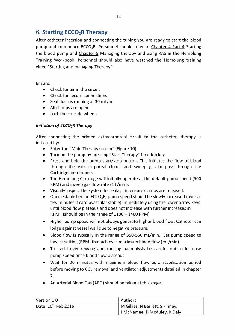
6. Starting ECCO2R Therapy After catheter insertion and connecting the tubing you are ready to start the blood
pump and commence ECCO2R. Personnel should refer to Chapter 4 Part 4 Starting
the blood pump and Chapter 5 Managing therapy and using RAS in the Hemolung
Training Workbook. Personnel should also have watched the Hemolung training
video “Starting and managing Therapy”
Ensure:
Check for air in the circuit
Check for secure connections
Seal flush is running at 30 mL/hr
All clamps are open
Lock the console wheels. Initiation of ECCO2R Therapy
After connecting the primed extracorporeal circuit to the catheter, therapy is initiated by:
Enter the “Main Therapy screen” (Figure 10)
Turn on the pump by pressing “Start Therapy” function key
Press and hold the pump start/stop button. This initiates the flow of blood through the extracorporeal circuit and sweep gas to pass through the Cartridge membranes.
The Hemolung Cartridge will initially operate at the default pump speed (500 RPM) and sweep gas flow rate (1 L/min).
Visually inspect the system for leaks, air; ensure clamps are released.
Once established on ECCO2R, pump speed should be slowly increased (over a few minutes if cardiovascular stable) immediately using the lower arrow keys until blood flow plateaus and does not increase with further increases in RPM. (should be in the range of 1100 – 1400 RPM)
Higher pump speed will not always generate higher blood flow. Catheter can
lodge against vessel wall due to negative pressure.
Blood flow is typically in the range of 350-550 mL/min. Set pump speed to
lowest setting (RPM) that achieves maximum blood flow (mL/min)
To avoid over revving and causing haemolysis be careful not to increase
pump speed once blood flow plateaus.
Wait for 20 minutes with maximum blood flow as a stabilisation period
before moving to CO2 removal and ventilator adjustments detailed in chapter
7.
An Arterial Blood Gas (ABG) should be taken at this stage.

Version 1.0 Authors
Date: 10th Feb 2016 M Gillies, N Barrett, S Finney, J McNamee, D McAuley, K Daly
15
Figure 10. Initiating Therapy on the Hemolung Console
From the last catheter connection screen, press the Start Therapy Function Key to enter Therapy Mode. You will be prompted with this screen. Press and hold the Pump Start/ Stop Key on the Hemolung Controller to initiate/resume therapy.
7. Ventilator and ECCO2R Settings During Trial Following a 20 minute stabilisation period, tidal volumes should be reduced in
tandem with increases in sweep gas flow rate slowly over 2 hours while the patient
is carefully monitored. A description of how the ventilator and ECCO2R settings
should be achieved is summarised in Figure 11 on the next page. It may be possible
to achieve maximum CO2 removal and tidal volume reduction over a shorter time
period. If pH < 7.20 at any stage then respiratory rate can be increased to a
maximum of 35 breaths per minute as tolerated. I:E ratio should remain 1:1-1:2. If
the arterial pH is < 7.20 and RR is maximally tolerated up to 35/min then no further
tidal volume reductions are recommended. Once ventilation goals are achieved and
stabilised on ECCO2R blood gases should usually be checked every 4-6 hours or
following any significant events or change in management.
The aim is to achieve the following goals:
tidal volume of ≤ 3mL/kg PBW
Pplat < 25cmH2O
Sweep gas 10
Respiratory Rate < 35 per minute
pH > 7.20
PaO2 7-10 kPa (SpO2 88-95%)

Version 1.0 Authors
Date: 10th Feb 2016 M Gillies, N Barrett, S Finney, J McNamee, D McAuley, K Daly
16
Figure 11. Adjusting ECCO2R Settings to Achieve Ventilator Goals
§The timings on this flow chart are as a guide only. Sweep gas flow and tidal volume may be adjusted more rapidly with an aim of 3ml kg-1 as the clinical condition of the patient permits.

Version 1.0 Authors
Date: 10th Feb 2016 M Gillies, N Barrett, S Finney, J McNamee, D McAuley, K Daly
17
8. Anticoagulation Management
All patients on ECCO2R should be systemically anticoagulated with intravenous heparin unless contraindicated (active bleeding, anticipated or recent surgery, thrombocytopenia). An APTTr of 1.5-2.0 will be targeted for the duration of therapy. Heparin infusions can be adjusted according to local policy. Management of Life Threatening Bleeding
If there is high risk of bleeding (e.g. planned or recent surgery) the following parameters should also be targeted: Platelets >80; Fibrinogen > 2.0. In the case of life threatening bleeding (either from catheter insertion site or elsewhere), inform responsible consultant and stop heparin. Blood component therapy (Cryoprecipitate, Platelets, fresh frozen plasma) should be given as to achieve the following targets: Platelets >80; Fibrinogen > 2.0, APTTr <1.5, INR <1.5. Tranexamic Acid administration (1g intravenously 6th hourly) should be considered. Source control (surgery, endoscopy, interventional radiology) should be considered as appropriate. If high risk or active bleeding, consider use of Thrombelastograph (TEG) to guide therapy if available. The Hemolung can continue to operate without heparin infusion with increased risk of the circuit clotting. Ongoing bleeding may necessitate discontinuation of ECCO2R therapy. Heparin-induced thrombocytopaenia and thrombosis (HITT) An isolated fall in platelets is common and not diagnostic. Advice from haematology is advised. Patients unable to have systemic heparin are excluded from this trial. If any patient develops HITT while in this trial specialist advice should be sought from a hematologist and the study team should be contacted. ECCO2R therapy should be discontinued.

Version 1.0 Authors
Date: 10th Feb 2016 M Gillies, N Barrett, S Finney, J McNamee, D McAuley, K Daly
18
9. Staffing and Maintenance while on Hemolung
9.1 Medical and Nursing Staff – General Principles
Patients will receive standard therapy as per local policy while they are in ICU
All patients in the REST trial must have decisions pertaining to ECCO2R
management undertaken by physicians and nurses who have undertaken
appropriate training in the use of the Hemolung system.
An ICU consultant with appropriate training and experience in ECCO2R should
review patients daily.
The nursing requirements specific to the patient on ECCO2R are primarily
related to the nursing responsibilities of maintaining a safe environment,
continuous monitoring and pressure area care.
Responsibility of the technical maintenance of the ECCO2R circuit (including
changes to circuit gas/blood flows) lies with appropriately trained medical
and nursing staff.
Patients on ECCO2R can be rolled and moved for chest x-rays, prone
ventilation, procedures and for pressure area assessment. However, a
Hemolung trained nurse or physician must be present to ensure that no
tension is transmitted to the catheter and that the circuit tubing is not kinked
and to designate a staff member to co-ordinate the move. Moves should only
be undertaken when staffing levels are appropriate (unless there is an urgent
patient need).
No procedures should be performed on an ECCO2R patient without the
consent of the ICU consultant.
If a surgical procedure is required while on the Hemolung, the heparin
infusion should be discontinued until it can safely be recommenced. It is
possible for patients to go to theatre with the Hemolung running but this
should be discussed with the local PI, the surgical team and if necessary a
member of the study team. In some circumstances it may be more
appropriate to discontinue Hemolung therapy prior to surgery. In the event
that the Hemolung is discontinued temporarily for a surgical procedure, the
device should be reprimed and left in re-circulation mode, and the catheter
flushed with heparinised saline and clamped.
For a more detailed account of Nursing Care, see section 12.

Version 1.0 Authors
Date: 10th Feb 2016 M Gillies, N Barrett, S Finney, J McNamee, D McAuley, K Daly
19
9.2 Criteria for Replacement of Hemolung Cartridge
The Hemolung should be discontinued after 7 days. If there is a decision by the treating physician that the Hemolung RAS therapy should last for longer than 7 days, the Cartridge may require replacement and, therefore, must be monitored for signs of reduced performance. This will be outside of the protocol and form part of clinical care. The following criteria are provided for determining the need for cartridge replacement:
1. If rate of CO2 removal falls below 50 mL/min AND cannot be explained by:
a. Reduced sweep gas flow ( < 7 L/min )
b. Reduction/correction of arterial PaCO2 (Note: magnitude of CO2 removal is directly proportional to partial pressure of CO2 in the blood; a hypercapnic patient will have a high rate of CO2 removal by the Hemolung, but as the Hemolung increases CO2 ventilation and PaCO2 begins to decrease, so will the rate of Hemolung CO2 removal. This is expected.)
c. Acceptable and minor reductions in blood flow which will occur transiently due to patient fluid volume
2. If circuit blood flow falls below 350 mL/min AND cannot be revived by:
a. Inspection of controller RPM setting (should be in the range of 1100 – 1400 RPM);
b. Inspection of circuit for pinching, bending, or twisting of tubing;
c. Repositioning of patient;
d. Minor ( < 1 cm ) adjustment of catheter position;
e. Increase in patient fluid volume, if low.
3. If severe hemolysis is indicated by observed hematuria and/or plasma free hemoglobin > 40 mg/dL, AND cannot be resolved within 2 hours by:
a. Identifying and correcting pinching or severe bends in circuit tubing;
b. Repositioning of patient and/or catheter ( < 1 cm );
c. Alternative sources (e.g. kinked or damaged infusion lines, dialysis catheters/systems);
d. Cessation or dilution of recently administered RBCs.

Version 1.0 Authors
Date: 10th Feb 2016 M Gillies, N Barrett, S Finney, J McNamee, D McAuley, K Daly
20
10. Weaning ECCO2R
After at least 48 hours of ECCO2R and lower tidal volume ventilation the patient will
be assessed daily to determine whether the following 3 criteria to progress to
ECCO2R weaning have been met:
1. Signs of clinical improvement of the underlying condition e.g. an
improvement in chest radiograph appearance or a reduction in the PEEP level
required to maintain acceptable oxygenation after the establishment of
ECCO2R.
2. PaO2 / FiO2 ratio ≥ 30kPa
3. Plateau pressure ≤ 25cmH2O when mechanically ventilated with a short trial
of Vt 6 mL kg-1 PBW (Be aware of the risk of hypocapnia due to increased
minute volume, this may be mitigated by reduction in respiratory rate)
Once the above weaning criteria are met then ECCO2R is weaned as per Figure 12.
Aim to reduce the sweep gas flow by 1 L/min every hour. It may be possible to
achieve sweep gas reduction over a shorter time period. Following 12 hours of
sweep gas flow at 1 L/min and an acceptable gas exchange, therapy is ended and
decannulation should follow as detailed in Section 11.
If the above three weaning criteria are not all met on daily assessment then ECCO2R
should be continued for a maximum of 7 days aiming for tidal volumes of ≤ 3mL/kg
PBW in mandatory ventilation or Vt ≤ mL kg-1 PBW if using pressure support
ventilation. It is recognised that at this stage that spontaneous breath tidal volumes
may be difficult to limit even with minimal pressure support.
An overview of the weaning process is outlined in Figure 11. The aim is to continue
lung protective ventilation regardless of whether mandatory ventilation (MV) or
pressure support ventilation is being used, i.e. Vt ≤ 6 mL kg-1 PBW if spontaneous
ventilation mode (pressure support) and Vt ≤ 3mL/kg PBW if mandatory mode.

Version 1.0 Authors
Date: 10th Feb 2016 M Gillies, N Barrett, S Finney, J McNamee, D McAuley, K Daly
21
Figure 12. Overview of weaning process for ECCO2R
*The timings on this flow chart are as a guide only. Sweep gas flow and tidal volume
may be adjusted more rapidly as the clinical condition of the patient permits.

Version 1.0 Authors
Date: 10th Feb 2016 M Gillies, N Barrett, S Finney, J McNamee, D McAuley, K Daly
22
11. Ending therapy and Decannulation Once the responsible consultant has decided that ECCO2R can be discontinued (see
above) decannulation should be undertaken. The A-Lung can be run with minimal or
no flow until a safe and appropriate time to decannulate the patient. There is 260mL
of blood in the circuit so prior to decannulation blood “rinse back” may be carried
out to return the blood to the patient if no thrombosis is suspected. Please refer to
A-Lung manual for the “rinse back” procedure and ensure staff have watched the
video “Ending Therapy”. There is a quick reference guide to “ending therapy with
blood rinse back” attached to the console
The process for removal of the Hemolung catheter is as follows:
Cease heparin for 2 hours
Reduce the RPM on the pump over a couple of minutes then Stop pump
Clamp both lumens of the Hemolung catheter
Clamp both blood tubes approximately 15cm from the catheter connection
The use of a deep vertical mattress suture(s) at the insertion site may be considered
Remove drainage catheter with direct pressure over the insertion site
Close mattress suture(s) if appropriate
Maintain digital pressure for 30 minutes after catheter removal (do not use a femstop device).
After 30 minutes of direct finger pressure the insertion site should be observed for bleeding.
Following this the insertion site should be observed every 15 minutes for 4 hours post removal. The area should NOT be covered over by a sheet during this time.
The mattress suture will need to be removed at 7 days, ensure this is documented in patient notes.

Version 1.0 Authors
Date: 10th Feb 2016 M Gillies, N Barrett, S Finney, J McNamee, D McAuley, K Daly
23
12. Nursing Care while on ECCO2R 12.1 Maintaining a Safe Environment
The following standards of nursing care should be met for patients on the Hemolung.
NURSING INTERVENTION RATIONALE
ECCO2R patients must be have a 1:1 bedside nurse: patient ratio. In addition, a nurse who has undertaken requisite training in Hemolung operation should be available on the unit 24/7 and should be present for any procedures requiring movement of the patient. Observations of the Hemolung should be performed hourly for the first 24 hours and then 2 hourly. Continuously monitor the patient for signs and symptoms of fluid imbalance, abnormal laboratory values, infection/sepsis, bleeding, thrombocytopenia, hemolysis, or other complications related to extracorporeal support systems.
The trained Hemolung nurse can provide support for the bedside nurse ensuring the safe management of the patient and the Hemolung circuit. Frequency of observations to maintain safe monitoring of patient, ventilation and operation of the Hemolung system.
Safety checks performed on commencement of shift to include:
Hemolung plugged into UPS plug
Check blood pump flow rate
Check sweep flow rate
Hemolung cartridge inspection
Check patient’s position and cannula sites
Check distal pulses and peripheral perfusion
Pump access line movement (shaking, swinging or still)
Check that all tubing is in the correct position to prevent kinks or restrictions.
Ensure Hemolung settings are correct and minimise the risk of circuit failure. Identify problems with patient perfusion, catheter, circuit or cartridge at an early stage.
The ECCO2R patient must NOT be left unattended at any time. Relief for breaks should be arranged so that a suitably experienced member of staff is monitoring the patient and Hemolung at all times.
Minimise risk of accidental decannulation or other system failure.

Version 1.0 Authors
Date: 10th Feb 2016 M Gillies, N Barrett, S Finney, J McNamee, D McAuley, K Daly
24
12.2 Monitoring the Patient/Circuit
NURSING INTERVENTION RATIONALE
Hemolung Flow Rate To be monitored continuously with circuit lines and membrane gas exchanger for any clot formation Document hourly for the first 24 hours then 2 hourly with RPM setting for trend monitoring Any drop in blood flow to be reported immediately and managed promptly (as per ECCO2R complications).
Blood flow through the Hemolung circuit is essential for maintenance of gaseous exchange and haemodynamic stability Immediately.
ECCO2R sweep flow rate To be monitored continuously. Document hourly for the first 24 hours then 2 hourly. Secure and label gas flow connections to membrane gas exchanger Ensure gas outlet is free from obstruction. Ensure correct sweep flow is given as prescribed. Regular ABGs
Any disruption to sweep flow will have significant effects on the patient’s stability and should therefore be identified and managed immediately
Inspection of Hemolung cartridge Perform once per shift looking for clot formation
Visible clots on the inflow side of the membrane may give an indication that the set is clotting
CO2 Clearance Record CO2 clearance hourly for the first 24 hours then 2 hourly.
A sustained decrease in CO2
extraction over time may indicate cartridge or circuit problems.
Haemodynamic Status Patient haemodynamic status should be monitored hourly.
Changes in haemodynamic status may affect both respiratory and circuit performance
Catheter Inspect catheter once per shift, for oozing of blood, and maintenance of secure dressings
Significant blood loss may occur from the catheter site. Dressings maintain catheter position and prevent infection.
Vascular Observation of Limbs (lower if femoral catheter used, upper if jugular catheter) Once per shift and should include limb temperature, colour and peripheral pulses.
Venous catheters may lead to DVT formation.

Version 1.0 Authors
Date: 10th Feb 2016 M Gillies, N Barrett, S Finney, J McNamee, D McAuley, K Daly
25
Change the sweep gas vacuum canister every 24 hours Must be changed daily to ensure adequate sweep gas flow.
Ensure that the seal flush is connected and volume administered is recorded
The seal flush is used to provide an infusion of saline at 30 mL/hr to provide a continuous flush of the blood pump seal. An infusion pump controls a continuous saline infusion to prevent the pump bearings from becoming damaged.
Perform safety checks once per shift (settings, circuit/line/canister inspection). Monitor for leaks, cracks, vibrations, air, or other system failures.
To ensure the safe monitoring of the including management of circuit emergencies
12.3 Patient Positioning, Patient Care and Pressure Area Care
NURSING INTERVENTION RATIONALE
All activities of nursing care involving the movement of the patient must be done with at least two nurses, one of who should be trained in use of the Hemolung.
Prevention of disruption of the circuit or accidental decannulation.
Patients should be positioned to optimise ventilation, ECCO2R blood flow rates and safety. Head of the bed may be elevated to 30 degrees
To improve respiratory and circuit function.
All moves are to be performed with nursing staff knowledgeable in ECCO2R immediately available in the unit
So any issues can be addressed immediately

Version 1.0 Authors
Date: 10th Feb 2016 M Gillies, N Barrett, S Finney, J McNamee, D McAuley, K Daly
26
13. Complications
Although fortunately rare, emergency complications involving the ECCO2R circuit can
be serious and demand immediate responses. They are largely preventable.
Possible complications are: low flow; bleeding; accidental decannulation; air
embolism; membrane failure.
13.1 Low Flow
Definition: A reduction in flow through the circuit e.g. an absolute blood flow of less
than 0.25L/min.
Effect: Hypercapnoea; membrane thrombosis; possible air embolus.
Causes: Hypovolaemia; increased intra-abdominal or intrathoracic pressure; circuit
kink; circuit thrombosis.
Prevention: Adequate circulating volume. Ensure that circuit not kinked or
obstructed. Ensure adequate anticoagulation.
Response: Ensure that the circuit is unobstructed. Assess abdomen to exclude raised
intra-abdominal pressure. Give a volume challenge. Ensure that circuit not
thrombosed (visual inspection and assess CO2 extraction). Ensure APTTr within
target range.
13.2 Bleeding
Definition: Bleeding from any source
Effect: Loss of blood and circulating volume
Causes: Anticoagulation; catheter insertion sites; other potential bleeding sites.
Accidental decannulation (partial or complete).
Prevention: Ensure patient not over-anticoagulated; where possible, avoid invasive
procedures whilst on ECCO2R. Catheter care on turns, positioning etc.
Response: Contact ECCO2R consultant to assess catheter position. Reduce or cease
heparin following discussion with consultant. Consider surgical and imaging options.
Consider tranexamic acid and desmopressin (DDAVP). Consider ceasing ECCO2R.

Version 1.0 Authors
Date: 10th Feb 2016 M Gillies, N Barrett, S Finney, J McNamee, D McAuley, K Daly
27
13.3 Decannulation
Definition: This is the accidental removal of the Hemolung catheter
Effect: Hypercapnia, blood loss.
Causes: tension on circuit, accidental dislodgement on turning or positioning.
Prevention: Secure catheter; adequate numbers of trained staff on turning or
positioning; circuit care on positioning or turning. A designated person should ensure
that lines remain free during patient manoeuvres
Response: Call for help. Clamp the circuit proximal to the disconnected catheter.
Apply pressure to the catheter insertion site. Adjust the ventilator settings to
compensate for loss of support. Contact ICU Consultant. Consider replacement of
circulating volume if indicated.
13.4 Circuit Air Embolism
Definition: This is the introduction of air into the ECCO2R circuit
Effect: Introduction of air embolus into the patient. Hypoxaemia and circulatory
collapse if large.
Causes: Introduction of air into the circuit via the catheter insertion site or via the
membrane gas exchanger.
Prevention: Only ECCO2R trained consultants to perform ECCO2R catheter insertion.
Care with circuit and catheter on patient movement (as outlined above).
Response: Clamp both arms of the catheter. Call for help. Contact ICU Consultant.
Assign roles for concurrent patient and circuit management.
Patient Management: Resuscitate A, B and C with 100% O2 and ventilation support
(increase minute ventilation as required). Consider positioning patient head down in
left lateral position and advancing CVC line into right atrium to aspirate air. Identify
source of air.
Circuit Management: Stop pump and discontinue therapy. Discard circuit.

Version 1.0 Authors
Date: 10th Feb 2016 M Gillies, N Barrett, S Finney, J McNamee, D McAuley, K Daly
28
13.5 Membrane gas exchanger failure
Definition: Over time thrombus may build up in the membrane. It is not unusual to
have some thrombus in the cartridge, particularly at points of turbulent flow.
Effect: Rise in PaCO2 despite constant minute ventilation, increased sweep gas flow
and pump speed. Overall reduction in flow through cartridge.
Causes: Thrombus, increase in biofilm, low blood flow.
Prevention: Ensure adequate anticoagulation. Ensure that the tubing is unobstructed.
Ensure adequate pump speed. Monitor CO2 removal and alert medical staff if
sustained reduction over time.
Response: Change the cartridge or circuit. Contact ICU Consultant.
Version 1.0 Authors
Date: 10th Feb 2016 M Gillies, N Barrett, S Finney, J McNamee, D McAuley, K Daly
29
14. Contact Numbers
Who do I contact for clinical advice?
Each ICU is encouraged to identify senior staff with a specific interest in ECCO2R.
Discuss the patient with these senior staff in your ICU. Consult with your REST
principal investigator or your REST research nurse.
If it is not possible to resolve your query locally you can contact the trial team as follows: Within office hours (8am - 5pm) please telephone: The NICTU trial office: 02890635794 who will contact one of the REST clinical team depending on the nature of the enquiry. If there is no reply please contact: REST research nurse in RVH on: 02890634247 / 02890633336 Dr James McNamee on: 07813776068 Prof Danny McAuley on: 07793525326
Out of hours (5pm to 8am) and you have an urgent clinical query, once the issue has been discussed with the senior staff in your ICU, the senior member of staff should call James or Danny on the numbers above. There may be additional contacts as the trial progresses.
Who do I contact for technical problems with the Alung / ECCO2R? In the first instance please refer to the REST abbreviated manual (laminated pages with ECCO2R containing troubleshooting tips). Your senior staff, REST principal investigator or your REST research nurse or technical staff may be able to help. If the issue cannot be resolved in-house, or you need urgent ECCO2R advice: Within office hours (8am - 5pm) please telephone: Garry Milner 07921587355 or
Debbie Bannister 07809458142 Out of hours (5pm to 8am): please telephone: Alung 08081891190
Who do I contact for administrative queries? Contact the trial office, telephone the REST trial office in NICTU on 02890635794, If you have no response, leave an answer phone message and we will get back to you as soon as possible. Or you can email us your request/query on: [email protected].

Version 1.0 Authors
Date: 10th Feb 2016 M Gillies, N Barrett, S Finney, J McNamee, D McAuley, K Daly
30
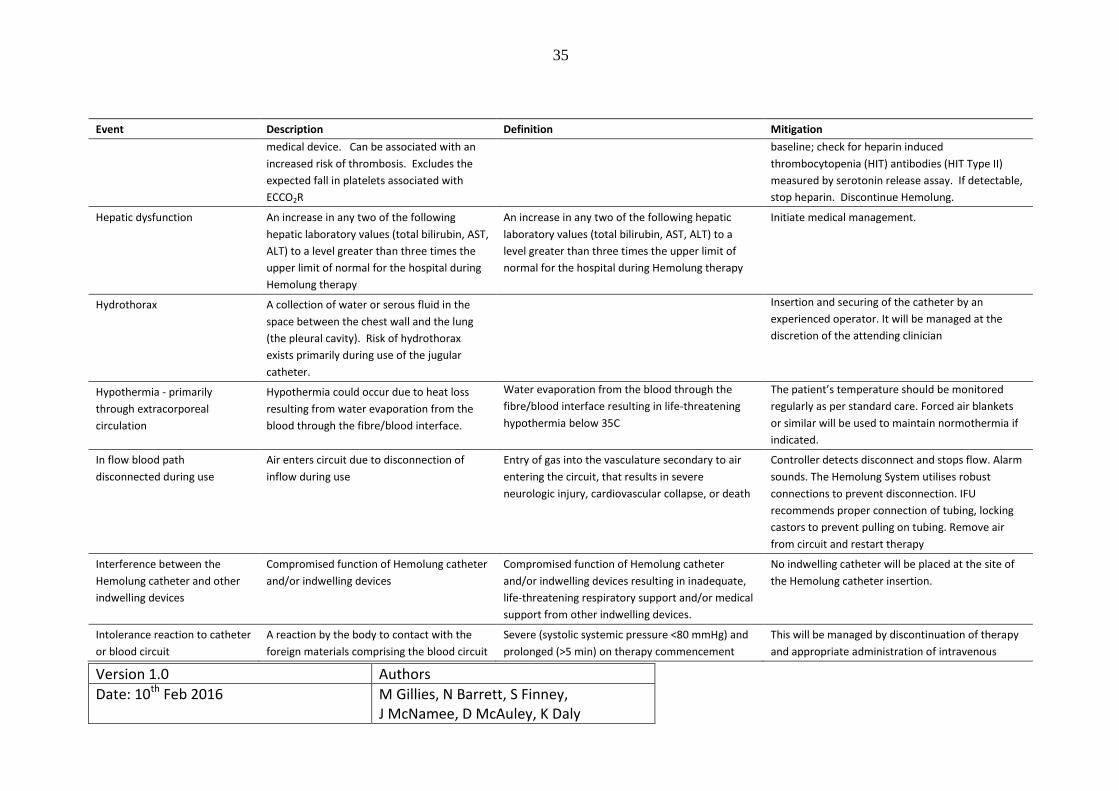
APPENDIX A - Complete list of anticipated adverse events and their mitigation Event Description Definition Mitigation
Access site contamination Contamination of access site. The Hemolung System requires only a single access
site, which reduces the number of access sites. The
catheter will be inserted employing the UK care
bundle for insertion and maintenance of central
venous catheters.
Acute Kidney Injury Abnormal kidney function. Abnormal kidney function requiring dialysis (include
hemofiltration) in patients who did not require this
procedure prior to Hemolung RAS therapy
initiation, or a rise in serum creatinine greater than
3 times baseline for greater than 48 hours.
Initiate medical treatment.
Air embolism Entry of gas into the vasculature. This may
occur during catheter insertion or during
therapy due to fibre leak, tubing connector
failure, cracked housing, breach of blood/air
interface, or console failure.
Entry of gas into the vasculature that results in
severe neurologic injury, cardiovascular collapse, or
death.
Standard blood aspiration techniques and Trust
protocols regarding the care and removal of central
venous access will be employed. The veno-venous
design minimises risk associated with air embolism,
since catheter is inserted into vein only.
Sweep gas pathway under vacuum prevents
possibility of air embolism from fibre failure. Low
and limited vacuum at the blood inlet, coupled with
a centrifugal pump design that limits the maximum
vacuum, and vertical pump position with impeller
on top, that traps the air at the top impeller and
stops the pump from pumping, are all safety
features of the design. Controller will alarm due to
signal from bubble detector and shutdown in a safe
mode. If this occurs, the device can be replaced by
the clinician.

Version 1.0 Authors
Date: 10th Feb 2016 M Gillies, N Barrett, S Finney, J McNamee, D McAuley, K Daly
31
Event Description Definition Mitigation
Arterial puncture with needle
during insertion
Incorrect insertion of needle during insertion
of catheter resulting in artery being
punctured instead of vein.
An arterial puncture requiring surgical intervention
as treatment.
The protocol mandates the use of real-time
ultrasound to locate the vein, and mandates that
experienced personnel with the appropriate
training will insert the catheter. If inadvertent
arterial puncture occurs then the needle will be
removed and local pressure applied.
Arteriovenous fistula. Abnormal connections or passageways
between an artery and vein.
Abnormal connections or passageways between an
artery and vein.
Initiate medical treatment. If unresolved,
discontinue therapy.
Battery Empty Controller running on battery and battery is
completely depleted
A controller failure resulting in inadequate, life-
threatening failure of respiratory support.
Controller displays battery status. Controller will
alarm for low battery when 20% battery power
remains. Plug in controller to continue therapy.
Bleeding at the insertion site Blood loss from placement site sufficient to
require blood transfusion or surgical/catheter
intervention.
> 100mLs blood loss from placement site sufficient
to require blood transfusion or surgical/catheter
intervention. Assessed by the investigator.
The insertion site will be examined and additional
stitches / local pressure applied. If the bleeding is
unresolved and excessive, then therapy will be
discontinued and formal surgical repair of the
catheter insertion site undertaken
Blood loss (Excessive) due to
disconnection from return
blood path
Excessive blood loss due to disconnection to
return blood path
Excessive blood loss due to disconnection to return
blood path
Controller detects disconnection and stops flow.
Alarm sounds. Hemolung RAS utilizes tight barbed
connectors to prevent disconnection. IFU
recommends proper connection of tubing, locking
castors to prevent pulling on tubing. Initiate medical
management.
Blood pressure decrease
(Hypotension)
Low blood pressure due to commencement
of therapy.
Systolic blood pressure of below MAP 60 mmHg
during the first two hours of therapy for more than
15 minutes.
Careful upregulation of the blood flow rate is
mandated in the Protocol. Initiate medical
treatment.
Brachial Plexus Injury Ipsilateral neurologic deficit in upper arm
secondary to catheter placement in
internal/external jugular vein
Catheters are placed using real-time ultrasound and
experienced operators.

Version 1.0 Authors
Date: 10th Feb 2016 M Gillies, N Barrett, S Finney, J McNamee, D McAuley, K Daly
32
Event Description Definition Mitigation
Bradycardia (low heart rate) Heart rate below 40 BPM during therapy Heart rate below 40 BPM during therapy Monitor heart rate (HR). If HR <40 BPM for greater
than 2 minutes, initiate medical treatment. If
unresolved, discontinue treatment.
Breach of device / environment
barrier
A breach in connection of tubing. This could
lead to bleeding or infection.
Investigators and nurses will be trained in the
proper connection of tubing, connectors, and
maintaining sterility. The system is designed to be
seamLess with robust alarms and connections.
Cardiac Arrhythmias Any documented arrhythmia requiring
defibrillation or cardioversion.
Any documented arrhythmia that results in clinical
compromise.
Medical management of arrhythmia, consider
discontinuation of therapy if arrhythmia is
recalcitrant.
Catheter and/or Blood Circuit
Occlusion
Occlusion of the catheter and/or blood circuit
due to thrombus formation. Reduced blood
flow and CO2 removal may result. Increased
risk of thromboembolism.
A Catheter and/or Blood Circuit Occlusion resulting
in inadequate, life-threatening respiratory support.
Hemolung device is coated with covalently bonded
heparin to minimise thrombus formation. Protocol
provides instruction regarding the anticoagulation
protocol. Catheter size limits embolism. Any
thrombus on the inflow side will be captured by
fibre mat. Junctions are designed to be smooth to
minimise potential for thrombus formation. The
exchange of the device and/or the re-siting of the
access catheter will be considered.
Catheter related blood stream
infection or cellulitis at
insertion area
A positive culture from the skin and/or tissue
surrounding the catheter insertion site, when
there is clinical evidence of infection such as
pain, fever, drainage, or leukocytosis.
Contamination of access site may lead to an
infection at the access site. Excessive
movement of the catheter can cause trauma
to the access site and increase risk of
infection
A positive culture from the skin and/or tissue
Hemolung catheter, coupled with the need to treat
with antimicrobial therapy, when there is clinical
evidence of infection such as pain, fever, drainage,
or leukocytosis.
Accessories included in catheter kit for sterile
insertion (e.g., drape, antiseptic swab, etc.). IFU
directions for aseptic technique.
All catheters will be inserted by experienced
personnel with appropriate competencies.
CO2 removal--excessive Excessive CO2 removal causing tetanus and Excessive CO2 removal resulting in tetanus Protocol for titration of therapy.

Version 1.0 Authors
Date: 10th Feb 2016 M Gillies, N Barrett, S Finney, J McNamee, D McAuley, K Daly
33
Event Description Definition Mitigation
hypocalcaemia
Chylothorax Chyle leak or chylothorax, a type of pleural
effusion, resulting from lymphatic fluid
(chyle) accumulating in the pleural cavity.
Chyle leak or chylothorax, a type of pleural
effusion, resulting from lymphatic fluid (chyle)
accumulating in the pleural cavity.
Initiate medical management.
Device Malfunction Failure of one or more of the components of
the Hemolung System which either directly
causes or could potentially induce a state of
inadequate respiratory support, but does not
include thrombus formation, or haemolysis,
or any other biological response to the
treatment (see Thrombus Formation event).
Failure of one or more of the components of the
Hemolung System which either directly causes or
could potentially induce a state of inadequate, life-
threatening respiratory support or death.
Controller will alarm for a variety of malfunctions.
When those alarms represent a safety concern for
the patient, the controller will shutdown in a safe
mode. The controller will be replaced by the back
up one held on-site if the patient has not met
weaning criteria.
Disseminated intravascular
coagulation (DIC)
Disseminated intravascular coagulation is a
disorder in which the proteins that control
blood clotting become abnormally active.
Small blood clots form that can cut off blood
supply to organs. Once clotting proteins are
consumed or used up, patients are at risk for
serious bleeding.
Disseminated intravascular coagulation is a
disorder in which the proteins that control blood
clotting become abnormally active. Small blood
clots form that can cut off blood supply to organs.
Once clotting proteins are consumed or used up,
patients are at risk for serious bleeding.
Initiate medical management. Discontinuation of
therapy should be considered.
Endocarditis Inflammation of the heart's inner lining. May
be caused by a catheter related infection.
Inflammation of the heart’s inner lining. May be
caused by a catheter-related infection.
Endocarditis requiring IV antibiotic therapy in the
hospital.
Medical management
Exit Site Necrosis At the catheter exit site due to injury Medical management
External leak in gas pathway A leak in the gas pathway to the device
reducing CO2 removal.
A leak in the gas pathway to the device reducing
CO2 removal resulting in inadequate, life-
threatening respiratory support.
Controller will alarm and indicate low CO2 removal.
The console can be replaced and tubing checked for
connections.
Extravasation Infusion of blood into the tissue surrounding
the accessed vessel due to retraction of the
Infusion of blood into the tissue surrounding the
accessed vessel due to retraction of the catheter,
Insertion and securing of the catheter by an
experienced operator.

Version 1.0 Authors
Date: 10th Feb 2016 M Gillies, N Barrett, S Finney, J McNamee, D McAuley, K Daly
34
Event Description Definition Mitigation
catheter, misplacement of the catheter, or
perforation of the vessel.
misplacement of the catheter, or perforation of the
vessel requiring surgical intervention for treatment.
Failure of controller to provide
sweep gas
Patient would receive insufficient respiratory
support due to controller failure.
A controller failure resulting in inadequate, life-
threatening respiratory support.
Controller will alarm. The controller will be
replaced by the back up one held on-site.
Haemorrhage not related to
insertion site bleeding.
Excessive external or internal bleeding not
related to insertion site.
Within any 24 hours period, > 1U packed red blood
cells (PRBC) required
The APTTr activities will be measured regularly and
the heparin infusion titrated in accordance with the
results. If uncontrolled internal bleeding occurs
then the heparin infusion will be reduced. The
patients haemoglobin will be supported by the
appropriate use of allogenic blood products.
Haematoma A mass of clotted blood (most likely to occur
at the catheter insertion site)
A mass of clotted blood (most likely to occur at the
catheter insertion site) requiring medical or surgical
treatment.
This will be managed at the discretion of the
attending clinician.
Haemothorax A collection of blood in the space between
the chest wall and the lung (the pleural
cavity). Risk of haemothorax exists primarily
during use of the jugular catheter.
A collection of blood in the space between the
chest wall and the lung (the pleural cavity). Risk of
haemothorax exists primarily during use of the
jugular catheter.
Insertion and securing of the catheter by an
experienced operator. It will be managed at the
discretion of the attending clinician
Haemolysis A plasma-free haemoglobin value that is
greater than 40 mg.dL-1
, in association with
clinical signs associated with haemolysis (e.g.
anaemia, low haematocrit,
hyperbilirubinaemia, raised LDH). Haemolysis
related to documented non-device-related
causes (e.g. transfusion or drug) is excluded
from this definition
A plasma-free haemoglobin value that is greater
than 40 mg.dL-1
, in association with clinical signs
associated with haemolysis (e.g., anaemia, low
haematocrit, hyperbilirubinaemia, raised LDH).
Haemolysis related to documented non-device-
related causes (e.g. transfusion or drug) is excluded
from this definition.
All known contributors to haemolysis have been
evaluated and optimised, RPM has been minimised,
surfaces have been smoothed, non haemolytic
materials utilised.
The revolutions per minute of the device are
controllable.
Plasma free haemoglobin will be assayed if
haemolysis suspected during therapy. The device
may be replaced/removed if haemolysis is high.
Heparin Induced
Thrombocytopenia (HIT)
Reduction in the number of platelets due to
the administration of heparin (an
anticoagulant), or use of heparin coated
A reduction of platelets due to the administration
of heparin requiring the infusion of any
replacement blood products as treatment.
Monitor platelet count during therapy. If platelet
count drops below 50 x 10.L-1
; or platelet count < 50
x 100.L-1
in conjunction with >50% decrease from
Version 1.0 Authors
Date: 10th Feb 2016 M Gillies, N Barrett, S Finney, J McNamee, D McAuley, K Daly
35
Event Description Definition Mitigation
medical device. Can be associated with an
increased risk of thrombosis. Excludes the
expected fall in platelets associated with
ECCO2R
baseline; check for heparin induced
thrombocytopenia (HIT) antibodies (HIT Type II)
measured by serotonin release assay. If detectable,
stop heparin. Discontinue Hemolung.
Hepatic dysfunction An increase in any two of the following
hepatic laboratory values (total bilirubin, AST,
ALT) to a level greater than three times the
upper limit of normal for the hospital during
Hemolung therapy
An increase in any two of the following hepatic
laboratory values (total bilirubin, AST, ALT) to a
level greater than three times the upper limit of
normal for the hospital during Hemolung therapy
Initiate medical management.
Hydrothorax A collection of water or serous fluid in the
space between the chest wall and the lung
(the pleural cavity). Risk of hydrothorax
exists primarily during use of the jugular
catheter.
Insertion and securing of the catheter by an
experienced operator. It will be managed at the
discretion of the attending clinician
Hypothermia - primarily
through extracorporeal
circulation
Hypothermia could occur due to heat loss
resulting from water evaporation from the
blood through the fibre/blood interface.
Water evaporation from the blood through the
fibre/blood interface resulting in life-threatening
hypothermia below 35C
The patient’s temperature should be monitored
regularly as per standard care. Forced air blankets
or similar will be used to maintain normothermia if
indicated.
In flow blood path
disconnected during use
Air enters circuit due to disconnection of
inflow during use
Entry of gas into the vasculature secondary to air
entering the circuit, that results in severe
neurologic injury, cardiovascular collapse, or death
Controller detects disconnect and stops flow. Alarm
sounds. The Hemolung System utilises robust
connections to prevent disconnection. IFU
recommends proper connection of tubing, locking
castors to prevent pulling on tubing. Remove air
from circuit and restart therapy
Interference between the
Hemolung catheter and other
indwelling devices
Compromised function of Hemolung catheter
and/or indwelling devices
Compromised function of Hemolung catheter
and/or indwelling devices resulting in inadequate,
life-threatening respiratory support and/or medical
support from other indwelling devices.
No indwelling catheter will be placed at the site of
the Hemolung catheter insertion.
Intolerance reaction to catheter
or blood circuit
A reaction by the body to contact with the
foreign materials comprising the blood circuit
Severe (systolic systemic pressure <80 mmHg) and
prolonged (>5 min) on therapy commencement
This will be managed by discontinuation of therapy
and appropriate administration of intravenous
Version 1.0 Authors
Date: 10th Feb 2016 M Gillies, N Barrett, S Finney, J McNamee, D McAuley, K Daly
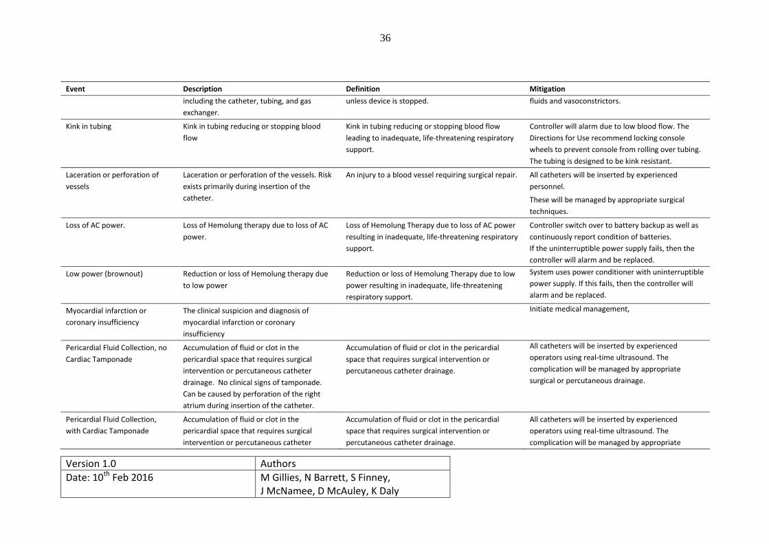
36
Event Description Definition Mitigation
including the catheter, tubing, and gas
exchanger.
unless device is stopped. fluids and vasoconstrictors.
Kink in tubing Kink in tubing reducing or stopping blood
flow
Kink in tubing reducing or stopping blood flow
leading to inadequate, life-threatening respiratory
support.
Controller will alarm due to low blood flow. The
Directions for Use recommend locking console
wheels to prevent console from rolling over tubing.
The tubing is designed to be kink resistant.
Laceration or perforation of
vessels
Laceration or perforation of the vessels. Risk
exists primarily during insertion of the
catheter.
An injury to a blood vessel requiring surgical repair. All catheters will be inserted by experienced
personnel.
These will be managed by appropriate surgical
techniques.
Loss of AC power. Loss of Hemolung therapy due to loss of AC
power.
Loss of Hemolung Therapy due to loss of AC power
resulting in inadequate, life-threatening respiratory
support.
Controller switch over to battery backup as well as
continuously report condition of batteries.
If the uninterruptible power supply fails, then the
controller will alarm and be replaced.
Low power (brownout) Reduction or loss of Hemolung therapy due
to low power
Reduction or loss of Hemolung Therapy due to low
power resulting in inadequate, life-threatening
respiratory support.
System uses power conditioner with uninterruptible
power supply. If this fails, then the controller will
alarm and be replaced.
Myocardial infarction or
coronary insufficiency
The clinical suspicion and diagnosis of
myocardial infarction or coronary
insufficiency
Initiate medical management,
Pericardial Fluid Collection, no
Cardiac Tamponade
Accumulation of fluid or clot in the
pericardial space that requires surgical
intervention or percutaneous catheter
drainage. No clinical signs of tamponade.
Can be caused by perforation of the right
atrium during insertion of the catheter.
Accumulation of fluid or clot in the pericardial
space that requires surgical intervention or
percutaneous catheter drainage.
All catheters will be inserted by experienced
operators using real-time ultrasound. The
complication will be managed by appropriate
surgical or percutaneous drainage.
Pericardial Fluid Collection,
with Cardiac Tamponade
Accumulation of fluid or clot in the
pericardial space that requires surgical
intervention or percutaneous catheter
Accumulation of fluid or clot in the pericardial
space that requires surgical intervention or
percutaneous catheter drainage.
All catheters will be inserted by experienced
operators using real-time ultrasound. The
complication will be managed by appropriate

Version 1.0 Authors
Date: 10th Feb 2016 M Gillies, N Barrett, S Finney, J McNamee, D McAuley, K Daly
37
Event Description Definition Mitigation
drainage. With clinical signs of tamponade
(e.g. increased central venous pressure and
decreased cardiac output). Can be caused by
perforation of the right atrium during
insertion of the catheter.
surgical or percutaneous drainage.
Pleural Effusion Excess fluid that accumulates between the
two pleural layers, the fluid-filled space that
surrounds the lungs.
Excess fluid that accumulates between the two
pleural layers, the fluid-filled space that surrounds
the lungs.
Initiate medical management.
Pneumothorax Presence of air in the space between the
chest wall and the lung (the pleural cavity).
This will be managed by appropriate drainage of the
pneumothorax.
Pneumomediastinum Presence of air in the mediastinum Initiate medical management
Pulmonary embolism Presence of a clot in a major pulmonary
artery
Evidence of a large clot in a first, or second order
pulmonary artery on CT angiography
This is unlikely in the presence of anticoagulation
and will be managed by on-going anticoagulation
Right Heart Failure Symptoms and signs of persistent right
ventricular dysfunction.
Symptoms and signs of persistent right ventricular
dysfunction not due to tamponade, ventricular
arrhythmias or pneumothorax requiring right
ventricular assist device, nitric oxide or inotropic
agents.
Initiate medical management.
Shock A life threatening medical condition as a
result of insufficient blood flow throughout
the body, including but not limited to
hypovolaemic or septic shock.
A life threatening medical condition as a result of
insufficient blood flow throughout the body,
including but not limited to hypovolaemic or septic
shock.
Initiate medical management. Discontinuation of
therapy may be considered.
Stroke
(Haemorrhagic/thrombotic) or
transient ischaemic attack.
Any new, temporary or permanent, focal or
global neurological deficit ascertained by a
standard neurological examination.
Any new, temporary or permanent, focal or global
neurological deficit ascertained by a standard
neurological examination (administered by a
neurologist or other qualified physician and
documented with appropriate diagnostic tests and
consultation note). The examining physician will
distinguish between a transient ischemic attack
This will be managed at the discretion of the
attending clinician.

Version 1.0 Authors
Date: 10th Feb 2016 M Gillies, N Barrett, S Finney, J McNamee, D McAuley, K Daly
38
Event Description Definition Mitigation
(TIA), which is fully reversible within 24 hours (and
without evidence of infarction), and a stroke, which
lasts longer than 24 hours (or less than 24 hours if
there is evidence of infarction).
Subcutaneous emphysema The presence of air in the subcutaneous
tissues.
Initiate medical management.
Severe Thrombocytopenia,
Non-HIT related
Reduction in the number of platelets due to
deposition and consumption within the blood
circuit. Severe thrombocytopenia is defined
as a platelet count drop below 20,000.mm-3
.
Can be associated with an increased risk of
thrombosis.
Monitor platelet count during therapy. If platelet
count drops below 50 x 10.L-1
, or platelet count <
100.L-1
in conjunction with >50% decrease from
baseline; check for heparin induced
thrombocytopenia (HIT) antibodies (HIT Type II)
measured by serotonin release assay. If detectable,
stop heparin. In these settings therapy should be
stopped.
Tachycardia Heart rate could increase during therapy HR > 140 beats per minute (BPM) for > 2 minutes Monitor the heart rate. If it exceeds 140 BPM for
more than 2 minutes, initiate medical management.
If unresolved, discontinue therapy.
Thoracic Duct Injury Injury to the thoracic duct during insertion of
the jugular catheter may result in the leakage
of chylous fluid.
All catheters will be inserted by experienced
operators using real-time ultrasound. The
complication will be managed by appropriate
surgical or medical therapies.
Thromboembolism, arterial
non-cns
An acute systemic arterial perfusion deficit in
any non-cerebrovascular organ system due to
thromboembolism confirmed by one or more
of the following: 1) standard clinical and
laboratory testing, 2) operative findings,
and/or, 3) autopsy findings. This definition
excludes neurological events.
An acute systemic arterial perfusion deficit in any
non-cerebrovascular organ system due to
thromboembolism confirmed by one or more of the
following: 1) standard clinical and laboratory
testing, 2) operative findings, and/or, 3) autopsy
findings. This definition excludes neurological
events.
Initiate medical management.
Thromboembolism, Venous Evidence of venous thromboembolic event Evidence of venous thromboembolic event (e.g. Only catheters appropriate to the vessel size will be
Version 1.0 Authors
Date: 10th Feb 2016 M Gillies, N Barrett, S Finney, J McNamee, D McAuley, K Daly
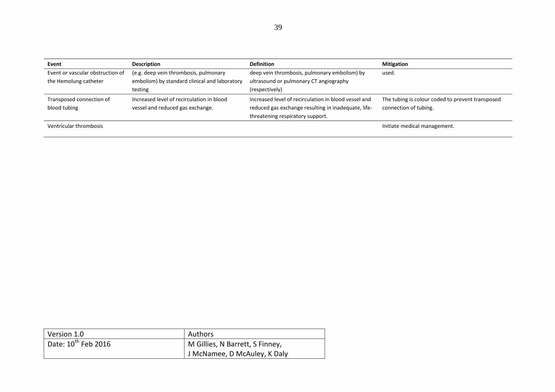
39
Event Description Definition Mitigation
Event or vascular obstruction of
the Hemolung catheter
(e.g. deep vein thrombosis, pulmonary
embolism) by standard clinical and laboratory
testing
deep vein thrombosis, pulmonary embolism) by
ultrasound or pulmonary CT angiography
(respectively)
used.
Transposed connection of
blood tubing
Increased level of recirculation in blood
vessel and reduced gas exchange.
Increased level of recirculation in blood vessel and
reduced gas exchange resulting in inadequate, life-
threatening respiratory support.
The tubing is colour coded to prevent transposed
connection of tubing.
Ventricular thrombosis Initiate medical management.

Version 1.0 Authors
Date: 10th Feb 2016 M Gillies, N Barrett, S Finney, J McNamee, D McAuley, K Daly
40
APPENDIX B –Predicted Body Weight Guide
Males: PBW = IBW (kg) = 50 + 0.91 (height (cm) – 152.4)

Version 1.0 Authors
Date: 10th Feb 2016 M Gillies, N Barrett, S Finney, J McNamee, D McAuley, K Daly
41
Females: PBW = IBW (kg) = 45.5 + 0.91 (height (cm) – 152.4)
APPENDIX C – PEEP Ladder
PEEP/FiO2 Combinations
FiO2 0.3 0.4 0.5 0.6 0.7 0.8 0.9 1.0
PEEP (cmH2O) 5 5-8 8-10 10 10-14 14 14-16 18-24
Version 1.0 Authors
Date: 10th Feb 2016 M Gillies, N Barrett, S Finney, J McNamee, D McAuley, K Daly
42
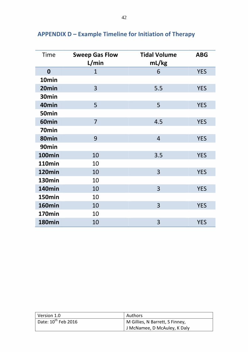
APPENDIX D – Example Timeline for Initiation of Therapy
Time Sweep Gas Flow L/min
Tidal Volume mL/kg
ABG
0 1 6 YES
10min 20min 3 5.5 YES
30min
40min 5 5 YES 50min
60min 7 4.5 YES
70min 80min 9 4 YES
90min
100min 10 3.5 YES 110min 10
120min 10 3 YES
130min 10 140min 10 3 YES
150min 10
160min 10 3 YES 170min 10
180min 10 3 YES
![research.unsw.edu.au Trial... · Web viewClinical Trial Protocol Template [Insert full study title] [Insert short study title] Clinical Trial Protocol and Protocol Amendment General](https://img.pdfslide.net/doc/110x75/5aaa0a097f8b9a9a188db405/trialweb-viewclinical-trial-protocol-template-insert-full-study-title-insert.jpg)