Embed Size (px)
Citation preview

1
STUDY OF CLINICAL, SONOLOGICAL
AND HISTOPATHOLOGICAL
CORRELATION OF OVARIAN TUMOR
DISSERTATION SUBMITTED FOR
M.D (BRANCH – II)
(OBSTETRICS & GYNAECOLOGY)
APRIL 2013
THE TAMILNADU
DR.M.G.R. MEDICAL UNIVERSITY
CHENNAI, TAMILNADU

2
CERTIFICATE
This is to certify that this dissertation titled ―STUDY OF CLINICAL,
SONOLOGICAL AND HISTOPATHOLOGICAL CORRELATION OF
OVARIAN TUMOR‖ submitted by DR. AISHWARYA JAGAN to the faculty of
Obstetrics and Gynecology, The TamilNadu Dr. M.G.R. Medical University,
Chennai in partial fulfillment of the requirement for the award of MD degree
Branch II Obstetrics and Gynecology, is a bonafide research work carried out by
her under our direct supervision and guidance from September 2011 to August
2012.
Dr.UMA, M.D.,D.G.O., Dr.P. ANGAYARKANNI M.D.(O.G), DCH,
Professor of Obstetrics and Professor and Head of the Department,
Gynecology, Department of Obstetrics and
Department of Obstetrics and Gynecology,
Gynecology, Madurai Medical College,
Madurai Medical College, Madurai.
Madurai.

3
DECLARATION
I, Dr. AISHWARYA JAGAN solemnly declare that the dissertation titled
―STUDY OF CLINICAL, SONOLOGICAL AND HISTOPATHOLOGICAL
CORRELATION OF OVARIAN TUMOR‖ has been prepared by me. This is
submitted to The Tamilnadu Dr. M.G.R. Medical University, Chennai, in
partial fulfillment of the regulations for the award of MD degree (Branch II)
Obstetrics & Gynaecology. I also declare that this bonafide work has not been
submitted in part or full by me or any others for any award, degree or diploma to
any other university or board either in India or abroad.
.
Place: Madurai Dr. AISHWARYA JAGAN.
Date:

4
ACKNOWLEDGEMENT
I owe my thanks to The Dean Dr. N. MOHAN M.S., Madurai Medical
College for allowing me to avail the facilities needed for my dissertation.
I am deeply indebted to DR. P. ANGAYARKANNI, M.D. (O.G.), D.C.H.,
Professor and Head of the Department of Obstetrics and Gynaecology, Madurai
Medical College, Madurai, for her able guidance, inspiration and the
encouragement she rendered at every stage of this study.
I am very grateful to my Former Professor and Head of Department
Dr. S. Dilshath M.D., D.G.O., and Prof. Dr. Uma M.D., D.G.O., for their
valuable guidance in conducting and completing the study. I express my gratitude
to other Professors, Dr. Ambigaimeena, M.D., D.G.O, Dr. S. Geetha, M.D.,
D.G.O., Dr. T. Uma Devi, M.D., D.G.O. and Dr. Revwathy Kailairajan, M.D.,
D.G.O, Department of Obstetrics and Gynaecology for allowing me and helping
me in conducting my study in their respective units.
I extend my heartful thanks to Dr. Usha Ravikumar, M.D., Professor and
Head of the Department of Pathology, Dr. N. Sundari M.D.R.D., Professor and
Head of Department of Radiology and Dr. Jayaraman D.M.R.D., for their
guidance throughout my study.

5
I thank all my Assistant Professors for their kind co-operation in helping
me to do this study.
Last but not the least I gratefully acknowledge my thanks to all my
Colleagues and the co-operation of the patients without whom this study would not
have been possible.

6
CONTENTS
S.NO. TITLE PAGE
1. INTRODUCTION 1
2. AIM AND OBJECTIVES 3
3 REVIEW OF LITERATURE 4
HISTORICAL ASPECTS 9
ANATOMY OF OVARY 11
PATHOLOGY OF OVARIAN TUMORS 19
4. MATERIALS AND METHODS 51
5. RESULTS 54
6. DISCUSSION 68
7. SUMMARY 80
8. CONCLUSION 83
ANNEXURES
Bibliography
Proforma
Master Chart
Ethical Committee Approval Certificate
Antiplagiarism Certificate

7
LIST OF CHARTS
NO. TITLE
1 Incidence of Benign, Borderline and Malignant tumors
as per HPE
2 Mean age distribution
3 Parity distribution
4 Per abdomen consistency of Ovarian Tumor
5 Laterality
6 USG Volume Score
7 USG Structural Score
8 Morphological score
9 Results as per USG total morphological score and HPE
10 Comparison of Sensitivity and Specificity with other
Studies

8
LIST OF TABLES
Table No. Title Page No.
1 Incidence of Benign, Borderline and Malignant
tumors as per HPE
54
2 Mean age distribution 55
3 Parity distribution 56
4 Mode Of Presentation 57
5 Per abdomen consistency of Ovarian Tumor 58
6 Laterality 59
7 USG Volume Score 60
8 USG Structural Score 61
9 Total Morphological Score 62
10 Results as per USG Total Morphological Score and
HPE
63
11 Efficacy of Morphological Score with HPE as Gold
Standard
63
12 Morphological Score and Malignancy 65
13 Types of tumor 66
14 Comparison of incidence of Ovarian Tumors 68
15 Comparison of age distribution of benign Ovarian
Tumors
69
16 Comparison of age distribution of malignant Ovarian
Tumors
70
17 Comparison Of Sensitivity And Specificity With
Other Studies
79

9
LIST OF PHOTOGRAPHS
Figure No. Title
1 Cells of Ovary
2 Histiogenesis of Ovarian tumors
3 Serous Cyst Adenoma
4 Papillary Serous Cyst Adenocarcinoma
5 Mucinous Cystadenoma
6 Papillary Mucinous Cyst Adenocarcinoma
7 Endometrioid Tumor
8 Malignant Brenner Tumor
9 Fibrothecoma
10 Granulosa Cell tumor
11 Benign Cystic Teratoma
12 Immature Teratoma
13 Krukenbergs Tumor
14 Ovarian Tumor complicating pregnancy
15 Morphological scoring System
16 Borderline Mucinous Ovarian Tumor
17 Leiomyosarcoma of ovary
18 Structural score
18a Smooth wall, Sonolucent
18b Smooth wall, Diffuse echogenicity
18c Wall thickening, <3mm fine septa
18d Papillary projections, septa> 3mm
18e Complex, predominantly solid
18f Complex, solid and cystic areas
18g Extratumoral fluid
10
INTRODUCTION
Ovary is a very important organ as it is concerned with reproduction.The
ovary consists of mesenchymal cells which are multipotential and sex cells which
are totipotent.So their stimulation may result in any type of tumor.1, 2
Of all gynaecological cancers, ovarian malignancies representsthe greatest
clinical challenge because of greater range and variety of tumors with uncertain
origin, with no known premalignant lesion and variability in the rate of disease
progression.1 Around 70% of patients with ovarian tumors are diagnosed only at
advanced stages due to unavailability of effective screening method and lack of
specific clinical presentations at early stage of the disease.
Ovarian cancer is the eighth leading cause of cancer and fifth leading cause
of cancer related death in females. Every year 204,000 women are diagnosed to
have ovarian cancer and almost 125,000 women die due to ovarian cancer
worldwide.1
The histiogenesis of ovarian tumor revolves around four main components,
mainly surface epithelium, germ cells, sex cord and specialized ovarian stroma.2
Ovarian tumors can occur in all ages, but there are differences in the
histological types during various decade of life. The predominant type of tumor

11
during younger age group arethe germ cell tumors. Sex cord stromal tumours
occurs in women of all ages.
In premenopausal women 7% of tumors are frankly malignant and 30 % of
themin postmenopausal women are malignant.
90 to 95% of malignant tumorsof the ovary are Surface Epithelial Ovarian
cancers and the remaining 5 to 10% constitute the Sex Cord Stromal tumors and
the Germ cell tumors of the ovary.
Ultrasonography is used extensively to differentiate benign and malignant
tumors of ovary.
This study is on the OVARIAN TUMOR MORPHOLOGICAL
INDEXING – a quantitative analysis relating tumor morphology from
sonographically generated images to risk malignancy.

12
AIM AND OBJECTIVES
1. To analyse the sensitivity and specificity of a morphological
scoring system in differentiating benign and malignant tumors of
the ovary.
2. To study the epidemiology of ovarian tumors.

13
REVIEW OF LITERATURE
Ovarian cancer is the second most common of all gynaecological cancers
and accounts for 10 – 15 % of gynaecological malignancies in developing
countries including India.
Ovarian cancer remains as the leading cause of all cancer related deaths
among gynaecological malignancies in United States. The incidence of tumors of
ovary is highest in Sweden (19.6/100,000) and the United States (15.4/100,000)
and lowest in Japan (10.1/100,000).3
Sonography is considered to be the investigation of choice for the evaluation
of ovarian tumors due of its high sensitivity, acceptability, and low cost.Campbell
et al in 1989 was the first to propose the use of sonography for ovarian cancer
screening.4He reported a high correlation coefficient of 97% between the
ultrasonographically detected ovarian volumes and the actual ovarian volume
measured after oophorectomy.
Several studies have been reported to evaluate the benefits of morphologic
scoring systems, in quantifying and standardizing the interpretation of sonographic
images.

14
Granberg et al in 1989 concluded that the ultrasound is highly reliable in
predicting the characteristics of an ovarian cyst.5They reported that the most
predictive characteristic feature of malignancy in an ovarian cyst is the papillary
projections seen on the inner surface of the cyst wall.
Sassone et al in 1991, suggested an index with four different morphologic
features of ovarian cyst architecture, which includes wall structure, septations, cyst
wall thickness and echogenicity.6 He gave a discrete score for each specific
character and was evaluated on 143 women with ovarian tumors. The
morphological index thus obtained was 100% sensitive and 83% specific in the
distinguishing benign tumor from that of a malignant one.
DePriest et al in 1994 recommended a morphologic index system, with only
3 structural characteristics - cyst wall, ovarian volume and septae.7This was
analysed on 213 women with ovarian tumors. The sensitivity was 89% and
specificity was only 70% in predicting ovarian cancer according to his study.
Lerner etalin 1994 used the Sassone classification system and defined the
relative importance of the structural components by a multiple regression analysis.8
Lerneretal appliedan individual score to each of the morphological components by
simplifying the indices. The new scoring system was evaluated in 350 women with
ovarian masses and reported a sensitivityof 97% and specificity of 77%.

15
Ferrazzi et al in 1997 designed a new scoring system that was evaluated on
330 women with ovarian masses.9According to the study the sensitivity of this
scoring system was 87% and the specificity was 67%.
In 2001 Mol et al conducted an external validation of different
morphological indices including those by Lerner et al, Granberg et al, Finkler et
al, De Priest et al, Sassone et al and Ferrazzi et al.10
Mol et al conducted a study on
170 ovarian tumor to compare the predictive accuracy of these scoring systems.
They reported a lower accuracy than the original scoring indices, with a high
sensitivity ranging from 77% to 93% and specificity ranging from 21% to 89 %.
Ueland and collegues in 2003 evaluated the Morphology indexing and
concluded it to be an inexpensive and accurate method in differentiating benign
ovarian tumors and malignant ovarian tumors, it can be used as an effective tool to
plan the management of ovarian tumors.11
The useof Doppler flow studies along
with this morphological indexing have not shown to improve the diagnostic
accuracy of morphological indexing.
Persistent ovarian masses and cysts studied in the Hirosaki and Kentucky
trials showed a very low likelihood ratio for malignancy tumors - 9.4% in
Kentucky trial and 7.0% in Hirosaki trial. By applying the morphological scoring
indices to ultrasonographic screening system in the Hirosaki and Kentucky trials,

16
the reported a high sensitivity ranging from 80% to 90% with a positive predictive
value of > 20%.
Since 1980s, the color Doppler flow sonography was considered as an
important tool for the predictor of malignancy in ovarian tumors because Doppler
flow gives indirect information on the metabolism and direct information on the
vascular anatomy of the ovaries.
Hata et al 1999, studied about the colour Doppler assessment of intratumoral
blood flow in tumors of the ovary.12
According to The National Cancer Data Base Report on Ovarian Cancer,the
risks for ovarian cancers were associated with age, nulliparity and a family history
of the ovarian tumors. They reported that the women‘s lifetime risk of developing
cancer of ovaryis 1 in 70 (1.4%). Women who have a family history of one or
more affected first-degree relatives will have an increase in risk upto 5% - 7%. It is
been reported that only 3% -9% of the women with family history will actually
manifest hereditary cancer syndromes.
Approximately 90% of malignant ovarian tumors in adult are of epithelial
tumors followed by sex cord stromal tumors (6%) and germ cell tumors (3%).

17
Shruti et al in 2008 studied on the incidence and management of ovarian
tumours in 75 cases and reported that 90% of ovarian tumours were of epithelial
origin, 75% were benign serous type, 20% of them were mucinous and 2% were
endometriod type.14
In 2010, GG Swamy et al did a study on 120 cases and concluded that 71.6%
were benign, 25% were malignant and 3% were borderline tumors.15
About two
third of all benign neoplasms was seen in patient between 20 to 40 years age,
whereas two third of all malignant neoplasms were seen after the age of 40 yrs.
The youngest patient in the study conducted by Swamy et al was 12 years of age
and the oldest women was of 70 years of age.

18
HISTORICAL ASPECTS
Herophilus first described the mammalian ovaries, he called it female testis.
Soranus describes the ovary as didymus [twin]. Vesalius and De Fabrica of
1543 labelled ovary as the testis of the uterus. Vesalius, first described the ovarian
follicle and corpus luteum.
In 1666, Janswammer dam explained that ―the human female testis are
comparable to ovaria of birds‖.
Esmond long in 1761, described all the common tumours of ovary.
Leonardo Da Vinci drew accurately the anatomy of the uterus and ovaries.
Carl von Rokintansky of Vienna, his name is attached to the Rokitansky
protuberance of dermoid tumour, also called ―dermoid mamilla‖, ―dermoid
protuberance‖ and ―embryonic node‖.
F.Von Werdt proposed the term ―granulosa cell tumour‖ in 1914.
Friedrich Ernst krukenerg described a ―fibrosarcoma ovarii mucocellular
carcinomatodes‖ in 1896 under the mistaken impression that the lesion was
primary malignancy of ovary. In 1902 Schlagenhauter corrected the error and
identified this lesion as metastatic carcinoma.

19
Struma ovary was first described by Ludwig pick in 1903.
Fritz brenners in 1907 published paper describing 3 cases of Brenner
tumour, which now bear his name.
The term ―Adenoacanthoma‖ was coined by Malcolm Dockerty of the Mayo
clinic in 1954.
The first histological description as germ cell tumours was given by M.
Cherassu in 1906.
Robert mayor first described the mixed and less differentiated forms of
tumours which produce viriliszing effect and named as ―Adreoblastoma‖, in 1930
it was replaced by ―Arrhenoblastoma‖.
In 1936 Joe Vincetmeigs presented 7 cases of syndrome of ovarian fibroma,
ascitie and hydrothorax. In 1937, Rhoads and Terrell suggested the eponym
―Meigs syndrome‖.

20
ANATOMY OF OVARY
The ovaries arenodular ovoid structures, located on eachside of the uterus. It
is related to the lateral pelvic wall and attached to the posterior layer of the broad
ligament, posteroinferior to the fallopian tubes.
They are pink in colour with a smooth surface in young adults. With
advancing age they become more greyish with a puckered and scarred surface due
to repeated ovulation.They areabout 3 cm long, 1.5 cm wide. The thickness of the
ovary is approximately 1cm. The weight of each ovary ranges from 2 - 3.5 gm.16
Each ovary has 2 ends – uterine and tubal end. There are two borders – free
border and mesoovarium. They have two surfaces – lateral and medial.
RELATIONS:
The upper end is also known as the tubal end. It gives attachment to the
fimbrial end of the tubes. It is also attached to the infundibulopelvic ligament. The
lower end is also known as the uterine end. It gives attachment to the ovarian
ligament.3.16
The free border is also called the posterior border. Upper part of free border
is related with the uterine tube while it is related to the ureter and internal iliac
vessels posteriorly. The mesoovarian border is also called the anterior border. It is

21
attached to the posterior leaf of the broad ligament. A cleft in this border called the
hilum, transmits the ovarian nerves and vessels.
The medial surface of the ovary is related to the fimbrial end of the uterine
tube. The lateral surface is convex and lies on the peritoneal depression of the
lateral pelvic wall, called the ovarian fossa. The lateral surface is related to the
obturator nerves and vessels.
BLOOD SUPPLY:
The ovarian artery is the main arterial supply of the ovary. Ovarian artery
arises directly from the abdominal aorta and enters the ovary through the
infundibulo pelvic ligament, mesoovarium and mesosalphinx. The ovaries are also
supplied by uterine artery.16,25
The pampiniform plexus of veins coming out of the hilum form a single vein
called the ovarian vein. The left ovarian vein drains into left renal vein and right
ovarian vein drains into inferior venacava.
LYMPHATIC DRAINAGE:
They mainly drain into pre and para aortic group of lymph nodes.

22
NERVE SUPPLY:
Twigs from the aortic plexus pass around the ovarian artery forms the
ovarian plexus. Ovarian plexus consists of sympathetic fibres from T10- T11 and
parasympathetic fibres from the vagus.16,25
1. The ovarian surface epithelium ( germinal epithelium) forms the outermost
layer
2. The cortex is covered by the tunica albuginea, which is lined by germinal
epithelium of waldeyer that consist of a layer of cuboidal cells.
3. The ovarian cortex consists of stroma and ovarian follicles.2,25
a. Ovarian follicle : Contains within it the membrana granulosa,
cumulus oophorus, the corona radiata, primary oocyte and the zona
pellucida. The zona pellucida, antrum, theca of follicle and liquor
folliculi are also included within the follicle.
b. Stromal tissue: The oocytes are included within the stromal tissue,
they are derived from the mesenchymal cells and are made up of
connective tissue and interstitial cells. They have the ability to
respond to hormones like leutinizing hormone or human chorionic
gonadotropin.

23
4. The inner most layer is the medulla. The medulla and cortex cannot be
distinctly differentiated. The medulla is made of loose connective tissue
derived mostly from mesonephric cells.
5. The hilum (rete ovarii): The hilum is the point where the mesoovarium is
attached to the ovary. It contains blood vessels and nerves, and hilus cells
which become active in response to steroidogenesis to form tumours.
CELLS OF THE OVARY3,16
(Fig – 1)
Germ cells: At birth the oocytes represents the germ cells. Germs cellsare capable
of reproducing tissue of all germ layers and are considered to be the cells of origin
of teratomas.
Granulosa cells: The granulosa cells lie in a single layer around the oocytes. They
proliferate under the influence of FSH, forming a fluid that contains the precursor
of the zona pellucida.
The Call-Exner bodies,small round masses of dense pink materials
surrounded by a rosette of granulosa cells are a specific product of granulosa cells,
normal and neoplastic. They synthesize estrogen and various intermediates,
including dehydroepiandrosterone. They enlarge at the time of ovulation to form
the corpus luteum.

24
Theca cells: As the maturing graffian follicle enlarge, the immediate surrounding
stromal cells enlarge to become rounded and plump, called luteinization. Follicle
associated theca cells when activated produce estrogen.
Corpus luteum: The graffian follicle ruptures in response to LH surge and expels
the oocyte to become a corpus luteum. The corpus luteum is the main source of
progesterone but it also synthesizes estrone and E2, and also androgen.
The corpus luteum producesrelaxin during gestation andpuerperium,
probably under the control of HCG.
Hilus cells (HilarLeydig cells): These are clusters of large cells with abundant
pink cytoplasm associated with nonmyelinated nerve fibers seen in the hilum of the
ovary. They contain crystalloid of Reinke. Their physiological significance of in
ovary has not been demonstrated.
Vestigial structure: Persistence of mesonephros as isolated small duct in the
mesovarium andaplexiform glandular structure, the rete ovarii are situated on the
margin of the ovarian-hilar junction.

25
ETIOLOGY OF OVARIAN TUMOUR1,2,3
The etiology of epithelial cancers of the ovary remains obscure.
1. Geographic factors-more common in women of European and North
American origin. Low incidence is seen in Japan.
3. Menstrual factors - early menarche and late menopause
4. Reproductive factors
Nulliparity and low parity are at high risk. There is 40% decrease in risk
following first pregnancy, and 14% decrease in risk following subsequent
pregnancy.
Elevation in circulating progestin levels during pregnancy protect from
ovarian cancer.
5. Oral contraceptives17
Long term use of combination of contraceptive reduces the risk of ovarian
cancer by 50%. The duration of protection last up to 25 years after the last use.
This is due to the inhibitory effect of the OCP on ovulation. OCP protect
against both malignant tumors of the ovaryand functional cysts of the ovarybut not
against the benign tumors of the ovary.

26
6. Dietary factors
There is increased risk with high fats, and low fiber, carotene and vitamin
intake.
7. Peritoneal irritants
Increased risk in women working in asbestos related industries and women
who used talc on the perineum as they reach ovaries by ascending through the
vagina and cervix.
8. Family history
Approximately 90% of inherited cancers of the ovary are due to mutations in
the genes BRCA1 or BRCA2.18
a. BRCA 1 gene – located on chromosome 17, long arm.Germlinemutation in
this gene are responsible for 80 – 90% of hereditary ovarian cancers.
b. BRCA 2 gene –located on chromosome 13, long arm .Germlinemutation in
this gene are responsible for 15% of hereditary ovarian cancers.
The lifetime risk of ovarian cancer is approximately 39% in BRCA 1 carriers
and 11% in BRCA 2 carriers.
c. Hereditary Site specific ovarian cancer (HSSOC) -this is characterized by
early onset <50 years) ovarian cancers especially if there are two or more

27
first or second degree relatives who have epithelial ovarian cancer.
Accounting for 5 – 10 % of all hereditary ovarian tumors.
d. Lynch syndrome II- Hereditary non polyposis colorectal cancer (HNPCC) -
this is characterized by early onset (<50 years of age) colon cancer (85%),
ovarian cancer (10to12%) and endometrial cancer (40to60%)t. It is caused
by mutation in mismatch repair MLH1 and MSH2 and makes up 5 – 10% of
all hereditary ovarian tumors.
9. ―Incessant ovulation theory‖19
In nulliparity ovarian surface epithelium form multiple cortical inclusion
cysts (CICs) as a result of rupture of follicles due to ovulation that occurs
cyclically. The ovarian surface epithelium, undergoes mulllerian metaplasia when
they get embedded in the cortex, which on exposure to inflammatory stimuli and
hormones results in DNA damage further leading to defined mutation and
formation of endometrioid, mucinous and serous tumors of low grade.
10. Other factors
a. Tubal ligation has decreased risk.
b. Increased incidence in women with bloodgroup ‗A‘.
c. Increased risk in women who had been treated with hormone replacement
therapy.

28
PATHOLOGY OF OVARIAN TUMORS1,2,3,20.25
The ovarian tumors are categorized into 3 main types:
1. Surface epithelial-stromal tumours
2. Sex cord-stromal tumours
3. Germ cell tumor
They are classified according to the anatomic structure of origin. Further,
each category is divided into a various subtypes. A combinations of these various
subtypes found adjacent or mixed together are possibly present with different
frequencies (Fig – 2).

29
WHOHISTOLOGIC CLASSIFICATION OF OVARIAN TUMORS2.3,20
I. Surface epithelial-stromal tumors
1. Serous tumors:
a. Benign
i. Cystadenoma and papillary cystadenoma
ii. Surface papilloma
iii. Adenofibroma and cystadenofibroma
b. Borderline malignant (carcinoma of low malignant potential)
i. Cystadenoma and papillary cystadenoma
ii. Surface papilloma
iii. Adenofibroma and cystadenofibroma
c. Malignant
i. Adenocarcinoma, papillary adenocarcinoma
ii. Surface papillary carcinoma
iii. Malignant adenofibroma and cystadenofibroma
2. Mucinous tumors, endocervical-like and intestinal-type:
a. Benign
i. Cystadenoma and papillary cystadenoma
ii. Surface papilloma
iii. Adenofibroma and cystadenofibroma
b. Borderline
i. Cystadenoma and papillary cystadenoma
ii. Surface papilloma
iii. Adenofibroma and cystadenofibroma

30
c. Malignant
i. Adenocarcinoma and cystadenocarcinoma
ii. Malignant adenofibroma and cystadenofibroma
3. Endometrioid tumors:
a. Benign
i. Adenoma and cystadenoma
ii. Adenofibroma and cystadenofibroma
b. Borderline malignant
i. Adenoma and cystadenoma
ii. Adenofibroma and cystadenofibroma
c. Malignant
i. Carcinoma
a. Adenocarcinoma
b. Adenoacanthoma
c. Malignant adenofibroma and cystadenofibroma
ii. Endometroid stromal sarcomas
iii. Mesodermal (Mullerian) mixed tumours, homologous
andHeterologous
4. Clear cell tumors:
a. Benign: Adenofibrom
b. Borderline malignant
c. Malignant: carcinoma and adenocarcinoma
5. Transitional cell tumors:
a. Brenner tumor
b. Brenner tumor of borderline malignancy

31
c. Malignant Brenner tumor
d. Transitional cell carcinoma (non-Brenner type)
6. Squamous cell tumors
7. Mixed epithelial tumors (specify components):
a. Benign
b. Borderline
c. Malignant
8. Undifferentiated carcinoma
II. Sex cord-stromal tumors
1. Granulosa-stromal cell tumors:
a. Granulosa cell tumors
b. Thecoma-fibroma group
2. Sertoli-stromal cell tumors, androblastomas:
a. Well differentiated
i. Sertoli cell tumour and tubular androblastoma
ii. Tubular androblastoma with lipid storage and Sertoli cell
tumour with lipid storage
iii. Sertoli-leydig cell tumour
iv. Leydig cell tumour and hilus cell tumour
b. Of intermediate differentiation
c. Poorly differentiated (sarcomatoid)
d. With heterogenous elements
3. Sex cord tumor with annular tubules
32
4. Gynandroblastoma
5. Unclassified
6. Steroid (lipid) cell tumors:
a. Stromal luteoma
b. Leydig cell tumor
c. Unclassified
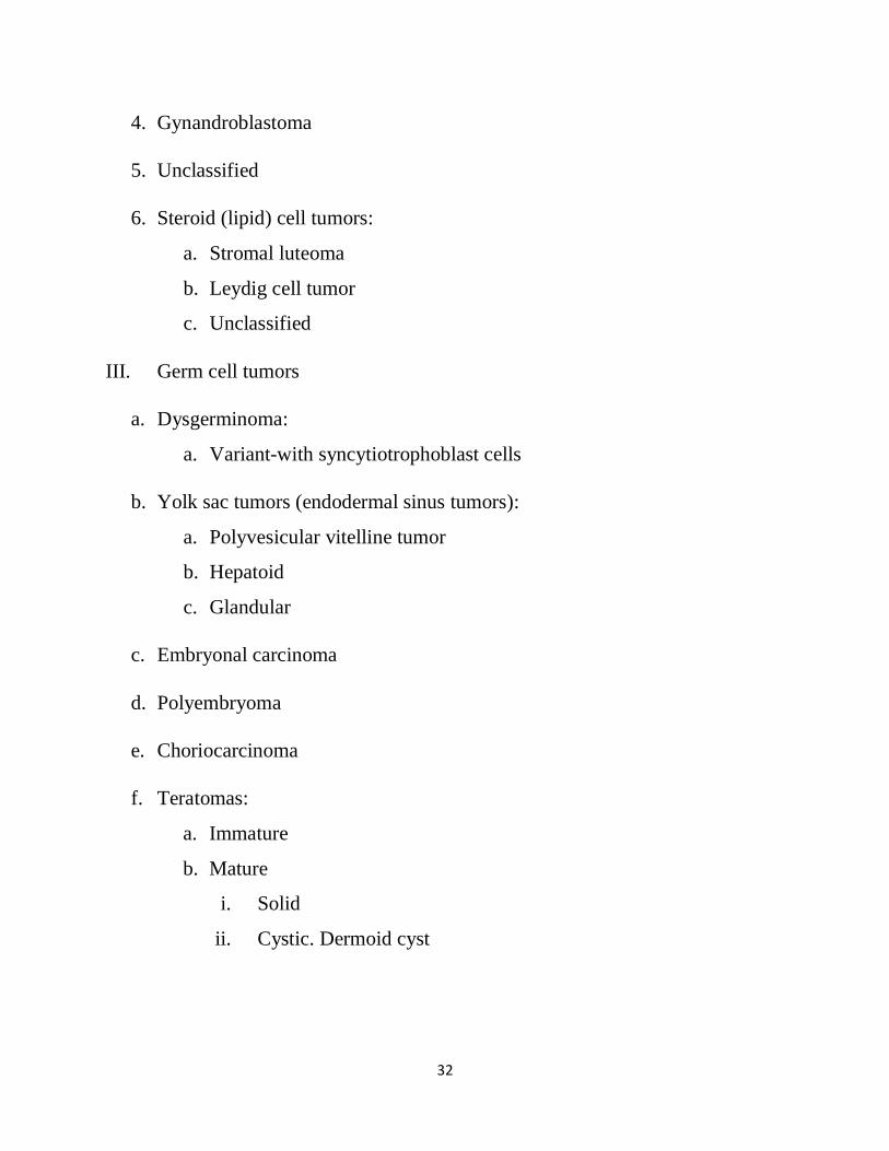
III. Germ cell tumors
a. Dysgerminoma:
a. Variant-with syncytiotrophoblast cells
b. Yolk sac tumors (endodermal sinus tumors):
a. Polyvesicular vitelline tumor
b. Hepatoid
c. Glandular
c. Embryonal carcinoma
d. Polyembryoma
e. Choriocarcinoma
f. Teratomas:
a. Immature
b. Mature
i. Solid
ii. Cystic. Dermoid cyst

33
c. Monodermal and highly specialized
i. Struma ovarii
ii. Carcinoid
iii. Struma ovarii and carcinoid
IV. Gonadoblastoma
a. Pure
b. Mixed with dysgerminoma or other form of germ cell tumour
V. Germ cell sex cord-stromal tumor of non gonadoblastoma type
VI. Tumors of rete ovarii
VII. Mesothelial tumors
VIII. Tumors of uncertain origin and miscellaneous tumors
a. Small cell carcinoma
b. Tumour of probable Wolifian origin
c. Hepatoid carcinoma
IX. Gestational trophoblastic diseases
X. Soft tissue tumors not specific to ovary
XI. Malignant lymphomas, leukemias, and plasmacytomas
XII. Unclassified tumors
XIII. Secondary (metastatic) tumors
XIV. Tumor like lesions

34
Surface Epithelial Tumors20,21
The surface epithelium of the ovary mimics the mesothelium, which forms
the inner epithelialliningof the abdomen and pelvis.
They are classified as
1. Benign - lack of invasive behaviour and exuberant cellular proliferation.
2. Borderline - no invasive behaviour but have exuberant cellular proliferation
3. Malignant- invasive behaviour
Approximately 60% of all tumors of ovary and nearly 90% of malignant
tumors of the ovary are Surface epithelial tumors. They usually occur in middle or
older women but very rarely can be seen in younger adults, evenbefore puberty.
Surface epithelial stromal tumorshas 5 subtypes:
a. Serous,
b. Mucinous,
c. Endometrioid,
d. Clear Cell and
e. Transitional Cell.

35
Malignant epithelial-stromal tumors those who lack specific differentiation are
grouped as Undifferentiated and those without any specific subtype are classified
as Adenocarcinomas Not Otherwise Specified (NOS).
1. SEROUS TUMORS20,22
They are formed by the invagination of the surface ovarian epithelium and
are lined by tall columnar ciliated epithelial cells resembling those of tubal
epithelium. Psammoma bodies characterize serous tumors. They account for nearly
30% of all tumors of the ovary.
a. Benign serous tumors:
They account for 40% of tumors of the ovary. It is bilateral 40% of cases.
Occurs in 4th and 5
th decade of life (Fig -3).
Gross: They may be cystic, papillary or adenofibromatous on macroscopic
appearance. Size may vary from 20 – 30 cms. Typically presents with smooth
walled cyst wall with small papillary projections.Papillary serous cystadenoma
occurs in combination with a cyst and the papillae on the inner wall or on the outer
surface (exophytic). Often associated with ascites and implantation of tumor
fragments on the peritoneal surfaces giving a false impression of malignancy.
Microscopy:Columnar epithelium with abundant cilia with microscopic papillae

36
b. Borderline serous tumors:
They account for 10 % – 15 % of seroustype of ovarian tumors. 30% of
tumors are bilateral, 40% of patients have small tumorlets in abdominal and pelvic
cavity.
Gross:They have an increased number of papillary projections.
Microscopy: Increased complexity of the papillae of the stromal tissue with
epithelial stratification and nuclear atypia.
c. Malignant serous tumors:
They account for 75% of epithelial tumors and 25 % of serous tumors are
malignant.60% of tumors are bilateral. Usually occurs in 6th decade (Fig -4).
Gross: They have multiple cystic loculations and solid areas. The tumor masses
are irregular with solid or papillary areas and capsular nodularity.
Microscopy: Even more complex growth with infiltration of the underlying stroma
by solid tumors. Individual tumor cells have extreme degrees of atypia.
Histologically, they may be papillary, adenopapillary or of diffuse pattern. Well
differentiated carcinoma form glands & have papillary structures. Psammoma
bodies are common. Poorly differentiated carcinoma has tumor cells with bizarre
37
large nuclei called giant cells, the papillae are more complex with a high degree of
cellular budding.
2. MUCINOUS TUMORS20,22
:
Mucinous tumors are made of cells which mimics those of the intestinal
epithelium orendocervical epithelium. They account for 20 % – 25 % of all ovarian
tumors.
a. Benign
They account for 20 – 25 % of all ovarian tumors. 10% of them are bilateral.
Usually occurs in 3rd
and 5th decade (Fig -5).
Gross:They are smooth walled, lobulated with whitish or bluish white hue. Cut
section shows thick, viscid mucin. The cyst is frequently multiloculated with
papillary growth arising from septum.
Microscopy:They are lined by a single layer of tall columnar epithelium with dark
staining basal nucleus but without any cilia. The epithelial characteristics are like
those of endocervix.
b. Borderline:
They account for 10 %- 15% of all mucinous type of ovarian tumors. These
tumors usually occur in 4th and 6
th decade of life. Bilaterality is seen in 40% of

38
endocervical type of borderline tumors where else only 10% of intestinal types are
bilateral.
They are similar to benign tumors but may have cystic chambers with
papillary areas and few solid areas. Intestinal variety of tumors may rarely be seen
in association with pseudo myxoma peritonei.
c. Malignant:
They account for 5 – 10 % of all ovarian tumors. Bilaterality is seen in 8 –
10% of tumors. Usually occurs in 6th decade of life.
Gross: They have larger cystic spaces and solid areas with more papillary
projections with large areas of hemorrhage and necrosis.
Microscopy:Tumors of well differentiated type are similar to mucin secreting
adenocarcinoma of intestinal origin.
3. ENDOMETRIOID TUMOR:20,21
They constitute 6 – 8 % of all epithelial tumors. They are formed by cells
which are similar to those of the endometrium. They are associated with
endometrial hyperplasia, endometriosis or endometrial carcinoma (Fig -7).

39
a. Benign:
Benign tumors are unusual and they account for only 2% of epithelial
tumors. These tumors are mostly unilateral in origin.
Gross: They are large usually measuring 10 – 25 cms. Outer surface is smooth
with firm fibrous cut surfaces with variably sized cystic spaces.
Microscopy:These tumors are formed of cudoidal or columnar epithelial cells
arranged in a glandular or acinar pattern. They are adenofibromas or
cystadenofibromas. The tumor stroma resembles that of ovary rather than that of
endometrium.
b. Proliferating:
They exhibit one of following features: multilocular cystadenofibromas or
cystic tumours with solid friable nodules.
Microscopy: They are low grade proliferating tumours has glands and cystic
spaces embedded in stroma. High grade proliferating tumour shows less stroma,
more intra cystic course, papillary and glandular proliferation.
c. Malignant :
Endometrioid adenocarcinoma: Accounts for 15-20% of epithelial ovarian
cancer and the second most common histological type. Occurs in 6th decade of life.

40
13-28% of these tumors have a bilateral origin.
Gross: These tumors have prominent solid areas with cystic spaces. The cystic
fluid is bloody or brown.
Microscopy: Well differentiated carcinoma have glandular growth pattern with
irregular budding and have good prognosis. Moderately differentiated carcinoma
show more complex glandular / micro glandular pattern.Poorly differentiated
carcinoma have round, punched out, broader longer papilla with squamous and an
adenomatous type of growth pattern.
4. CLEAR CELL TUMORS:20,21
Clear cell tumors are uncommon, with most of tumors having malignant
potential. They account for 4-5% of all epithelial tumors of the ovary. They are
usually seen in the 5th
decade of life. Approximately 15 – 20% of tumors are
bilateral in origin.
The hall mark of clear cell tumors are large polyhedral cells with eosinophilic
granular cytoplasm or peg like ‗hob nail‘ cells with little cytoplasm and bulbous
nuclei that bulge into lumen of cystic spaces.
a. Benign:
Gross: These tumor areusually cystic with mural nodules. Cystic spaces contain

41
clear fluid. Average size is 10-12 cm.
Microscopy: Histologically, adenofibromas with tubular spaces lined by one or
two layers of hobnail cells, clear cells, or indifferent cuboidal cells. Nuclei are
regular in size and shape. Cells contain intracytoplasmic glycogen.
b. Malignant :
Gross: They are large (15-20 cm), thick walled unilocular cysts containing blood
stained fluid.
Microscopy: The common type is small to large sheets of polyhedral clear cells
separated by delicate fibrovascular septa. Second type is tubulo papillary type.
Mitosis are less frequent than in other epithelial carcinoma (2/10 HPF).
Eosinophilic hyalinised stroma and hobnail cells are the characteristic features.
5. TRANSITIONAL CELL TUMORS ( BRENNERS): 20,21
Transitional cell tumors are made up of cells that mimicsthe bladder epithelium.
They are formed from ovarian surface epithelium that transforms into urothelium
like cells.
They accounts for 2% of all primary ovarian neoplasms and are mostly unilateral.

42
a. Benign
Most brenner tumors are benign. Occur in 5th
and 6th decade of life.
Gross:Most of them are very small, well circumscribed firm, solid bosselated
tumours with rubbery consistency and faintly lobulated grey white cut surface.
Microscopy: They have sharply demarcated epithelial cellnests set in fibrous
stroma (walthard nests), large cystic spaces and large blunt papillae. The epithelial
cells are round with clear margins and eosinophilic to clear cytoplasm. The oval
nuclei have obvious nucleoli and longitudinal grooving (Coffee bean appearance).
Microspaces are characteristic feature of transitional cell carcinomas.
b. Borderline :
Gross: Cut surfaces have variable multicystic component with watery fluid into
which friable papillary or polypoid nodules project.
Microscopy: Coarse papillary projections and cysts are lined by multilayered
atypical epithelium cells are similar to benign tumours.
c. Malignant :
Gross: They are partly cystic and contain watery or mucinous fluid and have
shaggy lining with friable masses projecting from the wall.

43
Microscopy: These show hetrogenous solid carcinomatous proliferation. Cysts are
lined by multilayered epithelium with hyperchromatic and pleomorphic
nucleoli.(Fig.-8)
6. MIXED EPITHELIAL TUMOURS:
TheWorld Health Organization follows the 10% rule – means that the minor
component must form at least 10% of the tumor mass to call it as a mixed tumour.
Common combinations include clear cell carcinoma and endometrioid
carcinoma as both these tumours usually originate rise endometriosis.
7. UNDIFFERENTIATED CARCINOMA:
They accounts for 5% of all cancers of the ovary and only 1% of surface
epithelial tumors. These tumors areformed by cells with high grade malignant
potentials, with high nuclear atypia and loss of cytoplasmic differentiation. Two
main types are recognized - the large cell and small cell types. About 50% are
bilateral.
II. SEX CORD TUMOURS OF THE OVARY 20,23
Sex cord-stromal tumors make up 5% of all neoplasms of the ovary. These
tumors arise from theca cells, granulose cells, leydig cells and the Sertoli cells.

44
Sexcord stromal tumors are usually seen in association with certain endocrine
manifestations.
A. GRANULOSA CELL TUMOURS
These tumors account for 2 % of all tumors of the ovary. They originate
from the rests of primitive granulosa cells unused in folliculogenesis. It is the
commonest ovarian stromal tumor. Rokitansky first described these tumors.
The hallmark of these tumours is the presence of cells that resemble those of
follicular granulosa or their luteinized variants.
Around 2% of these tumor are bilateral in origin. Inhibin, secreted by
granulosa cells to regulate FSH secretion is tumour marker.
There are two majorforms:
The adult type and
The juvenile type
a. Adult granulosa cell tumors:
Constitutes 95% of GCT, usually occurs in postmenopausal women. Clinical
presentations mainly depends on the hormone production, estrogen, they may be
associated with endometrial hyperplasia (50%). Unopposed estrogenic stimulation

45
leads to development of endometrial carcinoma in about 5- 10 % of cases. These
tumors are of low grade malignant potential. (Fig.-9)
b. Juvenile granulosa cell tumors:
These tumors constitute 5% of all ovarian GCT. These tumors are usually
unilateral, and almost 50% of themoccur before puberty. Due to their estrogen
production, most tumors result in precocious puberty.
Gross: The external surface is smooth or bosselated. Cut surface appears partly
cystic andpartly solid. Solid areas are soft or rubbery in consistency and cystic
spaces usually contain some proteinaceous fluid or altered blood.
Microscopy: They exhibitinsular or trabecular, macro follicular or micro follicular
patterns. Micro follicular is common - has multiple rounded small spaces made of
cystic degeneration of granulosa cells and they contain PAS positive material.
These spaces are known as call-exner bodies (30-50%).
B. THECOMA — FIBROMA GROUP OF TUMOURS
These tumors arise from the stromal tissue of the ovary. The thecoma,
fibroma and fibrothecoma constitute this group of tumors. These tumors account
for 4% of all tumors of the ovaryand they occur in both pre as well as in post
menopausal women (Fig -10).

46
Thecoma:
These tumor cells resemble the ovarian theca interna, mainly composed of
spindle cells with lipid elements contained within the cytoplasm. Thecomas are
usually seen in postmenopausal age group and are unilateral in origin. Thecomas
usually have estrogenic features, suchas postmenopausal bleeding, in some cases
may have hyperplasia of theendometrium, and even endometrial cancer. Most of
the thecomas are characteristically benign in nature.
Gross: Thecomas vary in size from a nodule to a large, firm rubbery solid tumour.
Cut section appears bright yellow to orange in colour. They are almost always
unilateral.
Microscopy: They consist of one or two patterns.
Typical thecomas consists of large ill-defined nodular masses of eosinophilic
or vacuolated cells with less fibrous connective tissue. Edema or myxoid
change is prominent.
Second pattern is ‗luteinized thecoma‘ — has the appearance of fibroma or
typical thecoma with lutein cells.

47
Fibroma:
Fibromas are the commonest neoplasm of the sex cord tumor group. Although
fibroma are said to arise from nonfunctioning stromal tissue with no estrogenic
activity, lipid-rich thecoma can have some estrogenic manifestations.
Fibromas usually occur in the middle aged women. Fibromas are mostly
benign and few with increase inmitotic activity are may have a malignant potential
called the fibrosarcomas.
On rare occasions these tumors may be bilateral and associated with basal
cell carcinoma of nevoid type called as the Gorlinsyndrome. They may be seen in
association with Meigs syndrome ( ascites, ovarian tumor and pleural effusion on
right side).
Gross: Fibromas are large tumours with slightly bosselated or smooth serosal
surface. They vary in consistency from rubbery to stony hard solid mass. The cut –
section appears white and whorled with few regions of cystic degeneration.5% of
cases are bilateral in origin.
Microscopy: They consist of variably cellular bundles and intersecting
collagenous fibrous tissue.

48
C. ANDROBLASTOMA (Sertoli — leydig cell tumours)
(Arrhenoblastoma) 35
They account for approximately only 1 % of all sex cord — stromal
tumours. They are characterized by presence of sertoli cells, leydig cells or
fibroblastic cells. Only <2% in them are bilateral in origin.
1. Sertoli cell tumours
Sertoli cell tumors are formed by proliferation of cells similar to rete ovarii.
They are a rare group of sex cord-stromal ovarian tumors constituting<0.5% of all
tumors of the ovary.They are usually seen in the 4th decade of life. Sertoli cell
tumors are non-function neoplasms, but may occasionally be able to induce sexual
developmentprecociouslyby hormone production or rarely may cause virilization
in girls.
Gross: Sertoli cell tumors are usually <10cm they are solid, firm, encapsulated
typically, yellow or brown, lobulated masses.
Microscopy: They are formed by highly differentiated uniform tubules lined by
single layer of cells with basal nuclei and clear cytoplasm. The epithelial cells
contain lipid as fine droplets.

49
d. Leydig cell Tumours (Hilus cell tumour)
Gross: These tumors are mostly unilateral and found in hilar region of ovary and
have soft or fleshy consistency.
Microscopy: They are formed of oval or polyhedral cells, which slender rod like
bodies with square or tapering ends known as Reinke crystals are present.
e. Sertoli — leydig cell tumours
Sertoli-Leydig cell tumors are made up of cells that mimic stromal and
epithelial cells of the testis. They are rare sex cord-stromal ovarian tumors make
up<0.5% of all ovarian neoplasms. They usually presents in the 3rd
decade of life.
These tumors usually cause virilization in about 30% of patients, but they can also
rarely produce estrogenic manifestations in few patients.
They are further classified into 5 subtypes:PoorlyDifferentiated,
Intermediate Differentiation, Well Differentiated, Mixed and Retiform Type.
Gross: These tumors are usually solid withcompletely or partial cystic areas. The
tumors have a yellowish tinge appearance with vesicular or polypoid structures in
their inner surface. Most ofthe tumors are unilateral in origin.
Microscopy: Well differentiated tumours have tubular structures lined by cells of
sertoli with variable numbers of mature cells of leydig between tubules.

50
Intermediate differentiation tumours contain cells of sertoli cells, arranged
incords, trabeculae or tubules with abundant mesenchymal stroma.
Poorly differentiated tumours are consists sheets of closely packed spindle
shaped cells.
D. GYNANDROBLASTOMA:
These tumours show intermingling of well differentiated ovarian and
testicular elements. They are usually unilateral in origin.
Gross: Gynandroblastoma are small and solid pole tumours. Cut section shows
pink to yellow fleshy nodules.
Microscopy: The ovarian elements resemble mature granulosa cells with call —
exner bodies and the testicular element resemble the tubules lined by typical leydig
and/ or sertoli cells with Reinke crystals.
E. STEROID (LIPID) CELL TUMOUR
Lipid cell tumors are classified as
Hilus cell tumor
Stromal luteomas

51
Steroid cell tumors not otherwise specified - NOS
Gross : These appear as a soft, yellowish or yellowish - brown nodules.
Microscopy : The cells are polyhedral or rounded, large mimicking the lutein
cells, adrenocortical cells and the leydig cells.
Stromal luteomas:
These are situated in the stroma of ovarian tissue and lack Reinke
crystalloids. These tumors are androgenic rarely.
Hilus cell tumours:
These tumors are usually andrognic and presence of Reinke crystalloids is a
characteristic feature.
Both Hilus cell tumor and Stromal luteomsa occur usually in
postmenopausal age group. These tumors are usually benign.
Steroid cell tumours not otherwise specified:
They are seen in younger age group. Approximately 25 – 40% of these
tumors are malignant. They typically produce androgen.

52
III. GERM CELL TUMOURS20,24
Germ cell tumours are the second most common of ovarian tumors only after the
surface epithelial tumours of ovary.
GCT arises from totipotent germ cells which are capable of both extra
embryonic and embryonic differentiation.
A.DYSGERMINOMA
These are the most common malignant germ cell tumour of the ovary. Peak
during 2nd
and 3rd
decades. Bilateral in 10% of cases. Serum lactate dehydrogenate
useful in monitoring individual for disease recurrence.
Gross: Pure dysgerminoma are solid, rapidly growing tumour and with glistening
smooth capsule. They may be oval, round or lobulated in appearance. On cut
section, solid and varying from firm soft rubbery and from grey pink to yellowish
tan colour.
Microscopy:They containaggregates or strands of uniform large cells with
lymphocytes, plasma cells, eosinophils, and show a granulomatous reaction with
foreign body giant cells and Langhan‘scells.

53
B. YOLK SAC TUMOURS
Yolk sac tumours in its pure form is the most frequent malignant germ cell
tumour only next to dysgerminoma. The average age ranges from 16-19 years.
Majority are unilateral.Alpha-fetoprotein is commonly produced by these tumours.
Gross: They resemble dysgerminoma with red and yellow areas of necrosis and
hemorrhage.
Microscopy: Microscopic features are highly variable. Glomeruloid Schiller —
Duval bodies are typical. The presence of diastase-resistant hyaline globules and
PAS positive is a characteristic feature of these tumors
C. EMBRYONAL CARCINOMA
Embryonal carcinoma is a raretumour. 60% of the tumours present with
hormonal manifestations.It typically produce HCG and 75% also secrete AFP.
Gross: They closely resembles those of yolk sac tumours. Most of the tumours are
unilateral.
Microscopy: They resembles testicular embryonal carcinoma.
D.POLYEMBRYOMA
It is rare organoid pattern of embryonal carcinoma and consists of numerous

54
embryoid bodies, closely resembling early, embryo. Serum AFP or HCG levels or
both may be elevated.
E.CHORIOCARCINOMA
Non gestational choriocarcinoma are extremely rare malignant tumours.
Choriocarcinomas secrete beta HCG, which are very useful tumour marker.
Gross: Choriocarcinoma are large, rapidly growing tumours and nearly always
hemorrhagic.
Microscopy: These tumour are composed of solid aggregates containing a
centrally located cytotrophoblast and a peripherally located syncytiotrophoblast.
F.TERATOMA
Benign cystic teratoma:
Ususally occurs in the age group of 20 years to 40 years. In adults they
account for 20% of all ovarian tumors and in children they account for 50% of all
ovarian tumors.
Usually unilocular with smooth surface. They contain hair and sebaceous
material and the wall is usually lined by skin (squamous epithelium). Other

55
structures commonly seen are teeth, cartilage, bone bronchial mucous membrane
and thyroid tissue. Rarely intestinal mucous membrane, pancreas and liver tissue
can be seen in the walls of dermoid cyst(Fig – 11).
Immature teratoma (Malignant teratoma):
Among germ cell tumor immature teratoma are the third most common
tumour. Usually occurs in children and young adults with a mean age of 18 yrs.
These tumors are usually solid with a trabeculated appearance on cut section. The
solid component of the tumor consist of bone and cartilage where else the
sebaceous material and hair form the cystic component of the tumor (Fig -12).
Monodermalteratoma
Primitive neuroectodermal tumours: These primitive neuroectodermal tumours
(PENTs) are highly malignant neoplasms. Collections of small undifferentiated
cells (primitive neuroepithelium) exhibiting varing degree of nuclear
pleomorphism characterise the microscopic appearance.
Sebaceous tumours: It‘s a rare form of a monodermal teratoma. The
characteristics necrotic ―Cheesy‖ cyst contents seen in typical teratoma are present
but hair is absent.

56
Cystic struma ovarii: These lesions are seen in women at any age, the average age
is 46 years. It is a smooth external surface with small foci of fibrous adhesions. A
clear to green down fluid fills the lumen of the cysts.
G.MIXED FORM
The prognosis of these tumors depend on the presence of components of
endodermal sinus tumor, teratoma grade III or choriocarcinoma. Tumors
accounting for more than one third of these components have a bad prognosis.
Tumors with less than one third of these components or when seen in association
with teratomagarde I &II, embryonal carcinoma or dysgerminoma have a better
prognosis.
IV. GERM CELL -SEX CORD STROMAL TUMOURS
Gonadoblastoma:
These are rare tumour that arise from a dysgenetic gonad. These tumour
consist of both sex cord -stromal cells, that have sertoli or granulose cell and
immature germ cells.
V. METASTATIC TUMOURS
Metastatic ovarian tumors are very common, accounting for 10% of all
malignant ovarian tumours (Fig -13).

57
Krukenbergs tumour are secondary growths in the ovary. The common sites
of primary are - stomach (70%), large intestine (15%) and breast (5%)
Gross :
These tumours are usually bilateral with smooth surface and at times slightly
bossed. They are freely mobile, not adherent to the adjacent structures and do not
show any capsular infilteration. These tumors maintatin the shape of the ovary. Cut
section shows a waxy consistency with occasional areas of cystic changes due to
degeneration.
Microscopy:
Stroma appears cellular or myxomatous. With large signetring cells which
are ovoid with eccentric nuclei and granular cytoplasm.
Metastatic melanomas and renal cell carcinoma may be confused with
granulosa cell tumor and clear cell carcinoma. Leukemias and lymphomas may
involve the ovaries usually in their late stage.
VI. OVARIAN TUMOURS ASSOCIATED WITH PREGNANCY20,26,27
Benign ovarian tumors are common in pregnancy. The incidence depends on
whether the tumor was noted on ultrasound examination – 1 in 50 live births or on
pelvic examination – 1 in 80 live birth or those requiring laparotomy – 1 in 1000 to

58
1500 live births (Fig – 14).
The most common benign ovarian tumor that occurs during pregnancy is
benign cystic teratoma (30%) followed by cystadenomas (15%). Malignant ovarian
tumors account for only 1 to 2% of all ovarian tumors that complicate pregnancy.
The most common malignant ovarian tumor complicating pregnancy is
dysgerminoma (60%) followed by epithelial tumors and sex cord stromal tumors.
Whether benign or malignant, most of the ovarian tumors complicating
pregnancy are unilateral in origin.
The optimal time for surgical intervention is 16 weeks to 18 weeks.
Conservative surgical management for benign tumors has an excellent outcome.
The overall 5 year survival rate for malignant ovarian tumors complicating
pregnancy depends on the cell type, stage of the disease and on the trimester in
which they were diagnosed.

59
CLINICAL MANIFESTATIONS
Difference between benign and malignant ovarian tumours
Benign Malignant
1. Age Reproductive age Extremes of age group (Premenarchal,
and post-menopausal)
2. Rapidity of
growth
Slow Rapid
3. Pain and
tenderness
Absent unless
complicated
Present
4. Surface Smooth Irregular
5. Consistency Usually cystic Solid nodular and irregular
6. Number Unilateral
15% bilateral
75% bilateral
7. Fixation Mobile Fixty present
8. Ascites Usually absent (Except
in fibroma)
Present
9. Oedema of
feet
Bilateral Unilateral with vulvaloedema
10. Metastasis Absent Present
11. Capsule Intact Ruptured
12. Appearance Uniform Variegated
13. Blood
vessels
No engorged vessels on
the surface
Large blood vessels present on the
surface
14. Features Unilocular cyst, few
papillary projection, no
solid areas
Multilocular, highly papillated solid
areas

60
MATERALS AND METHODS
This is a prospective study conducted in Department of Obstetrics and
Gynaecology, in Govt. Rajaji Hospital, Madurai from September 2011 to August
2012.
The study group includes patients who were admitted at Government Rajaji
Hospital with an ovarian tumor confirmed by transadbominal ultrasound
examination. Totally 136 patients were evaluated during this study period. A
standard proforma was used for collection of data.
Transabdominal sonography was performed on all patients using a 3.5 –
5MHztransducer. The ovary was measured in all its three dimensions, and the
volume of the ovary was calculated with the use of the ellipsoid formula (length x
width x height x 0.523). Cystic ovarian tumor with a papillary projections and
solid areas, echogenicity, presence of septum and the presence or absence of free
fluid in the extra tumoral space were noted. Morphology indexing was performed
according to Ueland and collegues in 2001.
Two different descriptive category were evaluated ( Fig15) :
1. Volume of tumor and
2. Morphologic features.

61
A score from 0-5 was assigned for each of the component. A total score ranging
from 0 and 10 for every tumor.
Observations of tumor septa, diffuse echogenicity and extra tumoral free
fluid were includedwithin the category of morphological features.
Following morphology indexing,eachtumor was surgically removed and
werehistologicallyclassified according to the WHO System of classifying ovarian
tumors.
Statistical Tools
The information collected regarding all the selected cases were recorded in a
Master Chart. Data analysis was done with the help of computer using
Epidemiological Information Package (EPI 2002).
Using this software, range, frequencies, percentages, means and standard
deviations were calculated.
Sensitivity, specificity, accuracy, positive predictive value and negative
predictive values were calculated using the following formulae.

62
Sensitivity = True positivex 100
True positive + False negative
Specificity = True negativex 100
False positive + True negative
Positive predictive value = True positive x 100
True positive + False positive
Negative predictive value = True negativex 100
True negative + False negative
Accuracy = True positive + True negative x 100
N

63
RESULTS
A total of 136 ovarian tumors were studied. Table 1 illustrates the incidence
benign, borderline and malignant tumors based on histopathological examination.
Of the 136 ovarian tumors 92 cases were benign (67.6%), 3 were borderline (2.2%)
and 41 were malignant (30.2%).
Table : 1 Incidence of benign , borderline and malignant tumors as per HPE
Type of tumor
Cases
No. %
Benign 92 67.6
Borderline 3 2.2
Malignant 41 30.2
Total 136 100
The mean age was 40.5 years (range, 13–85 years).60 patients belong to the
age group of 40 years or older and 76patients were less than 40 years of age. The
age distribution of the patients are Illustrated in Table 2

64
Table 2 : Age distribution
Age group
(in years)
Number of cases
Total cases Benign Borderline Malignant
No. % No. % No. % No. %
11 – 20 6 6.6 - - 3 7.1 9 6.6
21 – 30 32 35.2 - - 3 7.1 35 25.7
31 – 40 24 26.4 - - 8 19 32 23.5
41 – 50 14 15.4 1 33.3 8 19 23 16.9
51 – 60 12 13 2 66.7 14 34.1 28 20.6
Above 60 4 4.4 - - 5 11.9 9 6.6
Total 92 100 3 100 41 100 136 100
Range 16 – 75 45 – 52 13 – 85 13 – 85
Mean 36.7 49.7 48.1 40.5
S.D. 13.1 4.0 15.1 14.6
‗p‘ 0.0001
Significant

65
Table 3 : Parity distribution
Table 3 illustrates the parity distribution of ovarian tumours. Ovarian tumors
were common in multipara of 2 which is considered as statistically significant.
Benign tumors were common in 2nd
parity women and malignant tumors were
common in women with parity 4 and above constituting 33% and 35.7%
respectively. Incidence of benign tumours in nulliparous women was 22% and
malignant tumours were 14.3%. There were 5 cases (5.4%) of ovarian tumor
complicating pregnancy.
Parity
Number of cases
Total cases Benign Borderline Malignant
No. % No. % No. % No. %
Pregnant 5 5.4 - - - - 5 5.4
Nulli 20 22 - - 6 14.3 26 19.1
1 8 8.6 - - 5 12.1 13 9.5
2 28 30.7 - - 7 16.7 35 25.7
3 12 13.2 3 100 8 19 23 16.9
4 & above 19 20.8 - - 15 35.7 34 25
Total 92 100 3 100 41 100 136 100

66
Table 4 : Mode of Presentation
* There was more than one mode of presentation in many cases.
Table 4 illustrates the mode of presentation of ovarian tumors. The most
common presenting features in both benign and malignant ovarian tumours were
pain abdomen with an incidence of 84.7% and 82.9% respectively.
Mode of
Presentation
Number of cases
Total cases Benign Borderline Malignant
No. % No. % No. % No. %
Mass Abdomen 23 25.3 1 33.3 13 31 37 27.2
Pain 78 84.7 1 33.3 34 82.9 113 83.1
Menstrual
Disturbances 3 3.3 2 66.7 5 11.9 10 7.4
Post-Menopausal
Bleeding 1 1.1 - - - - 1 0.7
Loss of
Weight/Appetite 5 5.5 - - 4 9.5 9 6.6
Urinary Symptoms 1 1.1 - - - - 1 0.7
White discharge 1 1.1 - - - - 1 0.7
Vomiting 4 4.4 - - - - 4 2.9
Asymptomatic 1 1.1 - - - - 1 0.7
Total 92* 100 3* 100 41* 100 136* 100

67
Table 5 : Per abdomen consistency of Ovarian Tumor
Consistency
Number of cases
Total cases Benign Borderline Malignant
No. % No. % No. % No. %
Cystic 85 93.4 3 100 8 19 96 70.6
Firm - - - - 5 11.9 5 3.7
Hard - - - - 17 40.5 17 12.5
Variable - - - - 10 23.8 10 7.4
Not palpable 7 7.7 - - 1 2.4 8 5.8
Total 92 100 3 100 41 100 136 100
Table5 illustrates the consistency of ovarian tumor on clinical examination.
Most benign tumors were cystic in consistency (93.4%) and malignant tumors
were hard in consistency (40.5%) and around 23.8% of malignant tumors have a
variable consistency.

68
Table 6 : Laterality
Laterality
Number of cases
Total cases Benign Borderline Malignant
No. % No. % No. % No. %
Unilateral 86 92.3 2 66.7 22 53.6 110 80.9
Bilateral 6 6.6 1 33.3 19 45.2 26 19.1
Total 92 100 3 100 41 100 136 100
Table 6 illustrates the unilateral or bilateral involvement of ovaries in benign
and malignant tumors. Most ovarian tumors were unilateral (80.9%). 92.3% of
benign tumors were unilateral and 53.6% of malignant tumors were unilateral.

69
Table 7 illustrates the score for tumors based on the tumor volume by ultrasound.
Score 0 1 2 3 4 5
Tumor
volume
(cm3)
< 10 10-50 50-100 100-200 200-500 >500
Table 7: USG Volume Score
USG Volume
Score
Number of cases
Total cases Benign Borderline Malignant
No. % No. % No. % No. %
0 1 100 - - - - 1 0.7
1 8 100 - - - - 8 5.9
2 8 66.7 2 16.7 2 16.7 12 8.8
3 23 85.2 - - 4 14.8 27 19.9
4 27 87.1 - - 4 12.9 31 22.8
5 25 43.9 1 1.8 31 54.4 57 41.9
Total 92 100 3 100 41 100 136 100
Range
Mean
S.D.
1-5
3.54
1.23
2-5
3.0
1.73
0-5
4.45
1.11
0-5
3.84
1.27
‗p‘ 0.0001 (Significant)
Most benign tumors have a score of < 4 and most of the malignant tumors are more
have a score of>4

70
Table8: illustrates the score for tumors based on the tumor structures by
ultrasound.
Score 0 1 2 3 4 5
Structure
Smooth
wall,
sonolucent
Smooth
wall, diffuse
echogenicity
Wall
thickening,
<3mm fine
septa
Papillary
projections,
septa>
3mm
Complex,
predominantly
solid
Complex,
solid& cystic
areas,
extratumoral
fluid
Table 8 : Structural Score
Structural
Score
Number of cases
Total cases Benign Borderline Malignant
No. % No. % No. % No. %
0 56 96.6 - - 2 3.4 60 44.1
1 19 90.5 - - 2 9.5 21 15.4
2 6 75 - - 2 25 8 5.9
3 5 62.5 - - 3 37.5 8 5.9
4 2 13.3 1 6.7 12 80 15 11
5 2 8.3 2 8.3 20 83.3 24 17.6
Total 92 100 3 100 41 100 136 100
Range
Mean
S.D.
0-5
0.7
1.18
4-5
4.67
0.58
0-5
3.88
1.53
0-5
1.77
2.0
‗p‘ 0.0001 (Significant)
Most benign tumors have a structural score <2 and most malignant tumors have a
structural score > 3

71
Table 9: Total Morphological Score
Total
Morphological
Score
Number of cases
Total cases Benign Borderline Malignant
No. % No. % No. % No. %
0 1 100 - - - - 1 0.7
1 6 100 - - - - 6 4.4
2 7 100 - - - - 7 5.1
3 19 100 - - - - 19 14
4 19 90.5 - - 2 9.5 21 15.4
5 20 95.2 - - 1 4.8 21 15.4
6 12 80 - - 3 20 15 11
7 3 42.9 2 28.6 2 28.6 7 5.1
8 1 12.5 - - 7 87.5 8 5.9
9 3 20 1 6.7 11 73.3 15 11
10 1 6.3 - - 15 93.8 16 11.8
Total 92 100 3 100 41 100 136 100
Range
Mean
S.D.
1- 10
4.25
1.9
7 – 9
7.9
1.2
0 – 10
8.5
2.1
0 – 10
5.6
2.7
‗p‘ 0.0001
Significant
Table 9 illustrates the total of volume and structural score, consolidated as the
morphological score.
Most of the benign tumors have a score of < 5 and score of > 5 suggests
malignancy.

72
Table 10: Results as per USG total morphological score and HPE
USG total
morphological
score
Number
of cases
Number of cases
Benign Borderline Malignant
No. % No. % No. %
0 – 4
(Benign)
54 52 96.2 - - 2 3.73
5 – 10
(Malignant)
82 40 48.8 3 3.7 39 47.6
Table 11 : Efficacy of Morphological score with HPE as Gold standard
Result as per
Morphological
score
Number
Of cases
Result as per HPE
Positive Negative
No. % No. %
Positive
(Score > 5)
82 42 51.2 40 48.8
Negative
(Score < 5)
54 2 3.73 52 96.2

73
True Positive = 42
False Positive = 40
True Negative = 51
False negative = 3
Sensitivity = 95.5
Specificity = 56.5
Positive predictive value = 51.2
Negative predictive value = 96.3
Accuracy = 69.1

74
Table 12 : Morphological Score and Malignancy
USG Morphological
Score
No. of cases
Positive cases (Malignant + Borderline)
As per HPE result
No. %
0 1 - -
1 6 - -
2 7 - -
3 19 - -
4 21 2 9.5
Less than
5(Negative)
54 2 3.7
5 21 1 4.8
6 15 3 20
7 7 4 57.1
8 8 7 87.5
9 15 12 80
10 16 15 93.8
5 & above (Positive) 82 42 51.2
Total 136 44 32.3

75
Table 13: Type of Tumor
Type of Tumor Cases
No. %
1) Epithelial tumors
a. Serous tumors
74 54.5
- Benign Serous cystadenoma
- Benign papillary serous Cystadenofibroma
- Borderline serous papillary cystadenoma
- Serous cystadenocarcinoma
- Papillary Serous cystadenocarcinoma
53
1
2
3
15
39
0.7
1.5
2.2
11.1
b. Mucinous tumor
31 22.7
- Benign Mucinous cystadenoma
- Borderline mucinous cystadenoma
- Mucinous cystadenocarcinoma
- Papillary mucinous cystadenocarcinoma
21
1
8
1
15.4
0.7
5.9
0.7
c. Endometroid tumour
- Benign
2
1.5
d. Brenner tumour
- Malignant
1 0.7
e. Undifferentiated tumor
8 5.7
- Poorly differentiated papillary carcinoma
- Poorly differentiated carcinoma
- Adenocarcinoma
-
2
1
5
1.5
0.7
3.5

76
f. Mixed tumour
- Benign papillaryseromucinous cystadenoma
1 0.7
2. Germ cell tumors
a. Teratoma
11 8.1
1. Immature
2. Mature (Cystic dermoid)
1
10
0.7
7.4
3. Sex cord stromal tumor
6 4.5
a. Granulosa cell tumors malignant
b. Fibrothecoma
2
4
1.5
3.0
4 Metastatic Carcinoma
Krukenbergstumor
1 0.7
5. Soft tissue tumours not specific to the ovary
(leiomyosarcoma)
1 0.7
Total 136 100
Table 13 illustrates the incidence of various subtypes of ovarian tumors
In the present study, epithelial tumors (117) were most common ovarian
tumors followed by germ cell tumors (11).

77
DISCUSSION
Ovarian tumours manifest a wide spectrum of clinical, morphological and
histological features.Clinically they may be misdiagnosed for other non-neoplastic
conditions.
In this study we have analyzed 136 ovarian tumors over a period of one year
and correlated their clinical presentation and sonographic finding with the
histopathology.
Benign vs. Malignant tumors:
Of the 136 ovarian tumors, according to histologic diagnosis
91(66.9%)of ovariantumors were benign and 42 (30.9%) weremalignant, including
3(2.2%) borderlinetumor masses.
Table 15: Comparison of Incidence of Ovarian Tumors
Study Benign Borderline Malignant
Pilli G et al 75.2% 2.8% 20.74%
Gupta et al 59.4% 0.58% 40%
Couto F et al 80.7% 2.3% 16.9%
Present study 66.9% 2.2% 30.9%

78
The incidence of benign ovarian tumor (66.9 %) in this study is less than that
reported by Pilli G et al and Couto et29
al but more than that observed by Gupta at
al.
Age incidence:
Ovarian cancer may occur at any age. In our study the age incidence was
between 13 years and 85 years.
Table 16: comparison of Age distribution of benign tumours [%]
Study 11 -20 21 – 30 31 – 40 41 – 50 51 – 60 >61
Bhatiya et al (1986) 10 38.5 36.6 27.7 15.5 4.4
RamachandranG et al (1988) 9.8 30.7 22 20.4 10.1 4.2
Jagadheswari et al. (1991) 0 16 36 32.3 10.5 5.2
Present study (2012) 6.6 35.2 26.4 15.4 12.1 4.4
In the present study, the peak incidence of benign tumor was between the
age group of 21-30 years (35.2 %). Similar observations were made by Bhatiya et
al (1986) and Ramachandran G et al(1988) but this incidence is more compared to

79
that reported by Jagadheswari et al.(1991) in whose study the peak incidence of
benign tumors was in the age group of 41 – 50 years.
Borderline tumours were commonly seen in 45- 50 years with a mean age of
49.7 years, as shown in table 2
Table 17: comparisons of Age distribution of malignant tumours [%]
Study 11 -20 21 – 30 31 – 40 41 – 50 51 – 60 >61
Bhatiya et al (1986) 9.8 17.3 27.1 21.8 16.5 3
Ramachandran G et al(1988) 10.5 26.3 29.5 21 9.5 3.2
Jagadheswari et al.(1991) 13.9 16.4 19.9 27.8 17.1 2.5
Present study (2012) 7.1 7.1 19 19 35.7 11.9
In the present study, malignant tumours were commonly seen between the
age group 51-60 years with a mean age of 48.1 years. In studies reported by
Bhatiya et al and Ramachandran G et al the maximum incidence is seen in 31- 40
years of age, but in Jagadheswari et al the peal incidence is seen in 41-50 years.

80
Parity:
In the present study ovarian tumours were common in 2nd
parity. Of the
benign tumours 33% were in 2nd
parity.Of the malignant tumours35.7% were in 4th
parity and above. Similar observation was found in study done by Shahin Rashid et
al and Shah, Vaidya et al in 1990. In the present study we have reported 5 cases of
ovarian tumors during pregnancy and all were benign tumors.
Clinical manifestations:
The ovarian tumors manifest with wide variety of clinical manifestation.
According to Sharma et al, 93.16% of the cases presented with mass abdomen and
64.9% presented with abdominal pain. In the present study the commonest
presenting symptom was pain abdomen (83.1%) in both benign and malignant
tumors. 27.2% of patients presented with mass per abdomen. Similar observations
were found in the study done by Bhattacharya MM et al30
&Shahin Rashid et al31
.
Few tumors which produce hormones may cause menstrual disturbances. In
our study, 10 cases (7.4%) presented with menstrual disturbances and 1 case
(0.7%) with post-menopausal bleeding. According to Pilli et al in 2002, 6.7% of
cases presented with menstrual abnormalities which were similar to our study but
in contrary to the study conducted by Gupta et al in 1986, in which nearly 40.2%
of cases had menstrual disorders as the presenting complaint.

81
Consistency:
The ovarian tumors vary from cystic to solid in consistency. In the present
study 92.3 % of benign tumors were cystic, 40.5 % of malignant tumors were hard
and 23.8% of malignant tumors were variable in consistency.In the study
conducted by Pilli et al in 2002, of the benign tumors 76% were cystic in the
malignant group, 49.2% cases were hard, 44.1% were variable in consistency.
Laterality:
Ovarian tumors may be unilateral or bilateral; bilaterality represents the
multicentric origin of the tumor. 6.6% of benign tumors were bilateral and 45.2%
of malignant tumors were bilateral. According to Gupta et al 30.2 % of malignant
tumors were bilateral.
Histopathological types:
Surface epithelial tumours are common tumours comprising 85.8% of all
ovarian tumours. Among epithelial tumors, serous tumors (54.5%) were most
common followed by mucinous tumors (22.7%). 53(39%) cases are serous
cystadenomas. Histologically they are lined by low cuboidal to columnar
epithelium.1 cases of cystadenofibroma was present. It was cystic and lined by low
cuboidal epithelium.

82
Only 2 cases (1.5%) of borderline serous tumours were reported. One was
unilateral and the other was bilateral, both were cystic in consistency.
Histologically it has stratification of epithelial cells with mitotic activity and
stromal penetration.
19 malignant serous tumours were present. 10 were unilateral and 9 were
bilateral. On gross examination 6 were cystic, 8 were solid and 4 were variable in
consistency. Microscopically they are lined by more than one layer of columnar
epithelium with nuclear polymorphism and hyperchromatic nuclei.
21 mucinous cystadenoma were present, out of which 16 were multilocular
and 5 were unilocular. The content was mucoid. The epithelial lining of the cyst
wall is columnar with basophilic cytoplasm and basal nuclei. There was 1 case of
borderline mucinous tumour, 8 cases of mucinous cystadenocarcinoma and 1 case
of mucinous cystadenocarcinoma with papillary differentiation (Fig 16).
Germ cell tumours
These are second most common group of tumours. In germ cell tumors,
mature benign cystic teratomas (Dermoid cyst) (7.4%) were most common.

83
The common age group was 20-30years. 2 patients were in their 5th decade
of life. Grossly they were cystic. Majority of tumours have stratified squamous
epithelium and dermal appendages like hair, sebaceous glands etc.
Sex cord stromal tumours:
2 case of granulosa cell tumour presented with menstrual disturbances and
both were malignant and solid in consistency.Histologically the cells appear
polygonal to round with hyperchromatic nuclei with central groove and a very
scanty cytoplasm.
4 cases of fibrothecoma presented with mass abdomen and menstrual
irregularities.Grossly it was variable in consistency and unilateral.Histologically it
consists of lipid rich cells resembling theca cells.
Mixed tumours:
Only 1 case of benign seromucinous cystadenoma with papillary
differentiation was reported.
Metastatic tumor:
There was one case of krukenbergs tumor; the patient was operated for
carcinoma stomach 4 years back following which she presented with mass
abdomen.

84
Soft tissue tumours not specific to the ovary:
1 case of leiomyosarcoma has been reported in a 40 year old P3L3 presented
with a mass abdomen. On examination, it was a irregular mass of size 20x20 cm
variable consistency and restricted mobility with no ascites, per operatively Left
ovary replaced by a fleshy vascular tumour of size 20x18 cm multiloculated with
variable consistency. On histopathology the Cells were spindle shaped cells with
elongated nuclei and eosinophilic cytoplasam of interlacing bundles with whorled
appearance. Pleomorphism, hyperchromatic nuclei with of mitoses 14/10 hp were
seen( Fig 17).
Ultrasound examination:
With the demand for Evidence Based Medicine it has become a challenging
task to develop a morphological index to predict the malignancy of ovarian mass
without any surgical intervention. An ideal scoring system must be accurate, easy
to interpret, time saving, less interobserver bias and cost effective. The use of such
a scoring system must help in triaging the women with malignancy to an
oncologists and avoid unwanted surgical intervention in women with benign
tumors.

85
Several investigators have put forth many scoring system to compare the
morphological features of the tumors. The present Morphological Indexing is a
modification ofthescoring system proposed by DePriest and colleagues32
.
Morphological scoring system:
In the present study, one hundred and thirty six patients with ovarian tumor
were evaluated sonographically and were operated during the study period from
September 2011 and august 2012. The morphological characters were evaluated in
two different descriptive characters:
1. Volume of the tumor volume and
2. Morphological features.(Fig.18a-g)
A score from 0-5 was assigned for each of the component.A total score
ranging from 0 and 10 for every tumor.
According to the present study the benign tumors have a mean volume score
of 3.58 which is statistically significant (table 6). This mean deviation to the higher
value is mainly contributed by the benign mucinous cystadenoma which were
larger in size with a volume of > 500. The malignant ovarian tumors have a mean
volume score of 4.45 which is statistically significant. Of the 42 malignant tumors
31 were of volume > 5, only 2 tumors had a score 2 which were both serous
cystadenocarcinoma.

86
In the present study, benign tumours had a mean structural score of 0.7 and
malignant tumors had a mean structural score of 3.88 (table 7). Of the benign
tumors most of them had a smooth wall with either sonolucent or diffuse
echogenicity accounting for a score of 0 or 1. Of the malignant tumors most of
them had a complex, solid and cystic areas with extramural fluid accounting for a
score of 5.
The total morphological score was calculated by the sum of volume and
structural score and tabulated in table 8. The benign tumors had a mean score of
4.25 and malignant tumors had a mean of 8.5 which was statistically significant
with a p value = 0.0001). The malignancy risk is directly related to the volume of
the tumor and the various structural components each with a p value = 0.0001.
There were only 2 cases of malignancy (3.7%) reported with a tumor
morphological score of< 5 of the 54 ovarian tumors with a morphological score <5
studied , compared to only 0.3% as stated in Ueland et al in 2003. In contrast there
were 42 cases (51.2%) of malignancy with a tumor morphological Score > 5 of the
82 tumors studied, compared to only 41% as stated by Ueland et al in 2003.
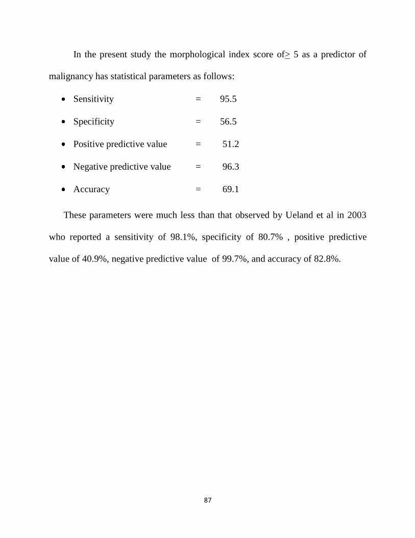
87
In the present study the morphological index score of> 5 as a predictor of
malignancy has statistical parameters as follows:
Sensitivity = 95.5
Specificity = 56.5
Positive predictive value = 51.2
Negative predictive value = 96.3
Accuracy = 69.1
These parameters were much less than that observed by Ueland et al in 2003
who reported a sensitivity of 98.1%, specificity of 80.7% , positive predictive
value of 40.9%, negative predictive value of 99.7%, and accuracy of 82.8%.

88
Table:18 Comparison of Sensitivity and Specificity with other Studies
Source, Year Sensitivity Specificity
Carter et al,1988 0.62 0.95
Granberg et al, 1991 0.82 0.92
Sassone et al, 1991 1.0 0.83
Lerner et al, 1994 0.97 0.77
DePriest et al, 1994 0.89 0.70
Yamashita et al, 1997 0.85 0.78
Ferrazzi et al,1997 0.75 0.67
Mol et al, 2001 0.77 – 0.93 0.21 – 0.89
Present study 0.93 0.56
Table 18 illustrates the sensitivity and specificity of various studies. These
individual studies to observer bias. Mol et al10
in 2001 did an external validation of
these sonological indices and revealed a lower accuracy with a high sensitivity
ranging from 77 - 93% only at the expense of a wide range in specificity ranging
from 21 - 89%. In the present study the sensitivity is 93% and specificity is 56%

89
SUMMARY
A total of 136 ovarian tumors were analysed and of these 136 ovarian
tumors91 cases were benign (66.9%), 3 were borderline (2.2%) and 42 were
malignant (30.9%) based on histopathology.
The mean age was 40.5 years (range, 13–85 years).60 patients belong to the
age group of 40 years or older and 76 patients were less than 40 years of age.
Ovarian tumors were common in multipara of 2 which is considered as
statistically significant. Benign tumors were common 2nd
parity and malignant
tumors were common in 4th parity and above constituting 33% and 35.7%
respectively.
Incidence of benign tumours in nulliparous women was 22% and malignant
tumours were 14.3%. There were 5 cases (5.4%) of ovarian tumor complicating
pregnancy.
The most common presenting features in both benign and malignant ovarian
tumours were pain abdomen with an incidence of 84.6% and 83.3% respectively.
Most benign tumors were cystic in consistency (93.4%) and malignant tumors
were hard in consistency (40.5%).Around 23.8% of malignant tumors have a
variable consistency.

90
Most ovarian tumors were unilateral (80.9%). 92.3% of benign tumors were
unilateral and 53.6% of malignant tumors were unilateral.
In the present study, 117 cases (85.8%) were epithelial tumors and were the
most common ovarian tumors followed by germ cell tumors, being reported in
only11cases (8.1%).
The morphological scoring system as assigned by Ueland et al was used to
calculate the morphological index.
A particular score is assigned to each depending on the tumor volume. Most
benign tumors have a score of <4 and most of the malignant tumors have a score
>4. A particular score is assigned to each depending on the tumor structure. Most
benign tumors have a structural score of <2 and most malignant tumors have a
structural score of > 3.The morphological index for Most of the benign tumors
was< 5 and score of > 5 suggests malignancy.

91
The present study has the following statistical parameters:
True Positive = 42
False Positive = 40
True Negative = 51
False negative = 3
Sensitivity = 95.5
Specificity = 56.5
Positive predictive value = 51.2
Negative predictive value = 96.3
Accuracy = 69.1

92
CONCLUSIONS
The findings of the present studyconclude the following
Morphological scoring system is helpful in identifying women with ovarian
tumors who are at risk for malignancy.
Morphological scoring system also effectively decreases the need for
unwanted surgery in benign ovarian tumor.
It is easy to perform.
This scoring system is subjected to interobserver variation.
Morphologic indexing helps to standardise the ultrasound reading without
adding costs.

93
Chart 1
Incidence of benign , borderline and malignant tumors as
per HPE
68%
2%
30%
.
BENIGN
BORDELINE

94
Chart 2 : Mean Age Distribution
Chart 3: Parity distribution
0
5
10
15
20
25
30
35
40
45
50
BENIGN BORDERLINE MALIGNANT TOTAL CASES
36.7
49.7 48.1
40.5
Mean
Ag
e (
in y
ears
)
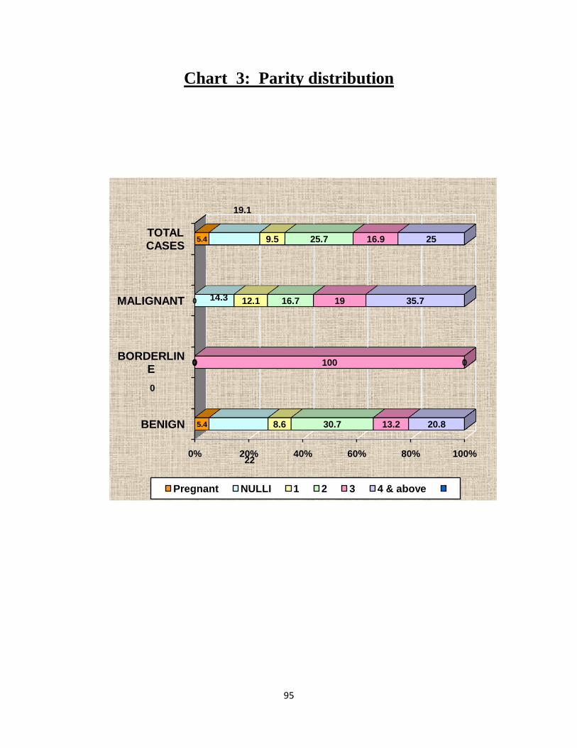
95
Chart 3: Parity distribution
0% 20% 40% 60% 80% 100%
BENIGN
BORDERLINE
MALIGNANT
TOTAL CASES
5.4
0
0
5.4
22
0
14.3
19.1
8.6
0
12.1
9.5
30.7
0
16.7
25.7
13.2
100
19
16.9
20.8
0
35.7
25
Pregnant NULLI 1 2 3 4 & above

96
Chart 4: Per abdomen c
onsistency of Ovarian Tumor
0%
10%
20%
30%
40%
50%
60%
70%
80%
90%
100%
BENIGN BORDERLINE MALIGNANT TOTAL CASES
93.4100
19
70.6
11.9
3.7
40.5
12.5
23.8
7.4
7.72.4 5.8
CYSTIC FIRM HARD VARIABLE NIL

97
Chart 5: Laterality
0%
10%
20%
30%
40%
50%
60%
70%
80%
90%
100%
BENIGN BORDERLINE MALIGNANT TOTAL CASES
92.3
66.7
53.6
80.9
6.6
33.3
45.2
19.1
UNILATERALITY BILATERALITY

98
Chart 6: USG Volume Score
0%
10%
20%
30%
40%
50%
60%
70%
80%
90%
100%
0 1 2 3 4 5
1 8
8 23 27
25
2
1
2 4 4
31
USG VOLUME SCORE
BENIGN BORDERLINE MALIGNANT

99
Chart 7: Structural Score
Chart 8 :Morphological score

100
Chart 8:
Morphological score

101
Chart 9:
Results as per USG total
morphological score and HPE
0% 20% 40% 60% 80% 100%
POSITIVE SCORE > 5
NEGATIVE SCORE <5
51.2
3.73
40
96.2
MALIGNANT

102
Chart 10:
Comparison of Sensitivity and
Specificity with other Studies
SENSITIVITY, 93
SPECIFICITY, 56
0 20 40 60 80 100 120
Carter et al
Granberg et al
Sassone et al
Lerner et al
De Priest et al
Yamashita et al
Ferrazzi et al
Present study
Axis Title

103
Fig 3.SEROUS CYST ADENOMA
Stromal papillae and columnar epithelium with abundant cilia

104
Fig 5.MUCINOUS CYSTADENOMA
Tall columnar cells with Cyst lined by columnar cells
Apical mucin and absence of cilia with bland basal nuclei and apical
mucin
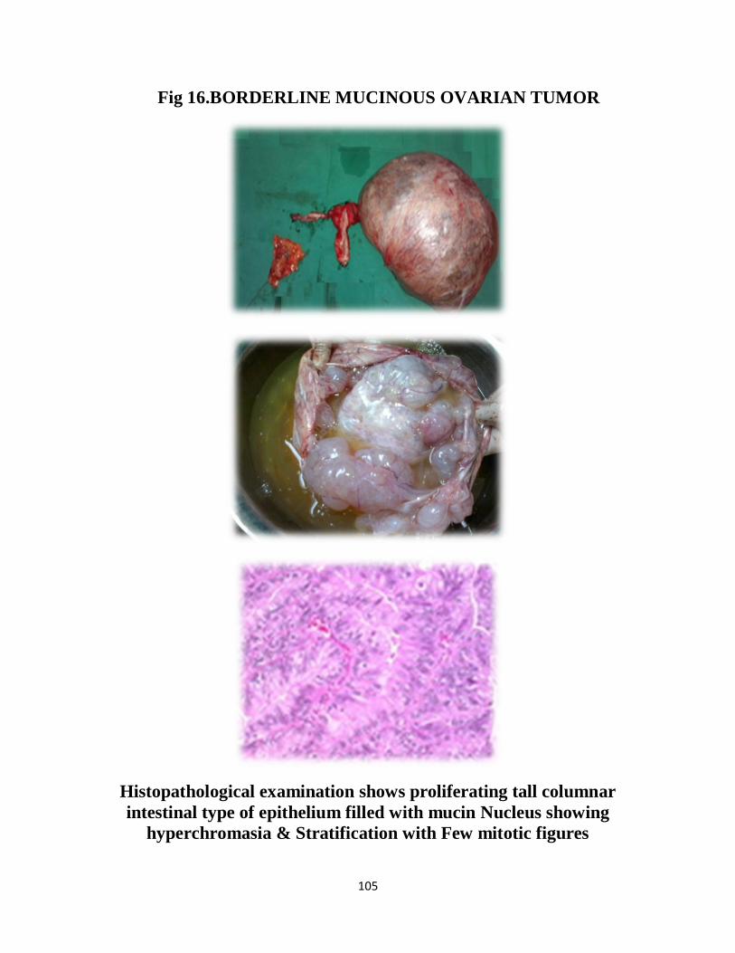
105
Fig 16.BORDERLINE MUCINOUS OVARIAN TUMOR
Histopathological examination shows proliferating tall columnar
intestinal type of epithelium filled with mucin Nucleus showing
hyperchromasia & Stratification with Few mitotic figures

106
Fig 4.PAPILLARY SEROUS CYST ADENOCARCINOMA
Ki67 MALIGNANT
Figure -
Tumor cells arranged in solid nests and papillary pattern

107
Fig.8 BILATERAL MALIGNANT BRENNER
Brenner tumor showing epithelial nests embedded within fibrous stroma
Nests and islands of tumor cells infiltrating the stroma
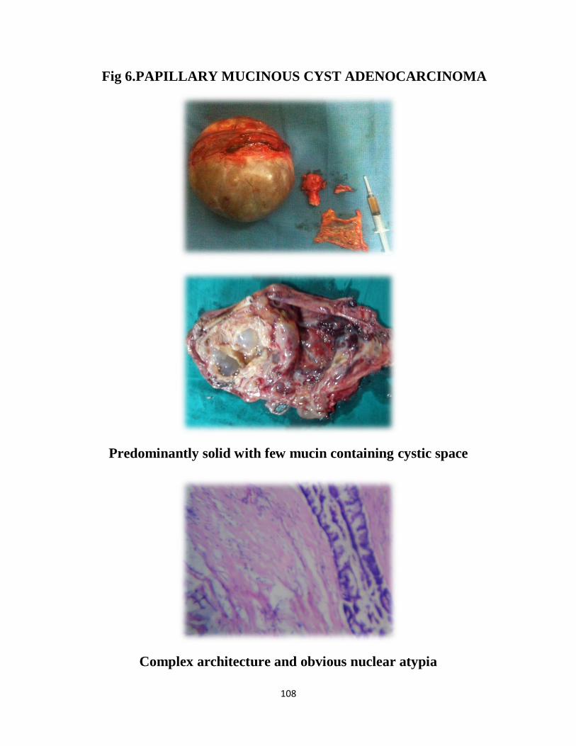
108
Fig 6.PAPILLARY MUCINOUS CYST ADENOCARCINOMA
Predominantly solid with few mucin containing cystic space
Complex architecture and obvious nuclear atypia

109
Fig 11.BENIGN CYSTIC TERATOMA
Areas of cartilage, hair and squamous epithelial lining
Sebum, well developed teeth and hair in a mature cystic teratoma

110
Fig 13.KRUKENBERGS TUMOR
Signet Ring Cells

111
Fig. 9 ADULT GRANULOSA CELL TUMOR
Call Exner bodies

112
Fig 17.LEIOMYOSARCOMA
Cells spindle shaped cells with elongated nuclei and eosinophilic
cytoplasam of interlacing bundles with whorled appearance
pleomorphism, hyperchromatic nuclei with mitoses 14/10 hp
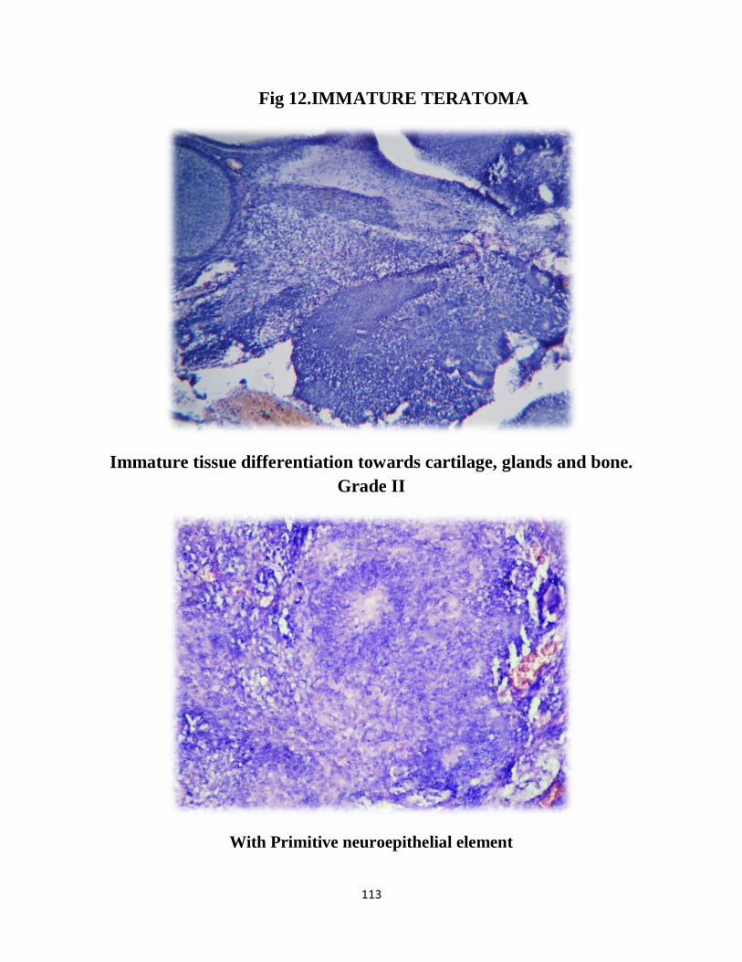
113
Fig 12.IMMATURE TERATOMA
Immature tissue differentiation towards cartilage, glands and bone.
Grade II
With Primitive neuroepithelial element

114
Fig 7.ENDOMETRIOID TUMOR
Well differentiated endometrioid tumor with focal villous architecture

115
Fig 10.FIBROTHECOMA
Pale lipid containing theca cells merge with spindle cell areas
characteristic of fibroma
g

116
Fig 14.OVARIAN TUMOR COMPLICATING PREGNANCY

117
Fig 18a. SMOOTH WALL, SONOLUCENT ( SCORE 0)
Fig 18b.SMOOTH WALL, DIFFUSE ECHOGENICITY ( SCORE 1)

118
Fig 18c. WALL THICKENING, <3mm FINE SEPTA ( SCORE 2)
Fig 18d.PAPILLARY PROJECTIONS, SEPTA>3mm ( SCORE 3)

119
Fig 18e. COMPLEX, PREDOMINANTLY SOLID ( SCORE 4)
Fig 18f.COMPLEX, SOLID AND CYSTIC AREAS WITH
EXTRATUMORAL FLUID ( SCORE 5)
Fig 18g.EXTRATUMORAL FLUID ( SCORE 5)

120
Fig 1. CELLS OF OVARY
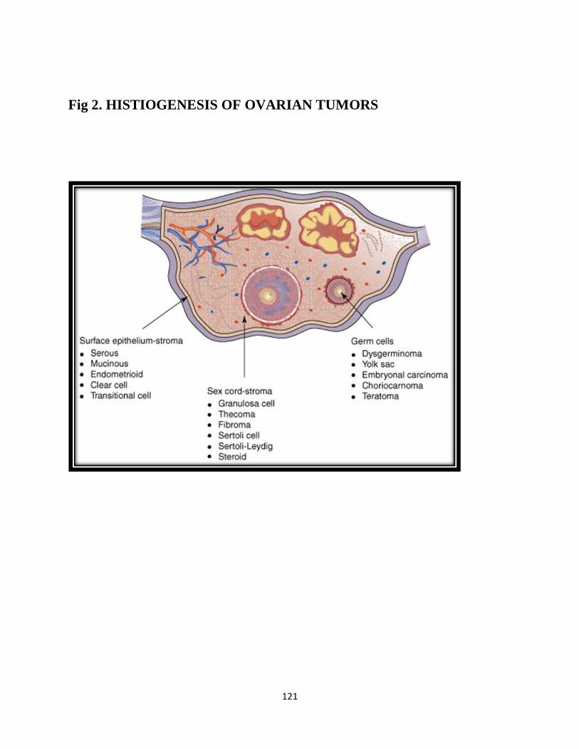
121
Fig 2. HISTIOGENESIS OF OVARIAN TUMORS

122
Fig 15. MORPHOLOGICAL SCORING INDEX

123
BIBLIOGRAPHY
1. Schorge, Schaffer, Halvorson: Williams Gynecology: 1st Ed, 2008; 716-753.
2. Berek and Novaks Gynecology: 14th Ed, 2007; 1457-1547.
3. TeLinde‘s Operative Gynaecology, 10th Ed, Lippincott - Raven, Philadelphia
1997; 1307-1339
4. Campbell S, Bhan V, Royston P: Transabdominal ultrasound screening for
early ovarian cancer. Br Med J 299:1363–1367, 1989
5. Granberg S, Wikland M, Jansson I: Macroscopic characterization of ovarian
tumors and the relation to the histological diagnosis: Criteria to be used for
ultrasound evaluation. GynecolOncol 35:139–144, 1989
6. Sassone AM, Timor-Tritsch IE, Artner A: Transvaginal sonographic
characterization of ovarian disease: Evaluation of a new scoring system to
predict ovarian malignancy. Obstet Gynecol 78:70–76, 1991
7. DePriest PD, Varner E, Powell J: The efficacy of a sonographic morphology
index in identifying ovarian cancer: A multi-institutional investigation.
Gynecol Oncol 55:174–178, 1994

124
8. Lerner JP, Timor-Tritsch IE, Federman A: Transvaginal ultrasonographic
characterization of ovarian masses with an improved, weighted scoring
system. Am J Obstet Gynecol 170:81–85, 1994
9. Ferrazzi E, Zanetta G, Dordoni D: Transvaginal ultrasonographic
characterization of ovarian masses: Comparison of five scoring systems in a
multicenter study. Ultrasound Obstet Gynecol 10:192–197, 1997
10. Mol B, Boll D, DeKanter M: Distinguishing the benign and malignant
adnexal mass: An external validation of prognostic models. Gynecol Oncol
80:162–167, 2001
11. Ueland FR , DePriest PD , Pavlik EJ , Kryscio RJ , van Nagell JR Jr.
Preoperative differentiation of malignant from benign ovarian tumors: the
efficacy of morphology indexing and Doppler flow sonography. Gynecol
Oncol. 2003 Oct;91(1):46-50.
12. Toshiyuki Hata, Yanagihara, Keiji Hayashi, Yamashir, Yohichi Ohnishi,
Akiyama, Atsushi Manabe and Kohji Miyazaki Human Three-dimensional
ultrasonographic evaluation of ovarian tumours: a preliminary study
Reproduction vol.14 no.3 pp.858–861, 1999

125
13. J. L. Alcazar, P. Royo, Jurado, M´inguez, Garcia-manero, C. Laparte, R.
Galvan and G. Lopez-garcia, Triage for surgical management of ovarian
tumors in Asymptomatic women: assessment of an ultrasound-based Scoring
system ultrasound obstetgynecol2008; 32: 220–225
14. Shruti Shah, Hishikar V. ―Incidence and management of ovarian tumor‖.
Bombay Hospital Journal 2008;50(1):30-33
15. Swamy G.S., Satyanarayana N: Clinicopathological analysis of ovarian
tumors; Nepal Med Coll Journal 2010; 12(4);221- 223
16. Williams PL, Dyson M: Gray‘s anatomy, 37th Ed, London, Churchill
Livingstone, 1993.
17. Carolyn Westhoff, Julie A. Britton, Marilie D,. ―Oral Contraceptives and
Benign Ovarian Tumors‖ Gammon American Journal of Epidemiology,
2005; Volume152, Issue3, 242-246.
18. Karen H. Lu, Judy E. Garber, ―Occult Ovarian Tumors in Women with
BRCA1 or BRCA2 Mutations Undergoing Prophylactic Oophorectomy‖
Journal of Clinical Oncology, Vol 18, Issue 14 (July), 2000: 2728-2732.

126
19. Charles N. Landen, Jr, Michael J. Birrer, Anil K. Sood, ―Early Events in the
Pathogenesis of Epithelial Ovarian Cancer‖, Journal of Clinical Oncology,
Vol 26, No 6 (February 20), 2008: pp. 995-1005.
20. Kumar and Cottran Robbins textbook of Pathology, 8th Edition, Chap 22,
Pages 1040 – 1052.
21. Fox H and Wells M: Haines and Taylor Obstetrical and Gynaecological
pathology, ovarian tumor, 4th Edn New York, Churchill Livingstone, 1996.
22. GezaAcs, MD, PhD, ―Serous and Mucinous Borderline (Low Malignant
Potential) Tumors of the Ovary‖, Journal of Clinical Oncology, November
10, 2008 vol. 26 no. 32 5284-5293
23. Vivien W. Chen Ph.D. Bernardo Ruiz M.D., Ph.D. Jeffrey L. Killeen M.D,
―Pathology and classification of ovarian tumors‖ Published 2003 by the
American Cancer Society. Volume 97, Issue S10, 15 May 2003: pages
2631–2642.
24. Gobel U, Schneider DT, Calaminus G, ―Germ-cell tumors in childhood and
adolescence‖ Annals of Oncology 11: 263-271, 2000.
25. Howkins and Bourne, Shaw‘s text book of Gynaecology: 2008; 14th
Ed 329-
349
127
26. Cunningham, Leveno, Bloom: William‘s obstetrics :23rd Ed, 2010: 29-30
27. Bakhidze E.V. N. Petrov Ovarian tumours associated with pregnancy
Journal of Clinical Oncology, 2006; Vol 24, No 18S (June 20 Supplement),
15063.
28. Jeffcoate‘s Principles of Gynaecology :2008; 7th Ed,524-560
29. Couto F, Nadkarni NS and Rebello MJP: ―Ovarian tumors in Goa: A
Clinicopathological study‖. J obstet and Gynecol India 1993;43(3):408-412.
30. Shahin Rashid, Ghulam Sarwar, Abid Ali ,A Clinico – Pathological Study of
Ovarian Cancer in departments of Radiotherapy & Oncology, Sir Ganga
Ram Hospital and Mayo Hospital, Lahore. Dec 1998; 13(4):117-125.
31. Malpani AN, Bhattacharya MS, Neuruukar N: Giant ovarian tumor. J Obstet
Gynecol India 39(2), 1989: 264-266.
32. DePriest P, Gallion HH, Pavlik EJ: Transvaginal sonography as a screening
method for the detection of early ovarian cancer. Gynecol Oncol 65:408–
414, 1997
33. Carter J, Lau M, Fowler JM: Blood flow characteristics of ovarian tumors:
Implications for ovarian cancer screening. Am J Obstet Gynecol 172:901–
907, 1995

128
34. Fleisher AC, Rodgers WH, Kepple DM: Color Doppler sonography of
ovarian masses: A multiparameter analysis. J Ultrasound Med 12:41–48,
1993
35. Roth LM, Anderson MC, and Govan ADT: Sertoli-Leydig cell tumors: A
clinico-pathologic study of 34 cases. Cancer 48(1), 1991; 187-197.

129
KEY TO MASTER CHART
IP NO : In patient Number
G/E : General physical Examination
P/S : Per speculum
P : Pain
M : Mass
D : Distension
U/O : Urine output
C : Constipation
PM : Post-Menopausal
NS : Normal Size
Cx, Vg H : Cervix & Vagina Healthy
WDPV : White discharge per vaginum
IR : Irregular Menstrual Irregularity
Nulli : Nulligravida
MPV : Mass per vaginum
PMB : Post Menopausal Bleeding
V : Vomiting
Amm : Ammenorrhoea
WL : Loss of Weight
LA : Loss of Appetite

130
DM : Diabetes Mellitus
MA : Menstrual Abnormalities
F : Fever
PE : Pedal Edema
R : Regular
NAD : No Abnormalities Detected
U : Unilateral
L : Bilateral
S : Smooth wall
T : Thickened
So : Sonolucent
DE : Diffuse echogenicity
ME : Mixed echogenicity
TAH : Total abdominal hysterectomy
BSO : Bilateral salpingo-oophorectomy
RSO : Right salpingo-oophorectomy
LSO : Left salpingo-oophorectomy
LOC : Left ovarian cystectomy
ROC : Right ovarian cystectomy
+ : Present
- : Absent

131
PROFORMA
Name : Marital status :
Age : Ip No :
Date of admission :
Address :
I) Present complaints:
Mass per abdomen
Abdominal pain
Any menstrual irregulartities
Urinary symptoms
GIT symptoms
Edema of lower limbs
Any discharge per vaginum
II) History of presenting illness
Mass per abdomen
Onset – insidious/acute
Pain – present / absent
Rapidity of growth – rapid / slow
Abdominal pain
Present/absent
Onset

132
Severity
associated with nausea/ vomiting
character of pain / squeezing / coliky / intermittent
aggravating / relieving factor
Menstrual irregularities
Menorrhagia – yes/no
Amenorrhea – yes/no
Polymenorrhea - yes/no
Dysmenorrhea - yes/no
Urinary symptoms
Frequency- yes/no
Acute retention of urine- yes/no
Burning micturition- yes/no
Difficulty in passing urine- yes/no
GIT symptoms
Nausea
Vomiting
Dyspepsia
Constipation
Edema of lower limbs
Bilateral/unilateral
Pitting / non pitting

133
Any discharge per vagina
Type / itching / foul smelling
III) Menstrual history
a) Age of menarche – years
b) Past menstrual cycle
Regular/irregular
Amount of flow- scanty/ moderate/ excessive
Dysmenorrhea – yes/no
Associated clots - yes/no
c) Attained menopause – yes/no
IV) Obstetric history
a) Parity
b) Last delivery
c) Sterilised/not
V) Past history
a) TB/DM/HTN/Bronchial asthma/any surgeries
b) H/O use of oral contraceptives in the past
VI) Family history
a) TB/DM/HTN/Bronchial asthma/any surgeries
b) Any similar complaints in the family
134
VII) Personal history
a) Diet – Veg/mixed
b) Appetite – Normal/ decreased
c) Sleep – Normal/ disturbed
d) Bowel – regular / irregular
e) Bladder – Normal/ increased/ decreased
VIII) General physical examination
a. Built / Nourishment
b. Clubbing/pallor/pedal edema
c. Lympadenopathy
d. Breast
e. Temperature – Febrile / afebrile
f. Pulse –
g. BP -
IX) Systemic examination
a) Cardiovascular system
b) Respiratory system
c) Abdominal examination
a. Inspection
a) Shape
b) Movement of quadrants with respiration
c) Mass / swelling
135
Size
Shape
Extent
d) Any engorged vein
e) Umblicus
f) Hernial sites
b. Palpation
a) Local raise of temperature
b) Tenderness
c) Mass
Situation
Size
Extent
Surface
Consistency
Borders
d) Movements with respiration
e) Any organomegaly
c. Percussion
Ascities – present / absent
d. Auscultation
Any bruit- present/absent

136
X) Per speculum examination
a) Vagina – pale/pink/discharge/rugosity
b) Cervix
position – anterior/middle/posterior
Erosion – Yes/no
Discharge – Yes/ No
XI) Pervaginal examination
Cervix- consistency/ position/ mobility/tenderness
Uterus – size/position/ mobility/tenderness
Mass felt bimanually separate from uterus/ not
Abdominal mass movement transmitted to cervix/not
Forniceal examination – full/ free, tender/non tender
XII) Per rectal examination
Nodulaity
Rectal wall
Pouch of douglas
XIII) Investigations
USG abdomen and pelvis
Bilateral
Solid/complex
Multiloculated
Thickness of cyst wall

137
Septal thickness
Papillary projections.
Doppler study
XIV) Treatment
Surgery
Peroperative finding
Tumour - Gross
Unilateral/ bilateral
Surface – nodular/ smooth
Capsule – thickened/ rupture/ hemorrhage
Cut section- cystic/ solid
Any papillary excrescens
XV) Histopathological examination
Diagnosis:

138
139

140

SIZ
E
CO
NS
IST
EN
CY
MO
BIL
ITY
TE
ND
ER
NE
SS
AS
CIT
ES
UT
ER
US
MA
SS
SIZ
E
VO
LU
ME
TU
MO
R W
AL
L
EC
HO
GE
NIC
ITY
PA
PIL
LA
RY
PR
OJE
CT
ION
S
CO
MP
ON
EN
TS
AS
CIT
ES
VO
LU
ME
S
CO
RE
ST
RU
CT
UR
AL
SC
OR
E
TO
TA
L
MO
RP
HO
LO
GIC
AL
SC
OR
E
1 Sundari 55 13232 P PM P6L6 NAD 6x8 Cystic + + - Cx,Vg H Atropic + 2.4x4x6.8 34.14 S So - Cystic - 1 0 1 U TAH BSOBenign Serous
Cystadenoma
2 Bhuavaneshwari 32 13751 P R P2L2 NAD 10x8 Cystic + + - Cx,Vg H NS + 9.2x5.1x6.2 152.1 S So - Cystic - 2 0 2 U ROBenign Serous
Cystadenoma
3 Pandiammal 55 15626P, M,
DIR P3L3 NAD 12x10 Cystic + + - Cx,Vg H NS + 9x12x5.4 305.01 S So + Cystic - 4 0 4 U TAH BSO
Benign Serous
Cyatadenoma
4 Rajathi 29 15188 P,M R Nulli NAD 12x12 Cystic + - - Cx,Vg H NS + 12.7 x 10.8x11.2 803.4 S So - Cystic - 5 1 6 U ROBenign Mucinous
Cystadenoma
5 Gundhammal 75 16724 P,D PM P6L6 CAC 30x30 Cystic + - - Cx,Vg H Atropic + 29.5x24x21.6 7998.1 S DE - Cystic - 5 1 6 UTAH BSO
ICO
Benign Mucinous
Cystadenoma
6 Mahamani 45 18044P,u/O,
CPM P5L2 NAD - - - - - Cx,Vg H NS + 2.1x3.9x4 17.13 S So - Cystic - 1 0 1 U LOC
Benign Serous
Cyatadenoma
7 Suseela 30 17626 P R P2L2 NAD 10x10 Cystic + - - Cx,Vg H NS + 10x5x7.5 196.12 S So - Cystic - 3 0 3 U LOCBenign Serous
Cystadenoma
8 Deivanai 35 1809 P IR Nulli NAD 10x10 Cystic + - + Cx,Vg H Bulky + 9.8x10x6 307.5 S So - Cystic * 4 5 9 U LOC IOCBenign Serous
Cystadenoma
9 Alagu 45 18550 P PM P3L3 NAD 10x8 Cystic + - - Cx,Vg H NS + 10x14.5x12 910.02 S So - Cystic - 5 0 5 U TAH BSOBenign Mucinous
Cystadenoma
10 Fathima 28 20992 P, D IR P2L2 NAD 20x20 Firm + + - Cx,Vg H NS + 22.5x20x13.5 3177.2 T DE + Complex - 5 4 9 U TAH BSO Granulosa Cell Tumor
11 Saroja 40 20128P,WL,L
AR P1L1 NAD 8x8 Cystic + - - Cx,Vg H NS + 8.2x6.8x5 145.8 S DE - Cystic - 3 2 5 U
TAH BSO
ICO
Benign Serous
Cystadenoma
12 Chinnapillai 38 21976 P, DM R P5L5 NAD 15x10 Cystic + - - Cx,Vg H NS + 14x12x7 615.04 S So - Cystic - 5 0 5 U ROBenign Mucinous
Cystadenoma
13 Dharmaboopathi 60 21058 P, C PM P3L3 OBS 30x25 Hard + - - Cx,Vg H Atropic + 30.2x28.1x22.5 9986.13 T ME + Complex - 5 4 9 BTAH BSO
ICOKrukenberg tumor
14 Rajathi 25 1607 P R P1L1 NAD 10x10 Cystic + - - Cx,Vg H NS + 10x8x5.2 217.56 S DE + Cystic - 3 3 6 U ROCBenign Cystic
Teratoma
15 Susila 40 24635 M IR P1L1 NAD 25x25 Cystic + - - Cx,Vg H Bulky + 30x21.5x18 6072.03 T DE + Complex - 5 4 9 UTAH BSO
ICOGranulosa Cell Tumor
16 Indirani 51 22634WD,
WLPM P4L4 NAD 15x15 Hard + - + Cx,Vg H Atropic + 13.5x14x10.2 1008.2 T ME + Complex + 5 5 10 B
TAH BSO
ICO
Papillary Serous
Cystadenocarcinoma
AG
E
I.P
.NO
.
MO
DE
OF
PR
SE
NT
AT
ION
ME
NS
TR
uA
L H
IST
OR
Y
PA
RIT
Y
G/E
PE
R S
PE
CU
LU
M
LA
TE
RA
LIT
Y
SU
RG
ER
Y
HP
E
S.N
o
US
G
NA
ME PE
R A
BD
OM
EN
PE
R V
AG
INA
L

17 Sumitha 38 24134 M R P1L1 NAD 18x15 Hard + - + Cx,Vg H NS + 14.8x20x18.5 2863.9 T DE - Complex + 5 5 10 UTAH BSO
ICOAdenocarcinoma
18 Selvi 30 1487P,WD,
V,LAR P3L3 NAD 10x8 Cystic + - - Cx,Vg H NS + 10x 6x5 156.9 S So - Cystic - 3 0 3 U LOC
Benign Serous
Cystadenoma
19 Alagammal 50 2508 D PM P4L3 NAD 30x30 Cystic + - - Cx,Vg H Atropic + 18x12.8x30 3614.9 T ME + Complex - 5 4 9 UTAH BSO
ICO
Benign Mucinous
Cystadenoma
20 Periyakka 50 4981 P,D R P1L1 NAD 13x12 Hard + - + Cx,Vg H NS + 10.5x9.8x11 591.9 T ME + Complex + 3 5 8 BTAH BSO
ICO
Mucinous
Cystadenocarcinoma
21 Mahalakshmi 20 4986 P,D R P2L0 CAC 10x10 Cystic + - - Cx,Vg H NS + 10.3x8x4.7 202.5 S DE - Cystic - 4 1 5 UTAH BSO
ICOOvarian Fibrothecoma
22 Sara 35 7125 P R P3L3 NAD 10x10 Cystic + - - Cx,Vg H NS + 10.5x7.2x5 32.9 T So - Cystic - 1 2 3 U LOBenign Serous
Cystadenoma
23 Mariyam Beevi 40 7107 P PM Nulli NAD - - - - - Cx,Vg H NS + 4.9x 3.1 x 5 39.7 S So - Cystic - 1 0 1 B TAH BSOBenign Serous
Cystadenoma
24 Sankarammal 60 5113 D PM P1L1 NAD 15x15 Hard + - + Cx,Vg H Atropic + 12.5x14x11.5 1052.5 T ME - Complex + 5 5 10 UTAH BSO
ICO
Mucinous
Cystadenocarcinoma
25 Nagalakshmi 35 9268 P R Nulli NAD 15x10 Firm + - - Cx,Vg H NS + 15x12.5x13 1274.81 T ME + Complex - 5 4 9 ULap omental
Biopey
Poorly Differentiated
Carcinoma
26 Selvi 42 10354 D R P4L3 NAD 20x20 Cystic + - - Cx,Vg H NS + 15x12.5x13 1274.8 S So - Cystic - 5 0 5 U TAH BSOBenign Serous
Cystadenoma
27 Lourdhmary 38 12571 D R P2L2 NAD 10x15 Cystic + - - Cx,Vg H NS + 10.2x9.1x6 291.26 S So - Cystic - 4 0 4 U TAH BSOBenign Serous
Cystadenoma
28 SubaithaBegam 40 11904 LA R Nulli NAD 20x20 Cystic + - - Cx,Vg H NS + 18.5x12x8.2 952 S So - Cystic - 5 0 5 U TAH BSOBenign Mucinous
Cystadenoma
29 Bakayalakshmi 55 13085 P PM P5L5 NAD 10x8 Cystic + - - Cx,Vg H NS + 10x6x8.5 266.73 S DE - Cystic - 4 1 5 U TAH BSOBenign Serous
Cystadenoma
30 Petchiyammal 40 39417 P R P3L3 NAD - - - - - Cx,Vg H NS + 3.4x4.2x5.8 43.31 S So - Cystic - 2 0 2 U TAH BSO Benign Serous
Cystadenoma
31 Valli 40 15765 P R P3L3 NAD 16x20Variabl
e- + + Cx,Vg H NS * 16.5x15x12 1553.31 T ME + Complex . 5 4 9 U
TAH BSO
ICO
Mucinous
Cystadenocarcinoma
32 Pandiselvi 21 8803 P R Nulli NAD 10x8 Cystic + - - Cx,Vg H NS + 8x5x6 125.52 S So - Cystic - 3 0 3 U LOCBenign Serous
Cyatadenoma
33 SajethaBegam 40 7226 M R P3L3 NAD 20x20 Firm + + - Cx,Vg H NS + 20x25x17.5 4576.2 S DE - Cystic - 5 1 6 UTAH BSO
ICOLeiomyosarcoma
34 Panchavarnam 26 22694 P R Nulli NAD 10x8 Cystic + - - Cx,Vg H NS + 10x8 x7 292.88 S So - Cystic - 4 0 4 U LOCBenign Serous
Cystadenoma
35 Selvi 24 2025 P R P2L2 NAD 9x10 Cystic + - - Cx,Vg H NS + 9x7x6.5 214.16 S ME - Cystic - 4 1 5 U ROBenign Cystic
Teratoma
36 Shajitha 26 4038 P,V R P2L2 NAD 10x12 Cystic + - - Cx,Vg H NS + 10.2x9.5x8 405.42 S So - Cystic - 5 0 5 U ROBenign Serous
Cystadenoma
37 Rakkammal 58 7236 P,D R P3L3 NAD 20x20 Hard + - + Cx,Vg H NS + 18.8x15x13.2 1946.8 T ME - Complex + 5 4 9 BTAH BSO
ICO
Mucinous
Cystadenocarcinoma

38 Veeralakshmi 47 9939 D,LA R P2L2 NAD 20x20 Cystic + - - Cx,Vg H NS + 16.5x15x12 1553.31 S So + Cystic + 5 5 10 UTAH BSO
ICO
Benign Serous
Cystadenoma
39 Guruvammal 60 9354 P,LA,D PM P3L3 NAD 20x18Variabl
e+ - - Cx,Vg H Atropic + 10.5x8x11 483.25 T ME + Complex - 4 4 8 B
TAH BSO
ICO
Poorly Differentiated
Papillary Carcinoma
40 Veeramuthu 17 12038 MA IR Nulli NAD 8x6 Cystic + + - Cx,Vg H NS + 8x6x5 125.52 S So - Cystic - 3 0 3 U LOCBenign Papillary
Serous Cystadenoma
41 Malarvizhi 20 13178 P R Nulli NAD 20x18 Variable + + + Cx,Vg H NS + 21.5x16x12 2158.94 T DE + Complex + 5 5 10 U LOC ICOMucinous
Cystadenocarcinoma
42 Rajeshwari 23 15308 P R Primi NAD 15x10 Hard + - - Cx,Vg H NS + 15x10x11 862.95 S So + Complex - 5 4 9 U LOBenign Mucinous
Cystadenoma
43 Rahmath nisha 55 15861 P PM P1L1 NAD - - - - - Cx,Vg H Atropic + 2x1.2x3 3.76 S So - Cystic - 0 0 0 UTAH BSO
ICO
Benign Serous
Cystadenoma
44 Shanthi 30 14373 P R P3L3 NAD - - - - - Cx,Vg H NS + 3.2x4.8x3 21.09 S So - Cystic - 1 0 1 U LOCBenign Serous
Cyatadenoma
45 Ammapillai 65 15320 P PM P4L4 NAD 10x8 Cystic + - - Cx,Vg H Atropic + 10x8.2x5 214.43 S So - Cystic - 4 0 4 U TAH BSO Benign Serous
Cyatadenoma
46 Sundareshwari 35 15786 P R P14 NAD 9x8 Cystic + - - Cx,Vg H NS + 9x5x6 141.21 S So - Cystic - 3 0 3 U ROBenign Serous
Cystadenoma
47 Subbulakshmi 30 16799P,D,WL
,FR P2L2 NAD 15x18 Cystic + - - Cx,Vg H NS + 18x16.5x12 1863.97 S ME - Complex - 5 3 8 U
TAH BSO
ICO
Benign Mucinous
Cystadenoma
48 Chellam 30 17277 P R Nulli NAD 10x7 Cystic + - - Cx,Vg H NS + 9x6x5.2 146.85 S So - Cystic - 3 0 3 U ROCBenign Serous
Cystadenoma
49 Sathya Bama 38 17754 P R P2L2 NAD 8x6 Cystic + - - Cx,Vg H NS + 8.6x6x4 107.94 S DE - Cystic - 3 1 4 U RSOBenign Serous
Cystadenoma
50 Rajam 55 18252 P R P6L6 NAD 10x8 Cystic + - - Cx,Vg H NS + 9x8x7.5 282.42 T ME - Cystic - 5 2 7 U TAH BSOBenign Mucinous
Cystadenoma
51 Jeyanthi 21 18756 P R Nulli NAD 14x15 Cystic + - - Cx,Vg H NS + 12x13x11.5 938.26 S DE - Cystic - 5 1 6 U LOBenign Endometriotic
Tumor
52 Valli 60 25253 P R P2L2 NAD 20x20 Hard + + + Cx,Vg H NS + 18x16.5x12 1863.97 T ME + Complex + 5 5 10 UTAH BSO
ICO
Papillary Serous
Cystadenocarcinoma
53 Murugeshwari 22 3576 P R G2P1L1 NAD - - - - - Cx,Vg H NS + 8x5x4.5 94.14 S So - Cystic - 3 0 3 U LOBenign Serous
Cyatadenoma
54 Serumathy 28 2915 P,V R P1L1 NAD 10x15 Cystic + - - Cx,Vg H NS + 10x8x6.5 271.96 S ME - Cystic - 4 1 5 U LOBenign Cystic
Teratoma
55 Vasantha 70 11286 P R P5L5 NAD 20x22 Hard + + - Cx,Vg H NS + 25x22x18.5 5321.5 S So - Cystic - 5 0 5 B BSOPapillary Serous
Cystadenocarcinoma
56 Chandra 38 4629 P,D R P2L2 NAD 10x8 Cystic + - - Cx,Vg H NS + 10x8 5x5 222.27 T DE - Cystic - 4 2 6 B TAH BSOSerous
cystadenocarcinoma
57 Rathinam 45 4146 P R P4L4 NAD 10x10 Cystic + - - Cx,Vg H NS + 10x8x6.5 271.96 S So - Cystic - 4 0 4 U TAH BSOBenign Serous
Cystadenoma
58 Muthu 35 7806 P R P5L5 NAD 10x10 Cystic + - - Cx,Vg H NS + 10x8x7.2 301.24 S So - Cystic - 4 0 4 U LOBenign Serous
Cystadenoma

59 Vanmathi 17 8368 P R Nulli Anemic 20x25 Hard + + - Cx,Vg H NS + 21x18x14.2 2807.25 T DE + Complex - 5 4 9 U LO ICO Immature Teratoma
60 Kamatchi 40 7799 P,D R P3L2 NAD 10x10 Cystic + - - Cx,Vg H NS + 10x6x4.5 141.21 S So - Cystic - 3 0 3 U ROBenign Mucinous
Cystadenoma
61 Ariyanatchi 50 11050 P,D R P4L4 NAD 15x17 Hard + + + Cx,Vg H NS + 20x18x15.5 2918.34 T So + Cystic + 5 3 8 BTAH BSO
ICO
Serous
Cystadenocarcinoma
62 Meenatchi 26 12128 D R P2L2 NAD . . . . - Cx,Vg H NS + 6x4x3.5 43.93 S So - Cystic . 1 0 1 U LOBenign Serous
Cystadenoma
63 Parameshwari 55 5572 P,D R P1L1 NAD 10X15 Cystic + + - Cx,Vg H NS + 9.2x5x7 168.4 S So - Cystic . 3 0 3 U TAH BSO Benign Serous
Cystadenoma
64 Ganesammal 35 12745 P R P2L2 NAD 10x12 Cystic + . . Cx,Vg H NS + 10x12.5x9 588.37 S So . Cystic . 5 0 5 U TAH RSOBenign Mucinous
Cystadenoma
65 Rani 42 13843 P,MA IR P4L2 Anemic 15x20 Variable + + + Cx,Vg H Bulky + 14.5x13x16 1577.368 T ME + Complex . 5 4 9 UTAH BSO
ICO
Papillary Serous
Cystadenocarcinoma
66 Mohana 54 47472 P,D R P2L2 NAD 10x8 Cystic + - - Cx,Vg H NS + 9x7x6.5 214.16 S So - Cystic - 4 0 4 U LOCBenign Serous
Cystadenoma
67 Thangammal 55 16473 PMB PM P4L3 NAD 6x8 Cystic + - - Cx,Vg H Atropic + 6x5x3.5 54.91 S DE - Cystic - 2 1 3 BTAH BSO
ICO
Benign Papillary
Serous
Cystadenofibroma
68 Nagalakshmi 26 17459 Asymp R A1 NAD 9x8 Cystic + - - Cx,Vg H NS + 9x8x4.5 169.45 S ME - Cystic - 3 1 4 U LOBenign Cystic
Teratoma
69 Sulochana 35 2363 P R P2L2 NAD 10x8 Cystic + - - Cx,Vg H NS + 10x7x5.2 190.37 S So - Cystic - 3 0 3 U LOBenign Serous
Cystadenoma
70 Muthulakshmi 20 2855 P R Nulli NAD 9x8 Cystic + - - Cx,Vg H NS + 9.5x4x6.2 123.2 S So - Cystic - 3 0 3 U ROCBenign Serous
Cystadenoma
71 Shenbagavalli 42 2857 P R P3L3 NAD 10x8 Cystic + - - Cx,Vg H NS + 10.2x7.2x6 230.45 S So - Cystic - 4 0 4 U TAH BSOBenign Serous
Cystadenoma
72 Devi 47 7017 P,D R P3L3 NAD 20x25 Variable - - + Cx,Vg H NS + 20x15.2x12 1907.9 T ME + Complex + 5 5 10 U TAH BSOSerous
Cystadenocarcinoma
73 Mathani 24 11270 P R P2L2 NAD 10x15 Cystic + - - Cx,Vg H NS + 10.8x7.4x8 334.38 S So - Cystic - 4 0 4 U ROCBenign Mucinous
Cystadenoma
74 Chinnammal 60 12442 P PM P7L5 NAD - Cystic + - - Cx,Vg H Atropic + 3x4.2x3.2 21.08 T So + Cystic - 1 3 4 B TAH BSOPapillary Serous
Cystadenocarcinoma
75 Murugeshwari 39 12961 P R P3L3 NAD - Variable + + - Cx,Vg H NS + 5x5.6x4 58.57 T So - Cystic - 2 2 4 BTAH BSO
ICOAdenocarcinoma
76 Rakku 50 14123 P PM P2L2 NAD 10x10 Variable + + - Cx,Vg H NS + 9.8x10x9 461.28 T So + Cystic - 4 3 7 BTAH BSO
ICOAdenocarcinoma
77 Ezuvakka 52 1670 P R P1L1 NAD 10x10 Firm + - - Cx,Vg H NS + 9x10x8.2 385.974 S So - Cystic - 4 0 4 U TAH BSOPapillary Serous
Cystadenocarcinoma
78 Bakkiyam 63 13467 P PM P6L5 NAD 8x6 Hard + + + Cx,Vg H Atropic + 5x6.5x4 67.99 T ME + Complex + 2 5 7 B TAH BSOPapillary Serous
Cystadenocarcinoma
79 Janaki 55 51053 P PM P2L2 NAD 15x18 Cystic + - - Cx,Vg H NS + 10x15x11.5 902.17 S ME - Cystic - 5 1 6 U TAH BSOBenign Cystic
Teratoma
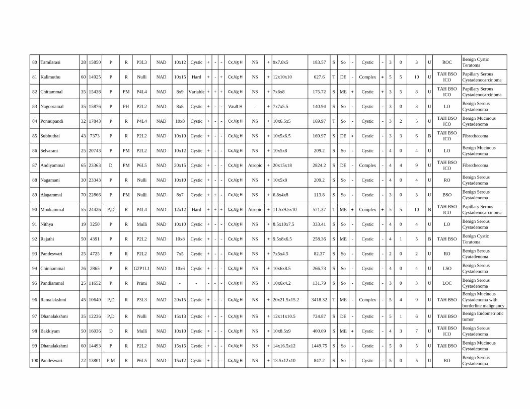
80 Tamilarasi 28 15850 P R P3L3 NAD 10x12 Cystic + - - Cx,Vg H NS + 9x7.8x5 183.57 S So - Cystic - 3 0 3 U ROCBenign Cystic
Teratoma
81 Kalimuthu 60 14925 P R Nulli NAD 10x15 Hard + - + Cx,Vg H NS + 12x10x10 627.6 T DE - Complex + 5 5 10 UTAH BSO
ICO
Papillary Serous
Cystadenocarcinoma
82 Chittammal 35 15438 P PM P4L4 NAD 8x9 Variable + + + Cx,Vg H NS + 7x6x8 175.72 S ME + Cystic + 3 5 8 UTAH BSO
ICO
Papillary Serous
Cystadenocarcinoma
83 Nagooramal 35 15876 P PH P2L2 NAD 8x8 Cystic + - - Vault H . + 7x7x5.5 140.94 S So - Cystic - 3 0 3 U LOBenign Serous
Cystadenoma
84 Ponnupandi 32 17843 P R P4L4 NAD 10x8 Cystic + - - Cx,Vg H NS + 10x6.5x5 169.97 T So - Cystic - 3 2 5 UTAH BSO
ICO
Benign Mucinous
Cystadenoma
85 Subbuthai 43 7373 P R P2L2 NAD 10x10 Cystic + - - Cx,Vg H NS + 10x5x6.5 169.97 S DE + Cystic - 3 3 6 BTAH BSO
ICOFibrothecoma
86 Selvarani 25 20743 P PM P2L2 NAD 10x12 Cystic + - - Cx,Vg H NS + 10x5x8 209.2 S So - Cystic - 4 0 4 U LOBenign Mucinous
Cystadenoma
87 Andiyammal 65 23363 D PM P6L5 NAD 20x15 Cystic + - - Cx,Vg H Atropic + 20x15x18 2824.2 S DE - Complex - 4 4 9 UTAH BSO
ICOFibrothecoma
88 Nagamani 30 23343 P R Nulli NAD 10x10 Cystic + - - Cx,Vg H NS + 10x5x8 209.2 S So - Cystic - 4 0 4 U ROBenign Serous
Cystadenoma
89 Alagammal 70 22866 P PM Nulli NAD 8x7 Cystic + + - Cx,Vg H NS + 6.8x4x8 113.8 S So - Cystic - 3 0 3 U BSOBenign Serous
Cystadenoma
90 Mookammal 55 24426 P,D R P4L4 NAD 12x12 Hard + + + Cx,Vg H Atropic + 11.5x9.5x10 571.37 T ME + Complex + 5 5 10 BTAH BSO
ICO
Papillary Serous
Cystadenocarcinoma
91 Nithya 19 3250 P R Mulli NAD 10x10 Cystic + - - Cx,Vg H NS + 8.5x10x7.5 333.41 S So - Cystic - 4 0 4 U LOBenign Serous
Cystadenoma
92 Rajathi 50 4391 P R P2L2 NAD 10x8 Cystic + - - Cx,Vg H NS + 9.5x8x6.5 258.36 S ME - Cystic - 4 1 5 B TAH BSOBenign Cystic
Teratoma
93 Pandeswari 25 4725 P R P2L2 NAD 7x5 Cystic + - - Cx,Vg H NS + 7x5x4.5 82.37 S So - Cystic - 2 0 2 U ROBenign Serous
Cyatadenoma
94 Chinnammal 26 2865 P R G2P1L1 NAD 10x6 Cystic + - - Cx,Vg H NS + 10x6x8.5 266.73 S So - Cystic - 4 0 4 U LSOBenign Serous
Cystadenoma
95 Pandiammal 25 11652 P R Primi NAD - - - - - Cx,Vg H NS + 10x6x4.2 131.79 S So - Cystic - 3 0 3 U LOCBenign Serous
Cystadenoma
96 Ramalakshmi 45 10640 P,D R P3L3 NAD 20x15 Cystic + - - Cx,Vg H NS + 20x21.5x15.2 3418.32 T ME - Complex - 5 4 9 U TAH BSO
Benign Mucinous
Cystadenoma with
borderline malignancy
97 Dhanalakshmi 35 12236 P,D R Nulli NAD 15x13 Cystic + - - Cx,Vg H NS + 12x11x10.5 724.87 S DE - Cystic - 5 1 6 U TAH BSOBenign Endometriotic
tumor
98 Bakkiyam 50 16036 D R Mulli NAD 10x10 Cystic + - - Cx,Vg H NS + 10x8.5x9 400.09 S ME + Cystic - 4 3 7 UTAH BSO
ICO
Benign Serous
Cystadenoma
99 Dhanalakshmi 60 14493 P R P2L2 NAD 15x15 Cystic + - - Cx,Vg H NS + 14x16.5x12 1449.75 S So - Cystic - 5 0 5 U TAH BSOBenign Mucinous
Cystadenoma
100 Pandeswari 22 13801 P,M R P6L5 NAD 15x12 Cystic + - - Cx,Vg H NS + 13.5x12x10 847.2 S So - Cystic - 5 0 5 U ROBenign Serous
Cystadenoma

101 Seethalakshmi 49 14778 P,D R P2L1 NAD 12x12 Cystic + - - Cx,Vg H NS + 12x11.5x9 649.56 T DE - Cystic - 5 1 6 UTAH BSO
ICO
Benign Papillary
Seromucinous
Cystadenoma
102 Lakshmi 40 18652 P,D R P2L1 NAD 10x13 Cystic + - - Cx,Vg H NS + 13x10.8x9 660.86 S So - Cystic - 5 0 5 UTAH BSO
ICO
Benign Mucinous
Cystadenoma
103 Chitra 23 22712 P R P1L1 NAD 10x8 Cystic + - - Cx,Vg H NS + 10.8x7.4x8 334.38 S So - Cystic - 4 0 4 U LOCBenign Mucinous
Cystadenoma
104 Muthulakshmi 16 23184 P R Nulli NAD 8x6 Cystic + - - Cx,Vg H NS + 4x7x6.2 90.79 S So - Cystic - 2 0 2 U LOCBenign Serous
Cystadenoma
105 Eswari 25 2813D,M, MDpv R P3L2 NAD 7x8 Cystic + - - Cystocele NS + 7x3.2x4 46.86 S So - Cystic - 1 0 1 U ROBenign Serous
Cystadenoma
106 Meenambal 62 5906 P R P4L5 NAD 25x20 Variable + + - Cx,Vg H NS + 20x18x19.5 3671.46 T ME + Complex - 5 4 9 B Inoperable Adenocarcinoma
107 Guruvu 85 5931 P R P4L4 NAD 10x10 Cystic + - - Cx,Vg H NS + 11.2x10x9.4 550.61 T DE - Complex - 5 4 9 U TAH BSOPoorly Differentiated
Papillary Carcinoma
108 Sudha 39 10703 P,M R P2L2 NAD 18x16 Cystic + - - Cx,Vg H NS + 16x18x16.5 2485.3 S So - Cystic - 5 0 5 U TAH BSO Benign Serous
Cystadenoma
109 Muthupandiammal 22 10866 D,M R P2L2 NAD 13x11 Cystic + - - Cx,Vg H NS + 11x6x7 462 S DE - Cystic - 4 1 5 UTAH BSO
ICO
Benign Serous
Cystadenoma
110 Kamalam 52 12860 P,D R P5L3 NAD 10x10 Cystic + - - Cx,Vg H NS + 10x9x6 282.42 S So - Cystic - 4 0 4 U TAH BSOBenign Serous
Cystadenoma
111 Kalaiammal 41 12905 P PH P3L3 NAD 10x6 Cystic + - - Vault H . + 8x6x5 125.52 S So - Cystic - 3 0 3 U TAH BSOBenign Serous
Cystadenoma
112 Lakshmi 32 59852 P, V R P2L2 NAD 10x12 Cystic + - - Cx,Vg H NS + 7.6x5x4 79.49 S So - Cystic - 2 0 2 U TAH BSOBenign Serous
Cyatadenoma
113 Jothi 20 18229 P,M R Primi NAD 12x7 Cystic + - - Cx,Vg H 16 wks + 10x8x6.5 271.96 S ME - Cystic - 4 1 5 U ROBenign Cystic
Teratoma
114 Vairakkal 56 17892 P,MA IR P4L4A1 NAD 25x15 Cystic + - - Cx,Vg H NS + 20x18.5x21.2 4102.41 S DE - Cystic - 5 1 6 UTAH BSO
ICO
Mucinous Papillary
Cystadenocarcinoma
115 Ambigavathy 30 61872 D, C, PE IR P2L2 NAD 20x15 Cystic + + + Cx,Vg H NS + 15.5x12x10 972.78 T ME + Complex + 5 5 10 UTAH BSO
ICO
Papillary Serous
Cystadenocarcinoma
116 Murugayee 60 19139 P, M, LA R P4L4 NAD 20x18 Cystic + + - Cx,Vg H NS + 15x16.5x18 2329.98 T ME + Complex + 5 5 10 UTAH BSO
ICO
Papillary Serous
Cystadenocarcinoma
117 Laxiammal 60 19116 D R P3L2 NAD 30x20 Cystic + + - Cx,Vg H NS + 20x18x16 3012.48 T So - Complex + 5 5 10 UTAH BSO
ICO
Papillary Serous
Cystadenocarcinoma
118 Mariammal 52 6309 MA IR P3L3 NAD 6x8 Cystic + - - Cx,Vg H Bulky + 6x6x5 94.14 T DE - Cystic + 2 5 7 BTAH BSO
ICO
Papillary serous tumor
of borderline
malignancy
119 panju 42 8617 P R P5L5 NAD 10x10 Hard + - - Cx,Vg H NS + 10x11x9 517.77 T DE + Complex + 5 5 10 BTAH BSO
ICO
Mucinous
cystadenocarcinoma
120 Dhanalakshmi 32 927 P,D R P2L2 NAD 20x15 Cystic + - - Cx,Vg H NS + 19.6x21x18 3874.8 S So - Cystic - 5 0 5 U ROBenign Mucinous
Cystadenoma
121 Lakshmi 35 2076 P R P5L1 NAD 10x10 Cystic + - - Cx,Vg H NS + 12x10x11 690.36 S So + Cystic - 5 2 7 U TAH BSOBenign Cystic
Teratoma

122 Meera 27 5782 P R P2L2 NAD 6x7 Cystic + - - Cx,Vg H NS + 5.2x5x4 54.39 S So - Cystic - 2 0 2 U RO LSOBenign Serous
Cystadenoma
123 Priya 23 9028 P R Nulli NAD 9x9 Cystic + - - Cx,Vg H NS + 9x8.2x7 270.18 S So + Cystic - 4 2 6 U ROBenign Cystic
Teratoma
124 Bakkiyam 30 9542 P R P1L1 NAD 7x7 Cystic + - - Cx,Vg H NS + 7.2x5x6 112.96 S So - Cystic - 3 0 3 U LOBenign Serous
Cystadenoma
125 Guruvammal 45 9033 P,D R P2L2 NAD 20x25 Cystic + - - Cx,Vg H NS + 21x18x19.5 3855.03 S ME - Cystic - 5 1 6 UTAH BSO
ICO
Benign Mucinous
Cystadenoma
126 Paapa 47 12433 P R P4L4 NAD 6x8 Cystic + - - Cx,Vg H NS + 4x6x8 100.42 S So - Cystic - 2 0 2 U LOCBenign Serous
Cystadenoma
127 Sundareshwari 35 15786 P R P1L1 NAD 9x10 Cystic + - - Cx,Vg H NS + 9x8x6 225.93 S So - Cystic - 4 0 4 U ROBenign Serous
Cystadenoma
128 Selvi 28 9013 P R P3L3 NAD 20x20 Cystic + - - Cx,Vg H NS + 20x21.5x16 3598.24 S DE - Cystic - 5 1 6 BTAH BSO
ICOOvarian Fibrothecoma
129 Chitra 25 9109 P R P2L2 NAD 20x15 Cystic + - - Cx,Vg H NS + 20x22.5x18 4257.22 S ME - Cystic - 5 1 6 U LSOBenign Mucinous
Cystadenoma
130 Lakshmi 55 8001 P R P2L0 NAD 8x6 Cystic + - - Cx,Vg H NS + 8x7x6.6 193.3 S So - Cystic - 3 5 8 BTAH BSO
ICO
Papillary Serous
Cystadenocarcinoma
131 Mariammal 52 6309 MA IR P3L3 NAD 6x8 Cystic + - - Cx,Vg H Bulky + 6x6x5 94.14 S So - Cystic - 2 5 7 UTAH BSO
ICO
Papillary serous tumor
of borderline
malignancy
132 Selvam 45 12834 P R P2L1 NAD 8x8 Cystic + - - Cx,Vg H NS + 8x7x6 175.7 S So - Cystic - 3 5 8 BTAH BSO
ICO
Papillary Serous
Cystadenocarcinoma
133 Vellathai 48 4744 P,D R P4L2A2 Cachetic 20x15 Variable - + + Cx,Vg H NS + 20x12x8.5 1066.92 T DE + Complex + 5 5 10 BTAH BSO
ICOAdenocarcinoma
134 Karupayee 60 65607 P PM P4L4 NAD 12x12 Hard - - + Cx,Vg H Atropic + 12x14x8.5 746.84 T ME - Cystic + 5 5 10 BTAH BSO
ICO
Mucinous
Cystadenocarcinoma
135 Ramalan 68 8234 P PM P4L4 NAD 10x15 Firm - - - Cx,Vg H Atropic + 13x8x10.5 571.11 T So + Cystic - 5 3 8 BTAH BSO
ICOBrenner tumor
136 Sathya 13 8078 D R Nulli NAD 20x15 Hard - - + Cx,Vg H NS + 18x15x12.5 1765.13 T ME + Complex + 5 5 10 U RSO ICOMucinous
Cystadenocarcinoma


Your digital receiptThis receipt acknowledges that Turnitin received your paper. Below you will find the receipt informationregarding your submission.
Paper ID 292276405
Paper title STUDY OF CLINICAL, SONOLOGICAL AND HISTOPATHOLOGICALCORRELATION OF OVARIAN TUMOR
Assignmenttitle Medical
Author Aishwarya Jagan 20101601 M.D. Obstetrics and GynaecologyE-mail [email protected]
Submissiontime 25-Dec-2012 07:14AM
Total words 18382
First 100 words of your submission
STUDY OF CLINICAL, SONOLOGICAL AND HISTOPATHOLOGICAL CORRELATION OFOVARIAN TUMOR DISSERTATION SUBMITTED FOR M.D (BRANCH – II) (OBSTETRICS &GYNAECOLOGY) APRIL 2013 THE TAMILNADU DR.M.G.R. MEDICAL UNIVERSITY CHENNAI,TAMILNADU 1 CERTIFICATE This is to certify that this dissertation titled “STUDY OF CLINICAL,SONOLOGICAL AND HISTOPATHOLOGICAL CORRELATION OF OVARIAN TUMOR”submittedby DR. AISHWARYA JAGAN to the faculty of Obstetrics and Gynecology, The TamilNadu Dr. M.G.R.Medical University, Chennai in partial fulfillment of the requirement for the award of MD degree BranchII Obstetrics and Gynecology, is a bonafideresearch work carried out by her under our directsupervision and guidance...
Copyright 2012 Turnitin. All rights reserved.