Embed Size (px)
Citation preview

RESEARCH Revista Mexicana de Fısica61 (2015) 137–148 MARCH-APRIL 2015
Study of dynamical properties inβ-TCP/CH layers
A. Minaa, J.C. Caicedoa,∗, and W. AperadorbaTribology, Powder Metallurgy and Processing of Solid Recycled Research Group,
Universidad del Valle, Cali-Colombia,∗Tel./Fax Number: 57+2+ 3122270 Ext 27
e-mail: [email protected] of Engineering,
Universidad Militar Nueva Granada, Bogota-Colombia.
Received 20 August 2014; accepted 5 February 2015
β-Tricalcium phosphate/Chitosan (β-TCP/Ch) coatings were deposited on 316L stainless steel (316L SS) substrates by a cathodic electro-deposition technique at different coating compositions. The crystal lattice arrangements were analyzed by X-Ray diffraction (XRD), andthe results indicated that the crystallographic structure ofβ-TCP was affected by the inclusion of the chitosan content. The changes in thesurface morphology as a function of increasing chitosan in the coatings via scanning electron microscopy (SEM) and atomic force microscopy(AFM) showed the root-mean squares hardness of theβ-TCP/Ch coatings decreased by further increasing chitosan percentage. The elastic-plastic characteristics of the coatings were determined by conducting nanoindentation test, indicating that the increase if chitosan percentageis directly related to increasing the hardness and elastic modulus of theβ-TCP/Ch coatings. Tribological characterization was performedby scratch test and pin-on-disk test to analyze the changes in the surface wear theβ-TCP/Ch coatings. Finally, the results indicated animprovement in the mechanical and tribological properties of theβ-TCP/Ch coatings as a function of increasing of the chitosan percentage.
Keywords: Surfaces; monolayers; coatings; electrochemical techniques; hardness.
PACS: 68.47.Gh; 75.70.Ak; 68.35.Ct; 82.45.Bb; 62.20.Qp
1. Introduction
In biomaterials devices has been reported that after long termimplantations, materials as stainless steel (316L SS) may po-tentially cause allergic reactions due to the release of nickel(Ni), molybdenum (Mo) and chromium (Cr) ions [1-3]. Asan innovative strategy, surface modification of this class ofmetallic implants with ceramic coatings represents the mostcommon solution to change the bulk properties of the metalsubstrate, and also improve the mechanical and tribologicalbehavior of the surface into interface bone/implant [4-7]. Inrecent studies, hydroxyapatite (HA), compounds of HA andβ-tricalcium phosphate (β-TCP), HA and chitosan andβ-TCP and chitosan have been frequently examined as coatingmaterials on metallic substrates for biomedical applicationsdue to their favorable biocompatibility, mechanical, and tri-bological characteristics [8-10].
β-TCP coatings are the newest application trying to im-prove the biocompatibility of metallic implants [11-13]. Onthe other hand, chitosan is a natural cationic polysaccharideproduced byalkaline N-deacetylationof chitin, a constituentof the exoskeletonfrom crabs and squid. It is widely usedin biomedical applications due to its bioactive characteris-tics that promote cell proliferation [14]. The combinationof β-TCP and chitosan as a bioactive compound can poten-tially improve the bone fixation and accelerate bone growth.So. The electrochemical deposition of biomaterials on metal-lic substrates has recently provided greater advantages inbiomedical applications [2]. Many bioactive compounds thataccelerate bone growth and improve bone fixation have beenused as coating materials. The incorporation of these bioac-
tive coatings to metallic implants represents great advancesin the development of orthopedic devices. The coatings canbe applied into the substrates by a variety of techniques, suchas sputtering [15], thermal evaporation [16], chemical vapordeposition [17], spin coating [18], spray pyrolysis [19], sol-gel deposition [20] and electrochemical deposition [21]. Theelectrochemical deposition has superior advantages compareto other techniques due to its simplicity, low cost and roomtemperature operation [22]. Furthermore, electrochemicaldeposition of biomaterials can provide functionalized coat-ing implants by modifying experimental solutions and condi-tions [23].
The time-dependent properties ofβ-TCP make it an idealcoating for implants, which supplies a temporary supportfor bone ingrowth, and eventually is replaced by naturaltissue [24]. Among several clinical applications involvingβ-TCP, posterior spinal fusion and craniomaxillofacial ap-plications have attracted considerable attentions [25]. Cal-cium phosphate-based bioceramics with improved mechan-ical properties and controlled resorbability can assist in de-signing optimal biodegradable bone substitutes for spinal fu-sion and craniomaxillofacial applications. The use of suchbone implants also avoids the second surgery required for au-tograft harvesting [26].
The biomaterials depends on the surface characteristicsthat directly influence the biocompatibility by affecting thesurface charge, surface topography, and etc. [27]. In addi-tion, some mechanical and tribologial properties are also ofimportance (hardness and friction coefficient). For example,materials used as catheters need excellent tensile and me-chanical characteristics. Therefore, both the biocompatibil-

138 A. MINA, J.C. CAICEDO, AND W. APERADOR
ity and the mechanical properties of biomaterials need to beimproved [28]. In this work,β-TCP/Ch coatings were de-posited on the 316L SS substrates by using cathodic electro-deposition technique in order to obtain a possible functional-ized coating implant. The influence of the chitosan percent-age in the coating structures, which improved the mechanicaland tribological response was characterized and discussed incurrent research. In this sense, theβ-TCP/Ch coatings couldbe considered as promising materials for biomedical indus-try in temporal invasive prosthesis applications as they caneffectively improve the bone/prosthesis interfaces.
2. Experimental details
The tricalcium phosphate, also known asβ-Ca3(PO4)2 or (β-TCP), and chitosan, also known as ((C6H11NO4)n) or (Poly-(1-4)-2-Amino-2-deoxy-β-D-Glucan) were used as materialcoatings.β-TCP-chitosan coatingsβ-TCP/Ch coatings weredeposited via cathodic electro-deposition on AISI 316L SSsubstrates (12 mm of diameter and 5 mm of thickness).Electro-deposition technique was performed using the fol-lowing conditions: current density of 260 mA, agitationvelocity of 250 rpm, pH electrolyte of 10.4, and a tem-perature deposition of 60◦C. The cylindrical 316L SS sub-strates were prepared by grinding to 600 grit water proofsilicon carbide papers, then they were activated by superfi-cial electrochemical etching using an acid solution of 1:1:1HCl:H2SO4:H2O, to improve the adhesion characteristics inthe substrate-coating interfaces. Finally, the substrates werecleaned in an ultrasonic bath with acetone and rinsed in de-ionized water in order to remove the organic contaminants.
The electro-deposition process was performed using anelectrolyte compound of two solutions. The first one wasβ-TCP dissolved in an ethanol-water 1:3 solution; and thesecond one was carried out with chitosan solution dissolvedin acetic acid 2%. The structural characterization was per-formed by X-Ray diffraction (XRD) Bruker D8 Advancewith Cu cathode and scintillation detector using the setup 0-20 and making a sweep of 20◦ to 80◦ with a step of 0.01degrees and a time per step of 2 seconds with Cu Ka ra-diation (λ = 1.5405 A). The diffraction patterns were ob-tained as a function of increase in the chitosan concentra-tion fromβ-TCP/Ch coatings, these patterns were further an-alyzed to determinate the changes in the crystal lattice ar-rangements. In addition, the different lattice arrangements ofcoating were compared and the changes in preferential peakswere determinate due to the inclusion of chitosan. The in-crease of intensity as a function of chitosan weight percent-age was determined after to be applied the full width at halfmaximum (FWHM) criterion for the XRD patterns [29]. Thesurface chemical characterization of theβ-TCP-Ch coatingswere performed using a scanning electron microscope (SEM)JEOLJSM-6490LV to determinate the different phases on thesurface coating. Energy Dispersive X-Ray (EDX) analysiswas carried out to investigate the distribution of the differ-ent elements in the composites and to evaluate the changes of
chemical composition as a function of chitosan weight per-centage.
Atomic Force Microscopy (AFM) was used for a quanti-tative study of the surface morphology inβ-TCP/Ch coatingsusing an AFM Asylum ResearchMFP − 3Dr and calcu-lated by a Scanning Probe Image Processor (SPIPr), whichis the standard program for processing and presenting AFMdata. Therefore, this software has become thede-factostan-dard for image processing at the nanoscale. The mechani-cal properties were defined using a Nanoindentation test, byusing a Nanoindenter Ubi1-Hysitron to obtain elastic-plasticproperties of the coatings. A three-side Berkovich diamondindenter with a tip radious of about 100 nm was used. Afternanoindentation measurements, the load-penetration depthcurves were performed to calculate the hardness and elas-tic modulus through the Oliver-Pharr method [30]. Tribo-logical characterization was developed with a Microtest,MT400 98tribometer, using a 6-mm diameter 100Cr6 steel-ball-like counter body slide. The load applied was 5 N with atotal running length of 1000 m (around 29000 cycles) and0.10 m/s. Scratch test was used to evaluate the adhesionstrength between theβ-TCP/Ch coatings and the 316L SSsubstrates. This technique was performed with a scratchingspeed of 4 mm/min and a continuous load rate of 1 N/s. Thescratch test was also used to calculate the critical load (Lc) asa function of chitosan percentage, and determine the cohesivefailure of the coatings.
3. Results and discussion
3.1. XRD Results
To study of crystalline structure of the preparedβ -TCP/Chcoatings, XRD technique was used to obtain the diffractionpatterns of each coating. Figure 1 shows the diffraction pat-terns ofβ -TCP peaks at low angles, preferential orienta-tions (0018), (1118) and (0502) for2θ = (43.55◦, 47.06◦
and 50.68◦), respectively. The results indicate that the pref-erential orientations, corresponding to the ceramic phase ofthe coating layers, are in agreement with the Joint Commit-tee on Powder Diffaction Standards (JCPDS) [31]. Figure 1also exhibits the changes in rhombohedral configurations ofβ-TCP as a function of chitosan percentage. From this figure,the compression stress was also identified due to the changesin the peak morphology with chitosan percentage increasing[31]. Moreover, the diffraction patterns show the influencebetween the peak intensity and the chemical composition forβ-TCP/Ch coatings. In this study, a displacement and widen-ing of the preferential peaks was observed for all the samples.
The diffraction patterns exposed in Fig. 1 show a varia-tion in the intensity of the preferential peaks oriented (0018)at 2θ = 43.6◦ as a function of chitosan percentage, whichis in an agreement with the results reported by other authors[10,31]. The variations in XRD patterns were appreciated us-ing thefull width at half maximum(FWHM) criterion for thepatterns. Therefore, the crystal structure deformation data
Rev. Mex. Fis.61 (2015) 137–148

STUDY OF DYNAMICAL PROPERTIES INβ-TCP/CH LAYERS 139
FIGURE 1. XRD patterns ofβ-TCP/Ch coatings deposited withdifferent chemical compositions. The diffraction patterns showrhombohedral configurations corresponding toβ-TCP and also adisplacement and widening of the preferential peaks that indicatethe compression stress due to the increase in the chitosan percent-age.
suggests an inversely proportional relationship between thechitosan percentage and the peak intensity. Analyzing thediffraction patterns in Fig. 2 shows a variation in the inten-sity of the preferential peaks oriented (0018) at2θ = 43.6◦
as a function of chitosan percentage in the coatings.The crystallographic behavior described above is shown
in Fig. 2a where it is analyzed the shape and size variation as-sociated to preferential peaks. Furthermore, the same shapeand size variation as a function of chitosan percentage wasobserved in Fig. 2b. This figure also exhibits a decreasingin peak area and peak height for the preferential peak (0018)which can be contributed to the changes in the internal resid-ual compression stress [10,23,31]. Consequently, it was pos-sible to observe that the peak variation suggests an inverserelationship between the chitosan percentage and the peak in-tensity. The observation of this peak variation demonstratesa crystal structure deformation due to the compression stressapplied by chitosan molecules in theβ-TCP structure.
3.2. SEM analysis
To analyze the surface characteristics of differentβ-TCP/Chcoatings SEM was used. Figure 3 exhibits SEM micrographsof different coatings as a function of chemical compositions.The micrographs show the changes in the surface morphol-ogy as a function of chitosan percentage, which is in ac-cordance with previous publications [10,30,32]. Therefore,Fig. 3a presents a fibrillar structure matrix associated to theβ-TCP particles, this figure also demonstrated irregular dis-persed particles associated to the particles which were notdissolved in the electrolyte solution deposited on the surfacecoating by ionic exchange from (Ca2+ and PO−3
4 ).
FIGURE 2. XRD patterns of differentβ-TCP/Ch coatings: (a) fullwidth at half maximum (FWHM) variations as a function of chi-tosan percentage change and (b) peak area and peak height changesof preferential peak (0018) as a function of chitosan percentage.The curves show the compression stress effect in crystal structuredue to the changes in chitosan percentage; it was evidenced by thedecrease of peak area and peak height while the chitosan percent-age increased.
Figure 3b exhibits the surface coating when it has 5% ofchitosan, it showed that the number of particles increasedassociated with the polymerization of chitosan moleculeswhich are not dissolved in the saturated electrolyte. Also,it was possible to observe that fibrillar structure matrix hasdisappeared, and it was replaced by a matrix with angu-lar particles dispersed on the surface coating without de-fined orientation. This effect visually demonstrates the func-tionalization effects between theβ-TCP particles and Chmolecules [24,26].
On the other hand, Fig. 3 shows that the thickness of thedeposited particles decrease as a function of the increasing ofchitosan percentage (Fig. 3b-e). Moreover, increasing of thechitosan percentage influences the deposited particles on the
Rev. Mex. Fis.61 (2015) 137–148

140 A. MINA, J.C. CAICEDO, AND W. APERADOR
FIGURE 3. SEM micrographs of differentβ-TCP/Ch coatings: (a)β-TCP100%/Ch0%, (b) β-TCP95%/Ch5%, (c) β-TCP90%/Ch10%, (d) β-TCP90%/Ch25%, (e) β-TCP65%/Ch35%, and (f)β-TCP50%/Ch50%. The irregular particles are related with the particles without a definedshape; Particles type needle are elongated particles and angular particles are agglomerated particles with angular shapes.
surface to be thinner than the other coatings with lower chi-tosan percentage and presents an angular shape similar toneedle shape.
Furthermore, these results exhibit progressive increasein the percentage and the size of particles deposited on thesurface coating. The chitosan particles have a tendency tohomogenize eliminating the surface area of the needle-likestructure. In this way, Fig. 6f (a typical micrograph of sur-
face coating with 50% of chitosan) shows a surface withoutneedle-like structures, probably due to increasing of irregularparticles.
Figure 3e shows a typical micrograph of the surface coat-ing with 50% of chitosan, this micrograph presents a surfacewithout needles structures probably due to the increasing ofirregular particles observed in the last figures (Fig. 3b-e). Al-lotropic changes in the surface morphology ofβ - TCP/Ch
Rev. Mex. Fis.61 (2015) 137–148

STUDY OF DYNAMICAL PROPERTIES INβ-TCP/CH LAYERS 141
FIGURE 4. Representative AFM images ofβ-TCP/Ch coatings on the 316L SS substrates: (a)β-TCP100%/Ch0%; (b) β-TCP95%/Ch5%;(c) β-TCP65%/Ch35%; and (d)β-TCP50%/Ch50% coatings.
coatings were described previously, which can be related tothe changes in the crystallographic arrangement presented inthe Fig. 1 and Fig. 2.
3.3. AFM analysis
AFM analysis was used to study the surface morphology ofdifferent β-TCP/Ch coatings. It is known that the surfacemorphology and roughness of coatings have a very importantrole on the final characteristics of the implants, since the re-actions at the tissue/coating interface is directly related to thesurface morphology. Taking this note into account, differentβ-TCP/Ch coatings as function of chitosan percentage wereanalyzed in an area of20 µm× 20 m for each sample with az-scale around86.30± 2.0 µm.
The morphology and the root-mean-square roughness oftheβ-TCP/Ch coatings are shown in Fig. 4, it suggests thatincreasing the chitosan percentage can cause major changesin the surface topography and roughness. In this way, byincreasing the chitosan percentage, the surface morphologychanged and the surface roughness decreased.
The AFM micrograph showed in Fig. 4a was obtainedfrom theβ-TCP coating surface, which shows rounded par-ticles overlapped between them, taking into account that thescale of measurements is a clear evidence that this coating isobtained from nanopowder. In addition, a qualitative anal-ysis indicates that the sample contains predominantly par-ticles of almost the same size and shape similar to the re-sults reported by previous publications [33]. The allotropicchanges in the surface morphology of theβ-TCP/Ch coatings
Rev. Mex. Fis.61 (2015) 137–148

142 A. MINA, J.C. CAICEDO, AND W. APERADOR
FIGURE 5. Morphological analysis ofβ-TCP/Ch coatings on the316L SS substrates: (a) Roughness as a function of chitosan per-centage curve, and (b) Grain size as a function chitosan percentage.
were described previously, indicating that these changes arerelated to the changing in the size and distribution of the par-ticles. It is possible to infer that the changes in the crystallo-graphic arrangement obtained by XRD analysis are related tothe growth, size and distribution of the particles, at the sametime the allotropic changes of the surface morphology is re-lated to the changes in the surface topography observed bythe SEM micrographs.
Applying statically analysis on AFM images using aScanning Probe Image Procesor (SPIPr), it was possible tomeasure the morphological changes of theβ-TCP/Ch coat-ings. Figure 5 shows the roughness and grain size trend as afunction of chitosan percentage. Similar to the results fromthe SEM analysis it was possible to observe a decrease ofcoating roughness by further increasing the chitosan content.The previous published studies indicated that the values ofgrain size are closely related to the values of roughness, there-fore, a decrease in the surface roughness implies a decreasein the grain size [34]. This effect is evidenced in Fig. 5 which
FIGURE 6. Nanoindentation test results for theβ-TCP/Ch coat-ings: load-displacement indentation curves as a function of increas-ing the chitosan percentage.
FIGURE 7. Mechanical properties of theβ-TCP/Ch coatings: (a)Hardness values of theβ-TCP/Ch coatings as a function of chitosanpercentage and (b) Elastic modulus of theβ-TCP/Ch coatings as afunction of chitosan percentage.
Rev. Mex. Fis.61 (2015) 137–148
STUDY OF DYNAMICAL PROPERTIES INβ-TCP/CH LAYERS 143
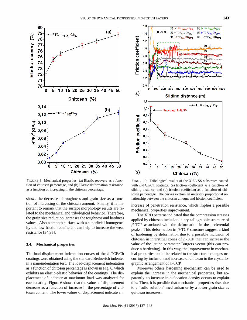
FIGURE 8. Mechanical properties: (a) Elastic recovery as a func-tion of chitosan percentage, and (b) Plastic deformation resistanceas a function of increasing in the chitosan percentage.
shows the decrease of roughness and grain size as a func-tion of increasing of the chitosan amount. Finally, it is im-portant to remark that the surface morphology results are re-lated to the mechanical and tribological behavior. Therefore,the grain size reduction increases the toughness and hardnessvalues. Also a smooth surface with a superficial homogene-ity and low friction coefficient can help to increase the wearresistance [34,35].
3.4. Mechanical properties
The load-displacement indentation curves of theβ-TCP/Chcoatings were obtained using the standard Berkovich indenterin a nanoindentation test. The load-displacement indentationas a function of chitosan percentage is shown in Fig. 6, whichexhibits an elastic-plastic behavior of the coatings. The dis-placement of indenter at maximum load was analyzed foreach coating. Figure 6 shows that the values of displacementdecrease as a function of increase in the percentage of chi-tosan content. The lower values of displacement indicate an
FIGURE 9. Tribological results of the 316L SS substrates coatedwith β-TCP/Ch coatings: (a) friction coefficient as a function ofsliding distance, and (b) friction coefficient as a function of chi-tosan percentage. The curves explain an inversely proportional re-lationship between the chitosan amount and friction coefficient.
increase of penetration resistance, which implies a possiblemechanical properties improvement.
The XRD patterns indicated that the compression stressesapplied by chitosan inclusion in crystallographic structure ofβ-TCP associated with the deformation in the preferentialpeaks. This deformation inβ-TCP structure suggest a kindof hardening by deformation due to a possible inclusion ofchitosan in interstitial zones ofβ-TCP that can increase thevalue of the lattice parameter Burgers vector (this can pro-duce a hardening). In this way, the improvement in mechan-ical properties could be related to the structural changes oc-curring by inclusion and increase of chitosan in the crystallo-graphic arrangement ofβ-TCP.
Moreover others hardening mechanism can be used toexplain the increase in the mechanical properties, but ap-parently no increase in dislocation density occurs to explainthis. Then, it is possible that mechanical properties rises dueto a “solid solution” mechanism or by a lower grain size asquitosan increases.
Rev. Mex. Fis.61 (2015) 137–148

144 A. MINA, J.C. CAICEDO, AND W. APERADOR
FIGURE 10. SEM micrographs for the wear tracks, evidencing the changes in the wear mechanism (abrasive y adhesive wear) as a functionof chitosan percentage for all theβ-TCP/Ch coatings
It was possible to determinate the values of hardness (H)and elasticity modulus (Er) using the indentation method de-veloped by Oliver and Pharr [36]. Figure 7a shows an in-crease of hardness while increase the chitosan percentage byusing Eq. (1) (which relate hardness with the ratio betweenmaximum applied load and contact area).
H =pmax
A(hc)(1)
wherePmax is the maximum applied load andA(hc) is thearea associated to the surface where the load is applied. Thisequation is according with [30,36]. Figure 7 shows the in-creasing of mechanical properties as a function of chitosanpercentage. As mentioned before, this increase ofEr and Hcould be related with little changes in the crystallography ori-
entation observed in the XRD patterns (Fig. 1) associated tothe changes in the area and height of the central peak.
Hajek and co-workers [37] proposed an expression to cal-culate the elastic recovery for the coatings. In this research,Fig. 8 shows the plastic deformation resistance (H3/Er2) andelastic recovery (R). The obtained results here are in agree-ment with other authors [38-42]. The elastic recovery forthe β-TCP/Ch coatings was calculated by using the follow-ing Eq. (2) [36].
R =δmax − δp
δmax(2)
whereδmax is the maximum displacement andδp is the resid-ual or plastic displacement. The equation data was taken
Rev. Mex. Fis.61 (2015) 137–148

STUDY OF DYNAMICAL PROPERTIES INβ-TCP/CH LAYERS 145
from the load-penetration depth curves of indentations foreach coating according to the results obtained from nanoin-dentation test and exhibited in Fig. 6. The best mechani-cal properties were reached by the coating with the highestamount of chitosan content. Figure 8 shows an inverse pro-portional relation between the mechanical properties and chi-tosan percentage. In this sense it was possible to observe thatthe elastic recovery and plastic deformation resistance valuesincrease as a function of chitosan amount increase, similar tothe results analyzed in elastic modulus and hardness.
From the load-displacement curves Fig. 6 its shows thatthe increase of the elastic recovery (Fig. 8) corresponds to areduction of the plasticity (or plastic deformation), therefore,it was reflected an increase in the Elastic modulus (Fig. 7),which could be interpreted as a hardening ofβ-TCP/Ch com-pound with increasing the addition of chitosan.
3.5. Tribological properties
The friction coefficient values were obtained from pin-on-disk test, in this way, the tribological properties were ana-lyzed into tribological pair between the 316L SS substrateandβ-TCP/Ch coatings system and a 100Cr6 steel balls. Fig-ure 9a shows the friction coefficient as a function of slidingdistance for the substrate and coatings. The curves displayedin Fig. 9a showed two different stages; the first stage is calledthe running-in period, where the friction coefficient began atlow levels; due to an interferential friction mechanism it iscaused by few contacts between the steel ball and the coat-ing surface through the roughness tips in both counterparts.As slide distance increase, the friction coefficients increasetoo due to the formation of wear debris by cracking of rough-ness tips on both counterparts according to the tribological
model proposed by Archard [43]. The second stage, alsocalled steady state is a zone of the curve where the rough-ness of tribological pair is smoothed and the friction coeffi-cient value is stabilized. In this stage, the surface coatings aremolded by the surface of a 100Cr6 steel ball, since the ballsurface is harder than the coating surface. Finally, it is possi-ble to suppose that the friction coefficients are closely relatedto the slide distance, and by increasing the slide distance, thefriction coefficient decreases [44,45].
A good combination between elastic-plastic properties(H, Er) and surface properties (Ra) could possibly guaranteea good tribological response. In fact, the tribological prop-erties depend mainly on the elastic-plastic properties, thus,while the chitosan percentage increasesH andEr, the rough-ness decreases. Therefore, the values of friction coefficientdecrease as is shown in Fig. 9b. As it was explained abovethe chitosan amount and friction are closely related, so Fig.9b shows the friction coefficient as a function of chitosanpercentage increase curve, this curve shows thatµ decreaseswhen the chitosan percentage increases.
3.5.1. Wear analysis
After a sliding distance of 1000 m in the pin-on-disk tests,SEM analysis was used to evaluate the wear properties ofthe surfaces. Figure 10 shows the micrographs of the sur-face wear for all the coating samples at 1000X and their wearmechanism, demonstrating a continuous and smooth weartrack, with a depth below the film thickness. Therefore, theabrasive wear is presented in each micrograph, which is re-lated to the generation of loose wear debris corresponding torupture, cohesive failure and delamination of the coatings dueto the interaction between the slider pair (steel ball) and the
FIGURE 11. Tribological results for the friction coefficient curves versus the applied load; showing the cohesive (LC1) and adhesive (LC2)failure mode for theβ-TCP/Ch coatings as a function of increasing in the chitosan percentage.
Rev. Mex. Fis.61 (2015) 137–148

146 A. MINA, J.C. CAICEDO, AND W. APERADOR
FIGURE 12. Critical load associated to the adhesion failure(Lc2) as a function of chitosan percentage for all the coat-ings: (β-TCP100%/Ch0%, β-TCP95%/Ch5%, β-TCP90%/Ch10%, β-TCP90%/Ch25%, β-TCP65%/Ch35%, andβ-TCP50%/Ch50%..
β-TCP/Ch coating surfaces. Thus, the wear debris interactagain with both surfaces, generating grooves and scratches onthe coating surfaces. In this sense, high friction coefficient,and other wear mechanisms associated with the mechanicalfatigue due to cyclical charges of pin on the surface is evi-denced by cracks. Figure 10a-f clearly show the effects ofchitosan percentages on the wear mechanism of the coatings.Therefore, it can be concluded that by increasing the chitosanpercentage, a decrease in the trace of abrasive particles andcracks could be observed. The decreasing of the wear mech-anism (adhesive and abrasive failure) for theβ-TCP/Ch coat-ings is associated with the morphological surface evolutionexhibited in the AFM results (Fig. 5) together with the evo-lution of the mechanical properties evidenced in Fig. 7 andFig. 8.
3.6. Adhesion behavior
Scratch-test technique was carried out to measure the adher-ence strength of theβ-TCP/Ch coatings. To identify the ad-hesion properties of the coatings, it is important to considera lower critical load (LC1). It is defined as the load wherethe first cracks occurred (cohesive failure) and the highestcritical load (LC2) where occurs the first delamination at theedge of scratch (adhesive failure) [46]. LC1 and LC2 valuescorrespond to the tribological zones where the friction is in-dependent on the applied load [47]. The critical load (LC1
and LC2) values for all theβ-TCP/Ch coatings are shown inFig. 11, which shows the critical load values experimentallydetermined from the friction coefficient as a function of ap-plied load.
From Fig. 11 it was possible to analyze the real adhesiveresponse associated with theβ-TCP/Ch coatings, hence, thecurves of friction coefficient as a function of load showed thatthe values of LC1 are similar in Fig. 11a - d. However, for
the coatings with higher percentages of chitosan (35% and50%), the critical load associated with the cohesive failure ishigher, as shown in Fig. 11e and f. On the other hand, thecritical load is associated with the adhesive failure, whichis closely related to the chitosan percentage. In this way,the friction coefficient curves as a function of load showedthat LC2 increased when the chitosan percentage increased.Moreover, the improvement in the adhesion behavior of theβ-TCP/Ch coatings could be related to the tribological anal-ysis showed in Fig. 9 from pin-on-disk results. Similarly, theimprovement in the mechanical behavior exhibited in Fig. 7and Fig. 8, could be also associated to the changes in thecrystal lattice arrangement studied in Fig. 1 and Fig. 2 andthe evolution in the topography and morphological surfaceobserved by SEM and AFM techniques, respectively.
As mentioned before, the chitosan percentage is influ-enced on the values of LC2, Fig. 12 shows the LC2 curve asa function of chitosan percentage. This curve indicates thatthe adhesive critical load of the coatings increase by furtherincreasing the chitosan percentage, in another word, a higherstress should be applied to reach the delamination of the coat-ings. From the data reported by other authors, it is possibleto conclude that the tribological results were governed by theelastic-plastic properties of theβ-TCP/Ch coatings and ad-hesion corresponding to the interface (β-TCP/Ch coatings-substrate) because theβ-TCP/Ch systems have a relativelyhigh elastic recovery (R%) values [47]. As can be seen inFig. 8a, the critical load changes occur due to the increas-ing of plastic deformation resistance (H3/E2) [47] (showedin Fig. 8b), which generates opposition to indenter penetra-tion producing an opposition to coating deformation. Thus,the last tribological and mechanical properties are related toa rhombohedral arrangement with the obtained results for allthe coatings.
4. Conclusions
In this study, different types ofβ-TCP/Ch coatings were de-posited via an electro-deposition method. It is shown that thepreferential orientation for rhombohedral arrangement were(0018), (1118) and (0502) orientations for2θ =(43.55◦,47.06◦ and 50.68◦), respectively. From the SEM micro-graphs, it was possible to observe the topography changes onthe surface coatings in relation with different chitosan per-centages. These changes were associated with the results ofthe morphological analysis ofβ-TCP/Ch coatings performedby using AFM analysis, which showed the decrease of grainsize and roughness as a function of chitosan percentage.
Different mechanical properties such as hardness wereobtained for theβ-TCP/Ch coatings. It was also determinedthat the elastic modulus (Er) and elastic recovery increased asfunction of increasing the chitosan percentage. These resultswere expected taking into account the changes in preferentialpeaks of XRD that suggested a deformation hardening.
The tribological behavior was studied, in which it wasobtained low friction coefficient into the tribological system
Rev. Mex. Fis.61 (2015) 137–148

STUDY OF DYNAMICAL PROPERTIES INβ-TCP/CH LAYERS 147
(steel pin andβ-TCP/Ch coatings) for all the coatings. Thisstudy suggested an improvement in tribological properties ofthe coatings evidencing an increase of adhesion resistance asfunction of chitosan increase. Therefore, the improvement intribological behavior could be expected taking into accountthe improvement of the elastic-plastic behavior exposed bythe β-TCP/Ch coatings by increasing the chitosan percent-age.
From the SEM results it was possible to identify the wearmechanisms in each coating, (abrasive and adhesive wear)with failure by fatigue were evidenced by micrographs whichare reduced when the chitosan percentage is increased. In this
sense is possible to observe a decrease in the evidence of wearmechanisms due to the improvement in elastic-plastic behav-ior of the coatings, which open up new features for future ofsurgical implants.
Acknowledgments
This research was supported by the Universidad MilitarNueva Granada, Bogota-Colombia project IMP-ING-1775and the Excellence Center for Novel Materials (CENM) atUniversidad del Valle in Colombia under Contract RC-043-2005 with Colciencias.
1. J.B. Brunski, in: B.D. Ratner, A.S. Hoffman, F.J. Schoen, andJ.E. Lemons (Eds.),Biomaterials Science an Introduction toMaterials in Medicine, 2nd ed., (Elsevier Academic Press, SanDiego, 2004) p. 137.
2. B. Karim, J. Jean, D. Mainard, P. Elisabeth, and N. Patrick,Biomaterials17 (1996) 491.
3. E. Salahinejadet al., PLoS ONE8 (2013) 1-8.
4. M. Mozafari et al., International Journal of Nanomedicine8(2013) 1665-1672.
5. S.M. Naghib, M. Ansari, A, Pedram, F. Moztarzadeh, and A.Feizpour, M. Mozafari,International Journal of Electrochemi-cal Science7 (2012) 2890-2903.
6. E. Salahinejadet al., Journal of Biomedical Nanotechnology9(2013) 1327-35.
7. M.J. Cross, E.N. Parish, and J. Bone,Joint Surg Br87 (2005)1073-6.
8. T. Jinno, D.T. Davy, and V.M. Goldberg,J. Arthroplasty.17(2002) 902-9.
9. X. Pang, and I. Zhitomirsky,Colloid and Interface Science. 330(2009) 323-329.
10. D. Pena, H. Estupinan, H. Cordoba, and C. Vasquez,Rev. Fac.Ing. Univ. Antioquia(54) (2010) 15-23.
11. Y. Abe, T. Kokubo and T. Yamamuro,Materials Science. Ma-terials in Medicine1 (1990) 233.
12. L. Pighinelli and M. Kucharska,Journal of Biomaterials andNanobiotechnology4 (2013) 20-29.
13. S.-J. Ding,Dental Materials Journal25 (2006) 706-712.
14. M. Kucharska, A. Niekraszewicz, M. Wisniewska-Wrona, E.Wesolowska and H. Struszczyk,Progress on Chemistry and Ap-plication of Chitin and Its Derivatives. 3 (2003) 69-72.
15. G.R. Bamwenda, K. Sayama, and H. Arakawa,J. Photochem.Photobiol., A Chem.122(1999) 175.
16. O. Bohnke, C. Bohnke, and G. Robert,Solid State Ion. 6 (1982)121.
17. D. Davazoglou, A. Donnadieu, and A. Donnadicu,Solar En-ergy Mat.71 (1988) 379.
18. E. Salahinejad, M.J. Hadianfard, D.D. Macdonald, M. Moza-fari, D. Vashaee, and L. Tayebi,Materials Letters88 (2012)5-8.
19. R. Hurdich,Electron Lett.11 (1975) 142.
20. K.D. Lee,Thin Solid Films302(1997) 84.
21. P.M.S. Monk, and L.S. Chester,Electrochim. Acta38 (1993)1521.
22. A.I. Inamdar, S.H. Mujawar, V. Ganesan and P.S. Patil,Nan-otechnol19 (2008) 325706.
23. E.M. Castro, H A. Estupinan, and D.Y. Pena,Scientia et Tech-nica13 (2007) 36.
24. D.S. Metsger, M.R. Rieger, and D.W. Foreman,J. Mater SciMater Med10 (1999) 9-17.
25. X. Guo, L. Lee, L.P. Law, H. Chow, R. Rosier, and C. Cheng,J. Orthop. Res20 (2002) 740-746.
26. S.S. Banerjee, S. Tarafder, N.M. Davies, A. Bandyopadhyay,and S. Bose,Acta Biomaterialia. 6 (2010) 4167-4174.
27. E.T. den Braber, J.E. de Ruijter, and L.A. Ginsel,Biomaterials17 (1996) 2037-2044.
28. M. Zhang, X.H. Li, Y.D. Gong, N.M. Zhao, and X.F. Zhang,Biomaterials. 23 (2002) 2641-2648.
29. R. Martınez H. Estupinan, D. Pena, and P. Mohan,Scientia etTechnica.36 (2007) 36.
30. W.C. Oliver and G.M. Pharr,Journal of Materials Research7-6(1992) 1564-1583.
31. W. Jiawei,Journal of Biomedical materials Research. Part. A.76 (2006) 503-511.
32. A. Tejada, C. Pina, S. Martınez, and G.Avila, Rev. Mex. Fis.5(2004) 187-192.
33. G. Tomoaia, A. Mocanu, I. Vida-Simiti, N. Jumate, L. DorelBobos, and O. Soritau, M. Tomoaia-Cotisel,Materials Scienceand Engineering: C, 37 (2014) 37-47.
34. G.S. Kim, S.Y. Lee, and J.H. Hahn,Surf. Coat. Technol.171(2003) 91-95.
35. J. Zhang, W. Liu, V. Schnitzler, F. Tancret, and J. Bouler,ActaBiomaterialia.10 (2014) 1035-1049.
36. G. Cabrera, J.C. Caicedo, C. Amaya, L. Yate, J. MunozSaldana, and P. Prieto,Materials Chemistry and Physics.125(2011) 576-586.
Rev. Mex. Fis.61 (2015) 137–148

148 A. MINA, J.C. CAICEDO, AND W. APERADOR
37. V.V. Hajek, K. Rusnak, J. Vlcek, L. Martinu and H.M.Hawthorne,Wear213(1997) 80-86.
38. J.C. Caicedoet al., Applied Surface Science.256(2010) 5898-5904.
39. G.S. Kim, S.Y. Lee, and J.H. Hahn,Surface and Coatings Tech-nology.171(2002) 91-95.
40. P.J. Burnett and D.S. Rickerby,Thin Solid Films.154 (1987)403-416.
41. J.C. Caicedo, G. Bejarano, M.E. Gomez, P. Prieto, C. Cortez,and J. Munoz,Phys. Stat. Sol.4 (2007) 4127-4133.
42. J. Romero, A. Lousa, J. Esteve, and E. Martınez, AppliedPhysics A: Materials Science & Processing.77(2003) 419-427.
43. J.F. Archard.Journal of Applied Physics.24 (1953) 981.
44. P. Nledengvist and S. Hogmark,Tribology International.30(1997) 507-516.
45. L. Ipaz, J.C. Caicedo, J. Esteve, F.J. Espinoza-Beltran, and G.Zambrano,Applied Surface Science258(2012) 3805- 3814.
46. S.J. Bull, D.S. Rickerby, A. Matthews, A. Leyland, A.R. Pace,and J. Valli,Surf. Coat. Technol.36 (1988) 503-517.
47. J.M Lackner, L. Major, and M. Kot,Bull. Pol. Ac.: Tech.59(2011) 343-355.
Rev. Mex. Fis.61 (2015) 137–148