Embed Size (px)
Citation preview
13
Ain Shams Journal of Forensic Medicine and Clinical Toxicology
January 2021, 36:13-30
Study of effects of repeated administration and withdrawal of cannabinoids on brain tissue of adult male albino rats: histopathological and biochemical study
Samy M. Badawy, Safaa A. Amin, Amira M. El-Seidy, Nagwa M. Habib and Reham Hassan. El- Farouny1,
1 Forensic Medicine and Clinical Toxicology Department, Faculty of Medicine, Menoufia University, Menoufia, Egypt.
All rights reserved
Abstract Introduction: Cannabis occupies the position of the most prevalent drug of abuse in Egypt. Chronic
cannabis abuse may cause degenerative pathological changes in the brain, also the effect of cannabis
on cognition, memory and learning is well known. The current work aimed to study the toxic effect
of chronic repeated doses of cannabis on rat behavior, brain dopamine level and histopathological
changes of the brain.
Subjects and Methods: The study was conducted on 50 male albino rats weighing 180-200 gm.
Rats were divided into 2 main groups: group 1 (control; each animal received 1ml/day 100%
sunflower oil (used as a vehicle); group 2 (received cannabis daily for one month "40 mg cannabis/
200 gm rat". At the end of the month this group was subdivided into 4 subgroups: subgroups 2a, 2b,
2c, and 2d and subjected to different periods of cannabis abstinence. The clinical manifestations due
to the effect of long-term cannabis administration and withdrawal were recorded. After scarification,
brain dopamine level was measured, and brain tissue was examined by light and electron
microscope (EM).
Results: Clinically; main manifestations of long-term cannabis administration found in subgroup 2a
were CNS depression; while main manifestations in subgroup 2c were irritability and fighting
aggression. Dopamine significantly increased in subgroup 2a compared to control group. Light and
EM examination showed degenerative changes.
Conclusion: Brain dopamine level was significantly high in subgroup 2a compared with control
group. long-term cannabis administration causes degenerative pathological changes in the brain,
which need long recovery periods.
Key words Cannabis, dopamine, brain, ∆9- THC
Introduction
annabis consists of the leaves and flowering parts
of the plant Cannabis Sativa and usually is smoked
in cigarettes ("joints" or "reefers") or pipes or
added to food (usually cookies or brownies).
Resin can be dried from the plant and packed int
o blocks called hashish. Delta 9- tetrahydrocannabinol
(∆9-THC) is the principal psychoactive ingredient of
cannabis (Radwan et al., 2015). Common receptors for
cannabinoids are CB1 and CB2. The receptors CB1 are
primarily present in brain tissue and have a wide
distribution. The highest densities are mainly located in
the frontal cerebral cortex (higher functioning),
hippocampus (memory, cognition), basal ganglia and
cerebellum (movement), and striatum (brain reward)
(Fitzgerald et al., 2013). In rodents CB1 receptor
agonists as ∆9-THC, induce what has been called the
“cannabinoid tetrad”; that is characterized by catalepsy,
hypothermia, anti-nociception (blocking sensory neural
perception of a harmful or injurious stimulus) and
motor activity suppression (Katsidoni et al., 2013).
Chronic cannabis use can lead to dependence w
hile stopping chronic use will l ad to symptoms of
withdrawal (Ramesh et al., 2011). The diagnostic and
statistical manual of mental disorders (DSM-5) listed
cannabis as one of the addictive drugs and established
medical criteria for "cannabis use disorder" (CUD) (American Psychiatric Association, 2013).
Human laboratory studies have shown that
cannabinoid withdrawal syndrome has arisen after abrupt
C
14 Badawya et al. / Ain Shams J Forensic Med Clin Toxicol, 1/2021 (36): 13-30
discontinuation of chronic oral ∆9-THC resulting in
disturbed sleep, restlessness, irritability, vomiting, chills
and nausea. It was therefore hypothesized that experience
of physical withdrawal symptoms or concerns linked to
abstinence may lead to the persistent use of marijuana in
cannabis-dependent people (Lichtman and Martin, 2002;
Allsop et al., 2011).
Cannabis use is associated with increased dopa
mine levels, as one of the dependence drugs. Cannabinoi
ds in fact cause their pharmacological activity by activati
ng dopaminergic neurons in the brain more precisely;
the mesostriatal dopaminergic system of cell bodies
situated within the ventral tegmental area (VTA) and
substantia nigra pars compacta (SNpc) by increasing the
dopamine release in their corresponding dopamine
terminal fields, nucleus accumbens (NAcc) and striatum
(Fanarioti et al., 2015). The dopaminergic system has a
crucial role in the production of addictive criteria of
drugs and reward processing (Volkow et al., 2011), and it
is involved in behavioral enhancement, motivated
behavior and the modulation of violent behaviors (Rosell
and Siever, 2015).
Chronic cannabis abuse can lead to
neurodegenerative alterations that can be clarified by
deposition of ∆9-THC in neurons leading to neurotoxic
and neuroanatomic changes (Monnet-Tschudi et al.,
2008). ∆9-THC is an important cause of oxidative cell
stress that may lead to cell injury, metabolic dysfunction
and neuronal harm (Sarafian and Marques, 1999).
The aim of the present study is to study the toxic
effect of long term repeated doses of cannabis on rat
behavior, brain dopamine level and histopathological
changes of the brain; this may help in minimizing the use
of this substance, which is considered safe by youth.
Patients and Methods Drugs: Cannabis was obtained from a patient
admitted to Dependence Treatment Unit- Forensic
Medicine and Clinical Toxicology Department, Faculty
of Medicine; Menoufia University for treatment.
Cannabis was found with the patient during the routine
inspection done before admission to Dependence
Treatment Unit. It was a solid to resinous brown paste,
about 10 gm. in weight. Cannabis was sent to the
Forensic and analytical chemistry lab, Faculty of
Medicine, Menoufia University to confirm its chemical
identity (extraction and detection by thin layer
chromatography (TLC) and was used in this study after
getting permission of the head of department.
Calculation of cannabis dose: ∆9-THC was initially
calculated at a dose of 10 mg/kg (i.e. 2 mg ∆9-THC/ 200
gm. rat) (Hložek et al., 2017). The doses used in the
current study were selected according to animal and
clinical studies in which ∆9-THC shows behavioral
locomotor effects and induces psychotic-like symptoms
in animals (Nagai et al., 2006; El-Alfy et al., 2010;
Katsidoni et al., 2013; Wiley and Burston, 2014).
Concentration of ∆9-THC in cannabis was measured by
TLC in comparison with standard curve; and was found
to be 50 mg ∆9-THC / 1 gm. cannabis (i.e. 5%). Then the
corresponding daily cannabis dose was = 40 mg
cannabis/ 200 gm. rat (Hložek et al., 2017).
Animals: Study was conducted on 50 male
albino rats weighing 180-200 gm., obtained from the
breeding animal house in Menoufia University. Rats
were kept in wide cages measuring 30 × 60 × 30 cm;
each one was able to accommodate 5 rats of
approximately the same weight to simplify drug dosing
calculation. They were left to acclimatize for one week.
They were housed at room temperature in metallic cages
and kept under constant healthy environmental and
nutritional conditions. Rats were also kept under a
schedule diurnal lightening conditions (12 hours for
darkness and 12 hours for light). They were fed on dry
food and housed under standard laboratory conditions.
Initial weight of each animal was recorded at the
beginning of the experiment and at its end (at the end of
the month) for comparison. The maintenance of animals
and the experimental procedures were in accordance with
the guiding principles of Ethical Committee of Faculty of
Medicine, Menoufia University.
Rats were divided into 2 main groups: group 1
(control group, consisted of 10 rats were kept throughout
the experiment under the same conditions without any
treatment and were given normal feeding to show the
normal values of the tested parameters. Each animal
received 1ml/day 100% sunflower oil (used as a vehicle)
by a curved needle – like oral tube that was introduced
directly into the stomach (a gavage process) (Hložek et
al., 2017); group 2 (40 rats, received cannabis daily for
one month "40 mg cannabis/ 200 gm rat, dissolved in
sunflower oil 100% as a vehicle and given by an oral
tube with gradually increasing dose by adding half of the
initial calculated dose every 3 days till the end of the
month". At the end of the month this group was further
subdivided into 4 subgroups: subgroups 2a (10 rats,
sacrificed after the last dose of cannabis i.e. under
pharmacological effect of cannabis); subgroup 2b (10
rats, left one day without giving cannabis after the last
cannabis dose, and then sacrificed); subgroup 2c (10 rats,
left 3 days without giving cannabis after the last dose,
and then sacrificed); subgroup 2d (10 rats, left 7 days
without giving cannabis after the last dose, and then
sacrificed). After scarification, brain dopamine level was
measured, and brain tissue was examined by both light
and electron microscope.
Measurement of brain dopamine level: The
hemisphere of the brain was homogenized each volume in
20 volumes of formic acid/ acetone (15/85, v/v), put in a
glass centrifuge tube and shacked, centrifuged and the
supernatant transferred to a glass centrifuge tubes. The
supernatant was washed by shaking with 3 ml of heptane/
chloroform (8/1, v/v) for every 1 ml of formic acid/ acetone.
The organic phase and lipid interfaces were aspirated, and
the samples were left to dry. The samples were reconstituted
15 Badawya et al. / Ain Shams J Forensic Med Clin Toxicol, 1/2021 (36): 13-30
in the dopamine assay buffer and analyzed by Infinite F50
TECAN ELISA (Smith et al., 1975).
Histopathology study:
Hematoxylin and Eosin staining (HX & E):
(Jaffe, 1969; Carlton, 1982). Histochemical Stain (Toluidine blue (T.B)
stain): Nuclei and acid carbohydrate components are
stained blue. Some carbohydrate- containing structures
are metachromatically stained red. Cytoplasm RNA (e.g.
Nissel bodies of neurons) is also blue (Kiernan, 1999).
Transmission electron microscope (EM) examination of
brain tissue was done in EM unit- Mansoura University
for confirmation of findings detected by HX & E and
Toluidine blue stains (Reynolds, 1963; Karnovsky, 1965).
Statistical Analysis:
Data were collected, organized, tabulated and
statistically analyzed using Statistical Package for Social
Science (SPSS) version 17 by Apple Mac computer.
Both statistical analysis and tabulation were done
according to (Dawson and Trapp, 2001). For quantitative
data, for comparison of means of more than two groups
one-way ANOVA test (f test) was used. the threshold of
significance was fixed at 5% level (P- value).
P- Value of > 0.05 = non-significant (NS).
P- Value < 0.05 = significant (*).
Results I) Observed Clinical Manifestations:
Group 1 (control group): No behavioral
abnormalities were noticed.
Subgroups 2a: A dose-related CNS-depression
within the first 10 days, progressed from inactivity to
prostration, and was associated with incoordination and
ataxia. Rats were hypoactive for 2-4 hours after giving
the dose in the form of being not interested to food or
looking from the cage to the foreigner as did the control
group. Craving to cannabis occurred in all animals about
10-20 days after starting cannabis administration and this
was known by observing the occurrence of mild
withdrawal manifestations before giving the morning
daily dose of cannabis (hyperactivity predominated in the
form of irritability and tremors), which decreased after
the dose intake. Decreased body weight was observed by
approximately l0% from the weight at the beginning.
Subgroups 2b: at the end of the day at the time
of cannabis dose; rats were hyperactive, irritable with
scratching movements, facial rubbing, and licking.
Subgroups 2c: main manifestations were
irritability, fighting aggression, beating each other.
Chewing movement and salivation were observed. Gasps
and shakes (both head shakes and body shakes) were
recorded. Cheek tremors and teeth chattering were also
seen.
Subgroups 2d: irritability markedly decreased,
rats were relatively more calm, with more improved
appetite for food.
Table 1 shows comparison of brain dopamine
level between different groups of experimental albino
rats: dopamine significantly increased in subgroup 2a
(under pharmacological effect of cannabis) compared to
control group; and significantly decreased in subgroup 2d
(withdrawal of cannabis for 7 days) compared to
subgroup 2a.
II) Brain dopamine level:
Table 1: Comparison of brain dopamine level between different groups in experimental albino rats using one-way
ANOVA test (f test):
Brain dopamine
level (ng/ml) G1 (control)
Subgroup 2a
(under effect)
Subgroup 2b
(withdrawal 1day)
Subgroup 2c
(withdrawal 3
days)
Subgroup 2d
(withdrawal 7
days)
Mean ± S.D 0.12 ± 0.1 1.27 ± 0.04 0.87 ± 0.43 0.76 ± 0.12 0.34 ± 0.14
Range 0.05 - 0.19 1.25 - 1.30 0.56 - 1.18 0.68 - 0.85 0.24 - 0.45
P value
P value * 0.003 * 0.019 * 0.032 0.353
P value 0.125 0.069 * 0.008
P value 0.659 0.061
P value 0.110
*P value <0.05 = significant P value >0.05 = non-significant
16 Badawya et al. / Ain Shams J Forensic Med Clin Toxicol, 1/2021 (36): 13-30
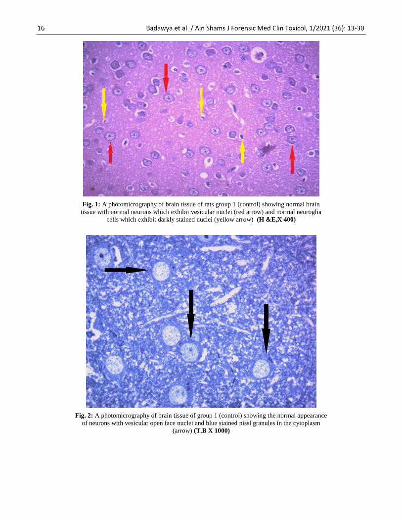
Fig. 1: A photomicrography of brain tissue of rats group 1 (control) showing normal brain
tissue with normal neurons which exhibit vesicular nuclei (red arrow) and normal neuroglia
cells which exhibit darkly stained nuclei (yellow arrow) (H &E,X 400)
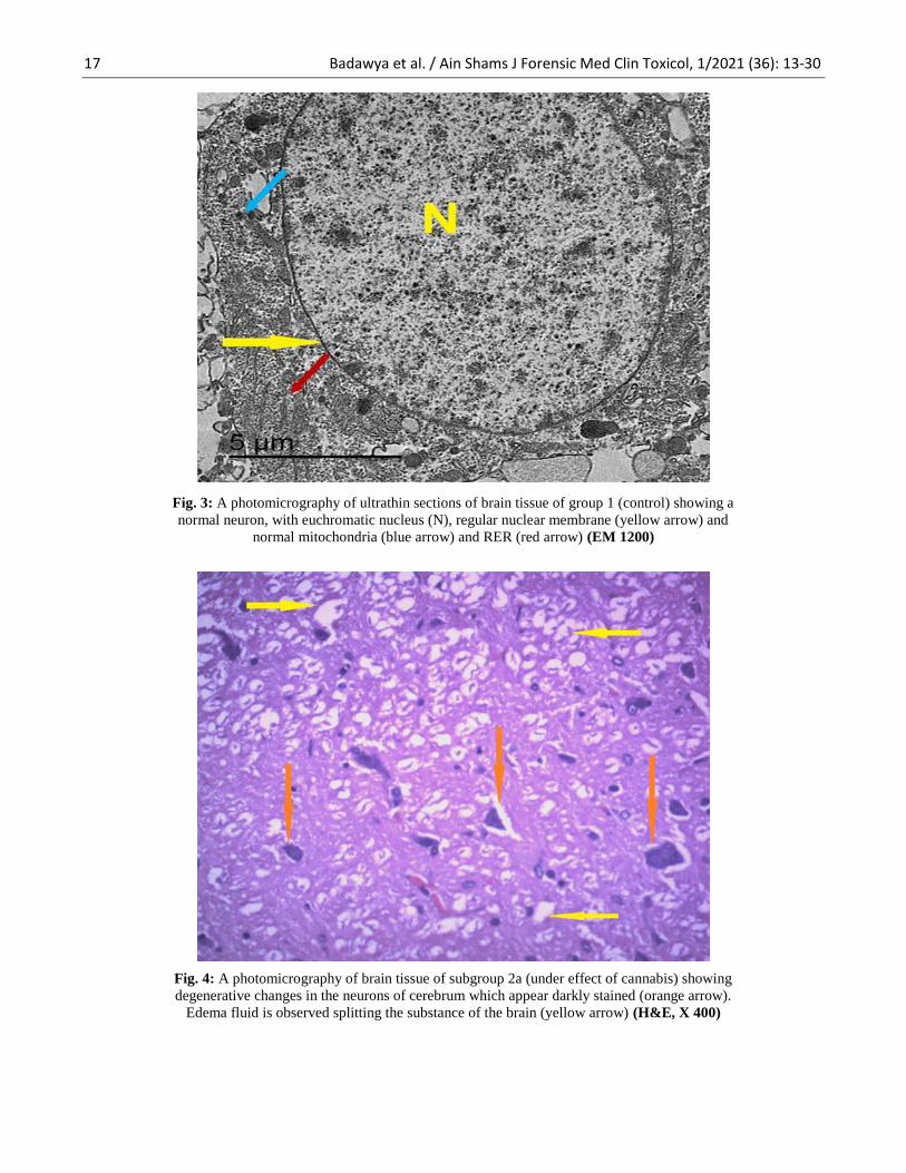
Fig. 2: A photomicrography of brain tissue of group 1 (control) showing the normal appearance
of neurons with vesicular open face nuclei and blue stained nissl granules in the cytoplasm
(arrow) (T.B X 1000)
17 Badawya et al. / Ain Shams J Forensic Med Clin Toxicol, 1/2021 (36): 13-30
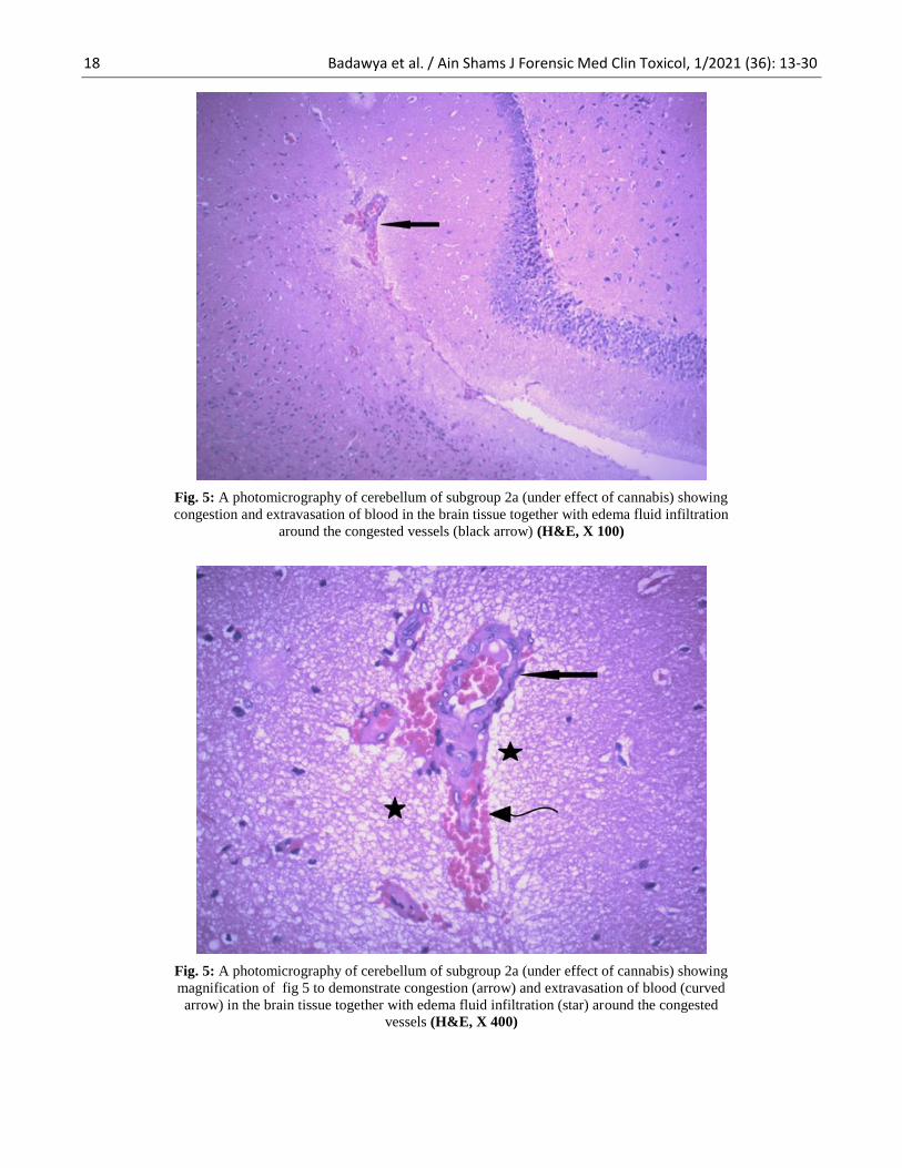
Fig. 3: A photomicrography of ultrathin sections of brain tissue of group 1 (control) showing a
normal neuron, with euchromatic nucleus (N), regular nuclear membrane (yellow arrow) and
normal mitochondria (blue arrow) and RER (red arrow) (EM 1200)
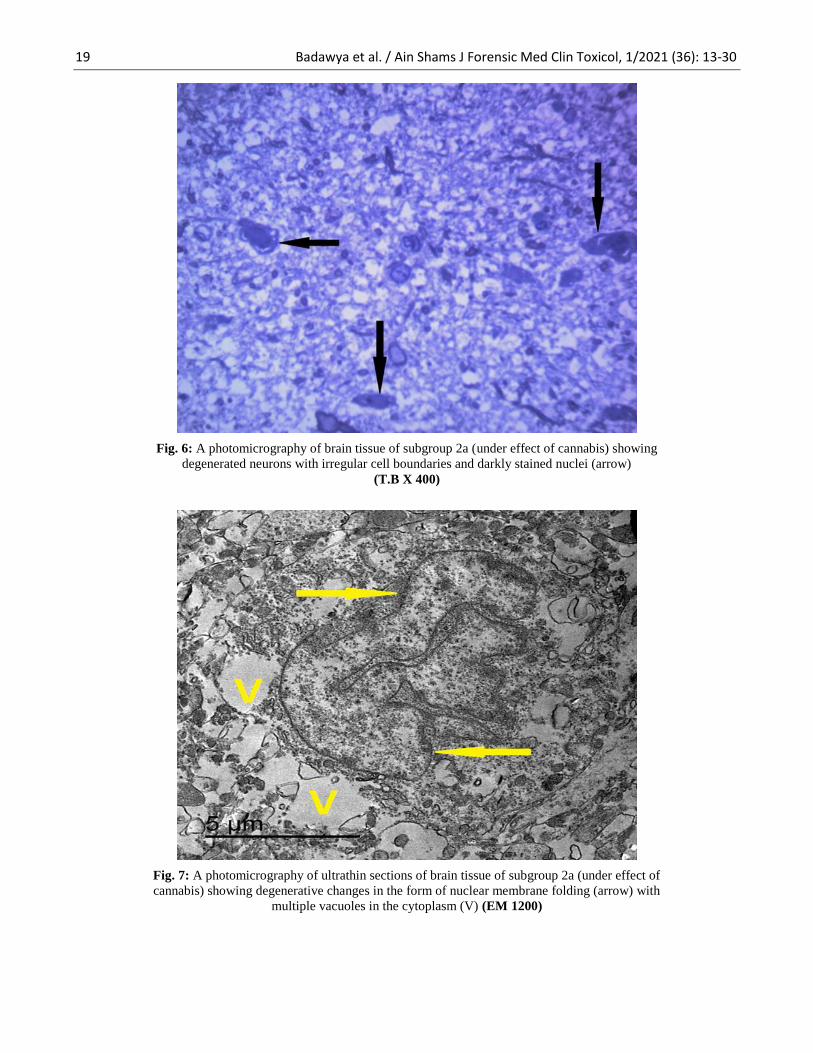
Fig. 4: A photomicrography of brain tissue of subgroup 2a (under effect of cannabis) showing
degenerative changes in the neurons of cerebrum which appear darkly stained (orange arrow).
Edema fluid is observed splitting the substance of the brain (yellow arrow) (H&E, X 400)
18 Badawya et al. / Ain Shams J Forensic Med Clin Toxicol, 1/2021 (36): 13-30
Fig. 5: A photomicrography of cerebellum of subgroup 2a (under effect of cannabis) showing
congestion and extravasation of blood in the brain tissue together with edema fluid infiltration
around the congested vessels (black arrow) (H&E, X 100)
Fig. 5: A photomicrography of cerebellum of subgroup 2a (under effect of cannabis) showing
magnification of fig 5 to demonstrate congestion (arrow) and extravasation of blood (curved
arrow) in the brain tissue together with edema fluid infiltration (star) around the congested
vessels (H&E, X 400)
19 Badawya et al. / Ain Shams J Forensic Med Clin Toxicol, 1/2021 (36): 13-30
Fig. 6: A photomicrography of brain tissue of subgroup 2a (under effect of cannabis) showing
degenerated neurons with irregular cell boundaries and darkly stained nuclei (arrow)
(T.B X 400)
Fig. 7: A photomicrography of ultrathin sections of brain tissue of subgroup 2a (under effect of
cannabis) showing degenerative changes in the form of nuclear membrane folding (arrow) with
multiple vacuoles in the cytoplasm (V) (EM 1200)
20 Badawya et al. / Ain Shams J Forensic Med Clin Toxicol, 1/2021 (36): 13-30
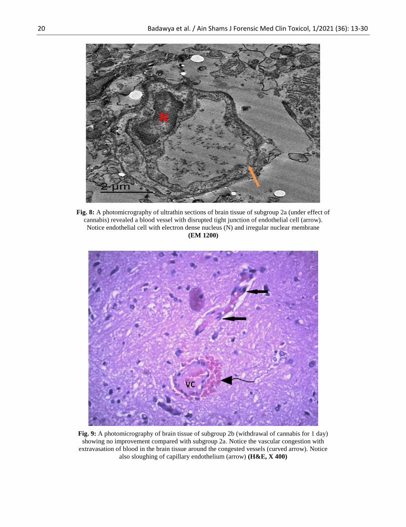
Fig. 8: A photomicrography of ultrathin sections of brain tissue of subgroup 2a (under effect of
cannabis) revealed a blood vessel with disrupted tight junction of endothelial cell (arrow).
Notice endothelial cell with electron dense nucleus (N) and irregular nuclear membrane
(EM 1200)
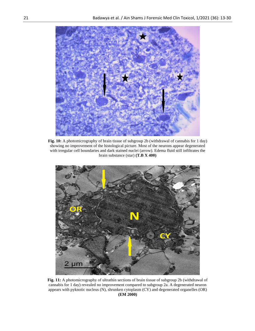
Fig. 9: A photomicrography of brain tissue of subgroup 2b (withdrawal of cannabis for 1 day)
showing no improvement compared with subgroup 2a. Notice the vascular congestion with
extravasation of blood in the brain tissue around the congested vessels (curved arrow). Notice
also sloughing of capillary endothelium (arrow) (H&E, X 400)
21 Badawya et al. / Ain Shams J Forensic Med Clin Toxicol, 1/2021 (36): 13-30
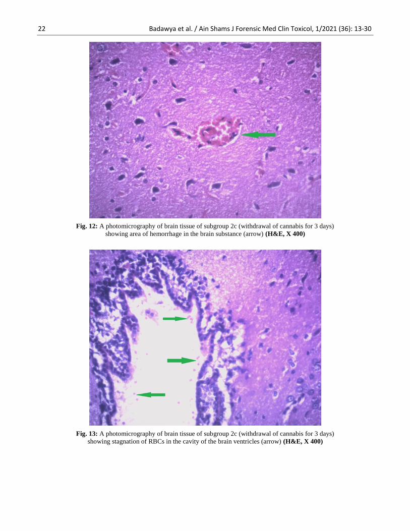
Fig. 10: A photomicrography of brain tissue of subgroup 2b (withdrawal of cannabis for 1 day)
showing no improvement of the histological picture. Most of the neurons appear degenerated
with irregular cell boundaries and dark stained nuclei (arrow). Edema fluid still infiltrates the
brain substance (star) (T.B X 400)
Fig. 11: A photomicrography of ultrathin sections of brain tissue of subgroup 2b (withdrawal of
cannabis for 1 day) revealed no improvement compared to subgroup 2a. A degenerated neuron
appears with pyknotic nucleus (N), shrunken cytoplasm (CY) and degenerated organelles (OR)
(EM 2000)
22 Badawya et al. / Ain Shams J Forensic Med Clin Toxicol, 1/2021 (36): 13-30
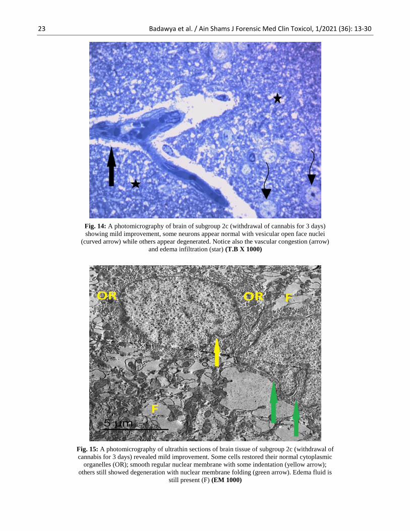
Fig. 12: A photomicrography of brain tissue of subgroup 2c (withdrawal of cannabis for 3 days)
showing area of hemorrhage in the brain substance (arrow) (H&E, X 400)
Fig. 13: A photomicrography of brain tissue of subgroup 2c (withdrawal of cannabis for 3 days)
showing stagnation of RBCs in the cavity of the brain ventricles (arrow) (H&E, X 400)
23 Badawya et al. / Ain Shams J Forensic Med Clin Toxicol, 1/2021 (36): 13-30
Fig. 14: A photomicrography of brain of subgroup 2c (withdrawal of cannabis for 3 days)
showing mild improvement, some neurons appear normal with vesicular open face nuclei
(curved arrow) while others appear degenerated. Notice also the vascular congestion (arrow)
and edema infiltration (star) (T.B X 1000)
Fig. 15: A photomicrography of ultrathin sections of brain tissue of subgroup 2c (withdrawal of
cannabis for 3 days) revealed mild improvement. Some cells restored their normal cytoplasmic
organelles (OR); smooth regular nuclear membrane with some indentation (yellow arrow);
others still showed degeneration with nuclear membrane folding (green arrow). Edema fluid is
still present (F) (EM 1000)
24 Badawya et al. / Ain Shams J Forensic Med Clin Toxicol, 1/2021 (36): 13-30
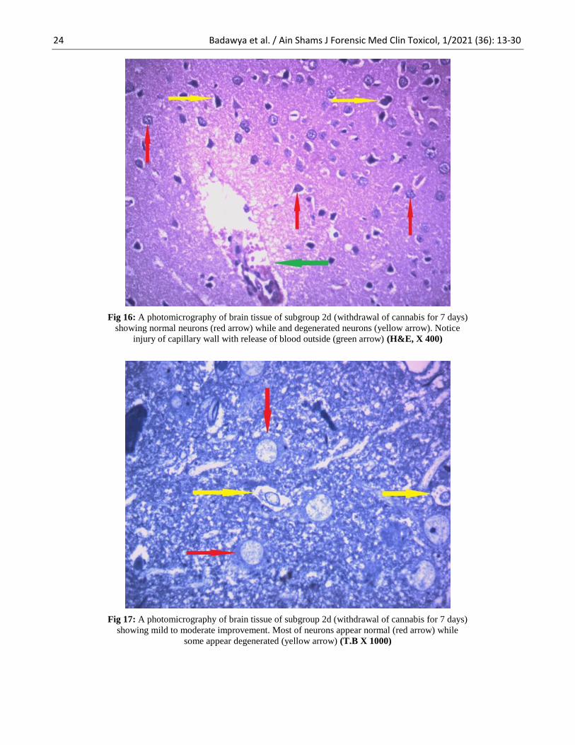
Fig 16: A photomicrography of brain tissue of subgroup 2d (withdrawal of cannabis for 7 days)
showing normal neurons (red arrow) while and degenerated neurons (yellow arrow). Notice
injury of capillary wall with release of blood outside (green arrow) (H&E, X 400)
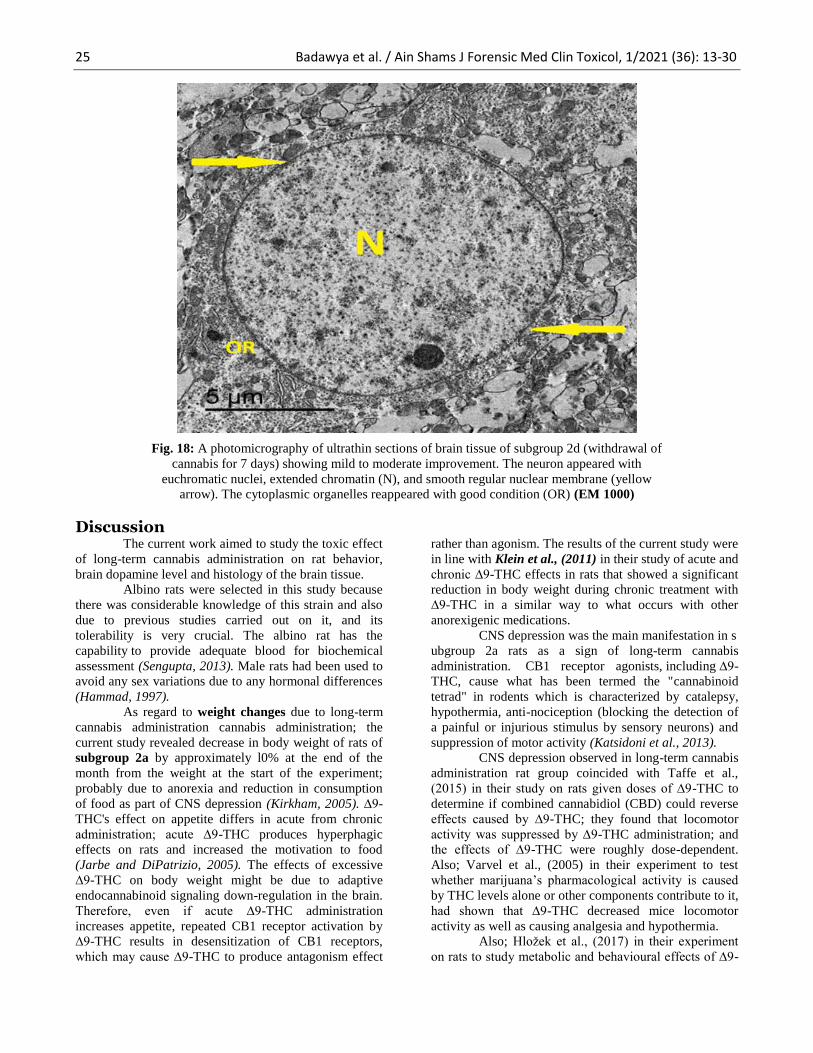
Fig 17: A photomicrography of brain tissue of subgroup 2d (withdrawal of cannabis for 7 days)
showing mild to moderate improvement. Most of neurons appear normal (red arrow) while
some appear degenerated (yellow arrow) (T.B X 1000)
25 Badawya et al. / Ain Shams J Forensic Med Clin Toxicol, 1/2021 (36): 13-30
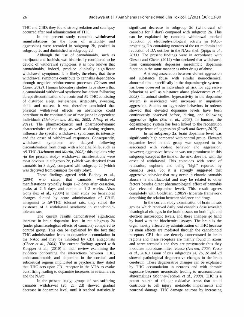
Fig. 18: A photomicrography of ultrathin sections of brain tissue of subgroup 2d (withdrawal of
cannabis for 7 days) showing mild to moderate improvement. The neuron appeared with
euchromatic nuclei, extended chromatin (N), and smooth regular nuclear membrane (yellow
arrow). The cytoplasmic organelles reappeared with good condition (OR) (EM 1000)
Discussion The current work aimed to study the toxic effect
of long-term cannabis administration on rat behavior,
brain dopamine level and histology of the brain tissue.
Albino rats were selected in this study because
there was considerable knowledge of this strain and also
due to previous studies carried out on it, and its
tolerability is very crucial. The albino rat has the
capability to provide adequate blood for biochemical
assessment (Sengupta, 2013). Male rats had been used to
avoid any sex variations due to any hormonal differences
(Hammad, 1997).
As regard to weight changes due to long-term
cannabis administration cannabis administration; the
current study revealed decrease in body weight of rats of
subgroup 2a by approximately l0% at the end of the
month from the weight at the start of the experiment;
probably due to anorexia and reduction in consumption
of food as part of CNS depression (Kirkham, 2005). ∆9-
THC's effect on appetite differs in acute from chronic
administration; acute ∆9-THC produces hyperphagic
effects on rats and increased the motivation to food
(Jarbe and DiPatrizio, 2005). The effects of excessive
∆9-THC on body weight might be due to adaptive
endocannabinoid signaling down-regulation in the brain.
Therefore, even if acute ∆9-THC administration
increases appetite, repeated CB1 receptor activation by
∆9-THC results in desensitization of CB1 receptors,
which may cause ∆9-THC to produce antagonism effect
rather than agonism. The results of the current study were
in line with Klein et al., (2011) in their study of acute and
chronic ∆9-THC effects in rats that showed a significant
reduction in body weight during chronic treatment with
∆9-THC in a similar way to what occurs with other
anorexigenic medications.
CNS depression was the main manifestation in s
ubgroup 2a rats as a sign of long-term cannabis
administration. CB1 receptor agonists, including ∆9-
THC, cause what has been termed the "cannabinoid
tetrad" in rodents which is characterized by catalepsy,
hypothermia, anti-nociception (blocking the detection of
a painful or injurious stimulus by sensory neurons) and
suppression of motor activity (Katsidoni et al., 2013).
CNS depression observed in long-term cannabis
administration rat group coincided with Taffe et al.,
(2015) in their study on rats given doses of ∆9-THC to
determine if combined cannabidiol (CBD) could reverse
effects caused by ∆9-THC; they found that locomotor
activity was suppressed by ∆9-THC administration; and
the effects of ∆9-THC were roughly dose-dependent.
Also; Varvel et al., (2005) in their experiment to test
whether marijuana’s pharmacological activity is caused
by THC levels alone or other components contribute to it,
had shown that ∆9-THC decreased mice locomotor
activity as well as causing analgesia and hypothermia.
Also; Hložek et al., (2017) in their experiment
on rats to study metabolic and behavioural effects of ∆9-
26 Badawya et al. / Ain Shams J Forensic Med Clin Toxicol, 1/2021 (36): 13-30
THC and CBD; they found strong sedation and catalepsy
occurred after oral administration of THC.
In the present study cannabis withdrawal
manifestations (in the form of irritability and
aggression) were recorded in subgroup 2b, peaked in
subgroup 2c and diminished in subgroup 2d.
Although the use of cannabinoids, such as
marijuana and hashish, was historically considered to be
devoid of withdrawal symptoms, it is now known that
cannabinoids, indeed, produce clinically significant
withdrawal symptoms. It is likely, therefore, that these
withdrawal symptoms contribute to cannabis dependence
through negative reinforcement processes (Oleson and
Cheer, 2012). Human laboratory studies have shown that
a cannabinoid withdrawal syndrome has arisen following
abrupt discontinuation of chronic oral ∆9-THC consisting
of disturbed sleep, restlessness, irritability, sweating,
chills and nausea. It was therefore concluded that
physical withdrawal effects on abstinence could
contribute to the continued use of marijuana in dependent
individuals (Lichtman and Martin, 2002; Allsop et al.,
2011). The pharmacokinetic and pharmacodynamic
characteristics of the drug, as well as dosing regimen,
influence the specific withdrawal syndrome, its intensity
and the onset of withdrawal responses. Consequently,
withdrawal symptoms are delayed following
discontinuation from drugs with a long half-life, such as
Δ9-THC (Lichtman and Martin, 2002); this explains why
-in the present study- withdrawal manifestations were
most obvious in subgroup 2c, (which was deprived from
cannabis for 3 days), compared with subgroup 2b (which
was deprived from cannabis for only 1day).
These findings agreed with Budney et al.,
(2004) who stated that cannabis withdrawal
manifestations typically begin 1–2 days after cessation,
peaks at 2–6 days and remits at 1–2 weeks. Also;
Gonz´alez et al., (2004) in their study on behavioral
changes elicited by acute administration of CB1R
antagonist to Δ9-THC tolerant rats, they stated the
existence of a withdrawal syndrome in cannabinoid-
tolerant rats.
The current results demonstrated significant
increase in brain dopamine level in rat subgroup 2a
(under pharmacological effects of cannabis) compared to
control group. This can be explained by the fact that
THC administration leads to dopamine accumulation in
the NAcc and may be inhibited by CB1 antagonists
(Cheer et al., 2004). The current findings agreed with
Kuepper et al., (2010) in their review examining the
evidence concerning the interactions between THC,
endocannabinoids and dopamine in the cortical and
subcortical regions implicated in psychosis; they stated
that THC acts upon CB1 receptor in the VTA to evoke
burst firing leading to dopamine increases in striatal areas
and the NAcc.
In the present study; groups of rats suffering
cannabis withdrawal (2b, 2c, 2d) showed gradual
decrease in dopamine level, until it reached statistically
significant decrease in subgroup 2d (withdrawal of
cannabis for 7 days) compared with subgroup 2a. This
can be explained by cannabis withdrawal marked
reduction of electrophysiological activity in NAcc-
projecting DA containing neurons of the rat midbrain and
reduction of DA outflow in the NAcc shell (Spiga et al.,
2011). The present findings were in accordance with
Oleson and Cheer, (2012) who declared that withdrawal
from cannabinoids depresses mesolimbic dopamine
function in the same manner as other drugs of abuse.
A strong association between violent aggression
and substance abuse with similar neurochemical
abnormalities - specifically in the dopaminergic system -
has been observed in individuals at risk for aggressive
behavior as well as substance abuse (Soderstrom et al.,
2003). In animal studies, hyperactivity in the dopamine
system is associated with increases in impulsive
aggression. Studies on aggressive behaviors in rodents
showed that elevated dopamine levels have been
continuously observed before, during, and following
aggressive fights (Seo et al., 2008). In humans, the
dopaminergic system has been linked to the recognition
and experience of aggression (Rosell and Siever, 2015).
In rat subgroup 2a; brain dopamine level was
significantly high compared with control group. Elevated
dopamine level in this group was supposed to be
associated with violent behavior and aggression;
however; aggressive behavior was not prominent in this
subgroup except at the time of the next dose i.e. with the
onset of withdrawal. This coincides with sense of
relaxation, euphoria and being "high" reported by
cannabis users. So; it is strongly suggested that
aggressive behavior that may occur in chronic cannabis
abusers is multifactorial and may be related to other
factors besides direct pharmacological effect of cannabis
(i.e. elevated dopamine level). This result agrees
completely with Goldstien, (1985's) tripartite framework
describing the relation between violence and drugs.
In the current study examination of brain in rats
groups which received daily oral cannabis dose revealed
histological changes in the brain tissues on both light and
electron microscopic levels, and these changes go hand
by hand with the biochemical assays. The brain is the
organ mostly affected by administration of THC because
its main effects are mediated through the cannabinoid
receptors CB1 that are densely concentrated in brain
regions and these receptors are mainly found in axons
and nerve terminals and they are presynaptic thus they
modulate neurotransmitter release (Iversen, 2003; Yassa
et al., 2010). Brain of rats subgroups 2a, 2b, 2c and 2d
showed pathological degenerative changes in the brain
cerebrum. These degenerative changes can be explained
by THC accumulation in neurons and with chronic
exposure becomes neurotoxic leading to neuroanatomic
abnormalities (Monnet-Tschudi et al., 2008). THC is a
potent source of cellular oxidative stress that could
contribute to cell injury, metabolic impairments and
neuronal damage. THC damage neurons by increasing
27 Badawya et al. / Ain Shams J Forensic Med Clin Toxicol, 1/2021 (36): 13-30
cyclooxygenase (COX) that catalyze the formation of
reactive oxygen spices (ROS), it also alters archidonic
acid metabolism and increases the production of
lipooxygenase products that are prooxidants (Sarafian
and Marques, 1999). Oxidative stress produced can also
lead to the opening of inter-endothelial junctions and
increased vascular permeability (Mittal et al., 2014). This
explain the current study evidence of vasculopathy in
cannabis treated rats; in the form of disturbance of the
tight junction between endothelial cells raveled by EM;
with subsequent increase in the vascular permeability and
filtration of edema fluid in between the nerve fibers of
the white matter.
The degenerative changes observed in the
current study as shrunken neurons with irregular cell
boundaries and darkly stained nuclei, nuclear membrane
folding, chromatolysis, condensed peripheral chromatin,
destruction of cellular organelles, multiple vacuoles in
the cytoplasm and loss of cell membrane; were reported
by Proskuryakov et al.,(2003) and Festjens et al., (2006)
as signs of necrosis and apoptosis. Since neurons are
permanent cells, they are most susceptible to hypoxic
injury as that occurs with cannabis. Cannabis causes
significant decrease in oxygen consumption and
significant increase in mitochondrial hydrogen peroxide
production and so changes in integrated mitochondrial
function and inhibitions of mitochondrial respiratory
chain (Athanasiou et al., 2007).
Current histopathological findings were in
accordance with Yassa et al., (2010) who reported that
the brain tissue is the organ mostly affected by cannabis
and they found noticeable brain tissue affection in the
form of irregular shrunken cells with dense nuclei and
vacuolated cytoplasm in all layers. Also; Nafea et al.,
(2016) found that in cannabis dependent rat group; HX
&E stained sections of rats' brain showed congested
meningeal blood vessels, irregular shrunken cells and
vacuolated cytoplasm. The cells were surrounded by
irregular wide spaces. Also, congested blood vessels and
dilated perivascular spaces were present.
Unfortunately, after withdrawal, the complete
reversibility wasn't achieved. This was in accordance
with Nafea et al., (2016) who stated that in cannabis–
withdrawn group; examination of rats' brain still showed
some pathological changes represented by inflammatory
cellular infiltration, vacuolated cytoplasm and area of
gliosis. The partial recovery of brain tissue after cannabis
withdrawal could be explained by the function of
endocannabinoid system (eCBs). However, non–eCBs
independent mechanism is superior in counteracting
neuropathological events (Bartsch et al., 2007).
Conclusion Long-term cannabis administration causes
degenerative pathological changes in the brain, which
may explain the literature well documented effect of
cannabis on cognition, memory and learning.
aggressive behavior was prominent in rat group
sacrificed after withdrawal of cannabis rather than rat
group sacrificed under the pharmacological effect of
cannabis; so; it is strongly suggested that aggressive
behavior that may occur in chronic cannabis abusers is
multifactorial and may be related to other factors besides
direct pharmacological effect of cannabis (i.e. elevated
dopamine level).
Also; degenerative histopathological changes
didn't completely returned normal; this may indicates the
need for longer recovery period after stoppage of
cannabis.
References Allsop, D.J.; Norberg, M.M.; Copeland, J. et al., (2011):
The Cannabis Withdrawal Scale development:
patterns and predictors of cannabis withdrawal
and distress. Drug Alcohol Depend., 119: 123–129
American Psychiatric Association (2013): Diagnostic and
Statistical Manual of Mental Disorders (DSM–
5®), 5th ed.; 571-4. American Psychiatric
Publishing: Washington, DC, USA.
Athanasiou, A.; Clarke, A.B.; Turner, A.E. et al., (2007):
Cannabinoid receptor agonist are mitochondrial
inibitors: A unified hypothesis of how
cannabinoids modulate mitochondrial function
and induce cell death. Biochemical and
Biophysical Research communications, 364: 131-
137
Bartsch, A.J.; Homola; G., Biller, A. et al., (2007):
Manifestations of early brain recovery associated
with abstinence from alcoholism. Brain,
130(1):36-47.
Budney, A.J.; Hughes, J.R.; Moore, B.A. et al., (2004):
Review of the validity and significance of
cannabis. Am. J. Psychiatry, 161: 1967–1977
Carlton, M.A. (1982): Histopathological techniques. 1st
edition. Oxford University Press. New York,
Toronto.
Cheer, J.F.; Wassum, K.M.; Heien, M.L.A.V. et al.,
(2004): Cannabinoids enhance subsecond
dopamine release in the nucleus accumbens of
awake rats. J. Neurosci, 24: 4393–4400.
Dawson, B. and Trapp, R.G. (2001): Basic and clinical
biostatistics: lange medical books. Oxford,
London. Boston, McGraw-Hill, Medical
Publishing Division. 3rd edition, Chapter 7-9,
pages: 161-218.
El-Alfy, A.T.; Ivey, K. and Robinson, K. (2010):
Antidepressant-like effect of Δ9-
tetrahydrocannabinol and other cannabinoids
isolated from Cannabis sativa L. Pharmacology,
Biochemistry and Behavior, 95: 434–442
Fanarioti, E.; Mavrikaki, M.; Panagis, G. et al., (2015):
Behavioral and Neurochemical Changes in
Mesostriatal Dopaminergic Regions of the Rat
after Chronic Administration of the Cannabinoid
28 Badawya et al. / Ain Shams J Forensic Med Clin Toxicol, 1/2021 (36): 13-30
Receptor Agonist WIN55,212-2. International
Journal of Neuropsychopharmacology,18(6): 1–
17
Festjens, N.; Vanden Berghe, T. and Vandenabeele, P.
(2006): "Necrosis, a well-orchestrated form of cell
demise: Signalling cascades, important mediators
and concomitant immune response". Biochimica
et Biophysica Acta (BBA) - Bioenergetics.
Mitochondria: from Molecular Insight to
Physiology and Pathology; 1757 (9–10): 1371–
1387
Fitzgerald, K.T.; Bronstein, A.C. and Newquist, K.L.
(2013): Marijuana poisoning. Top Companion
Anim Med., 28(1):8-12
Goldstein, P.J. (1985): The drugs/violence nexus: a
tripartite conceptual framework. Journal of Drug
Issues, 15: 493–506.
Gonz´alez, S.; Fernández-Ruiz, J.; Di Marzo, V. et al.,
(2004): Behavioral and molecular changes elicited
by acute administration of SR141716 to 9-
tetrahydrocannabinol-tolerant rats: an
experimental model of cannabinoid abstinence.
Drug and Alcohol Dependence, 74: 159–170
Hammad, S.A. (1997): Experimental study on toxicity of
acetaminophen in normal and bilharzially infested
animals. Doctorate thesis in forensic medicine and
toxicology. Faculty of medicine, Menoufia
University: 290-294
Hložek, T.; Uttl, L.; Kadeřábekc, L. et al., (2017):
Pharmacokinetic and behavioural profile of THC,
CBD, and THC+CBD combination after
pulmonary, oral, and subcutaneous administration
in rats and confirmation of conversion in vivo of
CBD to THC. European
Neuropsychopharmacology, 27: 1223–1237
Iversen, L. (2003): Cannabis and the brain. Brain.,
126(6):1252–70.
Jaffe, M. (1969): Jaffe Method. In Manual clinical
laboratory diagnosis. Levinson C, Mac Fate C. 7th
edition. Page: 117.
Jarbe, T.U. and DiPatrizio, N.V. (2005): Delta9-THC
induced hyperphagia and tolerance assessment:
interactions between the CB1 receptor agonist
delta9-THC and the CB1 receptor antagonist SR-
141716 (rimonabant) in rats. Behav Pharmacol,
16(5–6):373–80.
Karnovsky, M.J. (1965): A formaldehyde -glutaraldehyde
fixative of high osmolality for use in electron
microscopy. J. Cell Biol, 27, 137-138.
Katsidoni, V.; Kastellakis, A. and Panagis, G. (2013):
Biphasic effects of Delta 9- tetrahydrocannabinol
on brain stimulation reward and motor
activity.Int.J.Neuropsychopharmacol., 16: 2273–
2284.
Kiernan, J.A. (1999): Histopathological and
histochemical methods: theory and practice. 3rd
edition. Page: 105. Buterworth Heimann.
Kirkham, T.C. (2005): Endocannabinoids in the
regulation of appetite and body weight. Behav
Pharmacol, 16(5–6):297–313.
Klein, C.; Karanges, E.; Spiro, A. et al., (2011):
Cannabidiol potentiates delta (9)-
tetrahydrocannabinol (THC) behavioural effects
and alters THC pharmacokinetics during acute
and chronic treatment in adolescent rats.
Psychopharmacology (Berl), 218(2): 443–57
Kuepper, R.; Morrison, P.D.; Os, J.V. et al., (2010): Does
dopamine mediate the psychosis-inducing effects
of cannabis? A review and integration of findings
across disciplines. Schizophrenia Research, 121:
107–117
Lichtman, A.H. and Martin, B.R. (2002): Marijuana
Withdrawal Syndrome in the Animal Model.
Journal of Clinical Pharmacology, 42:20S-27S
Mittal, M.; Siddiqui, M.R.; Tran, K. et al., (2014):
Reactive Oxygen Species in Inflammation and
Tissue Injury Antioxidants & Redox Signaling,
20(7): 1126- 1176
Monnet-Tschudi, F.; Hazekamp, A.; Perret, N. et al.,
(2008): Delta-9-tetrahydrocannabinol
accumulation, metabolism and cell-type-specific
adverse effects in aggregating brain cell cultures.
Toxicol Appl Pharmacol, 228:8–16.
Nafea, O.E; ElKhishin IA, Awad OA et al., (2016): A
study of the neurotoxic effects of tramadol and
Cannabis in adolescent male albino rats Int J Sci
Rep., 2(7):143-154
Nagai, H.; Egashira, N.; Sano, K. et al., (2006):
Antipsychotics improve Delta 9-
tetrahydrocannabinol- induced impairment of the
prepulse inhibition of the startle reflex in mice.
Pharmacol.Biochem.Behav.; 84, 330–336.
Oleson, E.B. and Cheer, J.F. (2012): A Brain on
Cannabinoids: The Role of Dopamine Release in
Reward Seeking. Cold Spring Harbor Perspectives
in Medicine, 2(8): a012229–a012229.
Proskuryakov, S.Y.; Konoplyannikov, A.G. and Gabai,
V.L. (2003): "Necrosis: a specific form of
programmed cell death?". Experimental Cell
Research, 283 (1): 1–16.
Radwan, M.M.; ElSohly, M.A.; El-Alfy, A.T. et al.,
(2015): Isolation and Pharmacological Evaluation
of Minor Cannabinoids from High Potency
Cannabis sativa. J Nat Prod., 78(6): 1271-6.
Ramesh, D.; Schlosburg, J.E.; Wiebelhaus, J.M.; et al.,
(2011): Marijuana dependence: not just smoke and
mirrors. ILAR journal / National Research
Council, Institute of Laboratory Animal
Resources., 52: 295–308.
Reynolds, E.S. (1963): The use of lead citrate at high pH
as an electron-opaque stain in electron
microscopy. J. Cell Biol., 17, 208-212
Rosell, D.R. and Siever, L.J. (2015): The neurobiology of
aggression and violence. CNS Spectrums, 20:
254–279
29 Badawya et al. / Ain Shams J Forensic Med Clin Toxicol, 1/2021 (36): 13-30
Sarafian, T.A. and Marques, J.A. (1999): Oxidative stress
produced by marijuana smoke: an adverse effect
enhanced by cannabinoids. Am.J.Respir. CellMol.
Biol., 20(6): 1286- 1293.
Sengupta, P. (2013): The Laboratory Rat: Relating Its
Age With Human's. Int J Prev Med., 4(6): 624–
630.
Seo, D.; Patrick, C.J. and Kennealy, P.J. (2008): Role of
Serotonin and Dopamine System Interactions in
the Neurobiology of Impulsive Aggression and its
Comorbidity with other Clinical Disorders
Aggress Violent Behav., 13(5): 383–395.
Smith, J.E.; Lane, J.D.; Shea, P.A. et al., (1975): A
method of concurrent measurement of picomol
quataties of acetylcholine, choline, dopamine,
norepinephrine, serotonin, 5- hydroxytryptophan,
tyrosine, glycine, aspartate, glutamate, analine and
gamma-amino-butyric acid in single tissue
samples from different areas of rat central nervous
system. Anal Biochem., 64: 149- 169
Soderstrom, H.; Blennow, K.; Sjodin, A.K. and Forsman,
A. (2003): New evidence for an association
between the CSF HVA:5-HIAA ratio and
psychopathic traits. Journal of Neurology
Neurosurgery and Psychiatry., 74:918–921.
Spiga, S.; Lintas, A. and Diana, M. (2011): Altered
Mesolimbic Dopamine System in THC
Dependence. Current Neuropharmacology, 9: 200-
204
Taffe, M.A.; Creehan, K.M. and Vandewater, S.A.
(2015): Cannabidiol fails to reverse hypothermia
or locomotor suppression induced by Δ9-
tetrahydrocannabinol in Sprague-Dawley rats.
British Journal of Pharmacology, 127(7): 1-9
Varvel, S.A.; Bridgen, D.T.; Tao, Q. et al., (2005): 9-
Tetrahydrocannbinol Accounts for the
Antinociceptive, Hypothermic, and Cataleptic
Effects of Marijuana in Mice. The journal of
pharmacology and experimental therapeutics,
314(1): 329- 337.
Volkow, N;D.; Wang, G.J.; Fowler, J.S. et al., (2011):
Addiction: beyond dopamine reward circuitry.
Proc. Natl Acad. Sci. USA 108: 15037-15042
Wiley, J.L. and Burston, J.J. (2014): Sex differences in
Delta (9)-tetra- hydrocannabinol metabolism and
in vivo pharmacology following acute and
repeated dosing in adolescent rats. Neurosci.Lett.,
576: 51–55.
Yassa, H.A., Dawood, A.E.A., Shehata, M.M. et al.,
(2010): Subchronic toxicity of cannabis leaves on
male albino rats. Human and Experimental
Toxicology, 29(1): 37–47

30 Badawya et al. / Ain Shams J Forensic Med Clin Toxicol, 1/2021 (36): 13-30
: الببلغة البيضبء الفئزان لذكور المخ أنسجة على للحشيش الإنسحبة تأثيز المتكزروكذلك التعبطى تأثيز دراسة
وبيوكيميبئية هستولوجية دراسة
1الفرعوني حسن ريهام حبيب، لزمود نجوى الصعيدى، لزمد أميرة أمين، الظاهر عبد صفاء بدوي، مصطفي سامى الملخص العزبي
:المقدمة الإدراك على القنب تأثير أن كما ، الدماغ في تنكسية مرضية تغيرات في الدزمن القنب تعاطي يتسبب قد. مصر في انتشاراً تعاطيًا لسدرات أكثر مكانة الحشيش يحتل
الدماغ في الدوبامين ومستوى الفئران سلوك على الحشيش من الدزمنة الدتكررة للجرعات السام التأثير دراسة إلى الحالي العمل يهدف. جيد معروف والتعلم والذاكرة .للدماغ الدرضية النسيجية والتغيرات : البحث وطرق الحالات
. الحالات بين شيوعا الإعتماد مواد كأكثر الدراسة من الأول الجزء انتهاء بعد الحشيش اختيار تم وقد البالغين، البيضاء الجرذان ذكور علي الحشيش لتأثير تجريبية دراسة :رئيسيتين لرموعتين إلي تقسيمهم وتم جم، 011-181 بين ما وزنهم يتراوح الذين البالغين الجرذان ذكور من خمسين علي الدراسة إجراء تم
فئران عشرة من تتكون(: الضابطة) 1 لرموعة لرموعة(: فئران عشرة منها كلا) لرموعات أربعة إلي المجموعة هذه تقسم ثم شهر، لددة متزايدة يومية بجرعات الحشيش اعطائهم تم فأرا أربعين من تتكون: 0 لرموعة
التعاطى عن الناتجة الإكلينيكية الأعراض متابعة تم(. الحشيش تعاطى توقف من لستلفة لفترات تعريضهم تم) د -0 لرموعة ج، -0 لرموعة ب، -0 لرموعة أ، -0 للفحص الدخ من عينة أخذ وتم الدخ انسجة في الدوبامين لنسبة تحليل عمل تم التجريبية الدراسة فترة بنهاية ، الانسحاب عن الناتجة وكذلك للحشيش الدتكرر
.الالكتروني الديكروسكوب الضوئى الديكروسكوب باستخدام الذستولوجي : النتائج
ملاحظتها تم التي الرئيئسية الأعراض بينما الدركزي، العصبي الجهاز تثبيط كان أ-0 لرموعة فى للحشيش الدتكرر التعاطى لتأثير الأساسية الأعراض أن وجد إكلينيكيا. الضابطة بالمجموعة مقارنة عالية كانت أ-0 المجموعة فئران فى الدخ بأنسجة الدوبامين نسبة. البعض بعضهم ،وضرب العدوان ، التهيج كانت ج-0 المجموعة فى
.العصبية بالخلايا ضموروانحلال الدخ لخلايا المجهري الفحص أظهر : الإستنتاج
والذى العصبية، بالخلايا ضموروانحلال الدخ لخلايا المجهري الفحص أظهر. الضابطة بالمجموعة مقارنة عالية كانت أ-0 المجموعة فئران فى الدخ بأنسجة الدوبامين نسبة .طويلة تعافى فترات إلى يحتاج ما غالبا
الونوفيه جاهعه الطب، كليه الاكلينيكيه، والسووم الشرعي الطب قسن .1


![The prolongation of the lifespan of rats by repeated oral ... · The prolongation of the lifespan of rats by repeated oral administration of [60] fullerene Tarek Baatia,b, Fanchon](https://img.pdfslide.net/doc/110x75/5b95ff4209d3f2c2678cf472/the-prolongation-of-the-lifespan-of-rats-by-repeated-oral-the-prolongation.jpg)