Embed Size (px)
Citation preview

STUDY OF HISTOLOGY OF THYMUS GLAND -
VARIOUS FOETAL AGE GROUPS
Dissertation Submitted for
M.D. Degree Branch - XXIII
[ANATOMY]
DEPARTMENT OF ANATOMY
THANJAVUR MEDICAL COLLEGE
THANJAVUR
THE TAMILNADU DR.MGR MEDICAL UNIVERSITY,
CHENNAI
APRIL - 2016

CERTIFICATE
This is to certify that dissertation titled “STUDY OF HISTOLOGY
OF THYMUS GLAND - VARIOUS FOETAL AGE GROUPS” is a
bonafide work done by Dr.J.GAYATHRI under my guidance and
supervision in the Department of Anatomy, Thanjavur Medical College,
Thanjavur during her post graduate course from 2013 to 2016.
(Dr.M.SINGARAVELU, M.D.,) DR.T.SIVAKAMI,(M.S)
THE DEAN Professor and Head
Thanjavur Medical College Department of Anatomy
Thanjavur - 4. Thanjavur Medical College,
Thanjavur -4.

DECLARATION
I, Dr.J.GAYATHRI hereby solemnly declare that the dissertation
title “STUDY OF HISTOLOGY OF THYMUS GLAND - VARIOUS
FOETAL AGE GROUPS” was done by me at Thanjavur Medical College
and Hospital, Thanjavur under supervision and guidance of my professor
and head Dr.T.Sivakami.M.S., This dissertation is submitted to Tamil
Nadu Dr.M.G.R Medical University, towards partial fulfillment of
requirement for the award of M.D. Degree (Branch-XXIII) in Anatomy.
Place : Thanjavur.
Date : Dr.J.GAYATHRI

GUIDE CERTIFICATE
GUIDE
PROF.DR.T.SIVAKAMI, M.S.,
THE PROFESSOR AND HEAD
Department of anatomy,
Thanjavur medical college & Hospital,
Thanjavur.
Remark of the guide:
The work done by DR.J.GAYATHRI on “STUDY OF
HISTOLOGY OF THYMUS GLAND - VARIOUS FOETAL AGE
GROUPS” is under my supervision and I assure that this candidate will
abide by the rules of the Ethical Committee.
GUIDE:Prof.DR.T.Sivakami, M.S.,
THE PROFESSOR AND HOD,
Department of Anatomy,
Thanjavur medical college & Hospital,
Thanjavur.


ACKNOWLEDGEMENT
I am extremely thankful to my teacher Dr.T.Sivakami M.S,
Professor and Head , Department of Anatomy, Thanjavur medical
college, Thanjavur.
I profoundly thank Dr.M. SINGARAVELU. M.D,The Dean,
Thanjavur medical College for permitting me to do this dissertation .
I express my heartiest thanks to the Associate Professors
Dr.M.Margaret, M.S, D.N.B., and Dr.K.Mohan, M.S., and The Assistant
Professors Dr.S.Sumathi, M.S Dr.S.Kalaiyarasi, M.S , Dr.K.Nithiya
Priya , M.S., for their valuable suggestions and help. I am very much
thankful to my senior and junior post graduates Dr.T.Anitha,
Dr.TSR.Anjana, Dr.K.Aruna, Dr.V.Shanmugapriya and
Dr. M.Bharatha Devi for their help and cooperation.
I extend my thanks to Dr.B.Thamarai selvi M.D, D.G.O Professor
and Head of the Department of Obstetrics and Gynaecology who permitted
me in collecting the dead fetuses. I also thank my friend Dr.V.Thendral
M.D( O.G) Assistant Professor and Mrs.Usha ,Maternity Assistant,
Department of Obstetrics and Gynaecology Raja Mirasudar Hospital,
Thanjavur who helped me in collecting the fetuses.
I would like to acknowledge the assistance rendered by Lab-
technicians Mr.D.Anandaraj, Mrs.S.Karuppayee and Mr.S.Chitiraiselvan,
Mrs.D.Bhavana, Mr.S.Gowrishankar & Mr.P.Balraj, who helped to perform
the study.
I also thank our department sub-ordinate staffs Mr.N.Ramasamy ,
Mr.P.Bojagarajan , Mr.I.Asim Sherif , Mr.T.Navaneetharaj.
I owe my special thanks to my husband Dr.J.Jayamurugavel M.D
and my daughter J.Kandhashree for their help and moral support.

CONTENTS
INTRODUCTION 1
AIMS AND OBJECTIVES 3
REVIEW OF LITERATURE 4
MATERIALS AND METHODS 53
OBSERVATIONS 65
PHOTOGRAPHS
DISCUSSION 86
CONCLUSION 91
ANNEXURES
BIBLIOGRAPHY
MASTER CHART

1
INTRODUCTION
The name Thymus is from the Latin derivation of the Greek word
Thymos meaning ‖warty excrescence‖ due to its resemblance to the flowers
of the thyme plant .The earliest known reference to the Thymus is attributed
to Rufus of Ephesus circa of 100 AD, a Greek anatomist renowned for his
investigations of the heart and eye .Rufus attributed the discovery of the
Thymus to the Egyptians. It was Galen, who first described the morphology
of the gland.
The Thymus is a lymphatic organ that exhibits certain unique
structural features. The supporting reticular stroma arises from endodermal
epithelium and produces a cellular reticulum. The cells ,designated as
epithelioreticular cells, serve as stroma.
Lymphocytes come to lie in the interstices of the cellular
reticulum,and these two cellular elements ,the lymphocytes and the
epithelioreticular cells, comprise the bulk of the organ. A Blood-thymus
barrier is formed by sheathing of perivascular connective tissue of the
thymus by the epithelioreticular cells. In addition, there are no afferent
lymphatic vessels to the thymus. Thus it cannot react to circulating
antigens1.

2
Groups of medullary epithelial cells become characteristically
arranged in the form of concentric whorls called thymic Hassall‘s
corpuscles.
The thymic components along with the micro environment of thymus
gland are responsible for terminal T-cell differentiation and the development
and maintenance of cellular immunity. So there is a specific and
characteristic histological alteration of thymus gland in the Acquired
Immune Deficiency Syndrome(AIDS).
The concept of the thymus as an endocrine gland is now generally
accepted and several of its biologically active substances have already been
isolated .Among them ,three circulating peptides,thymosin@1,thymopoietin
and thymulin have been chemically characterized and obtained in synthetic
form. These thymic hormones were shown to play a major role in several
intra- and extra-thymic steps of T cell differentiation.2,3
Awareness of the anatomical features and a precise knowledge of the
histogenesis and histodifferentiation of the various components of the
normal thymus is essential in analyzing the different pathologies like
thymic neoplasia, myasthenia gravis and certain other autoimmune
disorders.

3
AIM AND OBJECTIVES
1. To study and record the histogenesis and histodifferentiation of the
components of human fetal thymus in various gestational ages by
haematoxylin and eosin staining.
2. To highlight the various elements in the micro architecture of fetal
thymus using special stains.
3. To demonstrate the localization and ultra structure of S-100
immunoreactive cells in the human fetal thymus.
4. To clinically apply the knowledge of histogenesis to diagnose
certain autoimmune disorders like myasthenia gravis.

4
REVIEW OF LITERATURE
HISTORIC REVIEW OF THYMUS
The name Thymus comes from the Latin derivation of the Greek
word thymos, meaning ‖warty excrescence‖ due to its resemblance to the
flowers of the thyme plant. The earliest known reference to the thymus is
attributed to Rufus of Ephesus circa of 100 AD, a Greek anatomist
renowned for his investigations of the heart and eye .Rufus attributed the
discovery of the thymus to the Egyptians.
Galen was the first to note that the size of the organ changed over the
duration of a person‘s life. After reaching its greatest weight in proportion to
body weight before birth, the thymus continues to grow, reaching its
maximum absolute weight at puberty.1
Cooper (1833) noted that there was wide variability in thymic size
and morphology and reconfirmed Galen‘s observation with fetal and infant
growth 5
. Hassall AH and Vanarsdale H (1846) used improvements in
compound microscope lens quality to study the thymus more thoroughly6.
They also described differences between the thymus and other lymphoid
tissues. It was in 1851, Hassall first described the solid concentric
corpuscles in the human thymus. Hassall‘s famous corpuscles have been
named after him. According to him these bodies were composed of mother

5
cells, which enclose newly formed daughter cells which are nucleated7.The
nature and origin of the Hassall‘s corpuscles has raised many doubts and
debates.
Watney(1881) described the structure of the thymus in the dog ,and
referred to Hassall‘s corpuscles as cysts lined by ciliated epithelium and felt
these cysts increased in size as the animal increased in age8. The myoid cells
were first noted in 1888 by Mayer,who saw them in frog thymus.They were
described as long, spindle shaped cells showing distinct striations and
closely resembling rudimentary skeletal muscle fibres.
Subsequently ,Bell (1906) describing the thymus in the pig, believed
that the primary function of the thymus lay in the colloid secretion found
within the cysts. This colloid formation is similar to one occurring in the
neighbouring thyroid gland was one stage in the subsequent formation of
the Hassall‘s corpuscles . In fact, he referred to the non-cystic concentric
corpuscles as the ‗abortive expressions‘ of the primary function of the
colloid formation9.According to Pappenheimer(1910) the Hassall‘s
corpuscles represent the sole cell rests of the original epithelial anlage10
.
Hammer(1921) described in detail the morphology and probable
functions of the thymus. He had dealt on thymus as early as 1905 and 1910
but his classical work on thymus was in 1921. He regarded the striated cells
in the thymus of frogs, chicks, dogs and cattle as hypertrophic reticular cells

6
.Because their cross-striated fibrils are similar to the fibrils of skeletal
muscle, He named them ‗Myoid Zellen’
Referring to the Hassall‘s corpuscles, he says,‖These bodies are
started as small fractions of 10 microns to 25 microns in diameter, often in
the neighbour hood of some small vessel. One or two reticular cells enlarge
in size and assume a spherical shape .More and more cells are added to the
periphery and they concentrically enclose like scales of an onion.
Compound corpuscles are formed by the union of two or more corpuscles.
Though the diameter of a corpuscle during fetal life varies between 25 and
50 microns during postnatal period the maximum goes
upto500microns.These bodies called Hassall‘s corpuscles form the
morphological expression of antitoxin activity11
.
Jaffe (1926) considered the corpuscles as ‗‘Spent reticulum cells‘‘12
.
Jordan (1927) described the origin of the corpuscles to stenosed
vein.13
Kingsbury (1928) contradicted Jordan‘s theory by tracing the origin
of the corpuscles to ‗‘expressions of growth transformation in an
epithelium as modified and determined through loss of surface relations,
and under conditions of marked reticulation.‘‘14
Dearth (1928) proposed a theory similar to that of Hammer.15
Norris
(1938) ascribed the origin of the corpuscle to ectodermal remnants of the
cervical sinus16
. In 1931, Wiseman conducted a series of experiments to

7
note the differential response of lymphoid tissues like tonsil, spleen, lymph
node and the thymus to foreign proteins. With repeated injection of egg
albumin, the lymphoid tissues, except the thymus undergo marked
hypertrophy.
The thymus differs in the following respects from other lymphoid
tissues. 1.Germinal centers, which form such a prominent feature in lymph
node, normally do not occur in thymus.2 . Whenever a substance with a low
molecular weight like trypan blue, is injected parenterally, it does not
penetrate into the thymus as readily as it does into the other lymphoid
tissues (Kostowieki 1963 & Clark 1963)17
3.Whenever as antigen is injected
parenterally and the antibody estimated at a scheduled period, it is found
that in the spleen and lymph nodes, the antibody titre is increased, whereas
there is no increase of antibody titre in the thymus. But if the said antigen is
injected directly into the thymus, in a live animal, the following `changes
are seen: i) lymphoid follicles with germinal centers appear.
ii) The antibody titre is also increased as in other lymphoid organs. These
findings go to prove that a haemato - thymic barrier does exist.
Simth (1949) tried to prove that the Hassall‘s corpuscle is a product
of degeneration of epithelial cells, by drawing comparison between the
staining characters of the Hassall‘s corpuscles and the thick skin of the

8
guinea pig.18
He also reported the presence of lipid laden foamy cells in the
cortex of the mouse thymus.
Metcalf (1956) proposed that the large reticular epithelial cells of the
medulla which later constitute the Hassall‘s corpuscles, had a secretory
function.19
These cells were positive for the PAS reaction and he called the
secretion as the LSF (Lymphocytosis stimulating factor) which is thought
to be regulator of the rate of lymphocyte production, within the thymus.
Arnesan (1958) described a secretory apparatus in the thymus of the
mice. In marked involution of the thymus, alveolar spaces are formed.
These spaces lined with cuboidal or columnar cells ,with or without cilia
,contain a colloid material giving a positive PAS reaction.‖20
A large number of research workers have studied the thymus of the
mouse under the electron microscope. Koka (1960) did ultra microscopic
studies on the thymus, especially on its epithelial cells.21
Miller (1961) by
doing neo-natal thymectomy in the mice showed that although the thymus
did not itself form antibodies, it played a crucial role in the development of
immune system.22
Almost simultaneously, Good, Archer and Pierce (1961)
had done neonatal thymectomy on rabbits and published similar results.

9
Marjan (1962) reported on the Hassall‘s corpuscles in the gunieapig.
Many views have been expressed regarding the functions of the thymus in
general and of the Hassall‘s corpuscle in particular. Hammer believed in an
antitoxic activity for the Hassall‘s corpuscles.
Tanaka (1962) observed the mesenchymal and epithelial reticulum in
the thymus of mice.23
Marshall & White (1962) first postulated the theory of
a barrier similar to blood-brain barrier.24
Burnet & Mackay (1962) have
suggested that a breakdown of the haemato - thymic barrier is responsible
for the onset of any auto-immune disease. When this barrier breaks down
the organisms own protein enter the thymus, for whose cells they then
provide an antigenic stimulus, which gives rise to the auto-immune disease.
This results in the formation of lymphoid nodules with germinal centers
within the thymus.25
Clark (1963) proved the presence of a secretion rich in
mucopolysaccharides within the Hassall‘s corpuscles. Analysing the
cellular constituents of the thymus, it has been observed that there are
lymphoid and non lymphoid types of cells. . He reported on the electron
microscopic appearance of the thymus in the mice.26
The epithelial reticular
cells within the thymus had received enormous attention by several workers.
10
Weiss (1963) postulated the presence of the epithelial reticular cell as
the peripheral element of the vessel wall in the thymus. These cells extend
processes\ which enclose a portion of the circumference of the vessel,
forming a boundary to the extra-cellular tissue and thus becoming the most
peripheral vascular elements. Two or three reticular cells enclose the whole
perimeter of the vessel. The reticular cells may form a complete or
incomplete covering for haemato thymic barrier.27
Electron microscope
studies by Clark and Weiss have more or less confirmed the presence of
such a barrier.
Hoshino (1963) almost simultaneously published his observations on
the epithelial reticular cells of the mouse thymus.28
Cells containing
tonofilaments and attached to each other by desmosomes, lining along the
inner surface of the capsule, and along the blood vessels have been observed
by Clark, Hoshino and Weiss, under the electron microscope.
Cells (1963) was the earliest author to describe the ultra structure of
the epithelial cell of the thymus. He described the barrier as interposed
between lymphoid cells and connective tissue and as consisting of a
continuous layer of epithelial cells, closely joined by desmosomes and
resting on a basement membrane. There was a perivascular space around the
venules, but a very narrow space around arterioles and capillaries.29

11
Kohnan and Weiss had conducted ultra structural studies on the
Hassall‘s corpuscles in the guinea pig and mouse and had observed
similarities between the two. Izard (1964) had reported on the ultra
structure of the intracytoplasmic bodies in the thymus of guinea pig. 30
Saint Marie and Leblond (1964) first described the existence of a
perivascular space between these reticular epithelial cells and the vessels
which these cells surround.31
Schoeider adopting method of fractionation
and thymocytolysis in the thymus of guinea pigs had studied the isolated
fractions of the Hassall‘s corpuscles and thymic stroma, and found that both
these elements increased during the involution period. Kohnen and Weiss
have reported on the highly variable electron microscopic appearance of the
reticular epithelial cells in the guinea pigs. They have described cell
junctions marked by complex interdigitating processes, a major portion of
the cell surface being involved in desmosomal formation.
Lundin and Schelin (1965) elaborated on the ultra-structure of the rat
thymus.32
Kamaya and Watnabe (1965) had presented his observations on
the human thymus and found them similar to those of any mammalian
thymus.33
Izard (1965) a, b, c had published three classical papers on the
electron microscopic appearance of the thymus in guinea pig. He also
reported on the ultra structure of the thymic reticulum in the guinea pig,
discussing the cytological aspects of the problem of thymic secretion.34,35,36

12
Clermont and Pereira (1965) reported on the distribution of the
epithelial reticular cells in the rat thymus with TPA(Tannic acid,
Phosphomolybdic acid and Amido black) technique of Leblond (1965).The
topography of the epithelial reticular cells have been studied by the presence
of the cell web within these cells being specifically stained by the TPA
technique.37
Ito and Hoshino (1966) had discussed the electron microscopic
observations on the vascular pattern of the thymus in the mouse.38
Izard
(1966) had dealt in detail on the desmosomal reticular cells in the thymus of
guinea pigs and described the reticular cells as being inter-connected by
typical desmosomes with tonofilaments extending from the desmosomes.39
Metcalf (1966) believed that the epithelial aggregates in the whorled
patterns gave rise to the Hassall‘s corpuscles ,but these bodies appear in
different morphological forms in different species .The significance of these
different forms may be related to different functional status of the cells
concerned. These epithelial cells which are connected by desmosomal
bridges are not phagocytic and appear to be secreting a PAS(periodic acid
Schiff) positive material. Metcalf said, that though at first sight, the
microscopic picture of thymus appeared simple, with detailed study, it is
found to be having a highly complex structure. He had quoted the presence

13
of cells specific for the thymus like the reticular epithelial cells. According
to him, the lymphocytes and macrophages are the non-specific cells in the
thymus.40
Mackay thought that the Hassall‘s corpuscles are complex tubular
structures with feature suggestive of derivation from either epithelial cells or
thick walled venules. Auerbach formulated the possibility of dual functions
for the thymus and suggested that it produced two factors (1) a diffusible
factor and (ii) a migratory factor. Blau (1967) found the localization of
antigen antibody in the Hassall‘s corpuscles suggesting an immunological
function41
.
Haelst (1967) dealt on the ultrastructural study of the normal and
pathological thymus of the rat.42
Bockman and Winborn (1967) had studied
the ultra structure of the thymus in two species of snakes.43
Blau (1973)
found that a substance like trypan blue did enter the thymus and was found
both in the macrophages and in the Hassall‘s corpuscles.44
Norris believes
that there is a partial haemato thymic barrier in all adult animals, but this
barrier is much less effective in the new born, and he has proved this fact by
auto-radiographic studies.
14
Mendel (1968) described the ultra structure of the Hassall‘s
corpuscles.45
Goldstein et al studying the ultra structure of the human
thymus believed that the basic structure of the mammalian thymus ,human
or not consisted of an epithelial ‗sponge‘ or ‗lattice‘, the interstices of which
were filled with lymphocytes and into which vessels had invaginated.
According to all these workers, the structure of the mammalian thymus is
similar in all species 3,4
Ito has studied the relationship of blood vessels to parenchyma of
thymus and found that a continuous layer of epithelial cells surround the
thymic capillaries and separate the capillaries from the parenchyma where
lymphocytosis is taking place, and thus suggesting a blood-thymus barrier.
When cortical capillaries are traced in low power, the perivascular spaces
are found to be continuous directly with the thymic parenchyma. In the
medulla, the vein is also surrounded by an incomplete layer of epithelial
cells. According to Ito, the blood thymus barrier is more a selective
functional entity than a structural one.
Goldstein and Mackay have done three dimensional reconstructions
of the Hassall‘s corpuscle from serial sections of human thymus. The
Hassall‘s corpuscle increase in size with the central cells undergoing
degeneration as evidenced by pyknotic nuclei. With further growth, a
central cavity containing cellular debris, polymorphs and lymphocytes are

15
found. This is the cystic type of Hassall‘s corpuscles. In a healthy human
thymus, the epithelial form of Hassall‘s corpuscles predominates with
approximately only three out of every ten having a cystic appearance.3,4
Kathiresan (1969) has described three stages in the formation of
Hassall‘s corpuscles, namely, 1) stage of secretion, ii) stage of absorption
and iii) stage of degeneration.46
Blau had discussed the relationship of
Hassall‘s corpuscles to the reticuloendothelial system.
Norris noted that the thymus is the first organ to contain lymphocytes
in an embryo, and that shortly after birth, it exports cells briskly into the
peripheral lymphoid system. According to Norris, the thymus produce a
humoral factor, which helps the bone marrow stem cells to differentiate into
immunocompetent lymphocytes, and these twin roles are indispensable over
the first few weeks of life.
Goldstein and Mackay reported on thymic substance affecting neuro-
muscular function and called this substance as the ‗thymin‘. According to
them ‗thymin‘ inhibits transmission at the neuromuscular synapse. In
myasthenia graves, it is excessive thymin which is considered to be
responsible for the lesion. Using immune fluorescent technique they also
described special cells called ‗myoid cells‘ seen close to Hassall‘s
corpuscles. Myoid cells with antigenic properties of striated muscle has

16
been demonstrated by these authors.3,4
These myoid cells are the same as
the epithelial cells of thymic medulla described by Vander Gold et al and
the ‗Myoid Zellen cells‘ quoted by Hammer.
Goldstein described these epithelial cells of the barrier under the
electron microscope.According to his study, the thymus consists of a
cytoreticulum of inter connected epithelial cells with numerous lymphocytes
in the interstices of the cytoreticulum. The epithelial cytoreticulum arises
from the third branchial cleft and the lymphoid tissues from the
mesenchyme. 3
The work done by Kathiresan on Human Foetal thymus (1970) shows
the epithelial reticular cells forming part of the perivascular sheath when
stained with the TPA (Tannic acid, Phosphomolybdic Acid and Amido
black) technique. The Electron microscopic picture, show the perivascular
epithelial cells with the basement membrane and the desmosomal junctions.
He also reported on the presence of mast cells in the thymus of the echidna
(an egg laying mammal) available in Australia.47
Hoshino (1970) had
mentioned about the presence of cells containing birbeck granules in the
human thymus.

17
Pereira and Clermont (1971) have described the topographical
distribution of the epithelial reticular cells in the thymus of young adult
rats. Such a distribution was revealed by the study of sections of thymus
stained with TPA technique, which is known to stain intracytoplasmic fibril
known as the cell-web (Puchtler and Leblond 1958). This descriptive work
by Pereira and Clermont on the spatial arrangement of the cell web
containing epithelial reticular cells led to a better classification of thymus
into two compartments: (1) Epithelial compartment, (2)Connective tissue
compartment.
The medulla itself consists of an outer medulla with an abundance of
T.P.A. stained reticular cells and an inner medulla which is faintly stained
and devoid of such cell. These epithelial cells of the inner medulla form a
discontinuous layer along the perivascular spaces enclosing the venules.
A basement membrane is seen underneath the epithelial sheet. At the
boundary between the two zones of the medulla, a large number of what is
called the ‗Stellate epithelial reticular cells‘ were seen. The Hassall‘s
corpuscles have their origin from the epithelial reticular cells, as proved by
T.P.A. technique. The deep cells near the centre of the Hassall‘s corpuscles
contain faint T.P.A. positive, cytoplasmic processes rich in tonofibrils. The
flattened epithelial reticular cells of outer medulla are morphologically
identical to those of the cortex and form a lining along the perivascular

18
spaces. This delicate layer of epithelial cells is not always continuous and as
such the so called haemato thymic barrier formed by the epithelial reticular
cells is not a complete barrier.
Haemato thymic barrier was investigated by Rappey et al
(1971).They reported on the fine structure, distribution and function of the
rat thymic reticular cells in the perinatal life.48
Schwarz (1971) observed the
epitheloid cells in thymus of the cat.49
Pereira and Clermont (1971) had
observed the Hassall‘s corpuscles to be mainly formed by reticular epithelial
cells and that newly formed corpuscles were oval structures consisting of
hyalinised epithelial reticular cells massed together in irregular fashion.
According to them, older corpuscles contained a homogenous colloid
substance or remnants of degenerating epithelial cells and lymphocytes.
Hayward (1972) had observed the myoid cells in the human foetal
thymus.50
Croxatto (1972) described the appearance of epithelial cords in
adult thymic remnants in man.51
Blau (1973) by auto radiographic studies
had provided evidence that the DNA from the thymocytes accumulated and
later disintegrated within the Hassall‘s corpuscle and thus making the
Hassall‘s corpuscle a grave-yard for the thymocytes.44
19
Ushiki.T et al(1984) in their study dealt with the localization and ultra
structure of S-100 immunoreactive cells in the human thymus.52
Lobach et al(1985)has suggested the appearance of mature T cell
antigens, T3 and p80 on thymocytes by 12 week of gestation. It implies that
the T cell antigen repertoire may be established in the thymus during the
first trimester. Thus, a critical period of T cell maturation appears to occur
between 7 and 12 week of human fetal gestation.53
Liberti et al(1994)classified the Hassall‘s corpuscles into solid and
cystic types,depending on the presence or absence of empty space inside
it.54
Ravinder K.Suniara et al(2000) have found that mesenchyme derived
fibroblasts are still required for early T cell development in the presence of
mature epithelial cells and hence mesenchyme might have a direct role in
lymphopoiesis. 55
According to Helen et al (2006) ,the epithelial reticular cells were the
predominant cells in the medulla during early gestational period in the
human foetal thymus. Hassall‘s corpuscles first appeared during the 17th
week and increased in size subsequently.56
Raica et al (2006) classified the Hassall‘s corpuscles into 4 types
;they are juvenile, premature, mature, senescent or advanced.57

20
R.K.Ajita et al (2006) in their study of structure of the thymus in
human foetus revealed that lobulation was completed by 12th
week and
differentiation of cortex and medulla possessing blood vessels was
completed between 12th and 14
th week.The presence of Hassall‘s corpuscles
was observed in 15th week ,which increase in number and size during 17
th
and 24th
week. Macrophage cells could be observed at 12th
week58
.
K.Karl et al(2012) has stated that fetuses with trisomy 18 or 21, but
not trisomy 13, have a small thymus, suggesting accelerated thymic
involution in utero . IUGR(intrauterine growth restriction) may contribute
to the reduced thymic size in trisomy 18 fetuses. Trisomy 21 fetuses seem to
have additional factors leading to a small thymus which could be a possible
confirmation of the reduced immune response observed in fetuses and
neonates with Down syndrome.59
Eviston DP et al (2012) were first to study and suggested that fetal
thymus growth is reduced before the clinical onset of preeclampsia and
precedes any described fetal anomalies or maternal immunological changes
associated with preeclampsia. They proposed that the fetal adaptive immune
system is either passively affected by maternal processes preceding clinical
preeclampsia or is actively involved in initiating preeclampsia in later
pregnancy.60
21
Interestingly, thymic mesenchyme is derived from neural crest cells,
and extirpation of the region of the neural crest involved results in impaired
thymic development and craniofacial abnormalities similar to the group of
clinical defects found in the DiGeorge syndrome.
Vijayalakshmi et al (2013) in her study on Histomorphogenesis of
Thymus in human fetuses found lobulation of the thymus was observed at
16th week ,cortex and medulla were differentiated at 16
th week .Hassall‘s
corpuscles were found at 18 weeks of gestation.61
Bashir khan et al(2013) in Histogenesis of endodermal component
of human fetal thymus concluded that the epithelial cells are observed at
10th week. Hassall‘s corpuscles appeared to be PAS positive .They are first
visible at 12th week .Maximum growth is observed between 18
th and 24
th
weeks, thereafter they increase in size and number with increase in
gestational age.62
Prabavathy(2014) in Histogenesis of human fetal thymus in different
gestational age groups stated ,lobules had started forming during the 9th
week and the formation of lobules become clearly evident after
12weeks,where as differentiation of cortex and medulla became well
distinguished from 14th
week onwards. Presence of Hassall‘s corpuscles was
observed from 14th week onwards and was present in all sections from 15
th
week onwards.63

22
Bashir khan et al(2014) in Histogenesis of mesodermal components
of human fetal thymus concluded that during development of human fetal
thymus, invasion of blood vessels , lymphocytic and other haemopoietic
cells is followed by lobular organization.The differentiation of human
thymus starts at 9th
week and all significant structural changes of thymus
such as lobulation and corticomedullary differentiation occur within 17th
week of gestation and thereafter thymus shows microscopic growth and
maturity in the form of increase in size of lobule and blood vessels.64
Aksh Dubey et al (2014) in their study of Estimation of gestational
age from histogenesis of the thymus in human fetuses concluded that the
lymphocytes first appeared in the thymus at the 9th
week,trabeculae
developed from the 9th
week onwards,lobulation started to develop at the
9th
week and continued till the 12th week .Corticomedullary differentiation
was apparent during the period of 9th
-14th week. Hassall‘s corpuscles first
appeared at the 15th
week.Other developmental features continued to occur
till 38th
week.65
Shunichi Suzuki et al (2014) detected the medulla formation more
clearly, they performed an immunohistochemical analysis with cytokeratin 5
(CK5), which is a marker protein for the thymic medulla.66

23
Sezin Erturk Aksakal et al(2014) found that both the transverse
diameter and area measurement of the thymus are more significant than
sedimentation and CRP(C reactive Protein) values in predicting histological
CA(chorioamnionitis). Fetal thymus measurements can be used in early
diagnosis of infections among high risk patients.67
Krishnamurthy et al (2015) in their study found that there is a delay
of 5 weeks in corticomedullary differentiation in South Indian fetuses when
compared to those of West Bengal region of India.There is a delay of 3
weeks in the time of At 36 weeks ‗starry- sky ‘appearance an indication of
emperipolesis was observed at the corticomedullary junction.68
GENERAL FEATURES
The Thymus is one of the two primary lymphoid organs;the other is
the bone marrow.It is responsible for the provision of the thymus processed
lymphocytes(T-lymphocytes)to the entire body and provides a unique
microenvironment in which T-cell precursors (thymocytes) undergo
development, differentiation and clonal expansion .During this process ,the
exquisite specificity of T cell responses is acquired, as is their immune
tolerance to the body‘s own components.These steps involve intimate
interactions between thymocytes and other cells( mainly epithelial cells and
antigen presenting cells) and chemical factors in the thymic environment.
The Thymus is also part of the neuroendocrine axis of the body,and it both

24
influences and is being influenced by the products of this axis.Its activity ,
therefore varies throughout life under the influence of different
physiological states ,disease conditions and chemical insults, such as drugs
and pollutants.
Fig.1 - Gross Anatomy of Fetal Thymus
POSITION AND RELATIONS
The greater part of thymus lies in the superior and anterior inferior
mediastinum and the lower border of the thymus reaches the level of the
fourth costal cartilages. Superiorly extensions into the neck are common
,reflecting the (bilateral ) embryonic origins of the thymus from the third
pharyngeal pouch. It sometimes reaches the inferior poles of the thyroid
gland or even higher.Its shape is largely moulded by the adjacent structures.
Inferiorly ,the lower end of right lobe is commonly between the right side

25
of the ascending aorta and the right lung,anterior to the superior
cava.Anterior to the gland in the neck are sternohyoid and sternothyroid and
fascia;in the thorax the gland is covered anteriorly by the manubrium ,the
internal thoracic vessels,the upper three costal cartilages,and laterally by the
pleura.
Posteriorly,it is in contact with the vessels of the superior
mediastinum especially the left brachiocephalic vein,which may be partly
embedded in the gland and with the upper part of the thoracic trachea and
the upper part of the anterior surface of the heart.
Ectopic thymic tissue is sometimes found. Small accessory nodules
may occur in the neck .They represent portions that have become detached
during their early descent .The thymus may be found even more superiorly
as thin strands along this path,reaching the thyroid cartilage or
above.Connective tissue marking the line of descent during early
development may occasionally run between the thymus and the
parathyroids.
VASCULAR SUPPLY
Arterial Supply-The thymus is supplied mainly from branches of the
internal thoracic and inferior thyroid arteries,which also supply the
surrounding mediastinal connective tissue.A branch from the superior

26
thyroid artery is sometimes present.There is no main hilum,but arterial
branches pass either directly through the capsule or more often into the
depths of the interlobar septa before entering the thymus at the junction of
the cortex and medulla.
Venous Drainage-Thymic veins drain to the left brachiocephalic
,internal thoracic and inferior thyroid veins.One or more veins often emerge
medially from each lobe of the thymus to form a common trunk opening
into left brachiocephalic vein.
LYMPHATIC DRAINAGE
The thymus has no afferent lymphatics.Efferent lymphatics arise
from the medulla and corticomedullary junction and drain through the
extravascular spaces in company with the arteries and veins entering and
leaving the thymus.Thymic lymphatic vessels end in the
brachiocephalic,tracheobronchial and parasternal nodes.It is not known
whether there is perithymic lymphatic drainage.
INNERVATION
The thymus is innervated by the sympathetic chain via the cervico -
thoracic (stellate) ganglion or ansa subclavia and by the vagus .Branches
from the phrenic nerve and the descending cervical nerve mainly innervate
the capsule of the thymus.

27
RADIOLOGICAL FEATURES
The appearance of the thymus varies considerably with age. It is
largest in the early part of life up to the age of 15 years,although it persists
actively into old age. It is a soft,bilobed ,and its two parts lie close together
side by side,joined in the midline by connective tissue that merges with the
capsule of each lobe.The thymus is visible on CT and MRI axial sections
just anterior to the aorta and inferior to the brachiocephalic vein.The CT
density in younger individuals is homogenous and similar to or greater than
that of muscle.With MRI on T2- weighted images,the signal intensity is
similar to or greater than that of fat.69
MICRO ARCHITECTURE
The thymus is derived from number of sources including epithelial
derivatives of the pharyngeal pouches, mesenchyme ,haemolymphoid cells
and vascular tissue.
The thymus is a primary lymphoid organ that is the site of
maturation of T lymphocytes.The capsule of the thymus composed of
dense,irregular collagenous connective tissue, sends septa into the
lobes,subdividing them into incomplete lobules. Each lobule is composed
of a cortex and a medulla,although the medullae of adjacent lobules are
confluent with each other. In section ,the thymus can be seen to consist of

28
an outer cortex of densely packed cells mainly of the T-lymphocyte
lineage,the thymocytes and an inner medulla ,with fewer lymphoid cells.
The thymic cortex serves as a lifelong source of T-lymphocytes,but it
is most active in fetal and early postnatal life. The thymus produces T-
lymphocyte continuously and its rate of lymphocyte production remains
unaffected by antigen levels or the number of lymphocytes in the peripheral
blood .Hence the thymus produces T- cells autonomously..
The supporting, tissue framework in the thymus consists of two
components. Reticular fibers (type III collagen) are noted in the
trabeculae,the septa and vessel adventitia but are absent from cortical
lobules and in central medulla.The other supporting network in the
lymphoid regions of the thymus is the epitheliocytes.
The main cellular constituents of the thymus are lymphocyte
(thymocytes),with characteristic small, round,dark staining nuclei and
epithelioreticular supporting cells with large pale-staining nuclei.The
thymus also contains macrophages,however ,they are difficult to distinguish
from the epithelioreticular cells.
By the proliferation of lymphocytes in the cortex ,immature T
lymphocytes are produced in large numbers and accumulate in this region.
Although most of these lymphocytes die in the cortex by apoptosis and are
29
removed by macrophages, small number migrate to the medulla and enter
the blood stream through the walls of the venules. These cells migrate to
nonthymic lymphoid structures and accumulate in specific sites as T-
lymphocytes.
In addition to the lymphocytes, the cortex houses macrophages and
epithelial reticular cells. The human epithelial reticular cells are derived
from endoderm of the third pharyngeal pouch. The three types of epithelial
reticular cells present in the thymic cortex are;
Type I cells- separate the cortex from the connective tissue capsule
and trabeculae and surround vascular elements in the cortex.These cells
form occluding junctions with each other, completely isolating the thymic
cortex from the remainder of the body.The nuclei of type I cells are
polymorphous and have a well-defined nucleolus.
Type II cells-located in the midcortex.These cells have long,wide
,sheath- like processes that form desmosomal junctions with each
other.Their processes form a cytoreticulum that subdivides the thymic
cortex into small,lymphocyte-filled compartments.The nuclei are large ,pale
structures with little heterochromatin.The cytoplasm is also pale and is
richly endowed with tonofilaments.

30
Type III cells -located in the deep cortex and at the cortico medullary
junction.The cytoplasm and the nuclei of these cells are denser than those of
type I and typeII epithelial reticular cells. The RER(rough endoplasmic
reticulum) of type III cells display dilated cisternae, which is indicative of
protein synthesis.
These three types of epithelial reticular cells completely isolate the
thymic cortex and thus prevent developing T cells from contacting foreign
antigens. Developing T lymphocytes whose TCRs recognize self-protein or
whose CD4 or CD8 molecules cannot recognize the MHCI or MHC II
molecules ,undergo apoptosis before they can leave the cortex. It is
interesting that 98% of developing T cells die in the cortex and are
phagocytosed by resident macrophages, which are referred to as tingible
body macrophages. The surviving cells enter the medulla of the thymus as
naïve T lymphocytes and from there they are distributed to secondary
lymphoid organs via vascular system.
The thymic medulla stains much lighter than the cortex because its
lymphocyte population is not nearly as profuse and because it houses a
large number of endothelially derived epithelial reticular cells. The medulla
contains Hassall‘s corpuscles,which are characteristic of this region.These
structures are concentrically arranged,flattened epithelial reticular cells that
become filled with keratin filaments, degenerate and sometimes

31
calcify.Their function is unknown.The medulla has the same cell population
as the cortex,with a large number of epithelial reticular cell. The three types
of epithelial reticular cells in medulla are;
Type IV cells - found in close association with type III cells of the
cortex and assist in the formation of the corticomedullary junction.The
nuclei of these cells have coarse chromatin network ,and their cytoplasm is
dark staining and richly endowed with tonofilaments.
Type V cells -form the cytoreticulum of the medulla.The nuclei of
these cells are polymorphous,with well-defined perinuclear chromatin
network and a conspicuous nucleolus.
Type VI cells - compose the most characteristic feature of the thymic
medulla.These large ,pale staining cells coalesce around each other,forming
whorl shaped thymic corpuscles ( Hassall‘s corpuscles),whose number
increase with a person‘s age.TypeVI cells may become highly cornified and
even calcified.Unlike types IV and V ,type VI epithelial reticular cells may
be ectodermal in origin.The function of thymic corpuscles is not known
although they may be the the site of T lymphocyte cell death in medulla.
The corticomedullary junction is a clear zone in the foetal thymus. The
cells seen here are Myoid cells also called Myoid-Zellen cells ,monocytes
and Hassall‘s corpuscle.70

32
Fig.2- Histology of Fetal Thymus (Haematoxylin & Eosin Staining)
Microvasculature of the thymus:
The arteries are from the internal thoracic and inferior thyroid. The
thymus receives numerous small arteries,which enter the capsule and are
distributed throughout the organ via the trabeculae between adjacent
lobules.The blood supply of the thymus first gains entry into the medulla
and forms a capillary bed at the junction of the cortex and medulla.
Branches of these capillaries enter the cortex and immediately become
surrounded by a sheath of type I epithelial reticular cells that are held to one
another by fasciae occludentes.These epithelial reticular cells form the
blood thymus barrier in thymic cortex,which ensures that macromolecules
carried in the blood stream cannot enter the cortex and interfere with the

33
immunologic development of T cells. The endothelial cells of the cortical
capillaries and the type I epithelial reticular cells possess their own basal
lamina, which adds support to the barrier. The space between the epithelial
sheath and the endothelium is patrolled by macromolecules that manage to
escape from the capillaries .
From these arterioles a series of radial capillaries pass into the cortex
and some into the medulla. Thymus capillaries have a nonfenestrated
endothelium and a very thick basal lamina.These capillaries are particulary
impermeable to proteins ,preventing most circulating antigens from reaching
the thymus cortex where T lymphocytes are being formed. The returning
post capillary vessels of corticomedullary junction present a thickened
endothelium, across which passage of lymphocytes occur frequently. The
endothelial venules have a cuboidal endothelium, specialized for the exit of
lymphocytes.These endothelial cells express on their surface Lymphocyte
Binding Molecules known as ―addressings‖ which allow lymphocytes to
bind to endothelium. This is the first step of migration of the lymphocyte
into the tissues from the blood vessel to bind to endothelium. Venous return
occurs via capsular venous plexus. The circulation from the arteriole to the
capillary continues its transcortical centrifugal path as radial venules and
small veins drain into capsular veins. These micro vascular routes may be
significant in the cell dynamics of the thymus, especially in relation to the

34
partial blood thymus barrier. It is the epithelial reticular cell and the
perivascular space forming part of the barrier. The cortex of the thymus
drains into the venous network of the medulla. Medullary veins penetrate
the connective tissue septa and leave the thymus through its capsule.There is
no blood thymus barrier in the medulla.71
Fig.3 - Microvasculature of Thymus Gland
Development of Thymus
The third pharyngeal pouch expands and develops a solid,dorsal
bulbar part and a hollow,elongated ventral part.Its connection with the
pharynx is reduced to a narrow duct that soon degenerates.By the sixth
week the epithelium of each dorsal bulbar part begins to differentiate into

35
an inferior parathyroid gland.The epithelium of the elongated ventral parts
of the third pair of pouches proliferates,obliterating their cavities.
These bilateral primordium of the thymus come together in the
median plane to form thymus, which descends into the superior
mediastinum. The bilobed form of this lymphatic organ remains throughout
life, discretely encapsulated; each lobe has its own blood supply.
The primordia of the thymus and parathyroid glands lose their
connections with the pharynx and migrate into the neck.Later the
parathyroid glands separate from the thymus and lie on the dorsal surface of
the thyroid gland.72
Histogenesis of Thymus
This primary lymphoid organ develops from epithelial cells derived
from endoderm of the third pair of pharyngeal pouches and from
mesenchyme into which epithelial tubes grow.The tubes soon become solid
cords that proliferate and give rise to side branches.Each side branch
becomes the core of a lobule of the thymus.Some cells of the epithelial
cords become arranged around a central point, forming small groups of
cells-the thymic corpuscles(Hassall‘s corpuscles).Other cells of the
epithelial cords spread apart but retain connections with each other to form
an epithelial reticulum.The mesenchyme between the epithelial cords forms
thin incomplete septa between lobules.Lymphocytes soon appear and fill the

36
interstices between the epithelial cells. The lymphocytes are derived from
hematopoietic stem cells.73
The thymic primordium is surrounded by a thin layer of mesenchyme
that is essential for its development.This mesenchyme as well as certain
epithelial cells in the thymus and a peculiar muscle cell in the medulla of
the organ is derived from neural crest cells.74
Cellular components: There are different types of cells seen in the thymus.
These are;
1. Lymphocyte
2. Epithelial reticular cells
3. Macrophages
4. Mast Cells.
5. Myoid Cells
6. Plasma Cells
7. Hassall`s Corpuscle
8. Adipose Cells
9. Eosinophils
1. Lymphocytes: The lymphocytes are small and are called as T-
lymphocytes and are responsible for cell-mediated immunity. They are
packed in the reticular mesh of the cortex. Stem cells in the bone marrow
travel to thymus. Here they lie on the superficial part of the cortex and
undergo repeated divisions to form small lymphocytes. The medulla also
has lymphocytes, but less densely packed. As a result, the cytoreticulum is

37
more clearly seen in the medulla. As the lymphocytes divide, they pass
deeper into the cortex and then into the medulla. After getting trained as
immune competent cells, these T cells pass into the blood vessels. These
comprise 20-50% of white cells in circulation. Some are small (6-9 µm)
and others large measuring 9-15µm. Small cells have a round nucleus
filling 90% of cell volume and only a thin rim of basophilic cytoplasm. 75
Lymphocytes are patrolling cells of the body, seen in circulation of
blood, lymph and extracellular fluid. It is in the lymphoid organs like the
spleen, lymph node, tonsil and lymphoid tissues of the
gastrointestinaltract.The primary lymphoid organs are the bone marrow and
the thymus. All the others are called secondary lymphoid organs. The
primary organs are responsible for lymphopoiesis, but the secondary organs
are responsible for the activation of a potentially reactive lymphocyte in
meeting an antigen. If an antigen binds to a lymphocyte surface receptor,
the lymphocyte will be activated and a specific response to that antigen is
triggered.
The immune response must be tightly controlled, so as to be very
active, when there is a potentially severe infection. On the other hand, the
immune response should not be there at all, against harmless component

38
parts of everyday life in foreign food protein and even against normal
components of the body itself. This is called auto-immunity.
There are a large number of pathogens but still the effusiveness of
immune system is the ability of the lymphocytes to produce a huge range of
antigen receptors -surface immunoglobulin (SIg) or ‗B‘ cells and T cell
receptors (TCR) for T cells. The ability of the antibody to bind the antigen
is determined by the physico-chemical properties of the antibody the closer
the fit of binding site to antigen, the stronger the bond formed and the more
likelihood of the lymphocyte being stimulated.
Immature T cells migrate from bone marrow to the thymus to
undergo maturation or schooling by the epithelial reticular cells of the
thymus. The process of maturation includes proliferation and
rearrangement of TCR genes and the acquisition of surface receptors and
necessary molecules of the mature T cells. At this stage, T cells with the
ability to react with self-antigens are removed by APOPTOSIS
(programmed cell destruction and phagocytosis) creating a state of self-
tolerance. Mature T cells (which do not react with self antigens) then reach
the secondary lymphoid tissues and from there as a continuous process re-
circulate via the blood stream in the quest for antigen.

39
Functional subsets in T cells are:
1. T helper cells (TH cells) - these secrete mediators called
INTERLEUKINS help β cells, cytotoxic T cells and macrophages
2. Cytotoxic (TC) T cells - able to kill virus infected and some cancer
cells. Require interaction with TH cells.
3.Suppressor T cells (TS cells) - may suppress immune
responsiveness to self antigens, switch off response, when antigen is
removed.
4. Memory T cells develop from activated T cells provide a RAPID
REACTING FORCE for a subsequent encounter with the same antigen.
2. Epithelial Reticular Cells: The origin of Epithelial Reticular Cells from
epithelium of third pharyngeal pouch is confirmed by (a) the presence of
basement membrance and (b) cell connections through tonofibrils and
desmosomes.
The identity of these cells has been established by their consistent
ultra-structural features being 1. presence of tonofilaments and desmosomes
2. basal laminar associated with cell membrane.
The cells have long cytoplasmic process getting connected with adjacent
cells. These connections could be seen in Electron microscope and also by
light microscope by the special staining called TPA technique.These cells
form a three dimensional frame work of thymic parenchyma.These reticular
cells are distinguished from the reticular cells of mesodermal origin in the

40
spleen and lymph nodes. Their epithelial origin is proved by the
keratinizing feature in the Hassall‘s corpuscle and by the presence of
tonofilaments and desmosomes. Hence these cells are also called
‗EPITHELIOCYTES‘. Some are named as THYMIC NURSE CELLS
because they play a role in maturation of the T-lymphocytes, making them
responsible for cell mediated immunity.
The cells are seen in the following areas:1. outside the capsule 2. Just
deep to the capsule in sub-capsular zone 3. within the trabeculae forming the
septae. 4. They form a sheath, covering blood vessels within the gland, and
probably play a role in the formation of the partial blood-thymic barrier. 5.
The cells are seen in the corticomedullary zone. Lastly, they form the lattice
like structure both in the cortex and medulla.
The lymphocytes lie in this network of epithelial reticular cells.Since
there is crowding of the lymphocytes in the cortex,the reticular cells are not
clearly visible in the cortex. But in the medulla, there a few lymphocytes
and hence epithelial cells are clearly seen and they form the Hassall‘s
corpuscles.
As forming part of the blood thymic barrier, these cells prevent the
antigens present in the blood from reaching the T-lymphocytes. The
epithelial cell also promote proliferation of T cells and T cell
differentiation. Several workers have described ultra structural differences

41
between the cortical and medullary epithelial reticular cells. It is not clear
whether the cells of the same origin and same type are seen differently
according to their functions in different situations.
In the electron microscope studies according to their electron density,
two main types are described, the pale and dark epithelial reticular cells
(DER) created by the increased density of cytoplasmic ground
substance.The pale epithelial cell shows the heterochromatin along the inner
nuclear membrane as a thin rim and rarely clumped. Nuclei are distinct and
there is space distribution of ribosomes. Some pale cells form the Hassall‘s
corpuscles.The dark cells are associated with collagen fibres. The long
cytoplasmic processes extend from the cell body to encompass the bundle of
collagen fibres. The collagen fibres are definitely extracellular in position.76
Both pale and dark epithelial reticular cells have in common the
rough endoplasmic reticulum, moderately developed golgi bodies,
membrane bound vesicles, electron dense granules and lysosome like
bodies. A few of these cells showed vacuoles and small cystic inclusions in
their cytoplasm.The vacuoles may contain degenerating material-may be
lymphocytes reacting with self antigens and hence getting destroyed.This
give the ‗Coffee seed appearance‘77

42
Fig.4 - Electron Microscopic view of Epithelial Reticular Cells
At least eight hormones have been isolated since 1966. But the
details of the synthesis of production and its transportation have not been
made clear. The proliferation of T-lymphocytes and their conversion into
cells capable of reacting to antigens are events dependent upon the
hormones produced by the epithelial reticular cells. The hormone affects
lymphopoises in the peripheral lymphoid organs. If thymus is removed
during neo-natal period, the peripheral lymphoid organs do not develop in
the normal way. Recent studies have identified some of these hormones
having origin from the epitheliocytes.
a. THYMULIN - enhances function of T-cells.
b. THYMOPOIETIN stimulates production of cytotoxic T-cells.
43
c. THYMOSIN - alpha 1- stimulates lymphocyte and antibody
production.
d. THYMOSIN BETA -4
e. Thymic humoral factor controls the multiplication of helper and
suppressor T-cells.
Apart from actions on the lymphocytes, hormones formed in the
thymus probably influence the adeno hypophysis and the ovaries.In turn, the
activities of thymus is influenced by the hormones of adenohypophysis ,
adrenal cortex and gonads.
3. Macrophages: Macrophages are cells belonging to the mononuclear
phagocytic system. They are large cells and are seen in the subcapsular
zone,cortico medullary junction and in the medulla.The central mass of
Hassall‘s corpuscles may contain degenerating macrophages.78
4. Mast Cells: Mast cells are seen in the sub capsular zone and also in the
septae. The cells appear big and dark and contain granules. The granules
are responsible for the three ‗H‘ substances Heparin, Histamine and Five
Hydroxyl tryptamine. During involution of the thymus due to stress, the
mast cells are more. The granules packed in the cytoplasm when stained

44
with toludine blue,bind to the dye and present red or dark red colour. Mast
cell is hence called metachromatic cell.
5. Myoid Cell : Myoid cell is a large cell in close relation with the Hassall‘s
corpuscle. Hammer called it as ‗Myoid Zellan Cell‘.The cell has a broad
head and a fusiform tail. These cells are seen at the corticomedullary
junction.The cells have the antigenic properties of striated muscle. The cells
increase with ageing of thymus. They appear to originate in the perithymic
mesenchyme and become secondarily included within the glandular
parenchyma.These elements are also found in thymic tumors and lesions
associated with myasthenia in human patients.They undergo involutionary
changes and may be phagocytosed by reticular cells.It is suggested that this
process might be related to the development of the antimuscle antibodies
which appear in the sera of myaesthenic patients.79
The number of myoid cells in the thymus varies from species to
species and even in members of the same class.As a rule,younger animals
contain more myoid elements per unit mass of thymic parenchyma than
older individuals.They are especially abundant in the thymuses of young
repitiles and amphibians.Two categories –the elongated cell and the rounded
variant—are commonly seen.

45
Fig.5 - Light Microscopic view of Myoid Cell
Elongated myoid elements usually appear as uninucleated ,fusiform
bodies but they can be multinucleated and branched. Generally they appear
as aggregates of independent cells or as single myocytes,partially
surrounded by reticular cells.Their fibrillar nature and similarity to striated
muscle fibres are clearly seen in thin plastic sections.Elongated forms are
usually located in the medullary region of the thymus,but are sometimes
found along the corticomedullary junction.
Occasionally they are noted in the cortex and in loose connective
tissue stroma surrounding the gland.The extramedullary location is
encountered more often in the early fetal thymus;it is not seen in adult
thymus.

46
Elongated striated fibrils resembling fibrils of somatic muscle are
seen by electron microscopy .Measurements indicate that the thick and thin
filaments are about 110A0and 50A
0in diameter and about1.5to2microns long
respectively. The sarcoplasm,which is enclosed in a cell membrane
,measuring 70-80 A0
in diameter and is abundant in the larger myoid
cells,contains some glycogen and some free ribosomes . Desmosomes are
occasionally observed,connecting adjacent myoid cells.Mitochondria
appear to be scattered,at random,peripheral to the cytoplasmic
fibrils,occasionally occurring with the sarcoplasmic cisternae between
individual fibres.Many are observed around the nucleus.The oval-shaped
nuclei situated at the periphery of the cell are enclosed by a smooth usually
homogeneous membrane.
Rounded myoid cells display considerable variation in size and shape
with some being pear shaped.The majority of fibrils are oriented along
long axis but at the point where the cell appear bulbous the fibrils are
curved and concentrically arranged about the nucleus80
No myoid cells were observed in the normal human
infantile,adolescent or adult thymus whereas these elements can be
identified in human thymic tumors at all ages. Myoid cells are common in
the early stages of human fetal thymic development but they are rarely seen
after the seventh month of gestation. A number of myoid cells were

47
observed in serial sections of the thymus of a 12-week old human fetus but
only 4 were seen in sections of the thymus from 7 month old still born
fetus.50
Myoid cells are present in the thymus and in thymomas from both
youthful and adult patients suffering from myasthenia gravis79
. The source
of acetylcholine receptors in the thymus is considered to be myoid
cells,which are in very close contact with antigen presenting interdigitating
cells.Thymectomy improves myasthenia gravis in some patients regardless
of whether thymoma is present.
6. Plasma Cells: Plasma cells are B lymphocytes which undergo changes
to become antibody forming cells, called plasma cells. Cells are big, have
cart wheel appearance of nucleus, seen in the medulla. These cells are
derived from PRECURSOR cells in the bone marrow .They undergo
maturation there.When stimulated B cells mature into plasma cells which
synthesise large amounts of antibodies. These immune globulins are
classified as; IgG, IgA,IgM,IgE and IgD Immunoglobulin is the antigen
receptor for the B-lymphocytes.When it binds, the B cell is activated,
generally with the help of TH cell responding to the same antigen.Once
activated, the B cell undergoes mitotic division to produce a clone of cells
able to synthesis immunoglobulin of the same antigen specificity. Most of
the B cells of such a clone mature into plasma cell .

48
7. Adipose cell: Infiltration of fat cells in seen even in foetal life. The
infiltration starts around the blood vessels in the septae. As age advances
the fat replaces large areas of the cortex. In some cases the fat invades the
medulla also.
8.Eosinophils: Eosinophils appear only in late foetal life. Rounded bilobed
nucleus is seen. In some cases eosinophils are seen within cysts of the
Hassall‘s corpuscles.
9. Hassall’s corpuscles: Hassall‘s corpuscles are characteristic components
of the medulla of mammalian thymus. These Hassall‘s corpuscles had
variable sizes from very small to very large. The smallest size class was
represented by corpuscles in early stages of organization, composed of one
or two hypertrophic Epithelial reticular cells(juvenile stage).Next was
represented by small groups of hypertrophic cells showing early processes
of keratinisation ,but without a flattened aspect or a tendency to concentric
disposition (pre mature stage).
In mature stage, the Epithelial reticular cells appeared flattened and
disposed concentrically around keratin and a mix of degenerated
lymphocytes and macrophages ,with or without empty space. In advance
stage (mainly observed in older fetuses) some Hassall‘s corpuscle showed
varying degrees of deposition of materials at their center or periphery,
where as Hassall‘s corpuscle with a distorted shape seemed to try and fuse
with other nearby one.57

49
The keratinization process of Epithelial reticularcells was triggered at
different moments from one corpuscle to another, with no obvious
correlation between the size of the Hassall‘s corpuscle and the
development of this process. Most Hassall‘s corpuscles showed a well-
organised peripheral zone, consisting of concentric Epithelial
reticularcells,with the central area occupied with material derived from both
keratinization and degeneration of Epithelial reticularcells,in different
proportion from one formation to another.Large corpuscles had same
general organization and structure as the medium size ones ,only
differences were in the dimensions and degree of degeneration of the
components in the central area.
Hassall‘s corpuscles are bodies made up of concentric cells derived
from the epithelial reticular cells. There are only two main types of the
corpuscles
a. the concentric type of corpuscle giving the onion peel appearance
b. the cystic pattern where the cysts are filled with degenerated
material or dead cells.54
Each corpuscle starts as a unicellular or bicellular body made up of
epithelial reticular cells.The size of the corpuscle is highly variable.The
variations of structure in the Hassall‘s corpuscle suggest a cyclic process.
50
1. Early alteration of the epithelial reticular cell
2. Migration and peripheral application of other cortical epithelial cells.
3. Formation of central cavity in the cell aggregate.
4. Process of cell intrusion into the central cavity.
5. Digestion of contents of cavity as evidenced by loss of intruded cell
outlines and loss of staining ability.
6. Rupture of cyst and after digestion, eosinophil enters the opened cyst.
HAEMATO - THYMIC BARRIER
The Haemato - thymic barrier is a concept arising from the
observation that materials injected intravascularly do not penetrate to the
extra vascular spaces of the thymic cortex, where the lymphocytes are
proliferating. This suggests that the walls of the blood vessel may act as
efficient barrier to the passage of antigens in to the thymic tissue (especially
to cortex) which could, thus be an ―IMMUNOLOGICALLY
SEQUESTERED SITE‖ allowing the untroubled differentiation of
lymphocytes - the T cells. Whenever epithelial cells borderd upon
connective tissue, a basement membrane separating the two was seen. The
barrier interposed between the lymphoid cells and connective tissue
consisted of a layer of epithelial cells, joined by desmosomes and resting on
a basement membrane. The barrier extended completely around the

51
periphery of each lobule and surrounded each of the penetrating blood
vessels.
The components of the haemato-thymic barrier are 1.The capillary
endothelium 2.The basement membrane 3.The perivascular space 4.The
epithelial reticular cell resting on basement membrane.
The main component being the reticular epithelial cell, a detailed
study of this cell had been done both under the light microscope and
electron microscope. The light microscope work done with the TPA
techique, a non specific staining method to bring out the cytoplasmic
fibrillar material found in epithelial reticular cells. The intra-cytoplasmic
protein fibrils referred to as ‗cell web‘ form the cytoskeleton giving rigidity
and resistence to the cytoplasm.These fibrils correspond to the bundles of
tonofilaments first described by Clark in the epithelial reticular cells of
mouse thymus under electron microscope. The epithelial reticular cells are
either stellate or flattened. The stellate epithelial reticular cells show a well
stained nuclear envelope due to accumulation of cell web filaments close to
the nucleus.The morphological characteristics of the tonofibril-containing
epithelial reticular cells suggest the fact the supporting frame work of the
thymic cortex is formed by both the stellate and flattened epithelial reticular
cell.

52
The reticular epithelial cell had a large polygonal nucleus,
mitochondria and golgi apparatus . Large inclusion bodies and organelles
were observed and vacuoles bounded by smooth membrane were seen.
These vacuoles were optically empty granules or related linear structures
varying in size . A charcteristic feature of these crystals was the very dense
peripheral component surrounding a large inner moderately dense material.
The barrier between the lymphoid cells and the capillary consists of a
layer of epithelial cells resting on a basement membrane. The continuous
layers of epithelial cells are joined by desmosomes. The two important
features which enable the epithelial reticular cell to act as the barrier are (1)
the desmosomal junctions connecting the adjoining epithelial reticular cells
and (2) the basement membrane in which they lie. The other features of the
barrier as seen in ultra thin sections are (1) the extension of the epithelial
reticular cells in between the lymphocytes (2) the extra-capillary space
otherwise known as the perivascular space.47

53
MATERIALS AND METHODS
20 human fetuses of different groups ranging from 10 to31
gestational weeks were procured from the Department of Obstetrics and
Gynaecology ,Raja Mirasudar Hospital ,Thanjavur Medical College. These
fetuses were the products of terminated pregnancies under the Medical
Termination of Pregnancy Act of India, 1971 and still births. Anomalous
fetuses and twins were excluded from the study.
Collection of Data
Fetuses were obtained within 4-5 hours of birth to avoid postmortem
changes and immediately fixed in 10% formalin .Gestational age of the
fetus was calculated from first day of last menstrual age(LMP).Fertilization
age was obtained by subtracting two weeks from gestational age.
Fertilization age was also determined from Crown Rump Length of fetus
and using table in the Moore and Persaud.72
Fig.6 - Dissection of Fetus

54
The fetuses were dissected and
the sternoclavicular joints were
disarticulated and costal cartilages were cut. Thus the entire thoracic cavity
was open and lower part of neck was also dissected for complete exposure
of thymus in its natural location. The tissue sample was fixed in, processed
to prepare paraffin embedded blocks and 4-5 micron thick sections were cut.
The slides were stained with Haematoxylin and Eosin, Mason‘s
Trichrome, Von-Gieson‘s, and Gomori‘s Reticulin stains and Periodic acid
Schiff were studied under light microscope.
Method of tissue processing
The formalin fixed thymus tissue was then processed using
Automated Tissue Processor (Leica TP1020).
Fig.7 - Automated Tissue Processor
55
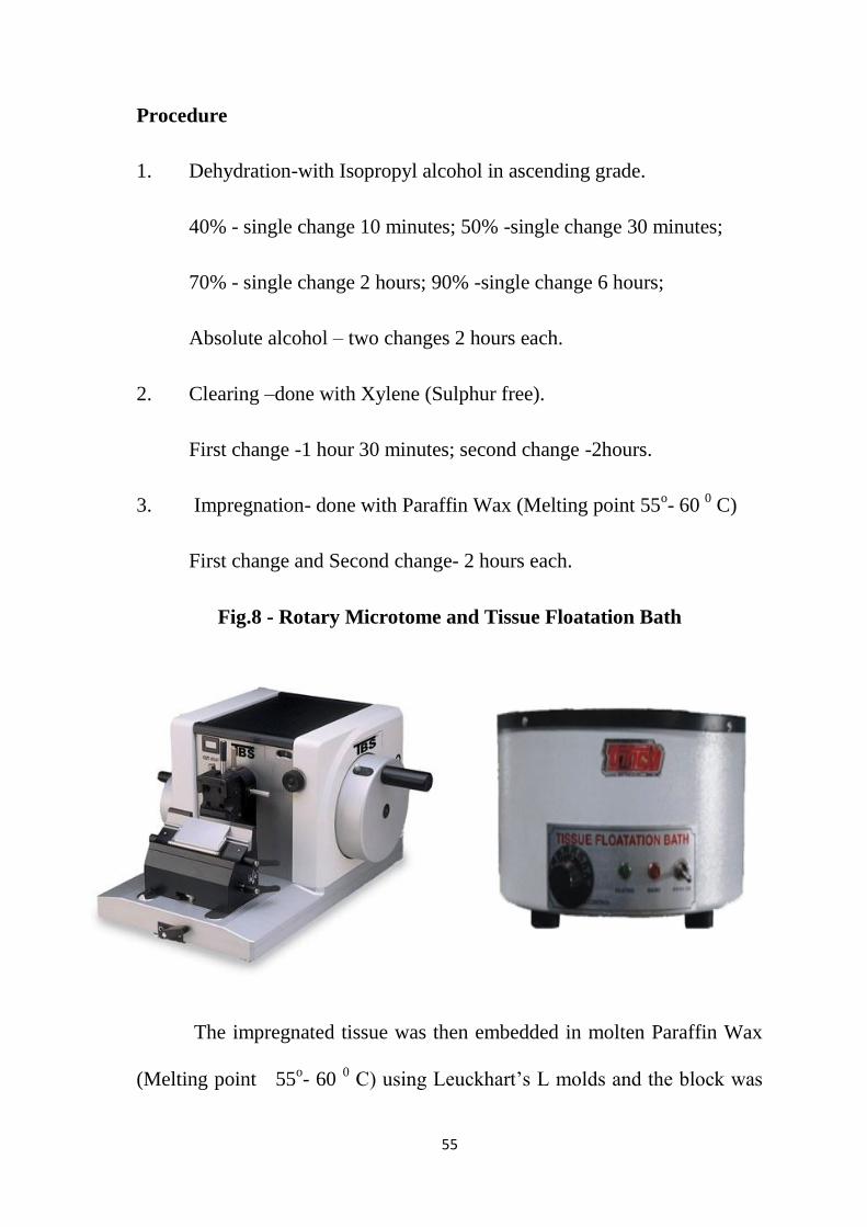
Procedure
1. Dehydration-with Isopropyl alcohol in ascending grade.
40% - single change 10 minutes; 50% -single change 30 minutes;
70% - single change 2 hours; 90% -single change 6 hours;
Absolute alcohol – two changes 2 hours each.
2. Clearing –done with Xylene (Sulphur free).
First change -1 hour 30 minutes; second change -2hours.
3. Impregnation- done with Paraffin Wax (Melting point 55o- 60
0 C)
First change and Second change- 2 hours each.
Fig.8 - Rotary Microtome and Tissue Floatation Bath
The impregnated tissue was then embedded in molten Paraffin Wax
(Melting point 55o- 60
0 C) using Leuckhart‘s L molds and the block was

56
cooled, trimmed and, labeled. The blocks are then cut into thin section of 3-
4 microns in thickness using Rotary Microtome (MT-1090A) and floated in
Tissue Floatation Bath (Dalal) at 48oC (below the melting point of wax)
and
mounted on the glass slide coated with Meyer‘s egg albumin.The mounted
slides were deparaffinized, dipped in Xylene and treated with descending
grades of isopropyl alcohol and brought to water.
STAINING PROCEDURE
HEMATOXYLIN AND EOSIN STAIN
After the slide was brought to water ,they were stained with
Hematoxylin and Eosin stains. Slides were kept in Hematoxylin (Erhlic‘s)
trough for 20 minutes and washed in water., then dipped in 1% acid alcohol
for differentiation and immediately washed in water Slides were kept for
blueing in running tap water for 10 minutes.Next the slides were dipped in
Eosin for 5 seconds,washed with water then air dried, and mounted using
DPX mountant.The slides were then studied under 4X,10X.40X
magnification,using binocular light microscope ( Magnus) and observation
noted and analyzed.
DIFFERENTIAL STAINING
Procedure for the differential staining of connective tissue fibres and
muscle are important part of histological technique and their use is often
helpful in the diagnosis of pathological changes in the tissues. Because of

57
this, many methods have been described for the demonstration of the
components , some of them selectively staining different types of fibres by
the use of several dyes, in combination or in sequence. Metallic
impregnation methods however, are necessary for the complete
demonstration of reticulin fibres.
Four special stains namely Van Gieson‘s ,Masson‘s Trichrome ,
Reticulin (Gomori‘s method ),Periodic acid Schiff (PAS) had been used to
bring out the arrangement of connective tissue elements composed of
collagen, muscle tissue,reticulin fibres and Hassall‘s corpuscles
respectively.81
1.VAN GIESON’S (1889) STAIN
Van Gieson‘s mixture of picric acid and acid fuchsin is the simplest
method for the differential staining of collagen. Its main disadvantages are
its inability to stain young fibrils the red that is imparted to mature collagen
and the tendency for the red colour to fade, whatever mounting medium is
used.
Fixation: formalin is used.
Sections: Paraffin embedded sections.

58
Preparation of stain:
Saturated aqueous picric acid-100ml mixed with 1 % acid fuchsin in
distilled water 5-10ml
Staining technique:
1. Take sections to water.
2. Stain nuclei either with Weigert‘s iron haematoxylin for 5 minutes,
wash with running water.
3. Dip in acid alcohol
4. Wash water for 5-10minutes.
5. Stain in Van Gieson‘s solution (picrofuchin) for 2 minutes.
6. Do not wash in alkaline tap water which extracts the red stain.
7. Dehydrate in absolute alcohol, clear in xylene and mount in a
synthetic resin
2. MASSON’S TRICHROME STAIN (MODIFIED FROM MASSON,
1929)
Fixation: Formalin
Sections : Thin paraffin sections.
Preparation:
Cytoplasmic (plasma) stain-1percent ponceau de xylidine (ponceau
2R) in 1 percent acetic acid 2 parts ,1percent acid fuchsin in 1 percent,and

59
acetic acid 1 part .Differentiator and Mordant-1Percent phosphomolybdic
acid in distilled water .Fibre stain -2 percent aniline blue in 2 percent acetic
acid.
Staining technique:
1. Take sections to water
2. Stain nuclei with Weigert‘s iron haematoxylin .
3. Wash well in water.
4. Differentiate nuclear stain with 1% acid alcohol.
5. Wash well in tap water, rinse in distilled water.
6. Stain in the red cytoplasmic stain 5-10 minutes
7. Rinse in distilled water.
8. Differentiate in 1percent phosphomolybdic acid until collagen is
decolorized, muscle, red blood cells and fibrin remaining red.
9. Rinse in distilled water.
10. Counterstain in aniline blue for 5 minutes
11. Wash well in 1 percent acetic acid (CH3COOH) for atleast 1 minute.
12. Blot, dehydrate in absolute alcohol, clear in xylene, mount in a
synthetic medium.
3. GOMORI’S RETICULIN METHOD
Fixation: formalin used
Sections : Thin paraffin sections.

60
Preparation of Silver Solution:
To 5ml. of 10.2 percent silver nitrate, add strong ammonia drop by
drop until the resulting precipitate is just dissolved. Add 5ml. of 3-1 percent
sodium hydroxide and re-dissolve the precipitate with a few more drops of
ammonia. Dilute to 0.5ml with distilled water.
Staining technique
1. Take sections to water
2. Oxidize for 1-2 minutes in 0-5 percent potassium permanganate,
3. Wash briefly in water.
4. Bleach in 2 percent oxalic acid.
5. Rinse in distilled water followed by thorough washing in tap water.
6. Sensitize in silver nitrate solution for 3 minutes
7. Wash well with distilled water.
8. Reduce with 10percent neutral formalin for 3 minutes.
9. Wash in tap water followed by distilled water.
10. Tone in 0.2 percent, gold chloride for 10 minutes (the sections turn a
purplish colour).
11. Wash briefly with distilled water.
12. Fix in 2 percent potassium metabisulfite for 2 minutes
13. Fix in 2percent, sodium thiosulphate (‗hypo‘) for 2 minutes.
14. Wash well in water.
15. Dehydrate, clear and mount in a synthetic resin medium.

61
4. PERIODIC ACID SCHIFF (PAS)
Reagent preparation:
1. 0.5% periodic acid -0.5gm periodic acid dissolved in 100ml of water.
2.Schiff‘s Reagent: Basic fuchsin- 1gm,sodium metabisulphate-1 gm
dissolved in hydrochloric acid 20ml and water 200ml.Boil the distilled
water, then add basic fuchsin and stir ,boil to 50oC,then filter and add 1N
hydrochloric ,cool to 25oC,then add sodium metabisulphate. This solution is
ready to use when it becomes nearly colourless, which may take upto two
days in the dark.
3. Sulphurous acid - Sodium metabisulphate 10%-6ml and 10% of
Hydrochloric acid 5ml mixed with 100ml 0f distilled water.
4. 0.2% light green or Mayer‘s hemalum.
Staining procedure:
1. Place the section to water.
2. Add 0.5% periodic acid(5-10 minutes)
3. Rinse in distilled water
4. Add Schiff agent (15 mins)
5. Rinse three changes of sulphurous acid between two minutes
6. Rinse in distilled or tap water for 5minutes.
7. Then add counter stain for 30 seconds.
8. Wash in running tap water for 5 minutes.
9. Air dry and mount in DPX
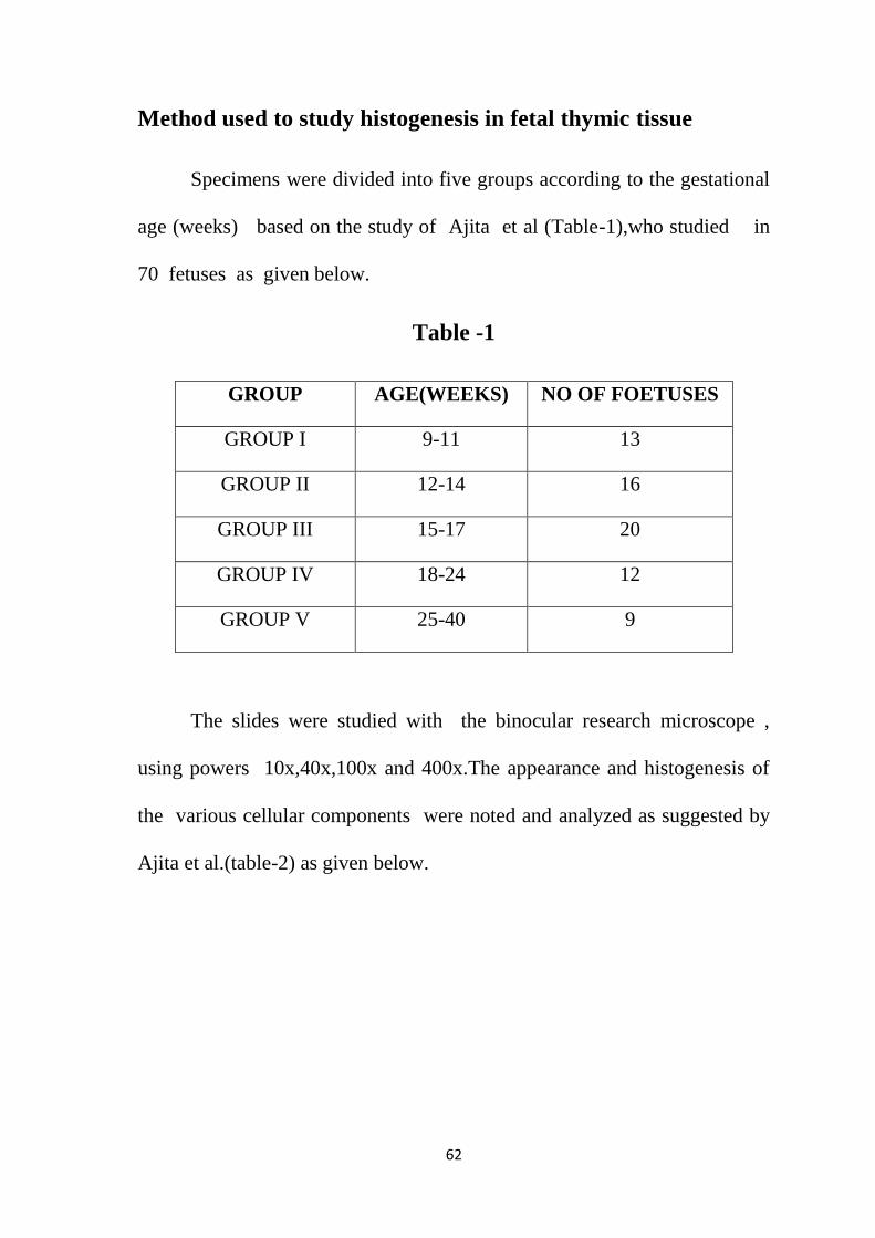
62
Method used to study histogenesis in fetal thymic tissue
Specimens were divided into five groups according to the gestational
age (weeks) based on the study of Ajita et al (Table-1),who studied in
70 fetuses as given below.
Table -1
GROUP AGE(WEEKS) NO OF FOETUSES
GROUP I 9-11 13
GROUP II 12-14 16
GROUP III 15-17 20
GROUP IV 18-24 12
GROUP V 25-40 9
The slides were studied with the binocular research microscope ,
using powers 10x,40x,100x and 400x.The appearance and histogenesis of
the various cellular components were noted and analyzed as suggested by
Ajita et al.(table-2) as given below.
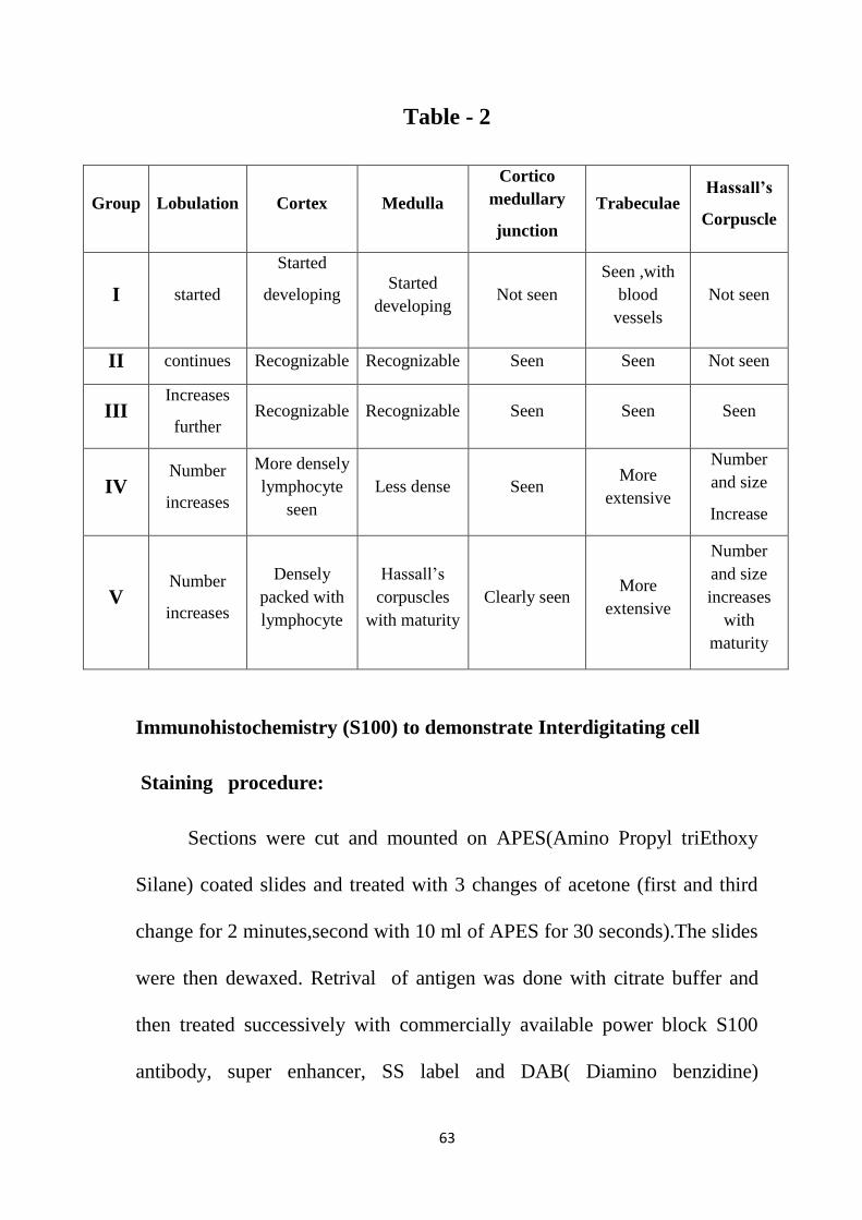
63
Table - 2
Group Lobulation Cortex Medulla
Cortico
medullary
junction
Trabeculae Hassall’s
Corpuscle
I started
Started
developing
Started
developing Not seen
Seen ,with
blood
vessels
Not seen
II continues Recognizable Recognizable Seen Seen Not seen
III Increases
further Recognizable Recognizable Seen Seen Seen
IV Number
increases
More densely
lymphocyte
seen
Less dense Seen More
extensive
Number
and size
Increase
V Number
increases
Densely
packed with
lymphocyte
Hassall‘s
corpuscles
with maturity
Clearly seen More
extensive
Number
and size
increases
with
maturity
Immunohistochemistry (S100) to demonstrate Interdigitating cell
Staining procedure:
Sections were cut and mounted on APES(Amino Propyl triEthoxy
Silane) coated slides and treated with 3 changes of acetone (first and third
change for 2 minutes,second with 10 ml of APES for 30 seconds).The slides
were then dewaxed. Retrival of antigen was done with citrate buffer and
then treated successively with commercially available power block S100
antibody, super enhancer, SS label and DAB( Diamino benzidine)

64
chromogen, after washing with buffer in each stage, Harris hematoxylin was
then used as counter stain(1min),washed ,dried and mounted with DPX.
To demonstrate the Interdigitating cells present in the medulla of
thymus. Three slides were stained from 12 weeks(sample no.19), 16
(sample no.13) and 31weeks (sample no.2) ,belonging to groups II,III and V
respectively.

65
OBSERVATIONS
20 fetal thymuses of 10 to 31weeks of gestational age were
considered and classified into five groups as described by R.K.Ajita et al .
All the specimens were analyzed and plotted against age groups.
Table- 3
Group Age(weeks) No of fetuses
Group I 9-11 1
Group II 12-14 3
Group III 15-17 3
Group IV 18-24 9
Group V 25-31 4
The appearance of various cellular components and their period of
development was noted and plotted against each group as mentioned by
Ajita et al in the following table.

66
Table 4
Group I - The Gland was seen to be composed of lymphocytes with
a delicate capsule.The lobulation and corticomedullary differentiation were
not seen. Trabeculae associated with blood vessels were observed .Spindle
shaped epithelial cells were noted .No Hassall‘s corpuscles were observed.
Group Lobulatio
n Cortex Medulla
Cortico
medullary
Junction
Trabeculae Hassall’s
Corpuscle
I Not seen
Not
recogniza
ble
Not seen
Not seen
Seen Not seen
II Started
appearing Not seen Not seen Not seen Seen Not seen
III
Increases
further in
number
Seen in
some
Seen in
some Seen in some Seen
Started
developing
IV
Number
increases
More
densely
packed
lympho
cyte seen
Less
dense Seen
More
extensive
Number and size
Increase
V Number
increases
Densely
packed
with
lympho
cytes
seen
Hassall‘s
corpuscle
with
maturity
Seen
Clearly seen More
extensive
Number and size
increases with
maturity
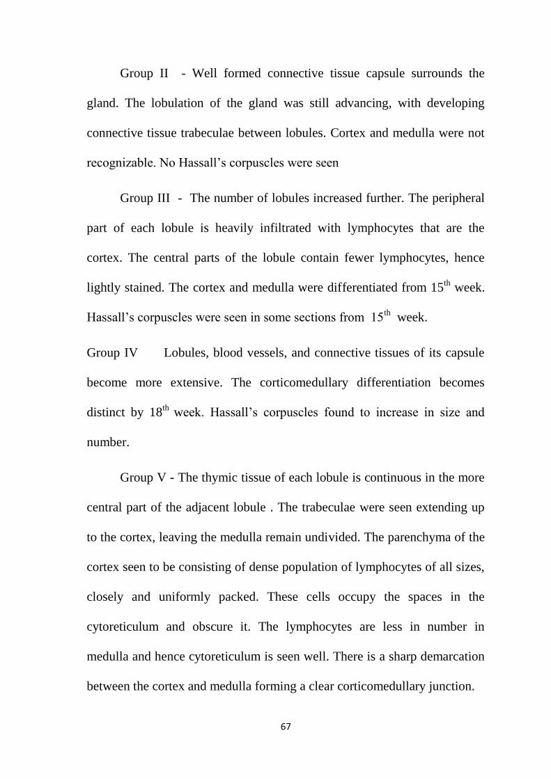
67
Group II - Well formed connective tissue capsule surrounds the
gland. The lobulation of the gland was still advancing, with developing
connective tissue trabeculae between lobules. Cortex and medulla were not
recognizable. No Hassall‘s corpuscles were seen
Group III - The number of lobules increased further. The peripheral
part of each lobule is heavily infiltrated with lymphocytes that are the
cortex. The central parts of the lobule contain fewer lymphocytes, hence
lightly stained. The cortex and medulla were differentiated from 15th week.
Hassall‘s corpuscles were seen in some sections from 15th
week.
Group IV Lobules, blood vessels, and connective tissues of its capsule
become more extensive. The corticomedullary differentiation becomes
distinct by 18th
week. Hassall‘s corpuscles found to increase in size and
number.
Group V - The thymic tissue of each lobule is continuous in the more
central part of the adjacent lobule . The trabeculae were seen extending up
to the cortex, leaving the medulla remain undivided. The parenchyma of the
cortex seen to be consisting of dense population of lymphocytes of all sizes,
closely and uniformly packed. These cells occupy the spaces in the
cytoreticulum and obscure it. The lymphocytes are less in number in
medulla and hence cytoreticulum is seen well. There is a sharp demarcation
between the cortex and medulla forming a clear corticomedullary junction.

68
Hassall‘s corpuscles of varying shapes and sizes , immature solid to
mature cystic types seen. The number increases with gestational age. The
gland during this stage had an internal architecture similar to that seen in the
adults.
Table- 5
Appearance of Lobulation in different gestational age groups
Group
Number of
specimens
Number seen Percentage (%)
Group I 1 0 0
Group II 3 1 33 %
Group III 3 3 100 %
Group IV 9 9 100 %
Group V 4 4 100 %
In the group I (9-11 weeks) lobulation was not seen.In the group II
(12-14 weeks) lobulations were seen in 1 out of 3 specimens (33%).In the
group III (15-17 weeks), the lobulations were seen in all the specimens
(100%).In group IV (18-24 weeks ) a total of 9 specimens were included
and lobulations were seen in all the specimens(100%). In the group V (25-
31 weeks) lobulations were seen in all the specimens (100%).

69
CHART-1
APPEARANCE OF LOBULATION IN DIFFERENT GESTATIONAL
AGE GROUPS
0
20
40
60
80
100
120
Group I Group II Group III Group IV Group V
LOBULATION

70
Table-6
Appearance of cortex in different gestational age groups
Group Number of
specimens Number seen Percentage
Group I 1 0 0
Group II 3 0 0
Group III 3 1 33%
Group IV 9 9 100%
Group V 4 4 100%
In the group I (9-11 weeks)cortex was not seen.In the group II (12-14
weeks) cortex was not recognizable in any of them.In the group III (15-17
weeks) out of 3 specimens, the cortex was recognizable in only one
specimen(33%).In group IV (18-24 weeks) the cortex was recognizable in
all the specimens(100%). In the group V (25-31 weeks) the cortex was
recognizable in all the specimens(100%).

71
CHART-2
APPEARANCE OF CORTEX IN DIFFERENT GESTATIONAL
AGE GROUPS
0
20
40
60
80
100
120
Group I Group II Group III Group IV Group V
CORTEX

72
Table-7
Appearance of medulla in different gestational age groups
Group Number of
specimens Number seen Percentage
Group I 1 0 0
Group II 3 0 0
Group III 3 1 33%
Group IV 9 9 100%
Group V 4 4 100%
In the group I (9-11 weeks) medulla was not seen. In the group II
(12-14 weeks) the medulla was not recognizable. In the group III(15-17
weeks) out of the 3 specimens, the medulla was recognizable in only one
specimen (33%).In group IV (18-24weeks),the medulla was recognizable in
all the specimens(100%).In the group V (25-31 weeks) it was recognizable
in all the specimens(100%).
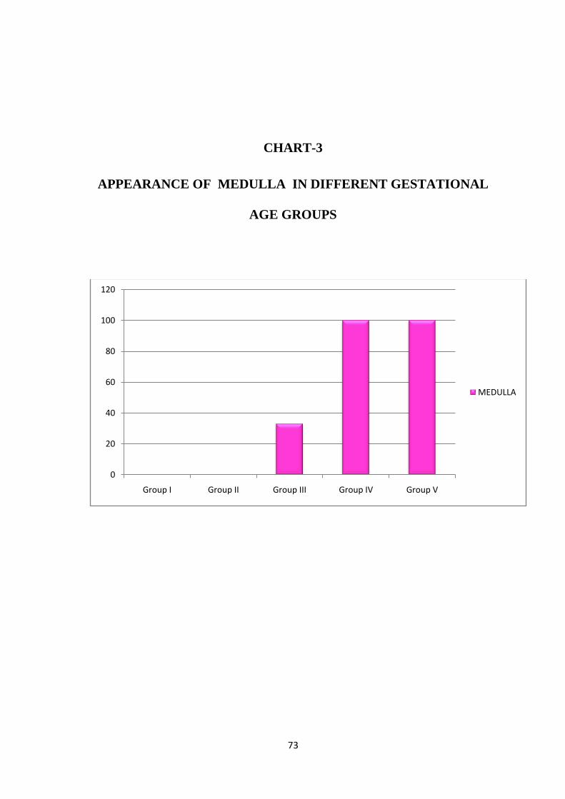
73
CHART-3
APPEARANCE OF MEDULLA IN DIFFERENT GESTATIONAL
AGE GROUPS
0
20
40
60
80
100
120
Group I Group II Group III Group IV Group V
MEDULLA

74
Table-8
Appearance of Corticomedullary junction in different gestational age
groups
Group Number of
specimens Number seen Percentage
Group I 1 0 0
Group II 3 0 0
Group III 3 1 33%
Group IV 9 6 67%
Group V 4 4 100%
In the group I (9-11 weeks) the corticomedullary junction was not
seen. In the group II (12-14 weeks) the corticomedullary junction was not
recognizable in any of them.In the group III (15-17 weeks) corticomedullary
junction was clearly seen in only one specimen (33%).In group IV (18-24
weeks) a total of 9 specimens were included and the corticomedullary
junction was clearly seen in 6 specimens(67%).In the group V (25-31
weeks) the corticomedullary junction was clearly seen in all the
specimens(100%).

75
CHART - 4
APPEARANCE OF CORTICOMEDULLARY JUNCTION IN
DIFFERENT GESTATIONAL AGE GROUPS
0
20
40
60
80
100
120
Group I Group II Group III Group IV Group V
Cortico Medullary Junction

76
Table-9
Appearance of trabeculae in different gestational age groups
Group Number of
specimens Number seen Percentage
Group I 1 1 100%
Group II 3 2 67%
Group III 3 3 100%
Group IV 9 9 100%
Group V 4 4 100%
In the group I (9-11 weeks) the trabeculae was seen(100%) In the
group II (12-14 weeks) the trabeculae noted in 2 specimens.(67%)In the
group III (15-17weeks),the trabeculae was seen in all the
specimens.(100%)In group IV (18-24 weeks ), the trabeculae noted in all
the specimens(100%). In the group V(25-31weeks) the trabeculae was
visible in all the specimens(100%).

77
CHART –5
APPEARANCE OF TRABECULAE IN DIFFERENT
GESTATIONAL AGE GROUPS
0
20
40
60
80
100
120
Group I Group II Group III Group IV Group V
Trabeculae

78
Table-10
Appearance of Hassall’s corpuscle in different gestational age groups
Group Number of
specimens Number seen Percentage
Group I 1 0 0
sGroup II 3 0 0
Group III 3 2 67%
Group IV 9 9 100%
Group V 4 4 100%
In the group I (9-11 weeks) the Hassall‘s corpuscle was not seen.In
the group II (12-14 weeks) the Hassall‘s corpuscle was not seen in any of
the specimens.
In the group III (15-17 weeks), it was seen in 2 specimens.(67%)In
group IV (18-24 weeks ), the Hassall‘s corpuscle was seen in all the
specimens(100%).
In the group V (25-31 weeks) , the Hassall‘s corpuscle was seen in
all the specimens(100%).

79
CHART - 6
APPEARANCE OF HASSALL’S CORPUSCLE IN DIFFERENT
GESTATIONAL AGE GROUPS
0
20
40
60
80
100
120
Group I Group II Group III Group IV Group V
HASSALL'S CORPUSCLE

80
Table11
Appearance of Lobulation, cortex and medulla in different groups
Group Lobulation cortex Medulla
Group I 0 0 0
Group II 33% 0 0
Group III 100% 33% 33%
Group IV 100% 100% 100%
Group V 100% 100% 100%
In Group I the lobulations ,cortex and medulla was not seen.No
cortex and medulla were seen in group II while the lobulations were
observed in 33%.
All the specimens in group III were lobulated(100%),while cortex and
medulla recognizable in 33% .Lobulations,cortex and medulla were found in
all the specimens which belong to group IV and group V(100%)

81
CHART- 7
APPEARANCE OF LOBULATION, CORTEX AND MEDULLA IN
DIFFERENT GROUPS
0
20
40
60
80
100
120
GROUP I GROUP II GROUP III GROUP IV GROUP V
Lobulation
cortex
medulla

82
Table-12
Appearance of corticomedullary junction,trabeculae and Hassall’s
corpuscles in different groups
Group Corticomedullary
Junction Trabeculae
Hassall’s
corpuscles
Group I 0 100% 0
Group II 0 66% 0
Group III 33% 100% 67%
Group IV 67% 100% 100%
Group V 100% 100% 100%
The corticomedullary junction and Hassall‘s corpuscles were not
seen, while the trabeculae was seen in group I(100%).In group II, the
corticomedullary junction and the Hassall‘s corpuscles were not seen but the
trabeculae was noted in 66%.The trabeculae was found in 100%,the
corticomedullary junction in 33% and the Hassall‘s corpuscles in 67% in
group III. The corticomedullary junction was seen in 67%, while the
trabeculae and Hassall‘s corpuscles were seen in 100% of group IV
specimens .In group V all the components were 100%.

83
CHART -8
APPEARANCE OF CORTICOMEDULLARY JUNCTION,
TRABECULAE AND HASSALL’S CORPUSCLES IN DIFFERENT
GROUPS
0
20
40
60
80
100
120
Group I Group II Group III Group IV Group V
CM junction
Trabeculae
Hassall's corpuscles

84
DIFFERENTIAL STAINING
1.Reticulin Staining
In the present study, reticulin stain is used to demonstrate the reticular
fibres in fetal thymus , which form the supporting framework of the gland
appearing black, while the collagen and cytoplasm appear purplish
grey.Photo17 which was taken from sample number 1(24 weeks), which
belongs to group IV shows the reticular fibres which appear
black.Lobulations and trabeculae are seen well. Blood vessels and Hassall‘s
corpuscles were seen in photo 18 which belongs to 31weeks .( group V,
sample no.2)
2. Masson’s Trichrome staining
In this study two samples were selected ,sample number 9 and 5
which belonged to gestational ages 26 weeks (group V) and 21 weeks
(group IV) respectively and stained. Photo 19 highlights , the blood vessel
in close relation to connective tissue trabeculae and the Hassall‘s corpuscle
.The parenchyma made up of reticular fibers and collagen appear
blue.Hassall‘s corpuscles appear reddish brown .In photo 20,Hassall‘s
corpuscle and trabeculae which appear in blue colour is seen.

85
3. Van Gieson’s stain -
In the present study ,sample number 14 which belonged to group V
of 25 weeks gestation was stained .The lobules appear yellow in colour due
to the cytoplasm of lymphocyte which take up the picric acid present in the
stain.(photo 21)
4.Periodic acid-
Schiff stain - In this study ,sample number 4 of 19 weeks fetus
belonging to group IV was stained. The Hassall‘s corpuscle take up the stain
and found to be PAS positive.(photo 22).
IMMUNOHISTOCHEMISTRY STAINING
Immunohistochemistry staining was done for 3 slides ,belonging to
group IIgroup III and groupV ,with S 100 marker to demonstrate the
proportion of Interdigitating cells(IDC) in the thymus. Observations were
made about staining pattern-regular or irregular and the intensity of staining
described as1+(mild) to 2+(moderate).The sample no;19 and 13 did not
show any staining pattern, while the sample no;2 showed,regular
staining pattern ,with the intensity of staining 2+.(photo23).

Photo-1
No Lobulation, no corticomedullary differentiation at 10 weeks (Group I)
S.NO.20 40x magnification
Photo-2
Lobules begin to appear at 12 weeks (Group II)
S.NO.19 40x magnification
TRABECULAE
TRABECULAE

Photo-3
Poorly formed lobules at 13 weeks (Group II)
S.NO.6 40x magnification
Photo-4
Lobulations seen, no corticomedullary differentiation at 14 weeks (Group II)
S.NO.8 100x magnification
TRABECULAE
LOBULE
TRABECULAE

Photo no- 5
Lobulations seen , corticomedullary differentiation ill defined at 15
weeks (Group III)
S.NO.10 40x Magnification
Photo no - 6
Epithelial reticular cells seen in trabeculae at 18weeks (Group IV)
S.NO: 7 40x Magnification
ERC
CM JUNCTION
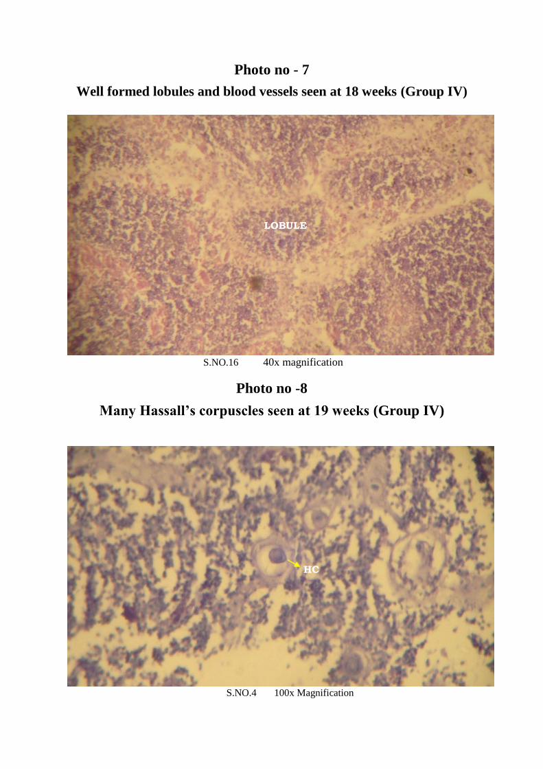
Photo no - 7
Well formed lobules and blood vessels seen at 18 weeks (Group IV)
S.NO.16 40x magnification
Photo no -8
Many Hassall’s corpuscles seen at 19 weeks (Group IV)
S.NO.4 100x Magnification
LOBULE
HC
CC
CC

Photo no -9
Many Hassall’s corpuscles seen at 20 weeks (Group IV)
S.NO.12 40x Magnification
Photo no -10 Many developing Hassall’s corpuscles seen at 20 weeks (Group IV)
S.NO.12 40x Magnification
CYSTIC HC
HC
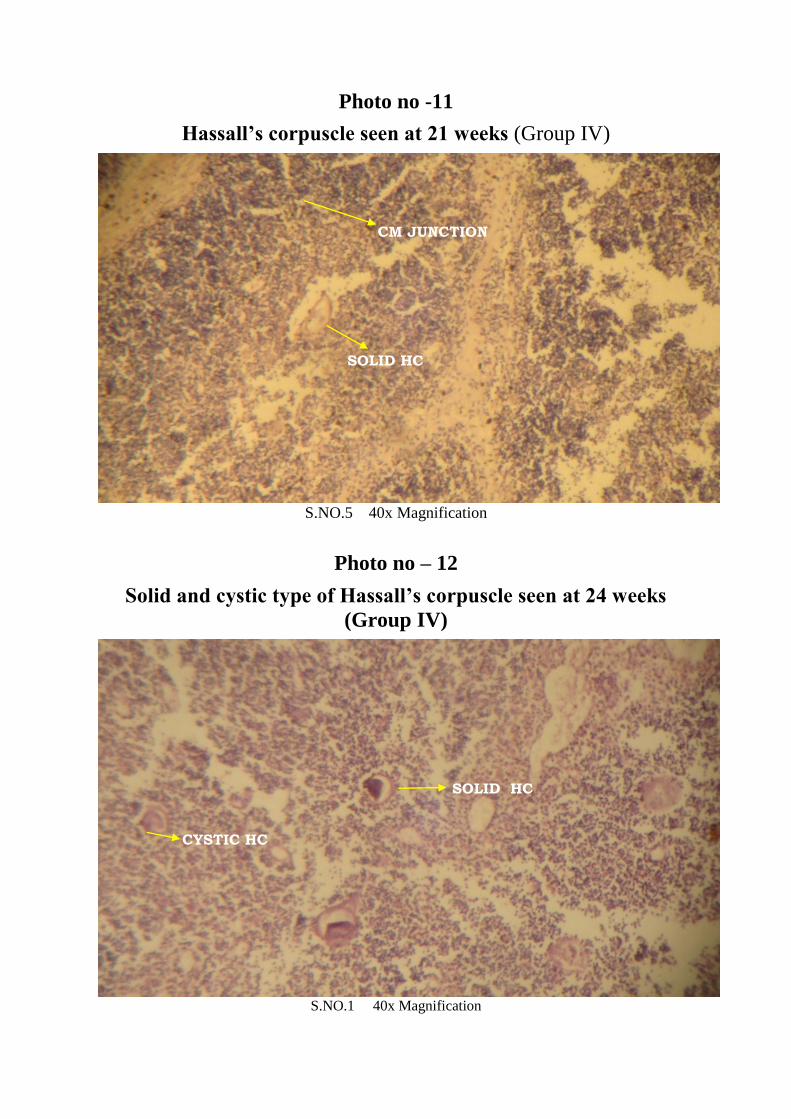
Photo no -11
Hassall’s corpuscle seen at 21 weeks (Group IV)
S.NO.5 40x Magnification
Photo no – 12
Solid and cystic type of Hassall’s corpuscle seen at 24 weeks
(Group IV)
S.NO.1 40x Magnification
SOLID HC
CM JUNCTION
CYSTIC HC
SOLID HC
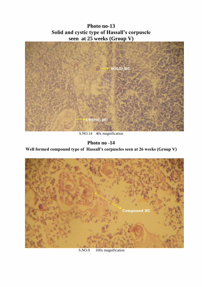
Photo no-13
Solid and cystic type of Hassall’s corpuscle
seen at 25 weeks (Group V)
S.NO.14 40x magnification
Photo no -14
Well formed compound type of Hassall’s corpuscles seen at 26 weeks (Group V)
S.NO.9 100x magnification
SOLID HC
CYSTIC HC
SOLID HC
CYSTIC HC
Compound HC
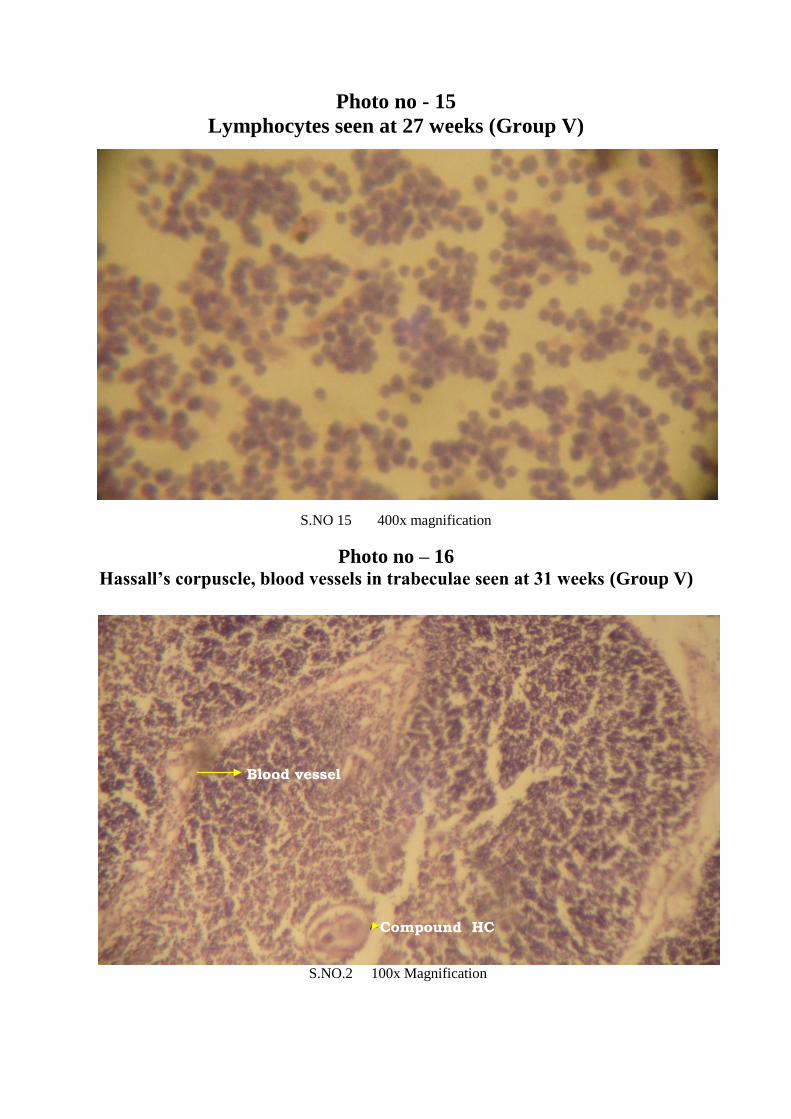
Photo no - 15
Lymphocytes seen at 27 weeks (Group V)
S.NO 15 400x magnification
Photo no – 16 Hassall’s corpuscle, blood vessels in trabeculae seen at 31 weeks (Group V)
S.NO.2 100x Magnification
Compound HC
Blood vessel

Photo- 17
Macrophages are seen -18 weeks (Group IV)
S.NO.16 400x Magnification
Photo no -18
RETICULIN STAIN-Lobulations well formed at 24 weeks (Group IV)
S.NO.1 100x Magnification
LOBULE
TRABECULAE

Photo no -19
RETICULIN STAIN-Blood vessels and Hassall’s corpuscle seen
at 31weeks (Group V)
S.NO.2 400x Magnification
Photo no- 20 MASON’S TRICHROME Lobules, Hassall’s corpuscle seen at
26 weeks (Group V)
S.NO.9 400x Magnification
BLOOD VESSEL
SOLID HC
CYSTIC HC
BLOOD VESSEL

Photo no -21
MASON’S TRICHROME- Lobules, Hassall’s corpuscle blood
vessels seen at 27 weeks (Group V)
S.NO15 100x Magnification
Photo no – 22 VAN GEISON’S STAIN Lobulation seen well at 25 weeks (Group V)
S.NO.14 100x Magnification
CYSTIC HC
TRABECULAE
LOBULE
TRABECULAE

Photo no - 23
PERIODIC ACID SCHIFF STAINING at 19 weeks (Group IV)
S.NO.11 100x Magnification
PHOTO – 24
IMMUNO HISTOCHEMISTRY WITH S-100 ANTIGEN AT 31 WEEKS
(GROUP V)
S.NO.2 100x Magnification
HC

86
DISCUSSION
In the present study the development of human fetal thymus is studied
under the following histological parameters.
1. Lymphocytes:
The time at which the lymphocytes were present in the thymus varies
in different studies. It appears by 8th
week according to Williams and
Standring S et al and Romanes GJ .Reported to appear by 9th
week
according to HaarJL, Hamilton and Mossman ,Von Gaudecker,Ritter and
Lampert,Ajita et al,prabhavathy, Bashir khan and Sanobar sheikh.
In the present study the lymphocytes were observed from 10th week
which have round purple nuclei with basophilic cytoplasm. It could not be
ascertained whether lymphocytic infiltration occurs from 8th
week onwards,
since the present study examined fetuses from 10 weeks.
2. Lobulation:
The epithelial cells grow as thumb like protrusions into the mass of
mesenchyme, which later forms a thin capsule around the organ. In the
region between epithelial protrusions, the mesenchyme remains and forms
thin incomplete septa, to give the lobular appearance.
Ghali et al, Bashir khan and Sanobar sheikh reported lobulation at
10th week, while Haar et al reported appearance of Lobulation at 12
th week.

87
In the study conducted by Ajita et al, Prabhavathy Lobulations started
at 9th
week and completed by 12th
week. According to Vijayalakshmi et al
Lobulations appeared by 16th
week.
In the present study Lobulation started by 12th
week and completed
by 15th
week.
3. Cortex and Medulla
The cells of Lymphatic series are more concentrated towards the
borders of each lobule .Hence, at the periphery of the lobules lymphocytes
are numerous and densely packed forming darkly stained cortex, whereas
the medulla is lightly stained due to less number of lymphocytes.
The Cortico medullary differentiation noted in the embryos of 40mm
crown-rump length by Hamilton and Mossman ,Ghali et al by 11th
week,Hayward, Arey and Muller – Hermelink et al by 12th week,Haar,
Lobach & Haynes, Prabhavathy by 14th week.Ajita et al reported that the
differentiation started at 9th week and more distinct at 12 to 14
weeks.According to Bashir khan and Sanobar sheikh the differentiation
started at 12th
week and more distinct by 14th
week.Vijayalakshmi et al
reported at 16th week.
In the present study the cortico medullary differentiation started at
15th week more distinct by 18
th week. This coincides with the study of
Vijayalakshmi et al

88
4. Blood Vessels
According to Haar, Hamilton and Mossman, Ajita et al. Bashir khan
and Sanobar sheikh vascularization started at 9th week, medullary vessels
seen at 12th
week.
Vascularity was reported by Williams et al by 10th week, by Ghali et
al at 11th
week.
In the present study blood vessels were seen by 10th
week in the
trabeculae. Since the fetus prior to 10th week was not examined, it could not
be ascertained whether blood vessels were present at early stages.
5. Macrophages
Haynes reported macrophages by 10th
week, while the appearance of
macrophages was reported at 12th week by Ajita et al. Stranding S et al ,
Bashir khan and Sanobar sheikh reported its appearance at 14th
week.
In the present study macrophages were seen from 12th
week, which
coincides with the study of Ajita et al.
6. Epithelial Reticular cells
Williams et al, Hamilton and Mossman, Von Gaudecker, Muller-
Hermelink and Standring S et al have described the appearance of epithelial
reticular cells by 8th
week.Ajita et al observed the cells at 9th week.Haywar,

89
Arey and Bashir khan et al reported at 10th week.Vijayalakshmi et al
reported the cells at 12th
week of gestation.
In the present study the epithelial cells were observed at 10th week.
Since the fetus prior to 10th week was not examined, it could not be
ascertained whether the epithelial cells were present at an earlier stage.
7. Hassall’s corpuscles:
The time of appearance of Hassall’s corpuscles varies in different
studies; Fawcett, Hamilton and Mossman reported its appearance as early as
8th
week,Gilhus et al from 9th
week,Williams and Stranding S et al and Arey
at 10th week. Ghali et al at , Sawant, Bashir khan et al at and Varga et al at
reported its appearance by 11th
,12th and 13
th week respectively.
Ajita et al and Krishnamurthy et al noted its appearance by 15th
week,Lobach and Haynes reported it between 15th
and 16th week while
Liberti et al noted at 16th week.According to Bodey and Kaiser Hassall’s
corpuscles develop in the 2nd
part of 3rd
lunar month.Vijayalakshmi et al
reported it at 18th week of gestation
In the present study the Hassall’s corpuscle was observed from 15th
week onwards,which coincides with the study of Ajita et al.

90
Growth of Hassall’s corpuscles
Bodey and Kaiser reported that the greatest development was
between 6th
and 10th
lunar month.Liberti et al mentioned that the mean area
of Hassall’s corpuscle increased with the fetal age with greatest difference
between 16th
-19th
week and 20th-23
rd week.Ajita et al and Krishnamurthy et
al observed the increase in number and size during 17th- 24
th weeks.Bashir
khan et al reported the growth to occur during 18th- 24
th week.
In the present study the number and size of the Hassall’s corpuscle
increased during 18th- 24
th week ,which coincides with the study of Bashir
khan et al.
Immunohistochemistry
The immunoreactive - interdigitating cells (IDCs) cells are distributed
mainly in the medulla with some scattered elements in the cortex. The
immunocytochemistry for S-100 visualizes the precise distribution and
extension of the IDCs under the light microscope and indicates that the
IDCs form no structural networks such as those established by the thymic
epithelial cells.

91
CONCLUSION
The present study concludes that in the histogenesis of human fetal
thymus, significant cellular events like lobulation ,corticomedullary
differentiation and the appearance of Hassall’s corpuscle all takes place
between 15th and 18
th week of gestational age. Thereafter the microscopic
growth and maturity takes place in the form of increase in size of lobules ,
blood vessels and increase in size and number of Hassall’s corpuscle.
Hence the period of gestation between 15 and 18 weeks is critical for
the development of fetal thymus .Any insult occurring to the developing
thymus in the form of radiation or drugs can affect its histogenesis leading
to impaired immunity.
As the above observations were made in a very limited sample size
statistical significance could not be determined. This precluded the
quantitative study of fetal thymic cellular components in relation to
gestational age and hence an attempt was made for qualitative study.
The clinical implication of this study is to provide the basis for more
accurate interpretation of the histogenesis of fetal thymic cellular
components in relation to gestational age .Individuals in whom there is a
persistence of myoid cell beyond 28 weeks of gestation, suggest they are
prone for myasthenia gravis.

92
Immunohistochemical study for demonstrating the Interdigitating
cells (IDC) are the sites where the Human Immunodeficiency Virus(HIV)
can remain latent, and gets activated by CD40 ligation or by the presence of
T-Helper cells. They play a role in transport of HIV .The virus derived
from infected IDCs carry T cell specific factor that make them highly
infectious.

List of Figures
Fig.1 Gross Anatomy of fetal thymus
Fig.2 Histology of fetal thymus (Haematoxylin & Eosin staining)
Fig.3 Microvasculature of thymus gland
Fig.4 Electron microscopic view of epithelial reticular cells
Fig.5 Light microscopic view of myoid cell
Fig.6 Dissection of fetus
Fig.7 Automated tissue processor
Fig.8 Rotary microtome and tissue floatation bath

List of Tables
1. Grouping of fetuses according to R.K.Ajita et al by gestational age
2. Gestational age related histogenesis of the various cellular components
according to R.K.Ajita et al.
3. Grouping of fetuses by gestational age in the present study.
4. Gestational age related histogenesis of cellular components in the present
study.
5. Appearance of lobulation in different gestational age groups.
6. Appearance of cortex in different gestational age groups.
7. Appearance of medulla in different gestational age groups.
8. Appearance of corticomedullary junction in different gestational age
groups.
9. Appearance of trabeculae in different gestational age groups.
10. Appearance of Hassall’s corpuscle in different gestational age groups
11. Appearance of lobulation, cortex and medulla in different groups
12. Appearance of corticomedullary junction, trabeculae and Hassall’s
corpuscles in different groups
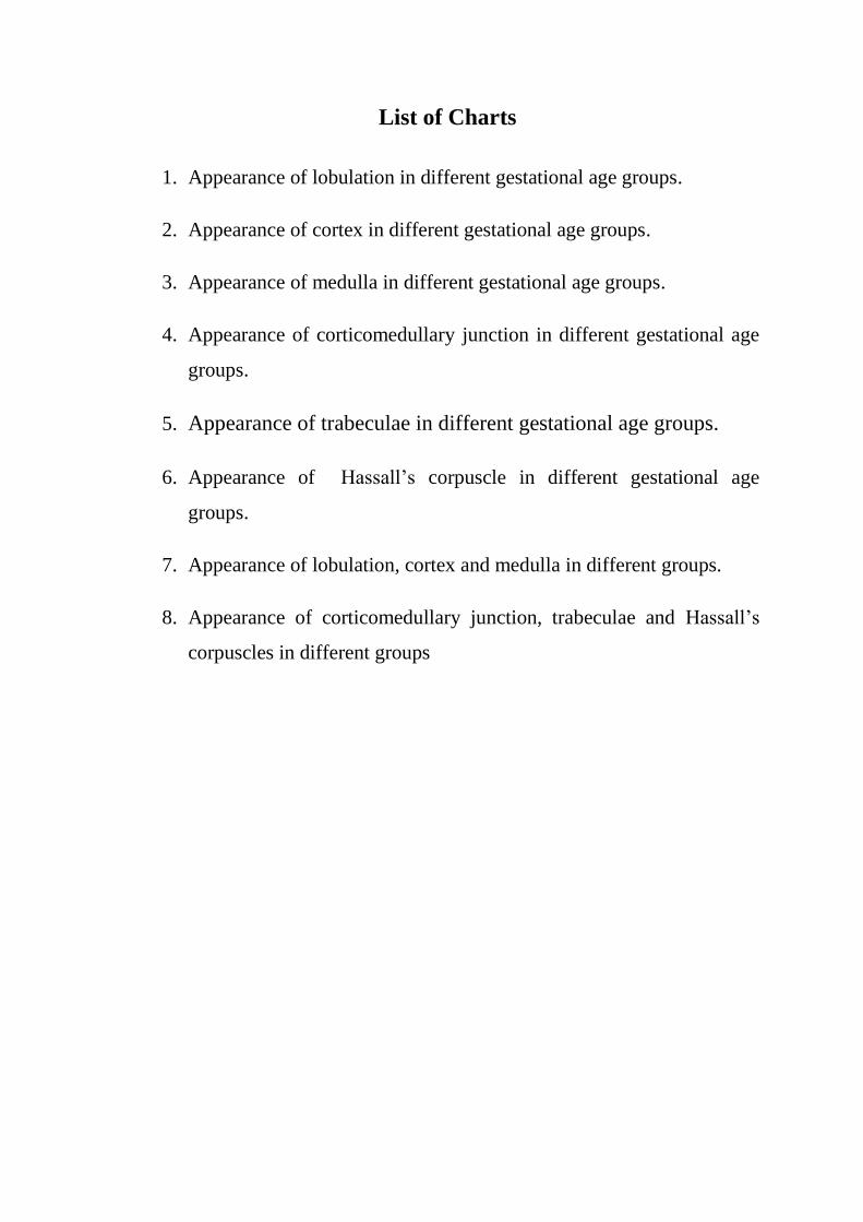
List of Charts
1. Appearance of lobulation in different gestational age groups.
2. Appearance of cortex in different gestational age groups.
3. Appearance of medulla in different gestational age groups.
4. Appearance of corticomedullary junction in different gestational age
groups.
5. Appearance of trabeculae in different gestational age groups.
6. Appearance of Hassall’s corpuscle in different gestational age
groups.
7. Appearance of lobulation, cortex and medulla in different groups.
8. Appearance of corticomedullary junction, trabeculae and Hassall’s
corpuscles in different groups

List of Photographs
1. No lobulation, no corticomedullary differentiation at 10 weeks (Group I)
2. Lobules begin to appear at 12 weeks (Group II)
3. Poorly formed lobules at 13 weeks (Group II)
4. Lobulations seen, no corticomedullary differentiation at 14 weeks
(Group II)
5. Lobulations seen , corticomedullary differentiation ill defined at 15
weeks (Group III)
6. Epithelial reticular cells seen in trabeculae at 18weeks (Group IV)
7. Well formed lobules and blood vessels seen at 18 weeks (Group IV)
8. Many Hassall’s corpuscles seen at 19 weeks (Group IV)
9. Many Hassall’s corpuscles seen at 20 weeks (Group IV)
10. Many developing Hassall’s corpuscles seen at 20 weeks (Group IV)
11. Hassall’s corpuscle seen at 21 weeks (Group IV)
12. Solid and cystic type of Hassall’s corpuscle seen at 24 weeks
(Group IV)
13. Solid and cystic type of Hassall’s corpuscle seen at 25 weeks (Group V)
14. Well formed compound type of Hassall’s corpuscles seen at 26 weeks
(Group V)
15. Lymphocytes seen at 27 weeks (Group V)
16. Hassall’s corpuscle, blood vessels in trabeculae seen at 31 weeks
(Group V)
17. Macrophages are seen at 18 weeks (Group IV)

18. Reticulin stain-lobulations well formed at 24 weeks (Group IV)
19. Reticulin stain-blood vessels and Hassall’s corpuscle seen at 31weeks
(Group V)
20. Mason’s trichrome - lobules, Hassall’s corpuscle seen at 26 weeks
(Group V)
21. Mason’s trichrome - lobules, Hassall’s corpuscle blood vessels seen at
27 weeks (Group V)
22. Van Geison’s stain - lobulation seen well at 25 weeks (Group V)
23. Periodic acid schiff staining at 19 weeks (Group IV)
24. Immuno histochemistry with S-100 antigen at 31 weeks (Group V)

BIBILIOGRAPHY
1. Galen . Galen on the usefulness of the parts of the body Ithaca. Cornell
University Press (New York) 1968: 30.
2. David H .Cormack. Hams histology, ninth edition J.B.Lippincott
Company 2010:242-247 .
3. Goldstein & Mackay Myoid Cells.The Human Thymus, Heinemann,
London. 1969:449-475.
4. Goldstein, G & I, Mackay Structure and development of the Human
Thymus.The Human Thymus .Heinemann-London.1969: 1-10.
5. Cooper AP . The anatomy of the thymus gland London, England:
Longman, Rees, Orem, Green, & Brown. 1833: 1-48.
6. Hassall AH and Vanarsdale H. Illustrations of the microscopic anatomy
of the human body in health and disease. In: Hassall AH, eds. Microscopic
Anatomy of the Human Body in Health and Disease ,London, England.
Wood 1846: 1-79.
7. Hassall, A.H. The Microscopical Anatomy of the Human body in health
and disease, 1851.
8. Watney. H. Minute Anatomy of the Thymus. Proceedings of the Royal
Society of London, 1881:Vol. XXXIII:11-17.
9. Bell E.T.The development of Thymus in the pig. American Journal of
Anatomy,1906;5:29-32.

10. Pappenheimer, A.M. Histology of Thymus.J. of Med. Research,
1910;22:1
11. Hammer. J.A. Morphology of Thymus and probable
functions.J.Endocrinology,1921;5: 543-550.
12. Jaffe,H.L.Thymus transplants with reference to regeneration of
Reticulum cells and formation of Hassall’s corpuscles.J.Exp. Med,1926;
44:523-525.
13. Jordan, H.E.The significance of the Hassall’s corpuscles.Amt. Rec.
1927;35:279-283.
14. Kingsbury On the nature and significance of Thymic corpuscles.Anat.
Rec. 1928;38:141-159.
15. Dearth. O.A..Late development of Thymus in the cat.Amer.J.Anat,1928;
41:321-323.
16. Norris, E.H. Morphogenesis and Histogenesis of Thymus in man, with
reference to origin of Hassall’s corpuscles.Contributions to Embryology -
Carnegic Institute, 1938; 166:193-195.
17. Kostowiecki, M. Development and degeneration of the second type of
Hassall’s corpuscles in the thymus of the guinea pig.Anat. Rec,1962;
142:195-203.
18. Smith, C. A comparison of the staining properties of Hassall’s
corpuscles and thick skin of guinea - pig.Anat. Rec,1949;103:649-653.
19. Metcalf, D. Thymic origin of LSF”.Brint. J. Cancer, 1956; 10:442-446.

20. Arnesen, K.The secretary apparatus in the Thymus of Mice. Acta,
pathol, Microbiol, Scand,1958; 43:339-42.
21. Koka, TElectron Microscopic studies on the thymus, especially on its
epithelial cells.Igaku Kenkyu, .1960;30:309-335.
22. Miller, J.F.A.P. Effect of Thymic ablation and replacement in The
Thymus in Immunobiology Eds. R.A. Good & Gabrielsen, Hayer and Row,
New York, 1961.
23. Tanaka, H. Mesenchymal and Epithelial reticulum in lymph nodes and
thymus of mice as revealed in the Electron Microscope.Ann. Report, Inst.
Virus Res. Kyoto Univ,1962; 5:146-169.
24. Marshall, A.H.E. & R.G.White The Immuological reactivity of the
thymus.Br. J.Exp. Path; 1962; 42:379-385.
25. Burnet, F.M. Role of Thymus and realted organs in Immunity Brit Med.
1962; J.2 : 807.
26. Clark, S.L. Jr The Thymus in mice of 129/J studied with the Elcetron
Microscope.AM. J. Anat. 1963;112:1-33.
27. Weiss, L.Electron Microscopic observation on the vascular barrier in the
cortex of the thymus of the mouse. Anat. Rec. 1963; 145:413-438.
28. Hoshino,TElectron Microscopic studies of the epithelial reticular cells of
the mouse Z.Zellfosch,1963;59:513-529.
29.Cells.Ultrastructure of Thymus .Journal of cell Biology,1963
May:375-412.

30. Izard, J., C.Bimes and A.Guillhern Ultrastructure d’use inclusion intra
cytoplasmique le coyos de Foa-Kurloff du thymus de cobaye.de cells
inclusion avec les bysosones, 1964.
31. Saint Marie, C. & C.P. Leblond Origin and fate of cells in the medulla
of rat thymus”.Proc. Soc. Exp. Biol. Med,1964; 98:909-915.
32. Lundian,P.M. and U.Schelin Ultrastructure of rat thymus Acta. Pathol.
Microbiol. Scnd,1965; 65:379-394.
33. Kameya, T. & Y. Watanabe Electron Microscopic observation on
human thymus and thymoma.Acta. Path. Jap,1965; 15:223-246.
34. Izard,J. Ultrastructure of the thymic reticular in the guinea pig -
cytological aspects of the problem of thymic secretion.Anat. Rec,1965a;
155 : 117-132.
35. Izard, J. Le a caracteristiques ultrastructurals de thymus a’ la periode
perinatale Exp. Cell Res,1965 b;37:487-490.
36. Izard,J. Ultrastructure des corpuscles de Hassall are course de 1’
involution experimental du thymus provoquee par la
folliculise.Z.Zellforsch,1965c; 66:276-292.
37. Clermont, Y & G. Pereira The distribution of reticular epithelial cells in
the rat thymus.Ant. Rec,1965; 151:337.
38. Ito, T. & T.Hoshino Light Microscopic and Electron Microscopic
observations on the vascular pattern of the thymus of the mouse Arch. Hist.
Jap,1966;27:351-361.
39. Izard, J.Ultrastructure des corpuscles de Hassal du thymus de
cobaye.J.Microscopic,1966; 4:148-149.
40. Metcalf, D. Recent results in cancer research - The Thymus(Heidelberg,
New York) ,1966;4-7.
41. Blau, J.N. The dynamic behavior of Hassall’s corpuscles in the transport
of particular matter in the thymus of guinea - pig.Immunology,1967;
13:281-292.
42. Haelst, U.VanLight Microscopic and Electron Microscopic study of the
normal and pathological thymus of the rat.Z.Zellofosch, 1967; 77:
534 - 553.
43. Bockman, D.E. & W.B. Winborn Electron Microscopic studies on the
thymus in two species of snakes - Crotalus,Atrox and Lampropeltis.
J.Morph. 1967; 121 :277-294.
44. Blau, Hassall’s corpuscles-A site of Thymocyte Death. Br.J.Exp.Path ,
1973; 54: 634.
45. Mendel, T. The development and structure of Hassall’s corpuscles in the
guinea pig - A Light Microscopic and Electron Microscopic
study.Z.Zellforsch, 1968; 89:180-192.
46. Kathiresan, S Morphological studies on the Thymus - Part II Echidna
Thymus Ind. J.Med. 1969; Res. 57: 939-943.
47. Kathiresan, S .Morphological studies on the thymus - Part II Echidna
Thymus. Indian J. Med,1970; Res. 58:1397-99.

48. Rappey, G.et al. Fine structure, distribution and function of the rat
thymic reticular cells in the perinatal life .Acta. Biol. Acad. Sci. Hung.
1971;22:187-196.
49. Schwarz, R. et al Epithelial cells in the thymus, interstritium of the cat.
Dtsch. Tiernerzti Wochenschr, 1971; 78:581-583.
50. Hayward, A.R. Myoid cells in the Human Foetal
Thymus.J.Path,1972;106 : 45-48.
51. Croxatto, O.C Epithelial cords of endocrine aspect observed in adult
thymic remains.Medicine (B. Aires) .,1972; 32:203-208.
52.Ushiki T.Iwanaga T. Masuda.T. Takahashi.Y. Fujita.T. Distribution and
ultrastructure of S-100 Immunoreactive cells in the human thymus 1.cell
tissue Res.1984;235(3):509-14.
53. Lobach DF and Haynes BF: Ontogeny of the human thymus during fetal
development.J. of clinical immunol ,1987;(7):81-97.
54. Liberti EA, Fagundas TP, Perito MA, Matson E, Konig Junior B. On the
size of Hassal’s corpuscles in human foetuses. Bull Assoc Anat (Nancy).
1994 Sep;78(242):15-8.
55.Ravinder K.Suniara,Jenkinson EJ,Owen JJ.An essential role for thymic
mesenchyme in early T cell development.J.Exp Med.2000 Mar
20;191(6):1051-6.

56. Helen H.Kay,E.Hayens et al .Early events in human T cell ontogeny
phenotypic characterization and Immunohistologic localization of T cell
precursors in early human fetal tissues.2006 .pub med.
57. Raica M, Encica S et al.Structural heterogeneity and
immunohistochemical profile of Hassall’s corpuscles in normal human
thymus ,Ann Anat.2006;188: 345-52.
58. RK Ajita, Naranbabu Singh TH, Ibochouba Singh Y and Chandramani
Singh L An Insight into the structure of the thymus in Human Foetus - A
Histological Approach .Journal of Anatomical Society of India, (2006);
55(1) : 45-49.
59.Karl.K ,Heling K.S et al .Thymic-thoracic ratio in fetuses with trisomy
21,18 or 13.Ultrasound Obstetrics Gynaecology,2012;Oct 40(4):412-7.
60. Eviston DP, Quinton AE et al.Impaired fetal thymic growth precedes
clinical preeclampsia: A case control study. Journal of Reproduction and
Immunology,2012 Jun;94 (2):183-9.
61. Vijayalakshmi .K, Narasinga Rao.B, Pramila Padmini .Histo-
Morphogenesis of Thymus in Human Foetuses, International Journal of
Basic and Applied Medical Sciences. 2012 Vol.2 ;(2): 78-82.
62. Bashir Khan, Vinaram Rukhmode et al .Histogenesis of Endodermal
components of human fetal thymus, International Journal of Recent Trends
in Science And Technology.2013 ,Vol .8(3):200-202.

63.Prabhavathy.G.Histogenesis of human fetal thymus in different
gestational age groups, National Journal of Clinical Anatomy.2014;Vol.3
(3):117-121.
64. Bashir Khan, Sanobar Shaikh.Histogenesis of Mesodermal Components
of Human Fetal Thymus, International Journal of Pharma and Bio
sciences.2014 Jan;5(1):(B)289 -295.
65. Aksh Dubey, S.L.Jethani, Deepa Singh. Estimation of gestational age
from histogenesis of the thymus in human fetuses.Journal of Anatomical
Society of India,2014;Vol.63(3):S25-S29.
66.Shunichi Suzuki,Misae Suzuki et al.Transcriptional and Histological
Analyses of the thymic developmental process in the fetal
pig.Exp.Anim,2014;63(2):215-225.
67.Sezin Erturk Aksakal,Kandemir et al.Fetal thymus size as a predictor of
histological chorioaminonitis in preterm premature rupture of
membranes.Journal of Maternal ,Fetal,Neonatal Medicine,2014 Jul;27
(11):1118-22.
68.Krishnamurthy J.V, Subhadra Devi V ,Vasudeva Reddy J.
Developmental Histology of Human Foetal Thymuses at different
gestational ages, Journal of Evolution of Medical and Dental
Sciences.2015;Vol 4(40):6944-53.

69. Standring S, Borely NR, Collins P, Crossman AR, Gatzzoulis MA,
Healy JC, Johnson D, Mahadevan V, Newell RLM, Wigley CB. Gray’s
Anatomy; 40th ed.; Churchill Livingstone Elsevier: 2008: 945-949.
70. .Leslie P.Gartner ,James .L.Hiatt .Color Textbook of Histology ,third
edition Saunders Elsevier.2011;12 :180-183.
71.Ghali WM, Abedl –Rahman S, Nagib M and Mahran ZY. Intrinsic
innervation and vasculature of pre and post-natal human thymus. Acta
Anatomica,1980; 108: 115-123.
72. Moore KL, Persaud TVN. The developing human. 8th ed .Philadelphia:
Saunders Elsevier, 2008; 96:167.
73. Hamilton WJ and Mossman HW: Hamilton, Boyd and Mossman’s
Human embryology. 4th ed London; The Macmillan Press Ltd, 1976:
291-376.
74. Arey LB. Developmental anatomy. 6th ed. Philadelphia and London:
WB Saunders company, 1956; 21-23: 234-236.
75.Michel H.Ross,Wojuech Paulina.A Text and Atlas with Correlated Cell
and Molecular Biology.5 th ed.Lippincott Williams& Wilkins.2006:486-7.
76.Haar JL Light and electron microscopy of the human fetal thymus.
Anatomical Record 179,1974: 463- 467.
77.Saratha Kathiresan :The human foetal thymus published by the author
in 2007;113-127.

78. Fawcett DW: A Text Book of Histology In: Thymus 12th ed. London:
Chapman and Hall 1994 :432-434.
79. Van de Velde RL,Nathan B.Friedman.Thymic Myoid Cells and
Myasthenia Gravis .American Journal of Pathology, 1970: 347-357.
80. Von Gaudecker B, Muller-Hermelink HK. Ontogeny and organization
of the stationary non-lymphoid cells in the human thymus. Cell Tissue Res,
1980; 207(2):287-306.
81 .John.D. Bancroft,Marilyn Gamble.Theory and Practice of Histological
Technique.6th edi 2008;(21):433-473.

Specimen
number
Gestational
Age in
weeks
group Lobulation cortex medulla
Cortico
medullary
junction
trabeculae Blood
vessel
Hassall’s
corpuscles
1 24 IV + + + + + + +
2 31 V + + + + + + ++
3 20 IV + + + + + + +
4 19 IV + + + + + +
5 21 IV + + + + + + +
6 13 II + +
7 18 IV + + + + + +
8 14 II + + +
9 26 V + + + + + + ++
10 15 III + + +
11 19 IV + + + + + + +
12 20 IV + + + + + + +
13 16 III + + + + + + +
14 25 V + + + + + + +
15 27 V + + + + + + ++
16 18 IV + + + + + +
17 16 III + + + +
18 19 IV + + + + + + +
19 12 II + +
20 10 I + +