Embed Size (px)
Citation preview
PART 10: Animal Structures and FunctionsSection A: Digestive System
1. Digestion: the breakdown of large food molecules into simpler compounds. These molecules are then absorbed by the body to carry out cell activities.
Intracellular digestion: digestion in basic multi-cellular organisms where the digestion itself takes place in the vacuoles of their cells.
Extracellular digestion: digestion in complex multi-cellular organisms where food is digested in a series of organs along a digestive tract in a gastrovascular cavity.
o Crop: a storage organ sometimes found in these organisms.
2. Human Digestive System: our digestive system is extracellular; its goal is to break down starch (a carbohydrate), proteins, fats, and nucleic acids.
Oral Cavity (Mouth): first step of the human digestive process.o Mechanical digestion (mastication): the physical breakdown and
softening of food using the teeth and tongue.o Chemical digestion: the chemical breakdown of food. In the mouth,
this breakdown is conducted by the enzyme salivary amylase which is found in the saliva. Saliva is produced by salivary glands found in the walls of the oral cavity.
o Bolus: chewed, salivated food in a ball shape that moves through the pharynx (cavity in the back of the throat) and on to the esophagus.
o Esophagus: long tube connecting the oral cavity to the stomach. It transports food by peristalsis (wavelike motion caused by contraction and relaxation of muscles).
Stomach: a thick, muscular sac in the gut that is highly acidic. It temporarily stores the ingested food, partially digests proteins, and kills bacteria.
o Gastric juices: a solution of digestive enzymes and hydrochloric acid (HCl) that is secreted into the stomach.
Pepsin: enzyme in gastric juices that breaks down proteins.o Chyme: partially digested food in the stomach.
Pancreas: organ that secretes enzymes.o Trypsin: breaks down proteins.
79

o Chymotrypsin: breaks down proteins.o Pancreatic lipase: breaks
down lipids.o Pancreatic amylase:
breaks down starch.o (Deoxy)ribonuclease:
breaks down nucleic acids.
o Pancreatic duct: tube whereby these hormones are transported from pancreas to the small intestine.
Small Intestine: long tube coiled below the stomach (average 23 feet long); it has three parts: duodenum, jejunum, and ileum. It is in the small intestine that all food is completely digested.
o Pyloric sphincter: “door” between the stomach and the small intestine.
o Bile: a substance of the small intestine that is an emulsifier, meaning that it mechanically breaks down fats. Bile is made in the liver and is stored in the gall bladder.
o (Micro)villi: folds in the small intestine that increase the surface area for food absorption (food absorbed by capillaries in the villi)
Lacteals: lymph vessels which absorb fatty acids in the small intestine.
Large Intestine: shorter and thicker tube that comes after the small intestine. Here, water and salt are absorbed and harmless bacteria break down potassium (Vitamin K).
o Feces: left over undigested food that moves to a storage chamber called the rectum. Bowel muscles then push the feces out through the anus to rid the body of the leftover toxins.
80
Section B: The Respiratory System
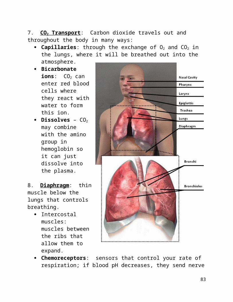
1. Trachea: “windpipe”; tube(s) with C-shaped rings of cartilage found in worms and vertebrates that intake air to retrieve oxygen.
Spiracles: small openings in worms that allow air to pass to the trachea. Epiglottis: a flap of tissue that covers the trachea while swallowing so food
will pass down the esophagus without disrupting breathing.
2. Lungs: air sacs that expand and contract in order to absorb oxygen into the bloodstream and release waste gases.
3. Gills: slits in the skin of an animal that use counter current-exchange in order to transfer water to their blood (e.g., fish). All mammals had gills at some point of development.
4. Nose: structure that cleans, warms, and moistens (same as the oral cavity) incoming air so it can easily pass through the pharynx and larynx (voice box).
5. Bronchi: two branches of the trachea that leads to each lung. Bronchioles: smaller tubes that branch from the bronchi. Alveoli: tiny air sacs found at the end of every bronchiole. They are the
site of gas exchange since they have an enormous surface area of about 100 square metres.
o Capillary: oxygen and carbon dioxide diffuse across the membrane between the alveoli and the capillary. The oxygen we breathe in oxidizes the deoxygenated blood as waste CO2 enters the alveoli from the blood to be exhaled.
6. Hemoglobin: a protein in red blood cells that transports 97% of inhaled oxygen throughout the body through the bloodstream. The other 3% of oxygen is absorbed into the plasma (fluid of the blood).
7. CO2 Transport: Carbon dioxide travels out and throughout the body in many ways:
Capillaries: through the exchange of O2 and CO2 in the lungs, where it will be breathed out into the atmosphere.
81
Bicarbonate ions: CO2
can enter red blood cells where they react with water to form this ion.
Dissolves – CO2 may combine with the amino group in hemoglobin so it can just dissolve into the plasma.
8. Diaphragm: thin muscle below the lungs that controls breathing.
Intercostal muscles: muscles between the ribs that allow them to expand.
Chemoreceptors: sensors that control your rate of respiration; if blood pH decreases, they send nerve impulses to the diaphragm and intercostal muscles to breathe faster.
9. Inspiration: the process of inhaling oxygen. The diaphragm relaxes, the ribs expand and the lungs expand.
10. Expiration: the process of exhaling carbon dioxide. The diaphragm contracts, the ribs return to resting position, and the lungs deflate to resting position.
82
Section C: Circulatory System
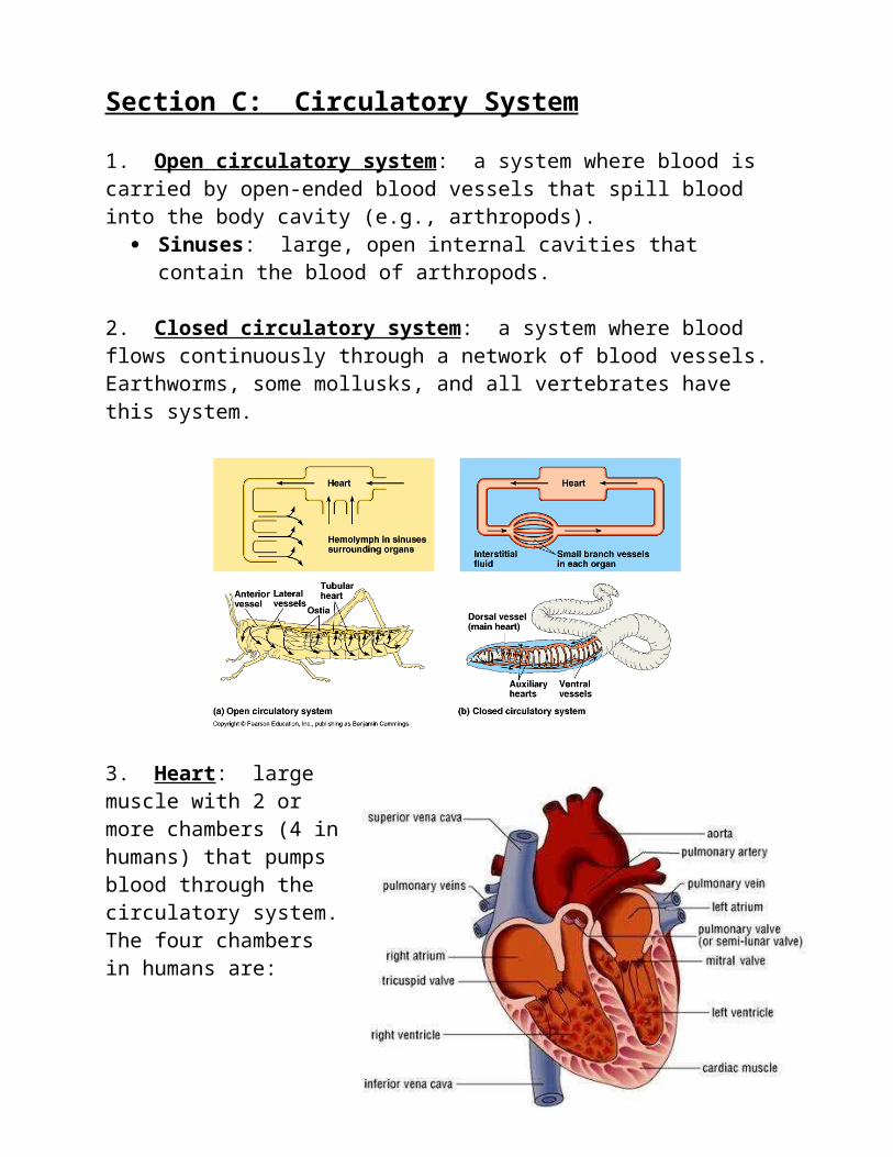
1. Open circulatory system: a system where blood is carried by open-ended blood vessels that spill blood into the body cavity (e.g., arthropods).
Sinuses: large, open internal cavities that contain the blood of arthropods.
2. Closed circulatory system: a system where blood flows continuously through a network of blood vessels. Earthworms, some mollusks, and all vertebrates have this system.
3. Heart: large muscle with 2 or more chambers (4 in humans) that pumps blood through the circulatory system. The four chambers in humans are: right atrium, right ventricle, left atrium, and left ventricle.
4. Blood: liquid substance that transports gases, nutrients and wastes throughout the body. It also transports proteins and immune responses where needed.
83
Plasma: liquid part of blood. Blood Cells: there are three types of blood cells (which are made in bone
marrow at the centre of bones):o Red Blood Cells (RBC’s): red cells that carry hemoglobin (the protein
that transports oxygen). Mature RBC’s lack a nucleus.o White Blood Cells (WBC’s): blood cells that fight infection.o Platelets: cell fragments that assist in blood clotting.
Blood Clotting: when a blood vessel is damaged, platelets stick to the collagen fibres of the vessel wall. The vessel and the platelets release substances that start the series of reactions in clotting. Prothrombin (a protein in the plasmas) converts to thrombin, which converts fibrinogen to fibrin. Fibrin threads then close the clot.
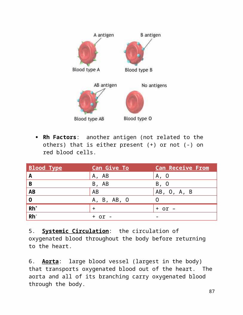
Blood Types: blood types are based on the type of antigen found on the outside of red blood cells.
o Antigen – define; there are four forms: O (no antigens), A (A antigens), B (B antigens), and AB (A and B antigens).
o Antibodies: immune substances of the white blood cells that attack foreign substances. Antibodies will attack any antigens that aren’t naturally in an individual’s body – that is why you can only give/receive blood to/from certain individuals.
Rh Factors: another antigen (not related to the others) that is either present (+) or not (-) on red blood cells.
84
Blood Type Can Give To Can Receive FromA A, AB A, OB B, AB B, OAB AB AB, O, A, BO A, B, AB, O ORh+ + + or –Rh- + or - -
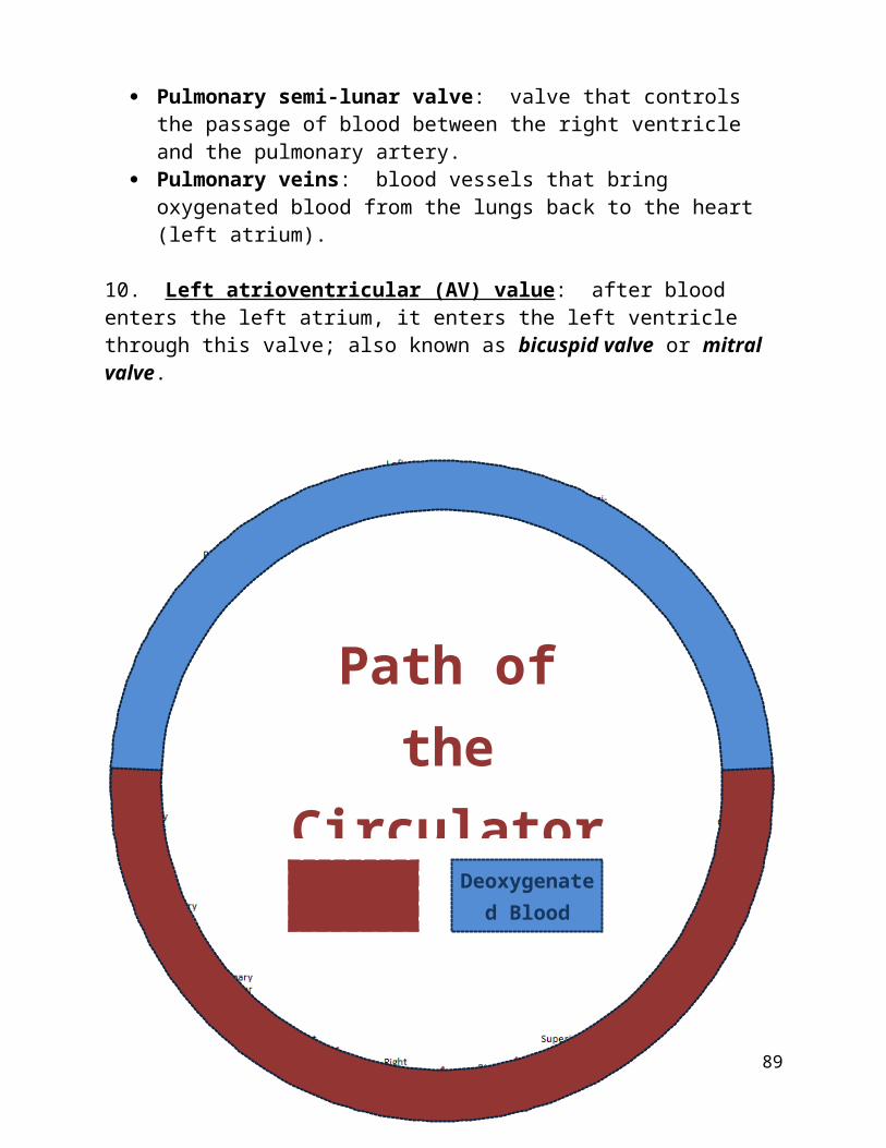
5. Systemic Circulation: the circulation of oxygenated blood throughout the body before returning to the heart.
6. Aorta: large blood vessel (largest in the body) that transports oxygenated blood out of the heart. The aorta and all of its branching carry oxygenated blood through the body.
Arteries: other large blood vessels. Aortic semi-lunar valve: valve that allows blood to flow from the left
ventricle to the aorta. Arterioles: small vessels that branch from arteries. Capillaries: the smallest blood vessels. As mentioned in Section B,
capillaries in the lungs are the sites of oxygen and carbon dioxide exchange. Because they are so thin (red blood cells must push through them in single file) the exchange of substances is easy here: oxygen and nutrients leave the capillaries while carbon dioxide and wastes enter the capillaries.
7. Veins: blood vessels that carry deoxygenated blood from capillaries, arterioles and arteries back to the heart.
Venules: branches of veins that attach to the arterioles because they are smaller.
Superior and inferior vena cava: the largest veins in the body which are attached to the right atrium of the heart. The superior brings blood from the upper body, while the inferior brings blood from the lower body.
8. Right atrioventricular (AV) valve: after blood enters the right atrium, it enters the right ventricle through this valve; also known as the tricuspid valve.
9. Pulmonary circulation: the process of oxygenating blood by sending it to the lungs from the right ventricle.
85
Pulmonary artery: blood vessel that brings blood from the heart to the lungs.
Pulmonary semi-lunar valve: valve that controls the passage of blood between the right ventricle and the pulmonary artery.
Pulmonary veins: blood vessels that bring oxygenated blood from the lungs back to the heart (left atrium).
10. Left atrioventricular (AV) value: after blood enters the left atrium, it enters the left ventricle through this valve; also known as bicuspid valve or mitral valve.
86
Path of the Circulatory
SystemOxygenated
BloodDeoxygenated
Blood
11. Thermoregulation: human homeostasis; there are two types: Ectotherms: “cold-blooded” animals; their body temperature is directly
affected by their environment. Endotherms: “warm-blooded” animals that regulate their own body
temperature; this is done through a process called counter current exchange.
o Counter current exchange: warm blood arteries run parallel to cooled veins to regulate the temperature of the body.
12. Heart regulation: the beat of the heart has to be regulated; in humans, the heart contracts and relaxes automatically at 72 beats per minute.
Sinoatrial (SA) node: “the pacemaker”; tissue in the right atrium that sends an impulse through the heart. This pulse activates the AV node.
Atrioventricular (AV) node: tissue that, in reaction to the SA node, pulses the left side of the heart.
Bundle of His and Purkinje Fibres: fibres in the walls of the ventricles that contract and relax the heart in response to the SA and AV nodes.
Systole: contraction part of the cycle Diastole: relaxation part of the cycle. Blood pressure: The pressure of the blood in the circulatory system, often
measured for diagnosis since it is closely related to the force and rate of the heartbeat and the diameter and elasticity of the arterial walls.
o Diastolic pressure: the blood pressure (as measured by a sphygmomanometer) after the contraction of the heart while the chambers of the heart refill with blood.
o Systolic pressure: the blood pressure (as measured by a sphygmomanometer) during the contraction of the left ventricle of the heart.
o High blood pressure: Hypertension or high blood pressure is a chronic medical condition in which the systemic arterial blood pressure is elevated.
o Low blood pressure: Hypotension or low blood pressure is a chronic medical condition in which the systemic arterial blood pressure is lower than normal.
o Pulse: pressure wave generated by blood flow flowing through the body. The average human pulse [heart rate] is 72 beats per minute.
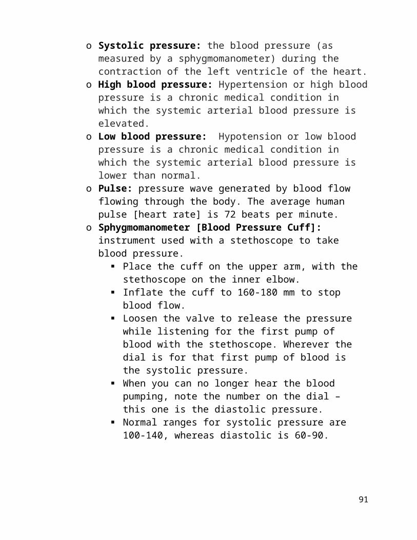
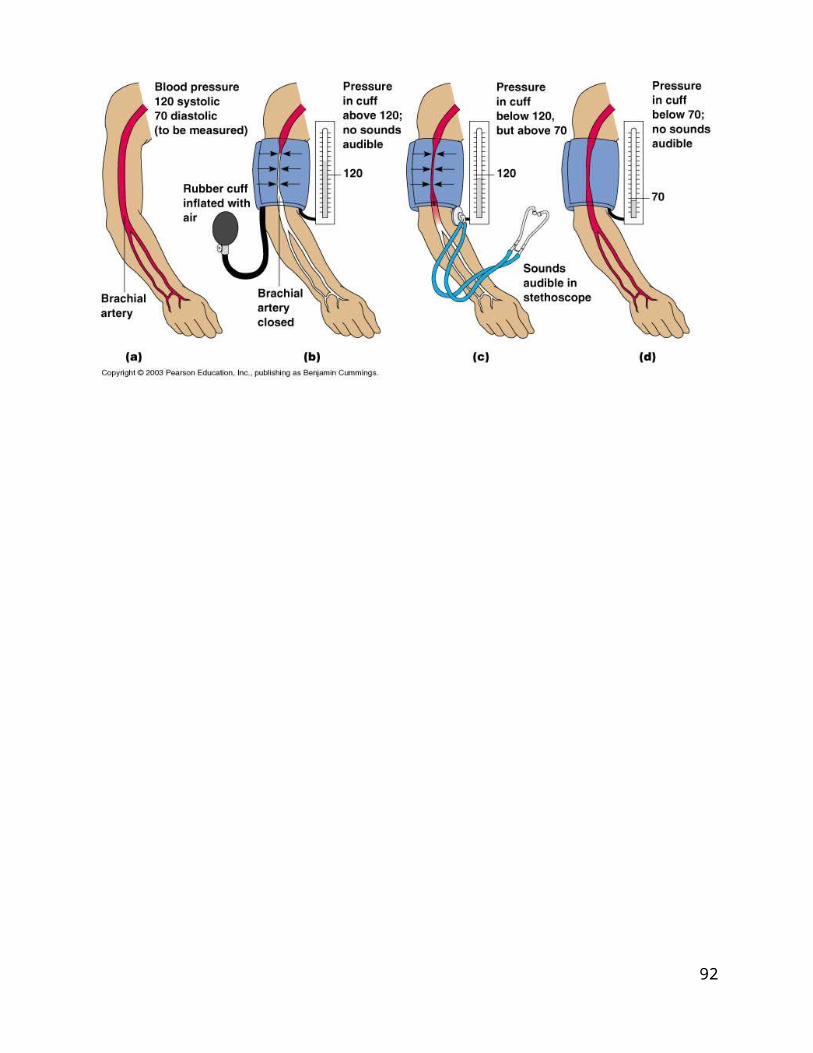
o Sphygmomanometer [Blood Pressure Cuff]: instrument used with a stethoscope to take blood pressure.
87
Place the cuff on the upper arm, with the stethoscope on the inner elbow.
Inflate the cuff to 160-180 mm to stop blood flow. Loosen the valve to release the pressure while listening for the
first pump of blood with the stethoscope. Wherever the dial is for that first pump of blood is the systolic pressure.
When you can no longer hear the blood pumping, note the number on the dial – this one is the diastolic pressure.
Normal ranges for systolic pressure are 100-140, whereas diastolic is 60-90.
88
Section D: Lymphatic System
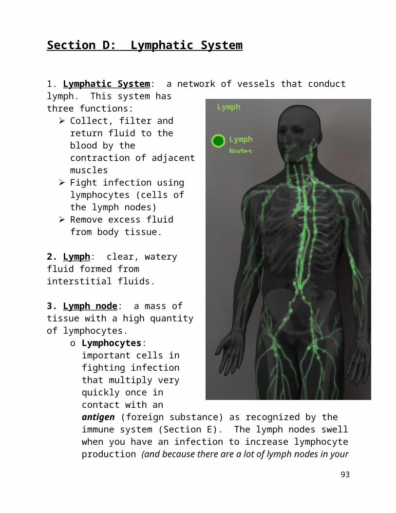
1. Lymphatic System: a network of vessels that conduct lymph. This system has three functions:
Collect, filter and return fluid to the blood by the contraction of adjacent muscles
Fight infection using lymphocytes (cells of the lymph nodes)
Remove excess fluid from body tissue.
2. Lymph: clear, watery fluid formed from interstitial fluids.
3. Lymph node: a mass of tissue with a high quantity of lymphocytes.
o Lymphocytes: important cells in fighting infection that multiply very quickly once in contact with an antigen (foreign substance) as recognized by the immune system (Section E). The lymph nodes swell when you have an infection to increase lymphocyte production (and because there are a lot of lymph nodes in your neck, doctors will touch your neck to see if your lymph nodes are swollen and fighting infection).
89
Lymph
Lymph Nodes
Section E: Immune System
1. Phagocyte: a substance that engulfs antigens (foreign substances).
2. Complement proteins: proteins that remove antigens from cell walls and membranes.
3. Interferons: inhibit viral replication by activating surrounding cells to fight it.
4. Inflammatory response: series of events that lead to swelling and/or irritation in response to an antigen or physical injury.
5. MHC (major histocompatibility complex) marker: substance that leaves markers on infected cells to identify them (they’ll be destroyed later).
6. T-lymphocytes: lymphocytes that fight infections and promote the reproduction of B-lymphocytes (lymphocytes that play a large role in the humoral immune response [as opposed to the cell-mediated immune response, which is governed by T cells]). They are made in the bone marrow. Helper T-cells: cells that activate B-lymphocytes. Memory T-cells: cells that recognize viruses and antigens they have
encountered before. Cytotoxic T-cells: cells that kill infected cells. Cell-mediated response: the activation of T-lymphocytes.
7. Humoral Immunity: Immunity referring to elements dissolved in the blood or body fluids, such as antibodies in the blood, rather than cells.
8. Macrophages: A large phagocytic cell found in stationary form in the tissues or as a mobile white blood cell.
9. AIDS (acquired immune deficiency syndrome): a syndrome that compromises the immune system as a result of HIV. There’s no cure and sufferers don’t die from the syndrome itself, but rather from infections their bodies can no longer fight.
90
Section F: The Excretory System
1. Nitrogenous wastes: toxic waste substances that are liquid (or dissolve in liquids) that are transported out of the body by the excretory system (system that excretes nitrogenous wastes). Ammonia (NH3): by-product of the breakdown of proteins that are acidic to
the body; fish excrete ammonia, just as it is, whereas other animals combine it with other substances to be excreted from the body.
Uric Acid: a conversion of ammonia and other waste substances that are excreted from birds (white, gooey).
Urea: a conversion of ammonia and other waste substances that are excreted from most mammals, including humans (clear yellow liquid).
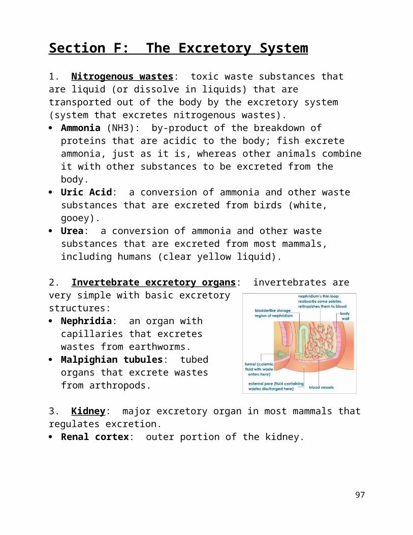
2. Invertebrate excretory organs: invertebrates are very simple with basic excretory structures: Nephridia: an organ with capillaries that
excretes wastes from earthworms. Malpighian tubules: tubed organs that
excrete wastes from arthropods.
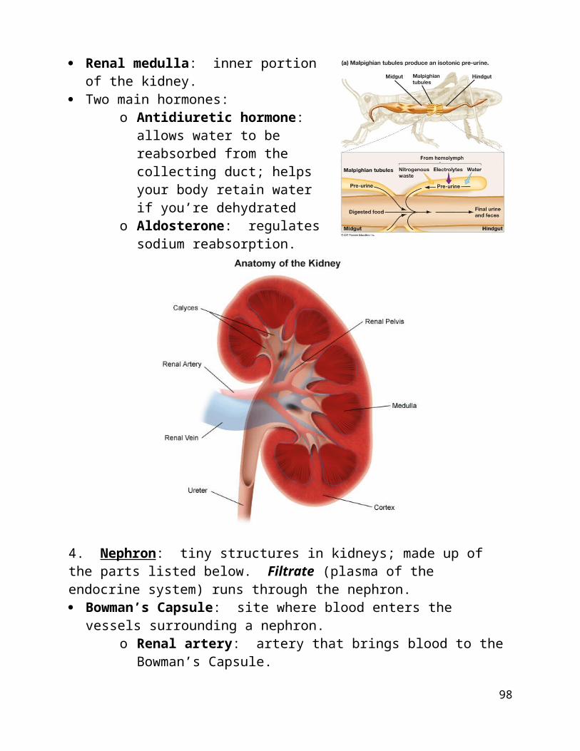
3. Kidney: major excretory organ in most mammals that regulates excretion. Renal cortex: outer portion of the kidney. Renal medulla: inner portion of the kidney. Two main hormones:
o Antidiuretic hormone: allows water to be reabsorbed from the collecting duct; helps your body retain water if you’re dehydrated
o Aldosterone: regulates sodium reabsorption.
4. Nephron: tiny structures in kidneys; made up of the parts listed below. Filtrate (plasma of the endocrine system) runs through the nephron. Bowman’s Capsule: site where blood enters the vessels surrounding a
nephron.
91
o Renal artery: artery that brings blood to the Bowman’s Capsule.o Glomerulus: ball of capillaries within the Bowman’s Capsule.
Proximal convoluted tubule: tube connecting the Bowman’s Capsule to the Loop of Henle.
Loop of Henle: loop of tubing surrounded by capillaries. Here, more wastes of the blood are transferred into the nephron.
Distal convoluted tubule: tube connecting the Loop of Henle to the collecting duct.
Collecting duct: here, the wastes are modified into urine.
There are three steps in the production of urine: Filtration: the blood is filtered in the glomerulus of the Bowman’s Capsule.
Proteins and blood cells stay in the blood while ions, water, glucose, urea, and amino acids pass into the filtrate.
Reabsorption: as the filtrate moves through, some materials are reabsorbed. Small solutes are reabsorbed by capillaries; remaining material is urine.
Secretion: as the filtrate moves through the tubules, some substances (e.g., potassium, hydrogen) are secreted from capillaries into the tubules.
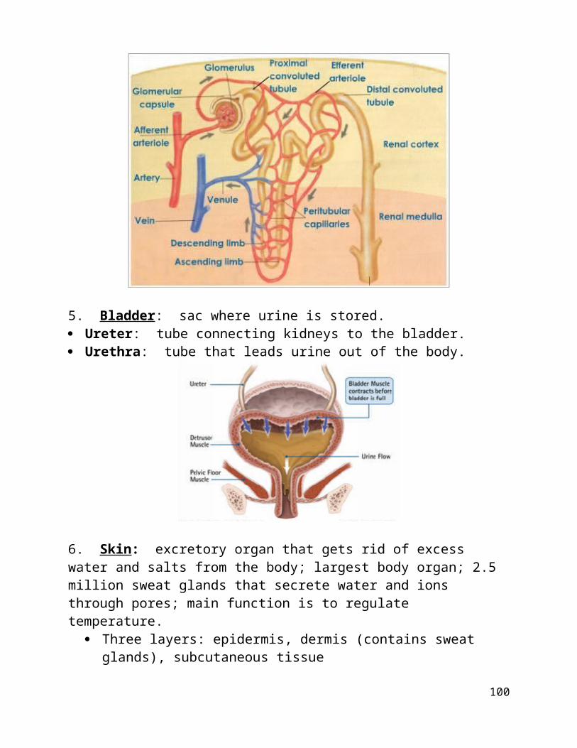
5. Bladder: sac where urine is stored. Ureter: tube connecting kidneys to the bladder. Urethra: tube that leads urine out of the body.
92
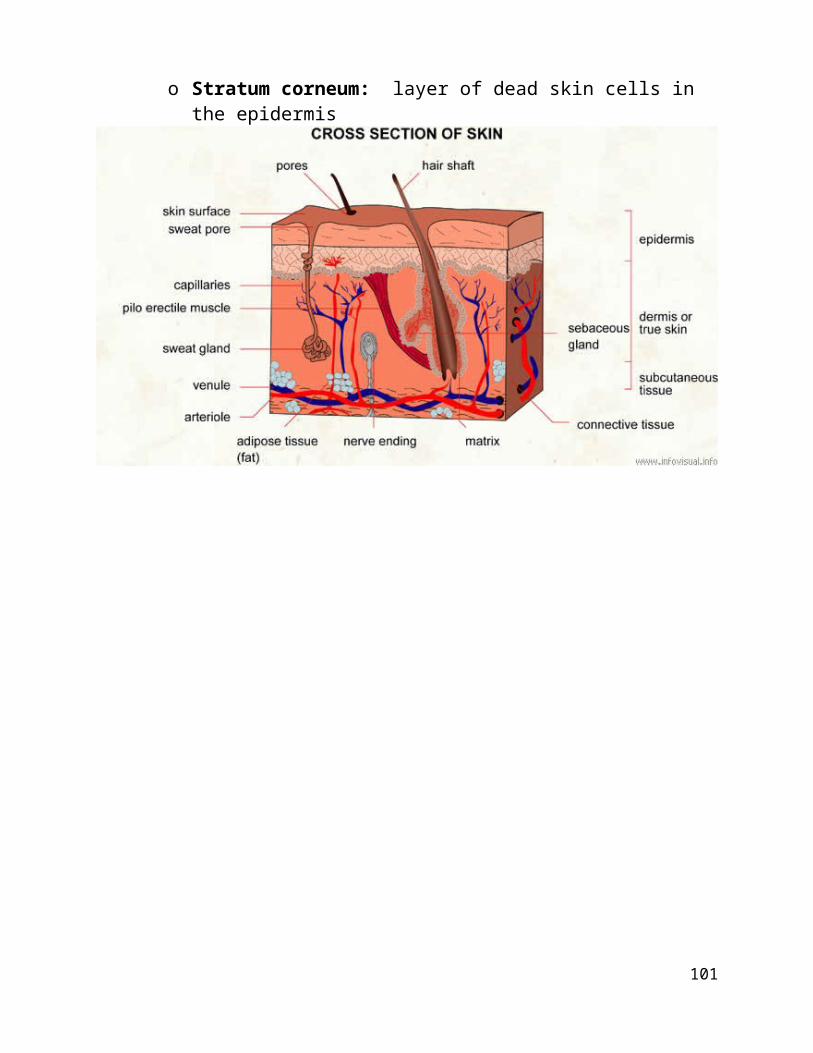
6. Skin: excretory organ that gets rid of excess water and salts from the body; largest body organ; 2.5 million sweat glands that secrete water and ions through pores; main function is to regulate temperature.
Three layers: epidermis, dermis (contains sweat glands), subcutaneous tissue
o Stratum corneum: layer of dead skin cells in the epidermis
93
Section G: Nervous System
1. Nervous System: The network of nerve cells and fibres that transmits nerve impulses between parts of the body.
Nerve Net: a simple nervous system found in simple organisms. Ganglia: a clump of nerve cells that developed in slightly more complex
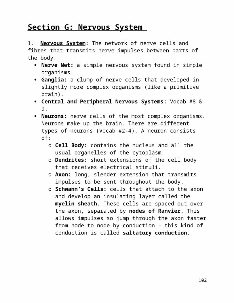
organisms (like a primitive brain). Central and Peripheral Nervous Systems: Vocab #8 & 9. Neurons: nerve cells of the most complex organisms. Neurons make up the
brain. There are different types of neurons (Vocab #2-4). A neuron consists of:
o Cell Body: contains the nucleus and all the usual organelles of the cytoplasm.
o Dendrites: short extensions of the cell body that receives electrical stimuli.
o Axon: long, slender extension that transmits impulses to be sent throughout the body.
o Schwann’s Cells: cells that attach to the axon and develop an insulating layer called the myelin sheath. These cells are spaced out over the axon, separated by nodes of Ranvier. This allows impulses so jump through the axon faster from node to node by conduction – this kind of conduction is called saltatory conduction.
94
2. Sensory Neurons: receive impulses from the environment (ex. a touch on skin) and sends messages back through the body. They are stimulated by the sense of touch.
3. Motor (Effector) Neurons: receive impulses from sensory neurons and produce a response in a muscle or a gland (ex. feel a bee sting in sensory neuron, motor neuron makes your muscles contract to pull away).
4. Interneurons: neurons that link motor neurons to sensory neurons.
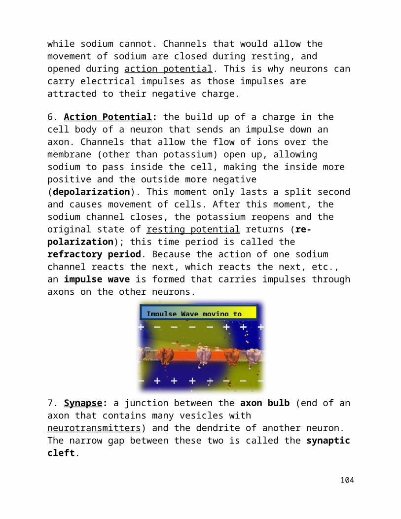
5. Resting Potential: when the inside of the plasma membrane of the axon has a negative charge, while there is a positive charge outside. This is caused by a sodium-potassium pump which is working backwards from most other sodium-potassium pumps. They are transporting sodium ions out, and potassium ions in. Many potassium ions can leave through open channels, while sodium cannot. Channels that would allow the movement of sodium are closed during resting, and opened during action potential. This is why neurons can carry electrical impulses as those impulses are attracted to their negative charge. 6. Action Potential: the build up of a charge in the cell body of a neuron that sends an impulse down an axon. Channels that allow the flow of ions over the membrane (other than potassium) open up, allowing sodium to pass inside the cell, making the inside more positive and the outside more negative (depolarization). This moment only lasts a split second and causes movement of cells. After this moment, the sodium channel closes, the potassium reopens and the original state of resting potential returns (re-polarization); this time period is called the refractory period. Because the action of one sodium channel reacts the next, which reacts the next, etc., an impulse wave is formed that carries impulses through axons on the other neurons.
95
Impulse Wave moving to the right.

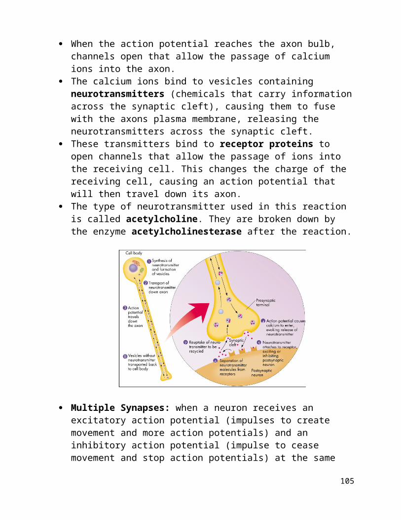
7. Synapse: a junction between the axon bulb (end of an axon that contains many vesicles with neurotransmitters) and the dendrite of another neuron. The narrow gap between these two is called the synaptic cleft.
When the action potential reaches the axon bulb, channels open that allow the passage of calcium ions into the axon.
The calcium ions bind to vesicles containing neurotransmitters (chemicals that carry information across the synaptic cleft), causing them to fuse with the axons plasma membrane, releasing the neurotransmitters across the synaptic cleft.
These transmitters bind to receptor proteins to open channels that allow the passage of ions into the receiving cell. This changes the charge of the receiving cell, causing an action potential that will then travel down its axon.
The type of neurotransmitter used in this reaction is called acetylcholine. They are broken down by the enzyme acetylcholinesterase after the reaction.
Multiple Synapses: when a neuron receives an excitatory action potential (impulses to create movement and more action potentials) and an inhibitory action potential (impulse to cease movement and stop action potentials) at the same time, they cancel each other out and nothing happens.
GABA & Norepinephrine: other types of neurotransmitters that work in the central areas of the human body.
96
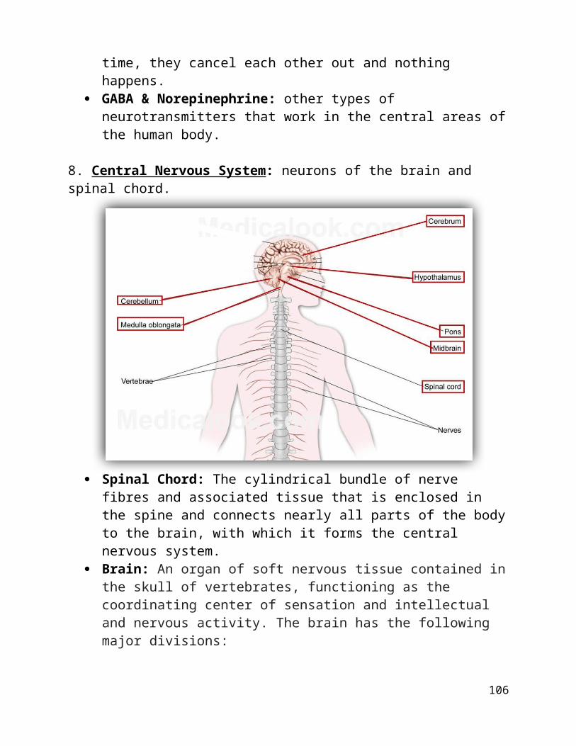
8. Central Nervous System: neurons of the brain and spinal chord.
Spinal Chord: The cylindrical bundle of nerve fibres and associated tissue that is enclosed in the spine and connects nearly all parts of the body to the brain, with which it forms the central nervous system.
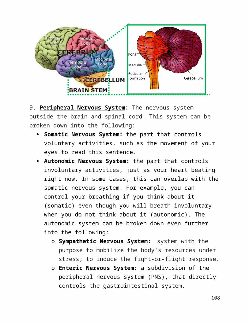
Brain: An organ of soft nervous tissue contained in the skull of vertebrates, functioning as the coordinating center of sensation and intellectual and nervous activity. The brain has the following major divisions:
o Cerebrum: largest part of the human brain that controls thoughts, coordination and voluntary activities. The cerebrum is made up of 4 lobes:
Frontal Lobe: the front of the brain Temporal Lobe: below the frontal lobe. Occipital Lobe: lower back of the brain. Parietal Lobe: upper back of the brain.
o Cerebellum: coordinates muscle activity, balance and refinement of movement.
o Brain Stem: the part of the brain continuous with the spinal cord. Consists of the medulla oblongata, pons, midbrain and parts of the hypothalamus.
Hypothalamus: regulates homeostasis and hormones (pituitary gland, growth, sexual, etc).
97
Medulla (oblongata): controls involuntary actions needed for survival (breathing, heart, etc.).
Pons: Controls part of the respiratory system and attaches parts of the brain together.
Midbrain: controls visual and auditory reflexes (blinking, jumping at loud noises, etc.).
Thalamus: controls sensory relay that connects impulses between the spinal chord and cerebrum.
9. Peripheral Nervous System: The nervous system outside the brain and spinal cord. This system can be broken down into the following:
Somatic Nervous System: the part that controls voluntary activities, such as the movement of your eyes to read this sentence.
Autonomic Nervous System: the part that controls involuntary activities, just as your heart beating right now. In some cases, this can overlap with the somatic nervous system. For example, you can control your breathing if you think about it (somatic) even though you will breath involuntary when you do not think about it (autonomic). The autonomic system can be broken down even further into the following:
o Sympathetic Nervous System: system with the purpose to mobilize the body's resources under stress; to induce the fight-or-flight response.
o Enteric Nervous System: a subdivision of the peripheral nervous system (PNS), that directly controls the gastrointestinal system.
o Parasympathetic Nervous System: opposes physiological effects of the sympathetic nervous system. It returns the body to homeostasis
98
(stimulates digestive secretions; slows the heart; constricts the pupils; dilates blood vessels).
99
Section H: Musculoskeletal System
1. Exoskeleton: a hard covering a shell of an organism with the purpose of physically supporting the organism’s systems.
2. Endoskeleton: an internal support mechanisms found in all vertebrates (organisms with backbones).
Human Skeletal System: an endoskeletal system found in humans, made up of cartilage and bone.
o Cartilage: a supportive tissue that is stiff but not solid that lacks nerves and blood vessels. All vertebrates have cartilage during embryonic development – some retain the cartilage skeleton (ex. Sharks), whereas others develop bones to replace some, but not all of the cartilage. In humans, we retain some cartilage between our bones, and our ears and noses are made of cartilage for our entire lives.
o Bone: a solid connective tissue that has nerves and blood vessels. Collagen: a fibrous protein in bone and cartilage and tendon
and other connective tissue; yields gelatin on boiling. Calcium Salts: a substance that makes up bones. Osteoblasts: bone-building cells. Osteoclasts: bone-breaking cells. Joints: where one bone connects to another. Joints are the
sites of movement between the bones. Ligaments: soft connective tissues that hold joints
together. Tendons: soft connective tissues that hold muscles to bones.
3. Human Muscular System: the anatomical system of a species that allows it to move. The muscular system in vertebrates is controlled through the nervous system, although some muscles (such as the cardiac muscle) can be completely autonomous.
100
Muscles: A band or bundle of fibrous tissue in a human or animal body that has the ability to contract, producing movement in or maintaining the position of parts of the body. It’s cells have multiple nuclei.
o Smooth Muscle: Muscle tissue in which the contractile fibrils are not highly ordered, occurring in the gut and other internal organs and not under voluntary control.
o Cardiac Muscle: the muscle tissue of the heart; adapted to continued rhythmic contraction.
Intercalated Discs: an undulating double membrane separating adjacent cells in cardiac muscle fibers. Intercalated discs support synchronized contraction of cardiac tissue. They can easily be visualized by a longitudinal section of the tissue.
o Skeletal Muscle: A muscle that is connected to the skeleton to form part of the mechanical system that moves the limbs and other parts of the body.
Striations: long parallel lineages. Skeletal muscles are characterized by striations.
Muscle Bundles: bundles of muscle tissue that create the visible striations. They contain the muscle fascicles.
Muscle Fascicles: Thin, protective layer of muscle that encloses the muscle fibre cells.
Muscle fibre cells: contractile cells made of fibres called myofibrils.
Myofibrils: contractile fibres that are subdivided into individual contractile units called sarcomeres. Myofibrils are surrounded motor neurons. An action potential from the motor neuron travels to the muscle cell and causes the myofibrils to contract.
Sarcoplasmic Reticulum: a storage of calcium ions that surrounds myofibrils.
Sarcomere: the functional unit of a muscle cell. Two proteins, actin and myosin, work together to contract and relax the muscles. Myosin filaments pulls actin filaments closer together, shortening the sarcomere. They do this when calcium is present (as released by the axon bulb of the neuron) – the calcium binds to
101
proteins on the actin, revealing their binding sites. The myosin head (which currently has ADP and a Phosphate attached to it) binds to the actin, releasing ATP. This release of ATP causes the myosin head to bend, pulling the actin. ATP then re attaches to the myosin head, breaking down to ADP and Phosphate, causing it to return to its original position. When calcium is present from
the neuron, this process repeats to contract the muscle. When the calcium is no longer released, the proteins on the actin return to position so the myosin head can no longer bind, and the muscle relaxes.
102
Section I: Endocrine System
1. Hormones: chemical messengers that can be produced in one region of the body to act on target cells in another part of the body.
Steroid Hormone: hormones made of steroids can easily pass into targeted cells to access the DNA and perform the task.
Protein/Peptide/Amine Hormones: hormones that are not steroids. They must bind to the plasma membranes of target cells to signal for tasks to be completed.
Ecdysone: hormone that promotes moulting and mating in butterflies. Brain Hormone: hormone in insects that promotes the production of other
hormones. Juvenile hormone: a hormone that causes larvae to retain certain
characteristics into adulthood. Endocrine Glands: specialized organs that make hormones.
o Hypothalamus: gland that regulates the anterior pituitary gland by secreting neurohormones.
o Pituitary Gland: the master gland that releases many hormones, many of which stimulate other glands to make other hormones. It has two parts:
I. Anterior Pituitary: the anterior pituitary produces six hormones:
Growth hormone (GH) – stimulates growth Adrenocorticotrophic Hormone (ACTH) – stimulates
adrenal cortex Thyroid-stimulating Hormone (TSH) – stimulates thyroid
gland Follicle-stimulating Hormone (FSH) – stimulates follicle
growth (females) and sperm development (males) Luteinizing Hormone (LH) – stimulates menstrual cycle
(females) and testosterone production (males) Prolactin (P) – stimulates mammary gland to produce
milkII. Posterior Pituitary: the posterior pituitary produces two
hormones: Antidiuretic hormone – regulate nephrons Oxytocin – stimulates uteral and mammary contraction
in females.
103
o Thyroid: located in the neck, it is the gland targeted the Thyroid-stimulating Hormone (TSH) of the pituitary gland. The thyroid secretes two hormones:
I. Thyroxin: hormone that regulates metabolic rate. Hyperthyroidism: individuals with an over production of
thyroxin. These people are generally thin and have trouble putting on weight.
Hypothyroidism: individuals with an under production of thyroxin. These people are generally overweight and have trouble losing that weight.
II. Calcitonin: hormone that lowers calcium levels in the blood.o Parathyroids: four small organs that rest on the thyroid. They secrete
parathyroid hormone (PTH), which increases blood calcium levels. It controls the breaking down and building of bones, which is called bone remodelling.
o Pancreas: as well as producing enzymes for the digestive systems, the pancreas also secrete insulin and glucagon.
Islets of Langerhans: cluster of cells where the pancreas produces hormones.
Glucagon: increases the levels of glucose in the blood. Insulin: decreases the levels of glucose in the blood.
Diabetics have extremely low insulin levels and must take it in order to survive.
o Adrenal: there are two adrenal glands, both found behind the kidneys – the cortex and medulla.
I. Adrenal Cortex: releases two hormones: Glucocorticoids: promote the release of glucose in the
liver. Mineralocorticoids: promote the retention of water in
the kidneys.II. Adrenal Medulla: releases two hormones:
Epinephrine & Norepinephrine: hormone that increases heart rate, breathing rate, blood pressure, etc. in response to high stress.
o Testes: glands in the male reproductive system that produce the male hormone of testosterone, a hormone that promotes sperm production. It also controls secondary male characteristics (ex. broad shoulders, facial hair, Adam’s Apple, etc.).
104
o Ovaries: glands in the female reproductive system that produce estrogen and progesterone, hormones that regulate the menstrual cycle. They also control secondary female characteristics (ex. breasts, wide hips, smaller limbs, etc.).
105
Section J: Reproductive System & Embryonic Development
1. Female Reproductive System: system in females that allows fertilization and embryonic development.
Uterus: structure which hosts the developing fetus, produces vaginal and uterine secretions, and passes the male's sperm through to the fallopian tubes. It is here that a fetus develops.
o Fetus: An unborn or unhatched offspring of a mammal, in particular an unborn human baby more than eight weeks after conception.
o Endometrium: the inner lining of the uterus.
Fallopian Tubes (Oviduct): a pair of tubes along which eggs travel from the ovaries to the uterus for fertilization.
Cervix: The narrow neck-like passage forming the lower end of the uterus. A baby must pass through the cervix in order to be born.
Vagina: The muscular tube leading from the external genitals to the cervix of the uterus in women and most female mammals.
Ovaries: structure that produces female hormones and female egg cells.
2. The Menstrual Cycle: a recurring cycle (beginning at menarche and ending at menopause) in which the endometrial lining of the uterus prepares for pregnancy; if pregnancy does not occur the lining is shed at menstruation; the average menstrual cycle is 28 days. There are three phases in the menstrual cycle:
I. The Follicular Phase: LH and FSH hormones from the pituitary gland are secreted, stimulating the growth of follicles in the ovaries. Estrogen now thickens the endometrium to prepare to carry a baby. LH is suddenly produced in mass amounts (Luteal surge) which triggers ovulation (the release of an egg from a follicle which is left behind in the ovary). The egg travels down the fallopian tube to the uterus.
II. The Luteal Phase: the follicle left in the ovary turns into a corpus luteum, a structure that continues to secrete estrogen and progesterone. The progesterone continues to ready the body for pregnancy by promoting the growth of glands and blood vessels of the endometrium so that a fertilized egg could latch on the tissue to develop. If the egg is no
106
fertilized in 13-15 days, phase three begins. If it is fertilized, then embryonic development begins and HCG (human chorionic gonadotropin) hormones are released to maintain the uterus lining for development.
III. The Menstruation (Flow) Phase: because fertilization did not occur, the uterus sheds some of its endometrium, causing bleeding. These monthly bleeding coming from the vagina is called menstruation. After menstruation, the cycle begins again to prepare for the possibility of fertilization and producing offspring.
3. Male Reproductive System: system in males with a purpose of fertilizing eggs in females to produce offspring.
Semen: male reproductive fluid that transports sperm in the uterus and fallopian tubes.
o Sperm Cells: gamete (haploid) cells that fertilize female eggs in order to produce offspring.
Prostate Gland: gland in males at the neck of the urethra; produces a viscid secretion that is the fluid part of semen.
Testes (Testicles): organ that produces sperm and hormones. Epididymis: tube by which the testes are connected to the sperm ducts. Sperm Duct (Vas Deferens): tube that transports sperm from the testes to
the prostate gland. Bladder: sac that contains urine. It is connected to the reproductive system
of males because both urine and semen are excrete from the body through the urethra.
Urethra: The duct by which urine is conveyed out of the body from the bladder, and which in male vertebrates also conveys semen.
Penis: The male genital organ of higher vertebrates, carrying the duct for the transfer of sperm during copulation. In humans and most other mammals, it consists largely of erectile tissue and serves also for the elimination of urine.
Seminal Vesicles: Each of a pair of glands that open into the vas deferens near its junction with the urethra and secrete many of the components of semen.
107
4. Puberty: the development of sexual characteristics in a human, usually in the early teens. Characteristics are primary (ex. sexual organs and hormones) and secondary (ex. facial hair in men, widening of hips in women).
5. Embryonic Development: the development of a fertilized egg in a female uterus.
Morphogenesis: the biological process that causes an organism to develop its shape.
Zygote: a fertilized egg. A sperm + an ovum (egg) = a zygote. Morula: a small ball of cells produced by mitosis of a fertilized egg. Blastula: the cells reproduce in such a way that a fluid cavity called a
blastocoel is created. Grastrula: the shape of the zygote begins to change as the three skin layers
begin to appear as single cell layers. The ectoderm will become skin, eyes and the nervous system. The mesoderm will become bones, muscles, excretory, reproductive and excretory systems. The endoderm will become the linings of the digestive and respiratory tracts, as well as other small organs.
Organogenesis (Neurula): the formation of the notochord and the neural tubes (neural plates form on cells and fold in on themselves). This leads to the production of all other organs of an embryo.
6. Embryo (Fetus): An unborn human baby, esp. in the first eight weeks from conception, after implantation but before all the organs are developed.
Extraembryonic Membranes: in other animals (ex. chickens) the following membranes are present for fetal development.
o Yolk Sac: provides food for the embryo.o Amnion: fluid filled sac that protects the embryo.o Allantois: Membrane that controls gas exchange for the embryo.o Charion: Membrane that encloses all other membranes.
Fetal Embryo: in humans, the following membranes are present for fetal development:
o Placenta: organ that provides the fetus with nutrients and oxygen, and removes the fetus’ wastes.
Umbilical Cord: the organ that connects the fetus to the placenta.
108


![study-guides.weebly.comstudy-guides.weebly.com/uploads/3/2/6/6/3266009/topic_8... · Translate this pageINDX( .•6œ(H è Ð È ~Æ 8vy]xÐ ä ßv Ð $»yy]xÐ 8vy]xÐ ð ¡ã 2003](https://img.pdfslide.net/doc/110x75/5b2242447f8b9ae4368b4644/study-translate-this-pageindx-6oeh-e-d-e-a-8vyxd-ae-ssv-d-yyxd.jpg)