Embed Size (px)
Citation preview
Caius G. Radu, M.D. Ahmanson Translational Imaging Division Department of Molecular and Medical Pharmacology David Geffen School of Medicine at UCLA Los Angeles, CA 90095 [email protected]
BIOMATH M263 Clinical Pharmacology Lecture
Studying tissue pharmacokinetics by Positron Emission Tomography (PET) Imaging tumor responses to therapy
1. What is PET and how does this technology work? 2. Using PET for imaging tumor responses to therapy 3. Studying tissue pharmacokinetics by PET
Overview
“~70% of the decisions made by physicians in the USA are based on the results of a diagnostic test… …yet only 2% of the US $2 trillion spent annually on healthcare goes into diagnostics”
Diagnostics vs. drugs
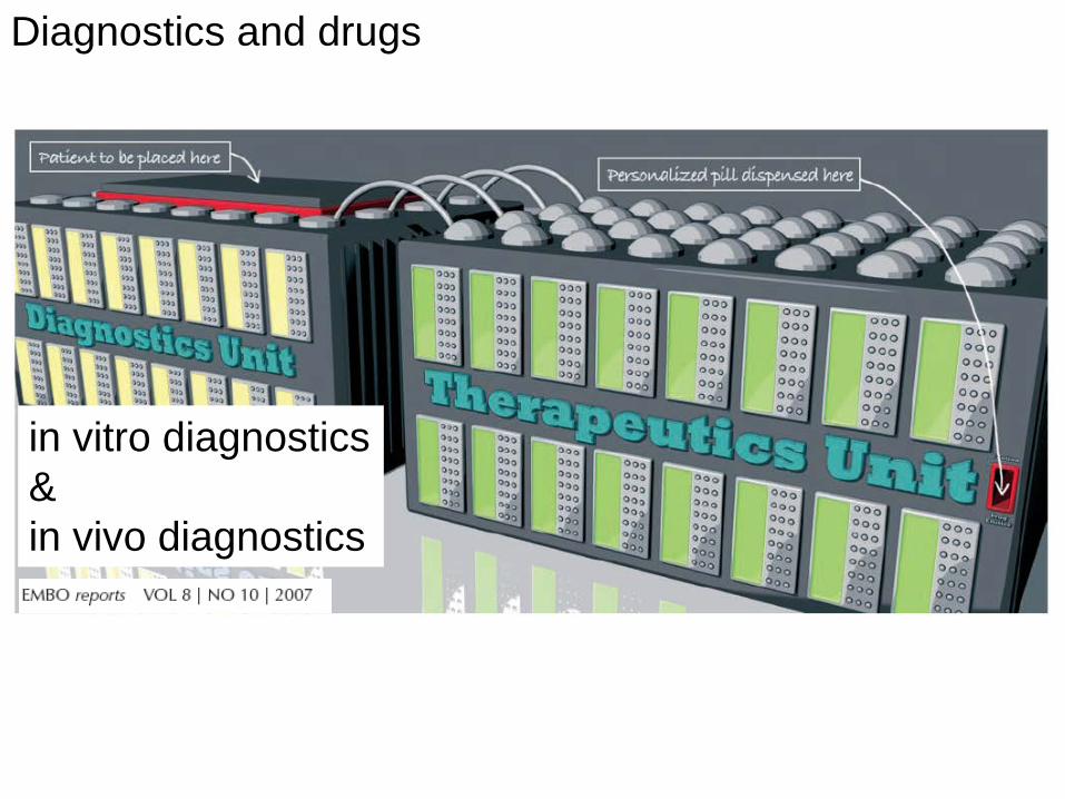
Diagnostics and drugs
in vitro diagnostics & in vivo diagnostics
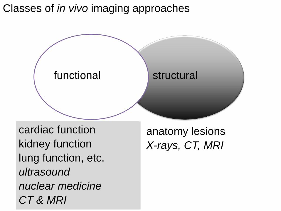
Classes of in vivo imaging approaches
structural functional
cardiac function kidney function lung function, etc. ultrasound nuclear medicine CT & MRI
anatomy lesions X-rays, CT, MRI
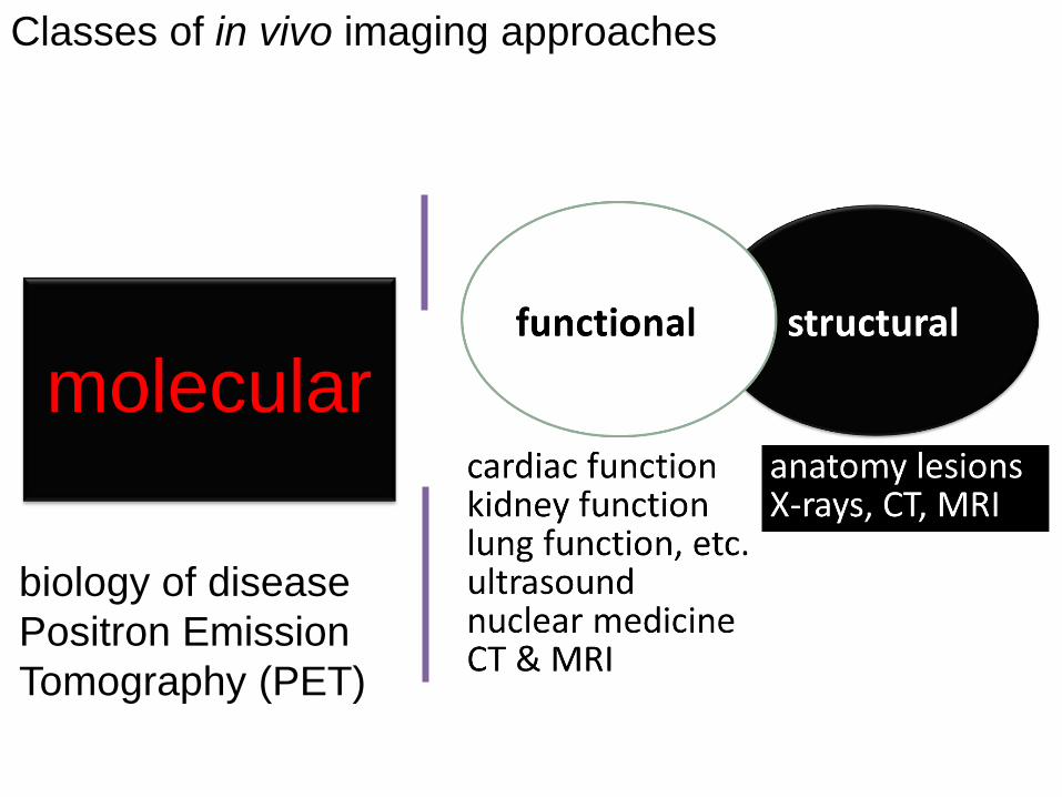
Classes of in vivo imaging approaches
molecular
biology of disease Positron Emission Tomography (PET)
Question: What is PET? Answer: A Molecular Camera
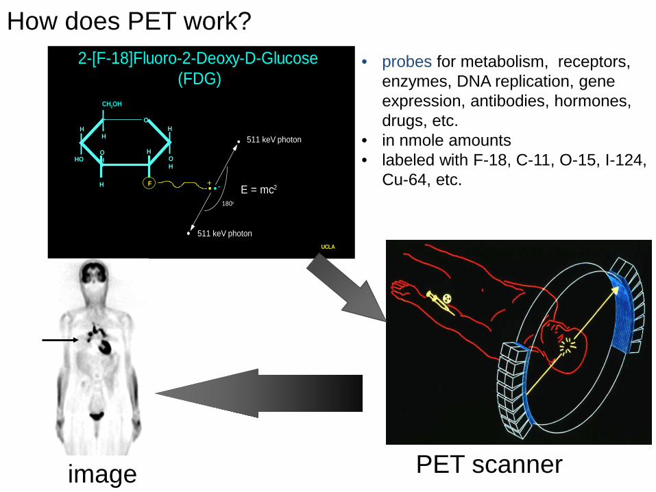
2-[F-18]Fluoro-2-Deoxy-D-Glucose (FDG)
OH
O
OH
HO
F
H
H
H
HH
CH2OH
511 keV photon
511 keV photon
E = mc2
180o
+ -
UCLA
PET scanner
• probes for metabolism, receptors, enzymes, DNA replication, gene expression, antibodies, hormones, drugs, etc.
• in nmole amounts • labeled with F-18, C-11, O-15, I-124,
Cu-64, etc.
image
How does PET work?
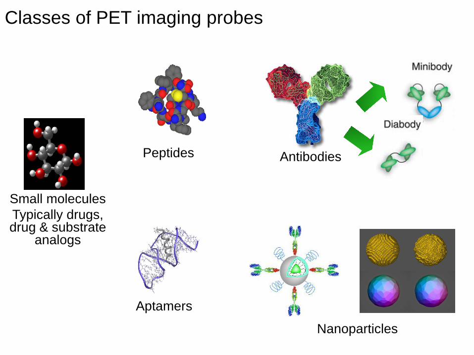
Antibodies Peptides
Aptamers
Small molecules Typically drugs, drug & substrate
analogs
Nanoparticles
Classes of PET imaging probes
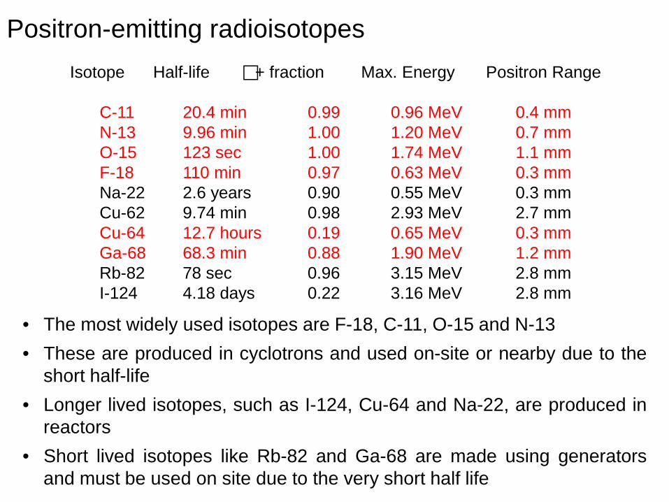
Positron-emitting radioisotopes Isotope Half-life + fraction Max. Energy Positron Range
C-11 20.4 min 0.99 0.96 MeV 0.4 mm N-13 9.96 min 1.00 1.20 MeV 0.7 mm O-15 123 sec 1.00 1.74 MeV 1.1 mm F-18 110 min 0.97 0.63 MeV 0.3 mm Na-22 2.6 years 0.90 0.55 MeV 0.3 mm Cu-62 9.74 min 0.98 2.93 MeV 2.7 mm Cu-64 12.7 hours 0.19 0.65 MeV 0.3 mm Ga-68 68.3 min 0.88 1.90 MeV 1.2 mm Rb-82 78 sec 0.96 3.15 MeV 2.8 mm I-124 4.18 days 0.22 3.16 MeV 2.8 mm
• The most widely used isotopes are F-18, C-11, O-15 and N-13 • These are produced in cyclotrons and used on-site or nearby due to the
short half-life • Longer lived isotopes, such as I-124, Cu-64 and Na-22, are produced in
reactors • Short lived isotopes like Rb-82 and Ga-68 are made using generators
and must be used on site due to the very short half life
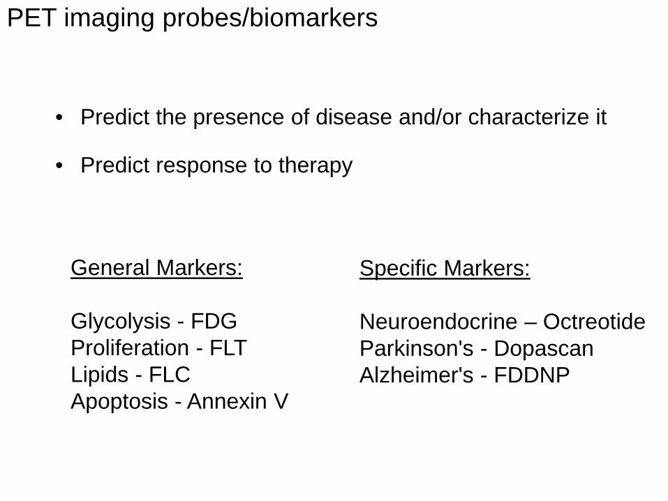
• Predict the presence of disease and/or characterize it • Predict response to therapy
General Markers:
Glycolysis - FDG Proliferation - FLT Lipids - FLC Apoptosis - Annexin V
Specific Markers:
Neuroendocrine – Octreotide Parkinson's - Dopascan Alzheimer's - FDDNP
PET imaging probes/biomarkers
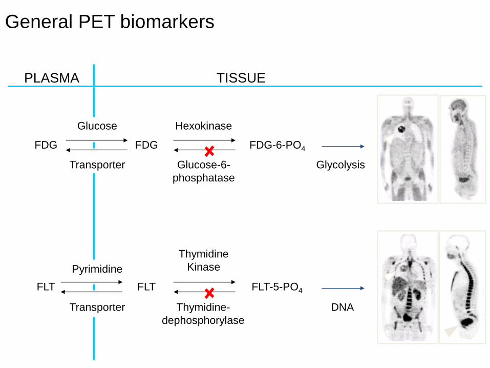
General PET biomarkers
PLASMA TISSUE
Glucose
Transporter
Pyrimidine
Hexokinase
Glucose-6- phosphatase
Glycolysis
DNA Transporter
FDG
FLT
FDG FDG-6-PO4
FLT-5-PO4
Thymidine Kinase
Thymidine-dephosphorylase
FLT
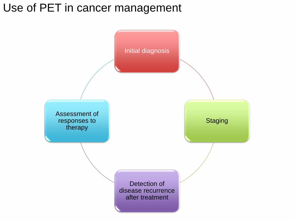
Initial diagnosis
Staging
Detection of disease recurrence
after treatment
Assessment of responses to
therapy
Use of PET in cancer management
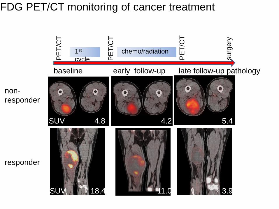
non-responder
responder
FDG PET/CT monitoring of cancer treatment
SUV 4.8 5.4 4.2
SUV 18.4 3.9 11.0
PE
T/C
T
1st cycle
baseline early follow-up late follow-up pathology
PE
T/C
T
PE
T/C
T
surg
ery
chemo/radiation

NSCLC: an example of a partial response
prior to therapy at three weeks
PET
CT
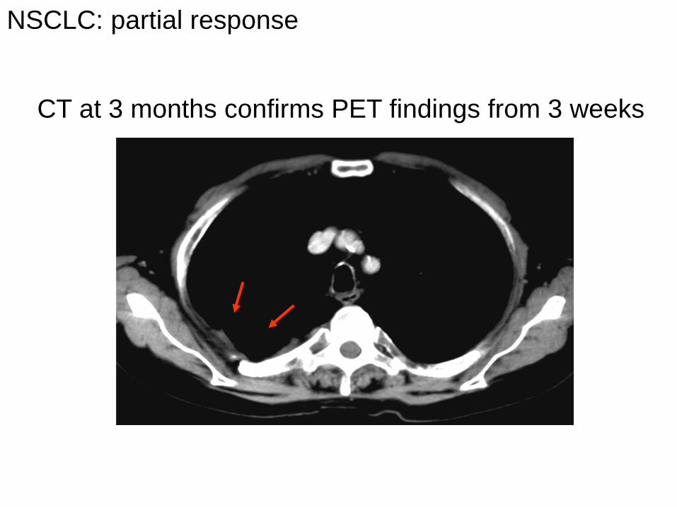
NSCLC: partial response
CT at 3 months confirms PET findings from 3 weeks
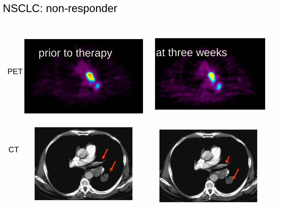
NSCLC: non-responder
prior to therapy at three weeks PET
CT
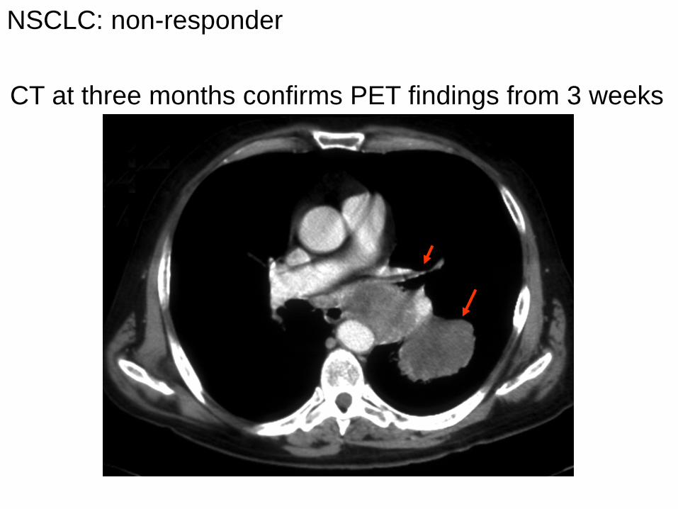
CT at three months confirms PET findings from 3 weeks
NSCLC: non-responder
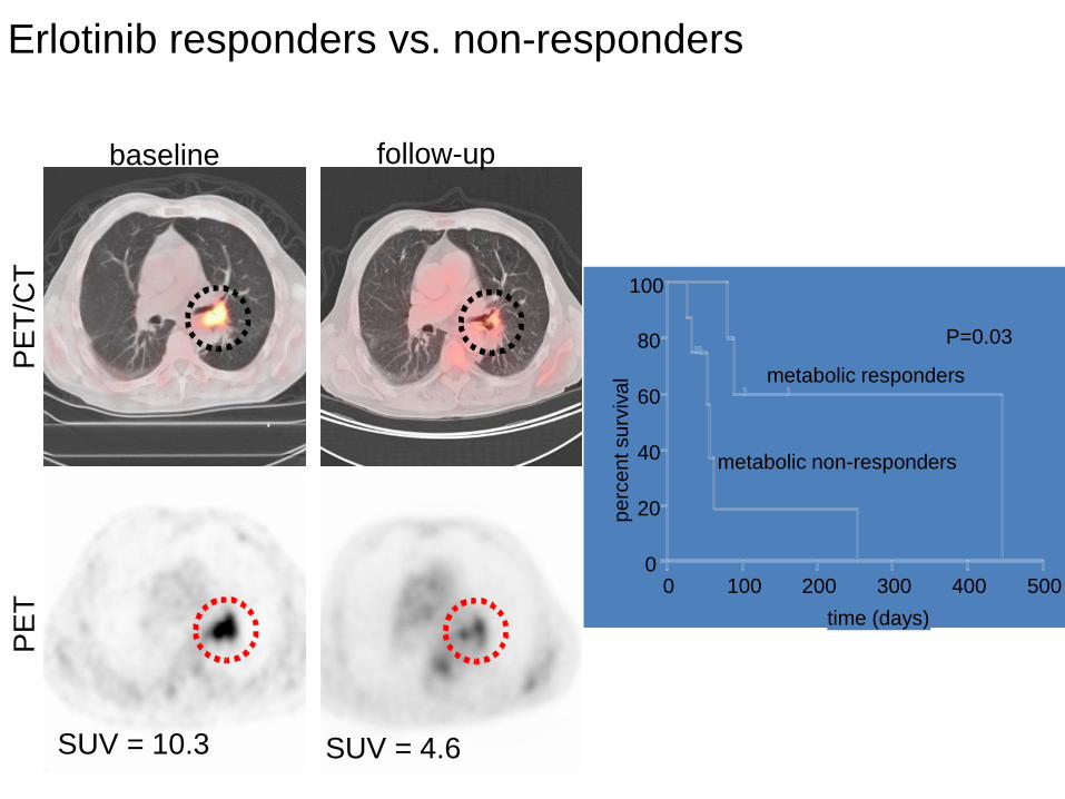
SUV = 10.3 SUV = 4.6
baseline follow-up
PET/
CT
PET
Erlotinib responders vs. non-responders
0 100 200 300 400 500 0
20
40
60
80
100
time (days)
metabolic responders
metabolic non-responders
P=0.03
perc
ent s
urvi
val
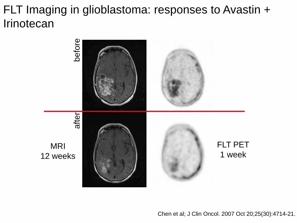
befo
re
afte
r
FLT PET 1 week
Chen et al; J Clin Oncol. 2007 Oct 20;25(30):4714-21.
MRI 12 weeks
FLT Imaging in glioblastoma: responses to Avastin + Irinotecan
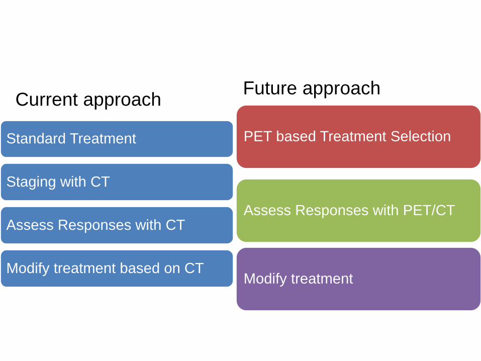
Standard Treatment
Staging with CT
Assess Responses with CT
Modify treatment based on CT
PET based Treatment Selection
Assess Responses with PET/CT
Modify treatment
Future approach Current approach
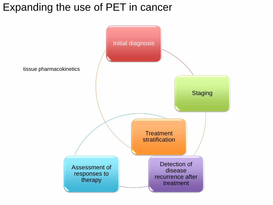
Expanding the use of PET in cancer
Initial diagnosis
Staging
Detection of disease
recurrence after treatment
Assessment of responses to
therapy
Treatment stratification
tissue pharmacokinetics

1-(2’ deoxy-2’,-18Fluoro-Arabinofuranosyl) Cytosine [18F]FAC
A new PET probe for visualizing DNA metabolism
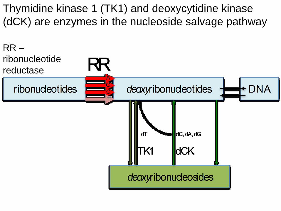
Thymidine kinase 1 (TK1) and deoxycytidine kinase (dCK) are enzymes in the nucleoside salvage pathway
RR – ribonucleotide reductase
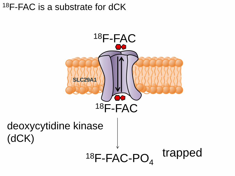
deoxycytidine kinase (dCK)
18F-FAC
18F-FAC-PO4
18F-FAC
trapped
SLC29A1
18F-FAC is a substrate for dCK
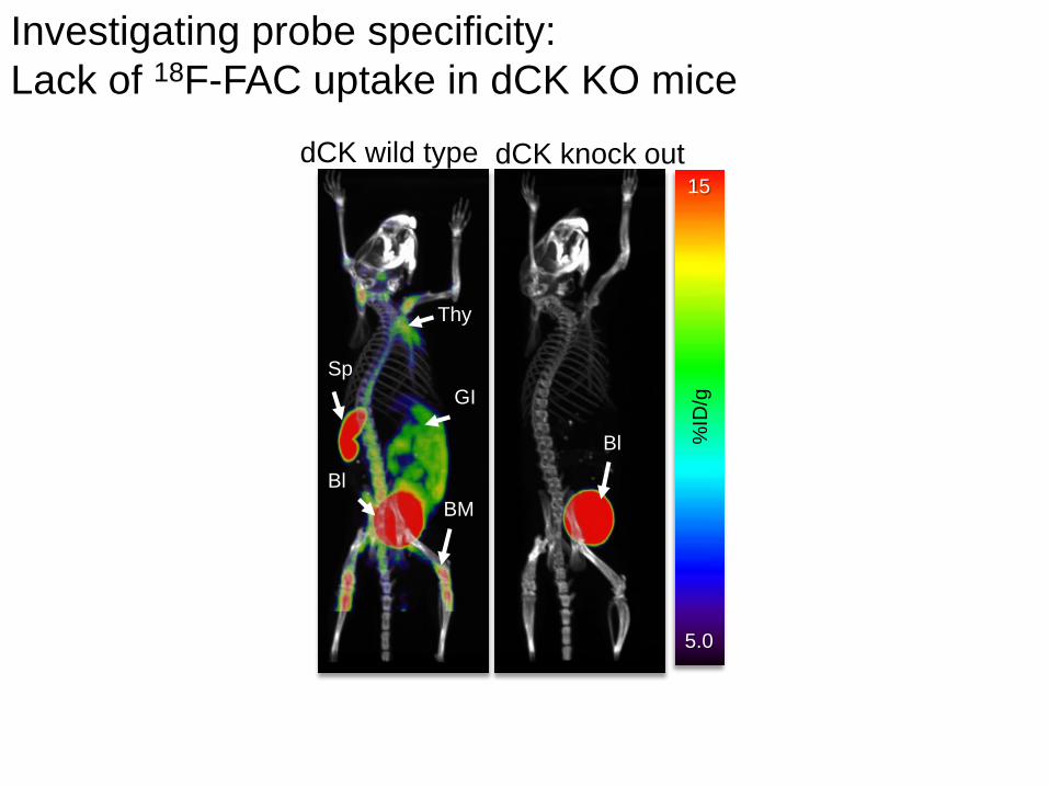
dCK wild type dCK knock out 15
5.0
%ID
/g
Thy
Sp GI
Bl BM
Bl
Investigating probe specificity: Lack of 18F-FAC uptake in dCK KO mice
accumulation in cancer
cells detected by PET
O
N
N
O
NH2
OH
OH
F
gemcitabine (dFdC) 18F-FAC
dCK 5’-NT
dFdC 18F-FAC
DNA
Nucleoside Transporter
(ENT1)
extracellular
intracellular
dFdC-TP 18F-FAC-TP
nucleus
antitumor effects
dFdC-MP 18F-FAC-MP
drug PET probe
O
N
N
O
NH2
OH
OH
F
F
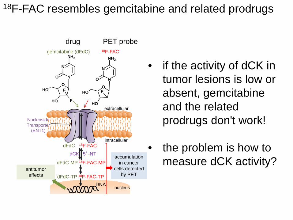
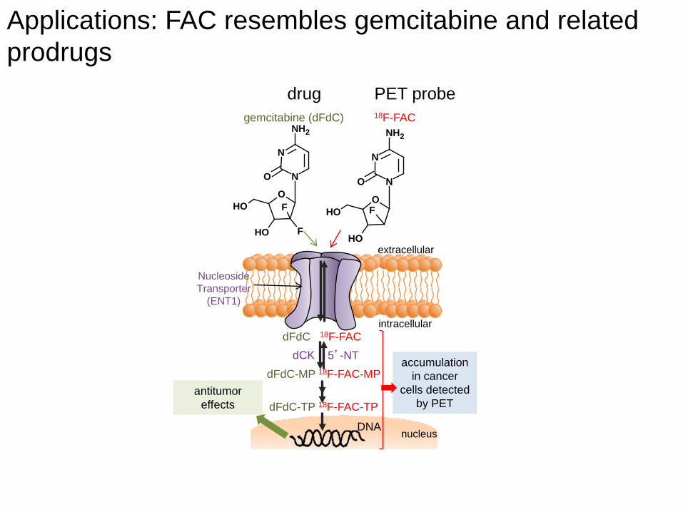
18F-FAC resembles gemcitabine and related prodrugs
• if the activity of dCK in tumor lesions is low or absent, gemcitabine and the related prodrugs don't work!
• the problem is how to
measure dCK activity?
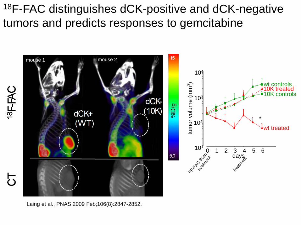
18F-FAC distinguishes dCK-positive and dCK-negative tumors and predicts responses to gemcitabine
tum
or v
olum
e (m
m3 )
101
103
104
0 1 2 3 4 5 6
wt treated
wt controls 10K treated 10K controls
days
* 102
Laing et al., PNAS 2009 Feb;106(8):2847-2852.
mouse 1 mouse 2
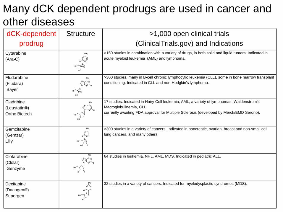
dCK-dependent prodrug
Structure >1,000 open clinical trials (ClinicalTrials.gov) and Indications
Cytarabine (Ara-C)
>150 studies in combination with a variety of drugs, in both solid and liquid tumors. Indicated in acute myeloid leukemia (AML) and lymphoma.
Fludarabine (Fludara) Bayer
>300 studies, many in B-cell chronic lymphocytic leukemia (CLL), some in bone marrow transplant conditioning. Indicated in CLL and non-Hodgkin's lymphoma.
Cladribine (Leustatin®) Ortho Biotech
17 studies. Indicated in Hairy Cell leukemia, AML, a variety of lymphomas, Waldenstrom's Macroglobulinemia, CLL currently awaiting FDA approval for Multiple Sclerosis (developed by Merck/EMD Serono).
Gemcitabine (Gemzar) Lilly
>300 studies in a variety of cancers. Indicated in pancreatic, ovarian, breast and non-small cell lung cancers, and many others.
Clofarabine (Clolar) Genzyme
64 studies in leukemia, NHL, AML, MDS. Indicated in pediatric ALL.
Decitabine (Dacogen®) Supergen
32 studies in a variety of cancers. Indicated for myelodysplastic syndromes (MDS).
O
N
N
O
NH2
OH
OH
OH
O
N
N
O
NH2
OH
OH
F
F
N
N
OOH
OH
OH
N
N
NH2
F
N
N
OOH
OH
N
N
NH2
Cl
N
N
OOH
OH
N
N
NH2
Cl
F
O
N
NN
O
NH2
OH
OH
Many dCK dependent prodrugs are used in cancer and other diseases
Translation of FAC to the clinic
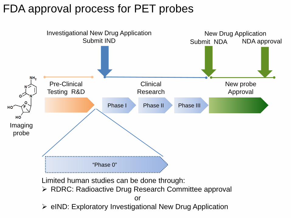
Pre-Clinical Testing R&D
Clinical Research
Phase I Phase II Phase III
New probe Approval
Submit NDA NDA approval
FDA approval process for PET probes
Investigational New Drug Application Submit IND
New Drug Application
“Phase 0”
Limited human studies can be done through: RDRC: Radioactive Drug Research Committee approval
or eIND: Exploratory Investigational New Drug Application
Imaging probe
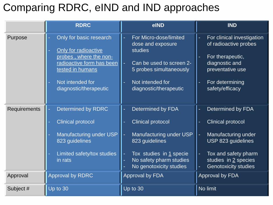
RDRC eIND IND
Purpose - Only for basic research - Only for radioactive
probes , where the non-radioactive form has been tested in humans
- Not intended for diagnostic/therapeutic
- For Micro-dose/limited dose and exposure studies
- Can be used to screen 2-5 probes simultaneously
- Not intended for
diagnostic/therapeutic
- For clinical investigation of radioactive probes
- For therapeutic, diagnostic and preventative use
- For determining safety/efficacy
Requirements - Determined by RDRC - Clinical protocol
- Manufacturing under USP
823 guidelines
- Limited safety/tox studies in rats
- Determined by FDA - Clinical protocol - Manufacturing under USP
823 guidelines
- Tox studies in 1 specie - No safety pharm studies - No genotoxicity studies
- Determined by FDA - Clinical protocol - Manufacturing under
USP 823 guidelines
- Tox and safety pharm studies in 2 species
- Genotoxicity studies
Approval Approval by RDRC Approval by FDA Approval by FDA
Subject # Up to 30 Up to 30 No limit
Comparing RDRC, eIND and IND approaches
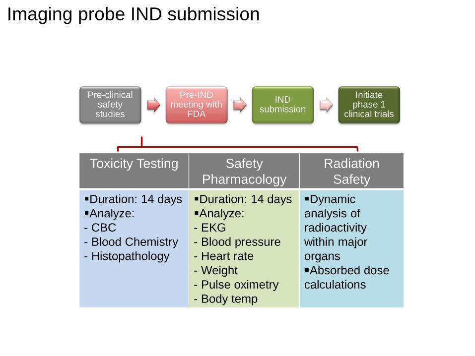
Pre-clinical safety studies
Pre-IND meeting with
FDA IND
submission Initiate
phase 1 clinical trials
Toxicity Testing Safety Pharmacology
Radiation Safety
Duration: 14 days Analyze: - CBC - Blood Chemistry - Histopathology
Duration: 14 days Analyze: - EKG - Blood pressure - Heart rate - Weight - Pulse oximetry - Body temp
Dynamic analysis of radioactivity within major organs Absorbed dose calculations
Imaging probe IND submission
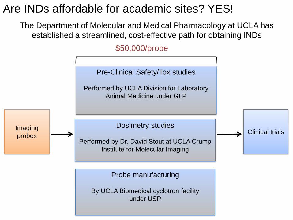
Are INDs affordable for academic sites? YES!
Imaging probes
Pre-Clinical Safety/Tox studies
Performed by UCLA Division for Laboratory Animal Medicine under GLP
$50,000/probe
Clinical trials Dosimetry studies
Performed by Dr. David Stout at UCLA Crump
Institute for Molecular Imaging
Probe manufacturing
By UCLA Biomedical cyclotron facility under USP
The Department of Molecular and Medical Pharmacology at UCLA has established a streamlined, cost-effective path for obtaining INDs
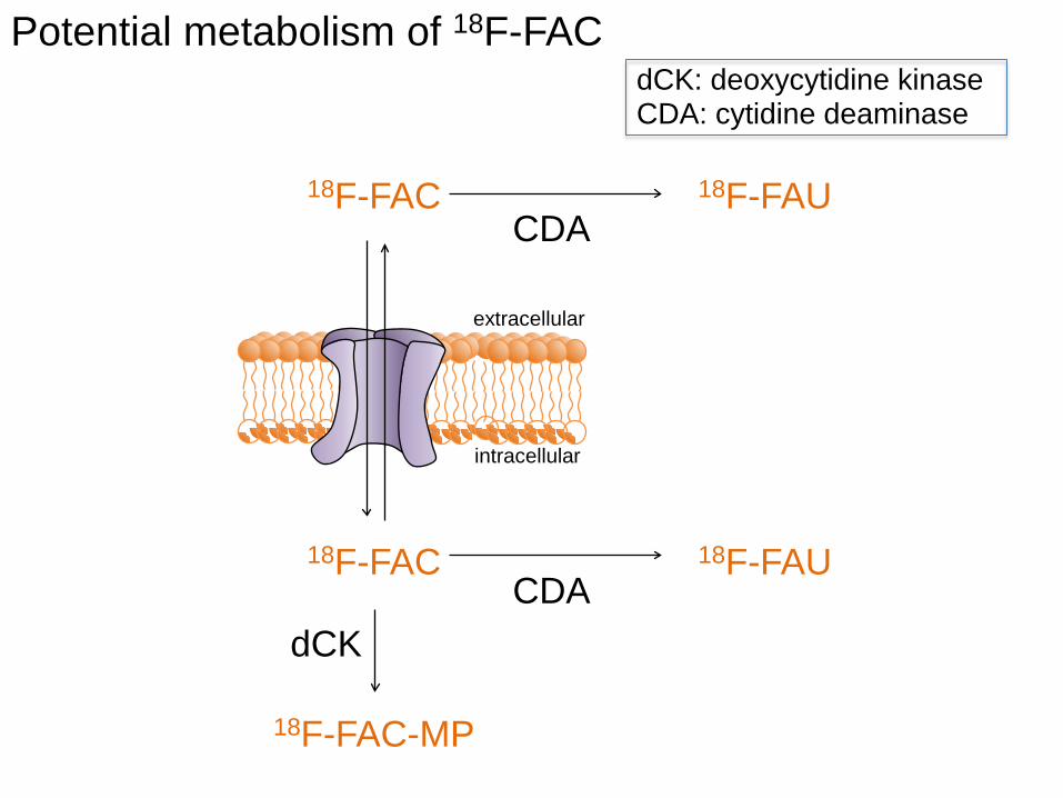
extracellular
intracellular
Potential metabolism of 18F-FAC
18F-FAC
18F-FAC-MP
dCK
CDA
CDA 18F-FAC
18F-FAU
18F-FAU
dCK: deoxycytidine kinase CDA: cytidine deaminase
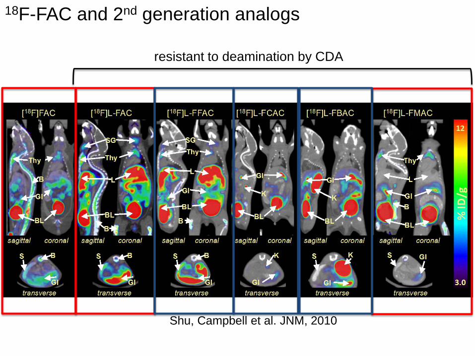
18F-FAC and 2nd generation analogs
Shu, Campbell et al. JNM, 2010
resistant to deamination by CDA
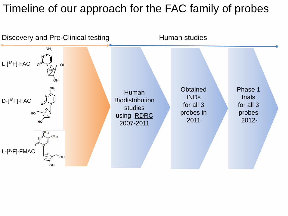
Human studies
Obtained INDs
for all 3 probes in
2011
Discovery and Pre-Clinical testing
Timeline of our approach for the FAC family of probes
Human Biodistribution
studies using RDRC
2007-2011
N
NO
NH2
O OH
OH
18F
L-[18F]-FAC
D-[18F]-FAC
L-[18F]-FMAC
Phase 1 trials
for all 3 probes 2012-
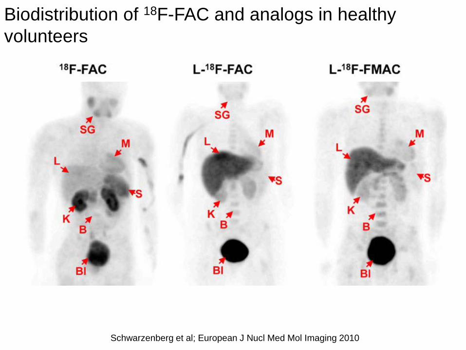
Biodistribution of 18F-FAC and analogs in healthy volunteers
Schwarzenberg et al; European J Nucl Med Mol Imaging 2010
accumulation in cancer
cells detected by PET
O
N
N
O
NH2
OH
OH
F
gemcitabine (dFdC) 18F-FAC
dCK 5’-NT dFdC 18F-FAC
DNA
Nucleoside Transporter
(ENT1)
extracellular
intracellular
dFdC-TP 18F-FAC-TP
nucleus
antitumor effects
dFdC-MP 18F-FAC-MP
drug PET probe
O
N
N
O
NH2
OH
OH
F
F
Applications: FAC resembles gemcitabine and related prodrugs
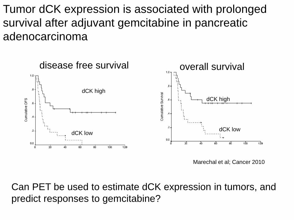
Tumor dCK expression is associated with prolonged survival after adjuvant gemcitabine in pancreatic adenocarcinoma
Marechal et al; Cancer 2010
disease free survival overall survival
dCK high
dCK low dCK low
dCK high
Can PET be used to estimate dCK expression in tumors, and predict responses to gemcitabine?
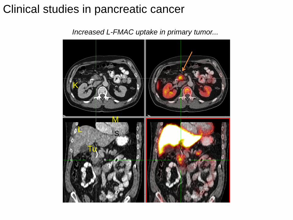
Clinical studies in pancreatic cancer
Increased L-FMAC uptake in primary tumor...
L M
S
Tu
K
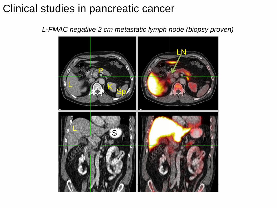
L-FMAC negative 2 cm metastatic lymph node (biopsy proven)
Clinical studies in pancreatic cancer
LN
P
L
L S
K Sp
Tu Tu
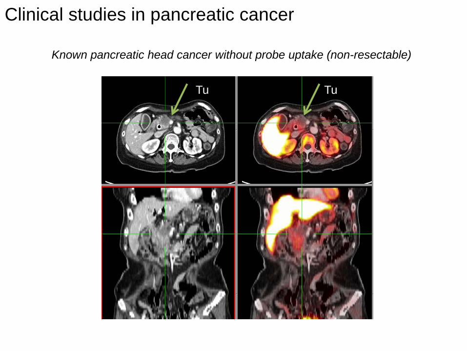
Known pancreatic head cancer without probe uptake (non-resectable)
Clinical studies in pancreatic cancer

dCK protein correlates with 18F-FAC PET imaging in an ovarian cancer patient
L-FMAC Scan FDG-Scan
Tumor site visualized on both scans Pre-therapy imaging
Tumor histology dCK expression is detected in epithelial tumor cells
α-dCK α-dCK H+E
IHC with anti-Hu-dCK monoclonal antibody M. Riedinger (Witte Lab)
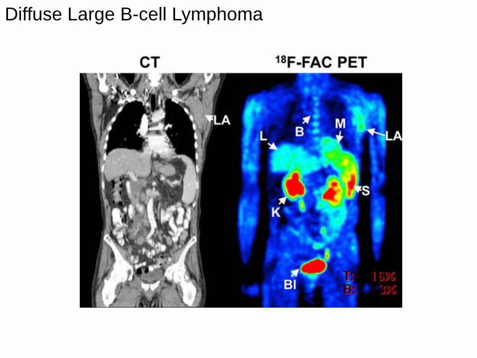
Diffuse Large B-cell Lymphoma
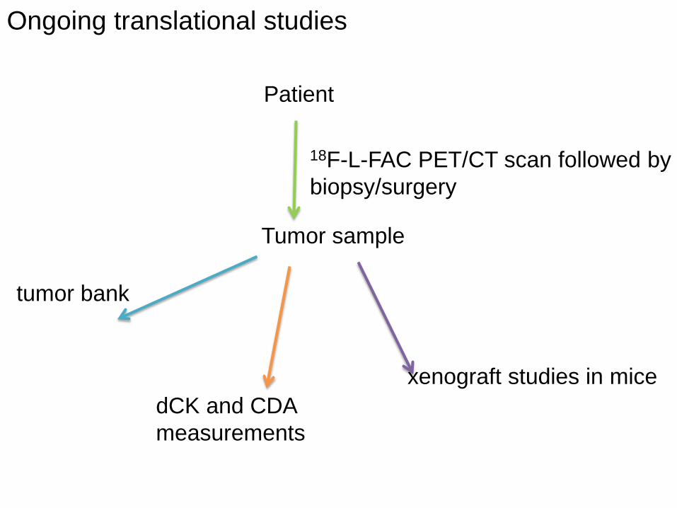
Ongoing translational studies
Patient
18F-L-FAC PET/CT scan followed by biopsy/surgery
Tumor sample
xenograft studies in mice
tumor bank
dCK and CDA measurements
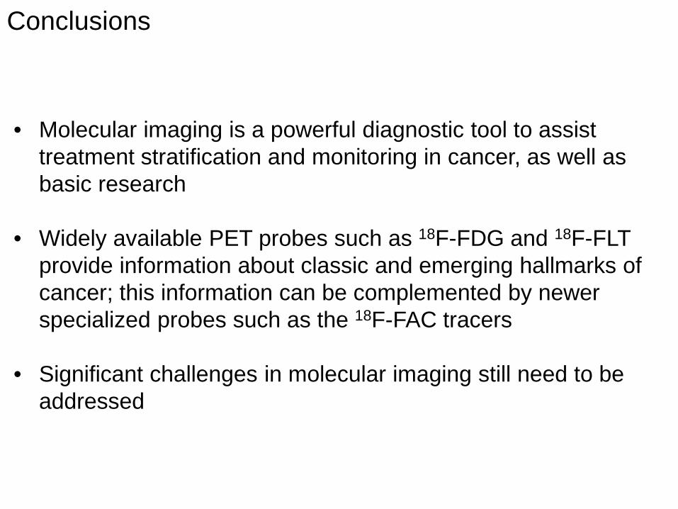
Conclusions
• Molecular imaging is a powerful diagnostic tool to assist treatment stratification and monitoring in cancer, as well as basic research
• Widely available PET probes such as 18F-FDG and 18F-FLT provide information about classic and emerging hallmarks of cancer; this information can be complemented by newer specialized probes such as the 18F-FAC tracers
• Significant challenges in molecular imaging still need to be addressed
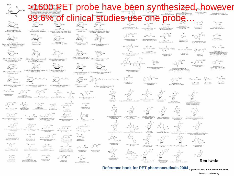
Reference book for PET pharmaceuticals 2004
>1600 PET probe have been synthesized, however 99.6% of clinical studies use one probe…
What explains the dominance of FDG? 1. FDG is very useful
2. The utility of the vast majority of probes has not been demonstrated. Why?
3. The cost of translating a new PET probe to the clinic can be substantial.
4. There may be many other useful probes, but the technology to radiolabel them does
not exist.
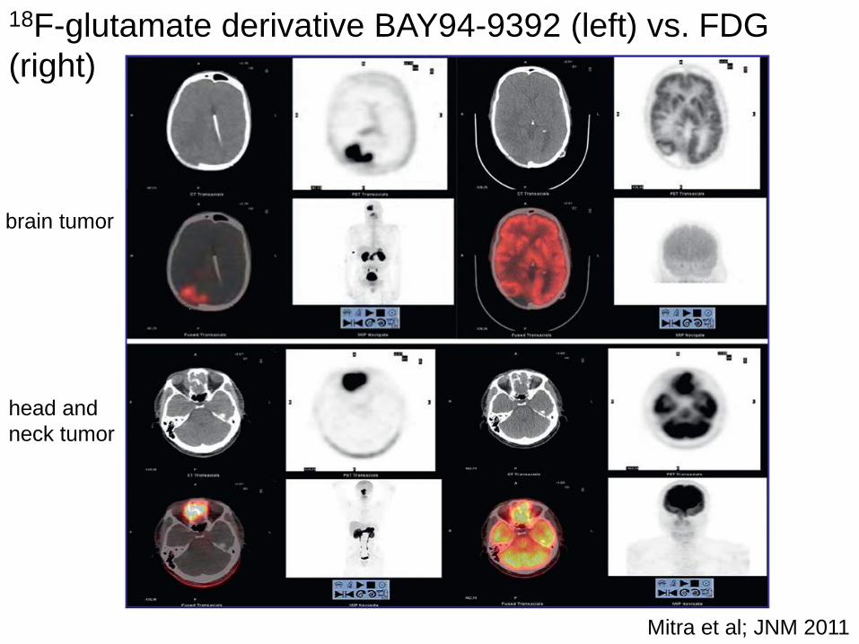
18F-glutamate derivative BAY94-9392 (left) vs. FDG (right)
Mitra et al; JNM 2011
brain tumor
head and neck tumor