Embed Size (px)
Citation preview

purified recombinant wild type (WT) and gain-of-function D374Y-PCSK9 from HEK 293Scells were added to the basolateral medium of intestinal Caco-2/15 cells grown on Transwellfilter plates. Results: An evident decrease was noted in LDLr in the whole-cell extracts andbasolateral cell-surface while exogenous WT- and D374Y-PCSK9 were accumulated in cells.Clearly, co-immunoprecipitation studies indicated an association of PCSK9 and LDLr. Bothforms of exogenous PCSK9 caused a significant enhancement in cholesterol uptake alsoevidenced by a raised protein expression of cholesterol transporters NPC1L1 and CD36,but SR-B-I, ABCA1, ABGC5 and ABCG8 expressions remained unchanged. Moreover, PCSK9altered the activity of HMG-CoA reductase and ACAT, two key proteins involved in cholesterolsynthesis and esterification, respectively. Finally, exogenous PCSK9 was able to enhancetriglyceride-rich lipoproteins by positively modulating lipid synthesis, Sar1GTPase expressionas well as apolipoprotein B-48 biogenesis. Conclusion: These data suggest that, in additionto its modulatory action on LDLr, PCSK9 significantly influences intracellular cholesterolhomeostasis and the machinery governing lipid transport in the gut. Acknowledgment: Thisstudy was supported by the Canadian Institutes of Health Research (EL, NS) and the J.A.DeSève Research Chair in Nutrition (EL).
Su2010
Chronic and Selective Inhibition of Na-K-ATPase Uniquely Regulates BrushBorder Na Dependent Nutrient Absorption in Intestinal Epithelial CellsSwapna Gayam, Palani Kumar Manoharan, Uma Sundaram
Background: Na-K-ATPase is an integral membrane protein in the mammalian cells whichis responsible for maintaining the favorable intracellular Na gradient necessary to promoteNa- coupled solute co-transport. In the chronically inflamed rabbit intestine Na-glucose co-transport (SGLT1) is inhibited on the brush border (BBM) of absorptive villus cells. Sincebasolateral membrane (BLM) Na-K-ATPase is also inhibited in these cells, we had postulatedthat at least part of the inhibition of SGLT1 is secondary to the loss of favorable Na gradientestablished by Na-K-ATPase. However, whether direct and specific chronic inhibition ofNa-K-ATPase may indeed affect BBM Na absorptive systems was unknown. Hypothesis:Direct, specific and chronic inhibition of BLM Na-K-ATPase modulates BBM Na absorptivepathways. Aim: Determine the effect of Na-K-ATPase silencing with α1siRNA transfectionon BBM SGLT1 and NH3 activity in IEC-18 cells. Methods: Rat small intestinal epithelialcells (IEC-18) were transfected with siRNA of Na-K-ATPase α1 subunit and grown in 12well plates for 5 days post transfection. 3H-OMG uptake and pH dependent, EIPA sensitive22Na uptake were performed for SGLT1 and NHE3 activity respectively. Na-K-ATPaseactivity was determined by measuring the inorganic phosphate released. RTQ-PCR wasperformed using SGLT1 specific primers and probes. Western Blot was performed using ratspecific antibodies. Results: Na-K-ATPase activity was significantly diminished by Na-K-ATPase-α1siRNA transfection on 5th day post transfection.( 18.5 ± 0.87 pmol/mg proteinin control and 8.2 ± 0.11 pmol/mg protein in Na-K-ATPase- α1siRNA transfected cells,n=5). However, SGLT1 activity was significantly up regulated in Na-K-ATPase-α1siRNAtransfected IEC-18 cells (294.86 ± 33.4 pmol/mg protein/ in control and 718.1 ± 96.6pmol/mg protein/ in Na-K-ATPase-α1siRNA transfected cells, n=3, p<0.05). NHE3 activityremained unaltered in Na-K-ATPase - α1siRNA transfected IEC-18 cells (1228 ± 95 pmol/mg protein/2 min in scrambled siRNA and 1026 ± 91 pmol/mg protein/2 min in Na-K-ATPase - α1siRNA transfected IEC-18 cells, n=3). Kinetics studies demonstrated that themechanism of stimulation of SGLT1 activity was secondary to increase in the affinity (1/Km) of the co-transporter for glucose without a change in the number of co-transporters(Km of SGLT1 is 5.8 ± 0.4 mM in Scrambled siRNA and 2.5 ± 0.35mM in Na-K-ATPase -α1siRNA transfected IEC-18 cells, n=3, p< 0.05). Further, Western blot and RTQ-PCR datadid not show a change in protein and mRNA expression respectively. Conclusion: Chronicand direct silencing of BLM Na-K-ATPase uniquely regulates BBM Na absorptive pathwaysin intestinal epithelial cells. Specifically, while BBM NHE3 is unaffected, SGLT1 is stimulatedsecondary to enhanced affinity of the co-transporter.
Su2011
Ablation of the Gut Microbiota Ameliorates Antipsychotic-Induced WeightGain and Associated Metabolic Dysfunction in the RatKieran J. Davey, Timothy G. Dinan, John F. Cryan, Siobhain M. O'Mahony
Introduction: In recent times, evidence from genetically and diet-induced animal modelsof obesity have demonstrated that the gut microbiota plays an important role in the regulationof body weight and metabolism. Aim: To investigate if the microbiota may be involved ina model of drug-induced obesity using olanzapine, a commonly prescribed antipsychoticwith well known, but poorly understood weight and metabolic side-effects.Methods: FemaleSprague-Dawley rats (n=10) received either vehicle, olanzapine (2 mg/kg/day) or olanzapine(4 mg/kg/day) I.P, B.I.D. for 21 days. In addition, rats were assigned to vehicle or anantibiotic cocktail consisting of neomycin (250 mg/kg/day), metronidazole (50 mg/kg/day)and polymyxin B (9 mg/kg/day). The antibiotic cocktail was administered by oral gavagedaily for 5 days prior to commencement of drug treatment and on all subsequent days. Onday 22 of drug treatment, a locomotor activity test was conducted on all rats. The animalswere overnight fasted prior to sacrifice. Animals were culled, uterine fat was carefullydissected and weighed, plasma, adipose and liver samples were collected, frozen and lateranalysed. Results: Co-administration of the antibiotic cocktail significantly ameliorated thebody weight gain induced by olanzapine (2 mg/kg), on days 8-21 inclusive (p < 0.05) andolanzapine (4 mg/kg) on days 11-16 inclusive (p < 0.05). Antibiotic treatment also preventedthe accretion of uterine adipose tissue induced by olanzapine (2 mg/kg) (p < 0.05) andolanzapine (4 mg/kg) (p < 0.01). Reduced inflammation was observed in the animals co-administered the antibiotic cocktail with reduced expression of CD68, a marker of macroph-age infiltration, seen in adipose tissue of animals receiving olanzapine and antibiotics com-pared to olanzapine only treated rats (p < 0.05). Conclusions: Our results demonstratewhat we believe to be for the first time, the involvement of the gut microbiota in a drug-induced obesity model. Ablation of the gut microbiota ameliorated a number of metaboliceffects associated with olanzapine treatment including increases in body weight and visceralfat. These side-effects are a serious clinical hurdle in the treatment of psychiatric patientsand hence these findings are of potentially huge benefit by offering a new therapeutic target
S-559 AGA Abstracts
for antipsychotic-induced metabolic dysfunction. Moreover, in line with recent reports, ourstudy demonstrates further the important role the gut flora has on host physiology inparticular the regulation of energy balance.
Su2012
A Study on Gender Differences in Feeding Behavior and Plasma Acyl GhrelinLevels in Aged Mice Following Exposure to a Novel Environmental StressShunsuke Ohnishi, Hiroshi Takeda, Shuichi Muto, Koji Nakagawa, Chiharu Sadakane,Miwa Nahata, Yayoi Saegusa, Tomohisa Hattori, Masahiro Asaka
Background/Aim: Response to stress during aging differs from that during youth, resultingin altered feeding behavior. Gender differences can sometimes have a major effect on theseresponses. We previously studied the food intake and expression of various appetite-relatedgenes in the central nervous system and peripheral organs in aged male and female mice,and demonstrated that hypothalamic neuropeptide Y expression was significantly decreasedin male mice, but it was increased in female mice (DDW 2011). In this study, we examinedthe response in aged mice following exposure to a novel environmental stress and evaluatedthe influence on central and peripheral ghrelin synthesis. Methods: Male and female agedmice (80 weeks old) were housed in groups (5 mice/cage) and acclimated to their environ-ment. They were fasted for 24 h, then isolated (1 mouse/cage). We examined the changesin the expression of genes related to the process of acyl ghrelin synthesis and oxytocin geneexpression 6 h after isolation. Results: Plasma acyl ghrelin level after 24 h of fasting in agedfemale mice was significantly lower than that of aged male mice. However, plasma acylghrelin level in aged male mice was significantly reduced after the exposure to the novelenvironmental stress. In contrast, ghrelin levels remained unaltered in aged female mice.The expression of preproghrelin gene in the gastric corpus was increased with age in bothsexes, but was not changed after exposure to the novel environmental stress. The geneexpression of MBOAT (GOAT), FOXO1, and PC1/3 in the gastric corpus and GHSR andleptin receptor mRNA in the hypothalamus remained unaffected. Although the expressionof hypothalamic preproghrelin and oxytocin genes were significantly increased in youngmice following stress, respectively, but no change were observed in the aged mice. Thenovel environmental stress caused a significant increase in the expression of hypothalamicNPY and AgRP genes in aged female mice only. Conclusion: The decreased feeding behaviorand plasma acyl ghrelin levels in aged male mice following exposure to stress may involvereduced secretion from the stomach, rather than changes in ghrelin biosynthesis in thestomach, including acylation. It is possible that regulation of ghrelin secretion from stomachis lacked in aged female mice during certain stress conditions.
Figure: Plasma acyl ghrelin levels in aged male and female mice
Su2013
A Chronic High Fat Diet Alters the Homologous and Heterologous Control ofSatiety Peptide Receptor ExpressionStephen J. Kentish, Gary A. Wittert, L Ashley Blackshaw, Amanda J. Page
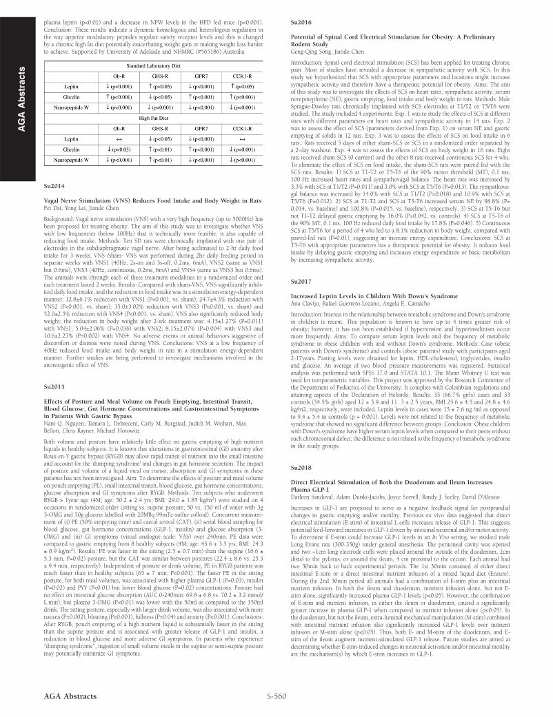
Background: Peripheral appetite signals from the gastrointestinal tract are conveyed to theCNS via the vagal nerves (Regul Pept. 2008: 149(1-3): 15-25). Leptin, ghrelin and NPWhave all been shown to have appetite modulator roles conveyed by vagal afferents and underhigh fat diet feeding conditions many of these signals are altered (Gastroenterology 2011:140(5): S332, S34, Gastroenterology 2010: 138(5) S45). They can also cause modulationof other appetite modulatory peptides (Endocrinology 2010: 151: 2200-2210); howevertheir effect on expression of their own receptor or other appetite peptide receptors isunknown. Aim: To determine whether leptin, ghrelin and neuropeptide W (NPW) cause achange in the expression of appetite modulatory receptor expression in nodose ganglia cellsand whether any modulation is changed in high fat diet fed mice. Methods: Female C57/BL6 mice (n= 4/group) were fed a standard laboratory diet (SLD: 7% energy from fat) or ahigh fat diet (HFD: 60% energy from fat) for 12 weeks. The mice were killed and nodoseganglia removed, dissociated and cultured in standard culture medium or culture mediumcontaining leptin (0.1, 1nM), ghrelin (1, 3nM) or NPW (1, 3nM) for 18 hours, after whichRNA was extracted and QRT-PCR run for leptin (Ob-R), ghrelin (GHS-R), NPW (GPR7)and CCK (CCK1R) receptors. Plasma samples were collected from the mice to measureendogenous levels of the peptides by ELISA. Results: The effect of gastric peptides on theexpression of peptide receptors in the nodose ganglia from mice fed either a standardlaboratory diet or a high fat diet is illustrated in the table. We also observed an increase in
AG
AA
bst
ract
s
AG
AA
bst
ract
splasma leptin (p<0.01) and a decrease in NPW levels in the HFD fed mice (p<0.001).Conclusion: These results indicate a dynamic homologous and heterologous regulation inthe way appetite modulatory peptides regulate satiety receptor levels and this is changedby a chronic high fat diet potentially exacerbating weight gain or making weight loss harderto achieve. Supported by University of Adelaide and NHMRC (#565186) Australia
Su2014
Vagal Nerve Stimulation (VNS) Reduces Food Intake and Body Weight in RatsFei Dai, Yong Lei, Jiande Chen
Background: Vagal nerve stimulation (VNS) with a very high frequency (up to 5000Hz) hasbeen proposed for treating obesity. The aim of this study was to investigate whether VNSwith low frequencies (below 100Hz) that is technically more feasible, is also capable ofreducing food intake. Methods: Ten SD rats were chronically implanted with one pair ofelectrodes in the subdiaphragmatic vagal nerve. After being acclimated to 2-hr daily foodintake for 3 weeks, VNS /sham- VNS was performed during 2hr daily feeding period inseparate weeks with VNS1 (40Hz, 2s-on and 3s-off, 0.2ms, 6mA), VNS2 (same as VNS1but 0.6ms), VNS3 (40Hz, continuous, 0.2ms, 6mA) and VNS4 (same as VNS3 but 0.6ms).The animals went through each of these treatment modalities in a randomized order andeach treatment lasted 2 weeks. Results: Compared with sham-VNS, VNS significantly inhib-ited daily food intake, and the reduction in food intake was in a stimulation energy-dependentmanner: 12.8±6.1% reduction with VNS1 (P<0.001, vs. sham); 24.7±4.3% reduction withVNS2 (P<0.001, vs. sham); 35.0±3.02% reduction with VNS3 (P<0.001, vs. sham) and52.0±2.5% reduction with VNS4 (P<0.001, vs. sham). VNS also significantly reduced bodyweight; the reduction in body weight after 2-wk treatment was: 4.15±1.27% (P=0.011)with VNS1; 5.04±2.06% (P=0.036) with VNS2; 8.15±2.07% (P=0.004) with VNS3 and10.6±2.23% (P=0.002) with VNS4. No adverse events or animal behaviors suggestive ofdiscomfort or distress were noted during VNS. Conclusions: VNS at a low frequency of40Hz reduced food intake and body weight in rats in a stimulation energy-dependentmanner. Further studies are being performed to investigate mechanisms involved in theanorexigenic effect of VNS.
Su2015
Effects of Posture and Meal Volume on Pouch Emptying, Intestinal Transit,Blood Glucose, Gut Hormone Concentrations and Gastrointestinal Symptomsin Patients With Gastric BypassNam Q. Nguyen, Tamara L. Debreceni, Carly M. Burgstad, Judith M. Wishart, MaxBellon, Chris Rayner, Michael Horowitz
Both volume and posture have relatively little effect on gastric emptying of high nutrientliquids in healthy subjects. It is known that alterations in gastrointestinal (GI) anatomy afterRoux-en-Y gastric bypass (RYGB) may allow rapid transit of nutrient into the small intestineand account for the ‘dumping syndrome' and changes in gut hormone secretion. The impactof posture and volume of a liquid meal on transit, absorption and GI symptoms in thesepatients has not been investigated. Aim: To determine the effects of posture and meal volumeon pouch emptying (PE), small intestinal transit, blood glucose, gut hormone concentrations,glucose absorption and GI symptoms after RYGB. Methods: Ten subjects who underwentRYGB > 1year ago (4M; age: 50.2 ± 2.4 yrs; BMI: 29.0 ± 1.85 kg/m2) were studied on 4occasions in randomized order (sitting vs. supine posture; 50 vs. 150 ml of water with 3g3-OMG and 50g glucose labelled with 20MBq 99mTc-sulfur colloid). Concurrent measure-ment of (i) PE (50% emptying time) and caecal arrival (CAT), (ii) serial blood sampling forblood glucose, gut hormone concentrations (GLP-1, insulin) and glucose absorption (3-OMG) and (iii) GI symptoms (visual analogue scale: VAS) over 240min. PE data werecompared to gastric emptying from 8 healthy subjects (4M; age: 45.6 ± 3.5 yrs; BMI: 24.3± 0.9 kg/m2). Results: PE was faster in the sitting (2.5 ± 0.7 min) than the supine (16.6 ±5.3 min, P=0.02) posture, but the CAT was similar between postures (22.4 ± 8.6 vs. 25.3± 9.4 min, respectively). Independent of posture or drink volume, PE in RYGB patients wasmuch faster than in healthy subjects (65 ± 7 min; P<0.001). The faster PE in the sittingposture, for both meal volumes, was associated with higher plasma GLP-1 (P=0.03), insulin(P=0.02) and PYY (P<0.01) but lower blood glucose (P=0.02) concentrations. Posture hadno effect on intestinal glucose absorption (AUC 0-240min: 69.8 ± 6.8 vs. 70.2 ± 3.2 mmol/L.min), but plasma 3-OMG (P<0.01) was lower with the 50ml as compared to the 150mldrink. The sitting posture, especially with larger drink volume, was also associated with morenausea (P=0.002), bloating (P<0.001), fullness (P=0.04) and anxiety (P<0.001). Conclusions:After RYGB, pouch emptying of a high nutrient liquid is substantially faster in the sittingthan the supine posture and is associated with greater release of GLP-1 and insulin, areduction in blood glucose and more adverse GI symptoms. In patients who experience“dumping syndrome”, ingestion of small volume meals in the supine or semi-supine posturemay potentially minimize GI symptoms.
S-560AGA Abstracts
Su2016
Potential of Spinal Cord Electrical Stimulation for Obesity: A PreliminaryRodent StudyGeng-Qing Song, Jiande Chen
Introduction: Spinal cord electrical stimulation (SCS) has been applied for treating chronicpain. Most of studies have revealed a decrease in sympathetic activity with SCS. In thisstudy we hypothesized that SCS with appropriate parameters and locations might increasesympathetic activity and therefore have a therapeutic potential for obesity. Aims: The aimof this study was to investigate the effects of SCS on heart rates, sympathetic activity, serumnorepinephrine (NE), gastric emptying, food intake and body weight in rats. Methods: MaleSprague-Dawley rats chronically implanted with SCS electrodes at T1/T2 or T5/T6 werestudied. The study included 4 experiments. Exp. 1 was to study the effects of SCS at differentsites with different parameters on heart rates and sympathetic activity in 14 rats. Exp. 2was to assess the effect of SCS (parameters derived from Exp. 1) on serum NE and gastricemptying of solids in 12 rats. Exp. 3 was to assess the effects of SCS on food intake in 6rats.. Rats received 5 days of either sham-SCS or SCS in a randomized order separated bya 2-day washout. Exp. 4 was to assess the effects of SCS on body weight in 16 rats. Eightrats received sham-SCS (0 current) and the other 8 rats received continuous SCS for 4 wks.To eliminate the effect of SCS on food intake, the sham-SCS rats were paired fed with theSCS rats. Results: 1) SCS at T1-T2 or T5-T6 of the 90% motor threshold (MT), 0.1 ms,100 Hz increased heart rates and sympathovagal balance. The heart rate was increased by3.3% with SCS at T1/T2 (P=0.011) and 3.0% with SCS at T5/T6 (P=0.013). The sympathova-gal balance was increased by 14.0% with SCS at T1/T2 (P=0.018) and 10.9% with SCS atT5/T6 (P=0.012). 2) SCS at T1-T2 and SCS at T5-T6 increased serum NE by 98.8% (P=0.014, vs. baseline) and 100.8% (P=0.015, vs. baseline), respectively. 3) SCS at T5-T6 butnot T1-T2 delayed gastric emptying by 16.0% (P=0.042, vs. controls). 4) SCS at T5-T6 ofthe 90% MT, 0.1 ms, 100 Hz reduced daily food intake by 17.8% (P=0.046). 5) ContinuousSCS at T5/T6 for a period of 4 wks led to a 8.1% reduction in body weight, compared withpaired-fed rats (P=0.01), suggesting an increase energy expenditure. Conclusions: SCS atT5-T6 with appropriate parameters has a therapeutic potential for obesity. It reduces foodintake by delaying gastric emptying and increases energy expenditure or basic metabolismby increasing sympathetic activity.
Su2017
Increased Leptin Levels in Children With Down's SyndromeAna Clavijo, Rafael Guerrero-Lozano, Angela E. Camacho
Introduction: Interest in the relationship between metabolic syndrome and Down's syndromein children is recent. This population is known to have up to 4 times greater risk ofobesity; however, it has not been established if hypertension and hyperinsulinism occurmore frequently. Aims: To compare serum leptin levels and the frequency of metabolicsyndrome in obese children with and without Down's syndrome. Methods: Case (obesepatients with Down's syndrome) and controls (obese patients) study with participants aged2-17years. Fasting levels were obtained for leptin, HDL-cholesterol, triglycerides, insulinand glucose. An average of two blood pressure measurements was registered. Statisticalanalysis was performed with SPSS 17.0 and STATA 10.1. The Mann Whitney U test wasused for nonparametric variables. This project was approved by the Research Committee ofthe Department of Pediatrics of the University. It complies with Colombian regulations andattaining aspects of the Declaration of Helsinki. Results: 33 (66.7% girls) cases and 33controls (54.5% girls) aged 12 ± 3.9 and 11. 3 ± 2.5 years, BMI 25.6 ± 4.5 and 24.8 ± 4.6kg/m2, respectively, were included. Leptin levels in cases were 15 ± 7.6 ng /ml as opposedto 9.4 ± 5.4 in controls (p = 0.001). Levels were not related to the frequency of metabolicsyndrome that showed no significant difference between groups. Conclusion: Obese childrenwith Down's syndrome have higher serum leptin levels when compared to their peers withoutsuch chromosomal defect; the difference is not related to the frequency of metabolic syndromein the study groups.
Su2018
Direct Electrical Stimulation of Both the Duodenum and Ileum IncreasesPlasma GLP-1Darleen Sandoval, Adam Dunki-Jacobs, Joyce Sorrell, Randy J. Seeley, David D'Alessio
Increases in GLP-1 are proposed to serve as a negative feedback signal for postprandialchanges in gastric emptying and/or motility. Previous ex vivo data suggested that directelectrical stimulation (E-stim) of intestinal L-cells increases release of GLP-1. This suggestspotential feed-forward increases in GLP-1 driven by intestinal neuronal and/or motor activity.To determine if E-stim could increase GLP-1 levels in an In Vivo setting, we studied maleLong Evans rats (300-350g) under general anesthesia. The peritoneal cavity was openedand two ~1cm long electrode cuffs were placed around the outside of the duodenum, 2cmdistal to the pylorus, or around the ileum, 4 cm proximal to the cecum. Each animal hadtwo 30min back to back experimental periods. The 1st 30min consisted of either directintestinal E-stim or a direct intestinal nutrient infusion of a mixed liquid diet (Ensure).During the 2nd 30min period all animals had a combination of E-stim plus an intestinalnutrient infusion. In both the ileum and duodenum, nutrient infusion alone, but not E-stim alone, significantly increased plasma GLP-1 levels (p<0.05). However, the combinationof E-stim and nutrient infusion, in either the ileum or duodenum, caused a significantlygreater increase in plasma GLP-1 when compared to nutrient infusion alone (p<0.05). Inthe duodenum, but not the ileum, extra-luminalmechanical manipulation (M-stim) combinedwith intestinal nutrient infusion also significantly increased GLP-1 levels over nutrientinfusion or M-stim alone (p<0.05). Thus, both E- and M-stim of the duodenum, and E-stim of the ileum augment nutrient-stimulated GLP-1 release. Future studies are aimed atdetermining whether E-stim-induced changes in neuronal activation and/or intestinal motilityare the mechanism(s) by which E-stim increases in GLP-1.