Embed Size (px)
Citation preview
CASE REPORT Open Access
Subarachnoidal Neurocysticercosisnon-responsive to cysticidal drugs: a case seriesGraciela Cárdenas1, Roger Carrillo-Mezo1, Helgi Jung1, Edda Sciutto2, Jose Luis Soto Hernandez1, Agnès Fleury1,2*
Abstract
Background: Neurocysticercosis (NC) is one of the most frequent parasitic diseases of the central nervous system.Cysticidal drugs, albendazole and praziquantel, are generally effective when parasites localize in the parenchyma. Incontrast, parasites lodged in the subarachnoid basal cisterns are less responsive to treatment.
Case Presentation: The clinical and radiological pictures of six Mexican patients non-respondent to cysticidaltreatment are presented.
Conclusions: The possible factors involved in the cysticidal non-response are discussed and hints are provided ofpotentially useful changes to therapeutic protocols.
BackgroundTaenia solium is a parasite which larvae (cysticercus)may localize in the central nervous system of humanscausing neurocysticercosis (NC). Most NC cases occurwith little or no neurological symptoms but others maypresent a variety of non-specific mild clinical symptoms(headache, partial seizures) or severe neurological syn-dromes with intracraneal hypertension and generalizedseizures [1].The introduction of cysticidal drugs, albendazole
(ABZ) and praziquantel (PZQ), for the treatment of NChave dramatically improved its prognosis [2]. In Mexico,ABZ is becoming the drug of choice due to its low costsand availability. Parasite localization is one of the mainfactors involved in the success of the treatment. Whencysticerci are lodged in the parenchyma, the use of thesedrugs allows generally a prompt radiological and clinicalimprovement in most of the patients although only amodest effect is reported in some cases [3,4]. When theparasites are located in the subarachnoid basal cisterns(SA-NC), the prognosis is more uncertain. Several caseseries have reported the effectiveness of these drugs inSA-NC treatment [5-7]. However, in our experience andthat of others [4], it is a frequent finding that the para-sites persist after treatment, even when a high dose ofABZ has been used [8].
In this report, six cases NC-SA patients who are non-respondent to conventional pharmacological treatmentand a brief review of literature are described.
Case PresentationPatient A (Figure 1, A0 to 1A3). A 46-year-old man withone year history of increasing incapacitating frontalheadache. Upon admission, bilateral papilledema andupward gaze paralysis were shown. Multiple vesicularcysticerci located in opto-chiasmatic and perimensence-phalic cisterns as well as basal meningeal enhancementwere observed by magnetic resonance imaging (MRI).Increased cellularity (89 cells/mm3) and anti-cysticercalantibodies (Abs) determined by ELISA were detected inthe cerebrospinal fluid (CSF). During one year, thepatient received two cycles of ABZ (30 mg/kg/day) andone course of PZQ (50 mg/kg), for eight days, associatedto corticosteroids. In the ninth month, he required aventriculoperitoneal shunt (VPS) due to hydrocephaly.In all of MRIs, which were performed four months aftereach treatment cycle, most of the vesicular parasitespersisted.Patient B (Figure 1, B0 to 1B3). A 60 year old man
with chronic headache. Nine months before hospitaladmission, headache frequency increased and lower limbmotor dysfunction appeared. Neurological examinationrevealed only papilledema. On MRI, vesicular subarach-noidal NC in posterior fossa was observed. CSF analysisshowed an inflammatory profile with abundant
* Correspondence: [email protected] Nacional de Neurología y Neurocirugía, Insurgentes Sur 3877,Colonia La Fama, Delegación Tlalpan, México DF, México, CP 14269
Cárdenas et al. BMC Neurology 2010, 10:16http://www.biomedcentral.com/1471-2377/10/16
© 2010 Cárdenas et al; licensee BioMed Central Ltd. This is an Open Access article distributed under the terms of the CreativeCommons Attribution License (http://creativecommons.org/licenses/by/2.0), which permits unrestricted use, distribution, andreproduction in any medium, provided the original work is properly cited.
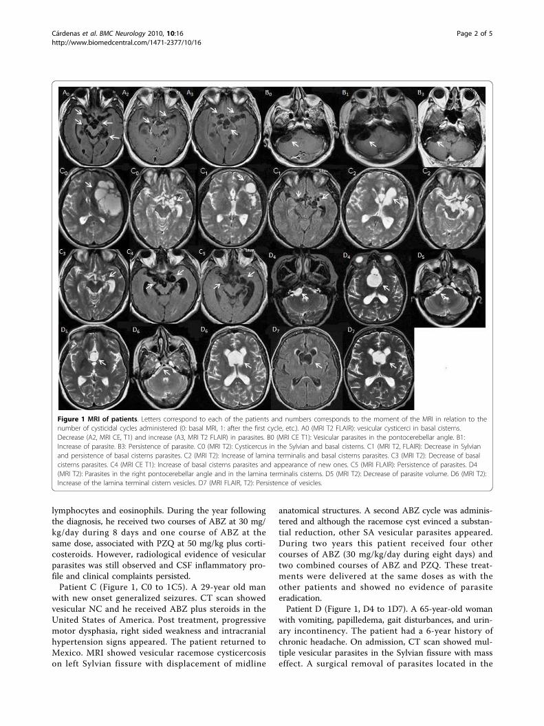
lymphocytes and eosinophils. During the year followingthe diagnosis, he received two courses of ABZ at 30 mg/kg/day during 8 days and one course of ABZ at thesame dose, associated with PZQ at 50 mg/kg plus corti-costeroids. However, radiological evidence of vesicularparasites was still observed and CSF inflammatory pro-file and clinical complaints persisted.Patient C (Figure 1, C0 to 1C5). A 29-year old man
with new onset generalized seizures. CT scan showedvesicular NC and he received ABZ plus steroids in theUnited States of America. Post treatment, progressivemotor dysphasia, right sided weakness and intracranialhypertension signs appeared. The patient returned toMexico. MRI showed vesicular racemose cysticercosison left Sylvian fissure with displacement of midline
anatomical structures. A second ABZ cycle was adminis-tered and although the racemose cyst evinced a substan-tial reduction, other SA vesicular parasites appeared.During two years this patient received four othercourses of ABZ (30 mg/kg/day during eight days) andtwo combined courses of ABZ and PZQ. These treat-ments were delivered at the same doses as with theother patients and showed no evidence of parasiteeradication.Patient D (Figure 1, D4 to 1D7). A 65-year-old woman
with vomiting, papilledema, gait disturbances, and urin-ary incontinency. The patient had a 6-year history ofchronic headache. On admission, CT scan showed mul-tiple vesicular parasites in the Sylvian fissure with masseffect. A surgical removal of parasites located in the
Figure 1 MRI of patients. Letters correspond to each of the patients and numbers corresponds to the moment of the MRI in relation to thenumber of cysticidal cycles administered (0: basal MRI, 1: after the first cycle, etc.). A0 (MRI T2 FLAIR): vesicular cysticerci in basal cisterns.Decrease (A2, MRI CE, T1) and increase (A3, MRI T2 FLAIR) in parasites. B0 (MRI CE T1): Vesicular parasites in the pontocerebellar angle. B1:Increase of parasite. B3: Persistence of parasite. C0 (MRI T2): Cysticercus in the Sylvian and basal cisterns. C1 (MRI T2, FLAIR): Decrease in Sylvianand persistence of basal cisterns parasites. C2 (MRI T2): Increase of lamina terminalis and basal cisterns parasites. C3 (MRI T2): Decrease of basalcisterns parasites. C4 (MRI CE T1): Increase of basal cisterns parasites and appearance of new ones. C5 (MRI FLAIR): Persistence of parasites. D4(MRI T2): Parasites in the right pontocerebellar angle and in the lamina terminalis cisterns. D5 (MRI T2): Decrease of parasite volume. D6 (MRI T2):Increase of the lamina terminal cistern vesicles. D7 (MRI FLAIR, T2): Persistence of vesicles.
Cárdenas et al. BMC Neurology 2010, 10:16http://www.biomedcentral.com/1471-2377/10/16
Page 2 of 5
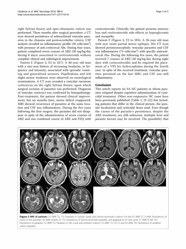
right Sylvian fissure and opto-chiasmatic cistern wasperformed. Three months after surgical procedure, a CTscan showed persistence of subarachnoid vesicular para-sites in the chiasma and pontocerebellar cistern. CSFanalysis revealed an inflammatory profile (20 cells/mm3)with presence of anti-cysticercal Abs. During four years,patient completed seven courses of ABZ (30 mg/kg/dayduring 8 days) associated to corticosteroids withoutcomplete clinical and radiological improvement.Patient E (Figure 2, E3 to 2E7). A 40-year old man
with a one-year-history of increasing headache, in fre-quency and intensity, associated with sporadic vomit-ing and generalized seizures. Papilledema and leftslight motor weakness were observed on neurologicalexaminations. A CT scan revealed a vesicular racemosecysticercus on the right Sylvian fissure, upon whichsurgical excision of parasites was performed. Diagnosisof vesicular cysticerci was confirmed by histopathology.Post-treatment, the patient showed clinical improve-ment, but six months later, motor deficit reappeared.MRI showed recurrence of parasites at the same loca-tion and CSF was inflammatory. During the five yearsfollowing the first surgery, the parasites did not disap-pear in spite of the administration of seven courses ofABZ and one combined course of ABZ and PZQ with
corticosteroids. Clinically, the patient presents memoryloss and corticosteroids side-effects as hyperglycemiaand myopathy.Patient F (Figure 2, F2 to 2F6). A 26-year old man
with new onset partial motor epilepsy. His CT scanshowed perimesencephalic vesicular parasites and CSFwas inflammatory (75 cells/mm3) with specific anticysti-cercal Abs. During the following five years, the patientreceived 7 courses of ABZ (30 mg/kg/day during eightdays with corticosteroids) and he required the place-ment of a VPS for hydrocephalus during the fourthyear. In spite of the received treatment, vesicular para-sites persisted on the last MRI, and CSF was stillinflammatory.
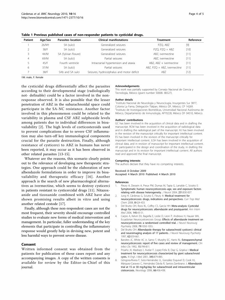
ConclusionThis article reports six SA-NC patients in whom para-sites relapsed despite repetitive administration of cysti-cidal treatment. Other non-responsive NC cases havebeen previously published (Table 1, [9-12]) but includ-ing patients that differ in the clinical picture, the para-site localization and cysticidal doses used. Even thoughthe causes of the parasite’s persistence, despite theABZ treatment, are still unknown, multiple host andparasite factors may be involved. The possibility that
Figure 2 MRI of patients. E3 (MRI T2, T1): Parasites in Sylvian, basal and lamina terminalis cisterns. E4 and E5 (MRI T2, FLAIR): Persistence ofsome of the parasites. E6 (MRI simple, CE T1): Persistence of lamina terminalis parasites and appearance of new ones. E7 (MRI FLAIR, T2):Persistence of parasites. F2 (MRI T1): Parasites in the crural and ambient cisterns. F3 (MRI T1), F4, F5 and F6 (MRI T2): Persistence of ambientcistern parasites.
Cárdenas et al. BMC Neurology 2010, 10:16http://www.biomedcentral.com/1471-2377/10/16
Page 3 of 5
the cysticidal drugs differentially affect the parasitesaccording to their developmental stage (radiologicallynot- definable) could be a factor involved in the non-response observed. It is also possible that the lesserpenetration of ABZ in the subarachnoidal space couldparticipate in the SA-NC resistance. Another factorinvolved in this phenomenon could be related to thevariability in plasma and CSF ABZ sulphoxide levelsamong patients due to individual differences in bioa-vailability [2]. The high levels of corticosteroids usedto prevent complications due to severe CSF inflamma-tion may also turn-off key immunological componentscrucial for the parasite destruction. Finally, althoughresistance of cysticerci to ABZ in humans has neverbeen reported, it may occur as it has been observed inother related parasites [13-15].Whatever are the reasons, this scenario clearly points
out to the relevance of developing new therapeutic stra-tegies. One approach could be the elaboration of newalbendazole formulations in order to improve its bioa-vailability and therapeutic efficacy [16]. Anotherapproach is the search of new pharmacological alterna-tives as ivermectine, which seems to destroy cysticerciin patients resistant to cysticercidal drugs [11]. Nitazox-anide and tizoxanide combined with ABZ have alsoshown promising results albeit in vitro and usinganother related cestode [17].Finally, although these non-respondent cases are not the
most frequent, their severity should encourage controlledstudies to evaluate new forms of medical intervention andmanagement. In particular, fuller understanding of the keyelements that participate in controlling the inflammatoryresponse would greatly help in devising new, potent andless harmful ways to prevent severe disease.
ConsentWritten informed consent was obtained from thepatients for publication of these cases report and anyaccompanying images. A copy of the written consents isavailable for review by the Editor-in-Chief of thisjournal.
AcknowledgementsThis work was partially supported by Consejo Nacional de Ciencia yTecnología, México (grant number S0008- 86527).
Author details1Instituto Nacional de Neurología y Neurocirugía, Insurgentes Sur 3877,Colonia La Fama, Delegación Tlalpan, México DF, México, CP 14269.2Instituto de Investigaciones Biomédicas, Universidad Nacional Autónoma deMéxico, Departamento de Inmunología, AP70228, México DF 04510, México.
Authors’ contributionsGC has been involved in the acquisition of clinical data and in drafting themanuscript. RCM has been involved in the acquisition of radiological dataand in drafting the radiological part of the manuscript. HJ has been involvedin the revision of the manuscript critically for important intellectual content.ES has been involved in the revision of the manuscript critically forimportant intellectual content. JLSH has been involved in the acquisition ofclinical data, and in revision of manuscript for important intellectual content.AF participated in the design and coordination of the study, in drafting themanuscript and in its revision for important intellectual content. All authorsread and approved the final manuscript.
Competing interestsThe authors declare that they have no competing interests.
Received: 8 October 2009Accepted: 4 March 2010 Published: 4 March 2010
References1. Fleury A, Dessein A, Preux PM, Dumas M, Tapia G, Larralde C, Sciutto E:
Symptomatic human neurocysticercosis–age, sex and exposure factorsrelating with disease heterogeneity. J Neurol 2004, 251:830-837.
2. Jung H, Cárdenas G, Sciutto E, Fleury A: Medical treatment forneurocysticercosis: drugs, indications and perspectives. Curr Trop MedChem 2008, 8:424-433.
3. Del Brutto OH, Roos KL, Coffey CS, García HH: Meta-analysis: Cysticidaldrugs for neurocysticercosis: albendazole and praziquantel. Ann InternMed 2006, 145:43-51.
4. Carpio A, Kelvin EA, Bagiella E, Leslie D, Leon P, Andrews H, Hauser WA,Ecuadorian Neurocysticercosis Group: Effects of albendazole treatment onneurocysticercosis: a randomised controlled trial. J Neurol NeurosurgPsychiatry 2008, 79:1050-1055.
5. Del Brutto OH: Albendazole therapy for subarachnoid cysticerci: clinicaland neuroimaging analysis of 17 patients. J Neurol Neurosurg Psychiatry1997, 62:659-661.
6. Bandres JC, White AC Jr, Samo T, Murphy EC, Harris RL: Extraparenchymalneurocysticercosis: report of five cases and review of management. ClinInfect Dis 1992, 15:799-811.
7. Proaño JV, Madrazo I, Avelar F, Lopez-Felix B, Diaz G, Grijalva I: Medicaltreatment for neurocysticercosis characterized by giant subarachnoidcysts. N Engl J Med 2001, 345:879-885.
8. Göngora-Rivera F, Soto-Hernández JL, González Esquivel D, Cook HJ,Márquez-Caraveo C, Hernández Dávila R, Santos-Zambrano J: Albendazoletrial at 15 or 30 mg/kg/day for subarachnoid and intraventricularcisticercosis. Neurology 2006, 66:436-438.
Table 1 Previous published cases of non-responder patients to cysticidal drugs.
Patient Age/Sex Parasites location Clinical manifestations Treatment Reference
1 26/M† SA (sulci) Generalized seizures PZQ, ABZ [9]
2 38/F SA (sulci) Generalized seizures PZQ, PZQ + ABZ [10]
3 44/M SA (Sylvian fissure) Generalized seizures ABZ, ivermectine [11]
4 69/M SA (sulci) Partial seizures ABZ, ivermectine [11]
5 45/F Fourth ventricle Intracranial hypertension and ataxia ABZ, ABZ + ivermectine [11]
6 37/M SA (sulci) Partial seizures ABZ, PZQ + ABZ, ivermectine [11]
7 38/F SAb and SA sulci Seizures, hydrocephalus and motor deficit ABZ [12]
†M: male, F: female
Cárdenas et al. BMC Neurology 2010, 10:16http://www.biomedcentral.com/1471-2377/10/16
Page 4 of 5
9. Cohen L, Belec L, Sanson M, Pierrot-Deseilligny C, Signoret JL: Selectivesensitivity of cysts to praziquantel and albendazole in a case of cerebralcysticercosis. Rev Neurol 1992, 148:58-61.
10. Chong MS, Hawkins CP, Cook GC, Hawkes CH, Kocen RS: A resistant caseof neurocysticercosis. Postgrad Med J 1991, 67:577-578.
11. Diazgranados-Sanchez JA, Barrios-Arrázola G, Costal JL, Burbano-Pabon J,Pinzón-Bedova J: Ivermectin aas therapeuthic alternative inneurocysticercosis that is resistant to conventional pharmacologicaltreatment. Rev Neurol 2008, 46:671-674.
12. Rocha MA Jr, Santos JM, Gomes EC, Rocha MA, Rocha CF, Carvalho GT,Costa BS: Treatment of cerebral cysticercosis with albendazole inelevated dosages. Arq Neuropsiquiatr 2008, 66:114-116.
13. Sissay MM, Asefa A, Uggla A, Waller PJ: Assessment of anthelminticresistance in nematode parasites of sheep and goats owned bysmallholder farmers in eastern Ethiopia. Trop Anim Health Prod 2006,38:215-222.
14. Equale T, Chaka H, Gizaw D: Efficacy of albendazole against nematodeparasites isolated from a goat farm in Ethiopia: Relationship betweendose and efficacy in goats. Trop Anim Health Prod 2009, 41:1267-1273.
15. Jimenez-Cardoso E, Eligio-Garcia L, Cortes-Campos A, Flores-Luna A,Valencia-Mayoral P, Lozada-Chavez I: Changes in beta-giardin sequence ofGardia intestinalis sensitive and resistant to albendazole strains. ParasitolRes 2009, 105:25-33.
16. Mittapalli PK, Yamasani MR, Shashank A: Improved Bioavailability ofAlbendazole Following Oral Administration of Nanosuspension in Rats.Current Nanoscience 2007, 3:191-194.
17. Palomares Alonso F, Piliado JC, Palencia G, Ortiz-Plata A, Jung-Cook H:Efficacy of nitazoxanide, tizoxanide and tizoxanide/albendazolesulphoxide combinations against Taenia crassi ceps cysts. J AntimicrobChemother 2007, 59:212-218.
Pre-publication historyThe pre-publication history for this paper can be accessed here:http://www.biomedcentral.com/1471-2377/10/16/prepub
doi:10.1186/1471-2377-10-16Cite this article as: Cárdenas et al.: Subarachnoidal Neurocysticercosisnon-responsive to cysticidal drugs: a case series. BMC Neurology 201010:16.
Submit your next manuscript to BioMed Centraland take full advantage of:
• Convenient online submission
• Thorough peer review
• No space constraints or color figure charges
• Immediate publication on acceptance
• Inclusion in PubMed, CAS, Scopus and Google Scholar
• Research which is freely available for redistribution
Submit your manuscript at www.biomedcentral.com/submit
Cárdenas et al. BMC Neurology 2010, 10:16http://www.biomedcentral.com/1471-2377/10/16
Page 5 of 5