Embed Size (px)
Citation preview
S U B M I T O C H O N D R I A L L O C A L I Z A T I O N A N D F U N C T I O N O F
E N Z Y M E S O F G L U T A M I N E M E T A B O L I S M I N A V I A N L I V E R
JEAN E. VORHABEN and JAMES W. CAMPBELL
From the Department of Biology, William Marsh Rice University, Houston, Texas 77001
ABSTRACT
Glutamine synthetase (EC 6.3.1.2) was localized within the matrix compartment of avian liver mitochondria. The submitochondrial localization of this enzyme was determined by the digitonin-Lubrol method of Schnaitman and Greenawalt (35). The matrix fraction contained over 74% of the glutamine synthetase activity and the major proportion of the matrix marker enzymes, malate dehydrogenase (71%), NADP-dependent isocitrate dehydrogenase (83 %), and glutamate dehy- drogenase (57%). The highest specific activities of these enzymes were also found in the matrix compartment .
Oxidation of glutamine by avian liver mitochondria was substantially less than that of glutamate. Bromofuroate, an inhibitor of glutamate dehydrogenase, blocked oxidation of glutamate and of glutamine whereas aminoxyacetate, a transaminase inhibitor, had little or no effect with either substrate. These results indicate that glutamine metabolism is probably initiated by the conversion of glutamine to glutamate rather than to an a-keto acid. The localization of a glutaminase activity within avian liver mitochondria plus the absence of an active mitochondrial glutamine transaminase is consistent with the differential effects of the transami- nase and glutamate dehydrogenase inhibitors. The high glutamine synthetase activity relative to glutaminase activity (40:1) suggests that mitochondrial catabo- lism of glutamine is minimal, freeing most of the glutamine synthesized for purine (uric acid) biosynthesis.
Glutamine synthetase and carbamyl phosphate synthetase-I appear to perform parallel functions in liver of uricotelic and ureotelic species, respec- tively (2, 3, 8, 39). These mitochondrial enzymes catalyze the primary ammonia-detoxifying step in pathways leading to the synthesis of urea or uric acid. During the catabolism of amino acids, am- monia released within the mitochondrion by the action of glutamate dehydrogenase is converted either to citrulline (via initial formation of carba- myl phosphate) in the case of ureoteles or to glutamine in uricoteles. Citrulline and glutamine then leave the mitochondrion and enter the cyto-
sol where they are converted to either urea or uric acid for excretion. Confirmation of this cellular mechanism for ammonia detoxication by urico- teles was recently obtained through heavy isotope tracer studies (3).
The results reported here further emphasize the analogy between uricotelic glutamine synthetase and ureotelic carbamyl phosphate synthetase-I by establishing yet another correlation between these enzymes, namely that of a common submitochon- drial localization. Additional studies indicate that avian mitochondria possess the capacity to oxidize glutamine, albeit at a slow rate, and that any
3 0 0 ThE JOURNAL OF CELL BIOLOGY' VOLUME 73, 1977.pages 300-310
on April 5, 2019jcb.rupress.org Downloaded from http://doi.org/10.1083/jcb.73.2.300Published Online: 1 May, 1977 | Supp Info:

mitochondrial metabol ism of glutamine probably occurs through the coupled react ions of a gtuta- minase and glutamate dehydrogenase .
M A T E R I A L S A N D M E T H O D S
Materials
Cytochrome c, succinic acid, oxalacetic acid, pyru- rate, a-ketoglutarate, isocitrate, glyoxylate, phenylpyru- vate, glutamine, glucose-6-phosphate, deoxycytidine tri- phosphate, ATP, ADP, AMP, digitonin, rotenone, ami- noxyacetic acid, glutamate dehydrogenase, hexokinase, and glucose-6-phosphate dehydrogenase were products of Sigma Chemical Co. (St. Louis, Mo.). 4-Hydroxy-3- methoxy-benzylamine was purchased from ICN Pharma- ceuticals, Inc. (Cleveland, Ohio); 5-bromofuroic acid, from Aldrich Chemical Co., Inc. (Milwaukee, Wis.); Luhrol WX, from I. C. I America, Inc: (Stanford, Conn.); L-amino acid oxidase, from Calbiochem (San Diego, Calif.); and the defatted bovine serum albumin for the mitochondrial isolating medium, from Miles Lab- oratories Inc. (Elkhart, Ind.). [u-laC]Glutamine was obtained from New England Nuclear (Boston, Mass.); the [U-J4C]a-ketoglutaramate used as a substrate in the glutamine transaminase assay was synthesized by the L- amino acid oxidase-catalyzed oxidation of [U- J4C]glutamine according to the procedure of Meister (29). All other reagents were of the highest commercial quality.
Methods
S U B M I T O C H O N D R I A L F R A C T I O N A T I O N
Mitochondria were prepared from 48-h-fasted white Leghorn chickens and were further fractionated by the digitonin-Lubrol procedure of Schnaitman and Greena- walt (35), as later modified by Greenawalt (16), except that a slightly higher digitonin concentration was used (0.15 mg digitonin/mg mitochondrial protein, the latter measured by the biuret method [27]). These mitochon- dria exhibited acceptor control ratios of 5-5.5 with succi- nate as substrate, and micorosomal contamination was only 2% as determined by assay of glucose-6-phospha- tase activity. Protein was determined by the method of Lowry et al. (28), except as indicated above, using bo- vine serum albumin as the standard.
C E L L U L A R F R A C T I O N A T I O N
The subcellular localization of phosphate-dependent glutaminase activity was accomplished by the differential fractionation procedure used in earlier studies on gluta- mine synthetase localization (39) except that the 0.25 M sucrose isolating medium was replaced by the medium of Greenawalt (16) which is a lightly buffered sucrose- mannitol medium supplemented with bovine serum albu- min.
ENZYME ASSAYS
A S S A Y S FOR THE MARKER ENZYMES: Enzymes used to identify the submitochondrial compartments were as follows: cytochrome oxidase, according to Whar- ton and Tzagoloff (40); succinate-cytochrome c reduc- tase and rotenone-insensitive NADH-cytochrome c re- ductase, according to Sottocasa et al. (36); monoamine oxidase, according to Christ et al. (5); adenylate kinase and nucleoside diphosphokinase, according to Schnait- man and Greenawalt (35), as modified by Kalra and Brosnan (22); isocitrate dehydrogenase, according to Plant and Sung (34); malate dehydrogenase, according to Ochoa (31), except that Tris-hydrochloride buffer was substituted for the glycylglycine buffer and the samples were activated by Lubrol as described by Schnaitman and Greenawalt (35); and glutamate dehydrogenase, according to Otson and Anfinsen (32).
A S S A Y S FOR THE ENZYMES OF GLUTAMINE
M E T A B O L I S M : Glutamine synthetase was assayed according to Pamiljans et al. (33); T-glutamyltranspepti- dase, according to Curthoys and Kuhlenschmidt (12) using glycylglycine, methionine, and glutamine as gluta- myl acceptors; glutamine transaminase (kidney form), according to Kupchik and Knox (26); and phosphate- dependent and phosphate-independent glutaminases, ac- cording to Curthoys and Weiss (13).
Phosphate-dependent glutaminase activity was also determined radiometrically. Reaction mixtures at pH 8.6 contained 20 mM [~4C]glutamine of specific radioactivity 0.002 p, Ci/p.mole, 0.15 M potassium phosphate, 0.2 mM EDTA, and 50 mM Tris-hydrochloride. The condi- tions were similar to those used by Curthoys and Weiss (13) in the spectrophotometric assay of phosphate-de- pendent glutaminase. Reactions were initiated by en- zyme addition and were terminated by perchloric acid addition. Incubations were carried out for 1 h at 37~ After centrifugation, the supernatant fluids were neutral- ized with potassium hydroxide and the precipitated per- chlorate was removed by centrifugation. Glutamate and unreacted glutamine were separated by means of electro- phoresis in 0.05 M ammonium formate, pH 7.2, and counted as previously described (38).
Glutamine transaminase (liver form) was measured by two radiometric procedures. In the first, enzyme samples were incubated with 20 mM [U-14C]glutamine (specific radioactivity 0.05 p.Ci//.~mol), 20 mM glyoxylate, and 50 mM Tris-hydrochloride buffer, pH 8.4, for 1 h at 37~ (9). Controls were reaction mixtures without enzyme or with heat-treated enzyme. The [~4C]a-ketoglutaramate was separated from unreacted [~4C]glutamine by elution from a Dowex 50 column (Dow Chemical Co., Midland, Mich.) according to procedure two of Cooper and Meis- ter (9). Instead of counting an aliquot of the column effluent as described, additional separation of ot-ketoglu- taramate and glutamine was achieved by paper electro- phoresis (38). Under these conditions, c~-ketoglutara- mate migrates 10.5 cm from the origin compared to 5.5
VORHABEN AND CAMPBELL Glutamine Metabolism in Avian Liver Mitochondria 301
cm for glutamine. The strips were cut into 1-cm pieces and counted in a Permablend-toluene mixture (38) (Packard Instrument Co., Inc., Downers Grove, Ill.). Heat-treated enzyme and reaction mixtures which did not contain enzyme were used as controls. A second procedure for assaying glutamine transaminase utilized [U-14C]a-keto-glutaramate (0.02 /xCi/p, mol) as the amino group acceptor and glutamine as the amino do- nor. Reaction conditions and controls were essentially those just described except that the Dowex column step was omitted. Instead, the reaction was terminated by adding absolute ethanol to precipitate protein. After centrifugation, unreacted [14C]a-ketoglutaramate and [~4C]glutamine were separated by electrophoresis as de- scribed above. Other enzymes assayed were glucose-6- phosphatase (18) and lactate dehydrogenase (39).
In assays where the activity was low or not detectable in chicken liver mitochondria (adenylate kinase, gluta- mine transaminase, and y-glutamyltranspeptidase), the same assays were applied to rat liver mitochondria or rat kidney preparations to verify reaction conditions.
OXYGEN UPTAKE
Oxygen consumption was measured polarographically by means of a Clark oxygen electrode (Yellow Springs Instrument Co., Yellow Springs, Ohio). The respiration medium was that of Greenawalt (16) except that the ADP concentration was 0.25 mM.
ELECTRON MICROSCOPY
Mitochondria were fixed for 1-2 h in 1.5 % glutaralde- hyde in a 1:1 dilution of MiUonig's buffer, pH 7.4, at 4~ (30); the osmolarity of this fixative was 300 mosM. The mitochondria were then washed four times with undi- luted Millonig's buffer, 300 mosM, pH 7.4. The mito- chondria were allowed to remain in the buffer solution for 15 rain at 40C before centrifuging. After fixing for I h in undiluted Millonig's buffer containing 1% osmium tetroxide, the sample was dehydrated with a graded series of ethanol-water mixtures, embedded in low vis- cosity epoxy embedding media and polymerized at 70~ for 12-24 h. Sections were made with a diamond knife on a Sorvall MT-2 ultramicrotome (DuPont Instruments, Sorvall Operations, Newtown, Conn.). These were stained with 2.5% aqueous uranyl acetate followed by 0.4% aqueous lead citrate. Osmolarity was determined with an Advanced Instruments osmometer (Advanced Instruments, Inc., Needham Heights, Mass.).
D I s c GEL ELECTROPHORESIS
Electrophoresis was carried out on gels prepared by chemical polymerization of solutions containing 7% acrylamide, 0.18% N~N-methylene-bisacrylamide, 0.375 M Tris-hydrochloride buffer, pH 8.9, and 0.07% ammonium persulfate as the catalyst. Samples were di- luted before application to the columns in 50% sucrose containing 0.0004% bromophenol blue as a tracking dye. The electrode buffer was 0.003 M Tris-glycine, pH
8.9, and the current applied was 2.5 mA/gel column. Electrophoresis was continued for 30 min after the dye had migrated off the gel. Previous experiments had shown the absence of proteins migrating faster than bovine serum albumin. Gels were stained for glutamine synthetase activity by incubating them at 37~ for 10-15 min in solutions containing 50 mM glutamate, 40 mM ATP, 125 mM hydroxylamine, 40 mM magnesium chlo- ride, and 50 mM imidazole buffer, pH 7.4, containing 2.5 mM 2-mercaptoethanol. After incubation, the reac- tion solution was decanted and the gels were washed once with distilled water before addition of the ferric chloride reagent. The chromogenic product formed be- tween ferric chloride and y-glutamylhydroxamate was visible almost immediately as a sharp brown band near the top of the gel. Gels were stained for protein with Coomassie brilliant blue, destained by diffusion in 7% acetic acid, and scanned with a Gilford model 2520 gel scanner (Gilford Instrument Laboratories, Inc., Oberlin, Ohio). Identification of the protein peak corresponding to glutamine synthetase was accomplished as follows. The brown band identifying glutamine synthetase migra- tion was marked by means of fine copper wire before diffusion had broadened the band. The gel was then washed with distilled water and stained with Coomassie blue. When scanned, the wire appeared as an off-scale spike in the spectrum and was readily correlated with a protein peak in gels stained only for protein. Gels treated with ferric chloride reagent could be scanned directly for glutamine synthetase activity at 500 nm. However, cor- relation of glutamine synthetase with a protein band was impeded by differences in swelling between gels treated with ferric chloride reagent and those treated with Coo- massie blue reagent. The percentage of the total matrix protein which glutamine synthetase represents was esti- mated by comparison of total peak area to glutamine synthetase peak area. Scans were traced on Albanene tracing paper (Keuffel & Esser Co., Morristown, N. J.) and integrated by weight. Corrections were made for bovine serum albumin present in the matrix sample.
RESULTS
The localization of glutamine synthetase within avian liver mitochondria was determined by the digitonin-Lubrol method of Schnaitman and Greenawalt (35). In this fractionation procedure,
centrifugation of digitonin-treated mitochondria yields a pellet representing the mitoplast fraction (inner membrane + matrix) and a supernatant fraction which may subsequently be separated by high-speed centrifugation into the intracristal space and outer membrane fraction. The inner membrane and matrix compar tments are sepa- rated by Lubrol t reatment of the mitoplast frac- tion. This method was applied to chicken liver mitochondria. The marker enzymes used to char-
302 THE JOURNAL OF CELL BIOLOGY" VOLUME 73, 1977
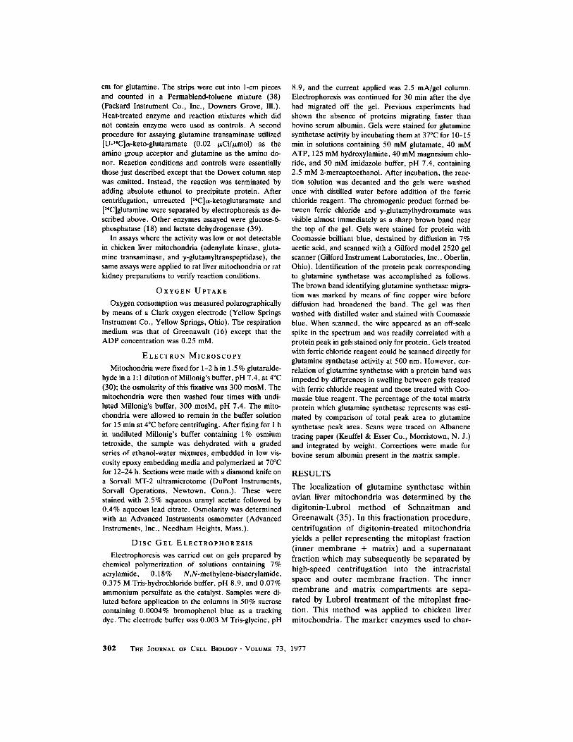
acterize the fractions obtained were those com- monly used for rat liver submitochondrial frac- tions: cytochrome oxidase and succinate-cyto- chrome c reductase for the inner membrane; mal- ate dehydrogenase, glutamate dehydrogenase, and NADP-dependent isocitrate dehydrogenase for the matrix; and rotenone-insensitive NADH- cytochrome c reductase and monoamine oxidase for the outer membrane. Unfortunately, no suita- ble marker for the intracristal space in avian mito- chondria has been found. Adenylate kinase, the most commonly used marker for this compart- ment in rat liver mitochondria, was below our level of detection in avian mitochondria, and nu- cleoside diphosphokinase, also reported to be an intracristal space enzyme, followed the distribu- tion of the matrix marker enzymes. Although adenylate kinase activity in avian liver mitochon- dria has been described by others (7), the activity reported is much too low to be of value as a marker enzyme. However, the matrix and inner membrane fractions obtained here were probably free of major contamination by the intracristal space because they were obtained by fractionation of isolated mitoplasts. Rat liver mitoplasts have a distinctive morphology characterized by pseudo- podlike extensions of the inner membrane and have been shown by biochemical techniques to be devoid of outer membrane and intracristal space enzymes (16, 35). An electron micrograph of an avian liver mitoplast pellet is shown in Fig. 1. These mitoplasts are similar in appearance to rat liver mitoplasts, displaying the characteristic pro- jections of the inner membrane.
Our initial attempts to fractionate avian liver mitochondria resulted in substantial solubilization of the outer membrane. In one such experiment, the digitonin concentration was sufficiently high (digitonin/protein ratio in excess of 0.16) to solu- bilize over 69 % of the monoamine oxidase activity and 59% of the rotenone-insensitive NADH-cyto- chrome c reductase activity. The high digitonin concentration did not, however, cause release of mitoplast enzymes since the major proportion of the matrix and inner membrane markers was re- covered in the mitoplast pellet and subsequently was present in the expected submitoplastal sites. Contamination of the mitoplast pellet by outer membrane was minor as indicated by a low per- centage of monoamine oxidase (12.5%) and rote- none-insensitive NADH-cytochrome c reductase (6.8%) recovered in this fraction. Distribution of glutamine synthetase activity followed that of the
matrix marker enzymes when mitoplasts were rup- tured with detergent. Although the fractionation was not absolute, based on the solubilization of the outer membrane markers, these initial results clearly indicated a matrix localization of glutamine synthetase. Parenthetically, it may be noted that monoamine oxidase activity of avian liver mito- chondria (23 nmol/h per mg protein) was about 3% that of rat liver mitochondria (35), and kynu- renine hydroxylase, another outer membrane marker for some mitochondria (16), could not be detected. An additional difference between avian and rat liver mitochondria observed in these ex- periments was the submitochondrial localization of nucleoside diphosphokinase. In rat liver this activity is found in the intracristal space (35), whereas in avian liver its submitochondrial distri- bution pattern followed that of the matrix marker enzymes.
Table I contains data from a fractionation ex- periment in which the digitonin/protein ratio was decreased to 0.15. The lower ratio resulted in a cleaner separation of the mitochondrial subfrac- tions as judged by enzyme assay. Outer mem- brane contamination of the mitoplast fraction was slightly lower (10.8%) and solubilization of the outer membrane was reduced. Although the rote- none-insensitive NADH-cytochrome c reductase activity was again distributed between the outer membrane and intracristal space, the greatest per- centage of this activity (55%) was associated with the outer membrane, in contrast to the results obtained in the initial studies. The higher recover- ies of isocitrate, malate, and glutamate dehydro- genases in the matrix fraction indicate that frac- tionation of the mitoplast was also cleaner al- though the same detergent/protein ratio was used to fractionate the mitoplast in all studies. As antic- ipated, cytochrome oxidase recovery and specific enzyme activity were highest in the inner mem- brane fraction. Similar observations regarding the localization of glutamine synthetase were noted: the highest percentage of the total as well as specific enzyme activity were found in the matrix compart- ment. This fraction also exhibited the highest spe- cific activities for the matrix marker enzymes. The data presented above represent results from two of six mitochondrial fractionations; in each of the six fractionations, the distribution of glutamine synthetase activity paralleled that of the matrix marker enzymes.
Fig. 2 is a densitometric scan of the matrix polypeptides separated by polyacrylamide gel
VORHABEN AND CAMPBELL Glutamine Metabolism in Avian Liver Mitochondria 303

..]
b"
Z
r < O
t- 7~
--d
Fm
u~
1 E
lect
ron
mic
rogr
aph
of
the
mit
opla
st
frac
tion
fr
om
digi
toni
n-
post
fixe
d in
OsO
4, a
nd s
tain
ed w
ith
uran
yl a
ceta
te a
nd l
ead
citr
ate
as d
e-
trea
ted
avia
n liv
er m
itoc
hond
ria.
The
mit
opla
sts
wer
e fi
xed
in g
luta
rald
ehyd
e,
scri
bed
in M
ater
ials
and
Met
hods
. x
23,3
00;
inse
t,
x 63
,875
.
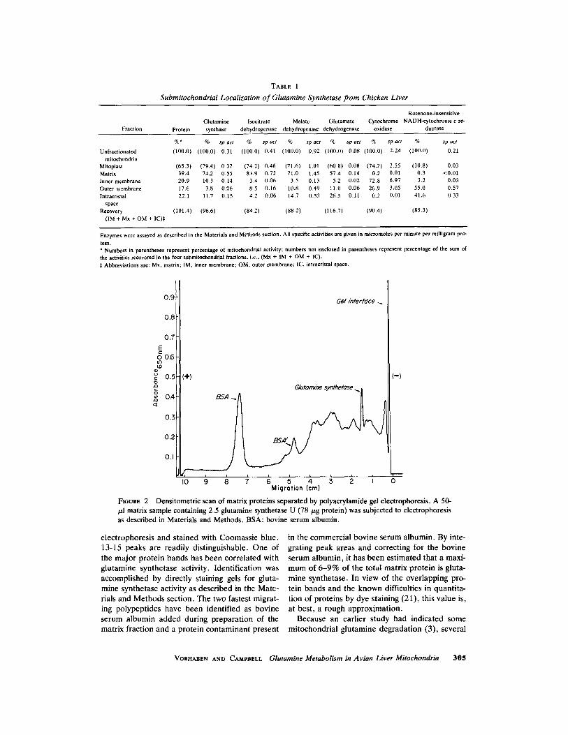
TABLE I
Submitochondrial Localization of Glutamine Synthetase from Chicken Liver
Rotenone-insensitive Glutamine Isocitrate Malatc Glutamate Cytochrome NADH-eytochrome c re-
Fraction Protein synthase dehydrogenase dehydrogenase dehydrogenase oxidase ductase
%* % sp act % sp act % sp act % sp act % sp act % sp act
(100.0) (100.0) 0.31 (100.0) 0.41 (100.0) 0.92 (100.0) 0.08 (100.0) 2.24 (100.0) 0.21
(65.3) (79.4) 0.37 (74.2) 0.46 (71.6) 1 . 0 1 (60.8) 0.08 (74.2) 2.55 (10.8) 0.03 39.4 74.2 0.55 83.9 0.72 71.0 1.45 57.4 0.14 0.2 0.01 0.3 <0.01 20.9 10.3 0,14 3.4 0.06 3.5 0.13 5.2 0.02 72.8 6.97 3.2 0,03 17.6 3.8 0,06 8.5 0.16 10.8 0.49 I1.0 0.06 26,9 3,05 55.0 0.57 22.1 11.7 0.15 4.2 0.06 14,7 0.53 26.5 0.I1 0.2 0,01 41,6 0.33
(101.4) (96.6) (84.2) (88.2) (116.7) (90.4) (85.3)
Unfractionated mitochondria
Mitoplast Matrix Inner membrane Outer membrane Intracrislal
space Recovery
(IM + Mx + OM + IC)~:
Enzymes were assayed as described in the Materials and Methods section. All specific activities are given in micramoles per minute per milligram prO-
tein. * Numbers in parentheses represent percentage of mitochondrial activity; numbers not enclosed in parentheses represent percentage of the sum of the activities recovered in the four submitoehondrial fractions, i.e., (Mx + IM + OM + IC). :~ Abbreviations are: Mx, matrix; IM, inner membrane; OM, outer membrane; IC, intracristal space.
0.9
0.8
0.7
E r
o O.6 u')
0.5
,,, 0.4
0.3
0.2
0.I
(§
Gel ~n/efface
BSA
~G/utarn~ (-)
§ & i 0 Migration (cm)
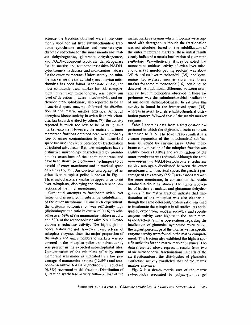
FIOURE 2 /xl matrix sample containing 2.5 glutamine synthetase as described in Materials and Methods. BSA: bovine
Densitometric scan of matrix proteins separated by polyacrylamide gel electrophoresis. A 50- U (78/zg protein) was subjected to electrophoresis serum albumin.
electrophoresis and stained with Coomassie blue. 13-15 peaks are readily distinguishable. One of the major protein bands has been correlated with glutamine synthetase activity. Identification was accomplished by directly staining gels for gluta- mine synthetase activity as described in the Mate- rials and Methods section. The two fastest migrat- ing polypeptides have been identified as bovine serum albumin added during preparation of the matrix fraction and a protein contaminant present
in the commercial bovine serum albumin. By inte- grating peak areas and correcting for the bovine serum albumin, it has been estimated that a maxi- mum of 6-9% of the total matrix protein is gluta- mine synthetase. In view of the overlapping pro- tein bands and the known difficulties in quantita- tion of proteins by dye staining (21), this value is, at best, a rough approximation.
Because an earlier study had indicated some mitochondrial glutamine degradation (3), several
VORHABEN AND CAMPBELL Glutamine Metabolism in Avian Liver Mitochondria 305
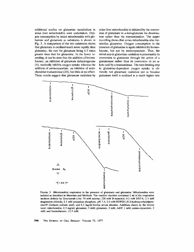
additional studies on glutamine metabolism in avian liver mitochondria were undertaken. Oxy- gen consumption by intact mitochondria with glu- tamine and glutamate as substrates is shown in Fig. 3. A comparison of the two substrates shows that glutamate is oxidized much more rapidly than glutamine, the rate for glutamate being 4.5 times greater than that for glutamine. In the lower re- cording, it can be seen that the addition of bromo- furoate, an inhibitor of glutamate dehydrogenase (4), markedly inhibits oxygen uptake whereas the addition of aminoxyacetate, an inhibitor of mito- chondrial transaminase (20), has little or no effect. These results suggest that glutamate oxidation by
avian liver mitochondria is initiated by the conver- sion of glutamate to a-ketoglutarate via deamina- tion rather than via transamination. The upper recording shows that avian mitochondria also me- tabolize glutamine. Oxygen consumption in the presence of glutamine is again inhibited by bromo- furoate, but not by aminoxyacetate. Thus, the initial step in glutamine oxidation is presumably its conversion to glutamate through the action of a glutaminase rather than its conversion to an t~- keto acid by a transaminase. The rate-limiting step in glutamine-dependent oxygen uptake is ob- viously not glutamate oxidation per se because glutamate itself is oxidized at a much higher rate
t 3 t
g a ~ 3 e~
~ 3 t o
x 3 "~ o
3 s t \ 3 t \ - . _ ~." ~- ~ ,
51 nmol Oi t "~
-T- 3 0
Q
N
FIGURE 3 Mitochondrial respiration in the presence of glutamate and glutamine." Mitochondria were isolated as described in Materials and Methods. The reaction chamber contained 2 ml of the respiration medium defined by Greenawait (16): 70 mM sucrose; 220 mM D-mannitol; 0.5 mM EDTA; 2.5 mM magnesium chloride; 2.5 mM potassium phosphate, pH 7.4; 2.0 mM HEPES (N-2-hydroxy-ethylpipera- zine-N'-2-ethane sulfonic acid); and 0.5 mg/ml bovine serum albumin. Additions shewn by the arrows were: mitochondria, 2.5 mg/ml; glutamine, 5 raM; glutamate, 5 raM; ADP, 1 raM; amino-oxyacetate, 2 raM; and bromofuroate, 12.5 raM.
306 T H E J O U R N A L OF C E L L B I O L O G Y " V O L U M E 7 3 , 1 9 7 7
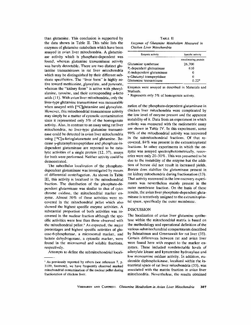
than glutamine. This conclusion is supported by the data shown in Table II. This table lists the enzymes of glutamine catabolism which have been assayed in avian liver mitochondria. A glutamin- ase activity which is phosphate-dependent was found, whereas glutamine transaminase activity was barely detectable. There are two distinct glu- tamine transaminases in rat liver mitochondria which may be distinguished by their different sub- strate specificities. The "liver form" is highly ac- tive toward methionine, glyoxylate, and pyruvate, whereas the "kidney form" is active with phenyl- alanine, tyrosine, and their corresponding a-keto acids (11). With avian liver mitochondria, only the liver-type glutamine transaminase was measurable when assayed with [~4C]glutamine and glyoxylate. However, this mitochondrial transaminase activity may simply be a matter of cytosolic contamination since it represented only 5% of the homogenate activity. Also, in contrast to an assay using rat liver mitochondria, no liver-type glutamine transami- nase could be detected in avian liver mitochondria using [14C]a-ketoglutaramate and glutamine. Be- cause ~/-glutamyltranspeptidase and phosphate-in- dependent glutaminase are reported to be cata- lytic activities of a single protein (12, 37), assays for both were performed. Neither activity could be demonstrated.
The subcellular localization of the phosphate- dependent gtutaminase was investigated by means of differential centrifugation. As shown in Table III, this activity is localized in the mitochondrial fraction. The distribution of the phosphate-de- pendent glutaminase was similar to that of cyto- chrome oxidase, the mitochondrial marker en- zyme. Almost 50% of these activities were re- covered in the mitochondrial pellet which also showed the highest specific enzyme activities. A substantial proportion of both activities was re- covered in the nuclear fraction although the spe- cific activities were less than those observed with the mitochondrial pel let) As expected, the major percentages and highest specific activities of glu- cose-6-phosphatase, a microsomal marker, and lactate dehydrogenase, a cytosolic marker, were found in the microsomal and soluble fractions, respectively.
Attempts to define the submitochondrial iocali-
i As previously reported by others (see reference 7, p. 3109, footnote), we have frequently observed marked mitochondrial contamination of the nuclear pellet during fractionation of chicken liver.
TABLE II Enzymes o f Glutamine Metabolism Measured in Chicken Liver Mitochondria
Enzymic activity Specific activity
nmollmin/mg protein
Glutamine synthetase 26,700 Pl-dependent glutaminase 610 P~-independent glutaminase 0 7-Glutamyl transpeptidase 0 Glutamine transaminase 0.22*
Enzymes were assayed as described in Materials and Methods. * Represents only 5% of homogenate activity.
zation of the phosphate-dependent glutaminase in chicken liver mitochondria were complicated by the low level of enzyme present and the apparent instability of it. Data from an experiment in which activity was measured with the radiometric assay are shown in Table IV. In this experiment, some 99% of the mitochondrial activity was recovered in the submitochondrial fractions. Of that re- covered, 84% was present in the extramitoplastal fractions. In other experiments in which the en- zyme was assayed spectrophotometrically, recov- eries were only 20-30%. This was presumed to be due to the instability of the enzyme but the addi- tion of borate did not result in increased yields. Borate does stabilize the glutaminase present in rat kidney mitochondria during fractionation (13). That activity recovered in the low-recovery experi- ments was nevertheless mainly present in the outer membrane fraction. On the basis of these results, the avian liver phosphate-dependent gluta- minase is tentatively assigned to the extramitoplas- tal space, specifically the outer membrane.
DISCUSSION
The localization of avian liver glutamine synthe- tase within the mitochondrial matrix is based on the methodology and operational definition of the various submitochondrial compartments described by Schnaitman and Greenawalt for rat liver (35). Certain differences between rat and avian liver were found here with respect to the marker en- zymes. These included nondetectable levels of adenylate kinase and kynurenine hydroxylase and low monoamine oxidase activity. In addition, nu- cleoside diphosphokinase, localized within the in- tracristal space of rat liver mitochondria (35), was associated with the matrix fraction in avian liver mitochondria. Nevertheless, the results obtained
VORHABEN AND CAMPBELL Glutamine Metabolism in Avian Liver Mitochondria 307
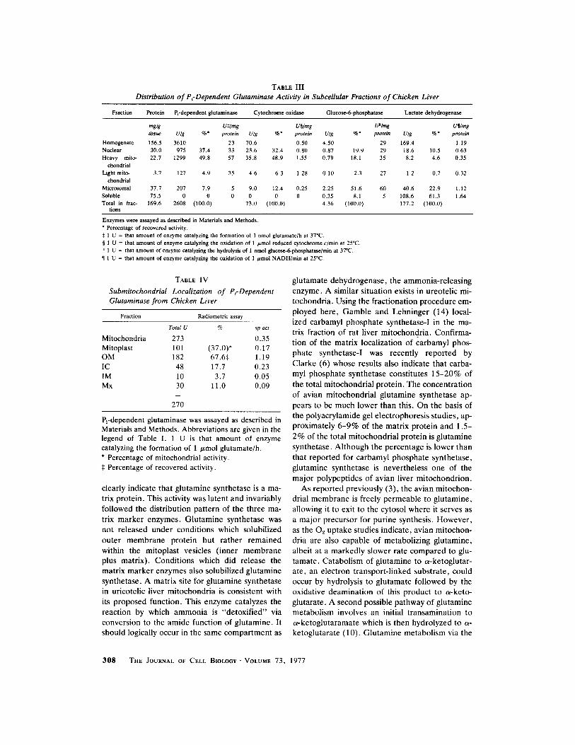
TABLE III Distribution of PcDependent Glutaminase Activity in Subcellular Fractions of Chicken Liver
Fraction Protein Prdepcndent glutaminase Cytochrome oxidase Glucose-6-phosphatase Lactate dehydrogenase
mglg U:~/mg Uw U"/mg U�82 tissue U/g %* protein U/g %* protein U/g %* protein U/g %" protein
Homogenate 156.5 3610 23 70.6 0.50 4.50 29 169.4 1.19 Nuclear 30.0 975 37.4 33 23.6 32.4 0.80 0.87 19.9 29 18.6 10.5 0.63 Heavy mito- 22.7 1299 49.8 57 35.8 48.9 1.55 0.79 18.1 35 8.2 4.6 0.35
ehondrial Light mito- 3.7 127 4.9 35 4.6 6.3 1.28 0.10 2.3 27 1.2 0.7 0.32
chondrial Microsomal 37.7 207 7.9 5 9.0 12.4 0.25 2.25 51.6 60 40.6 22.9 1.12
Soluble 75.5 0 0 0 0 0 0 0.35 8.1 5 108.6 61.3 1.64 Total in frac- 169.6 2608 (100.0) 73.0 (100.0) 4.36 (100.0) 177.2 (100.0)
tions
Enzymes were assayed as described in Materials and Methods. ~ Percentage of recovered activity. :~ 1 U = that amount of enzyme catalyzing the formation of 1 nmol glutamate/h at 37~ w 1 U = that amount of enzyme catalyzing the oxidation of 1 /~mol reduced cytochrome c/rain at 25"C. ii 1 U = that amount of enzyme catalyzing the hydrolysis of 1 nmol glueose-6-phosphatase/min at 370C.
�82 1 U = that amount of enzyme catalyzing the oxidation of 1 /~mol NADH/min at 25~
TABLE IV
Submitochondrial Localization of Pi-Dependent Glutaminase from Chicken Liver
Fraction Radiometric assay
Total U % sp act
Mitochondria 273 0.35 Mitoplast 101 (37.0)* 0.17 OM 182 67.6:~ 1.19 IC 48 17.7 0.23 IM 10 3.7 0.05 Mx 30 11.0 0.09
270
Prdependent glutaminase was assayed as described in Materials and Methods. Abbreviations are given in the legend of Table I. 1 U is that amount of enzyme catalyzing the formation of 1 /~mol glutamate/h. * Percentage of mitochondrial activity. :~ Percentage of recovered activity.
clearly indicate that glutamine synthetase is a ma- trix protein. This activity was latent and invariably followed the distribution pattern of the three ma- trix marker enzymes. Glutamine synthetase was not released under conditions which solubilized outer membrane protein but rather remained within the mitoplast vesicles (inner membrane plus matrix). Conditions which did release the matrix marker enzymes also solubilized glutamine synthetase. A matrix site for glutamine synthetase in uricotelic liver mitochondria is consistent with its proposed function. This enzyme catalyzes the reaction by which ammonia is "detoxified" via conversion to the amide function of glutamine. It should logically occur in the same compartment as
glutamate dehydrogenase, the ammonia-releasing enzyme. A similar situation exists in ureotelic mi- tochondria. Using the fractionation procedure em- ployed here, Gamble and Lehninger (14) local- ized carbamyl phosphate synthetase-I in the ma- trix fraction of rat liver mitochondria. Confirma- tion of the matrix localization of carbamyl phos- phate synthetase-I was recently reported by Clarke (6) whose results also indicate that carba- myl phosphate synthetase constitutes 15-20% of the total mitochondrial protein. The concentration of avian mitochondrial glutamine synthetase ap- pears to be much lower than this. On the basis of the polyacrylamide gel electrophoresis studies, ap- proximately 6-9% of the matrix protein and 1.5- 2% of the total mitochondrial protein is glutamine synthetase. Although the percentage is lower than that reported for carbamyl phosphate synthetase, glutamine synthetase is nevertheless one of the major polypeptides of avian liver mitochondrion.
As reported previously (3), the avian mitochon- drial membrane is freely permeable to glutamine, allowing it to exit to the cytosol where it serves as a major precursor for purine synthesis. However, as the 02 uptake studies indicate, avian mitochon- dria are also capable of metabolizing glutamine, albeit at a markedly slower rate compared to glu- tamate. Catabolism of glutamine to a-ketoglutar- ate, an electron transport-linked substrate, could occur by hydrolysis to glutamate followed by the oxidative deamination of this product to a-keto- glutarate. A second possible pathway of glutamine metabolism involves an initial transamination to a-ketoglutaramate which is then hydrolyzed to a- ketoglutarate (10). Glutamine metabolism via the
3 0 8 THE JOURNAL OF CELL BIOLOGY" VOLUME 7 3 , 1 9 7 7
first pathway is suggested by our results. Respira- tion in the presence of glutamine as well as gluta- mate was blocked by bromofuroate, an inhibitor of glutamate dehydrogenase, but not by aminoxy- acetate, a transaminase inhibitor. Hydrolysis rather than transamination as the initial step in glutamine metabolism has been observed in sev- eral other mitochondrial systems. In addition to those of the well-characterized kidney system (13, 15), mitochondria from Ehrlich ascites tumor cells and rat liver metabolize glutamine via an initial hydrolysis to glutamate catalyzed by glutaminases localized in the mitochondria (1, 17, 19, 24, 25).
Consistent with the above interpretation of the 02 uptake studies was the finding of a glutaminase activity but little or no transaminase activity in avian liver mitochondria. The glutaminase activity was phosphate-dependent and confirms an earlier report of this activity in chicken liver homogenates (23). In agreement with the response of this en- zyme from kidney (13) and Ehrlich ascites tumor cells (25), disruption of the avian mitochondrion with detergent resulted in enzyme inactivation in most cases. Unlike the other systems, however, addition of borate failed to protect the avian liver glutaminase against inactivation. In this regard, it may be noted that borate does not protect against all methods of mitochondrial disintegration since it does not stabilize the activity in sonically ruptured tumor cell mitochondria (25).
It is generally accepted that the physiological role of the kidney glutaminase is to provide am- monia in the regulation of acid-base balance (15) and that of the rat liver enzyme to provide ammo- nia for urea synthesis during glutamine catabo- lism. Yet another function for this enzyme in Ehr- lich ascites tumor cells has been suggested (25), and that is the intramitochondrial generation of reducing equivalents in the form of NAD(P)H via the oxidation of glutamate. A physiological role for the avian liver mitochondrial glutaminase is not readily evident. In one experiment in which essentially all of the mitochondrial activity was recovered in the submitochondrial fractions, the enzyme was localized in the extramitoplastal com- partment, mainly the outer membrane. This com- partment also consistently contained the highest specific enzyme activity, even where overall recov- eries were low. An extramitoplastal localization of phosphate-dependent glutaminase is the opposite of that found in kidney (13) and tumor cells (25) but is nevertheless consistent with the function of glutamine metabolism in avian liver mitochondria.
The localization of glutaminase in the outer mem- brane of these mitochondria suggests a "vectorial" metabolism of glutamine. Glutamine normally ex- its and may be channeled directly into purine biosynthesis. However, if entering the mito- chondrion, glutamine may be broken down by glutaminase.
This work was supported by grants from the National Science Foundation (BMS75-13161) and the U. S. Pub- lic Health Service (AI 05006).
Received for publication 16 August 1976, and in revised form 20 December 1976.
REFERENCES
1. BLACKBURN, E. H., and F. J. R. HIRD. 1972. Metabolism of glutamine and glutamate by rat liver mitochondria. Arch. Biochem. Biophys. 152:258- 264.
2. CAMPBELL, J. W. 1973. Nitrogen metabolism. In Comparative Animal Physiology. C. L. Prosser, ed- itor. W. B. Saunders Co., Philadelphia. 279-316.
3. CAMPBELL, J. W., and J. E. VORHABEN. 1976. Avian mitochondrial glutamine metabolism. J. Biol. Chem. 251:781-786.
4. CAUGHEY, W. S., J. D. SMILEY, and L. HELLEB- MAN. 1957. L-Glutamic acid dehydrogenase: struc- tural requirements for substrate competition. Effect of thyroxine. J. Biol. Chem. 224:591-607.
5. CHRIST, W., D. RAKOW, M. FERANDES, and S. MAGOUR. 1973. A simple and sensitive spectropho- tometric determination of monoamine oxidase ac- tivity. Z. Klin. Chem. Klin. Biochem. 11:367-370.
6. CLARKE, S. 1976. A major polypeptide component of rat liver mitochondria: carbamyl phosphate syn- thetase. J. Biol. Chem. 251:950-961.
7. CLINKENBEARD, K. D., W. D. REED, R. A. MOO- NEY, and M. D. LANE. 1975. Intracellular localiza- tion of the 3-hydroxy-3-methyl-glutaryl coenzyme A cycle enzymes in liver. J. Biol. Chem. 250:3108- 3116.
8. COHEN, P. O., and G. W. BROWN, JR. 1960. Am- monia metabolism and urea biosynthesis. In Com- parative Biochemistry. M. Florkin and H. S. Ma- son, editors, Academic Press, Inc., New York. 2:161-244.
9. COOPER, A. J. L., and A. MEISTER. 1972. Isolation and properties of highly purified glutamine transam- inase. Biochemistry. 11:661-671.
10. COOPER, A. J. L., and A. MEISTER. 1973. Gluta- mine transaminases from liver and kidney. In The Enzymes of Glutamine Metabolism. S. Prusiner and E. S. Stadtman, editors. Academic Press, Inc., New York. 207-226.
11. COOPER, A. J. L., and A. MEIS~R. 1974. Isolation
VORHABEN AND CAMPBELL Glutamine Metabolism in Avian Liver Mitochondria 3 0 9

and properties of a new glutamine transaminase from rat kidney. J. Biol. Chem. 249:2554-2561.
12. CURTHOYS, N. P., and T. KUHLENSCHMIDT. 1975. Phosphate-independent glutaminase from rat kid- ney. Partial purification and identity with ~/-gluta- myltranspeptidase. J. Biol. Chem. 250:2099-2105.
13. CURTHOVS, N. P., and R. F. WEISS, 1974. Regula- tion of renal ammoniagenesis. Subcellular localiza- tion of rat kidney glutaminase isoenzymes. J. Biol. Chem. 249:3261-3266.
14. GAMBLE, J. G., and A. L. LEHNINGER. 1973. Transport of ornithine and citrulline across the mi- tochondrial membrane. J. Biol. Chem. 248:610- 618.
15. GOLDSTEIN, L. 1975. Regulation of renal glutamine deamination. Med. Clin. N. Amer. 59:667-680.
16. GREENAWALT, J. W. 1974. The isolation of outer and inner mitochondrial membranes. Methods En- zymol. 10:310-323.
17. GUHA, S. R. 1971. Intracellular localization of glu- taminase I in rat liver. Enzymologia 23:94-100.
18. HARPER, A. E. 1963. Glucose-6-phosphatase. In Methods of Enzymatic Analysis. H.-U. Bergmeyer, editor. Academic Press, Inc., New York. 788-792.
19. HIRD, F. J. R., and M. A. MURGINSON. 1968. The formation of ammonia from glutamine and gluta- mate by mitochondria from rat liver and kidney. Arch. Biochem. Biophys. 127:718-724.
20. HOPPER, S., and H. L. SEGA. 1962. Kinetic studies of rat liver glutamic-alanine transaminase. J. Biol. Chem. 237:3189-3195.
21. HSIEn, W. C., and R. E. ANDERSON. 1975. Quanti- tation of stained proteins in SDS polyacrylamide gels with lysozyme as internal standard. Anal. Bio- chem. 69:331-338.
22. KALRA, J., and J. T. BROSNAN. 1974. The subcellu- lar localization of glutaminase isoenzymes in rat kidney cortex. J. Biol. Chem. 249:3255-3260.
23. KATUNAMA, N., Y. MATSUDA, and Y. KURODA. 1970. Phylogenic aspects of different regulatory mechanisms of glutamine metabolism. Adv. En- zyme Regul. Vol. 8:73-81.
24. KOVACEVI6, Z. 1971. The pathway of glutamine and glutamate oxidation in isolated mitochondria from mammalian cells. Biochem. J. 125:757-763.
25. KOVACEVlt~, Z. 1974. Properties and intracellular localization of Ehrlich ascites tumor cell glutamin- ase. Cancer Res. 34:3403-3407.
26. KUPCHIK, H. Z., and W. E. KNOX. 1970. Phenyl-
pyruvate glutamine aminotransferase (Rat kidney). Methods Enzymol. 17A:951-954.
27. LAYNE, E. 1957. Spectrophotometric and turbidi- metric methods for measuring proteins. Methods Enzymol. 3:450-451.
28. LOWRY, O. H., N. J. ROSEBROUGH, A. L. FARR, and R. J. RANDALL. 1951. Protein measurement with the Folin phenol reagent. J. Biol. Chem. 193:265-275.
29. MEISTER, A. 1953. Preparation and enzymatic reac- tions of the keto analogues of asparagine and gluta- mine. J. Biol. Chem. 200:571-589.
30. MILLONIG, G. 1961. Advantages of a phosphate buffer for OsO4 solutions in fixation. J. Appl. Phys. 32:1637.
31. OCHOA, S. 1955. Malic dehydrogenase from pig heart. Methods Enzymol. 1:735-739.
32. OLSON, J. A., and C. B. ANFINSEN. 1953. Kinetic and equilibrium studies on crystalline L-glutamic acid dehydrogenase. J. Biol. Chem. 202:841-856.
33. PAMILIANS, V., P. KRISHNASWAMY, G. DUN- VILLE, and A. MEISTER. 1962. Studies on the mech- anism of glutamine synthesis; isolation and proper- ties of the enzyme from sheep brain. Biochemistry. 1:153-158.
34. PLANT, G. W. E., and S. C. SUNG. 1955. Diphos- phopyridine nucleotide isocitric dehydrogenase from animal tissues. Methods Enzymol. 1:710-714.
35. SCHNAITMAN, C., and J. W. GREENAWALT. 1968. Enzymatic properties of the inner and outer mem- branes of rat liver mitochondria. J. Cell Biol. 38:158-175.
36. SOITOCASA, G. L., I . DYLENSTIERNA, L. ERNSTER, and A. S. WEISBERGER. 1967. An electron-trans- port system associated with the outer membrane of liver mitochondria. J. Cell Biol, 32:415-438.
37. TATE, S. S., and A. MEISTER. 1975. Identity of maleate-stimulated glutaminase with 3,-glutamyl transpeptidase in rat kidney. J. Biol. Chem. 250:4619-4627.
38. VORHABEN, J. E., L. WONG, and J. W. CAMPBELL. 1973. Assay for glutamine synthetase activity. Bio- chem. J. 135:893-896.
39. VORHAREN, J. E., and J. W. CAMPBELL. 1972. Glutamine synthetase, a mitochondrial enzyme in uricotelic species. J. Biol. Chem. 247:2763-2767.
40. WHARTON, D. C., and A. TZAGOLOFF. 1967. Cyto- chrome oxidase from beef heart mitochondria. Methods Enzymol. 10:245-250.
310 TeE JOURNAL OF CELL BIOLOGY" VOLUME 73, 1977