Embed Size (px)
Citation preview
SREE CHITRA TIRUNAL INSTITUTE FOR
MEDICAL SCIENCES AND TECHNOLOGY TRIVANDRUM, KERALA
MORPHOLOGICAL ABNORMALITIES IN HYPERTROPHIC
CARDIOMYOPATHY - A CARDIAC MRI BASED STUDY
THESIS
Submitted during the course of
DM Cardiology
Dr. VIJAYAN.G DM Trainee
DEPARTMENT OF CARDIOLOGY
July 2019
DECLARATION
I, Dr. Vijayan.G, hereby declare that the project in this book titled ―Morphological
abnormalities in Hypertrophic cardiomyopathy – A cardiac MRI based study‖ was
undertaken by me under the supervision of the faculty, Department of Cardiology, Sree
Chitra Tirunal Institute for Medical Sciences and Technology.
Date: Dr.Vijayan.G
DM cardiology
CERTIFICATE
I, Dr. Vijayan.G, hereby declare that the project in this book was undertaken by me under
the supervision of the faculty, Department of Cardiology, Sree Chitra Tirunal Institute for
Medical Sciences and Technology.
Thiruvananathapuram Prof. Dr. Ajit Kumar.V.K
Date: Head of Department of cardiology
CERTIFICATE
I hereby certify that the work in this project titled ―Morphological abnormalities in
Hypertrophic cardiomyopathy – A cardiac MRI based study‖ is a certified record of
original research work undertaken by Dr. Vijayan.G in partial fulfillment of requirement for
the purpose of award of DM cardiology degree under my guidance and supervision.
Guide:
Dr. Ajit Kumar.V.K
Professor and Head
Department of Cardiology
Sree Chitra Tirunal institute for Medical Sciences and Technology
Trivandrum – 695011
MORPHOLOGICAL ABNORMALITIES IN HYPERTROPHIC
CARDIOMYOPATHY - A CARDIAC MRI BASED STUDY
PRIMARY INVESTIGATOR
Dr. Vijayan.G
Senior Resident
Department of Cardiology,
Sree Chitra Tirunal Institute for Medical sciences and Technology
Thiruvananthapuram
GUIDE
Dr. Ajit Kumar.V.K
Professor and Head
Department of Cardiology,
Sree Chitra Tirunal Institute for Medical sciences and Technology
Thiruvananthapuram
CO-GUIDES
Dr. Sanjay.G
Additional Professor
Department of Cardiology,
Sree Chitra Tirunal Institute for Medical sciences and Technology
Thiruvananthapuram
Dr. Anoop.A
Assistant Professor
Department of Cardiology,
Sree Chitra Tirunal Institute for Medical sciences and Technology
Thiruvananthapuram
TITLE

INDEX
Page No.
INTRODUCTION 1
HYPOTHESIS 3
AIMS AND OBJECTIVES 5
REVIEW OF LITERATURE 7
MATERIALS AND METHODS 20
RESULTS 25
DISCUSSION 40
LIMITATIONS 45
CONCLUSIONS 47
BIBLIOGRAPHY 49
APPENDIX 53
ACKNOWLEDGEMENT
At the outset I would like to thank my mentor, guide Dr. Ajit Kumar.V.K,
Professor and Head, Department of cardiology, to whom I am greatly indebted to, for his
immense support, encouragement and inspiring attitude – not only for this project, throughout
my DM period.
I deeply thank my co-guide Dr. Sanjay.G, Additional Professor, Department of
cardiology, for his constant guidance, valuable inputs and motivation throughout this project.
Special thanks to my co-guide Dr. Anoop.A, Assistant Professor Department of
imaging sciences & intervention radiology, for his valuable suggestions.
I deeply thank Dr. Mukhund Prabhu, Assistant Professor, Department of
cardiology, for his valuable inputs in analysis of this project.
My sincere thanks to Dr. Ajay Alex, Senior resident, Department of cardio-vascular
imaging sciences & intervention radiology, for helping me in interpreting MRI images.
Finally I express my gratitude to all my patients, and all those who directly and
indirectly helped me do this study.
Vijayan.G

Acronyms and Abbreviations
ACRONYMS AND ABBREVIATIONS:
AML – Anterior mitral leaflet
ASA – Alcohol Septal Ablation
CMRI – Cardiac Magnetic Resonance Imaging
HCM – Hypertrophic cardiomyopathy
IVS – Inter ventricular Septum
LGE – Late gadolinium enhancement
LV – Left ventricle
LVOT – Left ventricular outflow tract
MR – Mitral regurgitation
MV - Mitral valve
PM – Papillary muscle
PML – Posterior mitral leaflet
PW - Posterior Wall
RV – Right ventricle
SAM – Systolic anterior motion of mitral valve
1
Introduction
2
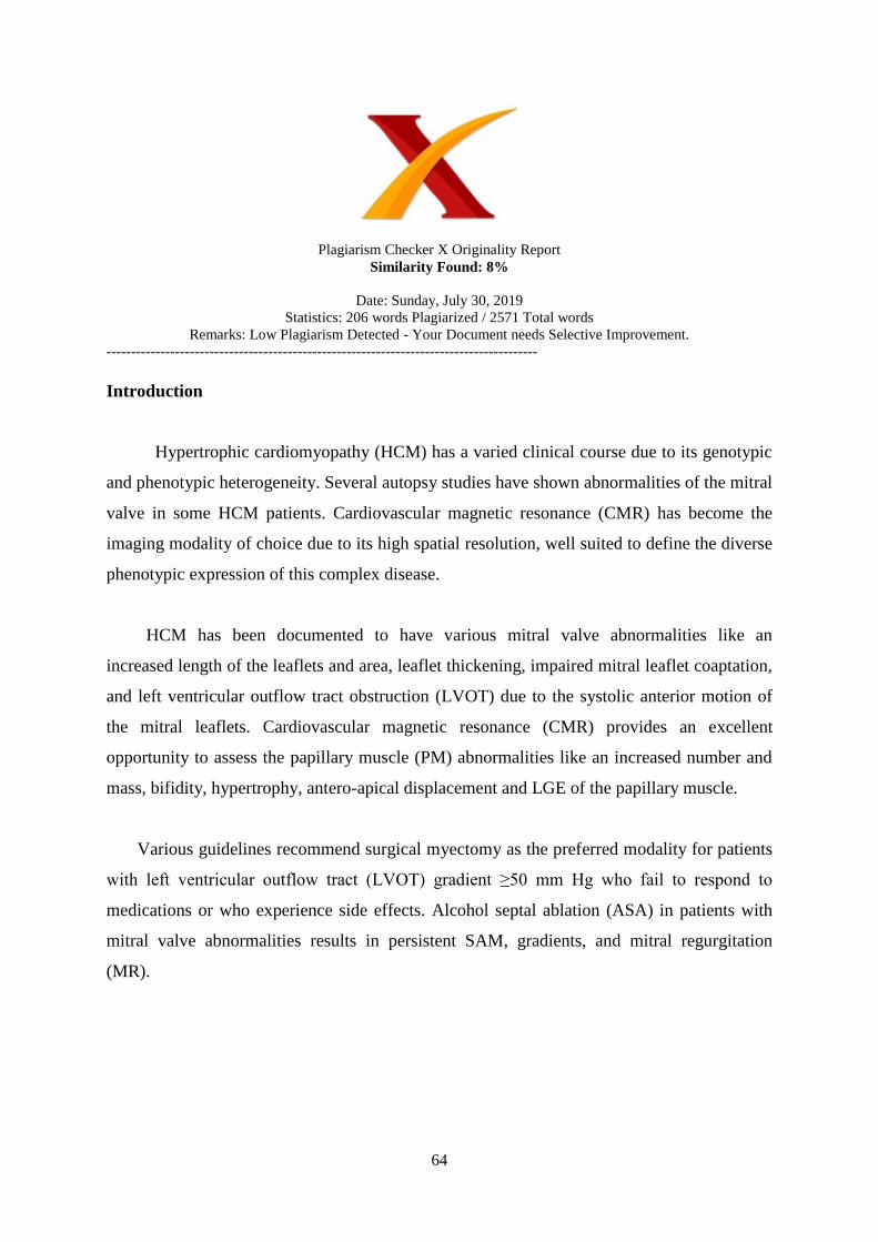
INTRODUCTION
Hypertrophic cardiomyopathy (HCM) has a varied clinical course due to its
genotypic and phenotypic heterogeneity. Several autopsy studies have shown
abnormalities of the mitral valve in some HCM patients. Cardiovascular magnetic
resonance (CMR) has become the imaging modality of choice due to its high spatial
resolution, well suited to define the diverse phenotypic expression of this complex
disease.
HCM has been documented to have various mitral valve abnormalities like an
increased length of the leaflets and area, leaflet thickening, impaired mitral leaflet
coaptation, and left ventricular outflow tract obstruction (LVOT) due to the systolic
anterior motion of the mitral leaflets. Cardiovascular magnetic resonance (CMR)
provides an excellent opportunity to assess the papillary muscle (PM) abnormalities like
an increased number and mass, bifidity, hypertrophy, antero-apical displacement and
LGE of the papillary muscle.
Various guidelines recommend surgical myectomy as the preferred modality for
patients with left ventricular outflow tract (LVOT) gradient ≥50 mm Hg who fail to
respond to medications or who experience side effects. Alcohol septal ablation (ASA) in
patients with mitral valve abnormalities results in persistent SAM, gradients, and mitral
regurgitation (MR).

3
Hypothesis

4
HYPOTHESIS
HCM is characterized by abnormalities of mitral valvular apparatus and
papillary muscles. The abnormal mitral valve and papillary muscle morphology are
associated with increased LVOT obstruction.

5
Aims & Objectives
6
AIMS & OBJECTIVES
To determine the pattern of hypertrophy of ventricles.
To determine the mitral valve and the papillary muscle abnormalities in hypertrophic
cardiomyopathy.
To determine the relationship between the mitral valve and the papillary muscle
abnormalities and obstruction in hypertrophic cardiomyopathy.
7
Review of Literature

8
REVIEW OF LITERATURE
LVOT obstruction in HCM was caused by systolic anterior motion (SAM) of the mitral
valve and mitral-septal contact1. 2011 American guidelines state: ―Mitral valve abnormalities
plays an important role in the generation of left ventricular outflow tract obstruction,
suggesting the potential value of additional surgical approaches (e.g., plication, valvuloplasty,
and papillary muscle relocation) and making myectomy more appropriate than alcohol septal
ablation in some patients‖2. 2015 European guidelines state: ―Septal myectomy, rather than
alcohol septal ablation, is recommended in patients with an indication for septal reduction
therapy and other lesions requiring surgical intervention (e.g., mitral valve
repair/replacement, papillary muscle intervention) Class I, Level of Evidence: C‖
Mitral regurgitation (MR) in obstructive HCM most commonly results from the poor
coaptation, due to the inadequate length or mobility of PML to move along with the anterior
mitral leaflet3. The SAM abolition either with optimal medical or surgical treatment results in
the reduction of MR.
PATTERN OF VENTRICULAR HYPERTROPHY:
Asymmetric septal hypertrophy is the commonest morphologic pattern of HCM and
the most commonly involved hypertrophied segment is the anteroseptal myocardium18
. The
prevalence of LVOT obstruction in asymmetric septal hypertrophy is 20–30%19
. Symmetric
HCM occurs in up to 42% of HCM and it has to be distinguished from other causes of
concentric left ventricular hypertrophy. Apical HCM was initially reported in the Japanese
population. Prevalence of apical HCM is variable in studies, ranging from 25% in Japanese
patients to fewer than 2% of all HCM patients in Western countries20
.
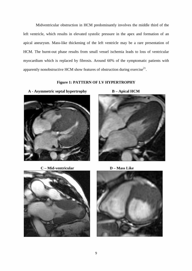
9
Midventricular obstruction in HCM predominantly involves the middle third of the
left ventricle, which results in elevated systolic pressure in the apex and formation of an
apical aneurysm. Mass-like thickening of the left ventricle may be a rare presentation of
HCM. The burnt-out phase results from small vessel ischemia leads to loss of ventricular
myocardium which is replaced by fibrosis. Around 60% of the symptomatic patients with
apparently nonobstructive HCM show features of obstruction during exercise21
.
Figure 1: PATTERN OF LV HYPERTROPHY
A - Asymmetric septal hypertrophy B – Apical HCM
C – Mid-ventricular D – Mass Like
10
LATE GADOLINIUM ENHANCEMENT:
Late gadolinium enhancement of the left ventricular myocardium occurs in around
80% of HCM patients and is located commonly in the mid-wall with a patchy distribution.
The most usual location of LGE is combined interventricular septum and the free wall
location (~30% of patients) but the free wall, septum, apex, and the right ventricular insertion
areas into the septum are the less commonly involved sites23
. LGE commonly involves the
anteroseptal area of the inter-ventricular septum from basal to mid segments. LGE in HCM
may be attributed histologically to plexiform fibrosis (fibrosis seen in areas of myocyte
disarray), expanded interstitial spaces, and replacement fibrosis due to microvascular
ischemia22
.
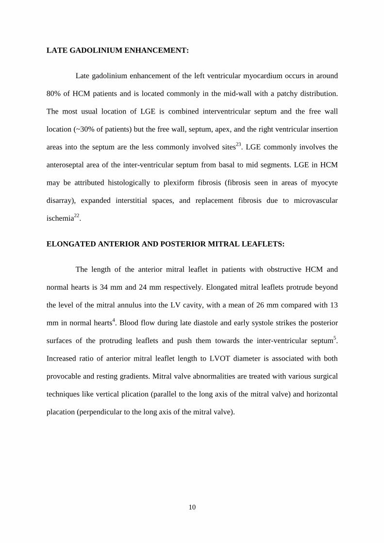
ELONGATED ANTERIOR AND POSTERIOR MITRAL LEAFLETS:
The length of the anterior mitral leaflet in patients with obstructive HCM and
normal hearts is 34 mm and 24 mm respectively. Elongated mitral leaflets protrude beyond
the level of the mitral annulus into the LV cavity, with a mean of 26 mm compared with 13
mm in normal hearts4. Blood flow during late diastole and early systole strikes the posterior
surfaces of the protruding leaflets and push them towards the inter-ventricular septum5.
Increased ratio of anterior mitral leaflet length to LVOT diameter is associated with both
provocable and resting gradients. Mitral valve abnormalities are treated with various surgical
techniques like vertical plication (parallel to the long axis of the mitral valve) and horizontal
placation (perpendicular to the long axis of the mitral valve).
11
Figure 2: Elongated AML and thickened inter-ventricular septum
Elongated mitral leaflets can displace the mitral-septal contact point (and site of
subaortic obstruction) distally creating the need for an extended muscular resection15
. The
mitral-septal contact (and obstruction) can persist even after adequate septal muscular
resection due to extremely elongated AML length. Various surgical reports of severely
symptomatic obstructive HCM patients advocates the combined approach of septal
myectomy and AML repair, with leaflet extension or shortening reconstruction or
plication16
.
SYSTOLIC ANTERIOR MOTION OF MITRAL VALVE (SAM):
The following factors may contribute to SAM in HCM (1) increased length of
the anterior or posterior leaflet; (2) aorto-mitral angle <120°; (3) elongation and buckling
of the chordae; (4) anteromedial displacement of the papillary muscles; (6) minimum
distance between the coaptation point to the septum (C-Sept, <2.5 cm) 14
. (7) Venturi
effect, which is defined as ―when fluid flows through a region of reduced cross-sectional
12
area, fluid pressure decreases, and velocity increases‖. Septal hypertrophy in HCM
creates the venturi effect through the reduction in the LVOT diameter, which leads to
increased velocity and reduced pressure of the ejected blood in the LVOT. The pressure
difference between the left atrium and the outflow tract may lead to the movement of the
mitral valve towards the septum. Other conditions causing SAM are hypertension,
diabetes mellitus, acute myocardial infarction, post mitral valve repair, and even in
asymptomatic patients during pharmacologic stress with dobutamine.
GRADING OF SAM:
Echo grading of systolic anterior motion of the mitral valve:
I: No mitral leaflet-septal contact, the minimum distance between the mitral valve and the
ventricular septum during systole = 10 mm;
II: No mitral leaflet-septal contact, the minimum distance between the mitral valve and
the ventricular septum during systole <10 mm;
III: Brief mitral leaflet-septal contact (<30% of systole time);
IV: Prolonged mitral leaflet-septal contact (>30% of systole time).
ELONGATED POSTERIOR LEAFLET WITH SAM:
SAM can be caused by isolated posterior leaflet elongation due to the protrusion of
the residual leaflet through inter-chordal space to contact the inter-ventricular septum11
.
13
MITRAL VALVE ABNORMALITIES CAUSED BY MUTATIONS IN
GENES CODING FOR SARCOMERIC PROTEINS:
Elongation of mitral leaflets has been documented in subjects with HCM-associated
mutations who have not yet developed thickening. Mitral leaflet elongation is considered
to a major phenotypic expression of HCM and is not obtained due to stretch from SAM.
ANTERIOR AND BASILAR DISPLACEMENT OF THE ANTEROLATERAL
PAPILLARY MUSCLE:
Anterior displacement of the papillary muscles results in the deviation of the
coaptation plane of the mitral valve anteriorly in the LV cavity6. Two most common papillary
muscles abnormalities in HCM are: 1) Antero-basilar displacement of the anterolateral
papillary muscle. 2) Abnormal muscular connections between the papillary muscular head
and the anterolateral wall, inserted into or near the A1 scallop7. CMR study by Kwon et al.
8
has shown a higher prevalence of bifid papillary muscles in HCM; Patients with LVOT
obstruction due to SAM had closer proximity of papillary muscle to the septum.
14
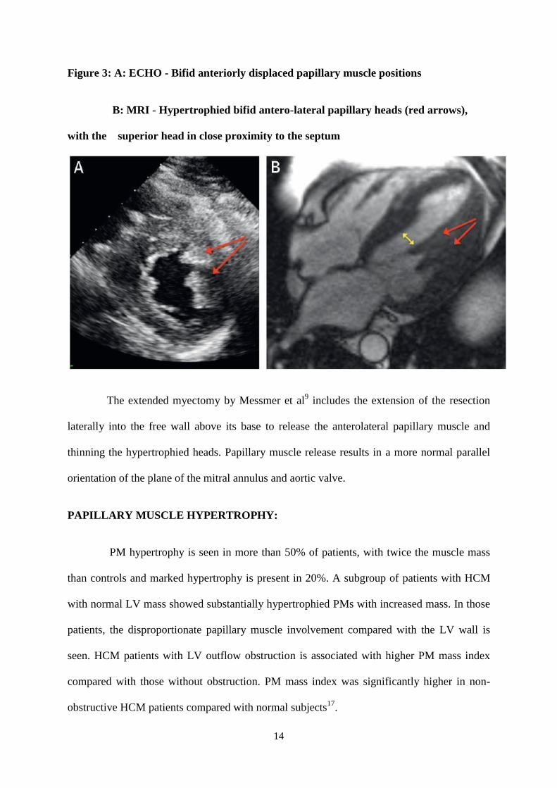
Figure 3: A: ECHO - Bifid anteriorly displaced papillary muscle positions
B: MRI - Hypertrophied bifid antero-lateral papillary heads (red arrows),
with the superior head in close proximity to the septum
The extended myectomy by Messmer et al9 includes the extension of the resection
laterally into the free wall above its base to release the anterolateral papillary muscle and
thinning the hypertrophied heads. Papillary muscle release results in a more normal parallel
orientation of the plane of the mitral annulus and aortic valve.
PAPILLARY MUSCLE HYPERTROPHY:
PM hypertrophy is seen in more than 50% of patients, with twice the muscle mass
than controls and marked hypertrophy is present in 20%. A subgroup of patients with HCM
with normal LV mass showed substantially hypertrophied PMs with increased mass. In those
patients, the disproportionate papillary muscle involvement compared with the LV wall is
seen. HCM patients with LV outflow obstruction is associated with higher PM mass index
compared with those without obstruction. PM mass index was significantly higher in non-
obstructive HCM patients compared with normal subjects17
.
15
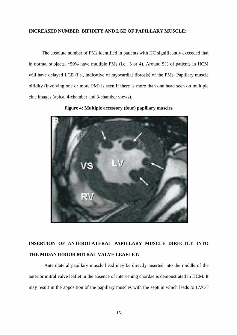
INCREASED NUMBER, BIFIDITY AND LGE OF PAPILLARY MUSCLE:
The absolute number of PMs identified in patients with HC significantly exceeded that
in normal subjects, ~50% have multiple PMs (i.e., 3 or 4). Around 5% of patients in HCM
will have delayed LGE (i.e., indicative of myocardial fibrosis) of the PMs. Papillary muscle
bifidity (involving one or more PM) is seen if there is more than one head seen on multiple
cine images (apical 4-chamber and 3-chamber views).
Figure 4: Multiple accessory (four) papillary muscles
INSERTION OF ANTEROLATERAL PAPILLARY MUSCLE DIRECTLY INTO
THE MIDANTERIOR MITRAL VALVE LEAFLET:
Anterolateral papillary muscle head may be directly inserted into the middle of the
anterior mitral valve leaflet in the absence of intervening chordae is demonstrated in HCM. It
may result in the apposition of the papillary muscles with the septum which leads to LVOT
16
obstruction10
. If a large anomalous papillary muscle could not be excised, longitudinal
resection to thin it, even to its base has been performed successfully.
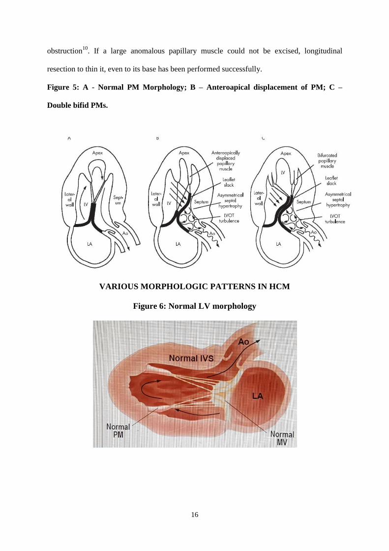
Figure 5: A - Normal PM Morphology; B – Anteroapical displacement of PM; C –
Double bifid PMs.
VARIOUS MORPHOLOGIC PATTERNS IN HCM
Figure 6: Normal LV morphology
17
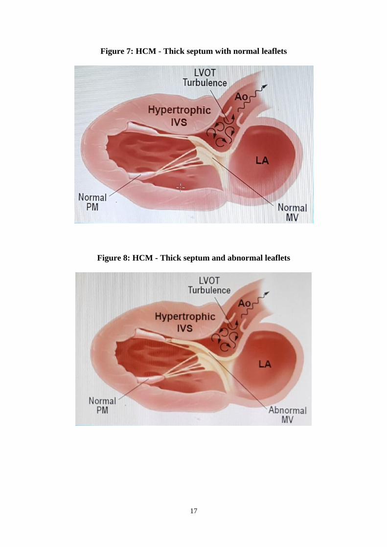
Figure 7: HCM - Thick septum with normal leaflets
Figure 8: HCM - Thick septum and abnormal leaflets
18
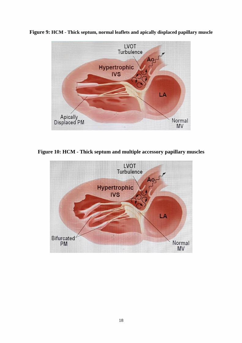
Figure 9: HCM - Thick septum, normal leaflets and apically displaced papillary muscle
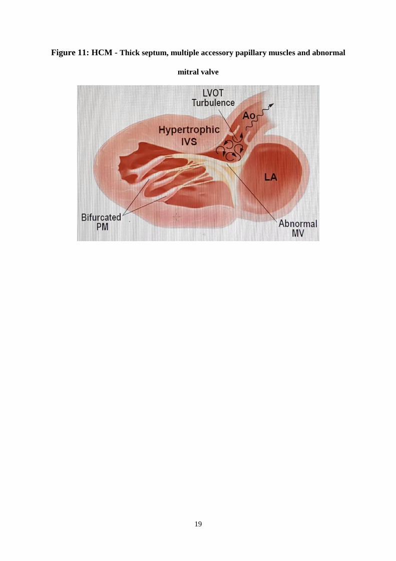
Figure 10: HCM - Thick septum and multiple accessory papillary muscles
19
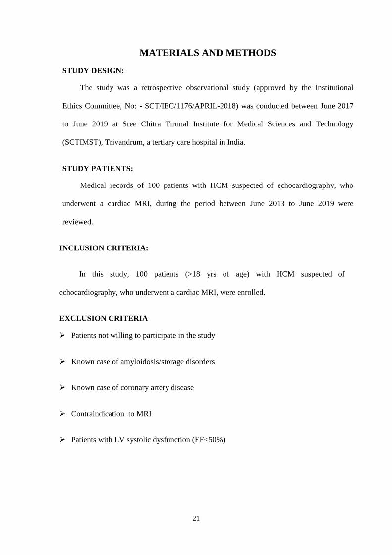
Figure 11: HCM - Thick septum, multiple accessory papillary muscles and abnormal
mitral valve

20
Materials & Methods
21
MATERIALS AND METHODS
STUDY DESIGN:
The study was a retrospective observational study (approved by the Institutional
Ethics Committee, No: - SCT/IEC/1176/APRIL-2018) was conducted between June 2017
to June 2019 at Sree Chitra Tirunal Institute for Medical Sciences and Technology
(SCTIMST), Trivandrum, a tertiary care hospital in India.
STUDY PATIENTS:
Medical records of 100 patients with HCM suspected of echocardiography, who
underwent a cardiac MRI, during the period between June 2013 to June 2019 were
reviewed.
INCLUSION CRITERIA:
In this study, 100 patients (>18 yrs of age) with HCM suspected of
echocardiography, who underwent a cardiac MRI, were enrolled.
EXCLUSION CRITERIA
Patients not willing to participate in the study
Known case of amyloidosis/storage disorders
Known case of coronary artery disease
Contraindication to MRI
Patients with LV systolic dysfunction (EF<50%)
22
METHODOLOGY:
Baseline demographic data of all enrolled patients were taken from medical records
including full clinical history, symptomatic status, electrocardiogram, echocardiography
details, catheterization study and CMR findings were also recorded.
TRANS-THORACIC ECHOCARDIOGRAPHY:
Detailed Echocardiography was done prior to CMR with emphasis on the following
parameters:
LV internal dimensions, septal and posterior wall thickness in end-diastole.
Patterns of hypertrophy of the ventricles.
LVOT and mid-cavity gradients.
Systolic anterior motion of the mitral valve and mitral regurgitation severity.
Maximum LV wall thickness in end-diastole.
CARDIAC MRI:
Cardiac MRI was done with SIEMENS 1.5 T somatom sensation, Seimens health
care Germany and GE 3 Tesla system (Discovery 750w; General electric GE healthcare;
USA). Following parameters were assessed:
Ventricular hypertrophy distribution
Assessment of the level of ventricular obstruction
LV function
Mitral valvular abnormalities
Papillary muscle abnormalities
23
MITRAL VALVE ABNORMALITIES:
The following parameters of the mitral valve were assessed.
AML and PML length
Measured at end-diastole in 3-chamber view, with the leaflets maximally
extended parallel to the anterior septum and LV free wall.
AML - distal most extent to its insertion into posterior aortic wall
PML – distal most extent to its insertion into basal LV posterior free wall.
SAM and MR severity
LGE of the mitral valve
Correlation of AML length with LV obstruction
Correlation of PML length with LV obstruction
Correlation of SAM with LV obstruction
PAPILLARY MUSCLE ABNORMALITIES:
The following parameters of the papillary muscle were assessed.
Number of papillary muscles in short-axis cine images at 3 levels (basal, mid-
ventricular and apical)
Bifidity of the papillary muscles (in apical 4-chamber and 3-chamber view)
Late gadolinium enhancement of the papillary muscles
Correlation of the papillary muscle abnormalities with LV obstruction
24
STATISTICAL ANALYSIS:
The data were analyzed with commercially available statistical software (SPSS) to
study the percentage of patients who had the mitral valve and papillary muscle abnormalities
in hypertrophic cardiomyopathy. Continuous variables are expressed as mean with standard
deviations and discrete variables as counts and percentages. For categorical variables, the chi-
square test and Fisher exact t-test were used, and for continuous variables, Student t-test was
used. HCM patients were divided into two groups such as HCM with obstruction and HCM
without obstruction. Mitral valve and papillary muscle abnormalities in both the groups were
noted and their correlation with left ventricular obstruction was assessed. A p-value less than
or equal to 0.05 was considered statistically significant.

25
Results
26
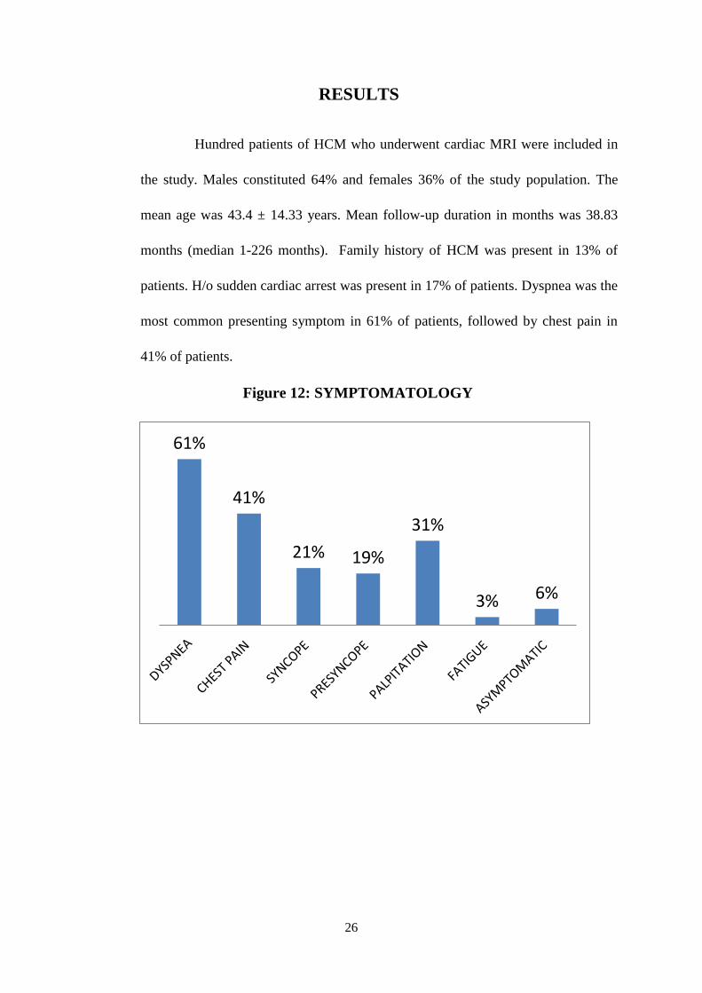
RESULTS
Hundred patients of HCM who underwent cardiac MRI were included in
the study. Males constituted 64% and females 36% of the study population. The
mean age was 43.4 ± 14.33 years. Mean follow-up duration in months was 38.83
months (median 1-226 months). Family history of HCM was present in 13% of
patients. H/o sudden cardiac arrest was present in 17% of patients. Dyspnea was the
most common presenting symptom in 61% of patients, followed by chest pain in
41% of patients.
Figure 12: SYMPTOMATOLOGY
61%
41%
21% 19%
31%
3% 6%
27
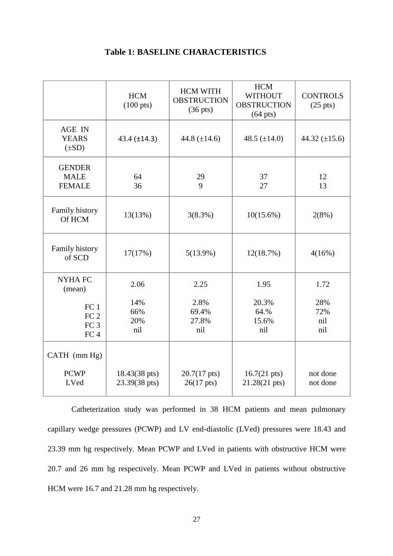
Table 1: BASELINE CHARACTERISTICS
Catheterization study was performed in 38 HCM patients and mean pulmonary
capillary wedge pressures (PCWP) and LV end-diastolic (LVed) pressures were 18.43 and
23.39 mm hg respectively. Mean PCWP and LVed in patients with obstructive HCM were
20.7 and 26 mm hg respectively. Mean PCWP and LVed in patients without obstructive
HCM were 16.7 and 21.28 mm hg respectively.
HCM
(100 pts)
HCM WITH
OBSTRUCTION
(36 pts)
HCM
WITHOUT
OBSTRUCTION
(64 pts)
CONTROLS
(25 pts)
AGE IN
YEARS
(±SD) 43.4 (±14.3) 44.8 (±14.6) 48.5 (±14.0) 44.32 (±15.6)
GENDER
MALE
FEMALE
64
36
29
9
37
27
12
13
Family history
Of HCM 13(13%) 3(8.3%) 10(15.6%) 2(8%)
Family history
of SCD 17(17%) 5(13.9%) 12(18.7%) 4(16%)
NYHA FC
(mean)
FC 1
FC 2
FC 3
FC 4
2.06
14%
66%
20%
nil
2.25
2.8%
69.4%
27.8%
nil
1.95
20.3%
64.%
15.6%
nil
1.72
28%
72%
nil
nil
CATH (mm Hg)
PCWP
LVed
18.43(38 pts)
23.39(38 pts)
20.7(17 pts)
26(17 pts)
16.7(21 pts)
21.28(21 pts)
not done
not done
28
Table 2: INVASIVE PROCEDURES
PROCEDURE No. of patients
Alcohol Septal Ablation (ASA) 10
Electrophysiology Study (EPS) 8
Permanent pacemaker implantation (PPI) 5
Implantable Cardioverter Defibrillator (ICD) 9
(1-primary,8-secondary
prophylaxis)
Cardiac Resynchronization Therapy (CRT-D) 1
No events were noted in HCM patients with either ICD or CRT-D over 38.83 months
of follow-up.
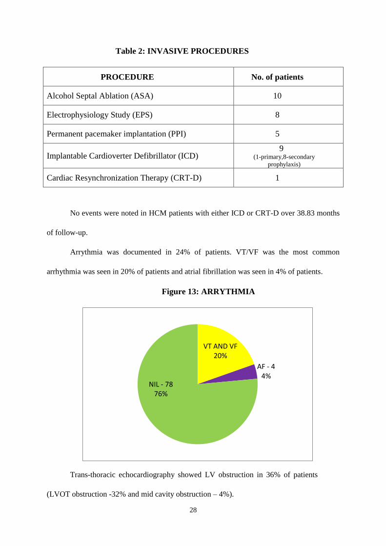
Arrythmia was documented in 24% of patients. VT/VF was the most common
arrhythmia was seen in 20% of patients and atrial fibrillation was seen in 4% of patients.
Figure 13: ARRYTHMIA
Trans-thoracic echocardiography showed LV obstruction in 36% of patients
(LVOT obstruction -32% and mid cavity obstruction – 4%).
VT AND VF 20%
AF - 4 4%
NIL - 78 76%
29
Figure 14: LEVEL OF LV OBSTRUCTION – BY ECHO
MRI CHARACTERISTICS:
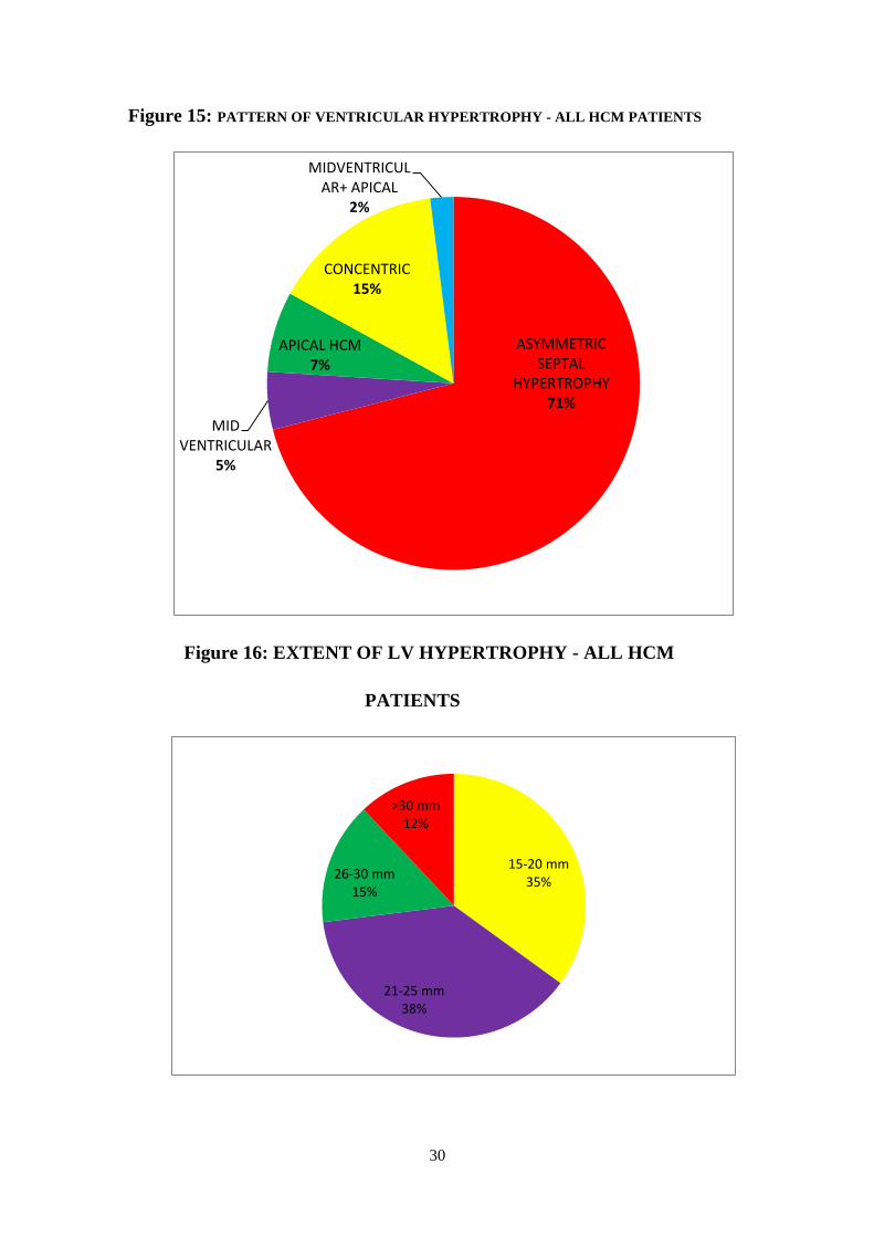
Mean LVEF was 70.1 ± 11%. Asymmetric septal hypertrophy was the most
common pattern of LV involvement seen in 71% of patients. Maximum LV thickness in
end-diastole is 22.88 ± 6.17 mm. Mean indexed myocardial mass is 99.5 ± 31.11 g/mm2.
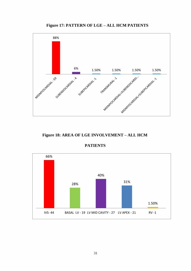
LGE of the myocardium is seen in 67% of patients. Mid-myocardial LGE is seen in 88%
and sub-endocardial LGE in 6% of patients. LGE of the inter-ventricular septum is the most
common involved segment in 66% of patients.
32%
4%
62%
LVOTO-32 MID CAVITY-4 NIL-62
30
Figure 15: PATTERN OF VENTRICULAR HYPERTROPHY - ALL HCM PATIENTS
Figure 16: EXTENT OF LV HYPERTROPHY - ALL HCM
PATIENTS
ASYMMETRIC SEPTAL
HYPERTROPHY 71%
MID VENTRICULAR
5%
APICAL HCM 7%
CONCENTRIC 15%
MIDVENTRICULAR+ APICAL
2%
15-20 mm 35%
21-25 mm 38%
26-30 mm 15%
>30 mm 12%
31
Figure 17: PATTERN OF LGE – ALL HCM PATIENTS
Figure 18: AREA OF LGE INVOLVEMENT – ALL HCM
PATIENTS
88%
6% 1.50% 1.50% 1.50% 1.50%
66%
28%
40%
31%
1.50%
IVS- 44 BASAL LV - 19 LV MID CAVITY - 27 LV APEX - 21 RV -1
32
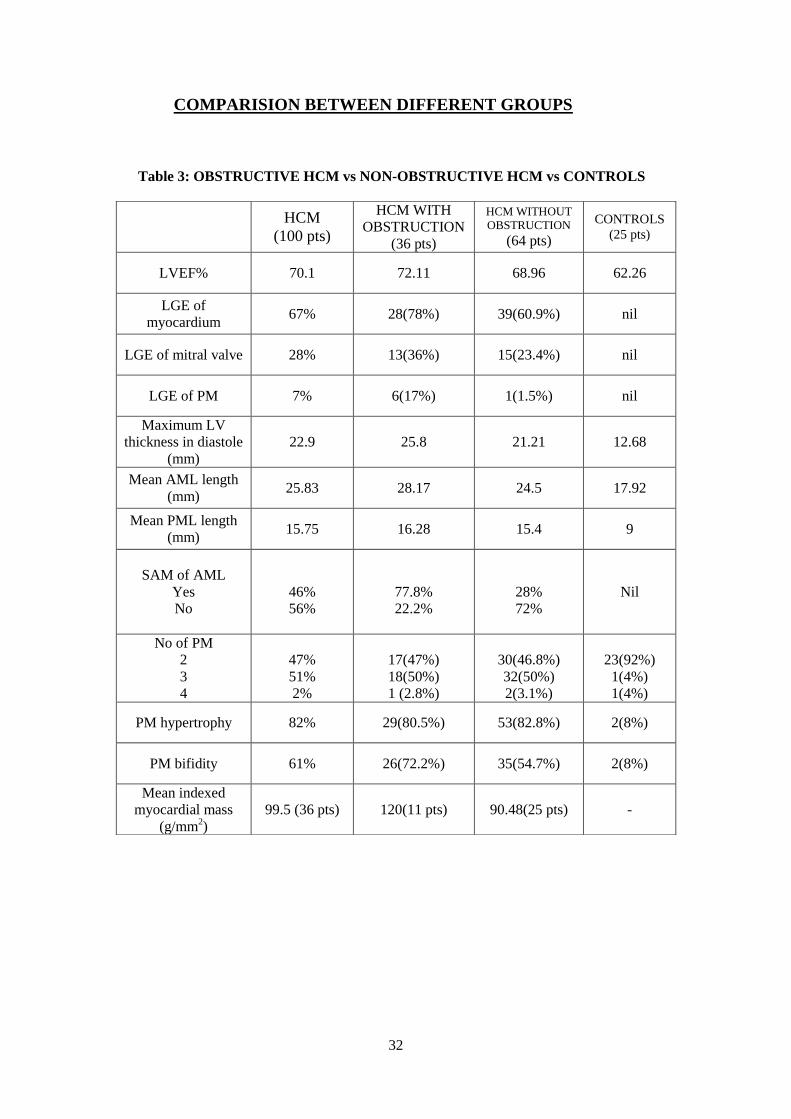
COMPARISION BETWEEN DIFFERENT GROUPS
Table 3: OBSTRUCTIVE HCM vs NON-OBSTRUCTIVE HCM vs CONTROLS
HCM
(100 pts)
HCM WITH
OBSTRUCTION
(36 pts)
HCM WITHOUT
OBSTRUCTION
(64 pts)
CONTROLS
(25 pts)
LVEF% 70.1 72.11 68.96 62.26
LGE of
myocardium 67% 28(78%) 39(60.9%) nil
LGE of mitral valve 28% 13(36%) 15(23.4%) nil
LGE of PM 7% 6(17%) 1(1.5%) nil
Maximum LV
thickness in diastole
(mm)
22.9 25.8 21.21 12.68
Mean AML length
(mm) 25.83 28.17 24.5 17.92
Mean PML length
(mm) 15.75 16.28 15.4 9
SAM of AML
Yes
No
46%
56%
77.8%
22.2%
28%
72%
Nil
No of PM
2
3
4
47%
51%
2%
17(47%)
18(50%)
1 (2.8%)
30(46.8%)
32(50%)
2(3.1%)
23(92%)
1(4%)
1(4%)
PM hypertrophy 82% 29(80.5%) 53(82.8%) 2(8%)
PM bifidity 61% 26(72.2%) 35(54.7%) 2(8%)
Mean indexed
myocardial mass
(g/mm2)
99.5 (36 pts) 120(11 pts) 90.48(25 pts) -
33
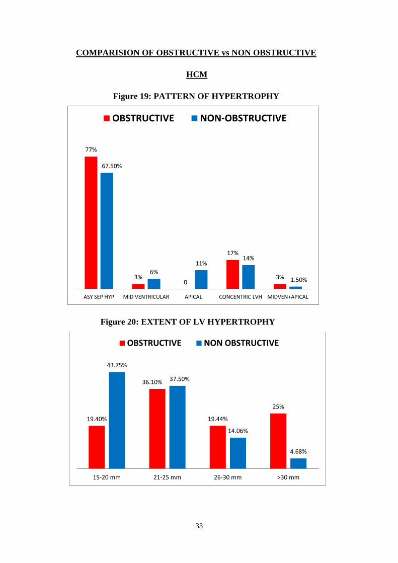
COMPARISION OF OBSTRUCTIVE vs NON OBSTRUCTIVE
HCM
Figure 19: PATTERN OF HYPERTROPHY
Figure 20: EXTENT OF LV HYPERTROPHY
77%
3% 0
17%
3%
67.50%
6%
11% 14%
1.50%
ASY SEP HYP MID VENTRICULAR APICAL CONCENTRIC LVH MIDVEN+APICAL
OBSTRUCTIVE NON-OBSTRUCTIVE
19.40%
36.10%
19.44%
25%
43.75%
37.50%
14.06%
4.68%
15-20 mm 21-25 mm 26-30 mm >30 mm
OBSTRUCTIVE NON OBSTRUCTIVE
34
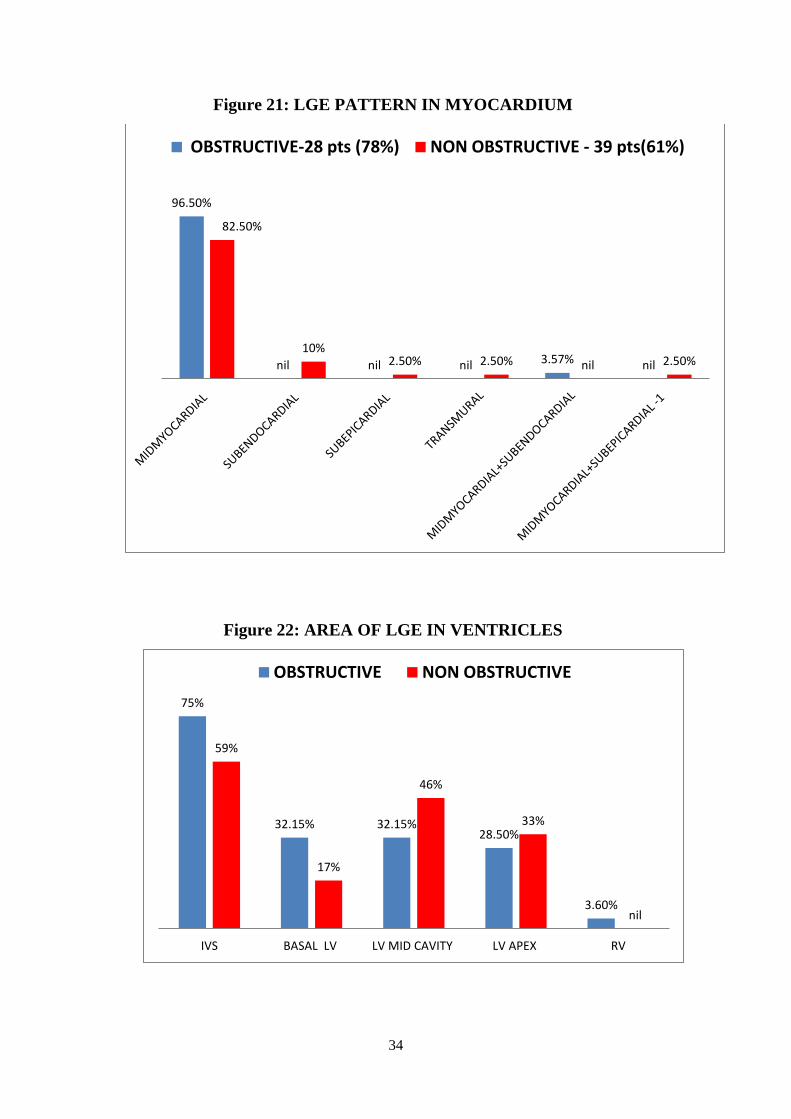
Figure 21: LGE PATTERN IN MYOCARDIUM
Figure 22: AREA OF LGE IN VENTRICLES
96.50%
nil nil nil 3.57% nil
82.50%
10% 2.50% 2.50% nil 2.50%
OBSTRUCTIVE-28 pts (78%) NON OBSTRUCTIVE - 39 pts(61%)
75%
32.15% 32.15% 28.50%
3.60%
59%
17%
46%
33%
nil
IVS BASAL LV LV MID CAVITY LV APEX RV
OBSTRUCTIVE NON OBSTRUCTIVE
35
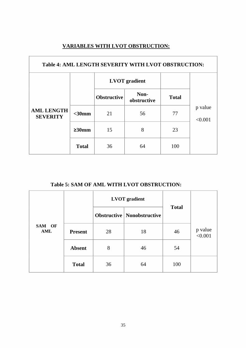
VARIABLES WITH LVOT OBSTRUCTION:
Table 4: AML LENGTH SEVERITY WITH LVOT OBSTRUCTION:
AML LENGTH
SEVERITY
LVOT gradient
p value
<0.001
Obstructive Non-
obstructive Total
<30mm 21 56 77
≥30mm 15 8 23
Total 36 64 100
Table 5: SAM OF AML WITH LVOT OBSTRUCTION:
SAM OF
AML
LVOT gradient
Total
p value
<0.001
Obstructive Nonobstructive
Present 28 18 46
Absent 8 46 54
Total 36 64 100
36
Table 6: PM HYPERTROPHY WITH LVOT OBSTRUCTION:
PM
HYPERTROPHY
LVOT gradient
Total
pvalue
1.0
Obstructive Nonobstructive
Present 29 53 82
Absent 7 11 18
Total 36 64 100
Table 7: PM BIFIDITY WITH LVOT OBSTRUCTION:
PM BIFIDITY
LVOT gradient
Total
p value
0.09
Obstructive Non-
obstructive
Present 26 35 61
Absent 10 29 39
Total 36 64 100
37
Table 8: EXTENT OF LV HYPERTROPHY WITH LVOT
OBSTRUCTION:
EXTENT OF LV
HYPERTRPHY
(mm)
LVOT gradient
Total
p value <
0.03
Obstructive Non-
obstructive
15-20 7 28 35
21-25 13 24 37
26-30 7 9 16
31-35 9 3 12
Total
36
64
100
Based on the LV thickness in end-diastole, patients were categorized into 4 groups
such as 15-20 mm, 21-25 mm, 26-30 mm, >30 mm. Among all HCM patients, LV thickness
of 15-20 mm is seen in 35%, 21-25 mm in 38%, 26-30 mm in 15% and >30 mm in 12%.
LGE of the mitral valve and papillary muscles was seen in 28% and 7% respectively. Mean
AML length in HCM patients was 25.83 ± 4.8 mm and the mean PML length was 15.75 ±
3.06 mm. Systolic anterior motion of AML is seen in 46% of patients. Papillary muscle
hypertrophy is seen in 82% of patients. Bifid papillary muscle is seen in 61% of patients.
38
COMPARISION OF HCM vs CONTROLS
Table 9: BASELINE PARAMETERS
HCM CONTROLS p value
LVEF (%) 70.1 62.26 0.38
LGE OF MYOCARDIUM 67% nil -
LGE OF MITRAL
VALVE 28% nil -
LGE OF PM 7% nil -
MEAN AML LENGTH
(mm) 25.83 17.92 <0.001
MEAN PML LENGTH
(mm) 15.75 9 <0.001
MAXXIMUM LV
THICKNESS IN
DIASTOLE (mm) 22.9 12.62 0.001
Table 10: SAM OF AML
SAM OF AML HCM CONTROLS
p value
< 0.001
PRESENT 46 1
ABSENT 56 24
TOTAL 100 25
39
Table 11: NUMBER OF PAPILLARY MUSCLES
No of PM HCM CONTROLS
p value
< 0.001
TWO 47 23
THREE 51 1
FOUR 2 1
TOTAL 100 25
Table 12: PM HYPERTROPHY
PM HYPERTROPHY HCM CONTROLS
p value
< 0.001
PRESENT 82 2
ABSENT 18 23
TOTAL 100 25
Table 13: PM BIFIDITY
PM BIFIDITY HCM CONTROLS
p value
< 0.001
PRESENT 61 2
ABSENT 39 23
TOTAL 100 25

40
Discussion
41
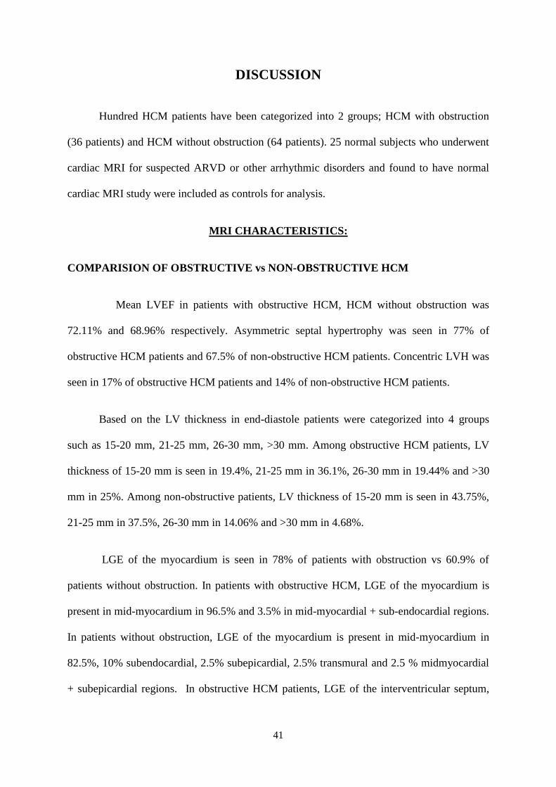
DISCUSSION
Hundred HCM patients have been categorized into 2 groups; HCM with obstruction
(36 patients) and HCM without obstruction (64 patients). 25 normal subjects who underwent
cardiac MRI for suspected ARVD or other arrhythmic disorders and found to have normal
cardiac MRI study were included as controls for analysis.
MRI CHARACTERISTICS:
COMPARISION OF OBSTRUCTIVE vs NON-OBSTRUCTIVE HCM
Mean LVEF in patients with obstructive HCM, HCM without obstruction was
72.11% and 68.96% respectively. Asymmetric septal hypertrophy was seen in 77% of
obstructive HCM patients and 67.5% of non-obstructive HCM patients. Concentric LVH was
seen in 17% of obstructive HCM patients and 14% of non-obstructive HCM patients.
Based on the LV thickness in end-diastole patients were categorized into 4 groups
such as 15-20 mm, 21-25 mm, 26-30 mm, >30 mm. Among obstructive HCM patients, LV
thickness of 15-20 mm is seen in 19.4%, 21-25 mm in 36.1%, 26-30 mm in 19.44% and >30
mm in 25%. Among non-obstructive patients, LV thickness of 15-20 mm is seen in 43.75%,
21-25 mm in 37.5%, 26-30 mm in 14.06% and >30 mm in 4.68%.
LGE of the myocardium is seen in 78% of patients with obstruction vs 60.9% of
patients without obstruction. In patients with obstructive HCM, LGE of the myocardium is
present in mid-myocardium in 96.5% and 3.5% in mid-myocardial + sub-endocardial regions.
In patients without obstruction, LGE of the myocardium is present in mid-myocardium in
82.5%, 10% subendocardial, 2.5% subepicardial, 2.5% transmural and 2.5 % midmyocardial
+ subepicardial regions. In obstructive HCM patients, LGE of the interventricular septum,
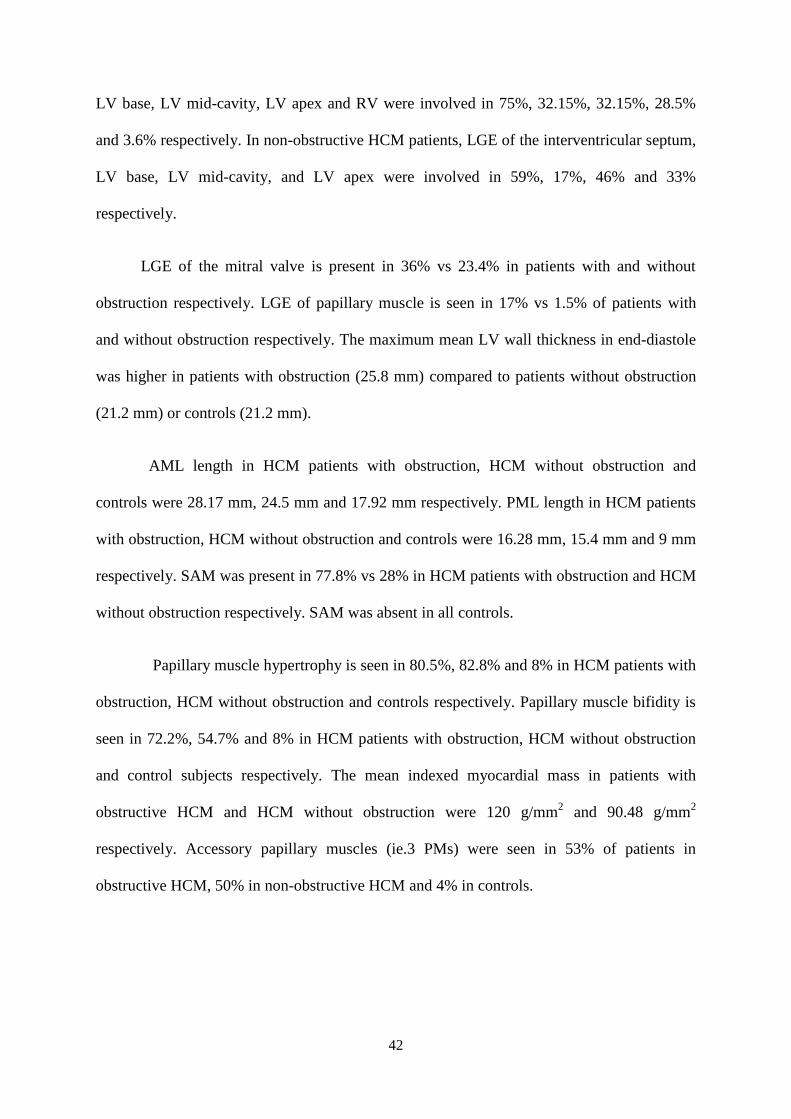
42
LV base, LV mid-cavity, LV apex and RV were involved in 75%, 32.15%, 32.15%, 28.5%
and 3.6% respectively. In non-obstructive HCM patients, LGE of the interventricular septum,
LV base, LV mid-cavity, and LV apex were involved in 59%, 17%, 46% and 33%
respectively.
LGE of the mitral valve is present in 36% vs 23.4% in patients with and without
obstruction respectively. LGE of papillary muscle is seen in 17% vs 1.5% of patients with
and without obstruction respectively. The maximum mean LV wall thickness in end-diastole
was higher in patients with obstruction (25.8 mm) compared to patients without obstruction
(21.2 mm) or controls (21.2 mm).
AML length in HCM patients with obstruction, HCM without obstruction and
controls were 28.17 mm, 24.5 mm and 17.92 mm respectively. PML length in HCM patients
with obstruction, HCM without obstruction and controls were 16.28 mm, 15.4 mm and 9 mm
respectively. SAM was present in 77.8% vs 28% in HCM patients with obstruction and HCM
without obstruction respectively. SAM was absent in all controls.
Papillary muscle hypertrophy is seen in 80.5%, 82.8% and 8% in HCM patients with
obstruction, HCM without obstruction and controls respectively. Papillary muscle bifidity is
seen in 72.2%, 54.7% and 8% in HCM patients with obstruction, HCM without obstruction
and control subjects respectively. The mean indexed myocardial mass in patients with
obstructive HCM and HCM without obstruction were 120 g/mm2 and 90.48 g/mm
2
respectively. Accessory papillary muscles (ie.3 PMs) were seen in 53% of patients in
obstructive HCM, 50% in non-obstructive HCM and 4% in controls.
43
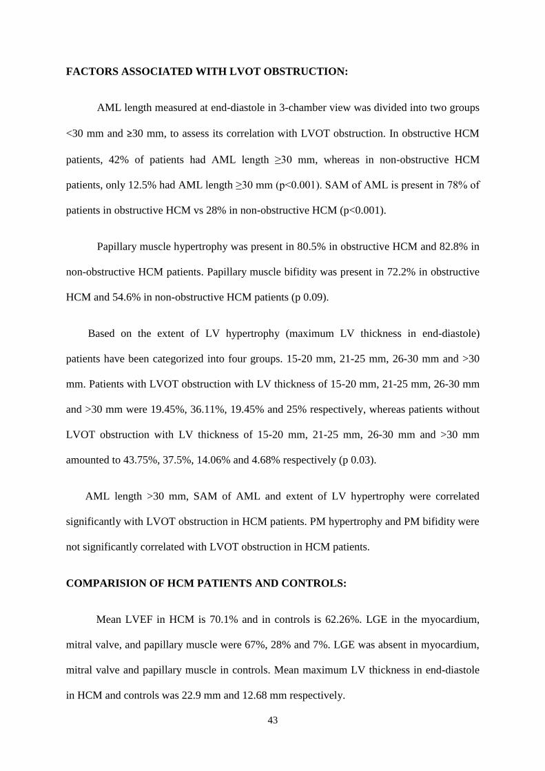
FACTORS ASSOCIATED WITH LVOT OBSTRUCTION:
AML length measured at end-diastole in 3-chamber view was divided into two groups
<30 mm and ≥30 mm, to assess its correlation with LVOT obstruction. In obstructive HCM
patients, 42% of patients had AML length ≥30 mm, whereas in non-obstructive HCM
patients, only 12.5% had AML length ≥30 mm (p<0.001). SAM of AML is present in 78% of
patients in obstructive HCM vs 28% in non-obstructive HCM (p<0.001).
Papillary muscle hypertrophy was present in 80.5% in obstructive HCM and 82.8% in
non-obstructive HCM patients. Papillary muscle bifidity was present in 72.2% in obstructive
HCM and 54.6% in non-obstructive HCM patients (p 0.09).
Based on the extent of LV hypertrophy (maximum LV thickness in end-diastole)
patients have been categorized into four groups. 15-20 mm, 21-25 mm, 26-30 mm and >30
mm. Patients with LVOT obstruction with LV thickness of 15-20 mm, 21-25 mm, 26-30 mm
and >30 mm were 19.45%, 36.11%, 19.45% and 25% respectively, whereas patients without
LVOT obstruction with LV thickness of 15-20 mm, 21-25 mm, 26-30 mm and >30 mm
amounted to 43.75%, 37.5%, 14.06% and 4.68% respectively (p 0.03).
AML length >30 mm, SAM of AML and extent of LV hypertrophy were correlated
significantly with LVOT obstruction in HCM patients. PM hypertrophy and PM bifidity were
not significantly correlated with LVOT obstruction in HCM patients.
COMPARISION OF HCM PATIENTS AND CONTROLS:
Mean LVEF in HCM is 70.1% and in controls is 62.26%. LGE in the myocardium,
mitral valve, and papillary muscle were 67%, 28% and 7%. LGE was absent in myocardium,
mitral valve and papillary muscle in controls. Mean maximum LV thickness in end-diastole
in HCM and controls was 22.9 mm and 12.68 mm respectively.
44
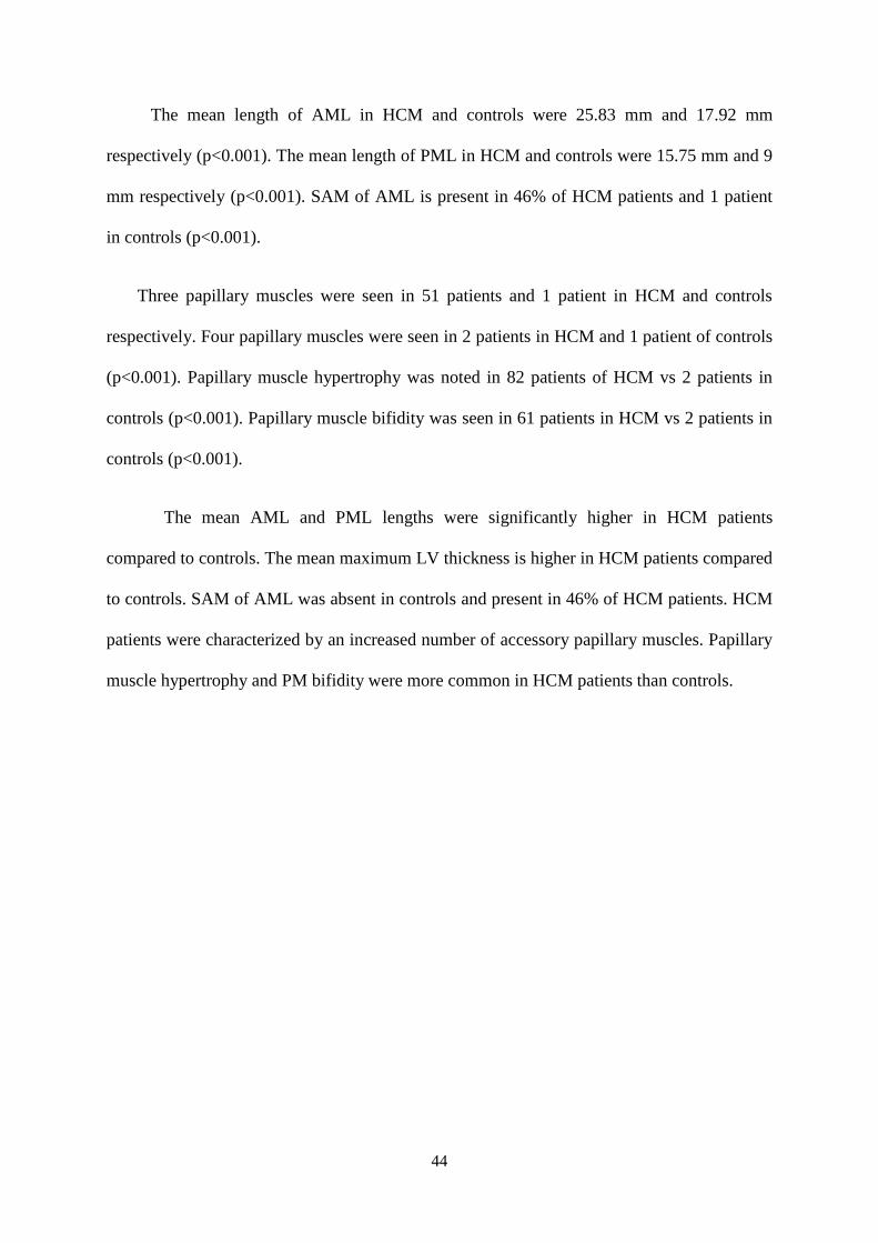
The mean length of AML in HCM and controls were 25.83 mm and 17.92 mm
respectively (p<0.001). The mean length of PML in HCM and controls were 15.75 mm and 9
mm respectively (p<0.001). SAM of AML is present in 46% of HCM patients and 1 patient
in controls (p<0.001).
Three papillary muscles were seen in 51 patients and 1 patient in HCM and controls
respectively. Four papillary muscles were seen in 2 patients in HCM and 1 patient of controls
(p<0.001). Papillary muscle hypertrophy was noted in 82 patients of HCM vs 2 patients in
controls (p<0.001). Papillary muscle bifidity was seen in 61 patients in HCM vs 2 patients in
controls (p<0.001).
The mean AML and PML lengths were significantly higher in HCM patients
compared to controls. The mean maximum LV thickness is higher in HCM patients compared
to controls. SAM of AML was absent in controls and present in 46% of HCM patients. HCM
patients were characterized by an increased number of accessory papillary muscles. Papillary
muscle hypertrophy and PM bifidity were more common in HCM patients than controls.
45
Limitations
46
LIMITATIONS
It was a single-center study conducted in a tertiary care center and there is a chance of
selection bias. Due to the dynamic nature of the LVOT gradient, it could be affected by a lot
of factors, including heart rate, arrhythmia, body position and volume status. LVOT gradient
at a particular point of time might not be a true representation of the dynamic LVOT gradient.
Since there are no proper standardized methods of assessment of PM morphology or
dimensions by MRI and it could be subjected to various errors. SAM of AML could be
influenced by various other factors in addition to the mitral valve, papillary muscle, and LV
morphology. PM displacement and PM mass of the HCM patients were not included in the
study.
.

47
Conclusions
48
CONCLUSIONS
The mean AML and PML lengths were significantly higher in HCM patients
compared to controls. SAM of AML was absent in controls and present in 46% of HCM
patients. HCM patients were characterized by an increased number of accessory papillary
muscles. Papillary muscle hypertrophy and PM bifidity were more common in HCM patients
than controls.
AML length >30 mm, SAM of AML and extent of LV hypertrophy were correlated
significantly with resting LVOT obstruction in HCM patients. PM hypertrophy and PM
bifidity were not significantly correlated with resting LVOT obstruction in HCM patients.

49
Bibliography
50
REFERENCES
1. Delling FN, Sanborn DY, Levine RA, et al. Frequency and mechanism of persistent
systolic anterior motion and mitral regurgitation after septal ablation in obstructive
hypertrophic cardiomyopathy. Am J Cardiol 2007;100:1691–5.
2. Gersh BJ, Maron BJ, Bonow RO, et al. 2011 ACCF/AHA guideline for the diagnosis
and treatment of hypertrophic cardiomyopathy: executive summary: a report of the
American College of Cardiology Foundation/American Heart Association Task Force
on Practice Guidelines. J Am Coll Cardiol 2011;58:2703–38.
3. Grigg LE, Wigle ED, Williams WG, et al. Transesophageal Doppler
echocardiography in obstructive hypertrophic cardiomyopathy: clarification of
pathophysiology and importance in intraoperative decision making. J Am Coll
Cardiol 1992;20:42–52.
4. Ro R, Halpern D, Sahn DJ, et al. Vector flow mapping in obstructive hypertrophic
cardiomyopathy to assess the relationship of early systolic left ventricular flow and
the mitral valve. J Am Coll Cardiol 2014; 64:1984–95.
5. Maron MS, Olivotto I, Harrigan C, et al. Mitral valve abnormalities identified by
cardiovascular magnetic resonance represent a primary phenotypic expression of
hypertrophic cardiomyopathy.Circulation 2011;124:40–7.
6. Levine RA, Vlahakes GJ, Lefebvre X, et al. Papillary muscle displacement causes
systolic anterior motion of the mitral valve. Experimental validation and insights into
the mechanism of subaortic obstruction. Circulation 1995;91:1189–95.
7. Halpern DG, Swistel DG, Po JR, et al. Echocardiography before and after resect-
plicaterelease surgical myectomy for obstructive hypertrophic cardiomyopathy. J Am
Soc Echocardiogr 2015;28:1318–28.
8. Kwon DH, Setser RM, Thamilarasan M, et al. Abnormal papillary muscle
morphology is independently associated with increased left ventricular outflow tract
obstruction in hypertrophic cardiomyopathy. Heart 2008;94:1295–301.
51
9. Messmer BJ. Extended myectomy for hypertrophic obstructive cardiomyopathy. Ann
Thorac Surg 1994;58:575–7.
10. Minakata K, Dearani JA, Nishimura RA, et al. Extended septal myectomy for
hypertrophic obstructive cardiomyopathy with anomalous mitral papillary muscles or
chordae. J Thorac Cardiovasc Surg 2004;127:481–9.
11. Hagège AA, Bruneval P, Levine RA, et al. The mitral valve in hypertrophic
cardiomyopathy: old versus new concepts. J Cardiovasc Transl Res 2011;4:757–66.
12. Roberts WC. Operative treatment of hypertrophic obstructive cardiomyopathy. The
case against mitral valve replacement. Am J Cardiol 1973;32:377–81.
13. Dearani JA, Ommen SR, Gersh BJ, et al. Surgery insight: Septal myectomy for
obstructive hypertrophic cardiomyopathy—the Mayo Clinic experience. Nat Clin
Pract Cardiovasc Med 2007;4: 503–12.
14. Ibrahim M, Rao C, Ashrafian H, Chaudhry U, Darzi A, Athanasiou T. Modern
management of systolic anterior motion of the mitral valve. Eur J Cardiothorac Surg.
2012;41:1260–1270. doi:10.1093/ejcts/ezr232
15. Yacoub MH. Surgical versus alcohol septal ablation for hypertrophic obstructive
cardiomyopathy: the pendulum swings. Circulation. 2005; 112:450–452.
16. Balaram SK, Sherrid MV, Derose JJ Jr, Hillel Z, Winson G, Swistel DG. Beyond
extended myectomy for hypertrophic cardiomyopathy: the resection-plication-release
(RPR) repair. Ann Thorac Surg. 2005;80: 217–223.
17. Casolo G OI, Manta R, Rega L, Nistri S, Petrone P, Gensini GF. Relationship of
echocardiographic maximum left ventricular wall thickness to cardiac mass assessed
by magnetic resonance in hypertrophic cardiomyopathy. Eur Heart J 2005;26:387.
18. Sipola P, Lauerma J, Jääskeläinen P, et al. Cine MR imaging of myocardial
contractile impairment in patients with hypertrophic cardiomyopathy attributable to
Asp175Asn mutation in the alpha-tropomyosin gene. Radiology 2005; 236:815–824
52
19. Elliott P, McKenna WJ. Hypertrophic cardiomyopathy. Lancet 2004; 363:1881–1891
20. Wigle ED. Cardiomyopathy: the diagnosis of hypertrophic cardiomyopathy. Heart
2001; 86:709–714
21. Hughes SE. The pathology of hypertrophic cardiomyopathy. Histopathology 2004;
44:412–427
22. Moon JC, McKenna WJ, McCrohon JA, Elliott PM, Smith GC, Pennell DJ. Toward
clinical risk assessment in hypertrophic cardiomyopathy with gadolinium
cardiovascular magnetic resonance. J Am Coll Cardiol 2003; 41:1561–1567
23. Maron MS, Appelbaum E, Harrigan CJ, Buros J, Gibson CM, Hanna C, Lesser JR,
Udelson JE, Manning WJ, Maron BJ: Clinical profile and significance of delayed
enhancement in hypertrophic cardiomyopathy. Circ Heart Fail 2008, 1:1
53
Appendix
54
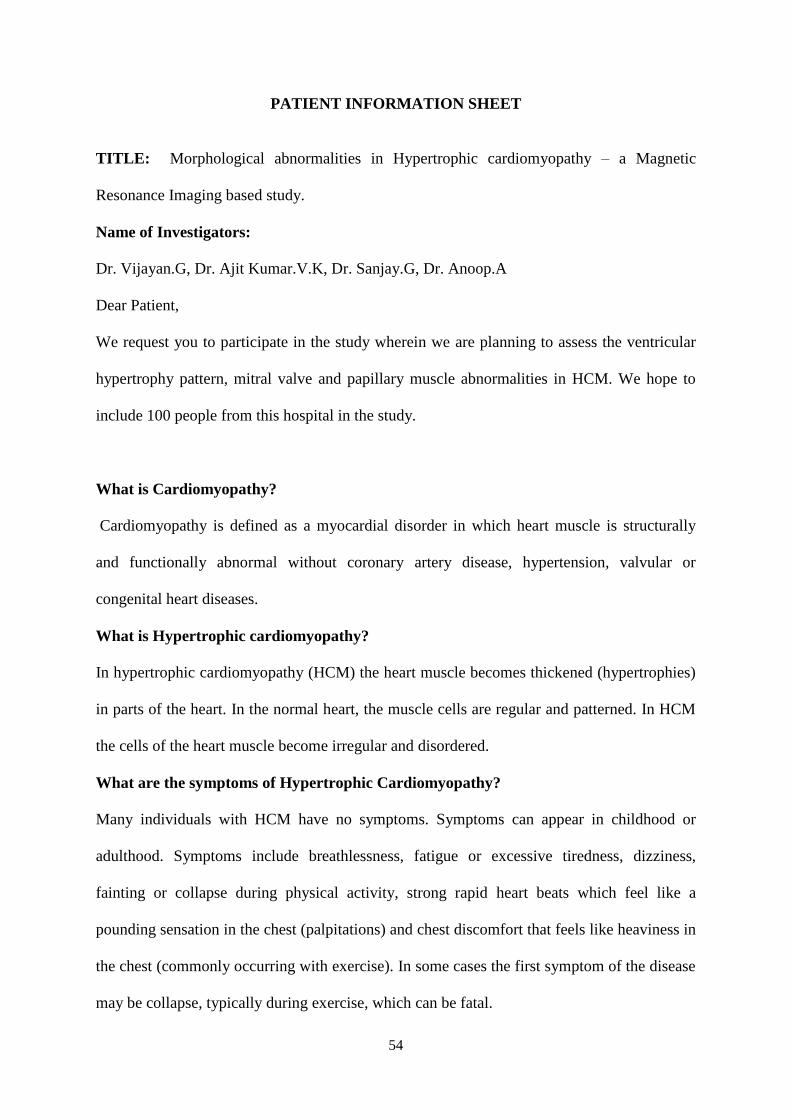
PATIENT INFORMATION SHEET
TITLE: Morphological abnormalities in Hypertrophic cardiomyopathy – a Magnetic
Resonance Imaging based study.
Name of Investigators:
Dr. Vijayan.G, Dr. Ajit Kumar.V.K, Dr. Sanjay.G, Dr. Anoop.A
Dear Patient,
We request you to participate in the study wherein we are planning to assess the ventricular
hypertrophy pattern, mitral valve and papillary muscle abnormalities in HCM. We hope to
include 100 people from this hospital in the study.
What is Cardiomyopathy?
Cardiomyopathy is defined as a myocardial disorder in which heart muscle is structurally
and functionally abnormal without coronary artery disease, hypertension, valvular or
congenital heart diseases.
What is Hypertrophic cardiomyopathy?
In hypertrophic cardiomyopathy (HCM) the heart muscle becomes thickened (hypertrophies)
in parts of the heart. In the normal heart, the muscle cells are regular and patterned. In HCM
the cells of the heart muscle become irregular and disordered.
What are the symptoms of Hypertrophic Cardiomyopathy?
Many individuals with HCM have no symptoms. Symptoms can appear in childhood or
adulthood. Symptoms include breathlessness, fatigue or excessive tiredness, dizziness,
fainting or collapse during physical activity, strong rapid heart beats which feel like a
pounding sensation in the chest (palpitations) and chest discomfort that feels like heaviness in
the chest (commonly occurring with exercise). In some cases the first symptom of the disease
may be collapse, typically during exercise, which can be fatal.
55
How is Hypertrophic Cardiomyopathy diagnosed?
HCM is usually diagnosed with echocardiography. This is an ultrasound scan of the heart.
Measurements of the thickness of the heart wall can be made. ECGs (Electrocardiograms) are
also used. In some cases cardiac MRI (Magnetic Resonance Imaging) is also now be utilized
especially where it is difficult to view the heart well with echocardiography.
What is a cardiac MRI?
A cardiac MRI scan is a non-invasive test where magnetic and radio waves are used to create
pictures of the inside of the heart. The key difference between echocardiograms and MRI are
the ability to better visualize the edges of the heart walls and the advantage of being able to
see in the walls of the heart to pick up areas of scar or fibrosis.
Why do I need this procedure?
People with this condition usually do not have symptoms. However, in some people with this
condition, there is a risk of developing complications such as failure of the heart to pump
blood and sudden death. Currently doctors do not know much about why some people
develop these complications. The purpose of this study is to find ways of predicting the risk
of developing these problems, so that appropriate treatment can be given. This study will
carry out a careful and thorough assessment of people with HCM using MRI to identify
markers that are associated with these complications. This information will help doctors to
identify people with HCM who are at higher risks of developing complications in the future
as a result of the disease.
How long does it take?
The procedure will take about 30-45 minutes.
Is MRI safe?
An MRI study utilizes radio waves to acquire pictures and therefore NO ionizing radiation is
required (as opposed to a CT scan, cardiac catherization or X-ray which does require the use
56
of ionizing radiation). As a result, MRI is a very safe test and no long term ill effects have
been reported. Claustrophobia may be problematic in about 2% of patients but often a mild
anxiolytic (prescribed by your doctor) prior to the test can prevent this from occurring.
What are the side-effects of Gadolinium contrast used in MRI?
Itchy skin rash may appear in approximately 1 in 1000 patients. Nephrogenic systemic
fibrosis (NSF) is a rare condition resulting in skin contractures (or localised skin thickening
and tightening) and internal organ damage. It has occurred with some gadolinium-based
contrast media in a minority of patients with pre-existing severe kidney function
abnormalities.
Do I need to take any precautions?
Patients with pacemakers, implantable cardiac defibrillators (ICD) or retained pacemaker
leads cannot undergo the procedure if the device is not MRI compatible.
Permission to collect information from medical records
Details of the treatment including current medical condition and treatment undergone will be
obtained for research purposes from your medical records. We seek your permission to take
this information.
If you have any further questions, please ask: Dr. Vijayan.G (Principal investigator), Senior
Resident, Department of Cardiology (Email: [email protected] Mobile No: 8334890131)
For any technical clarifications, please contact - Dr. Mala Ramanathan, Member
Secretary(Study independent contact person), IEC, SCTIMST and Additional
Professor, AMCHSS, SCTIMST (Email: [email protected], Phone no. 0471-
2524234

57
INFORMED CONSENT FORM
TITLE OF THE STUDY: Morphological abnormalities in Hypertrophic
Cardiomyopathy - a Magnetic Resonance Imaging based study
Study number: 100.
Participant’s name: Date of Birth / Age (in years):
I _____________________________________________________________ ___________,
son/daughter of ___________________________________ (Please tick boxes).
[ ] Declare that I have read the above information provide to me regarding the study:
Morphological abnormalities in Hypertrophic cardiomyopathy- an Magnetic Resonance
Imaging based study and have clarified any doubts that I had.
[ ] I also understand that my participation in this study is entirely voluntary and that I am free
to withdraw permission to continue to participate at any time without affecting my usual
treatment or my legal rights.
[ ] I also understand that cardiac MRI is a part of the routine workup; I need not have to pay
for using this information for this study.
[ ] I understand that the study staff and institutional ethics committee members may be not
need my permission to look at my health records even if I withdraw from the trial. I agree to
this access.
[ ] I understand that my identity may be not be revealed in any information released to third
parties or published.
[ ] I voluntarily agree to take part in this study
.[ ] I received a copy of this signed consent form.
Name: Name of the witness:
Signature: Relationship to participant:
58
Date: Date:
I attest that the requirements for informed consent for the medical research project described
in this form have been satisfied. I have discussed the research project with the participant and
explained to him or her in nontechnical terms all of the information contained in this
informed consent form, including any risks and adverse reactions that may reasonably be
expected to occur. I further certify that I encouraged the participant to ask questions and that
all questions asked were answered.
________________________________ ___________________ Name and Signature of
Person Obtaining Consent
Dr. Vijayan.G
Senior resident
Mobile no: 8334890131
Dept. of cardiology SCTIMST
22-1-2018
Contact details:
Dr.Vijayan.G Senior resident, Department of cardiology,
Room no 321,
Shristy hostel, SCTIMSTquarters,
Poonthi road, Kumarapuram,
Trivandrum, Kerala.

59
For any technical clarifications, please contact Dr. Mala Ramanathan, Member
Secretary, IEC, SCTIMST and Additional Professor, AMCHSS, SCTIMST (Email:
[email protected], Phone no. 0471-2524234)
Expenses:
60
61
62
63
64
Plagiarism Checker X Originality Report
Similarity Found: 8%
Date: Sunday, July 30, 2019
Statistics: 206 words Plagiarized / 2571 Total words
Remarks: Low Plagiarism Detected - Your Document needs Selective Improvement.
----------------------------------------------------------------------------------------
Introduction
Hypertrophic cardiomyopathy (HCM) has a varied clinical course due to its genotypic
and phenotypic heterogeneity. Several autopsy studies have shown abnormalities of the mitral
valve in some HCM patients. Cardiovascular magnetic resonance (CMR) has become the
imaging modality of choice due to its high spatial resolution, well suited to define the diverse
phenotypic expression of this complex disease.
HCM has been documented to have various mitral valve abnormalities like an
increased length of the leaflets and area, leaflet thickening, impaired mitral leaflet coaptation,
and left ventricular outflow tract obstruction (LVOT) due to the systolic anterior motion of
the mitral leaflets. Cardiovascular magnetic resonance (CMR) provides an excellent
opportunity to assess the papillary muscle (PM) abnormalities like an increased number and
mass, bifidity, hypertrophy, antero-apical displacement and LGE of the papillary muscle.
Various guidelines recommend surgical myectomy as the preferred modality for patients
with left ventricular outflow tract (LVOT) gradient ≥50 mm Hg who fail to respond to
medications or who experience side effects. Alcohol septal ablation (ASA) in patients with
mitral valve abnormalities results in persistent SAM, gradients, and mitral regurgitation
(MR).
65
MASTER CHART S
l.N
o
AG
E
GE
ND
ER
(MA
LE
-1,F
EM
AL
E-2
)
LA
TE
Gd
EN
HA
NC
EM
EN
T O
F
MY
OC
AR
DIU
M (
YE
S-1
, N
O-2
)
LA
TE
Gd
EN
HA
NC
EM
EN
T I
N
MIT
RA
L V
AL
VE
(Y
ES
-1,
NO
-2)
LA
TE
Gd
EN
HA
NC
EM
EN
T I
N
PA
PIL
LA
RY
MU
SC
LE
(Y
ES
-1,
NO
-
2)
MA
XIM
UM
WA
LL
TH
ICK
NE
SS
IN
MM
at
EN
D D
IAS
TO
LE
EX
TE
NT
OF
LV
HY
PE
RT
RO
PH
Y
(15
-20
=1
, 21
-25
=2
, 2
6-3
0=
3, >
30
=4
)
AM
L L
EN
GT
H I
N M
M
AM
L L
EN
GT
H S
EV
ER
ITY
(<
30
-1,
≥3
0-2
)
PM
L L
EN
GT
H I
N M
M
SA
M(A
ML
-1,
PM
L-2
, B
OT
H-3
,
AB
SE
NT
-4)
NU
MB
ER
OF
PA
PIL
LA
RY
MU
SC
LE
PA
PIL
LA
RY
MU
SC
LE
HY
PE
RT
RO
PH
Y Y
ES
-1,N
O-2
BIF
IDIT
Y O
F P
AP
ILL
AR
Y
MU
SC
LE
(YE
S-1
, N
O-2
)
Ind
exed
my
oca
rdia
l m
ass
in
g
1 38 1 1 1 2 24 2 21 1 21 4 4 2 1 125
2 55 1 1 2 2 16 1 30 2 20 4 3 1 1 69
3 41 2 1 2 2 22 2 26 1 12 4 3 1 1 65
4 62 2 1 2 2 27 3 18 1 10 4 2 2 2 127
5 19 1 1 2 2 21 2 17 1 11 4 3 1 2 85
6 23 1 2 2 2 23 2 33 2 14 1 3 1 2 55
7 54 2 1 2 2 18 1 28 1 21 4 3 2 1 72
8 61 1 2 2 2 16 1 27 1 19 4 3 1 1 78
9 31 1 1 2 2 29 3 27 1 21 1 2 1 2 132
10 52 1 1 2 2 17 1 30 2 15 4 3 1 1 94
11 31 1 1 2 2 35 4 26 1 22 1 2 1 1 118
12 19 2 1 1 1 32 4 30 2 20 1 2 1 1 147
13 45 1 1 2 2 24 2 24 1 16 4 2 2 2 90
14 39 1 1 1 2 22 2 21 1 14 4 2 1 1 125
15 58 2 1 2 2 22 2 26 1 12 4 3 1 2 78
16 33 1 1 2 2 27 3 17 1 26 4 3 1 1 108
17 47 1 1 2 2 20 1 23 1 16 4 3 2 2 132
18 20 1 1 2 2 25 2 27 1 16 1 2 1 1 81
19 58 1 1 2 2 18 1 13 1 17 1 3 1 1 78
20 59 1 2 2 2 17 1 29 1 17 4 3 1 1 79
21 26 1 1 2 2 25 2 27 1 16 4 3 2 2 106
22 42 2 2 2 2 25 2 21 1 18 4 3 1 1 84
23 70 2 1 2 2 27 3 18 1 15 4 3 1 1 77
24 32 1 1 2 2 37 4 22 1 15 4 2 2 2 296
25 52 2 1 2 2 15 1 28 1 18 4 3 1 2 67
26 50 1 2 2 2 26 3 25 1 18 4 2 1 1 118
27 45 1 1 2 2 25 2 31 2 16 1 3 1 1 144
28 17 1 1 2 2 29 3 21 1 14 1 2 1 1 96
29 57 2 1 1 2 23 2 22 1 16 4 3 2 1 85
30 56 1 1 1 2 23 2 30 2 17 1 3 1 1 150
31 37 1 2 2 1 25 2 28 1 17 1 3 1 2
32 45 1 2 2 1 17 1 33 2 17 1 3 1 1
33 61 1 1 1 1 26 3 30 2 14 1 3 1 1
34 42 1 1 1 1 35 4 29 1 14 1 2 2 2
35 35 1 1 1 2 29 3 27 1 14 1 2 1 1
36 42 1 1 1 2 22 2 21 1 16 1 3 1 1
37 41 2 1 2 2 20 1 33 2 15 1 2 2 2
38 57 2 1 1 2 30 3 28 1 16 1 2 1 1
39 38 1 1 1 2 26 3 29 1 16 1 2 1 2
66
Sl.
No
AG
E
GE
ND
ER
(MA
LE
-1,F
EM
AL
E-2
)
LA
TE
Gd
EN
HA
NC
EM
EN
T O
F
MY
OC
AR
DIU
M (
YE
S-1
, N
O-2
)
LA
TE
Gd
EN
HA
NC
EM
EN
T I
N
MIT
RA
L V
AL
VE
(Y
ES
-1,
NO
-2)
LA
TE
Gd
EN
HA
NC
EM
EN
T I
N
PA
PIL
LA
RY
MU
SC
LE
(Y
ES
-1,
NO
-
2)
MA
XIM
UM
WA
LL
TH
ICK
NE
SS
IN
MM
at
EN
D D
IAS
TO
LE
EX
TE
NT
OF
LV
HY
PE
RT
RO
PH
Y
(15
-20
=1
, 21
-25
=2
, 2
6-3
0=
3, >
30
=4
)
AM
L L
EN
GT
H I
N M
M
AM
L L
EN
GT
H S
EV
ER
ITY
(<
30
-1,
≥3
0-2
)
PM
L L
EN
GT
H I
N M
M
SA
M(A
ML
-1,
PM
L-2
, B
OT
H-3
,
AB
SE
NT
-4)
NU
MB
ER
OF
PA
PIL
LA
RY
MU
SC
LE
PA
PIL
LA
RY
MU
SC
LE
HY
PE
RT
RO
PH
Y Y
ES
-1,N
O-2
BIF
IDIT
Y O
F P
AP
ILL
AR
Y
MU
SC
LE
(YE
S-1
, N
O-2
)
Ind
exed
my
oca
rdia
l m
ass
in
g
40 50 1 1 1 2 21 2 27 1 18 1 3 1 1
41 61 1 2 2 2 17 1 28 1 10 4 2 1 1
42 71 1 1 2 2 22 2 27 1 13 4 2 2 2
43 59 1 1 2 2 17 1 24 1 14 1 2 2 2
44 45 1 1 2 2 19 1 18 1 12 4 3 1 2
45 31 2 2 2 2 19 1 25 1 10 4 2 1 2
46 59 2 2 2 2 17 1 26 1 12 1 3 1 1
47 35 1 2 2 2 26 3 27 1 19 1 2 1 2
48 27 2 2 2 2 18 1 21 1 8 4 2 1 2
49 58 2 2 2 2 16 1 19 1 17 4 4 2 1
50 50 1 1 1 2 24 2 21 1 17 1 3 1 1
51 52 1 1 2 2 22 2 21 1 12 4 2 1 1
52 47 1 1 1 2 17 1 22 1 12 4 2 1 1
53 69 1 1 2 2 25 2 20 1 13 4 2 1 1
54 67 1 1 1 1 23 2 31 2 16 4 3 1 1
55 50 2 1 2 2 17 1 25 1 18 4 2 2 2
56 68 1 1 1 2 19 1 30 2 13 1 2 1 1
57 41 1 2 2 2 20 1 22 1 11 4 2 1 1
58 28 1 1 2 2 22 2 34 2 16 1 2 1 2
59 55 2 2 2 2 22 2 23 1 16 1 2 1 2
60 55 2 2 2 2 18 1 20 1 15 4 3 1 2
61 61 2 1 2 2 23 2 25 1 16 1 3 1 1
62 34 2 2 1 2 17 1 25 1 10 1 3 1 2 54
63 63 2 2 1 2 16 1 25 1 10 1 3 1 2 56
64 48 1 1 2 2 24 2 26 1 16 1 3 1 1
65 52 1 1 2 2 21 2 28 1 17 4 3 1 1
66 58 2 1 2 2 25 2 20 1 15 4 2 2 2
67 76 1 2 1 2 25 2 20 1 15 1 2 1 2
68 55 2 2 2 2 18 1 24 1 12 1 2 1 1
69 28 1 1 1 2 35 4 38 2 13 4 2 1 1
70 67 1 1 2 2 25 2 29 1 13 4 3 1 2
71 66 1 1 2 2 20 1 31 2 15 4 2 1 1
72 52 1 2 2 2 15 1 30 2 13 4 3 2 1
73 27 2 2 2 2 21 2 23 1 15 1 2 1 2
74 46 2 1 2 2 27 3 30 2 16 1 3 1 1
75 64 1 1 2 2 25 2 35 2 22 1 2 1 1
76 51 1 1 1 2 35 4 25 1 18 1 2 1 1
77 36 1 1 1 2 34 4 38 2 16 1 2 1 1
78 25 1 1 1 2 32 4 32 2 18 1 2 1 1
79 63 2 2 2 2 21 2 33 2 17 1 3 1 1
67
Sl.
No
AG
E
GE
ND
ER
(MA
LE
-1,F
EM
AL
E-2
)
LA
TE
Gd
EN
HA
NC
EM
EN
T O
F
MY
OC
AR
DIU
M (
YE
S-1
, N
O-2
)
LA
TE
Gd
EN
HA
NC
EM
EN
T I
N
MIT
RA
L V
AL
VE
(Y
ES
-1,
NO
-2)
LA
TE
Gd
EN
HA
NC
EM
EN
T I
N
PA
PIL
LA
RY
MU
SC
LE
(Y
ES
-1,
NO
-
2)
MA
XIM
UM
WA
LL
TH
ICK
NE
SS
IN
MM
at
EN
D D
IAS
TO
LE
EX
TE
NT
OF
LV
HY
PE
RT
RO
PH
Y
(15
-20
=1
, 21
-25
=2
, 2
6-3
0=
3, >
30
=4
)
AM
L L
EN
GT
H I
N M
M
AM
L L
EN
GT
H S
EV
ER
ITY
(<
30
-1,
≥3
0-2
)
PM
L L
EN
GT
H I
N M
M
SA
M(A
ML
-1,
PM
L-2
, B
OT
H-3
,
AB
SE
NT
-4)
NU
MB
ER
OF
PA
PIL
LA
RY
MU
SC
LE
PA
PIL
LA
RY
MU
SC
LE
HY
PE
RT
RO
PH
Y Y
ES
-1,N
O-2
BIF
IDIT
Y O
F P
AP
ILL
AR
Y
MU
SC
LE
(YE
S-1
, N
O-2
)
Ind
exed
my
oca
rdia
l m
ass
in
g
80 66 2 2 1 2 18 1 20 1 17 1 3 1 1
81 34 2 2 2 2 22 2 24 1 16 4 2 1 1
82 63 1 1 1 2 17 1 21 1 18 1 3 1 1
83 53 1 2 2 2 18 1 37 2 17 1 3 1 2
84 43 1 1 2 2 31 4 27 1 18 1 3 1 1
85 25 1 1 2 2 23 2 24 1 17 4 2 1 2
86 40 1 1 2 2 23 2 25 1 17 4 3 1 1
87 19 2 2 2 2 37 4 21 1 14 4 3 1 2
88 19 2 2 2 2 29 3 24 1 18 4 2 1 1
89 44 2 1 2 2 22 2 28 1 16 4 2 1 2
90 70 2 2 2 2 17 1 21 1 16 4 3 1 2
91 62 1 1 2 2 40 4 31 2 15 4 3 2 1
92 40 2 2 2 2 29 3 22 1 16 4 3 1 1
93 38 1 1 2 2 33 4 26 1 17 1 3 1 1
94 63 2 2 1 2 15 1 25 1 15 1 3 1 2 54
95 52 1 1 2 2 18 1 27 1 12 4 2 2 2
96 53 2 1 2 2 15 1 28 1 20 4 2 1 1 65
97 55 2 1 1 2 18 1 31 2 15 4 3 1 1
98 47 1 1 2 2 25 2 27 1 19 1 2 1 2
99 50 1 2 2 2 26 3 25 1 20 4 2 1 2 118
100 37 1 2 1 1 21 2 25 1 21 4 3 1 1 74