Embed Size (px)
Citation preview
ACTA NEUROBIOL. EXP. 1971, 31: 203--212
SUBTHALAMUS IN DOG BRAIN
Antoni SMIAEOWSKI
Department of Comparative Neuroanatomy, Jagiellonian University, Cracov, Poland
The aim of this paper is to describe, on the basis of myelo-and cytoar- chitectonics, the division, topography and structure of the subthalamic nuclei in the dog. The first author who described the subthalamus was Forel (1877). He proposed this term for the area lying orally to the me- sencephalon including the subthalamic nucleus, zona incerta and fields HI and Hz. Some authors add to the subthalamus the entopeduncular nucleus (lying below the internal capsule), the tegmental field H of Forel or the red nucleus and substantia nigra. According to our observations, the subthalamus includes the subthalamic nucleus of Luys, the zona incerta, the nucleus of field of Forel, fasciculus thalamicus Foreli (HI) and fasciculus lenticularis (Hz). The subthalamus in the dog is a small region lying in a ventrolateral part of the diencephalon squeezed between the thalamus (dorsally), the hypothalamus (medially, ventromedially) and internal capsule (ventrolaterally). Caudally it borders on the tegmentum.
MATERIAL AND METHOD
The study was car.-ied out on a series of frontal sections of the dog brain stained by the Weigert-Wolters method (50 ,u sections), as well as series of frontal sections, stained alternately by the Kliiver-Barrera and the Schultze silver methods (20 p sections). The results were compared with sagittal and horizontal sections of the dog brain, stained by the Weigert-Wolters, Kliiver-Barrera and Schultze methods. A total of seven series were used for this study.
OBSERVATIONS
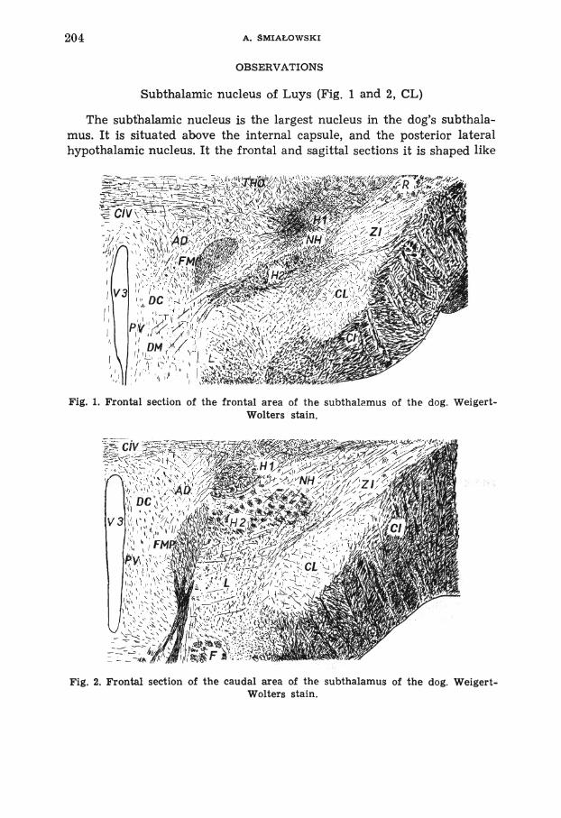
Subthalamic nucleus of Luys (Fig. 1 and 2, CL)
The subthalamic nucleus is the largest nucleus in the dog's subthala- mus. It is situated above the internal capsule, and the posterior lateral hypothalamic nucleus. It the frontal and sagittal sections it is shaped like
Fig. 1. Frontal section of the frontal area of the subthalamus of the dog. Weigert- Wolters stain.
Fig. 2. Frontal section of the cau,dal area of the subthalamus of the dog. Weigert- Wolters stain.

SUBTHALAMUS IN DOG BRAIN 205
a biconvex lens lying horizontally. The dimensions of this nucleus in the dog are 2.2 mm (orocaudally), 1.2 mm (dorsoventrally) and 2 mm (medio- laterally). Dorsally, the nucleus borders on the zona incerta, laterally, lateroventrally and oraly adjoins to internal capsule, and ventrally to the posterior lateral hypothalamic nucleus. Behind the nucleus (ventrocau- dally) we can easily found the substantia nigra well visible on sagittal sections.
Weigert-Wolters sections show a set of well myelinized delicate fibers running in various directions in the subthalamic nucleus in the dog's brain. Only few long bundles of fibers may be seen among more nume- rous single fibers. Nine systems of connections have been distinguished, but their directions of conductions are not established.
The first system of fibers well visible in the sagittal sections consist of bundles of fibers running orocaudally across this nucleus. The dia- meters of the bundles are about 30 p and they contain 7-10 thin nerve fibers each. These bundles leave the nucleus through the caudal boundary and while running dorsally they disperse in the tegmentum. The bundles situated on the ventral part of the nucleus concentrate together coming out as the subthalamotegmental tract (Singer 1962). This tract separates the subthalamic nucleus from the substantia nigra situated ventrocau- dally.
The second system is made up of the fibers which enter subthalamic nucleus from the lenticular ansa. They penetrate through the lateral and ventral border after passing across the internal capsule. This system consists of a few thick fibers 4 p in diameter. The fibers are thicker than in the first system and are stained stronger by the myelin method.
The third system is built of thin fibers 1-2 p in diameter taking origin in the internal capsule. These fibers run out from among the bundles of the internal capsule and after penetrating through the oral or lateral border along the whole subthalamic nucleus they assume an oromedial course. This system disperses mainly within the lateral part of the nucleus, but some fibers also disperse in the medial part.
The fourth system includes the fibers extending dorsoventrally bet- ween the subthalamic nucleus and the internal capsule (Fig. 1). The fibers come from the nucleus, and after entering the internal capsule run paralelly to its fibers, so it is impossible to trace them.
The fifth system is composed of a few single fibers which penetrate the nucleus from its orocaudal side. They belong to the system of the medial forebrain bundle well visible in the horizontal sections.
There are also abundant connections between the subthalamic nuc- leus and the pallidum. In the horizontal sections we can see the system of orocaudal fibers penetrating into the nucleus. They converge in the

206 A. SMIAEOWSKI
caudal area of the nucleus and form a loose wide tract which runs out- side, dorsolaterally above the internal capsule. Afterwards the fibers turn anteroventrally and scatter the pallidum.
The subthalamic nucleus is also connected with the posterior lateral hypothalamic nucleus. The connection is made up of thin fibers (1-2 ,u
in diameter) slightly myelinated. They leave the nucleus through its ventral and ventromedial border (Fig. 1).
The next system of fibers leaves the nucleus throughout the medial and mediodorsal border. After passing through the H1 field of Fore1 (Fig. 1) they enter the dorsal hypothalamic nucleus, but some fibers running more caudally sink into the dorsal supramammillary commis- sure.
The last connection is built of the fibers disorderly dispersed between the H, field of Fore1 and the subthalamic nucleus.
Only few fibers enter the nucleus from the dorsal supraoptic commis- sure. They penetrate in the anterior area of the nucleus from its ventro- medial side. The fibers run singly and are strongly stained in Weigert sections.
Inside the subthalamic nucleus of Luys of the dog the nerve cells are intensely stained. They are oval in shape and their diameters in paraffin sections are about 20-25 p.
In the dog's subthalamic nucleus of Luys is the largest nucleus in the subthalamic area. I t is homogenous, whereas in the Primates (Whittier and Mettler 1949, Carpenter et al. 1968) the subthalamic nucleus is dis- tinctly divided into two parts: medial and lateral. Both parts are different in cytoarchitectonics and have different connections with the globus pallidus.
The observed connections of subthalamic nucleus with the fibers running from the internal capsule may correspond to the connection described by Carpenter et al. (1968) in monkey brain, which pass from the globus pallidus and may also concern the connection with the puta- men and caudate nucleus mentior.?J '!v Bochenek and Reicher (1963). These authors mention another conneL,:Jn with the frontal cortex (area 6) which was found in our material.
Nucleus of field of Fore1 (Fig. 1 and 2, K9)
Nucleus of field of Fore1 is the second structure in the dog's subthala- mus. It is situated between the H1 and H2 fields of Forel. The zona incerta and subthalamic nucleus mark out the lateral boundary of the nucleus. Medially in the oral portion it touches the dorsal hypothalamic nucleus.
Nucleus of field of Fore1 lies in a frontal part of subthalamic area.

SUBTHALAMUS IN DOG BRAIN 307
The end of the nucleus is situated in region where the HI and Hz fields of Fore1 unite together.
Nucleus of field of Fore1 has a connection with the thalamic external medullary lamina. This connection is represented by the bundle of fibers extending laterally from the nucleus to the zona incerta (Fig. 1 and 2). Some fibers sink into the nucleus of field of Forel but the rest of them comes down to the area Hz of Fore1 lying below. This fascicle shows a ventromedial course and lies in the frontal plane. Its fibers penetrate the whole area of nucleus.
The next connections of the nucleus of field of Forel is made up of delicate fibers coming from the ventral portion of the thalamus. They enter the nucleus through the area HI of Forel. The fibers of this system scatter inside the whole nucleus, but they are less abundant than the first system.
The anterior portion of the nucleus receives the fibers laterally from the zona incerta (Fig. 1). In the nucleus the fibers turn medially and ventromedially. Most of them disperse in it, but some fibers passing through the medial border of the nucleus enter the dorsal hypothalamic nucleus.
The ansa lenticular fibers sink into nucleus of field of Forel ventro- caudally after penetrating among the fibers of the internal capsule. They are thick (4 p) and strongly stained in the Weigert method. Most of them disappear in the nucleus, but some penetrate dorsally, and after passing through the H, field of Fore1 they turn orally terminating in the regicrl of ventral thalamic nuclei.
Inside the nucleus of field of Forel there are oval cells (12x20 p), triangular cells (15x25 p) and circular cells (20 p), well stained by the Nissl method.
The nucleus of field of Forel occupies the center of the dog's subtha- lamic area, between the fibers of both fields of Forel.
Some authors do not distinguish this nucleus as a separate part of the subthalamus. Rioch (1929) described it together with the zona incerta. We can not agree with the Rioch's opinion because in the dog, zona incer- ta and nucleus of field of Fore1 are different; therefore in this paper they have been described separately.
Zona incerta (Fig. 1 and 2, ZI)
Zona incerta is the most dorsally situated nucleus in the dog's subtha- lamus. It lies above the capsula interna and the subthalamic nucleus. In the frontal sections this zona is shaped as a horizontally lying rhomb with easily traceable borders.

208 A. SMIAEOWSKI
Laterally, zona incerta borders on the thalamic reticular nucleus, ventrally on the nucleus of field of Forel, and dorsally it separated from the thalamus by the fibers of thalamic external medullary lamina. Dor- somedially in the anterior subthalamic region the zona incerta comes into contact with Forel's HI field. Because of a nearly complete lack of orocaudal fibers, it is easy to distinguish the zona incerta from the Forel's H fields.
In frontal sections the zona incerta is composed of a regular set of thin fibers extended slantingly medioventrally, loosely penetrating through the whole nucleus. This is the main system of fibers in this nucleus. In the lateral side it sinks into the thalamic reticular nucleus, and medial part of its fibers enters the nucleus field of Fore1 and field H, of Forel. In myelin sections the fibers of this system are poorly stainable.
The other systems in the zona incerta are less developed. There are a few fibers of the lenticular ansa penetrating ventrally into the anterior region of the zona incerta after passing through the internal capsule. This fibers are thicker (3-4 ,L! in diameter) and show a stronger staining than the fibers if the first system.
Inside the zona incerta there are also a few bundles of fibers from the ventral portion of the thalamus. The diameters of these bundles are 75 p, while their lenghts are about 170 ,LL in the 50 p sections. These fibers extend ventrocaudally, and they do not leave the borders of the nucleus.
The connection between the zona incerta and the posterior lateral hypothalamic nucleus has also been investigated. These connections are formed by the tract which runs ventromedially outside the zona incerta. After leaving the zona incerta the tract comes along the dorsal surface of the subthalamic nucleus and then reaches the laterodorsal margin of the posterior hypothalamic nucleus (Fig. 2).
The zona incerta lies in the dorsolateral region of the dog's subthala- mus surrounded by the thalamus, internal capsule and subthalamic nu- cleus and has connections with them. According to Gurdjian (1927) the zona incerta in the rat brain receives cortical fibers through the internal capsule and sends the fibers to the tectum, tegmentum and supra- mammillary commissure. These connections have not been anatomically found in this study.
Zyo et al. (1963) found a bilateral connections of the zona incerta through the dorsal supramammillary commissure. This agrees with our observations.
Rioch (1929) divided the z'ona incerta in the dog into two parts: zona

SUBTHALAMUS IN DOG BRAIN 209
incerta proper and caudalis. In our material the zona incerta shows a homogenous structure which does not agree with Rioch's division.
Fields of Fore1 Hi and H, (Fig. 1 and 2, H1 Hz)
The fields of Fore1 are two concentration of nerve fibers. They fuse together a t the back of subthalamus, forming the single tegmental H field of Forel situated as far as the mesencephalon (not studied in this paper). Between the fibers of Forel's H1 and H2 fields there are small numbers of nerve cells stained intensely in Nissl method.
1. Hi field of Forel occupies the dorsal area of the subthalamus between the thalamus (dorsally), and the nucleus of field of Fore1 (ven- trally). I t forms the medial prolongation of the thalamic external me- dullary lamina. Medially the HI field of Fore1 touches the dorsal hypo- thalamic area, caudally the H field of Forel (tegmental), and laterally the zona incerta. In the anterior region the HI field shows an oval shape (lying horizontally) while caudally it becomes irregular.
The H1 field of Forel is made up of well myelinated nerve fibers directed orocaudally. They run caudally to the tegmentum. This system is composed of fibers coming from the medial portion of the thalamic external medullary lamina as well as fmm the ventral thalamic nuclei.
The fibers from the thalamus sink into the H1 field singly or forming bundles directed lateroventrally or laterocaudally. Some of them pene- trate into the zona incerta (laterally) or to the nucleus of field of Fore1 (ventrally). Remaining fibers run caudally to the tegmentum. The thala- mic fibers are poorly stainable in the Weigert-Wolters method.
The lateral border of the H1 field is crossed by the fibers from the main system of the zona incerta. They disperse among the other fibers of this field.
In the caudal area of the subthalamus, the H1 fields are connected bilaterally by means of the commissural fibers which pass by the ventral portion of the adhesio interthalamica area (in the ventral part of com- missura interventralis), (Rioch 1929).
2. Hz field of Forel is a region of many neuronal connections. Rost- rally it border on the inferior thalamic peduncle and posterior lateral hypothalamic nucleus. Ventrally it neighbours on the lateral hypothala- mic nucleus and the subthalamic nucleus, medially on the dorsal hypo- thalamic nucleus and fasciculus mammillaris princeps. Laterally it comes into contact with the zona incerta, dorsolaterally with the nucleus of field of Fore1 and caudally with H1 field.
The H2 field is characterized by orocaudal and caudodorsal sets of fibers running singly or more frequently forming bundles which fill up compactly the whole area of the field.
210 A. SMIALOWSKI
The diameter of these bundles amounts to 70 p while the diameter of the single fibers amounts to about 4-5 p. In the caudal region of the field the bundles are mxch more numerous and they sink into the teg- mental area.
The fibers from the lenticular ansa enter the Hz field on its oral side. After passing between the fibers of the internal capsule or posterior lateral hypothalamic nucleus they reach the lateroventral margin of the Hz field and dwindle away inside it.
The Hz field receives a distinct set of fibers from the internal capsule. The fibers of this system separate from the capsula interna in the region between the subthalamic nucleus and the zona incerta. Then they lie along the border of these two structures (Fig. 1 and 2), and partly in the ventral part of the zona incerta. These fibers from the tract which con- tains the poorly stainable elongated nerve fibers running medially in the frontal plane. They enter the field through its lateral boundary.
The fibers from the zona incerta enter the Hz field on its lateral border, dorsally to the system from the internal capsule. The fibers of this system run ventromedially and penetrate the whole Hz field of Fore1 with no change of direction.
There is a abundant connection between the Hz field of Fore1 and posterior lateral hypothalamic nucleus lying ventrally to it. This con- nection is made up of numerous thin fibers penetrating the border bet- ween these two structures. The fibers are not ordered, but run in various directions through the border region, and mingle with the fibers of other systems of the field.
In the medial direction the Hz field sends off a tract of thick, well myelinated fibers. After passing above the fornix, it turns ventrally and medially. The ventral fibers from the connection of Hz field with the nuclei of the pars intermedia hypothalami (ventromedial, dorsomedial and posterior lateral hypothalamic nucleus), and the medial fibers from the connection between the Hz field and the dorsal hypothalamic nucleus.
In the caudal region of the subthalamus there is a bilateral con- nection of both Fore1 Hz fields through the medium of the dorsal sup- ramammillary commissure fibers. They sink medioventrally into the Hz field and join medially the hypothalamotegmental tract, which runs cau- dally together with the fibers of the Hz fields and soon scatters in the tegmentum.
The HI and Hz fields of Forel form two concentration of nervous fibers in the subthalamus. In the literature the H field of Fore1 is also distinguished (Singer 1962), but this field lies already in the tegmentum,

SUBTHALAMUS IN DOG BRAIN 211
and in the dog it is composed of fibers passing caudally from the sub- thalamus to mesencephalon.
According to Bochenek and Reicher (1963), the HI field sends off fibers to the thalamus, and Rioch (1929) considers that the H, fields is crossed by the rubrothalamic tract.
The H2 field of Fore1 occupies a bigger area than HI field and con- ?air.s m ,re fibers, mainly orocaudal. The H2 field remains in relation with the extrapisamida! system, and has strong connections with the globus pallidus (Carpenter et al. 1968). According to Everret (1966) this f i ~ l d c,onducts impulses to the red nucleus and medial longitudinal fas- cicle. The H, field in the dog's brain is also connected with the tuberal portion of the hypothalamus and lateral hypothalamic nucleus, but its influence upon vegetative functions has not ben described in the acces- sjble papers.
SUMMARY
The term "subthalamus" is here used for the region situated between the thalamus, the hypothalamus and the internal capsule. The paper is based on a seven complete series from the dog brains sectioned in three principal planes and stained by Weigert-Wolters, Kliiver-Barrera and Schultze methods.
The following parts have ben differentiated in the subthalamus of the dog: the subthalamic nucleus of Luys (crossed by a nine fibers systems), the zona incerta (composed of a four systems of thin fibers), the nucleus of field of Fore1 (between the fibers of the HI and H2 fields of Forel, includes a four systems of fibers), fasciculus thalamicus Foreli (HI) (four systems of connections), and the fasciculus lenticularis (H2) (seven sys- tems of connections).
This investigations was partially supported by Foreign Research Agreement No. 287 707 of the U.S. Department of Health, Education and Welfare under PL480.
Abbreviations
AD Dorsal hypothalamic nucleus HI Hi field of Fore1 CL Subthalmic nucleus of Luys HZ HZ field of Fore1 CI Internal capsule Posterior lateral hypothalamic nu-
NH cleus CIV Thalamic interventral commissure L Nucleus of field of Fore1 DC Dorsocaudal hypothalamic nucleus PV Periventricular area DM Dorsomedial hypothalamic nucleus R Reticular thalamic nucleus F Fornix THO Thalamus FM Mammillothalamic tract V3 Third ventricle FMP Fasciculus mammiliaris princeps ZI Zona incerta

REFERENCES
BOCHENEK, A. AND REICHER, M. 1963. Anatomia czlowieka. Tom VI. PZWL, Warszawa. 466 p.
CARPENTER, M. B., FRASER, R. A. R. AND SCHRIVER, J. E. 1968. The orga- nization of pallidosubthalamic fibers in the monkey. Brain Res. 11: 522- 559.
EVERRET, N. B. 1966. Functional anatomy. Lea and Febiger Press, Philadelphia. FOREL, A. 1877. Untersuchungen iiber die Haubenregion. Arch. Psychiat. 7: 399-
495. GURDJIAN, E. S. 1927. The diencephalon of the albino rat. J. Comp. Neurol.
43: 1-114. RIOCH, D. M. 1929. Certain nuclear configuration and fiber connections of the
subthalamus and midbrain of the dog and cat. J. Comp. Neurol. 49: 121- 154.
SINGER, M. 1962. The brain of the dog in sections. Saunders Comp., Philadelphia. WHITTIER, J. W. AND METTLER, F. A. 1949. Studies on the subthalamus of
the rhesus monkey. I. Anatomy and fiber connections of the subthalamic nucleus of Luys. J. Comp. Neurol. 90: 281-319.
ZYO, K., OKI, T. AND BAN, T. 1963. Experimental studies on the medial fore- brain bundle, medial longitudinal fasciculus and supraoptic deccussations in the rabbit. Med. J. Osaka Univ. 13: 193-239.
Received 27 May 1970


![Sant Antoni - Stella Maris - Orosol Hotel · [ ]7 1 2 Sant Antoni - Stella Maris Sant Antoni - Port des Torrent Sant Antoni - Sant Rafel - Eivissa [ ]13 4 5 6 Sant Antoni - Cala de](https://img.pdfslide.net/doc/110x75/5f3d90e6d6626a2ec83570af/sant-antoni-stella-maris-orosol-7-1-2-sant-antoni-stella-maris-sant-antoni.jpg)