Embed Size (px)
Citation preview
8/10/2016
1
Subtle Deformities and The Role of the Joint Capsule Joshua Harris, MD
August 7, 2016
DisclosuresResearch support: Smith & Nephew, Depuy Synthes, Ossur;
Consultant: Smith & Nephew, NIA Magellan;Royalties: SLACK, Inc.;
Editorial board: Arthroscopy, Arthritis Research UK;Committees: AANA, AOSSM, AAOS
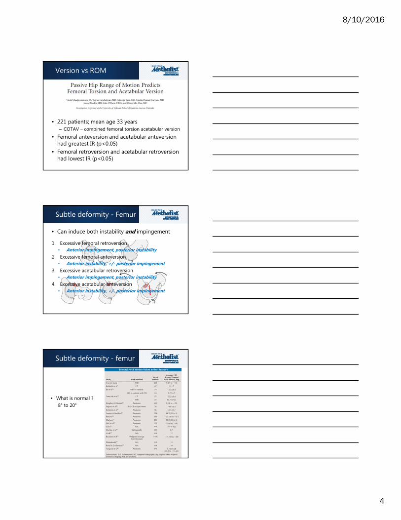
Goals
• Subtle deformities– Acetabular
• Borderline dysplasia– Femoral
• Version• Neck-shaft angle
• Capsule– Normal anatomy– Pathomechanics
• Iatrogenic• Hypermobility

8/10/2016
2
Subtle deformities
Understand normal anatomyMinimum Maximum
Neck‐shaft angle 120° 135°
Lateral CEA 25° 40°
Anterior CEA 25° 40°
Tonnis angle 0° 10°
Extrusion index 0% 25%
Alpha angle n/a 50‐55°
Head‐neck offset 8 mm n/a
Offset ratio 17% n/a
Sharp’s angle 33° 38°
Center‐troch distance ‐17 mm 10 mm
Joint space 2 mm n/a
Hip‐center position n/a 10 mm
Offset 41 mm 44 mm
Minimum Maximum
Crossover sign + ‐
Ischial spine sign + ‐
Posterior wall sign + ‐
Coxa profunda + ‐
Protrusio acetabulae + ‐
Tonnis OA 0 3
AIIS type 1 3
Congruency Congruent Incongruent
Shenton’s line Intact Broken
• Treat patients, not x-rays
Asymptomatic X-rays
• Cam: 37% prevalence – Using 50-55° threshold– 55% athletes– 23% non-athletes
• Pincer: 67% prevalence – 2/6 radiographic markers

8/10/2016
3
Laxity = AsymptomaticInstability = Symptomatic
Laxity ≠ Instability
Understand pathology
Subtle deformity - Femur
• Version – both femoral and acetabular – McKibbin index (version), Omega surface (5 parameters)
8/10/2016
4
Version vs ROM
• 221 patients; mean age 33 years– COTAV – combined femoral torsion acetabular version
• Femoral anteversion and acetabular anteversion had greatest IR (p<0.05)
• Femoral retroversion and acetabular retroversion had lowest IR (p<0.05)
Subtle deformity - Femur
• Can induce both instability and impingement
1. Excessive femoral retroversion• Anterior impingement, posterior instability
2. Excessive femoral anteversion• Anterior instability, +/- posterior impingement
3. Excessive acetabular retroversion• Anterior impingement, posterior instability
4. Excessive acetabular anteversion• Anterior instability, +/- posterior impingement
Subtle deformity - femur
• What is normal ?8° to 20°

8/10/2016
5
Version outcomes
• Excessive version ↑↑↑ OA risk
Version outcomes
• 100 M, 88 F; mean age 35 years• Mean femoral version 9°
– Version vs ER (r= -.21; weak, but p<0.05)– Version vs IR (r= +.23; weak, but p<0.05)
• Femoral version >15° 2.2X more likely to have anterior tears beyond 3 o’clock
• Higher version (p<0.05) in psoas release patients
Version outcomes

8/10/2016
6
Version outcomes
• 180 hips; mean femoral version 10°– >15° - more anterior 3 o’clock labral tears– 5° - 15°– <5°
• Higher (p<0.05) version in psoas release patients• No difference (p>0.05) in PRO’s
Version outcomes
• 67 hips; – Low/normal version = <25°– High version = >25°
• High version >> worse pre-op HOS-SSS (p<0.05)• High version >> worse post-op mHHS (p<0.05)
Version outcomes
• 278 hips; mean femoral version 8°• Anterior 3 o’clock tears
– Version >18° = 30%– Version -2° to 18° = 78%– Version <-2° = 73%
• No difference in PRO’s (p>0.05)
8/10/2016
7
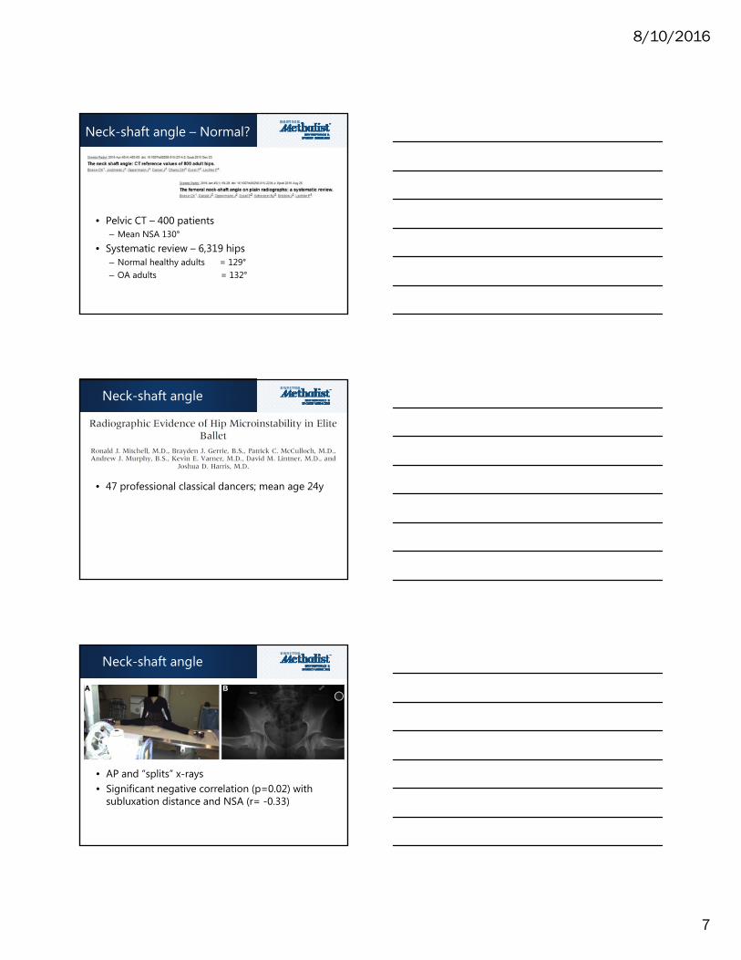
Neck-shaft angle – Normal?
• Pelvic CT – 400 patients– Mean NSA 130°
• Systematic review – 6,319 hips– Normal healthy adults = 129°– OA adults = 132°
Neck-shaft angle
• 47 professional classical dancers; mean age 24y
Neck-shaft angle
• AP and “splits” x-rays• Significant negative correlation (p=0.02) with
subluxation distance and NSA (r= -0.33)

8/10/2016
8
NSA – Troch-pelvic FAI
Permissive limb ER
Intentional limb IR
NSA Outcomes
Borderline dysplasia –What is it?

8/10/2016
9
Borderline dysplasia
• Define it:– LCEA: 20° - 25°– ACEA: 20° - 25°– Tonnis angle: 10° - 15°
Borderline dysplasia
• Potential hip instability– Pre-op – Post-op
Borderline dysplasia -Outcomes

8/10/2016
10
Borderline dysplasia -Outcomes
• Non-arthritic dysplasia – PAO +/- arthroscopy• If arthroscopic manage BD:
– Preserve labrum– Preserve capsule– Preserve iliopsoas
• No iatrogenic over-resection of the rim• Do not ignore femur !!! (combined effects)• Outcomes may be as good
– Possibly worse – Possibly catastrophic
Borderline dysplasia -Outcomes
• Pre-operative or iatrogenic dysplasia or BD present in 5/11 (45%) of macro-instability cases
• Capsular closure performed in 2/10 interportal and 1/1 “T” capsulotomy
Role of the capsule

8/10/2016
11
Capsule – In the lab
Take-home points:*Unrepaired capsulotomies – instability*Repaired capsulotomies – normalizes stability
Capsule - Pathology
• Native– EDS, hypermobility
• Iatrogenic– Unrepaired capsulotomies
Capsule - Presentation
• Pain• Apprehension/fear• Difficult RTS
*** Extended, ER*** Abducted, ER

8/10/2016
12
Capsule – Indications
Capsule – Technique
Capsule – Technique
8/10/2016
13
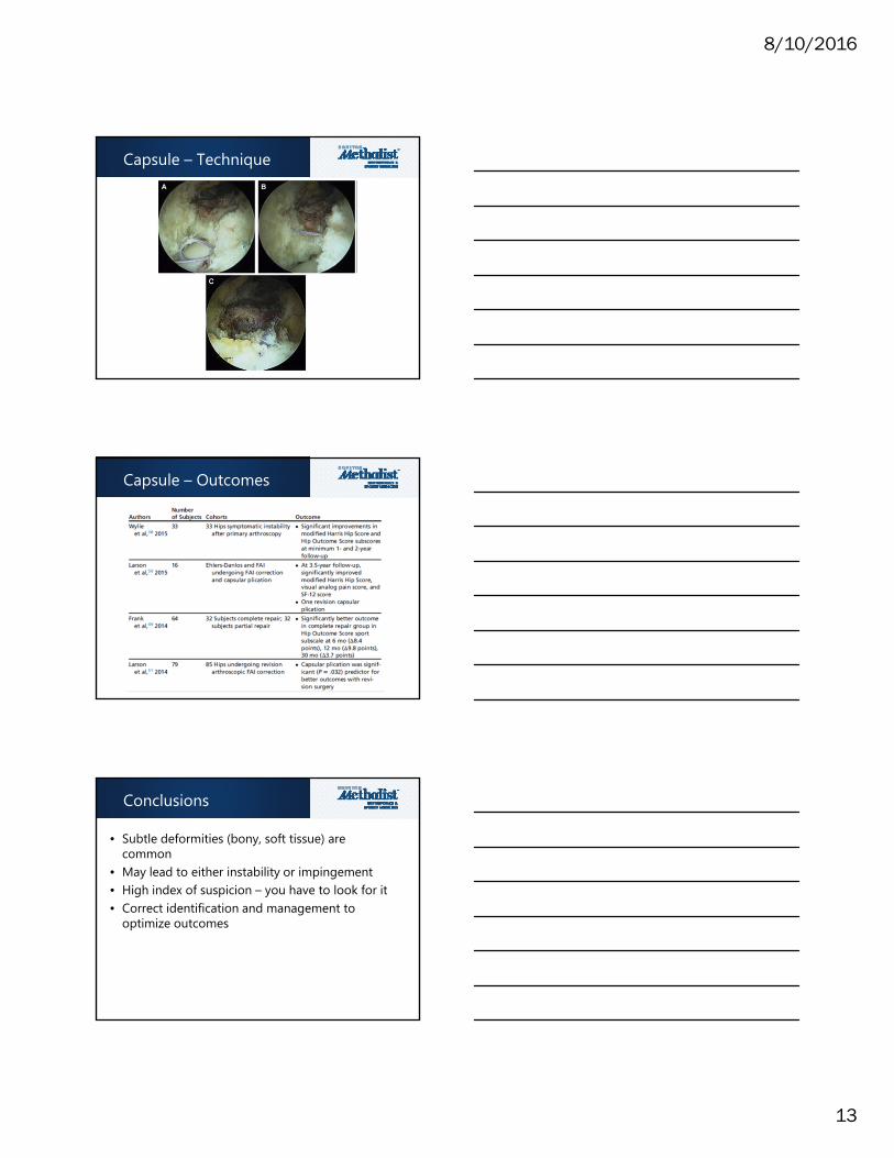
Capsule – Technique
Capsule – Outcomes
Conclusions
• Subtle deformities (bony, soft tissue) are common
• May lead to either instability or impingement • High index of suspicion – you have to look for it• Correct identification and management to
optimize outcomes

8/10/2016
14