Embed Size (px)
Citation preview
Suitable combination of noble/ferromagnetic
metal multilayers for enhanced magneto-
plasmonic biosensing
David Regatos,* Borja Sepúlveda, David Fariña, Laura G. Carrascosa,
and Laura M. Lechuga
Research Center on Nanoscience and Nanotechnology CIN2 (CSIC) and CIBER-BBN,
Campus UAB, 08193 Bellaterra, Barcelona, Spain
Abstract: We present a theoretical and experimental study on the
biosensing sensitivity of Au/Co/Au multilayers as transducers of the
magneto-optic surface-plasmon-resonance (MOSPR) sensor. We
demonstrate that the sensing response of these magneto-plasmonic (MP)
transducers is a trade-off between the optical absorption and the magneto-
optical activity, observing that the MP multilayer with larger MO effect
does not provide the best sensing response. We show that it is possible to
design highly-sensitive MP transducers able to largely surpass the limit of
detection of the conventional surface-plasmon-resonance (SPR) sensor.
This was proved comparing the biosensing performance of both sensors for
the label-free detection of short DNA chains hybridization. For this
purpose, we used and tested a novel label-free biofunctionalization protocol
based on polyelectrolytes, which increases the resistance of MP transducers
in aqueous environments.
©2011 Optical Society of America
OCIS codes: (310.4165) Multilayer design; (250.5403) Plasmonics; (240.6680) Surface
plasmons; (230.3810) Magneto-optic system; (280.4788) Optical sensing and sensors; (280.1415) Biological sensing and sensors.
References and Links
1. J. S. Yuk, and K. S. Ha, “Proteomic applications of surface plasmon resonance biosensors: analysis of protein
arrays,” Exp. Mol. Med. 37(1), 1–10 (2005).
2. J. Treviño, A. Calle, J. M. Rodríguez-Frade, M. Mellado, and L. M. Lechuga, “Determination of human growth hormone in human serum samples by surface plasmon resonance immunoassay,” Talanta 78(3), 1011–1016
(2009). 3. L. G. Carrascosa, A. Calle, and L. M. Lechuga, “Label-free detection of DNA mutations by SPR: application to
the early detection of inherited breast cancer,” Anal. Bioanal. Chem. 393(4), 1173–1182 (2009).
4. E. Mauriz, A. Calle, J. J. Manclús, A. Montoya, and L. M. Lechuga, “Multi-analyte SPR immunoassays for environmental biosensing of pesticides,” Anal. Bioanal. Chem. 387(4), 1449–1458 (2007).
5. A. A. Bergwerff, and F. van Knapen, “Surface plasmon resonance biosensors for detection of pathogenic
microorganisms: strategies to secure food and environmental safety,” J. AOAC Int. 89(3), 826–831 (2006). 6. S. A. Maier, Plasmonics: Fundamentals and Applications (Springer, 2007).
7. J. Homola, Surface Plasmon Resonance Based Sensors (Springer, 2006).
8. S. Patskovsky, M. Maisonneuve, M. Meunier, and A. V. Kabashin, “Mechanical modulation method for ultrasensitive phase measurements in photonics biosensing,” Opt. Express 16(26), 21305–21314 (2008).
9. Y. Shin, H. M. Kim, Y. Jung, and B. H. Chung, “A new palm-sized surface plasmon resonance (SPR) biosensor
based on modulation of a light source by a rotating mirror,” Sens. Actuators B Chem. 150(1), 1–6 (2010). 10. P. P. Markowicz, W. C. Law, A. Baev, P. N. Prasad, S. Patskovsky, and A. Kabashin, “Phase-sensitive time-
modulated surface plasmon resonance polarimetry for wide dynamic range biosensing,” Opt. Express 15(4),
1745–1754 (2007). 11. A. V. Kabashin, S. Patskovsky, and A. N. Grigorenko, “Phase and amplitude sensitivities in surface plasmon
resonance bio and chemical sensing,” Opt. Express 17(23), 21191–21204 (2009).
12. B. Sepúlveda, A. Calle, L. M. Lechuga, and G. Armelles, “Highly sensitive detection of biomolecules with the magneto-optic surface-plasmon-resonance sensor,” Opt. Lett. 31(8), 1085–1087 (2006).
13. P. E. Ferguson, O. M. Stafsudd, and R. F. Wallis, “Enhancement of the transverse kerr magneto-optic effect by
#141724 - $15.00 USD Received 25 Jan 2011; revised 15 Mar 2011; accepted 23 Mar 2011; published 15 Apr 2011(C) 2011 OSA 25 April 2011 / Vol. 19, No. 9 / OPTICS EXPRESS 8336
surface magnetoplasma waves,” Physica 89B, 91–94 (1977).
14. B. Sepulveda, L. M. Lechuga, and G. Armelles, “Magnetooptic Effects in Surface-Plasmon-Polaritons Slab Waveguides,” J. Lightwave Technol. 24(2), 945–955 (2006).
15. V. I. Safarov, V. A. Kosobukin, C. Hermann, G. Lampel, J. Peretti, and C. Marlière, “Magneto-optical Effects
Enhanced by Surface Plasmons in Metallic Multilayer Films,” Phys. Rev. Lett. 73(26), 3584–3587 (1994). 16. C. Hermann, V. A. Kosobukin, G. Lampel, J. Peretti, V. I. Safarov, and P. Bertrand, “Surface-enhanced
magneto-optics in metallic multilayer films,” Phys. Rev. B 64(23), 235422 (2001).
17. V. V. Temnov, G. Armelles, U. Woggon, D. Guzatov, A. Cebollada, A. Garcia-Martin, J. Garcia-Martin, T. Thomay, A. Leitenstorfer, and R. Bratschitsch, “Active magneto-plasmonics in hybrid metal–ferromagnet
structures,” Nat. Photonics 4(2), 107–111 (2010).
18. N. Bonod, R. Reinisch, E. Popov, and M. Nevière, “Optimization of surface-plasmon-enhanced magneto-optical effects,” J. Opt. Soc. Am. B 21, 791–797 (2004).
19. J. B. González-Díaz, A. Garcia-Martin, G. Armelles, J. M. Garcia-Martin, C. Clavero, A. Cebollada, R. A.
Lukaszew, J. R. Skuza, D. P. Kumah, and R. Clarke, “Surface-magnetoplasmon nonreciprocity effects in noble-metal/ferromagnetic heterostructures,” Phys. Rev. B 76(15), 153402 (2007).
20. E. Ferreiro-Vila, J. B. Gonzalez-Diaz, R. Fermento, M. U. Gonzalez, A. Garcia-Martin, J. M. Garcia-Martin, A.
Cebollada, G. Armelles, D. Meneses-Rodriguez, and E. M. Sandoval, “Intertwined magneto-optical and plasmonic effects in Ag/Co/Ag layered structures,” Phys. Rev. B 80(12), 125132 (2009).
21. D. Regatos, D. Fariña, A. Calle, A. Cebollada, B. Sepúlveda, G. Armelles, and L. M. Lechuga, “Au/Fe/Au
multilayer transducers for magneto-optic surface plasmon resonance sensing,” J. Appl. Phys. 108(5), 054502 (2010).
22. C. Clavero, K. Yang, J. R. Skuza, and R. A. Lukaszew, “Magnetic field modulation of intense surface plasmon
polaritons,” Opt. Express 18(8), 7743–7752 (2010). 23. E. Kretschmann, “Radiative decay on nonradiative surface plasmons excited by light,” Z. Naturforsch. B 23A,
2135 (1968).
24. R. Schasfoort, and A. Tudos, eds., Handbook of Surface Plasmon Resonance (RSC Publising, 2008). 25. B. Sepúlveda, J. B. González-Díaz, A. García-Martín, L. M. Lechuga, and G. Armelles, “Plasmon-Induced
Magneto-Optical Activity in Nanosized Gold Disks,” Phys. Rev. Lett. 104(14), 147401 (2010). 26. J. B. González-Díaz, A. García-Martín, J. M. García-Martín, A. Cebollada, G. Armelles, B. Sepúlveda, Y.
Alaverdyan, and M. Käll, “Plasmonic Au/Co/Au nanosandwiches with enhanced magneto-optical activity,”
Small 4(2), 202–205 (2008). 27. J. B. Gonzalez-Diaz, J. M. Garcia-Martin, A. Garcia-Martin, D. Navas, A. Asenjo, and M. Vazquez, “M.
Hernandez-Velez, and G. Armelles, “Plasmon-enhanced magneto-optical activity in ferromagnetic membranes,”
Appl. Phys. Lett. 94, 263101 (2009). 28. M. Schubert, “Polarization-dependent optical parameters of arbitrarily anisotropic homogeneous layered
systems,” Phys. Rev. B Condens. Matter 53(8), 4265–4274 (1996).
29. P. B. Johnson, and R. W. Christy, “Optical Constants of the Noble Metals,” Phys. Rev. B 6(12), 4370–4379
(1972).
30. M. J. Weber, Handbook of Optical Materials (CRC, 2002).
31. L. G. C. Melo, A. D. Santos, L. M. Alvarez-Prado, and Y. Souche, “Optimization of the TMOKE response using
the ATR configuration,” J. Magn. Magn. Mater. 310(2), 947–949 (2007).
1. Introduction
In the last two decades label-free optical biosensors have turned into essential tools for the
real-time detection and identification of chemical and biological species with high sensitivity
and selectivity. Surface-plasmon-resonance (SPR) devices are the landmark sensors within
photonic biosensing technology and, nowadays, these devices are basic tools for the analysis
of biospecific interactions. Different SPR biosensors are commercialized by more than twelve
companies around the world, demonstrating remarkable applications in wide areas as
proteomics [1], medical diagnostic [2], genomics [3], environmental monitoring [4], food
safety and security [5]. These types of sensors are based on the well-known properties of
surface plasmon polaritons (SPP) [6], which can be excited by light at the interface of a metal
and a dielectric. There are several SPR biosensor configurations [7], depending on the
excitation and interrogation layouts. But due to its simplicity, low cost, and high-performance,
the intensity-interrogated SPR biosensor is one of the most widely employed, and it is
currently an essential analytical tool for real-time and label-free detection. However, its
sensitivity is not enough for the label-free detection of low concentrations of small molecular
weight analytes. Therefore, the development of any technique which could improve the
sensitivity of the SPR biosensor while keeping all its advantages can constitute a very
attractive tool for biosensing applications.
#141724 - $15.00 USD Received 25 Jan 2011; revised 15 Mar 2011; accepted 23 Mar 2011; published 15 Apr 2011(C) 2011 OSA 25 April 2011 / Vol. 19, No. 9 / OPTICS EXPRESS 8337
For that reason, during last years, different SPR modulation configurations have been
proposed to improve the sensitivity of the standard SPR sensors, as for example mechanical
[8,9] or phase [10,11] modulated SPR sensors. These configurations use external modulation
techniques to improve the signal-to-noise (SNR) ratio of the sensing measurements, thus
increasing their limit of detection (LOD). However, the main drawback of these
configurations is the high-cost and complexity of the final devices. As an alternative, our
group proposed the magneto-optic surface-plasmon-resonance (MOSPR) biosensor [12],
based on the non-reciprocal variation of the SPP wave vector with an external magnetic field
[13,14]. The so-called magneto-plasmonic (MP) modulation arises from the simultaneous
excitation of magneto-optic (MO) effects and the SPP in structures with plasmonic and MO
activity. The most suitable structures to generate large MP modulation are multilayers
composed of ferromagnetic and noble metals [15,16], since they combine the large MO
activity of ferromagnetic materials and the exceptional plasmonic properties of noble metals.
Compared to the conventional SPR sensor, the MOSPR only requires a low field
electromagnet and a MP transducer instead of the typical gold layer. Although we
demonstrated that the MOSPR sensor can surpass the sensitivity of the conventional SPR
sensor, its final performance strongly depends on the MP features of the multilayer structures
employed as transducer. Therefore, the optimization of the sensor response through the most
suitable combination of multilayers becomes essential, not only for the development of
MOSPR biosensors, but also for future MP active devices [17].
So far, the MP features of multilayered structures made of noble and ferromagnetic metals
have been extensively studied [18–22], but not their sensing properties. For that reason, in this
work we present a combined theoretical and experimental analysis of the sensing response of
MP multilayered transducers, confirming that the sensing response is a complex balance
between optical absorption and MO activity. Thanks to this analysis it is possible to design
high-sensitive MP transducers, showing that the MOSPR biosensor with an optimized
Au/Co/Au multilayer transducer can exhibit a four-fold improvement of the LOD with respect
to conventional SPR biosensors.
2. Operating principle of the MOSPR biosensor
In the case of monochromatic p-polarized light in the Kretschmann configuration [23] (or
prism-coupling), the excitation of the SPP results in a sharp decrease of the reflected intensity
at a specific angle of incident (θspp). The principle of detection of the conventional SPR sensor
[24] is based on the strong dependence of the SPP wave vector (kspp) on the refractive index
of the dielectric (nd) medium. The changes of refractive index in the close proximity of the
metal layer, as those generated by biochemical interactions, induce an angular shift of the
reflectance resonance dip, which can be employed to quantify the biosensing interactions. In
contrast, the MOSPR measurement is based on the simultaneous excitation of the SPP and the
transversal magneto-optic Kerr effect (TMOKE), which is generated by applying an external
magnetic field parallel to the metal layer but perpendicular to the direction of propagation of
the SPP. Such magnetic field induces a non-reciprocal shift of kspp, which is translated into
small variations of the SPP excitation angle, Δθspp, for monochromatic light (Fig. 1(a)) and,
subsequently, into variations of the angular reflectance curve:
ΔR R H R H 0pp pp pp
(1)
where Rpp is the reflectance of p-polarized light and H the external magnetic field. TMOKE
gives access to the derivative of the reflectance resonance dip, given by:
Rpp
ΔR (H) Δθ (H)pp sppθ
(2)
#141724 - $15.00 USD Received 25 Jan 2011; revised 15 Mar 2011; accepted 23 Mar 2011; published 15 Apr 2011(C) 2011 OSA 25 April 2011 / Vol. 19, No. 9 / OPTICS EXPRESS 8338
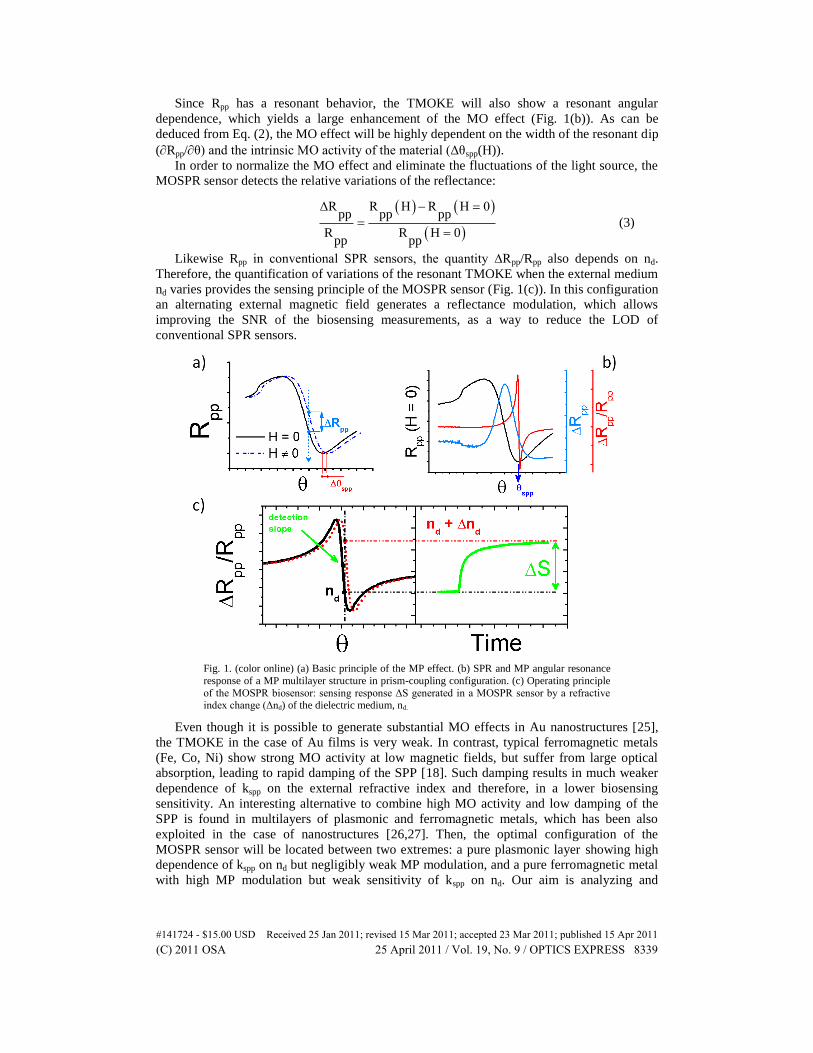
Since Rpp has a resonant behavior, the TMOKE will also show a resonant angular
dependence, which yields a large enhancement of the MO effect (Fig. 1(b)). As can be
deduced from Eq. (2), the MO effect will be highly dependent on the width of the resonant dip
(Rpp/θ) and the intrinsic MO activity of the material (Δθspp(H)).
In order to normalize the MO effect and eliminate the fluctuations of the light source, the
MOSPR sensor detects the relative variations of the reflectance:
ΔR R H R H 0pp pp pp
R R H 0pp pp
(3)
Likewise Rpp in conventional SPR sensors, the quantity ∆Rpp/Rpp also depends on nd.
Therefore, the quantification of variations of the resonant TMOKE when the external medium
nd varies provides the sensing principle of the MOSPR sensor (Fig. 1(c)). In this configuration
an alternating external magnetic field generates a reflectance modulation, which allows
improving the SNR of the biosensing measurements, as a way to reduce the LOD of
conventional SPR sensors.
Fig. 1. (color online) (a) Basic principle of the MP effect. (b) SPR and MP angular resonance
response of a MP multilayer structure in prism-coupling configuration. (c) Operating principle
of the MOSPR biosensor: sensing response ΔS generated in a MOSPR sensor by a refractive index change (Δnd) of the dielectric medium, nd.
Even though it is possible to generate substantial MO effects in Au nanostructures [25],
the TMOKE in the case of Au films is very weak. In contrast, typical ferromagnetic metals
(Fe, Co, Ni) show strong MO activity at low magnetic fields, but suffer from large optical
absorption, leading to rapid damping of the SPP [18]. Such damping results in much weaker
dependence of kspp on the external refractive index and therefore, in a lower biosensing
sensitivity. An interesting alternative to combine high MO activity and low damping of the
SPP is found in multilayers of plasmonic and ferromagnetic metals, which has been also
exploited in the case of nanostructures [26,27]. Then, the optimal configuration of the
MOSPR sensor will be located between two extremes: a pure plasmonic layer showing high
dependence of kspp on nd but negligibly weak MP modulation, and a pure ferromagnetic metal
with high MP modulation but weak sensitivity of kspp on nd. Our aim is analyzing and
#141724 - $15.00 USD Received 25 Jan 2011; revised 15 Mar 2011; accepted 23 Mar 2011; published 15 Apr 2011(C) 2011 OSA 25 April 2011 / Vol. 19, No. 9 / OPTICS EXPRESS 8339
maximizing the response of such MP modulation to the changes of refractive index. We focus
our analysis in the dependence of the TMOKE, i.e. ∆Rpp, on the refractive index of the
external medium nd for different combinations of plasmonic and ferromagnetic metals. Since
we are interested in the biosensing applications, we restrict our study to Au as plasmonic
material due to its chemical stability. On the other hand, we select Co as ferromagnetic
material owing to its much larger MO activity compared to Fe when it is integrated within Au
layers [21].
3. Theoretical analysis of the sensor response of MP multilayer transducers
In any intensity-interrogated sensor as a function of the angle of incidence, the sensitivity can
be defined as:
θS S spp
ηn θ n
d d
(4)
where S represents the measured signal (either optic or magneto-optic). Therefore, the
maximum sensitivity will be achieved for transducers that are able to combine high slopes in
the resonant angular curves and large displacements of the resonance position when the
refractive index changes. As a result, we propose the following parameter for the study and
optimization of the MOSPR sensor:
ΔRpp
ηMOSPR n
d
(5)
Another important aspect in the SPP based sensors in Kretschmann configuration is the
coupling efficiency of the SPP and, closely related, the intensity of the electromagnetic field
at the sensing interface. Both parameters are highly dependent on the thickness of the metal
multilayer. The optimal coupling and maximum electromagnetic field at the sensing interface
are obtained for multilayers yielding to reflectance values close to 0 at the resonant angle.
Therefore, we employed the Transfer Matrix Method [28] to analyze the MO and sensing
properties of Au/Co/Au multilayers (Fig. 2(a)) as a function of the thicknesses of the Au top
layer (dAuTop) and the Co layer (dCo), when the thickness of the Au bottom layer (dAuBottom) is
selected to provide a reflectance Rpp at the resonance angle is as close to 0 as possible.
Depending on the values of dAuTop and dCo, the previous reflectance criterion will be fulfilled
for dAuBottom = 0 (bilayer structures) or dAuBottom 0 (trilayer structures). The thickness of the
Au bottom layer that optimizes the coupling efficiency of the SPP can also be determined
through the Transfer Matrix Method. In all the calculations, the incident medium is glass (ni =
1.516 RIU) and the external medium is water (nd = 1.3323 RIU). Between glass and the MP
multilayer we include a 2 nm thick Ti layer, since it will be required to improve the
mechanical adhesion of the experimental multilayers. We assume that the incident light
wavelength is 660 nm and, at this wavelength, the dielectric constants of the different
materials are [29,30]: εxxAu = 13.7 + 1.04·i, εxxCo = 12.12 + 19.75·i, and εxxTi = 4 + 13.55·i,
respectively. The MO constant of cobalt are εxzCo = 0.470 + 0.0010·i.
#141724 - $15.00 USD Received 25 Jan 2011; revised 15 Mar 2011; accepted 23 Mar 2011; published 15 Apr 2011(C) 2011 OSA 25 April 2011 / Vol. 19, No. 9 / OPTICS EXPRESS 8340
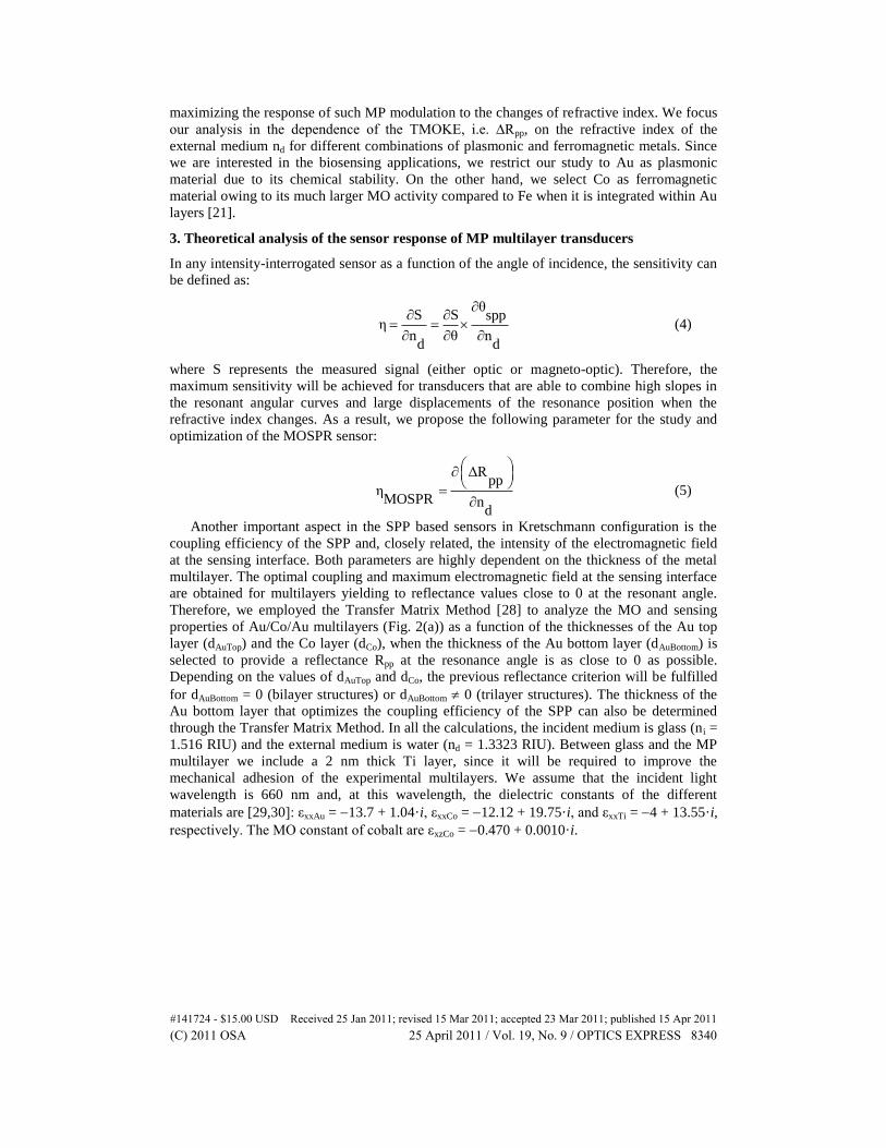
Fig. 2. (color online) (a) Schematic view of the analyzed MP structure in Kretschmann configuration. Contour plots of: (b) the maximum amplitude of the ΔRpp and (c) ηMOSPR as
function of the upper gold layer (dAuTop) and cobalt thicknesses (dCo). The value of the gold bottom layer thickness (dAuBottom) was previously optimized to achieve the maximum coupling
efficiency of the SPP (see appendix A). The blue line divides the region of bilayer structures
(those in which the optimization occurs for dAuBottom = 0) and the region of trilayer structures
(where occurs for dAuBottom 0). Finally, the red numbers of the figure (c) are the combinations
of dAuTop-dCo experimentally evaluated in this work.
Once the combination of layers satisfying the reflectance criterion is obtained (see
appendix A), we study their TMOKE amplitude. In the theoretical analysis, the maximum
thicknesses for the Au top and Co layers were of 40 and 20 nm, respectively. Thicker Au top
layers are discarded from the analysis since the reflectivity criterion imposes Co layers whose
small thickness give rise a very weak MO effect. Figure 2(b) clearly shows that the maximum
amplitude of ∆Rpp is obtained for the layers with higher content of Co. In particular, the
highest MO effect is exhibited by the pure 20 nm Co layer. According to a previous
publication [31], when gold is introduced in the multilayer, the MO effect is always
maximized when cobalt is in contact with the external dielectric medium (dAuTop = 0), i.e., in
the region where the electromagnetic field associated to the SPP is maximized. However, as
we have previously discussed, a larger MO effect does not necessarily mean a higher
sensitivity for the biosensing measurements. Indeed, Fig. 2(c), depicting the sensitivity of
TMOKE ηMOSPR shows a very different scenario. The TMOKE sensitivity presents two clearly
defined regions of optimal sensitivity, one in the bilayers region (dAuTop ~40 nm: dCo ~12 nm)
and one for trilayers (dAuTop ~25 nm: dCo ~6 nm; dAuBottom ~10 nm). The ηMOSPR parameter was
numerically determined for a refractive index change of the external medium of 5·104
RIU.
4. Experimental verification and discussion
To corroborate the theoretical study with experimental results, we fabricated twelve different
Au/Co/Au structures on glass substrates, previously coated with a 2 nm thick Ti layer, in
order to show the main features of the sensitivity described in Fig. 2(c). The nominal
thickness values of the fabricated samples correspond to the twelve points displayed in Fig.
2(c), also gathered in appendix B. The transducers were fabricated by electron-beam
#141724 - $15.00 USD Received 25 Jan 2011; revised 15 Mar 2011; accepted 23 Mar 2011; published 15 Apr 2011(C) 2011 OSA 25 April 2011 / Vol. 19, No. 9 / OPTICS EXPRESS 8341
evaporation at room temperature with a base pressure of 106
mbar and the thickness was
monitored using a calibrated quartz crystal sensor. Due to the fast oxidation of cobalt in the
sensing aqueous environment, we used a minimum of 15 nm as upper gold layer. The
magnetization of the Co layer is reversed at a frequency of 64Hz in the transversal
configuration with an alternating magnetic field of 50 Oe. The optic (Rpp) and MO (∆Rpp)
signals are extracted via FFT analysis of the reflected light intensity.
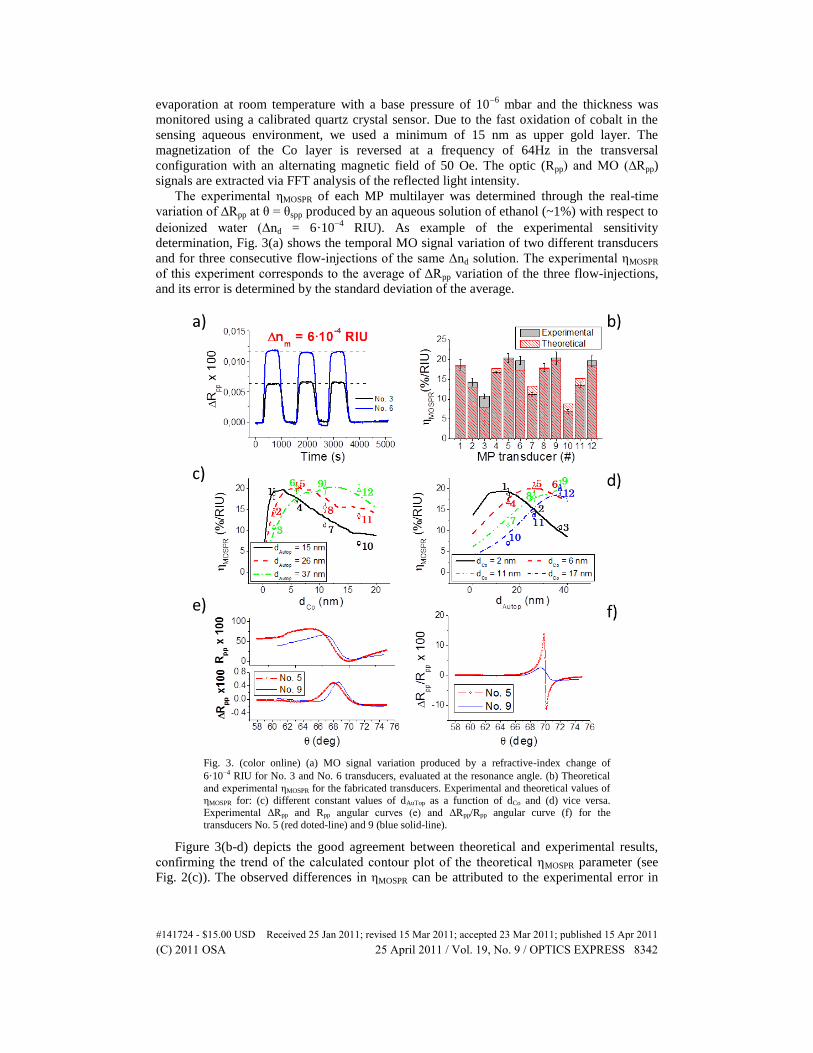
The experimental ηMOSPR of each MP multilayer was determined through the real-time
variation of ∆Rpp at θ = θspp produced by an aqueous solution of ethanol (~1%) with respect to
deionized water (Δnd = 6·104
RIU). As example of the experimental sensitivity
determination, Fig. 3(a) shows the temporal MO signal variation of two different transducers
and for three consecutive flow-injections of the same Δnd solution. The experimental ηMOSPR
of this experiment corresponds to the average of ΔRpp variation of the three flow-injections,
and its error is determined by the standard deviation of the average.
Fig. 3. (color online) (a) MO signal variation produced by a refractive-index change of
6·104 RIU for No. 3 and No. 6 transducers, evaluated at the resonance angle. (b) Theoretical
and experimental ηMOSPR for the fabricated transducers. Experimental and theoretical values of
ηMOSPR for: (c) different constant values of dAuTop as a function of dCo and (d) vice versa. Experimental ΔRpp and Rpp angular curves (e) and ΔRpp/Rpp angular curve (f) for the
transducers No. 5 (red doted-line) and 9 (blue solid-line).
Figure 3(b-d) depicts the good agreement between theoretical and experimental results,
confirming the trend of the calculated contour plot of the theoretical ηMOSPR parameter (see
Fig. 2(c)). The observed differences in ηMOSPR can be attributed to the experimental error in
#141724 - $15.00 USD Received 25 Jan 2011; revised 15 Mar 2011; accepted 23 Mar 2011; published 15 Apr 2011(C) 2011 OSA 25 April 2011 / Vol. 19, No. 9 / OPTICS EXPRESS 8342
the thickness determination and the roughness of the thinner Au layers. The lower
experimental ηMOSPR values of the samples with smaller Au top layer (No. 1, 4, 7 and, 10) are
probably due to the poor protection of this layer from Co partial oxidation. Despite of these
subtle deviations, we corroborate the existence of two regions with a maximum value of
ηMOSPR. One corresponds to a bilayer structure (No. 9) with 11 nm of Co located at 37 nm of
the SPP surface, and the other to a trilayer structure (No. 5) with 6 nm of Co located at 26 nm
of the SPP surface. These results confirm that the most suitable transducer is a trade-off
between a structure with high MO signal (high content of magnetic material in the vicinity of
the SPP interface) and low damping of the excited SPP (low content of magnetic material and
far from the SPP interface).
5. MOSPR vs. SPR as biosensor devices
The final objective of this work is comparing the biosensing performance of the optimized
MOSPR and standard SPR sensors for the label-free detection of DNA hybridization of very
short DNA chains. The detection of these targets is challenging in standard SPR sensors due
to their very low molecular weight. The previous experimental analysis has shown that
structure No. 5 exhibits the maximum sensitivity (Fig. 3(b)) and, in addition, the lowest
reflectance at the resonance angle (Fig. 3(e)). This combination allows the generation of a
larger enhancement of ΔRpp/Rpp (Fig. 3(f)), helping to improve even more the signal-to-noise
ratio (SNR) of the biosensing measurement. We used 48 nm Au on
2 nm Ti as transducer of the SPR sensor, since this gold thickness provides the optimal
coupling between the incident light (λ = 660 nm) and the SPP (previous reflectivity criterion).
Both transducers were fabricated on the same type of glass substrates and using the same
growth conditions (Fig. 4(a)).
The sensitivity comparison must be done taking into account the SNR due to the different
nature of the MOSPR and conventional SPR measurements. We defined the system noise as
the RMS deviation of the sensing signal acquired during 1000 s with a sampling rate of
1 Hz when nd is constant. The evaluation of the biosensing capabilities of both sensors was
done using the same experimental set-up and biofunctionalization protocols.
.
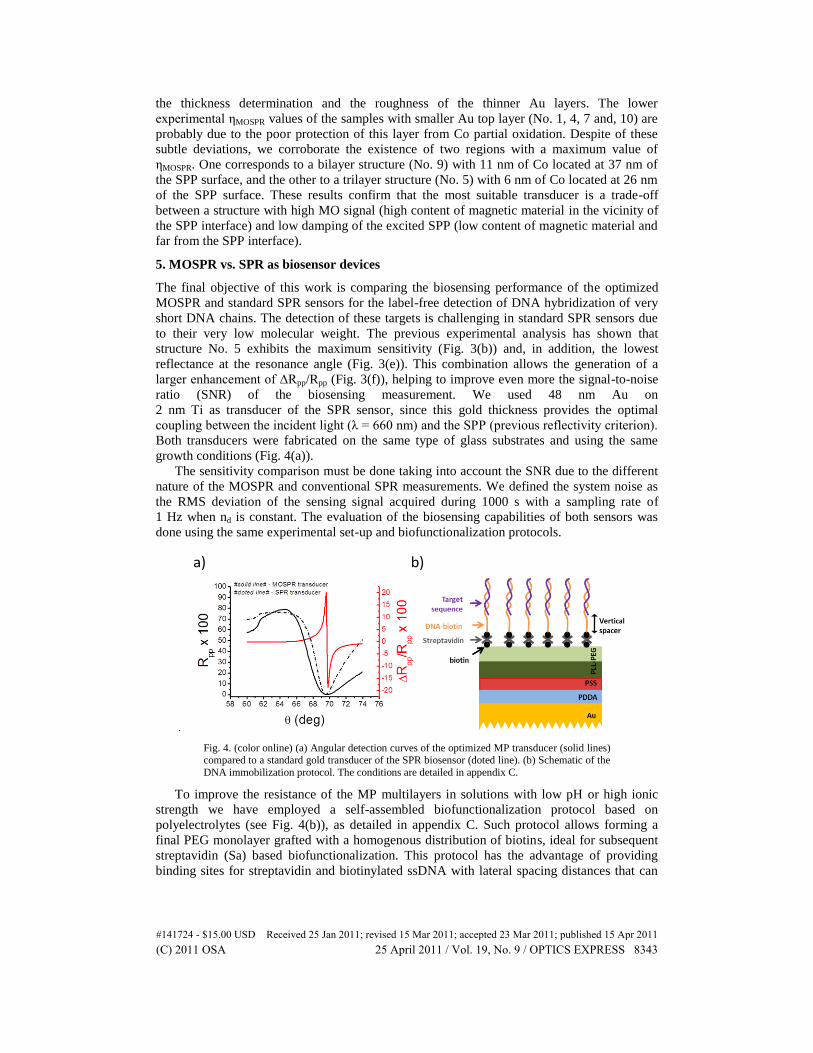
Fig. 4. (color online) (a) Angular detection curves of the optimized MP transducer (solid lines) compared to a standard gold transducer of the SPR biosensor (doted line). (b) Schematic of the
DNA immobilization protocol. The conditions are detailed in appendix C.
To improve the resistance of the MP multilayers in solutions with low pH or high ionic
strength we have employed a self-assembled biofunctionalization protocol based on
polyelectrolytes (see Fig. 4(b)), as detailed in appendix C. Such protocol allows forming a
final PEG monolayer grafted with a homogenous distribution of biotins, ideal for subsequent
streptavidin (Sa) based biofunctionalization. This protocol has the advantage of providing
binding sites for streptavidin and biotinylated ssDNA with lateral spacing distances that can
#141724 - $15.00 USD Received 25 Jan 2011; revised 15 Mar 2011; accepted 23 Mar 2011; published 15 Apr 2011(C) 2011 OSA 25 April 2011 / Vol. 19, No. 9 / OPTICS EXPRESS 8343
help to maximize the hybridization efficiency. On the other hand, the remaining PEG surface
permits the minimization of unspecific adsorptions.
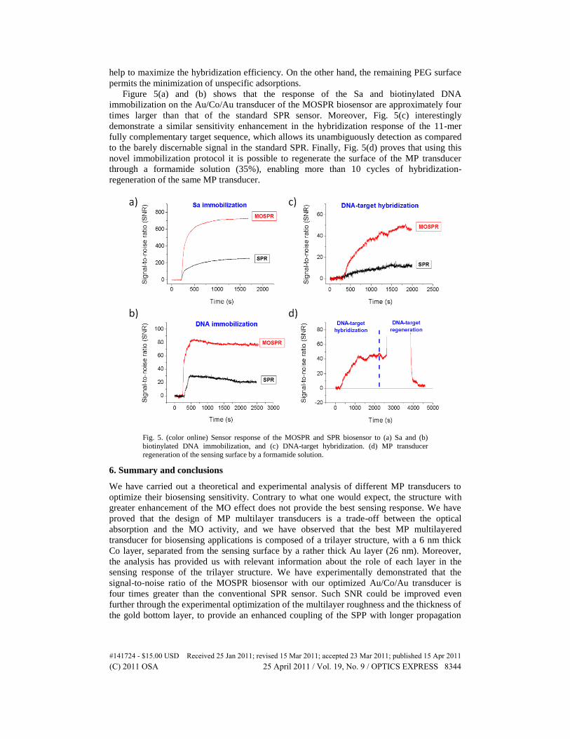
Figure 5(a) and (b) shows that the response of the Sa and biotinylated DNA
immobilization on the Au/Co/Au transducer of the MOSPR biosensor are approximately four
times larger than that of the standard SPR sensor. Moreover, Fig. 5(c) interestingly
demonstrate a similar sensitivity enhancement in the hybridization response of the 11-mer
fully complementary target sequence, which allows its unambiguously detection as compared
to the barely discernable signal in the standard SPR. Finally, Fig. 5(d) proves that using this
novel immobilization protocol it is possible to regenerate the surface of the MP transducer
through a formamide solution (35%), enabling more than 10 cycles of hybridization-
regeneration of the same MP transducer.
Fig. 5. (color online) Sensor response of the MOSPR and SPR biosensor to (a) Sa and (b)
biotinylated DNA immobilization, and (c) DNA-target hybridization. (d) MP transducer regeneration of the sensing surface by a formamide solution.
6. Summary and conclusions
We have carried out a theoretical and experimental analysis of different MP transducers to
optimize their biosensing sensitivity. Contrary to what one would expect, the structure with
greater enhancement of the MO effect does not provide the best sensing response. We have
proved that the design of MP multilayer transducers is a trade-off between the optical
absorption and the MO activity, and we have observed that the best MP multilayered
transducer for biosensing applications is composed of a trilayer structure, with a 6 nm thick
Co layer, separated from the sensing surface by a rather thick Au layer (26 nm). Moreover,
the analysis has provided us with relevant information about the role of each layer in the
sensing response of the trilayer structure. We have experimentally demonstrated that the
signal-to-noise ratio of the MOSPR biosensor with our optimized Au/Co/Au transducer is
four times greater than the conventional SPR sensor. Such SNR could be improved even
further through the experimental optimization of the multilayer roughness and the thickness of
the gold bottom layer, to provide an enhanced coupling of the SPP with longer propagation
#141724 - $15.00 USD Received 25 Jan 2011; revised 15 Mar 2011; accepted 23 Mar 2011; published 15 Apr 2011(C) 2011 OSA 25 April 2011 / Vol. 19, No. 9 / OPTICS EXPRESS 8344
distance due to the reduced scattering. On the other hand, the increase of the modulation
frequency using ferrite based electromagnets is another important factor that can contribute to
amplify the SNR of the biomeasurements. Finally, we have also proposed and tested a novel
label-free biofunctionalization protocol based on polyelectrolytes. This protocol increases the
resistance of the MP transducers in aqueous environment thanks to the protective effect of the
polyelectrolyte multilayer. Such protective multilayer can be biotinylated, which offers
different strategies of further biofunctionalization. The protocol could be used in other types
of magnetic or MP transducers, such as magnetic nanoparticles or MP nanostructures.
Appendices
A. Complementary theoretical results
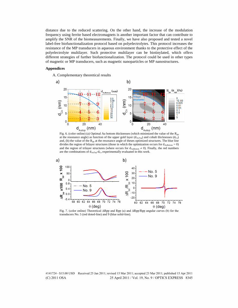
Fig. 6. (color online) (a) Optimal Au bottom thicknesses (which minimized the value of the Rpp
at the resonance angle) as function of the upper gold layer (dAuTop) and cobalt thicknesses (dCo) and, (b) the value of the Rpp at the resonance angle of theses optimized structures. The blue line
divides the region of bilayer structures (those in which the optimization occurs for dAuBottom = 0)
and the region of trilayer structures (where occurs for dAuBottom 0). Finally, the red numbers are the combinations of dAuTop-dCo experimentally evaluated in this work.
Fig. 7. (color online) Theoretical ΔRpp and Rpp (a) and ΔRpp/Rpp angular curves (b) for the
transducers No. 5 (red doted-line) and 9 (blue solid-line).
#141724 - $15.00 USD Received 25 Jan 2011; revised 15 Mar 2011; accepted 23 Mar 2011; published 15 Apr 2011(C) 2011 OSA 25 April 2011 / Vol. 19, No. 9 / OPTICS EXPRESS 8345
B. Complementary experimental results
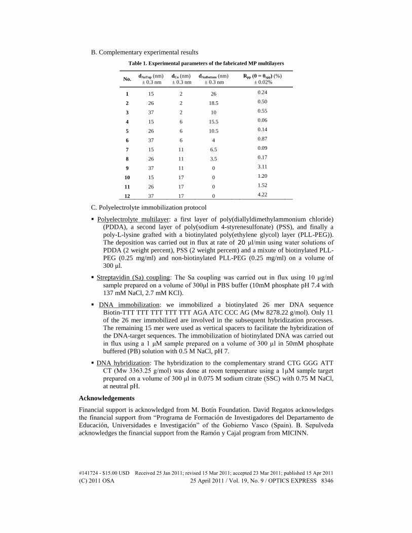
Table 1. Experimental parameters of the fabricated MP multilayers
No. dAuTop (nm)
± 0.3 nm
dCo (nm)
± 0.3 nm
dAuBottom (nm)
± 0.3 nm Rpp (θ = θspp) (%)
± 0.02%
1 15 2 26 0.24
2 26 2 18.5 0.50
3 37 2 10 0.55
4 15 6 15.5 0.06
5 26 6 10.5 0.14
6 37 6 4 0.87
7 15 11 6.5 0.09
8 26 11 3.5 0.17
9 37 11 0 3.11
10 15 17 0 1.20
11 26 17 0 1.52
12 37 17 0 4.22
C. Polyelectrolyte immobilization protocol
Polyelectrolyte multilayer: a first layer of poly(diallyldimethylammonium chloride)
(PDDA), a second layer of poly(sodium 4-styrenesulfonate) (PSS), and finally a
poly-L-lysine grafted with a biotinylated poly(ethylene glycol) layer (PLL-PEG)).
The deposition was carried out in flux at rate of 20 μl/min using water solutions of
PDDA (2 weight percent), PSS (2 weight percent) and a mixute of biotinylated PLL-
PEG (0.25 mg/ml) and non-biotinylated PLL-PEG (0.25 mg/ml) on a volume of
300 μl.
Streptavidin (Sa) coupling: The Sa coupling was carried out in flux using 10 μg/ml
sample prepared on a volume of 300μl in PBS buffer (10mM phosphate pH 7.4 with
137 mM NaCl, 2.7 mM KCl).
DNA immobilization: we immobilized a biotinylated 26 mer DNA sequence
Biotin-TTT TTT TTT TTT TTT AGA ATC CCC AG (Mw 8278.22 g/mol). Only 11
of the 26 mer immobilized are involved in the subsequent hybridization processes.
The remaining 15 mer were used as vertical spacers to facilitate the hybridization of
the DNA-target sequences. The immobilization of biotinylated DNA was carried out
in flux using a 1 μM sample prepared on a volume of 300 μl in 50mM phosphate
buffered (PB) solution with 0.5 M NaCl, pH 7.
DNA hybridization: The hybridization to the complementary strand CTG GGG ATT
CT (Mw 3363.25 g/mol) was done at room temperature using a 1μM sample target
prepared on a volume of 300 μl in 0.075 M sodium citrate (SSC) with 0.75 M NaCl,
at neutral pH.
Acknowledgements
Financial support is acknowledged from M. Botín Foundation. David Regatos acknowledges
the financial support from “Programa de Formación de Investigadores del Departamento de
Educación, Universidades e Investigación” of the Gobierno Vasco (Spain). B. Sepulveda
acknowledges the financial support from the Ramón y Cajal program from MICINN.
#141724 - $15.00 USD Received 25 Jan 2011; revised 15 Mar 2011; accepted 23 Mar 2011; published 15 Apr 2011(C) 2011 OSA 25 April 2011 / Vol. 19, No. 9 / OPTICS EXPRESS 8346