Embed Size (px)
Citation preview
Sulfonylurea Therapy BenefitsNeurological and PsychomotorFunctions in Patients WithNeonatal Diabetes Owing toPotassium Channel MutationsDiabetes Care 2015;38:2033–2041 | DOI: 10.2337/dc15-0837
OBJECTIVE
Neonatal diabetes secondary tomutations in potassium-channel subunits is a raredisease but constitutes a paradigm for personalized genetics-based medicine, asreplacing the historical treatment with insulin injections with oral sulfonylurea(SU) therapy has been proven beneficial. SU receptors are widely expressed in thebrain, and we therefore evaluated potential effects of SU on neurodevelopmentalparameters, which are known to be unresponsive to insulin.
RESEARCH DESIGN AND METHODS
We conducted a prospective single-center study. Nineteen patients (15 boys aged0.1–18.5 years) were switched from insulin to SU therapy. MRI was performed atbaseline. Before and 6 or 12 months after the switch, patients underwent quan-titative neurological and developmental assessments and electrophysiologicalnerve and muscle testing.
RESULTS
At baseline, hypotonia, deficiencies in gesture conception or realization, andattention disorders were common. SU improved HbA1c levels (median change21.55% [range 23.8 to 0.1]; P < 0.0001), intelligence scores, hypotonia (in 12 of15 patients), visual attention deficits (in 10 of 13 patients), gross and fine motorskills (in all patients younger than 4 years old), and gesture conception and re-alization (in 5 of 8 older patients). Electrophysiological muscle and nerve testswere normal. Cerebral MRI at baseline showed lesions in 12 patients, suggestingthat the impairments were central in origin.
CONCLUSIONS
SU therapy in neonatal diabetes secondary to mutations in potassium-channelsubunits producesmeasurable improvements in neuropsychomotor impairments,which are greater in younger patients. An early genetic diagnosis should always bemade, allowing for a rapid switch to SU.
Neonatal diabetes is a rare condition that develops during the first months of lifewith an estimated incidence of 1 in 90,000 newborns (1,2). Neonatal diabetes can bepermanent or transient, relapsing around puberty after a period of remission. Werecently reported that 42% of patients in a large cohort had a heterozygous
1Service Endocrinologie, Gynecologie et DiabetologiePediatrique, Hopital Universitaire Necker EnfantsMalades Paris, Assistance Publique-Hopitaux deParis, Paris, France2Faculte de Medecine, Paris Descartes–UniversiteSorbonne Paris Cite, Paris, France3Inserm U1016, Institut Cochin, Paris, France4InsermUMR1163, Institut Imagine, ParisDescartes–Universite Sorbonne Paris Cite, Paris, France5Unite de Recherche Clinique et Centre d’Investi-gation Clinique 1419, Unite de Pharmacologie EA7323, Paris Descartes–Universite Sorbonne ParisCite, Hopital Universitaire Cochin, AssistancePublique-Hopitaux de Paris, Paris, France6Departement de Neurophysiologie Clinique,Hopital Pitie-Salpetriere, Assistance Publique-Hopitaux de Paris, Paris, France7UMR S 1127, Centre de Reference des Canalo-pathies Musculaires, Universite Pierre et MarieCurie, Universite Paris 06, Paris, France8Service d’ImagerieMedicale, InsermU1000, HopitalUniversitaire Necker Enfants Malades Paris, Assis-tance Publique-Hopitaux de Paris, Paris, France
Corresponding author: Michel Polak, [email protected].
Received 20 April 2015 and accepted 16 August2015.
Clinical trial reg. no. NCT00610038, clinicaltrials.gov.
This article contains Supplementary Data onlineat http://care.diabetesjournals.org/lookup/suppl/doi:10.2337/dc15-0837/-/DC1.
J.B., C.E., and K.B. contributed equally to themanuscript and should be considered co-first au-thors. L.V.-D. and M.P. should be considered co-last authors.
†Deceased.
*A list of the GlidKir Study Group members canbe found in the APPENDIX.
© 2015 by the American Diabetes Association.Readersmayuse this article as longas thework isproperly cited, the use is educational and not forprofit, and the work is not altered.
Jacques Beltrand,1,2,3,4 Caroline Elie,5
Kanetee Busiah,1,2,3,4
Emmanuel Fournier,6,7
Nathalie Boddaert,2,4,8
Nadia Bahi-Buisson,2,4,9 Miriam Vera,1
Emmanuel Bui-Quoc,10
Isabelle Ingster-Moati,11,12†
Marianne Berdugo,2,13 Albane Simon,1,14
Claire Gozalo,15 Zoubir Djerada,15
Isabelle Flechtner,1 Jean-Marc Treluyer,5
Raphael Scharfmann,3 Helene Cave,12,16
Laurence Vaivre-Douret,2,4,17,18 and
Michel Polak,1,2,3,4 on behalf of the GlidKir
Study Group*
Diabetes Care Volume 38, November 2015 2033
CLIN
CARE/ED
UCATIO
N/N
UTR
ITION/PSYC
HOSO
CIAL
activating mutation in the coding se-quences of the KCNJ11 or ABCC8 gene(3), both of which encode the Kir6.2 sub-unit and SUR1 subunit, respectively, ofthe KATP channel. In b-cells, this channelinduces membrane depolarization,thereby triggering insulin granule exo-cytosis (4,5). Sulfonylureas (SUs), whichare widely used to treat type 2 diabetes,bind specifically to SUR1, closing theKATP channel via an ATP-independentmechanism and therefore increasingthe release of insulin. We and othershave shown that SUs are effectivewhen used instead of subcutaneousinsulin in children and adults with Kir6.2-or SUR1-activating mutations (5,6). SUtherapy provided excellent metabolic con-trol without the hypoglycemic episodescommonly seen with insulin.Studies have established that ;20%
of patients with mutations in KATP geneshave abnormalities of the standard neu-rological evaluation ranging from mildto severe developmental delay. Theconcomitant presence of treatment-resistant epilepsy and muscle weaknessis known as developmental delay, epi-lepsy and neonatal diabetes (DEND)syndrome (3,7); intermediate DENDis a less severe phenotype without epi-lepsy. However, we recently reportedthat appropriate testing methods de-tected developmental impairments in.70% of patients with KATP gene muta-tions (3). These impairments adverselyaffect academic performance, socialfunctioning, and quality of life. Theymight be improved by SU therapy, sinceKATP channels are found in many tissues,including the brain andmuscle (8,9), andplay a role in membrane polarizationand cell functions. Anecdotal case re-ports support this possibility (2,7,10).We hypothesized that a successful
switch from insulin to SU in patientswith neonatal diabetes owing to KATPchannel mutations would improve
developmental parameters. We conducteda prospective single-center cohort studyof patients successfully switched frominsulin to SU therapy. In-depth neuro-developmental assessments were per-formed just before the switch and then12–18 months later.
RESEARCH DESIGN AND METHODS
The appropriate ethics committee (CPPIle-de-France 1) approved the study. SUsare not licensed for use in children inFrance, and we therefore obtained ap-proval for SU therapy in our patientsfrom both the CPP Ile-de-France 1 andthe French Healthcare Agency (ANSM).Written informed consent was obtainedfrom the parents or patients beforestudy inclusion.
Study PopulationWe prospectively included 19 patientsseen at the Pediatric Endocrinology andDiabetology Unit of Hopital UniversitaireNecker Enfants Malades Paris between10 July 2006 and 4 February 2009 whohad neonatal diabetes owing to docu-mented mutations in the coding se-quence of KCNJ11 or ABCC8. Exclusioncriteria were known hypersensitivity toSUs, severe renal failure (creatinineclearance ,30 mL/min), severe hepaticfailure (prothrombin rate ,70%), por-phyria, imidazole therapy, pregnancy, orno coverage by the statutory health careinsurance system. Of the 19 patients, 18completed the study and 1 was with-drawn when the parents decided to de-cline the switch from insulin to SU.
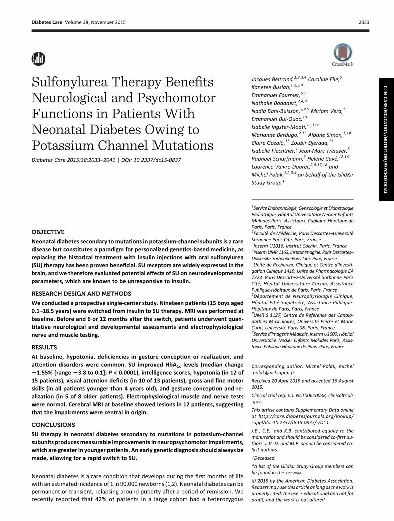
Table 1—Characteristics of the 18 study patients
Mutations n of patients; patient no.
KCNJ11G228A n = 1; 13E227K n = 1; 14E292G n = 1; 10H186D n = 1; 5I182T n = 1; 9Q51G n = 1; 8R201C n = 2; 16, 18R201H n = 7; 1, 2, 3, 7, 11, 15, 17V59M n = 1; 12
ABCC8R1183W n = 1; 4R1380H n = 1; 6
Characteristics Before SU therapyDuring SU therapy,month 12 or 18§
Age at SU initiation, years, median (range) 5.3 (0.1–18.5)
Males, n (%) 13 (72)
HbA1c, %, median (range) 7.75 (5.5–12.8) 6.4 (5.4–10)
Basal C-peptide, ng/mL, median (range)* 0.07 (0.02–0.51) 0.28 (0.12–0.82)**
Stimulated C-peptide, ng/mL,median (range)* 0.1 (0.05–1.44) 0.74 (0.2–1.99)**
Glibenclamide dosage, mg/kg/day,median (range) 0.2 (0–1.43)
*C-peptide measured using a glucagon stimulation test; **P , 0.01; §month 12 for basal andstimulated C-peptide and month 18 for HbA1c and glibenclamide dosage.
9Service de Neurologie Pediatrique, Hopital Uni-versitaire Necker Enfants Malades Paris, Assis-tance Publique-Hopitaux de Paris, Paris, France10Service d’Ophtalmologie, Hopital UniversitaireRobert Debre, Assistance Publique-Hopitaux deParis, Paris, France11Service d’Ophtalmologie, Hopital UniversitaireNecker Enfants Malades Paris, Assistance Publique-Hopitaux de Paris, Paris, France12Faculte de Medecine Paris-Diderot, UniversiteSorbonne-Paris-Cite, Paris, France
13Inserm U1138, Centre de Recherche desCordeliers, Universite Pierre etMarie Curie, Paris,France14Service de Pediatrie, Centre Hospitalier de Ver-sailles, Le Chesnay, France15Laboratoire de Pharmacologie-Toxicologie,Hopital Maison Blanche, Centre Hospitalier etUniversitaire de Reims, Reims, France16Departement de Genetique, Assistance Publi-que-Hopitaux de Paris, Hopital Universitaire
Robert Debre, Assistance Publique-Hopitaux deParis, Paris, France17Service d’Obstetrique et de Gynecologie,Hopitaux Universitaires Paris Centre, Cochin PortRoyal, Assistance Publique-Hopitaux de Paris,Paris, France18Inserm UMR 1178, Service de Pedopsychiatrie,Hopital Universitaire Necker Enfants MaladesParis, Universites Paris Sud et Paris Descartes,Paris, France
2034 Neuropsychomotor Improvements With SU Diabetes Care Volume 38, November 2015
The switch from insulin to oral SU(glibenclamide) was performed as previ-ously described (5,6). Patients were evalu-ated2, 6, 12, and18months after inclusion.
French Neuromotor Functions inChildren BatteryThe French Neuromotor Functions inChildren (NP-MOT) battery (11) was per-formed at baseline and then after12 months to evaluate developmentvia qualitative (movements) and quanti-tative (speeds) assessments of muscletone, gross motor control, laterality,praxis, gnosopraxis (12), digital and man-ual dexterity, body spatial integration,rhythmic tasks, and an auditory atten-tional task (12,13). (See SupplementaryData for details.) The NP-MOT batteryis a standardized normative instrumentwith identical subtests for all ages (andexpected saturation for patients aged8 years or older) developed and validatedby L.V.-D. For most of the tests, the cut-offs vary with age. Test scores are stan-dardized according to scoring guidelinesand expressed as SD of the population
mean (failure if,1 SD) or as the percen-tile (failure if,20th percentile).
Overall test-retest reliability of the NP-MOT has been reported to range from 70to 98% (11), and correlation coefficientswith the Lincoln-Oseretsky motor devel-opment scale (similar to the Bruininks-Oseretsky Test of Motor Proficiency [14]for upper-limb coordination, balance, andbilateral coordination subtests) were 0.72and 0.84 in two studies (15,16).
Developmental, Language, andSociability AssessmentThe same pediatric neurologist conducteda thorough neurological evaluation at base-line and then 6 and 12 months after SUinitiation. An electroencephalogram wasrecorded at baseline in all patients.
Intellectual performance was eval-uated at baseline and then after12 months by the same examiner. Allchildren completed all subtests of a stan-dard measure of intelligence (Brunet-Lezine test, Wechsler Preschool andPrimary Scale of Intelligence–Revised[17], Wechsler Intelligence Scale for
Children–Fourth Edition [18], orWechslerAdult Intelligence Scale–Third Edition).Other specific standardized tests con-sisted of visual-perceptual-motor testsassessing visual construction skills (repro-duction of a block design [19]), visual-spatial structuring (manual copy followedby visual-spatial memory of a complex geo-metric figure [20]), and the DevelopmentTest of Visual-Motor Integration (21),which involves manually copying 24 geo-metric drawings of progressively increas-ing complexity. Visual-spatial attentionwas assessed using a bell-crossing test(22) similar to that developed by Gauthier,Dehaut, and Joanette. Mental execu-tive function was evaluated using thePorteus Maze Test (23) and the Towerof London test (24). In addition, the pa-tients performed visual perception (vi-sual gnosis) tasks (recognizing tangledlines and naming animals seen in outlinefrom the rear) (25) and a language screen-ing battery (22). Hyperactivity was definedusing theDSMcriteria. Children’s behaviorwas observed by the same examiner atinclusion and 12 months after the switch.The examiner recorded the symptomscharacterizing the disorder: inattentive-ness, impulsivity, and overactivity. Pa-rents were administered an interviewcovering a broad range of child behaviors.As a part of this interview, parents wereasked about the presence/absence of hy-peractivity symptoms.
Electrophysiological Assessment ofVisual FunctionEach patient underwent an electroretino-gram and visual evoked potential record-ings, as recommended by the InternationalSociety for Clinical Electrophysiology of Vi-sion (26,27), at baseline and then6monthslater. Flash visual evoked potentials wererecorded in younger patients (monocular,patient no. 10, or binocular if patching aneye was not possible, patient nos. 1, 6, 18,15, 7, 12, and 14) and pattern reversalvisual evoked potentials in older patients(100% contrast; reversal at 1.0 Hz; 60’, 30’,or 15’ squares, patient nos. 4, 9, 11, 17, 3,5, and 16). No recordings were performedin patient nos. 2 and 13.
Electrophysiological Muscle andNerve TestingElectrophysiological testing was per-formed at baseline and then after 6and 12 months in children older than 6years of age and carrying a KCNJ11 mu-tation (as SUR1 expressed by ABCC8 is
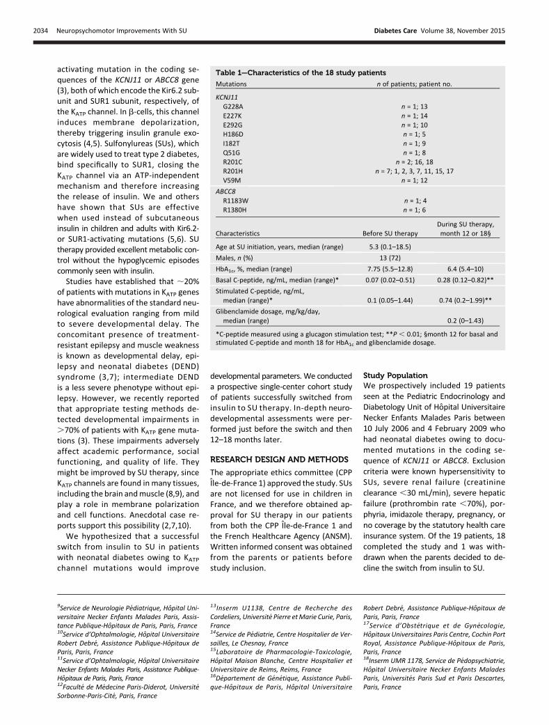
Table 2—Assessment using NP-MOT at baseline and then 12 months afterstarting SU therapy in 8 patients younger than 4 years of age at baseline(patient nos. 1–8)
Abnormal atbaseline
Abnormal after12 months of SU
Median scoredifference (range)
TonePassive tone 6 (75) 1 (12.5) 21 (21 to 0), P = 0.06**
Improved n = 5Stable n = 1
Still normal n = 2Standing tone (score ,8) 6 (75) 0 (0%) 2 (026), P = 0.03**
Improved n = 6Still normal n = 2
MotricityGross motor skills
(delay .2 months) 3 (37.5) 1 (12.5) 2 (27 to 3), P = 0.19**Improved n = 3
Degradation n = 1Still normal n = 4
Fine motor skills(delay .2 months) 5 (62.5) 2 (25) 1 (25.5 to 3.5), P = 0.07**
Improved n = 3Stable n = 2
Still normal n = 3
AttentionAttention 3 (37.5) 1 (12.5) P = 0.48*
Improved n = 2Stable n = 1
Still normal n = 5Hyperactivity 2 (33.3) 0 d
Improved n = 2Still normal n = 6
Data are n (%) unless otherwise indicated. The value presented in the parentheses in the lastcolumn is the range of score differences in values for the patients. The lowest score is21 and 0 isthe highest. *McNemar x2 test; **Paired Wilcoxon test on continuous scores.
care.diabetesjournals.org Beltrand and Associates 2035
not expressed in the muscle). A stan-dardized protocol ensuring reproducibleand painless electrophysiological testingof skeletal muscle excitability was used(28). Briefly, compound muscle action
potentials were recorded from the rightand left abductor digiti minimi muscles af-ter supramaximal electrical stimulation ofthe ulnar nerves at the wrists. Recordingswere repeated before and after voluntary
contraction of the recorded muscle. If theresponse changed after exercise, care wastaken to check that the electrode positionswereunchangedand thatnerve stimulationremained supramaximal.
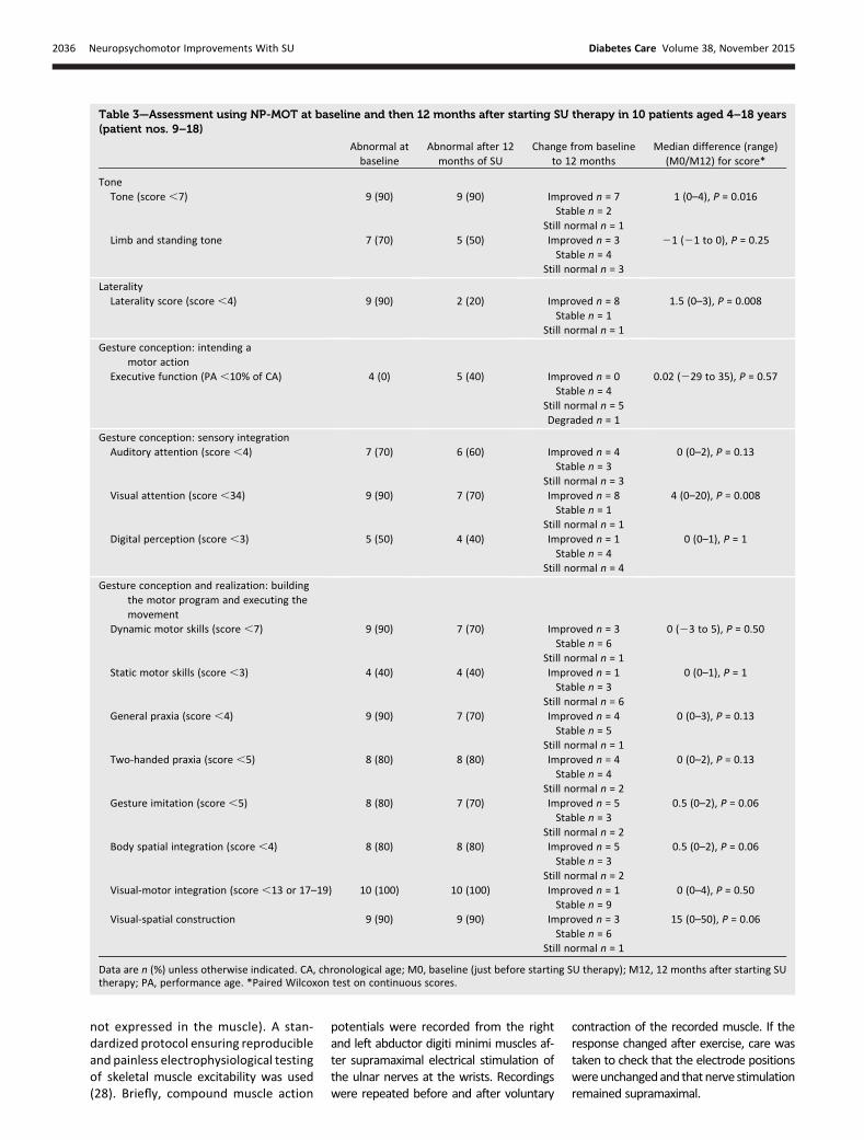
Table 3—Assessment using NP-MOT at baseline and then 12 months after starting SU therapy in 10 patients aged 4–18 years(patient nos. 9–18)
Abnormal atbaseline
Abnormal after 12months of SU
Change from baselineto 12 months
Median difference (range)(M0/M12) for score*
ToneTone (score ,7) 9 (90) 9 (90) Improved n = 7 1 (0–4), P = 0.016
Stable n = 2Still normal n = 1
Limb and standing tone 7 (70) 5 (50) Improved n = 3 21 (21 to 0), P = 0.25Stable n = 4
Still normal n = 3
LateralityLaterality score (score ,4) 9 (90) 2 (20) Improved n = 8 1.5 (0–3), P = 0.008
Stable n = 1Still normal n = 1
Gesture conception: intending amotor action
Executive function (PA ,10% of CA) 4 (0) 5 (40) Improved n = 0 0.02 (229 to 35), P = 0.57Stable n = 4
Still normal n = 5Degraded n = 1
Gesture conception: sensory integrationAuditory attention (score ,4) 7 (70) 6 (60) Improved n = 4 0 (0–2), P = 0.13
Stable n = 3Still normal n = 3
Visual attention (score ,34) 9 (90) 7 (70) Improved n = 8 4 (0–20), P = 0.008Stable n = 1
Still normal n = 1Digital perception (score ,3) 5 (50) 4 (40) Improved n = 1 0 (0–1), P = 1
Stable n = 4Still normal n = 4
Gesture conception and realization: buildingthe motor program and executing themovement
Dynamic motor skills (score ,7) 9 (90) 7 (70) Improved n = 3 0 (23 to 5), P = 0.50Stable n = 6
Still normal n = 1Static motor skills (score ,3) 4 (40) 4 (40) Improved n = 1 0 (0–1), P = 1
Stable n = 3Still normal n = 6
General praxia (score ,4) 9 (90) 7 (70) Improved n = 4 0 (0–3), P = 0.13Stable n = 5
Still normal n = 1Two-handed praxia (score ,5) 8 (80) 8 (80) Improved n = 4 0 (0–2), P = 0.13
Stable n = 4Still normal n = 2
Gesture imitation (score ,5) 8 (80) 7 (70) Improved n = 5 0.5 (0–2), P = 0.06Stable n = 3
Still normal n = 2Body spatial integration (score ,4) 8 (80) 8 (80) Improved n = 5 0.5 (0–2), P = 0.06
Stable n = 3Still normal n = 2
Visual-motor integration (score ,13 or 17–19) 10 (100) 10 (100) Improved n = 1 0 (0–4), P = 0.50Stable n = 9
Visual-spatial construction 9 (90) 9 (90) Improved n = 3 15 (0–50), P = 0.06Stable n = 6
Still normal n = 1
Data are n (%) unless otherwise indicated. CA, chronological age; M0, baseline (just before starting SU therapy); M12, 12 months after starting SUtherapy; PA, performance age. *Paired Wilcoxon test on continuous scores.
2036 Neuropsychomotor Improvements With SU Diabetes Care Volume 38, November 2015
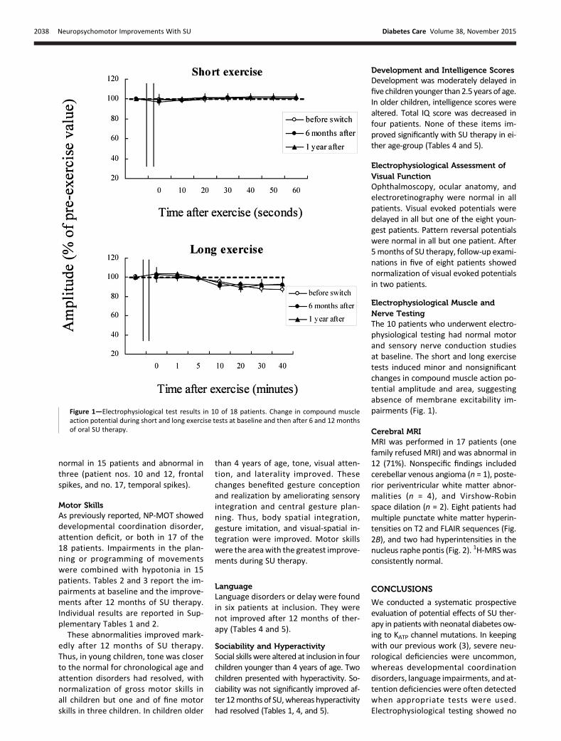
Two provocative testswere performed.The first was a repeated short exercisetest at room temperature on the lefthand (three maximal, isometric, 10-s ab-ductor digiti minimi contractions sepa-rated by 50-s rest periods during whichcompound muscle action potentialswere recorded every 5–10 s). Then, along exercise test was performed atroom temperature on the right hand(maximal, isometric, 5-min abductor dig-iti minimi contraction followed by com-pound muscle action potential recordingevery 5min for 40min). Compoundmus-cle action potential amplitude, total du-ration, and total area were expressed aspercentages of pre-exercise values.Needle electrode recordings were
obtained from five muscles (deltoid, ex-tensor digitorum communis, first inter-osseus dorsalis, vastus medialis, andtibialis anterior) to look for myotonic dis-charges or evidence of myopathy. Fi-nally, motor conduction of the ulnarand peroneal nerves and sensory con-duction of the right and left superficialperoneal nerves were measured. Ampli-tude, latency, and conduction velocity ofthe electrophysiological signals werecompared with normal values for thelaboratory.
Cerebral MRIMRI was performed at baseline. High-resolution images were acquired using a1.5-T Signa Systemmachine (GEHealthcare,Milwaukee, WI) with a three-dimensionalT1-weighted fast spoiled gradient recalledimaging sequence (repetition time [TR]/echo time [TE]/inversion time/NEX: 10.5/2.2/600/1, 108, matrix 256 z 192; 124 axialslices, 1.2-mm thickness, 124 contigu-ous slices, 22 cm field of view), an axialfast spin echo T2-weighted sequence(TR/TE: 6,000/120, 4-mm slices, 0.5 mmgap, 22 cm field of view), coronal fluid-attenuated inversion recovery (FLAIR)sequences (TR/TE/TI: 10,000/150/2,250,4-mm slices, 1-mm gap, 24 cm field ofview), and 1H-MRS.
Pharmacological AssessmentPlasma glibenclamide concentrationswere determined after standard liquid-liquid extraction by liquid chromatography–ion-trap tandem mass spectrometry aspreviously described (29).
Statistical AnalysisData were described as median (range orinterquartile range where indicated) forquantitative variables and as number(percentage) for qualitative variables.Comparisons of data at different time
points were performed with the pairedWilcoxon or McNemar test. The nonlinearmixed-effect modeling programNONMEM(version VII, release 1) was used to com-pute the area under the curve of gliben-clamide concentrations over 24 h.
RESULTS
Study PopulationWe studied 18 patients aged 5 months to18 years; eight were younger than 4 yearsold.Metabolic control improved after theswitch to glibenclamide therapy, and nopatients experienced hypoglycemia. Onepatient had a remission allowing gliben-clamide discontinuation after the 12-month evaluation (Table 1).
Median glibenclamide dosage was0.37 mg/kg/day (range 0–1.4) at month 18.Medianplasmaglibenclamideconcentrationwas 50 mg/L (interquartile range 21.5–118), andmedian area under the time curveover 24 h was 1,335 mg z L/h (511–2,122).
Neurological EvaluationNeurological impairments were foundin a single patient (patient no. 17) and con-sisted of a global pyramidal syndromewithspasticity and mild walking disability, mildmental retardation, and seizures (DEND).The baseline electroencephalogram was
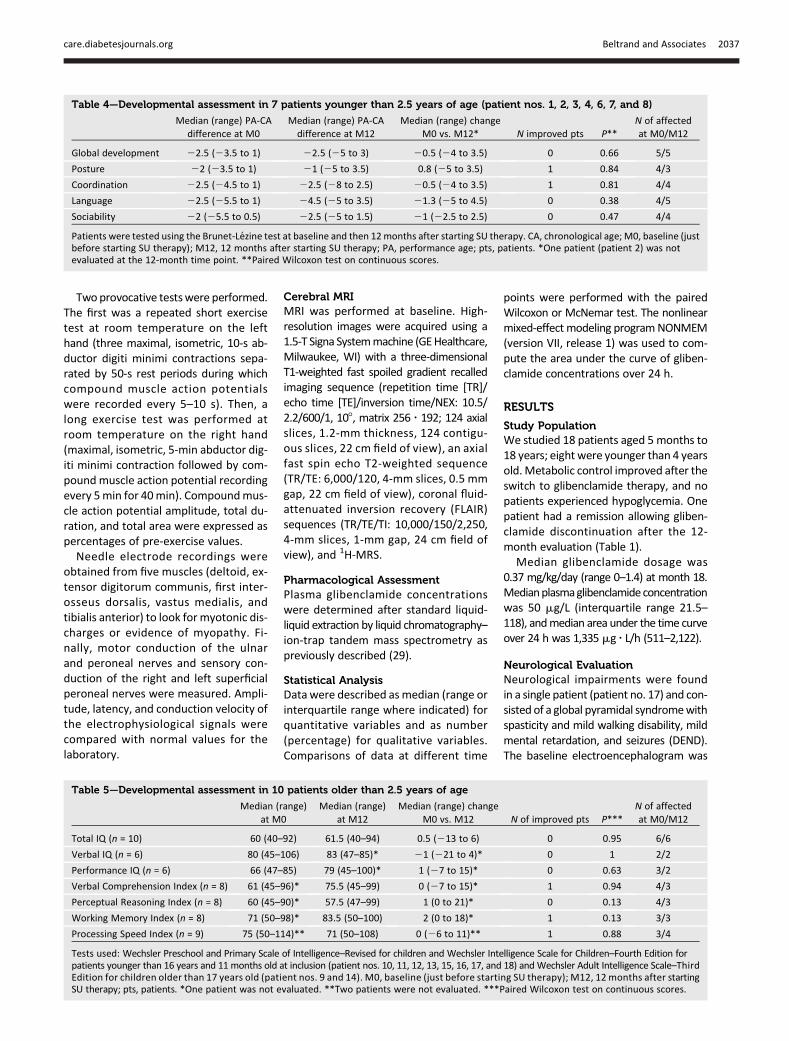
Table 4—Developmental assessment in 7 patients younger than 2.5 years of age (patient nos. 1, 2, 3, 4, 6, 7, and 8)
Median (range) PA-CAdifference at M0
Median (range) PA-CAdifference at M12
Median (range) changeM0 vs. M12* N improved pts P**
N of affectedat M0/M12
Global development 22.5 (23.5 to 1) 22.5 (25 to 3) 20.5 (24 to 3.5) 0 0.66 5/5
Posture 22 (23.5 to 1) 21 (25 to 3.5) 0.8 (25 to 3.5) 1 0.84 4/3
Coordination 22.5 (24.5 to 1) 22.5 (28 to 2.5) 20.5 (24 to 3.5) 1 0.81 4/4
Language 22.5 (25.5 to 1) 24.5 (25 to 3.5) 21.3 (25 to 4.5) 0 0.38 4/5
Sociability 22 (25.5 to 0.5) 22.5 (25 to 1.5) 21 (22.5 to 2.5) 0 0.47 4/4
Patients were tested using the Brunet-Lezine test at baseline and then 12months after starting SU therapy. CA, chronological age; M0, baseline (justbefore starting SU therapy); M12, 12 months after starting SU therapy; PA, performance age; pts, patients. *One patient (patient 2) was notevaluated at the 12-month time point. **Paired Wilcoxon test on continuous scores.
Table 5—Developmental assessment in 10 patients older than 2.5 years of age
Median (range)at M0
Median (range)at M12
Median (range) changeM0 vs. M12 N of improved pts P***
N of affectedat M0/M12
Total IQ (n = 10) 60 (40–92) 61.5 (40–94) 0.5 (213 to 6) 0 0.95 6/6
Verbal IQ (n = 6) 80 (45–106) 83 (47–85)* 21 (221 to 4)* 0 1 2/2
Performance IQ (n = 6) 66 (47–85) 79 (45–100)* 1 (27 to 15)* 0 0.63 3/2
Verbal Comprehension Index (n = 8) 61 (45–96)* 75.5 (45–99) 0 (27 to 15)* 1 0.94 4/3
Perceptual Reasoning Index (n = 8) 60 (45–90)* 57.5 (47–99) 1 (0 to 21)* 0 0.13 4/3
Working Memory Index (n = 8) 71 (50–98)* 83.5 (50–100) 2 (0 to 18)* 1 0.13 3/3
Processing Speed Index (n = 9) 75 (50–114)** 71 (50–108) 0 (26 to 11)** 1 0.88 3/4
Tests used: Wechsler Preschool and Primary Scale of Intelligence–Revised for children and Wechsler Intelligence Scale for Children–Fourth Edition forpatients younger than 16 years and 11 months old at inclusion (patient nos. 10, 11, 12, 13, 15, 16, 17, and 18) andWechsler Adult Intelligence Scale–ThirdEdition for children older than 17 years old (patient nos. 9 and 14). M0, baseline (just before starting SU therapy); M12, 12 months after startingSU therapy; pts, patients. *One patient was not evaluated. **Two patients were not evaluated. ***Paired Wilcoxon test on continuous scores.
care.diabetesjournals.org Beltrand and Associates 2037
normal in 15 patients and abnormal inthree (patient nos. 10 and 12, frontalspikes, and no. 17, temporal spikes).
Motor SkillsAs previously reported, NP-MOT showeddevelopmental coordination disorder,attention deficit, or both in 17 of the18 patients. Impairments in the plan-ning or programming of movementswere combined with hypotonia in 15patients. Tables 2 and 3 report the im-pairments at baseline and the improve-ments after 12 months of SU therapy.Individual results are reported in Sup-plementary Tables 1 and 2.These abnormalities improved mark-
edly after 12 months of SU therapy.Thus, in young children, tone was closerto the normal for chronological age andattention disorders had resolved, withnormalization of gross motor skills inall children but one and of fine motorskills in three children. In children older
than 4 years of age, tone, visual atten-tion, and laterality improved. Thesechanges benefited gesture conceptionand realization by ameliorating sensoryintegration and central gesture plan-ning. Thus, body spatial integration,gesture imitation, and visual-spatial in-tegration were improved. Motor skillswere the areawith the greatest improve-ments during SU therapy.
LanguageLanguage disorders or delay were foundin six patients at inclusion. They werenot improved after 12 months of ther-apy (Tables 4 and 5).
Sociability and HyperactivitySocial skills were altered at inclusion in fourchildren younger than 4 years of age. Twochildren presented with hyperactivity. So-ciability was not significantly improved af-ter 12months of SU,whereas hyperactivityhad resolved (Tables 1, 4, and 5).
Development and Intelligence ScoresDevelopment was moderately delayed infive children younger than2.5 years of age.In older children, intelligence scores werealtered. Total IQ score was decreased infour patients. None of these items im-proved significantly with SU therapy in ei-ther age-group (Tables 4 and 5).
Electrophysiological Assessment ofVisual FunctionOphthalmoscopy, ocular anatomy, andelectroretinography were normal in allpatients. Visual evoked potentials weredelayed in all but one of the eight youn-gest patients. Pattern reversal potentialswere normal in all but one patient. After5months of SU therapy, follow-up exami-nations in five of eight patients showednormalization of visual evoked potentialsin two patients.
Electrophysiological Muscle andNerve TestingThe 10 patients who underwent electro-physiological testing had normal motorand sensory nerve conduction studiesat baseline. The short and long exercisetests induced minor and nonsignificantchanges in compound muscle action po-tential amplitude and area, suggestingabsence of membrane excitability im-pairments (Fig. 1).
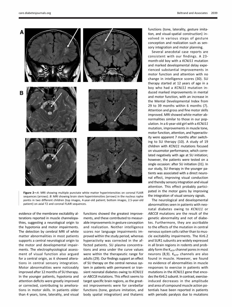
Cerebral MRIMRI was performed in 17 patients (onefamily refused MRI) and was abnormal in12 (71%). Nonspecific findings includedcerebellar venous angioma (n = 1), poste-rior periventricular white matter abnor-malities (n = 4), and Virshow-Robinspace dilation (n = 2). Eight patients hadmultiple punctate white matter hyperin-tensities on T2 and FLAIR sequences (Fig.2B), and two had hyperintensities in thenucleus raphe pontis (Fig. 2). 1H-MRSwasconsistently normal.
CONCLUSIONS
We conducted a systematic prospectiveevaluation of potential effects of SU ther-apy in patients with neonatal diabetes ow-ing to KATP channel mutations. In keepingwith our previous work (3), severe neu-rological deficiencies were uncommon,whereas developmental coordinationdisorders, language impairments, and at-tention deficiencies were often detectedwhen appropriate tests were used.Electrophysiological testing showed no
Figure 1—Electrophysiological test results in 10 of 18 patients. Change in compound muscleaction potential during short and long exercise tests at baseline and then after 6 and 12 monthsof oral SU therapy.
2038 Neuropsychomotor Improvements With SU Diabetes Care Volume 38, November 2015
evidence of the membrane excitability al-terations reported in muscle channelopa-thies, suggesting a neurological origin tothe hypotonia and motor impairments.The detection by cerebral MRI of whitematter abnormalities in most patientssupports a central neurological origin tothe motor and developmental impair-ments. The electrophysiological assess-ment of visual function also arguedfor a central origin, as it showed altera-tions in central sensory integration.Motor abnormalities were noticeablyimproved after 12months of SU therapy.In the younger patients, hypotonia andattention deficits were greatly improvedor corrected, contributing to ameliora-tions in motor skills. In patients olderthan 4 years, tone, laterality, and visual
functions showed the greatest improve-ments, and these contributed to measur-able improvements in gesture conceptionand realization. Neither intelligencescores nor language impairments im-proved within the study period, whereashyperactivity was corrected in the af-fected patients. SU plasma concentra-tions and area under the curve valueswere within the therapeutic range foradults (29). Our findings support an effectof SU therapy on the central nervous sys-tem in patients with permanent or tran-sient neonatal diabetes owing to KCNJ11or ABCC8mutations. This effect seems totarget specific brain regions, as the great-est improvements were for cerebellarfunctions (tone, gesture imitation, andbody spatial integration) and thalamic
functions (tone, laterality, gesture imita-tion, and visual-spatial construction) in-volved in various steps of gestureconception and realization such as sen-sory integration and motor planning.
Several anecdotal case reports areconsistent with our findings. A 23-month-old boy with a KCNJ11 mutationand marked developmental delay expe-rienced substantial improvements inmotor function and attention with nochange in intelligence scores (30). SUtherapy started at 12 years of age in aboy who had a KCNJ11 mutation in-duced marked improvements in mentaland motor function, with an increase inthe Mental Developmental Index from29 to 39 months within 6 months (7).Attention and gross and fine motor skillsimproved. MRI showed white matter ab-normalities similar to those in our pop-ulation. In a 6-year-old girl with a KCNJ11mutation, improvements in muscle tone,motor function, attention, and hyperactiv-ity were apparent 7 months after switch-ing to SU therapy (10). A study of 19children with KCNJ11 mutations focusedon visuomotor performance, which corre-lated negatively with age at SU initiation;however, the patients were tested on asingle occasion: after SU initiation (31). Inour study, SU therapy in the younger pa-tients was associated with a direct neuro-nal effect, improving visual conductionand thereby sensory integration and visualattention. This effect probably partici-pated in the motor gains by improvingthe integration of visual sensory signals.
The neurological and developmentalabnormalities seen in patients with neo-natal diabetes owing to KCNJ11 orABCC8 mutations are the result of thegenetic abnormality and not of diabe-tes. Furthermore, they are ascribableto the effects of the mutation in centralnervous system cells rather than tomus-cle excitability impairments. The Kir6.2and SUR1 subunits are widely expressedin all brain regions in rodents and prob-ably form the KATP channel pores inmostneurons (8,9). KATP channels are alsofound in muscle. However, we foundno evidence of abnormalities in muscleresponses to exercise in patients withmutations in the KCNJ11 gene that enco-des theKir6.2 subunit. In contrast, exercise-induced decreases in the amplitudeand area of compoundmuscle action po-tentials have been reported in patientswith periodic paralysis due to mutations
Figure 2—A: MRI showing multiple punctate white matter hyperintensities on coronal FLAIRsequences (arrows). B: MRI showing brain stem hyperintensities (arrows) in the nucleus raphepontis in two different children (top images, 4-year-old patient; bottom images, 2.5-year-oldpatient) on axial T2 and coronal FLAIR sequences.
care.diabetesjournals.org Beltrand and Associates 2039
in sodium (SCN4A), calcium (CACNA1S), orKir2.1 potassium (KCNJ2) channel genes(28,32). Thus, the muscle disorders in pa-tients with KCNJ11mutations do not seemascribable to changes in muscle mem-brane excitability. Furthermore, they areascribable to the effects of the mutationin central nervous system cells rather thanto muscle excitability impairments (33).None of our patients had a history of re-current severe hypoglycemia. The MRIchanges in our patients differ from thosereported in children with type 1 diabetes(34,35). These last two facts support thedirect effect on the central nervous systemrather than an indirect effect due to im-proved metabolic control.Limitations of our study include the
small sample size, although we includedmost of the patients in France who hadneonatal diabetes owing to potassium-channel subunit mutations and had notyet been switched to SU. A controlgroup would have been ethically unac-ceptable. The short study period wassufficient to see substantial coordina-tion disorders improvement allowingcomplex tasks such as handwriting tobe better performed. If we found nomajor changes in language, intelligence,or sociability, these functions might im-prove with time, as this short study wassufficient to correct hyperactivity signs,or, alternatively, SUs may fail to targetthe brain regions involved. A majorstrength of our study is the use of a stan-dardized normative neurodevelopmen-tal test battery validated in normalFrench children and performed by a sin-gle examiner. This battery allowed us toobtain accurate information about SUeffects on developmental parameters.SUs have already been suggested as a
neuroprotective drug after strokes (36,37).Our findings strongly suggest that SUtherapy improves neurodevelopmentalparameters in patients with neonataldiabetes owing to potassium-channel sub-unit mutations and acts via a centralmechanism. The greater improvements inyounger patients indicate a need for estab-lishing the diagnosis early to allow promptinitiation of SU therapy. Further follow-upof our patients will provide informationabout the kinetics of SU effects on neuro-developmental parameters.
Acknowledgments. The authors thank PaulCzernichow and Jean Jacques Robert for their
long-standing support to studies on neonataldiabetes and Philippe Froguel, Martine Vaxil-laire, and Amelie Bonnefont, Institut Pasteur,Lille, France, for their cooperation with workon identifying genes responsible for neonataldiabetes. The authors thank Myriam Faivreand the nursing team of the Pediatric Endocri-nology and Diabetology Department, as well asSandra Colas for her help in managing the pro-tocol and the clinical research unit teamdbothat Hopital Universitaire Necker Enfants MaladesParis.Funding. This study was sponsored by Assis-tance Publique-Hopitaux de Paris and received agovernment grant managed by Agence Nationalede la Recherche under the “Investments for theFuture” program (reference ANR-10-IAHU-01).The work was performed within DepartementHospitalo-Universitaire AUToimmune and HOR-monal diseaseS. It was partly funded by AgenceNationale de la Recherche–Maladies RaresResearch Program grant ANR-07-MRAR-000(to M.P.), Transnational European ResearchGrant on Rare Diseases grant ERANET-09-RARE-005 (to M.P.), and Societe Francophonedu Diabete–Association Française du Diabete(to M.P.). K.B. received a CIFRE grant from theFrench government and was supported by theFrench Ministry of Higher Education and Re-search and Societe Française de Pediatrie.Support was also received from LabEx Reviveand from the Bettencourt-Schueller Founda-tion (R.S.) and Aide aux Jeunes Diabetiques(to M.P.).Duality of Interest. K.B. was supported by HRAPharma. No other potential conflicts of interestrelevant to this article were reported.Author Contributions. J.B., K.B., and A.S.collected data. J.B., C.E., K.B., and M.P. wrotethe manuscript. J.B., C.E., M.B., and J.M.-T.analyzed data. K.B., A.S., I.F., and M.P. designedthe study. E.F. performed the electromyographyand analyzed the results. N.B. performed theMRI and analyzed the results. N.B.-B. performedthe neurological examination. M.V. performedthe intelligence tests. E.B.-Q., I.I.-M., and M.B.performed the electrophysiological assessmentof visual function and analyzed the results. C.G.and Z.D. performed the pharmacological assess-ment. R.S. reviewed the manuscript. H.C. per-formed the genetic analysis. L.V.-D. performedthe neuropsychomotor tests. M.P. is the guar-antor of this work and, as such, had full access toall the data in the study and takes responsibilityfor the integrity of the data and the accuracy ofthe data analysis.Prior Presentation. Parts of this study werepresented in abstract form at the Annual Meetingof the European Society for Paediatric Endocrinol-ogy,Dublin, Ireland, 18–21September2014, andatthe 40th International Society for Pediatric andAdolescent Diabetes Conference, Toronto, ON,Canada, 3–6 September 2014.
AppendixThe GlidKir Study Group members are as follows:Claire Le Tallec and Nicole Ser, Departement dePediatrie, CHU Toulouse, Toulouse, France; SylvieNivot-Adamiak and Marc de Kerdanet, Service dePediatrie, CHU Rennes, Rennes, France; MaryseCartigny and Jacques Weill, Service de Pediatrie,CHU Jeanne de Flandre, Lille, France; Sabine Baronand Emmanuelle Ramos-Caldagues, Service de
Pediatrie, CHU Nantes, Nantes, France; HenriBruel, Service de Pediatrie, Hopital de Le Havre,Le Havre, France; Anne Lienhardt-Roussie, Servicede Pediatrie, CHU Limoges, Limoges, France; Guy-Andre Loeuille, Service de Pediatrie, Hopitalde Dunkerque, Dunkerque, France; BertheRazafimahefa, Pediatrie-Nouveaux nes, HopitalGeorges Sand, La Seyne surMer, France; and RachelReynaud and Gilbert Simonin, Service de Pediatrie,Hopital La Timone, Marseilles, France.
References1. Iafusco D, Massa O, Pasquino B, et al.; EarlyDiabetes Study Group of ISPED.Minimal incidenceof neonatal/infancy onset diabetes in Italy is 1:90,000 live births. Acta Diabetol 2012;49:405–4082. Slingerland AS, Shields BM, Flanagan SE, et al.Referral rates for diagnostic testing support an in-cidence of permanent neonatal diabetes in threeEuropean countries of at least 1 in 260,000 livebirths. Diabetologia 2009;52:1683–16853. Busiah K, Drunat S, Vaivre-Douret L, et al.;French NDM study group. Neuropsychologicaldysfunction and developmental defects associ-ated with genetic changes in infants with neo-natal diabetes mellitus: a prospective cohortstudy [corrected]. Lancet Diabetes Endocrinol2013;1:199–2074. Gloyn AL, Pearson ER, Antcliff JF, et al. Acti-vating mutations in the gene encoding the ATP-sensitive potassium-channel subunit Kir6.2 andpermanent neonatal diabetes. N Engl J Med2004;350:1838–18495. Babenko AP, Polak M, Cave H, et al. Activatingmutations in theABCC8gene inneonatal diabetesmellitus. N Engl J Med 2006;355:456–4666. Pearson ER, Flechtner I, Njølstad PR, et al.;Neonatal Diabetes International CollaborativeGroup. Switching from insulin to oral sulfonylureasin patients with diabetes due to Kir6.2 mutations.N Engl J Med 2006;355:467–4777. Slingerland AS, Hurkx W, Noordam K, et al.Sulphonylurea therapy improves cognition in apatient with the V59MKCNJ11mutation. DiabetMed 2008;25:277–2818. Li B, Xi X, Roane DS, Ryan DH, Martin RJ.Distribution of glucokinase, glucose transporterGLUT2, sulfonylurea receptor-1, glucagon-likepeptide-1 receptor and neuropeptide Y messen-ger RNAs in rat brain by quantitative real time RT-PCR. Brain Res Mol Brain Res 2003;113:139–1429. Thomzig A, Laube G, Pruss H, Veh RW. Pore-forming subunits of K-ATP channels, Kir6.1 andKir6.2, display prominent differences in regionaland cellular distribution in the rat brain. J CompNeurol 2005;484:313–33010. Mlynarski W, Tarasov AI, Gach A, et al. Sulfo-nylurea improves CNS function in a case of inter-mediate DEND syndrome caused by amutation inKCNJ11. Nat Clin Pract Neurol 2007;3:640–64511. Vaivre-Douret L. Batterie d’evaluation desfonctions neuro-psychomotrices (NP-MOT) del’enfant. Paris, France, Editions du Centre dePsychologie Appliquee, 2006 [in French]12. Vaivre-Douret L. A more robust predictor ofideomotor dyspraxia: study on an alternative scor-ingmethod of the Berges-Lezine’s Imitation of Ges-tures test. Arch Clin Neuropsychol 2002;17:37–4813. Vaivre-Douret L. Evaluation of the DistalMotricity Control, Revision and AdaptationBerges-Lezine’s Test. Paris, France, Editions du Cen-tre de Psychologie Appliquee, 1997 [in French]
2040 Neuropsychomotor Improvements With SU Diabetes Care Volume 38, November 2015
14. Bruininks H. Bruininsks-Oserestky Test ofMotor Proficiency. Circles Pines, MN, AmericanGuidance Service, 197815. Vaivre-Douret L. Developmental and cognitivecharacteristics of “high-level potentialities” (highlygifted) children. Int J Pediatr 2011;2011:42029716. Vaivre-Douret L, Lalanne C, Ingster-Moati I,et al. Subtypes of developmental coordinationdisorder: research on their nature and etiology.Dev Neuropsychol 2011;36:614–64317. Wechsler D. Wechsler Preschool and Pri-mary Scale of Intelligence. San Antonio, TX,The Psychological Corporation, 198918. Wechsler D. Manual for the Wechsler Pre-school and Primary Scale of Intelligence-Revised.San Antonio, TX, The Psychological Corporation, 198919. Khos C. Test des Cubes de Khos. Paris, France,Editions du Centre de Psychologie Appliquee,1972 [in French]20. Rey A. Manuel test de copie d’une figurecomplexe. Paris, France, Editions du Centre dePsychologie Appliquee, 1959 [in French]21. Beery KE. Revised Administration Scoring andTeaching Manual for the Development Test ofVisual-Motor Integration (VMI). Toronto, ON,Canada, Modern Curriculum Press, 198222. Jaquier-Roux M, Valdois S, Zorman M, et al.ODEDYS Outil de depistage des dyslexies, version2, Cogni-Sciences, IUFM, Paris, France, 2005[in French]23. Porteus SD. Test des Labyrinthes de Por-teus. Paris, France, Editions du Centre de Psy-chologie Appliquee, 1952 [in French]
24. KorkmanMKU.Developmental Neuropsycho-logical AssessmentManual. Paris, France, Editionsdu Centre de Psychologie Appliquee, 200325. Rey A. L’examen psychologique dans le casd’encephalopathie traumatique. Arch Psychol1941;112:286–340 [in French]26. Odom JV, BachM, BrigellM, et al. ISCEV stan-dard for clinical visual evoked potentials (2009update). Doc Ophthalmol 2010;120:111–11927. Marmor MF, Fulton AB, Holder GE, MiyakeY, Brigell M, Bach M; International Society forClinical Electrophysiology of Vision. ISCEVstandard for full-field clinical electroretinogra-phy (2008 update). Doc Ophthalmol 2009;118:69–7728. Fournier E, Arzel M, Sternberg D, et al. Elec-tromyography guides toward subgroups of mu-tations in muscle channelopathies. Ann Neurol2004;56:650–66129. Hoizey G, Lamiable D, Trenque T, et al. Iden-tification andquantificationof 8 sulfonylureaswithclinical toxicology interest by liquid chromatogra-phy-ion-trap tandem mass spectrometry and li-brary searching. Clin Chem 2005;51:1666–167230. Slingerland AS, Nuboer R, Hadders-Algra M,Hattersley AT, Bruining GJ. Improved motor de-velopment and good long-term glycaemic con-trol with sulfonylurea treatment in a patientwith the syndrome of intermediate develop-mental delay, early-onset generalised epilepsyand neonatal diabetes associated with theV59M mutation in the KCNJ11 gene. Diabetolo-gia 2006;49:2559–2563
31. Shah RP, Spruyt K, Kragie BC, Greeley SA,MsallME. Visuomotor performance in KCNJ11-relatedneonatal diabetes is impaired in children withDEND-associated mutations and may be improvedby early treatment with sulfonylureas. DiabetesCare 2012;35:2086–208832. Bendahhou S, Fournier E, Sternberg D, et al.In vivo and in vitro functional characterization ofAndersen’s syndrome mutations. J Physiol 2005;565:731–74133. Clark RH, McTaggart JS, Webster R, et al.Muscle dysfunction caused by a KATP channelmutation in neonatal diabetes is neuronal inorigin. Science 2010;329:458–46134. Barnea-Goraly N, Raman M, Mazaika P,et al.; Diabetes Research in Children Network(DirecNet). Alterations in white matter struc-ture in young children with type 1 diabetes. Di-abetes Care 2014;37:332–34035. Marzelli MJ, Mazaika PK, Barnea-Goraly N,et al.; Diabetes Research in Children Network(DirecNet). Neuroanatomical correlates of dys-glycemia in young children with type 1 diabetes.Diabetes 2014;63:343–35336. Kunte H, Schmidt S, Eliasziw M, et al. Sulfo-nylureas improve outcome in patients withtype 2 diabetes and acute ischemic stroke.Stroke 2007;38:2526–253037. Simard JM, Yurovsky V, Tsymbalyuk N,Melnichenko L, Ivanova S, Gerzanich V. Protectiveeffect of delayed treatment with low-dose gliben-clamide in threemodels of ischemic stroke. Stroke2009;40:604–609
care.diabetesjournals.org Beltrand and Associates 2041