Embed Size (px)
Citation preview
1
Annex no. 2
SUMMARY OF PROFESSIONAL ACCOMPLISHMENTS
1. NAME AND SURNAME: Alicja UTRATA-WESOŁEK
2. DIPLOMAS AND SCIENTIFIC DEGREES:
June 2001 Master of Science, specialization Chemical Technology and Polymer Chemistry Faculty of Mathematics, Physics and Chemistry, Basic and Applied Chemistry, University of Opole
diploma thesis granted by PTChem “Janina Janikowa” award:
„Copolymerization of ethylene with 1-hexene using zirconocene catalyst supported
on MgCl2(THF)2”
Supervisor: Prof. dr hab. Krystyna Czaja
July 2006 Doctor of Chemical Sciences
Silesian University of Technology in Gliwice, Poland Faculty of Chemistry
Ph.D. thesis: “Stimuli sensitive polymers based upon reactive polyethers”, thesis grant-ed by the faculty the degree of excellency “summa cum laude”
Supervisors: Prof. dr hab. Andrzej Dworak; Prof. dr hab. Brigitte Voit
3. COURSE OF THE EMPLOYMENT – INFORMATION ABOUT PAST AND CURRENT EM-PLOYMENTS IN SCIENTIFIC UNITS
Positions:
november 2001 – october 2005 Ph.D. student at the Faculty of Chemistry, Silesian University of Technology
november 2005 – july 2007 specialist in Institute of Coal Chemistry Polish Academy of Sciences
july 2007 – march 2017 research scientist (adiunkt) in Centre of Polymer and Carbon Materials Polish
Academy of Sciences in Laboratory of Nano- and Microstructural Materials
march 2017 to date assistant in Centre of Polymer and Carbon Materials Polish Academy of Sciences
in Laboratory of Nano- and Microstructural Materials
Functions:
2011-2014 Member of the Scientific Council of the Centre of Polymer and Carbon Materials
Polish Academy of Sciences

2
4. PRESENTATION OF THR SCIENTIFIC ACHIEVEMENT
resulting from the article 16.2 of the act on scientific degrees and scientific titles and degrees and title in the field of art (Act no. 65, item 595 as amended)
(a) TITLE OF SCIENTIFIC ACHIEVEMENT The scientific achievement is the series of monothematic articles entitled:
„BIOCOMPATIBLE POLYMER LAYERS OF CONTROLLED AFFINITY FOR WATER. SYNTHESIS AND APPLICATION”
(b) List of selected publications constituting the scientific achievement in chronological order, with percentage contribution of habilitant to each work
Impact Factor (IF) – appropriate to the year of publication
[H1] Biocompatible cryogels of thermosensitive polyglycidol derivatives with ultra-rapid swelling properties
P. Petrov, A. Utrata-Wesołek, B. Trzebicka, Ch. B. Tsvetanov, A. Dworak, J. Anioł, A. Sieroń
European Polymer Journal 2011, 47, 981-988
IF=2.739
(contribution = 45%)
[H2] Photodegradation of polyglycidol in aqueous solutions exposed to UV irradiation
A. Utrata-Wesołek, R. Trzcińska, K. Galbas, B. Trzebicka, A. Dworak
Polymer Degradation and Stability 2011, 96, 907-918
IF=2.769
(contribution = 60 %)
[H3] Antifouling surfaces in medical application
A. Utrata-Wesołek*
Polimery 2013, 58, 685-695
IF=0.617
(contribution = 100 %)
[H4] Poly[tri(ethylene glycol) ethyl ether methacrylate] - coated surfaces for controlled fibroblasts culturing
A. Dworak, A. Utrata-Wesołek, D. Szweda, A. Kowalczuk, B. Trzebicka, J. Anioł, A. L. Sieroń, A. Klama-Baryła, M. Kawecki
ACS Applied Materials & Interfaces 2013, 5, 2197-2207
IF=5.900
(contribution = 40 %)
[H5] Modified polyglycidol based nanolayers of switchable philicity and their interactions with skin cells
A. Utrata-Wesołek, N. Oleszko, B. Trzebicka, J. Anioł, M. Zagdańska, M. Lesiak, A. Sieroń, A. Dworak
European Polymer Journal 2013, 49, 106-117
IF=3.242
(contribution = 60 %)
[H6] (Co)polymers of oligo(ethylene glycol) methacrylates – temperature-induced aggre-gation in aqueous solution
B. Trzebicka, D. Szweda, S. Rangelov, A. Kowalczuk, B. Mendrek, A. Utrata-Wesołek, A. Dworak
Journal of Polymer Science Part A-Polymer Chemistry 2013, 51, 614-623
IF=3.245
(contribution = 10 %)
[H7] Poly(2-substituted-2-oxazoline) surfaces for dermal fibroblasts adhesion and de-tachment
A. Dworak, A. Utrata-Wesołek, N. Oleszko, W. Wałach, B. Trzebicka, J. Anioł, A. L. Sieroń, A. Klama-Baryła, M. Kawecki
Journal of Materials Science-Materials in Medicine 2014, 25, 1149-1163
IF=2.587
(contribution = 50 %)
[H8] Controlling the crystallinity of thermoresponsive poly(2-oxazoline)-based nanolayers to cell adhesion and detachment
N. Oleszko, W. Wałach, A. Utrata-Wesolek, A. Kowalczuk, B. Trzebicka, A. Klama-Baryła, D. Hoff-Lenczewska, M. Kawecki, M. Lesiak, A. L. Sieron, A. Dworak
Biomacromolecules 2015, 16, 2805-2813
IF=5.583
(contribution = 40 %)
3
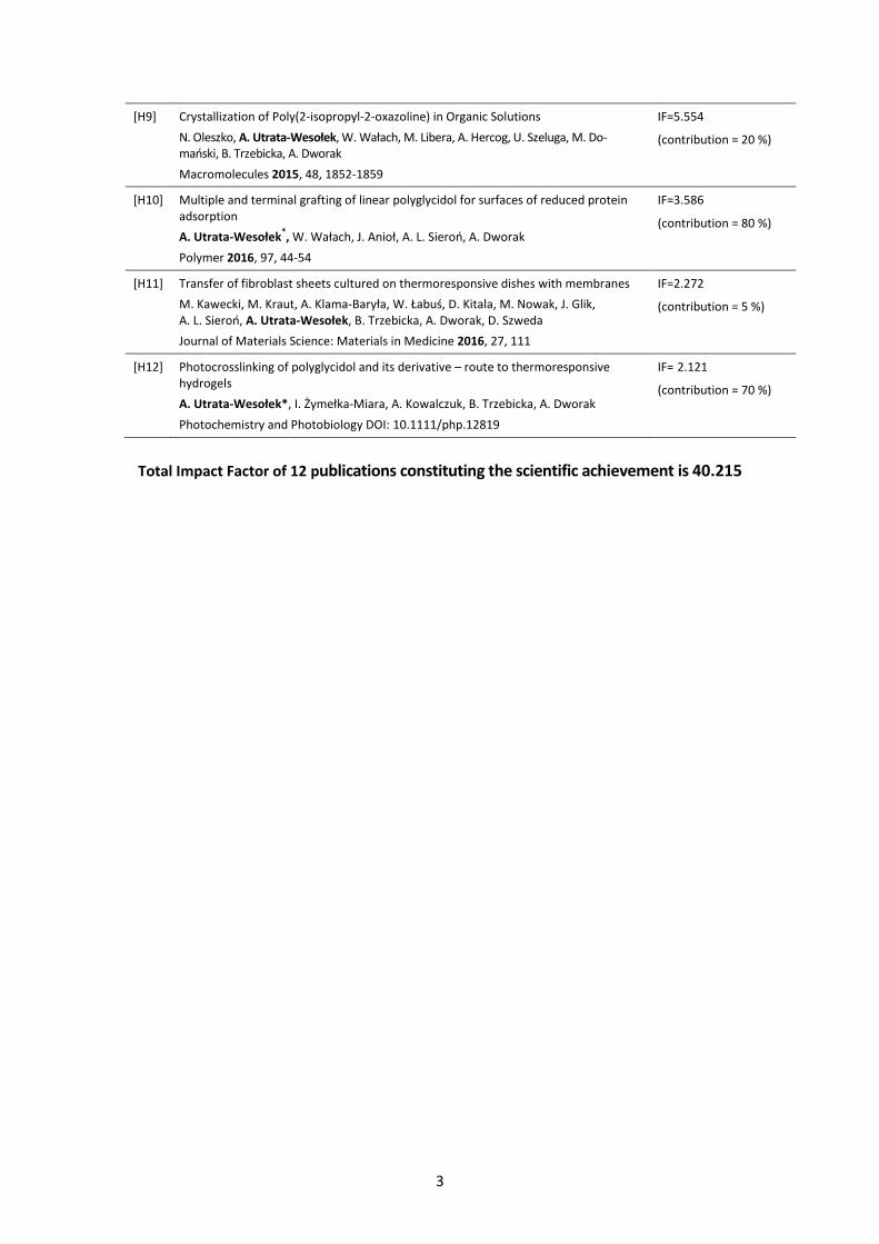
[H9] Crystallization of Poly(2-isopropyl-2-oxazoline) in Organic Solutions
N. Oleszko, A. Utrata-Wesołek, W. Wałach, M. Libera, A. Hercog, U. Szeluga, M. Do-mański, B. Trzebicka, A. Dworak
Macromolecules 2015, 48, 1852-1859
IF=5.554
(contribution = 20 %)
[H10] Multiple and terminal grafting of linear polyglycidol for surfaces of reduced protein adsorption
A. Utrata-Wesołek*, W. Wałach, J. Anioł, A. L. Sieroń, A. Dworak
Polymer 2016, 97, 44-54
IF=3.586
(contribution = 80 %)
[H11] Transfer of fibroblast sheets cultured on thermoresponsive dishes with membranes
M. Kawecki, M. Kraut, A. Klama-Baryła, W. Łabuś, D. Kitala, M. Nowak, J. Glik, A. L. Sieroń, A. Utrata-Wesołek, B. Trzebicka, A. Dworak, D. Szweda
Journal of Materials Science: Materials in Medicine 2016, 27, 111
IF=2.272
(contribution = 5 %)
[H12] Photocrosslinking of polyglycidol and its derivative – route to thermoresponsive hydrogels
A. Utrata-Wesołek*, I. Żymełka-Miara, A. Kowalczuk, B. Trzebicka, A. Dworak
Photochemistry and Photobiology DOI: 10.1111/php.12819
IF= 2.121
(contribution = 70 %)
Total Impact Factor of 12 publications constituting the scientific achievement is 40.215
4
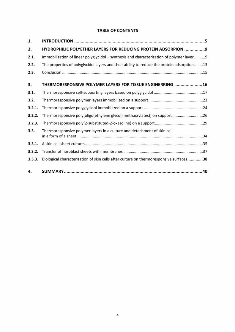
TABLE OF CONTENTS
1. INTRODUCTION ......................................................................................................5
2. HYDROPHILIC POLYETHER LAYERS FOR REDUCING PROTEIN ADSORPION ................9
2.1. Immobilization of linear polyglycidol – synthesis and characterization of polymer layer .......... 9
2.2. The properties of polyglycidol layers and their ability to reduce the protein adsorption ........13
2.3. Conclusion .................................................................................................................................15
3. THERMORESPONSIVE POLYMER LAYERS FOR TISSUE ENGINERRING ..................... 16
3.1. Thermoresponsive self-supporting layers based on polyglycidol .............................................17
3.2. Thermoresponsive polymer layers immobilized on a support ..................................................23
3.2.1. Thermoresponsive polyglycidol immobilized on a support ......................................................24
3.2.2. Thermoresponsive poly[oligo(ethylene glycol) methacrylates)] on support ............................26
3.2.3. Thermoresponsive poly(2-substituted-2-oxazoline) on a support ............................................29
3.3. Thermoresponsive polymer layers in a culture and detachment of skin cell in a form of a sheet....................................................................................................................34
3.3.1. A skin cell sheet culture .............................................................................................................35
3.3.2. Transfer of fibroblast sheets with membranes ........................................................................37
3.3.3. Biological characterization of skin cells after culture on thermoresponsive surfaces ............. 38
4. SUMMARY ............................................................................................................ 40
5
(c) OVERVIEW OF THE SCIENTIFIC AIMS AND OBTAINED RESULTS PRESENTED IN THE SERIES OF PUBLICATIONS „BIOCOMPATIBLE POLYMER LAYERS OF CONTROLLED AFFINITY FOR WATER. SYNTHESIS AND APPLICATION”
1. INTRODUCTION
Materials made of metals, ceramics, carbon materials and polymers (both natural and synthetic)
are often used in modern medicine for the treatment of various diseases [1, 2]. Coronary stents, vas-
cular, knee or hip prostheses, artificial heart valves, various implants or contact lenses are products
derived from such biomaterials and play a significant role in improving the health and quality
of patients life. In recent years, significant advances have also been achieved in using a biomaterials
for targeted drug transport, gene therapy, reconstructive medicine, or tissue engineering.
Biomedical materials that come into contact with body fluids or tissues must possess a specific prop-
erties that are adapted to the particular application. Thus it is essential to understand the interaction
of biomaterial with biologically active substances such as proteins or cells. For these interactions
the surface of biomaterial is responsible [3]. Therefore, the ability to control the biomaterials
surface properties, e.g. by coating them with layer made of polymer, is of particular importance
for the applications in medicine.
The main research problem that forms the basis of the habilitation procedure, to which this series
of publications is dedicated, is the development and characterization of new, biocompatible
polymer layers with properties that allow to use the obtained biomaterial in reconstructive medi-
cine as coatings reducing the protein adsorption or in tissue engineering for culture and detach-
ment of cell sheets. To achieve this, the interactions of proteins and cells with polymer coatings
should be determined.
The works with the use of synthetic materials for biomedical applications are intensive. Synthesis
of polymer layers (e.g. of a linear structure in the form of a polymer brush or crosslinked, dendritic,
or layer-on-layer coatings) are described [3, 4]. Unfortunately, based on the presented results,
the correlation between the properties of polymer layers resisting protein adsorption or favoring cell
culture with macromolecule structure or topology, surface coating method or polymer layer thick-
ness is not known or not sufficiently clarified.
The aim of this work was to develop and compare new biocompatible polymer layers with different
composition and structure, and to determine the relationship between the properties of the surface
covered with polymers and their interaction with proteins or cells.
In order to obtain the polymer layers reducing the protein adsorption a biocompatible, hydrophilic
polymers of glycidol were used. In the laboratory of Nano- and Microstructural Materials CMPW
PAN, where the research was conducted, methods of controlled synthesis of polyglycidol of different
macromolecular architecture [5,6] were developed. Based on this knowledge and on the fact that
there was no data on the use of polyglycidol, except for dendritic one, as anti-protein coatings [7-9],
the studies to obtain coatings made of linear polyglycidol and its copolymers with ethylene glycol
and to determine their interaction with proteins were undertaken.
The biocompatible polymer layers have also been tested for their use as a cell culture surfaces.
For this purpose a thermoresponsive polymers were used. The control of the affinity for water
(hydrophilic-hydrophobic balance) of the layers made of these polymers, only by changing the envi-
6
ronmental temperature, is possible. This property allow the thermoresponsive layers to be used
in regenerative medicine for culture and detachment of cells in the form of a sheet. As a result of our
previous work, a method for modifying the hydrophilic polyglycidol to thermoresponsive polymers
has been developed [10]. Based on this knowledge, a research to obtain thermoresponsive coatings
based on modified polyglycidol and the estimation of their interaction with cells has been under-
taken. Other thermoresponsive polymers have also been used in this work: poly[oligo(ethylene
glycol) methacrylates] and poly(2-substituted-2-oxazoline)s, as poly(N-isopropylacrylamide)
(PNIPAM) or its copolymers used to this point for cell sheet culture have some disadvantages
(e.g. lack of functional groups available for modification, phase hysteresis or aggregation) [11, 12].
To synthesis the polymers, living and controlled anionic and cationic polymerization as well as atom
transfer radical polymerization (ATRP) were applied. These methods allow for precise control
of the polymer layer structure, which further ensures the control of properties of the resultant mate-
rials under given conditions.
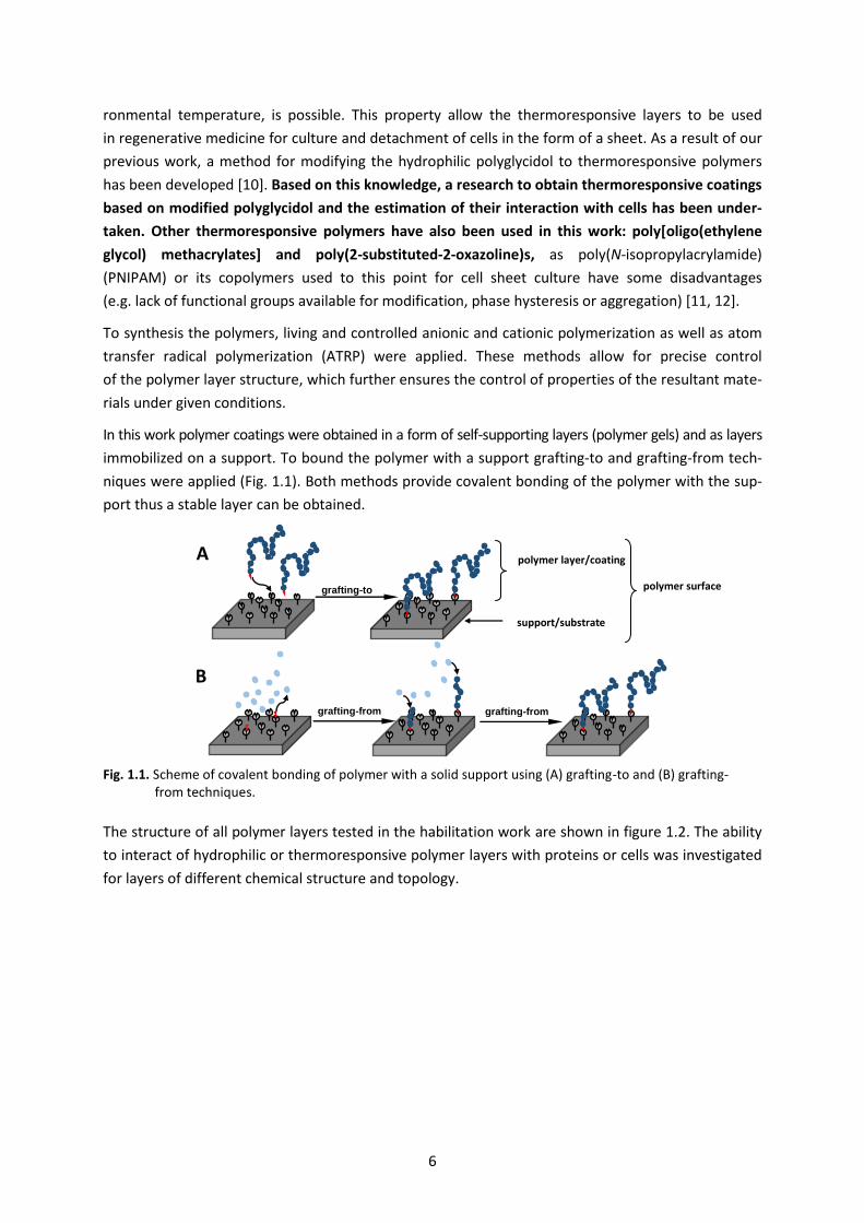
In this work polymer coatings were obtained in a form of self-supporting layers (polymer gels) and as layers
immobilized on a support. To bound the polymer with a support grafting-to and grafting-from tech-
niques were applied (Fig. 1.1). Both methods provide covalent bonding of the polymer with the sup-
port thus a stable layer can be obtained.
Fig. 1.1. Scheme of covalent bonding of polymer with a solid support using (A) grafting-to and (B) grafting- from techniques.
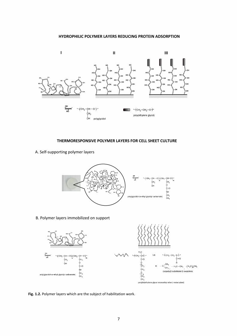
The structure of all polymer layers tested in the habilitation work are shown in figure 1.2. The ability
to interact of hydrophilic or thermoresponsive polymer layers with proteins or cells was investigated
for layers of different chemical structure and topology.
polymer surface
A
grafting-to
B
grafting-from grafting-from
polymer layer/coating
support/substrate
7
Fig. 1.2. Polymer layers which are the subject of habilitation work.
HYDROPHILIC POLYMER LAYERS REDUCING PROTEIN ADSORPTION
I II III
A. Self-supporting polymer layers
THERMORESPONSIVE POLYMER LAYERS FOR CELL SHEET CULTURE
B. Polymer layers immobilized on support

8
The habilitation dissertation describes the author's work including:
1. Synthesis and characterization of hydrophilic layers based on polyglycidol [H3, H10]
2. An estimation of the influence of polyglycidol structure and polyglycidol layers properties on the
reduction of protein adsorption [H10]
3. Synthesis and characterization of thermoresponsive polymer layers:
a) self-supporting layers [H1, H2, H12]
b) layers based on polyglycidol [H5], poly[oligo(ethylene glycol) methacrylate] [H4, H6]
and (co)poly(2-substituted-2-oxazoline)s [H7, H8, H9] immobilized on supports
4. An estimation of the influence of polymers composition and structure and properties
of thermoresponsive polymer layers on the ability of cell sheet culture and detachment taking
into account:
a) an adhesion, proliferation and detachment of cell sheet [H1, H4, H5, H7, H8]
b) transfer of cell sheet [H8, H11]
c) cell characterization after culture on thermoresponsive polymer layers [H7]
All of the works described in this dissertation are focused on the ability to control the synthesis
of polymers and their grafting with the support in order to influence the properties of the resulting
polymer layers. Particular emphasis was placed on determining the effect of the polymer layers
properties on their capability to reduce protein adsorption or to culture of cell sheets. Such analysis
enabled, within the presented habilitation work, to define the potential use of received materials
in reconstructive medicine and tissue engineering.
A monothematic articles [H1-H12] described in this dissertation are collective works in which the role
of each author is defined and described in the table in Chapter 4B of this summary.

9
2. HYDROPHILIC POLYETHER LAYERS FOR REDUCING PROTEIN ADSORPION
The uncontrolled adhesion of biological compounds on the surface of biomaterials is a harmful phe-
nomenon. Medical implants, regardless of their structure, will become coated with a layer of pro-
teins within a few seconds of contact with physiological fluids and tissues. As a result, the host
defense mechanisms is activated, which can lead to inflammatory reactions, thromboembolic
complications or deterioration of the functioning of the device [13]. Therefore, materials with anti-
fouling properties have been the subject of much interest and extensive research within the last
few years [14-16]. This antifouling behavior is usually achieved by coating the surface with polymer
layer with appropriate properties. Generally, two major classes of polymers have been investigated
for the minimization of nonspecific adsorption: hydrophilic and zwitterionic. Among these polymers,
the most widely studied layers are based on poly(ethylene glycol) [3] which is non-toxic,
non-immunogenic and water-soluble. Polyglycidol (PGl) – a poly(ethylene glycol) analogue, is also
a biocompatible, hydrophilic polymer and additionally possess functional groups capable for further
modification. It can therefore be a polymer suitable for this kind of application.
In the author labs, methods for controlled polymerization of glycidol and its derivatives to obtain
polymers with linear [17], branched [6], dendritic of “pom-pom” type [18] or “bottle-brush” [19]
structure have been developed. Despite the high level achieved in polyglycidol synthesis and in attempts
to apply it in biomedicine [20], few data on its use as an antifouling coating have been published
at the time of the habilitation work [7-9]. These studies concerned only dendritic polyglycidol,
so it was not possible to determine the relationship between the polymer structure, the polymer
layer structure and properties, and the resistance of obtained materials for protein adsorption.
In the habilitation work, the research issues related to the development of polymer layers based
on linear polyglycidol and its copolymers with ethylene glycol capable for reducing protein adsorp-
tion have been taken [H3, H10].
2.1. Immobilization of linear polyglycidol – synthesis and characterization of polymer layer [H10]
Polymer surfaces containing a layer of polyglycidol or its block copolymers with ethylene glycol
of linear structure (scheme in Fig. 1.2), described in [H10], were obtained using a grafting-to method.
The synthesis involved two steps: modification of solid supports (silica) to introduce reactive
functional groups followed by immobilization of previously synthesized (co)polymers of glycidol.
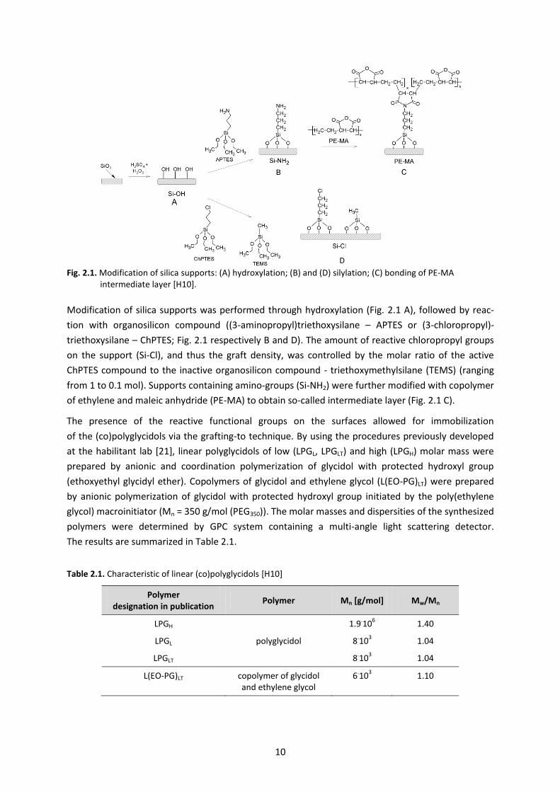
The scheme of silica support modification is shown in figure 2.1.
10
Fig. 2.1. Modification of silica supports: (A) hydroxylation; (B) and (D) silylation; (C) bonding of PE-MA intermediate layer [H10].
Modification of silica supports was performed through hydroxylation (Fig. 2.1 A), followed by reac-
tion with organosilicon compound ((3-aminopropyl)triethoxysilane – APTES or (3-chloropropyl)-
triethoxysilane – ChPTES; Fig. 2.1 respectively B and D). The amount of reactive chloropropyl groups
on the support (Si-Cl), and thus the graft density, was controlled by the molar ratio of the active
ChPTES compound to the inactive organosilicon compound - triethoxymethylsilane (TEMS) (ranging
from 1 to 0.1 mol). Supports containing amino-groups (Si-NH2) were further modified with copolymer
of ethylene and maleic anhydride (PE-MA) to obtain so-called intermediate layer (Fig. 2.1 C).
The presence of the reactive functional groups on the surfaces allowed for immobilization
of the (co)polyglycidols via the grafting-to technique. By using the procedures previously developed
at the habilitant lab [21], linear polyglycidols of low (LPGL, LPGLT) and high (LPGH) molar mass were
prepared by anionic and coordination polymerization of glycidol with protected hydroxyl group
(ethoxyethyl glycidyl ether). Copolymers of glycidol and ethylene glycol (L(EO-PG)LT) were prepared
by anionic polymerization of glycidol with protected hydroxyl group initiated by the poly(ethylene
glycol) macroinitiator (Mn = 350 g/mol (PEG350)). The molar masses and dispersities of the synthesized
polymers were determined by GPC system containing a multi-angle light scattering detector.
The results are summarized in Table 2.1.
Table 2.1. Characteristic of linear (co)polyglycidols [H10]
Polymer designation in publication
Polymer Mn [g/mol] Mw/Mn
LPGH
polyglycidol
1.9.10
6 1.40
LPGL 8.10
3 1.04
LPGLT 8.10
3 1.04
L(EO-PG)LT copolymer of glycidol and ethylene glycol
6.10
3 1.10
11
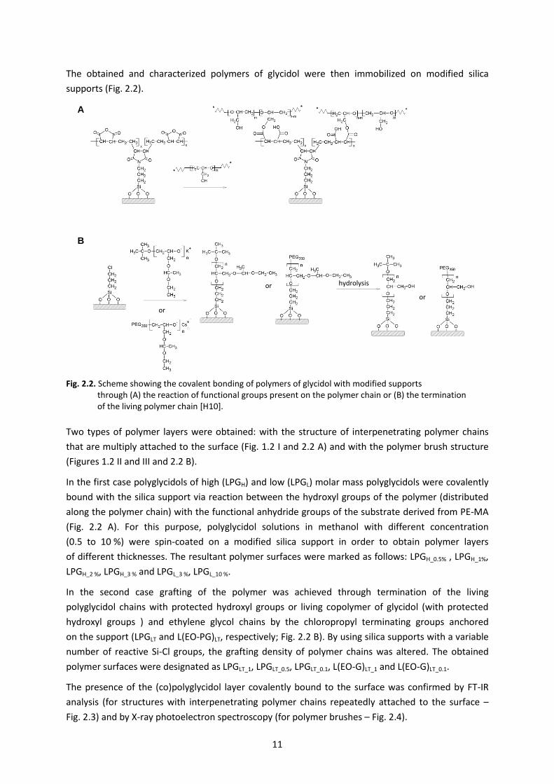
The obtained and characterized polymers of glycidol were then immobilized on modified silica
supports (Fig. 2.2).
Fig. 2.2. Scheme showing the covalent bonding of polymers of glycidol with modified supports through (A) the reaction of functional groups present on the polymer chain or (B) the termination of the living polymer chain [H10].
Two types of polymer layers were obtained: with the structure of interpenetrating polymer chains
that are multiply attached to the surface (Fig. 1.2 I and 2.2 A) and with the polymer brush structure
(Figures 1.2 II and III and 2.2 B).
In the first case polyglycidols of high (LPGH) and low (LPGL) molar mass polyglycidols were covalently
bound with the silica support via reaction between the hydroxyl groups of the polymer (distributed
along the polymer chain) with the functional anhydride groups of the substrate derived from PE-MA
(Fig. 2.2 A). For this purpose, polyglycidol solutions in methanol with different concentration
(0.5 to 10 %) were spin-coated on a modified silica support in order to obtain polymer layers
of different thicknesses. The resultant polymer surfaces were marked as follows: LPGH_0.5% , LPGH_1%,
LPGH_2 %, LPGH_3 % and LPGL_3 %, LPGL_10 %.
In the second case grafting of the polymer was achieved through termination of the living
polyglycidol chains with protected hydroxyl groups or living copolymer of glycidol (with protected
hydroxyl groups ) and ethylene glycol chains by the chloropropyl terminating groups anchored
on the support (LPGLT and L(EO-PG)LT, respectively; Fig. 2.2 B). By using silica supports with a variable
number of reactive Si-Cl groups, the grafting density of polymer chains was altered. The obtained
polymer surfaces were designated as LPGLT_1, LPGLT_0.5, LPGLT_0.1, L(EO-G)LT_1 and L(EO-G)LT_0.1.
The presence of the (co)polyglycidol layer covalently bound to the surface was confirmed by FT-IR
analysis (for structures with interpenetrating polymer chains repeatedly attached to the surface –
Fig. 2.3) and by X-ray photoelectron spectroscopy (for polymer brushes – Fig. 2.4).
A
B
or
or
or hydrolysis
12
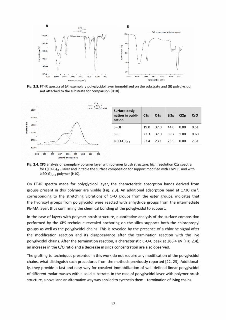
Fig. 2.3. FT-IR spectra of (A) exemplary polyglycidol layer immobilized on the substrate and (B) polyglycidol not attached to the substrate for comparison [H10].
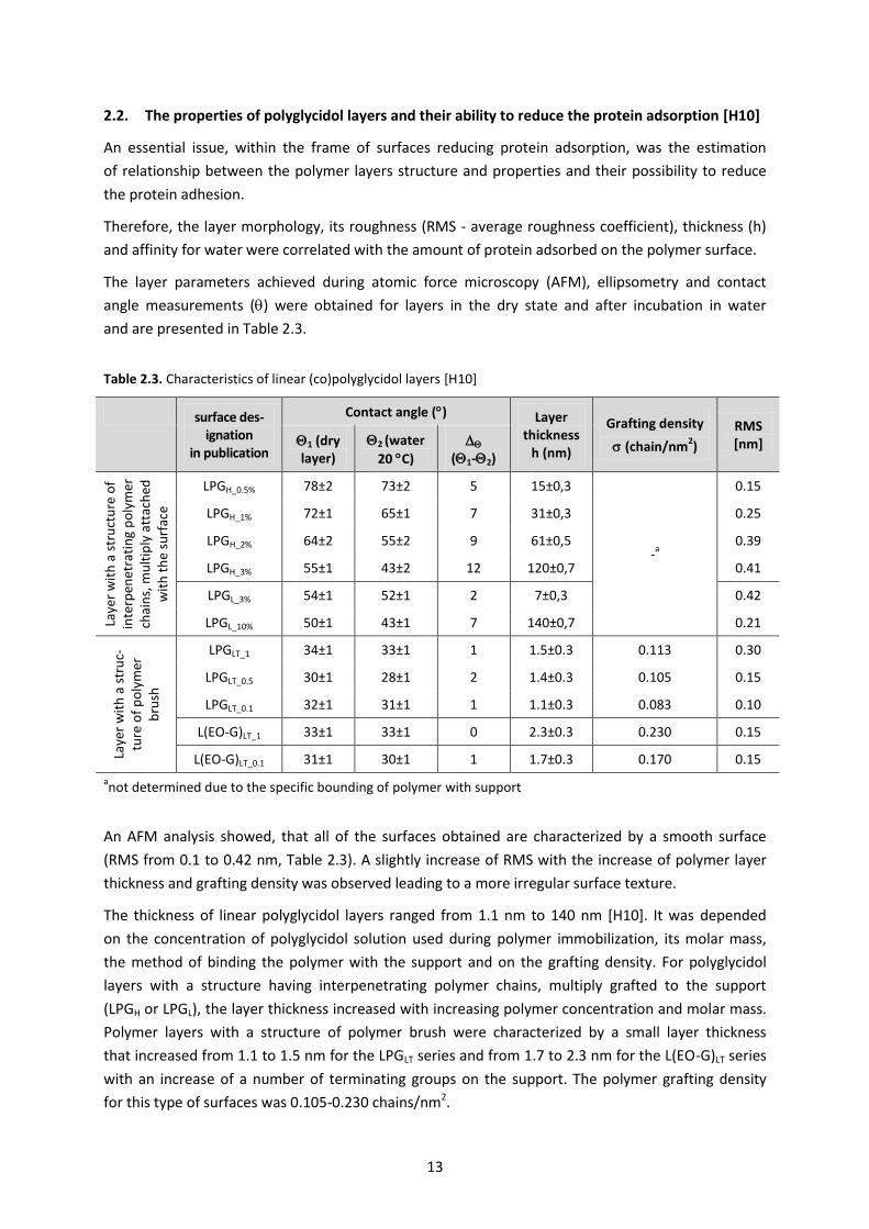
Fig. 2.4. XPS analysis of exemplary polymer layer with polymer brush structure: high resolution C1s spectra for L(EO-G)LT_1 layer and in table the surface composition for support modified with ChPTES and with L(EO-G)LT_1 polymer [H10].
On FT-IR spectra made for polyglycidol layer, the characteristic absorption bands derived from
groups present in this polymer are visible (Fig. 2.3). An additional adsorption band at 1730 cm-1,
corresponding to the stretching vibrations of C=O groups from the ester groups, indicates that
the hydroxyl groups from polyglycidol were reacted with anhydride groups from the intermediate
PE-MA layer, thus confirming the chemical bonding of the polyglycidol to support.
In the case of layers with polymer brush structure, quantitative analysis of the surface composition
performed by the XPS technique revealed anchoring on the silica supports both the chloropropyl
groups as well as the polyglycidol chains. This is revealed by the presence of a chlorine signal after
the modification reaction and its disappearance after the termination reaction with the live
polyglycidol chains. After the termination reaction, a characteristic C-O-C peak at 286.4 eV (Fig. 2.4),
an increase in the C/O ratio and a decrease in silica concentration are also observed.
The grafting-to techniques presented in this work do not require any modification of the polyglycidol
chains, what distinguish such procedures from the methods previously reported [22, 23]. Additional-
ly, they provide a fast and easy way for covalent immobilization of well-defined linear polyglycidol
of different molar masses with a solid substrate. In the case of polyglycidol layer with polymer brush
structure, a novel and an alternative way was applied to synthesis them – termination of living chains.
Surface desig-nation in publi-cation
C1s O1s Si2p Cl2p C/O
Si-OH 19.0 37.0 44.0 0.00 0.51
Si-Cl 22.3 37.0 39.7 1.00 0.60
L(EO-G)LT_1 53.4 23.1 23.5 0.00 2.31
B A
13
2.2. The properties of polyglycidol layers and their ability to reduce the protein adsorption [H10]
An essential issue, within the frame of surfaces reducing protein adsorption, was the estimation
of relationship between the polymer layers structure and properties and their possibility to reduce
the protein adhesion.
Therefore, the layer morphology, its roughness (RMS - average roughness coefficient), thickness (h)
and affinity for water were correlated with the amount of protein adsorbed on the polymer surface.
The layer parameters achieved during atomic force microscopy (AFM), ellipsometry and contact
angle measurements () were obtained for layers in the dry state and after incubation in water
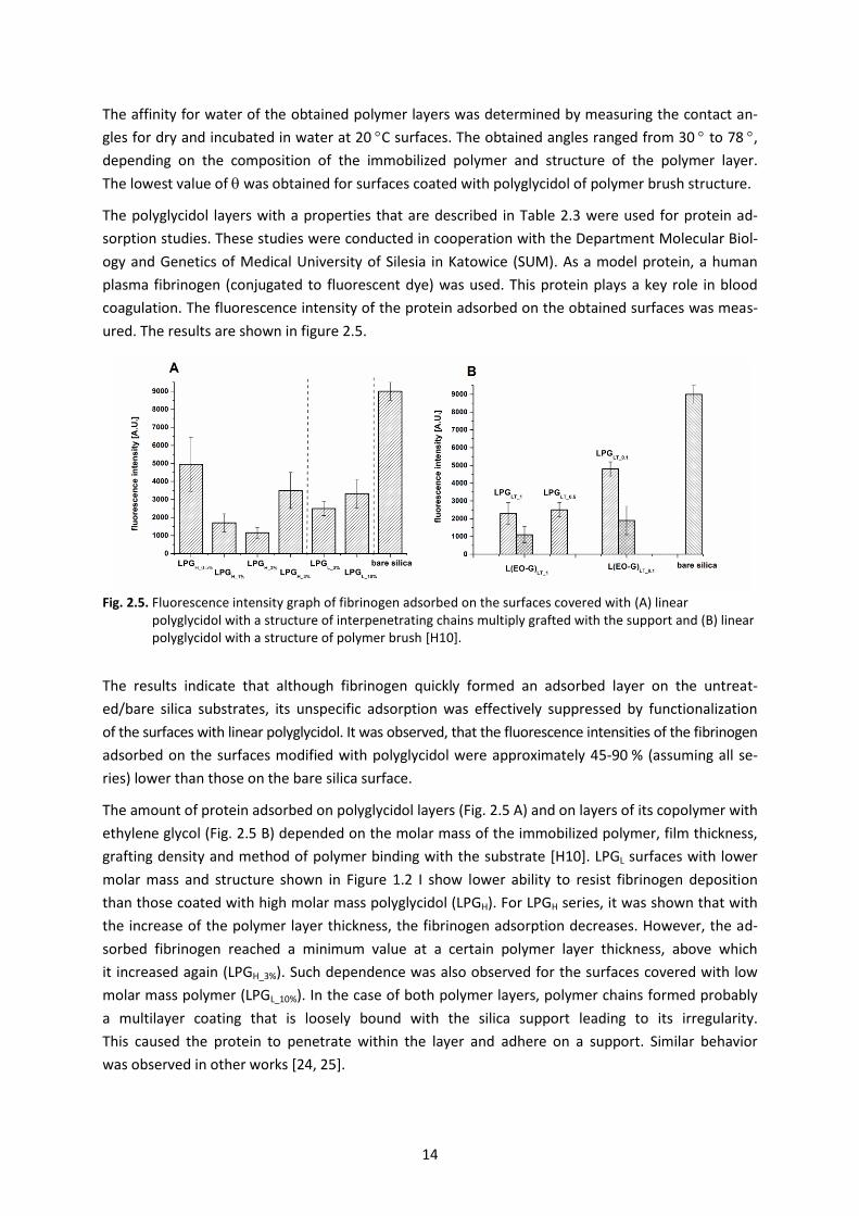
and are presented in Table 2.3.
Table 2.3. Characteristics of linear (co)polyglycidol layers [H10]
surface des-ignation
in publication
Contact angle () Layer thickness
h (nm)
Grafting density
(chain/nm2)
RMS [nm] 1 (dry
layer) 2 (water
20 C)
(1-2)
Laye
r w
ith
a s
tru
ctu
re o
f in
terp
enet
rati
ng
po
lym
er
chai
ns,
mu
ltip
ly a
ttac
hed
w
ith
th
e su
rfac
e
LPGH_0.5% 78±2 73±2 5 15±0,3
-a
0.15
LPGH_1% 72±1 65±1 7 31±0,3 0.25
LPGH_2% 64±2 55±2 9 61±0,5 0.39
LPGH_3% 55±1 43±2 12 120±0,7 0.41
LPGL_3% 54±1 52±1 2 7±0,3 0.42
LPGL_10% 50±1 43±1 7 140±0,7 0.21
Laye
r w
ith
a s
tru
c-tu
re o
f p
oly
mer
b
rush
LPGLT_1 34±1 33±1 1 1.5±0.3 0.113 0.30
LPGLT_0.5 30±1 28±1 2 1.4±0.3 0.105 0.15
LPGLT_0.1 32±1 31±1 1 1.1±0.3 0.083 0.10
L(EO-G)LT_1 33±1 33±1 0 2.3±0.3 0.230 0.15
L(EO-G)LT_0.1 31±1 30±1 1 1.7±0.3 0.170 0.15
anot determined due to the specific bounding of polymer with support
An AFM analysis showed, that all of the surfaces obtained are characterized by a smooth surface
(RMS from 0.1 to 0.42 nm, Table 2.3). A slightly increase of RMS with the increase of polymer layer
thickness and grafting density was observed leading to a more irregular surface texture.
The thickness of linear polyglycidol layers ranged from 1.1 nm to 140 nm [H10]. It was depended
on the concentration of polyglycidol solution used during polymer immobilization, its molar mass,
the method of binding the polymer with the support and on the grafting density. For polyglycidol
layers with a structure having interpenetrating polymer chains, multiply grafted to the support
(LPGH or LPGL), the layer thickness increased with increasing polymer concentration and molar mass.
Polymer layers with a structure of polymer brush were characterized by a small layer thickness
that increased from 1.1 to 1.5 nm for the LPGLT series and from 1.7 to 2.3 nm for the L(EO-G)LT series
with an increase of a number of terminating groups on the support. The polymer grafting density
for this type of surfaces was 0.105-0.230 chains/nm2.
14
The affinity for water of the obtained polymer layers was determined by measuring the contact an-
gles for dry and incubated in water at 20 C surfaces. The obtained angles ranged from 30 to 78 ,
depending on the composition of the immobilized polymer and structure of the polymer layer.
The lowest value of was obtained for surfaces coated with polyglycidol of polymer brush structure.
The polyglycidol layers with a properties that are described in Table 2.3 were used for protein ad-
sorption studies. These studies were conducted in cooperation with the Department Molecular Biol-
ogy and Genetics of Medical University of Silesia in Katowice (SUM). As a model protein, a human
plasma fibrinogen (conjugated to fluorescent dye) was used. This protein plays a key role in blood
coagulation. The fluorescence intensity of the protein adsorbed on the obtained surfaces was meas-
ured. The results are shown in figure 2.5.
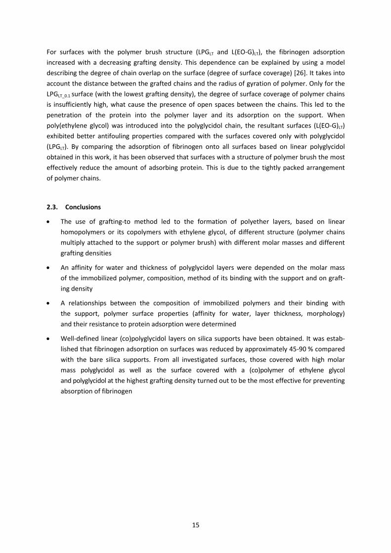
Fig. 2.5. Fluorescence intensity graph of fibrinogen adsorbed on the surfaces covered with (A) linear polyglycidol with a structure of interpenetrating chains multiply grafted with the support and (B) linear polyglycidol with a structure of polymer brush [H10].
The results indicate that although fibrinogen quickly formed an adsorbed layer on the untreat-
ed/bare silica substrates, its unspecific adsorption was effectively suppressed by functionalization
of the surfaces with linear polyglycidol. It was observed, that the fluorescence intensities of the fibrinogen
adsorbed on the surfaces modified with polyglycidol were approximately 45-90 % (assuming all se-
ries) lower than those on the bare silica surface.
The amount of protein adsorbed on polyglycidol layers (Fig. 2.5 A) and on layers of its copolymer with
ethylene glycol (Fig. 2.5 B) depended on the molar mass of the immobilized polymer, film thickness,
grafting density and method of polymer binding with the substrate [H10]. LPGL surfaces with lower
molar mass and structure shown in Figure 1.2 I show lower ability to resist fibrinogen deposition
than those coated with high molar mass polyglycidol (LPGH). For LPGH series, it was shown that with
the increase of the polymer layer thickness, the fibrinogen adsorption decreases. However, the ad-
sorbed fibrinogen reached a minimum value at a certain polymer layer thickness, above which
it increased again (LPGH_3%). Such dependence was also observed for the surfaces covered with low
molar mass polymer (LPGL_10%). In the case of both polymer layers, polymer chains formed probably
a multilayer coating that is loosely bound with the silica support leading to its irregularity.
This caused the protein to penetrate within the layer and adhere on a support. Similar behavior
was observed in other works [24, 25].
15
For surfaces with the polymer brush structure (LPGLT and L(EO-G)LT), the fibrinogen adsorption
increased with a decreasing grafting density. This dependence can be explained by using a model
describing the degree of chain overlap on the surface (degree of surface coverage) [26]. It takes into
account the distance between the grafted chains and the radius of gyration of polymer. Only for the
LPGLT_0.1 surface (with the lowest grafting density), the degree of surface coverage of polymer chains
is insufficiently high, what cause the presence of open spaces between the chains. This led to the
penetration of the protein into the polymer layer and its adsorption on the support. When
poly(ethylene glycol) was introduced into the polyglycidol chain, the resultant surfaces (L(EO-G)LT)
exhibited better antifouling properties compared with the surfaces covered only with polyglycidol
(LPGLT). By comparing the adsorption of fibrinogen onto all surfaces based on linear polyglycidol
obtained in this work, it has been observed that surfaces with a structure of polymer brush the most
effectively reduce the amount of adsorbing protein. This is due to the tightly packed arrangement
of polymer chains.
2.3. Conclusions
The use of grafting-to method led to the formation of polyether layers, based on linear
homopolymers or its copolymers with ethylene glycol, of different structure (polymer chains
multiply attached to the support or polymer brush) with different molar masses and different
grafting densities
An affinity for water and thickness of polyglycidol layers were depended on the molar mass
of the immobilized polymer, composition, method of its binding with the support and on graft-
ing density
A relationships between the composition of immobilized polymers and their binding with
the support, polymer surface properties (affinity for water, layer thickness, morphology)
and their resistance to protein adsorption were determined
Well-defined linear (co)polyglycidol layers on silica supports have been obtained. It was estab-
lished that fibrinogen adsorption on surfaces was reduced by approximately 45-90 % compared
with the bare silica supports. From all investigated surfaces, those covered with high molar
mass polyglycidol as well as the surface covered with a (co)polymer of ethylene glycol
and polyglycidol at the highest grafting density turned out to be the most effective for preventing
absorption of fibrinogen
16
3. THERMORESPONSIVE POLYMER LAYERS FOR TISSUE ENGINERRING
The studies described so far have shown that it is possible to obtain hydrophilic polymer layers
based on polyglycidol that efficiently reduce protein adsorption. That is why, it was interesting
to determine the ability to control the affinity for water of the obtained polymer layers (so called
hydrophilic-hydrophobic balance) and also the ability to "switch" this affinity depending on the
external conditions. Such behavior of coatings can be used for controlled cell adhesion. It is known
that the cells more likely adhere to hydrophobic surfaces, while hydrophilic layers resist their adhe-
sion. Thus the obtainment of layers with switchable affinity for water opens the way for their use
in tissue engineering.
Substrates made of modified polystyrene (TCPS) are generally used for cell cultures. Good cell adhe-
sion to these substrates is achieved by appropriate control of their hydrophilic-hydrophobic balance.
Separation of proliferated cells from such surfaces is traditionally done enzymatically what however,
destroy a number of cells and disturb the cell sheet integrity. In that case, a single cell suspension
is obtained. The use of such a suspension in regenerative medicine is common. However, some re-
strictions have been observed in its application, for example, difficulties in controlling the location
of introduced cells or the formation of cells aggregates often not attached to the host tissue.
In the case of scaffolds, that are populated by cell suspension, an inflammatory responses are often
triggered or in the case of biodegradable scaffolds the products of their degradation are toxic.
In many cases, these problems could be avoided by culturing the cells in the form of a sheets fol-
lowed by separating them from the scaffold and transferring to the desired place in an intact form.
Polymers sensitive to temperature changes (so-called thermoresponsive polymers) can be used
to develop such scaffolds. A temperature change allows to change the affinity for water of such pol-
ymers. They are soluble below a certain temperature and, when it is exceeded (so-called phase sepa-
ration temperature – TCP), polymer precipitate as a result of strong intra- and intermolecular interac-
tions. The immobilization of thermoresponsive polymers on the support lead to surfaces with prop-
erties, such as affinity for water, morphology and thickness, that can be reversible changed
by the temperature alteration [27].
At the beginning of habilitation work, thermoresponsive substrates based mainly on poly(N-iso-
propylacrylamide) (PNIPAM) and its copolymers were used for cell sheets culture and their non-
invasive detachment. This idea, called cell sheet engineering, was developed by T. Okano [11].
The cell culture is carried out at a temperature above the phase separation temperature
of the thermoresponsive polymer, when the surface is hydrophobic. When the cells form a sheet,
the temperature is lowered, the surface becomes hydrophilic and the cell sheet spontaneously sepa-
rates from the substrate. It is not necessary to use enzymatic methods of cell separation.
Initially, the attention has been focused on PNIPAM-based polymers. These polymers are character-
ized by a TCP around the physiological temperature which makes them attractive for biomedical
applications. However, PNIPAM does not have functional groups available for modifications,
its phase transition curves show a hysteresis, it can aggregate and additionally it irreversibly interacts
with biological compounds, e.g. amino acids or proteins. While a lot of studies on intelligent
polymers have been made thus many thermoresponsive polymers have been identified, in many
respects more promising [28]. In the laboratory of Nano- and Microstructural Materials CMPW PAN,
a modification of polyglycidol to thermoresponsive copolymers with TCP ranging from 10 C to 90 C
17
was developed (with my leading participation) [10, 29]. By appropriate modification, it is possible
to control the affinity for water of obtained the glycidol copolymers only by changing the environ-
mental temperature.
By using the obtained knowledge, the studies on the development of polymer layers based on mod-
ified polyglycidol, with affinity for water controlled by a temperature, were undertaken in the ha-
bilitation work. The determination of interaction with cells of such obtained polymer layers was
of significant importance. The researches have also been extended to other thermoresponsive polymers:
poly[oligo(ethylene glycol) methacrylates] and poly(2-substituted-2-oxazoline)s. The aim of the study
was to determine the suitability of these layers for culture and non-invasive detachment of the cells
in a form of a sheet.
By using grafting-from and -to techniques, a thermoresponsive polymer layers immobilized on a solid
(glass or silica) supports were obtained. Works to determine whether thermoresponsive layers
of sufficient mechanical parameters in a form of self-supporting layers (without immobilizing
the polymer on the support) have also been undertaken.
For the clarity of the dissertation, syntheses and properties of thermoresponsive polymer layers,
both self-supporting and immobilized on the substrate will be described first (chapters 3.1 and 3.2).
Section 3.3 will be related to the determination of the influence of the polymer composition
and structure and the properties of the thermoresponsive surface on the ability of the cell adhesion
and detachment.
3.1. Thermoresponsive self-supporting layers based on polyglycidol – synthesis
and properties [H1, H2, H12]
Thermoresponsive self-supporting layers (in form of polymer gels) based on polymers of glycidol
were obtained using photocrosslinking. As compared to chemical crosslinking, this method of gels
preparation is simple and does not require the use of many reagents. The aim of the work was
to develop the conditions of photocrosslinking of polyglycidol in such a way that thermoresponsive
layers with optimum mechanical parameters will be obtained.
The work with the use of UV-radiation required first an estimation of the UV effect on the stability
and possible degradation of the polyglycidol chain. This issue is also important because this type
of radiation is used in sterilization processes of biomaterials. The behavior of polyglycidol under UV
irradiation and its possible photodegradation have not been studied so far, so in [H2] this issue has
been undertaken.
At work [H2], the behavior of aqueous polyglycidol solutions (at different concentrations) was de-
termined by exposure to UV radiation. A changes in physical and chemical properties of polymer
was monitored by gel permeation chromatography (SEC-MALLS), FT-IR and NMR spectroscopy.
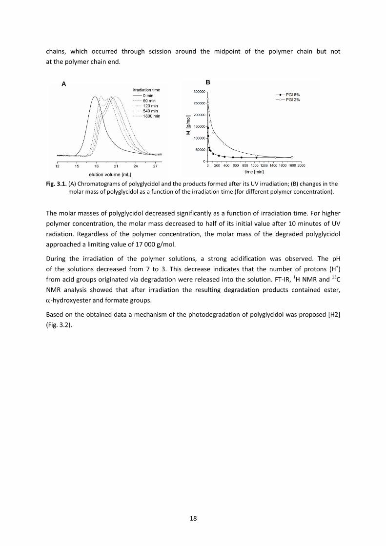
It was observed that during irradiation, the shape of the chromatograms changed from monomodal
to bimodal and the quantity of the lower molar mass fractions increased with an increase
in the photodegradation time. The molar masses for the both maxima of bimodal chromatograms
differed in half. In all of the chromatograms, the lack of signals at higher elution volumes pointed
to the absence of low molar mass oligomers as photodegradation products (Fig. 3.1 A). This could
indicate that UV irradiation of polyglycidol solutions led to the fragmentation of the polymer
18
chains, which occurred through scission around the midpoint of the polymer chain but not
at the polymer chain end.
Fig. 3.1. (A) Chromatograms of polyglycidol and the products formed after its UV irradiation; (B) changes in the molar mass of polyglycidol as a function of the irradiation time (for different polymer concentration).
The molar masses of polyglycidol decreased significantly as a function of irradiation time. For higher
polymer concentration, the molar mass decreased to half of its initial value after 10 minutes of UV
radiation. Regardless of the polymer concentration, the molar mass of the degraded polyglycidol
approached a limiting value of 17 000 g/mol.
During the irradiation of the polymer solutions, a strong acidification was observed. The pH
of the solutions decreased from 7 to 3. This decrease indicates that the number of protons (H+)
from acid groups originated via degradation were released into the solution. FT-IR, 1H NMR and 13C
NMR analysis showed that after irradiation the resulting degradation products contained ester,
-hydroxyester and formate groups.
Based on the obtained data a mechanism of the photodegradation of polyglycidol was proposed [H2]
(Fig. 3.2).
B A
19
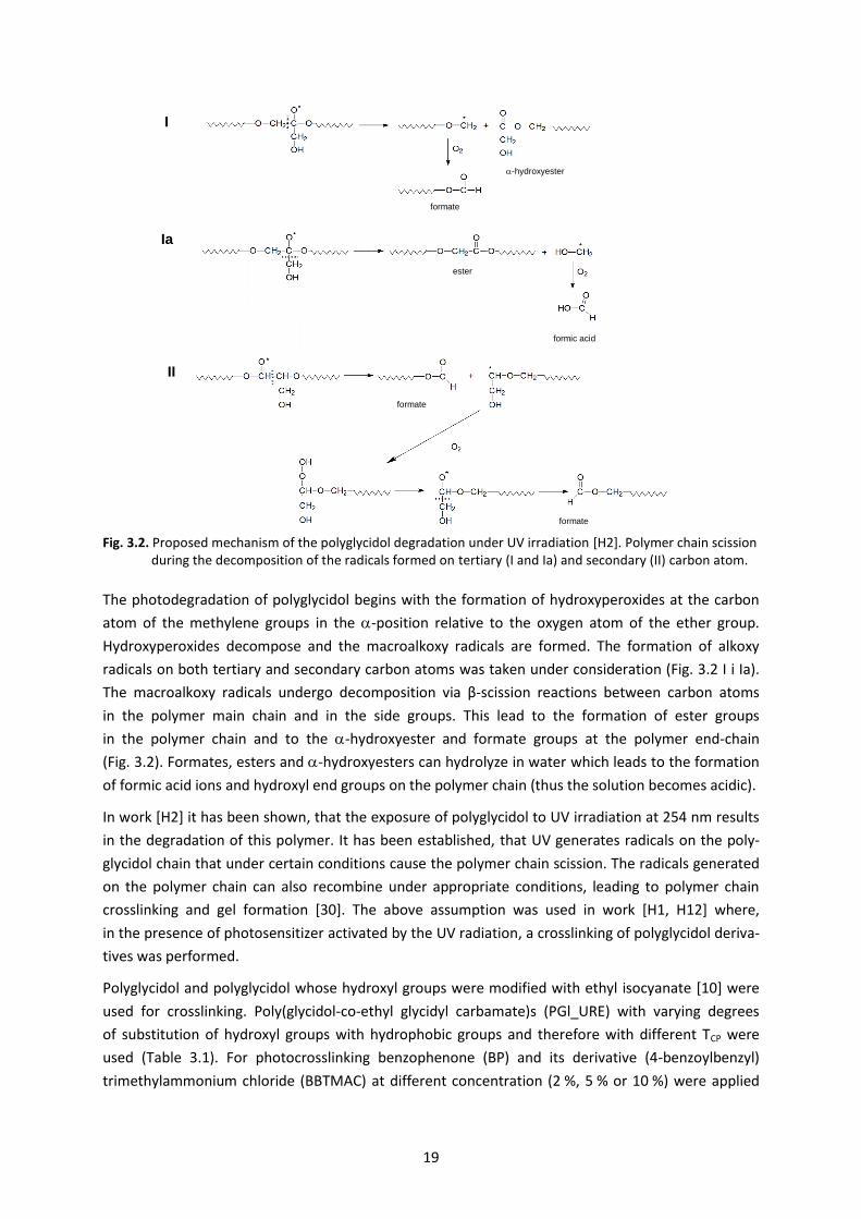
Fig. 3.2. Proposed mechanism of the polyglycidol degradation under UV irradiation [H2]. Polymer chain scission during the decomposition of the radicals formed on tertiary (I and Ia) and secondary (II) carbon atom.
The photodegradation of polyglycidol begins with the formation of hydroxyperoxides at the carbon
atom of the methylene groups in the -position relative to the oxygen atom of the ether group.
Hydroxyperoxides decompose and the macroalkoxy radicals are formed. The formation of alkoxy
radicals on both tertiary and secondary carbon atoms was taken under consideration (Fig. 3.2 I i Ia).
The macroalkoxy radicals undergo decomposition via β-scission reactions between carbon atoms
in the polymer main chain and in the side groups. This lead to the formation of ester groups
in the polymer chain and to the -hydroxyester and formate groups at the polymer end-chain
(Fig. 3.2). Formates, esters and -hydroxyesters can hydrolyze in water which leads to the formation
of formic acid ions and hydroxyl end groups on the polymer chain (thus the solution becomes acidic).
In work [H2] it has been shown, that the exposure of polyglycidol to UV irradiation at 254 nm results
in the degradation of this polymer. It has been established, that UV generates radicals on the poly-
glycidol chain that under certain conditions cause the polymer chain scission. The radicals generated
on the polymer chain can also recombine under appropriate conditions, leading to polymer chain
crosslinking and gel formation [30]. The above assumption was used in work [H1, H12] where,
in the presence of photosensitizer activated by the UV radiation, a crosslinking of polyglycidol deriva-
tives was performed.
Polyglycidol and polyglycidol whose hydroxyl groups were modified with ethyl isocyanate [10] were
used for crosslinking. Poly(glycidol-co-ethyl glycidyl carbamate)s (PGl_URE) with varying degrees
of substitution of hydroxyl groups with hydrophobic groups and therefore with different TCP were
used (Table 3.1). For photocrosslinking benzophenone (BP) and its derivative (4-benzoylbenzyl)
trimethylammonium chloride (BBTMAC) at different concentration (2 %, 5 % or 10 %) were applied
II
formate
formate
I
formate
-hydroxyester
Ia
formic acid
ester
20
as photosensitizers. The polymer/photosensitizers films were prepared by film-casting and then
subjected to UV irradiation [H12].
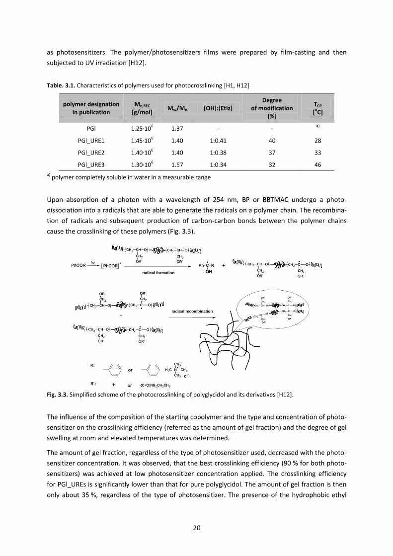
Table. 3.1. Characteristics of polymers used for photocrosslinking [H1, H12]
polymer designation in publication
Mn,SEC [g/mol]
Mw/Mn [OH]:[EtIz] Degree
of modification [%]
TCP [
oC]
PGl 1.25·106
1.37 - - a)
PGl_URE1 1.45·106 1.40 1:0.41 40 28
PGl_URE2 1.40·106 1.40 1:0.38 37 33
PGl_URE3 1.30·106 1.57 1:0.34 32 46
a) polymer completely soluble in water in a measurable range
Upon absorption of a photon with a wavelength of 254 nm, BP or BBTMAC undergo a photo-
dissociation into a radicals that are able to generate the radicals on a polymer chain. The recombina-
tion of radicals and subsequent production of carbon-carbon bonds between the polymer chains
cause the crosslinking of these polymers (Fig. 3.3).
Fig. 3.3. Simplified scheme of the photocrosslinking of polyglycidol and its derivatives [H12].
The influence of the composition of the starting copolymer and the type and concentration of photo-
sensitizer on the crosslinking efficiency (referred as the amount of gel fraction) and the degree of gel
swelling at room and elevated temperatures was determined.
The amount of gel fraction, regardless of the type of photosensitizer used, decreased with the photo-
sensitizer concentration. It was observed, that the best crosslinking efficiency (90 % for both photo-
sensitizers) was achieved at low photosensitizer concentration applied. The crosslinking efficiency
for PGl_UREs is significantly lower than that for pure polyglycidol. The amount of gel fraction is then
only about 35 %, regardless of the type of photosensitizer. The presence of the hydrophobic ethyl
radical formation
radical recombination
21
carbamate groups in the polymer chain seems to prevent the formation of radicals on the polymer
backbone, thus suppressing photocrosslinking.
A relatively low crosslinking efficiency for PGl_URE and an atypical behavior associated with a decrease
in gel fraction with an increase in the amount of photosensitizer was observed. The studies of the
soluble gel fraction indicated that the molar mass of the polymers was reduced and the molar
mass distribution increased. The presented results suggest that the UV irradiation of PGl
and PGl_URE films led to their crosslinking; however, this process competes with the process of deg-
radation of the polyether chains [H12].
In [H12] an optimal crosslinking parameters have been also determined for which the degradation
is minimal and the obtained gels are characterized by a relatively high degree of crosslinking.
The behavior of these materials in water at temperatures ranging from 25 C to 75 C (example
data in Fig. 3.4) has been investigated, which is important because of their potential use as a cell
culture surfaces.
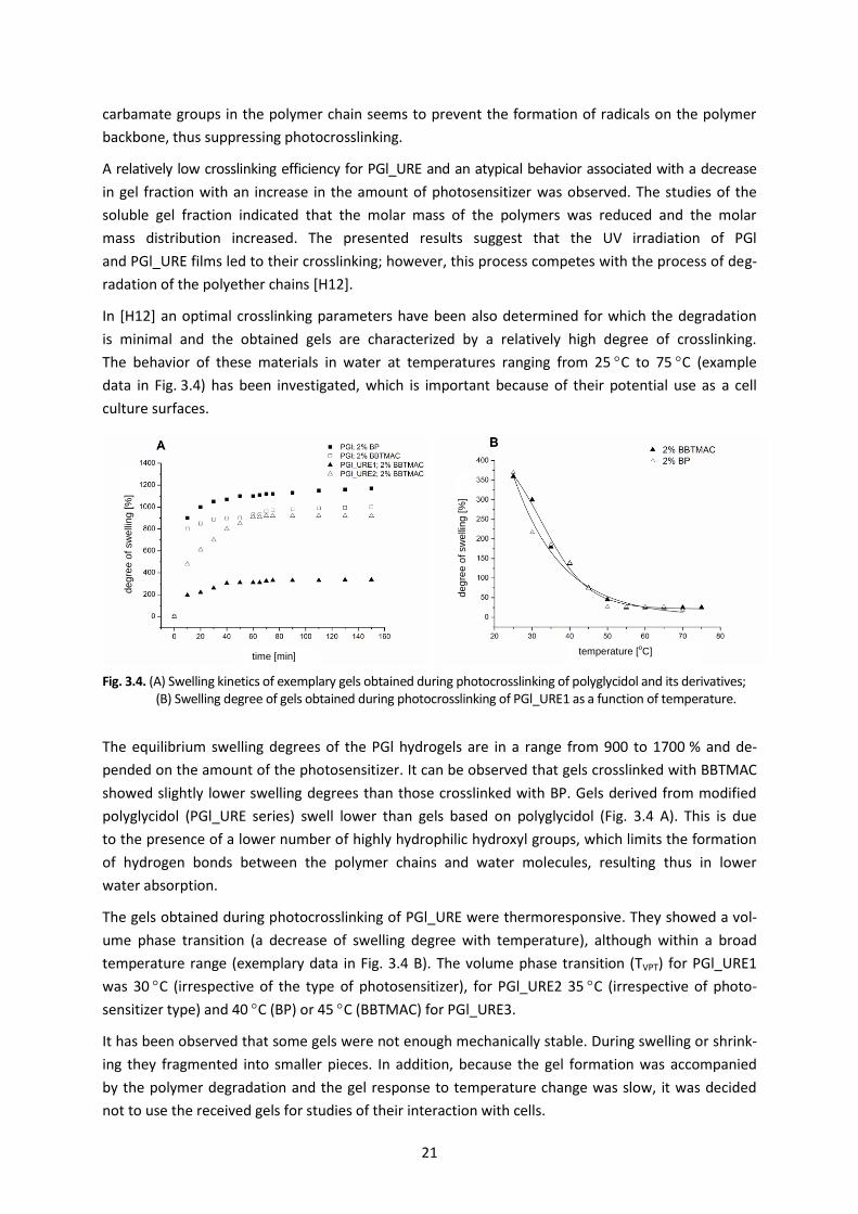
Fig. 3.4. (A) Swelling kinetics of exemplary gels obtained during photocrosslinking of polyglycidol and its derivatives; (B) Swelling degree of gels obtained during photocrosslinking of PGl_URE1 as a function of temperature.
The equilibrium swelling degrees of the PGl hydrogels are in a range from 900 to 1700 % and de-
pended on the amount of the photosensitizer. It can be observed that gels crosslinked with BBTMAC
showed slightly lower swelling degrees than those crosslinked with BP. Gels derived from modified
polyglycidol (PGl_URE series) swell lower than gels based on polyglycidol (Fig. 3.4 A). This is due
to the presence of a lower number of highly hydrophilic hydroxyl groups, which limits the formation
of hydrogen bonds between the polymer chains and water molecules, resulting thus in lower
water absorption.
The gels obtained during photocrosslinking of PGl_URE were thermoresponsive. They showed a vol-
ume phase transition (a decrease of swelling degree with temperature), although within a broad
temperature range (exemplary data in Fig. 3.4 B). The volume phase transition (TVPT) for PGl_URE1
was 30 C (irrespective of the type of photosensitizer), for PGl_URE2 35 C (irrespective of photo-
sensitizer type) and 40 C (BP) or 45 C (BBTMAC) for PGl_URE3.
It has been observed that some gels were not enough mechanically stable. During swelling or shrink-
ing they fragmented into smaller pieces. In addition, because the gel formation was accompanied
by the polymer degradation and the gel response to temperature change was slow, it was decided
not to use the received gels for studies of their interaction with cells.
time [min] temperature [
oC]
deg
ree o
f sw
elli
ng [
%]
deg
ree o
f sw
elli
ng [
%]
22
In order to increase the mechanical strength of thermoresponsive gels, to improve their behavior
in water under temperature changes and above all to eliminate the degradation of the polymeric
chain under UV irradiation, in habilitation work [H1] the photocrosslinking process was carried out
for a water polymer/photosensitizer mixture after its freezing. So called cryogels were then formed.
During freezing, water forms ice crystals, whereas soluble substances accumulate in a non-frozen
liquid microphase. The irradiation with UV lead to the crosslinking and gel formation in this microphase
and the ice crystals act as porogens. Therefore, the exposure time can be minimized thus limiting
the possible degradation of the polymer. Additional cryogels, compared to conventional gels, exhibit
a much faster reaction to hydration and dehydration.
Polyglycidol and its thermoresponsive derivatives PGl_URE1, PGl_URE2 and PGl_URE3 described
earlier (Table 3.1) were frozen in the presence of a BBTMAC photosensitizer and then shortly exposed
to UV radiation. The mechanism of polymer crosslinking in the non-frozen liquid phase is the same
as in the case of photocrosslinking without freezing (Fig. 3.3). In the [H1] work, the effect of copoly-
mer composition, concentration during crosslinking and exposure time on the efficiency of cryogel
formation (gel fraction) and properties (equilibrium swelling degree and TVPT) was investigated.
Cryogels with a yield of 73-88 % were obtained. They were characterized by a relatively high swelling
degree in water. The cryogels obtained from polyglycidol swelled up to 7000 % while the thermo-
responsive cryogels PGl_URE up to 5200 %. The lower degree of swelling of PGl_URE materials
was attributed to the presence of hydrophobic ethyl carbamates groups in the polymer chain.
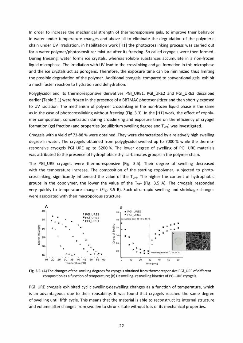
The PGl_URE cryogels were thermoresponsive (Fig. 3.5). Their degree of swelling decreased
with the temperature increase. The composition of the starting copolymer, subjected to photo-
crosslinking, significantly influenced the value of the TVPT. The higher the content of hydrophobic
groups in the copolymer, the lower the value of the TVPT (Fig. 3.5 A). The cryogels responded
very quickly to temperature changes (Fig. 3.5 B). Such ultra-rapid swelling and shrinkage changes
were associated with their macroporous structure.
Fig. 3.5. (A) The changes of the swelling degrees for cryogels obtained from thermoresponsive PGl_URE of different composition as a function of temperature; (B) Deswelling–reswelling kinetics of PGl-URE cryogels.
PGl_URE cryogels exhibited cyclic swelling-deswelling changes as a function of temperature, which
is an advantageous due to their reusability. It was found that cryogels reached the same degree
of swelling until fifth cycle. This means that the material is able to reconstruct its internal structure
and volume after changes from swollen to shrunk state without loss of its mechanical properties.
A B
PGl_URE3 PGl_URE2 PGl_URE1
PGl_URE2 PGl_URE3

23
On the base of performed studies, an optimum exposure time of UV radiation and concentration
of polymer, required to obtain cryogels with high amount of gel fraction, high swelling rate and rapid
reaction to temperature changes, were determined. These cryogels were therefore used as a self-
supporting layers for cell culture.
The research described in the work [H1, H2, H12] allowed for:
determination of UV radiation limit parameters (such as concentration of polymer, UV exposure
time) under which the polymer chain scission occurs. The obtained results should be taken into
account when planning the potential application of polyglycidol
obtaining a self-supporting layers of polyglycidol and its thermoresponsive derivatives
(poly(glycidol-co-ethyl glycidyl carbamate)s) during the UV irradiation of a photosensitiz-
er/polymer mixture. The obtained gels were thermoresponsive and their TVPT values can be con-
trolled by the composition of the polymer. These gels, however, were not mechanically stable,
besides during their irradiation the polymer chain degradation was observed
receiving the so-called cryogel, as a result of photocrosslinking of a mixture of thermoresponsive
polyglycidol/photosensitizer after its freezing. These gels were characterized by high mechanical
strength, did not degrade during irradiation and, importantly, had a rapid reaction (seconds)
to hydration and dehydration under temperature changes
the thermoresponsive cryogels, obtained in habilitation works, with phase separation tempera-
ture equal to 25 C, were used for the study of their interaction with cells. This choice was justi-
fied by the fact that this cryogel is hydrophobic under cell culture conditions (37 C) and may
therefore promote cell adhesion and proliferation.
3.2. Thermoresponsive polymer layers immobilized on a support
As mentioned earlier, in the habilitation work the suitability of thermoresponsive polymer layers
based on modified polyglycidol immobilized on a solid support for cell sheet culture and detachment
has also been undertaken. Due to the significant progress made in the field of chemistry of tempera-
ture-sensitive polymers, these studies have also been extended to other thermoresponsive polymers:
poly[oligo(ethylene glycol) methacrylates] and poly(2-substituted-2-oxazolines).
This chapter will first describe the synthesis and properties of layers based on modified polyglycidol
immobilized on a support (subsection 3.2.1), followed by layers of poly[oligo(ethylene glycol) meth-
acrylates] (subsection 3.2.2) and poly(2-substituted-2-oxazoline)s (Section 3.2.3). Chapter 3.3 shows
the use of these thermoresponsive layers in a cell sheet culture and detachment.
24
3.2.1. Thermoresponsive polyglycidol immobilized on a support [H5]
Polymer surfaces containing the layer made of thermoresponsive polyglycidol derivative (Fig. 1.2 B)
were obtained using a grafting-to technique. The synthesis of thermoresponsive layers was analo-
gous to that described in Section 2.1 for polyglycidol layers tested for protein adhesion. It included
two steps: modification of solid support to introduce reactive anhydride groups (Fig. 2.1 A, B and C)
and then immobilization of pre-synthesized polymers of glycidol (Fig. 2.2 A), in this case
thermoresponsive ones.
Poly(glycidol-co-ethyl glycidyl carbamate) (Mn = 2.106 g/mol, Mn/Mw = 1.3), having 40 % of ethyl
carbamate groups in the polymer chain, was used for the study. This polymer (mPGl – the polymer
designation in the publication) showed a transition temperature at 25 C. At this temperature
the polymer was hydrophobic, which can promote adhesion and cell proliferation.
The above polymer at different concentrations (0.25% to 10%) was applied onto silica or glass sub-
strates containing PE-MA layer. Due to the reaction of anhydride groups with hydroxyl groups
derived from mPGl, a polymer layer, in which the polymer chains were repeatedly attached with
the support, was obtained.
The presence of the polymer on the substrate was confirmed by FT-IR. A characteristic absorption
bands derived from groups present in mPGl have been observed in the spectrum. After grafting reac-
tion, the absorption band at 1850 cm-1 derived from the C=O groups of the PE-MA layer disappears.
This proves that most of the anhydride groups of the PE-MA layer have reacted with the hydroxyl
groups of poly(glycidol-co-ethyl glycidyl carbamate).
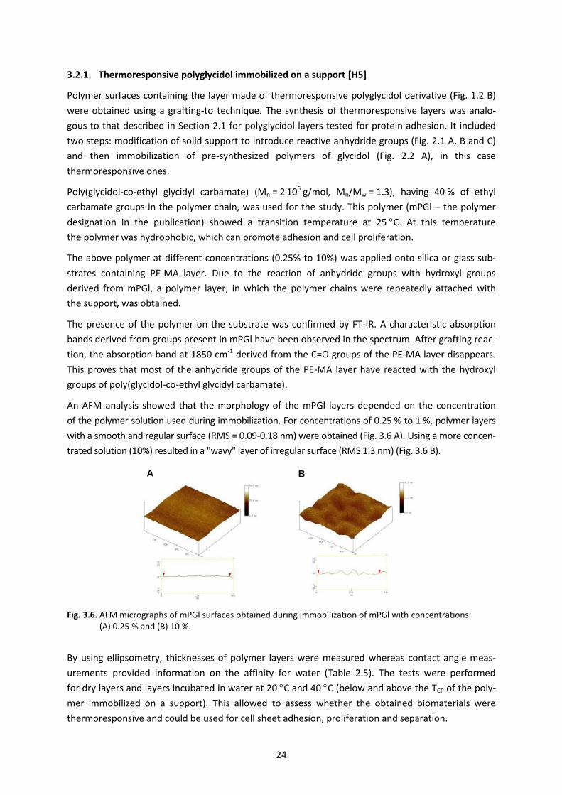
An AFM analysis showed that the morphology of the mPGl layers depended on the concentration
of the polymer solution used during immobilization. For concentrations of 0.25 % to 1 %, polymer layers
with a smooth and regular surface (RMS = 0.09-0.18 nm) were obtained (Fig. 3.6 A). Using a more concen-
trated solution (10%) resulted in a "wavy" layer of irregular surface (RMS 1.3 nm) (Fig. 3.6 B).
Fig. 3.6. AFM micrographs of mPGl surfaces obtained during immobilization of mPGl with concentrations: (A) 0.25 % and (B) 10 %.
By using ellipsometry, thicknesses of polymer layers were measured whereas contact angle meas-
urements provided information on the affinity for water (Table 2.5). The tests were performed
for dry layers and layers incubated in water at 20 C and 40 C (below and above the TCP of the poly-
mer immobilized on a support). This allowed to assess whether the obtained biomaterials were
thermoresponsive and could be used for cell sheet adhesion, proliferation and separation.
A B
25
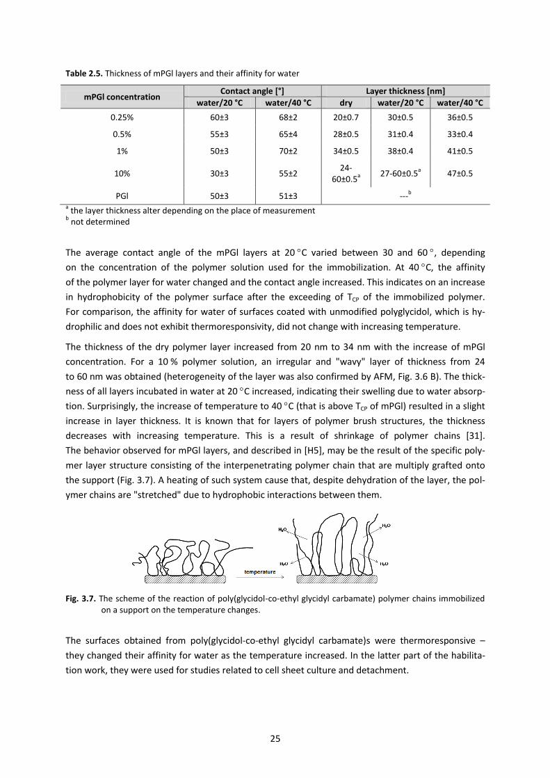
Table 2.5. Thickness of mPGl layers and their affinity for water
mPGl concentration Contact angle [°] Layer thickness [nm]
water/20 °C water/40 °C dry water/20 °C water/40 °C
0.25% 60±3 68±2 20±0.7 30±0.5 36±0.5
0.5% 55±3 65±4 28±0.5 31±0.4 33±0.4
1% 50±3 70±2 34±0.5 38±0.4 41±0.5
10% 30±3 55±2 24-
60±0.5a 27-60±0.5
a 47±0.5
PGl 50±3 51±3 ---b
a the layer thickness alter depending on the place of measurement
b not determined
The average contact angle of the mPGl layers at 20 C varied between 30 and 60 , depending
on the concentration of the polymer solution used for the immobilization. At 40 C, the affinity
of the polymer layer for water changed and the contact angle increased. This indicates on an increase
in hydrophobicity of the polymer surface after the exceeding of TCP of the immobilized polymer.
For comparison, the affinity for water of surfaces coated with unmodified polyglycidol, which is hy-
drophilic and does not exhibit thermoresponsivity, did not change with increasing temperature.
The thickness of the dry polymer layer increased from 20 nm to 34 nm with the increase of mPGl
concentration. For a 10 % polymer solution, an irregular and "wavy" layer of thickness from 24
to 60 nm was obtained (heterogeneity of the layer was also confirmed by AFM, Fig. 3.6 B). The thick-
ness of all layers incubated in water at 20 C increased, indicating their swelling due to water absorp-
tion. Surprisingly, the increase of temperature to 40 C (that is above TCP of mPGl) resulted in a slight
increase in layer thickness. It is known that for layers of polymer brush structures, the thickness
decreases with increasing temperature. This is a result of shrinkage of polymer chains [31].
The behavior observed for mPGl layers, and described in [H5], may be the result of the specific poly-
mer layer structure consisting of the interpenetrating polymer chain that are multiply grafted onto
the support (Fig. 3.7). A heating of such system cause that, despite dehydration of the layer, the pol-
ymer chains are "stretched" due to hydrophobic interactions between them.
Fig. 3.7. The scheme of the reaction of poly(glycidol-co-ethyl glycidyl carbamate) polymer chains immobilized on a support on the temperature changes.
The surfaces obtained from poly(glycidol-co-ethyl glycidyl carbamate)s were thermoresponsive –
they changed their affinity for water as the temperature increased. In the latter part of the habilita-
tion work, they were used for studies related to cell sheet culture and detachment.
26
3.2.2. Thermoresponsive poly[oligo(ethylene glycol) methacrylates)] on support [H4, H6]
Poly[oligo(ethylene glycol) methacrylates)] (POEGMA), except of the modified polyglycidol, were also
used to obtain thermoresponsive polymer coatings. POEGMA is a large group of polymers,
which in recent years has gained considerable interest [32]. Most monomers of oligo(ethylene glycol)
methacrylates are commercially available and easily polymerizable using controlled radical polymeri-
zation techniques (especially ATRP – controlled atom transfer radical polymerization). The amphiphilic
structure of these polymers (where the side chain of oligo(ethylene glycol) is responsible for the sol-
ubility and formation of hydrogen bonds with water molecules whereas the main chain for compet-
ing hydrophobic interactions) cause that many of them exhibit thermoresponsive behavior.
Compared to the commonly used PNIPAM, the thermoresponsive POEGMAs have many advantages,
for example, they are characterized by a narrow phase transition with a slight hysteresis,
and the influence of the external factors on their TCP values is small. At the time of the habilitation
work, the use of poly[oligo(ethylene glycol) methacrylates)] immobilized on the support for cell
sheet culture and detachment was not described.
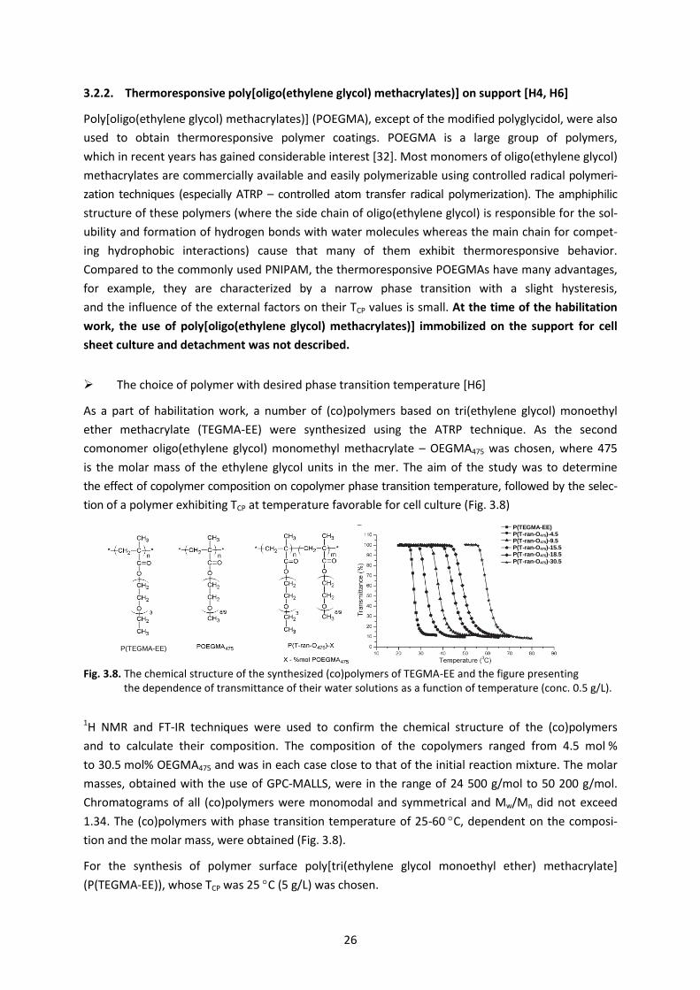
The choice of polymer with desired phase transition temperature [H6]
As a part of habilitation work, a number of (co)polymers based on tri(ethylene glycol) monoethyl
ether methacrylate (TEGMA-EE) were synthesized using the ATRP technique. As the second
comonomer oligo(ethylene glycol) monomethyl methacrylate – OEGMA475 was chosen, where 475
is the molar mass of the ethylene glycol units in the mer. The aim of the study was to determine
the effect of copolymer composition on copolymer phase transition temperature, followed by the selec-
tion of a polymer exhibiting TCP at temperature favorable for cell culture (Fig. 3.8)
Fig. 3.8. The chemical structure of the synthesized (co)polymers of TEGMA-EE and the figure presenting the dependence of transmittance of their water solutions as a function of temperature (conc. 0.5 g/L).
1H NMR and FT-IR techniques were used to confirm the chemical structure of the (co)polymers
and to calculate their composition. The composition of the copolymers ranged from 4.5 mol %
to 30.5 mol% OEGMA475 and was in each case close to that of the initial reaction mixture. The molar
masses, obtained with the use of GPC-MALLS, were in the range of 24 500 g/mol to 50 200 g/mol.
Chromatograms of all (co)polymers were monomodal and symmetrical and Mw/Mn did not exceed
1.34. The (co)polymers with phase transition temperature of 25-60 C, dependent on the composi-
tion and the molar mass, were obtained (Fig. 3.8).
For the synthesis of polymer surface poly[tri(ethylene glycol monoethyl ether) methacrylate]
(P(TEGMA-EE)), whose TCP was 25 C (5 g/L) was chosen.
P(TEGMA-EE)
P(TEGMA-EE) P(T-ran-O475)-4.5 P(T-ran-O475)-9.5 P(T-ran-O475)-15.5 P(T-ran-O475)-18.5
P(T-ran-O475)-30.5
27
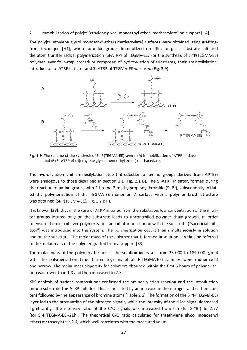
Immobilization of poly[tri(ethylene glycol monoethyl ether) methacrylate] on support [H4]
The poly[tri(ethylene glycol monoethyl ether) methacrylate] surfaces were obtained using grafting-
from technique [H4], where bromide groups immobilized on silica or glass substrate initiated
the atom transfer radical polymerization (SI-ATRP) of TEGMA-EE. For the synthesis of Si~P(TEGMA-EE)
polymer layer four-step procedure composed of hydroxylation of substrates, their aminosilylation,
introduction of ATRP initiator and SI-ATRP of TEGMA-EE was used (Fig. 3.9).
Fig. 3.9. The scheme of the synthesis of Si~P(TEGMA-EE) layers: (A) immobilization of ATRP initiator and (B) SI-ATRP of tri(ethylene glycol monoethyl ether) methacrylate.
The hydroxylation and aminosilylation step (introduction of amino groups derived from APTES)
were analogous to those described in section 2.1 (Fig. 2.1 B). The SI-ATRP initiator, formed during
the reaction of amino groups with 2-bromo-2-methylpropionyl bromide (Si-Br), subsequently initiat-
ed the polymerization of the TEGMA-EE monomer. A surface with a polymer brush structure
was obtained (Si-P(TEGMA-EE), Fig. 1.2 B II).
It is known [33], that in the case of ATRP initiated from the substrates low concentration of the initia-
tor groups located only on the substrate leads to uncontrolled polymer chain growth. In order
to ensure the control over polymerization an initiator non-bound with the substrate (“sacrificial initi-
ator") was introduced into the system. The polymerization occurs then simultaneously in solution
and on the substrate. The molar mass of the polymer that is formed in solution can thus be referred
to the molar mass of the polymer grafted from a support [33].
The molar mass of the polymers formed in the solution increased from 23 000 to 189 000 g/mol
with the polymerization time. Chromatograms of all P(TEGMA-EE) samples were monomodal
and narrow. The molar mass dispersity for polymers obtained within the first 6 hours of polymeriza-
tion was lower than 1.3 and then increased to 2.3.
XPS analysis of surface compositions confirmed the aminosilylation reaction and the introduction
onto a substrate the ATRP initiator. This is indicated by an increase in the nitrogen and carbon con-
tent followed by the appearance of bromine atoms (Table 2.6). The formation of the Si~P(TEGMA-EE)
layer led to the attenuation of the nitrogen signals, while the intensity of the silica signal decreased
significantly. The intensity ratio of the C/O signals was increased from 0.5 (for Si~Br) to 2.77
(for Si-P(TEGMA-EE)-21h). The theoretical C/O ratio calculated for tri(ethylene glycol monoethyl
ether) methacrylate is 2.4, which well correlates with the measured value.
H2SO
4/H
2O
2
100 oC, 2h
Si
OHOH
Si
OH
Si
Si
OHOH
Si
OH
Si
OSi
O
O
NH2
CH3
CH3
CH3
EtOH, RT, 2h
O
Si
Si O Si
O
Si
NH2
Si
O
SiO
NH
O
Br
O
Si
Si O Si
O
Si Si
O
SiOO
Br
Br
CH2Cl
2, Et
3N, RT, 4h
O
Si
Si O Si
O
Si
NH2
Si
O
SiO
NH
O
Br
O
Si
Si O Si
O
Si Si
O
SiO
NH
O
CH2 C
COO
CH2CH2
O
CH2
CH3
CH3
Br
O
Si
Si O Si
O
Si Si
O
SiO
CH2 C
COO
CH2CH2
O
CH2
CH3
CH3
3
3
n
CuCl, Bpy, MeOH/H2O,
EBiB
+
O
O
CH2 C
COO
CH2CH2
O
CH2
CH3
CH3
Br
CH3
3
n
2n
A
B
C
D
Si/SiO2 Si-OH
Si~NH2
Si~Br
Si~P(TEGMA-EE)
P(TEGMA-EE)
A
B
28
Table 2.6. Composition of modified substrate and substrate coated with polymer P(TEGMA-EE)
Designation of the layer in publication
C1s O1s Si2p N1p Br
Si~OH 17.2 57.4 24.7 0.7 0.0
Si~NH2 28.8 47.7 20.8 2.7 0.0
Si~Br 25.9 51.3 20.5 2.2 0.1
Si~P(TEGMA-EE) 72.5 26.2 1.23 0.0 0.0
Analysis of Si~P(TEGMA-EE) layer properties [H4]
The roughness of the Si~P(TEGMA-EE) layers changed with the polymerization time, which was relat-
ed to the increase of polymer chains. RMS increased from an initial value of 0.24 nm (after one hour
of polymerization) to a maximum of 0.62 nm for the layer after 4 hours of polymerization. A further
increase in the polymerization time resulted in the formation of a homogeneous film (due to the
elongation of the polymer chains), thereby decreasing the RMS to 0.21 nm was observed.
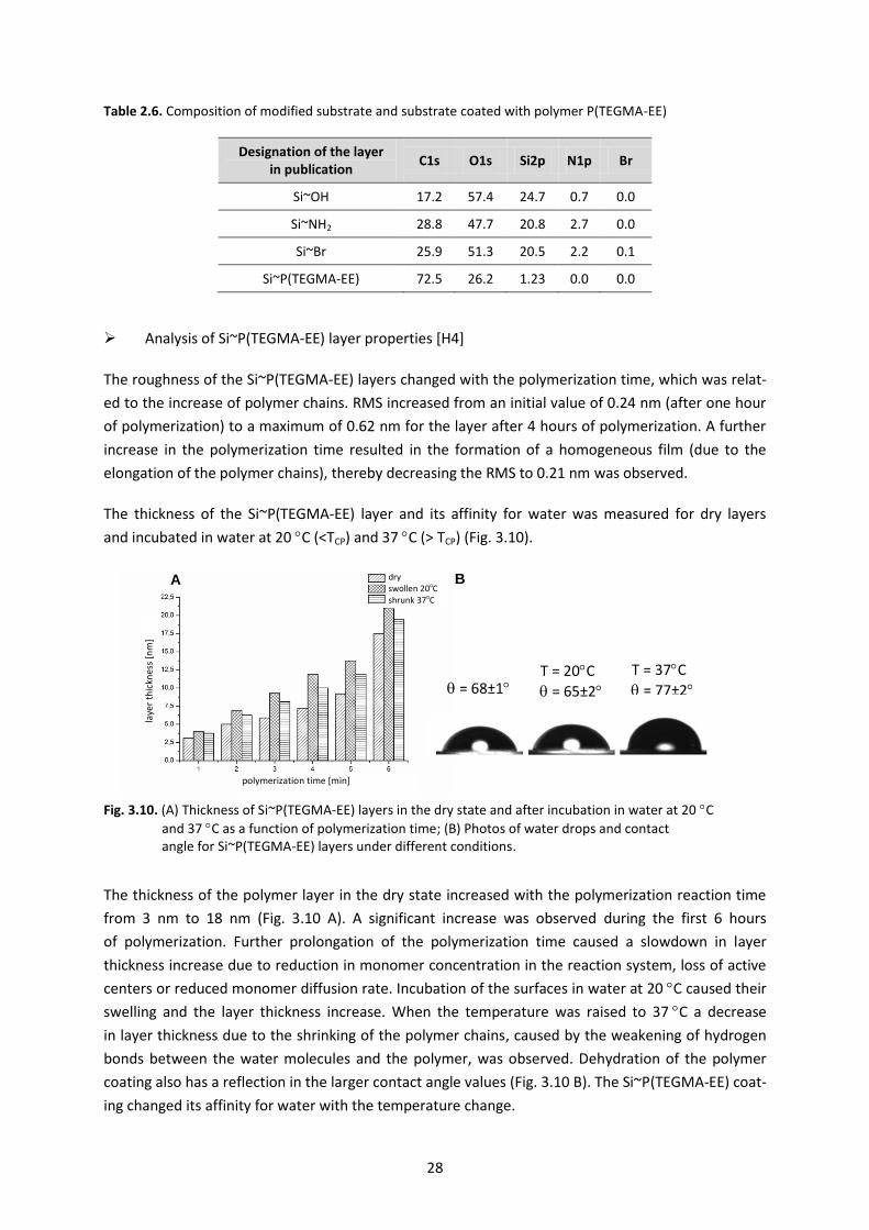
The thickness of the Si~P(TEGMA-EE) layer and its affinity for water was measured for dry layers
and incubated in water at 20 C (<TCP) and 37 C (> TCP) (Fig. 3.10).
Fig. 3.10. (A) Thickness of Si~P(TEGMA-EE) layers in the dry state and after incubation in water at 20 C
and 37 C as a function of polymerization time; (B) Photos of water drops and contact angle for Si~P(TEGMA-EE) layers under different conditions.
The thickness of the polymer layer in the dry state increased with the polymerization reaction time
from 3 nm to 18 nm (Fig. 3.10 A). A significant increase was observed during the first 6 hours
of polymerization. Further prolongation of the polymerization time caused a slowdown in layer
thickness increase due to reduction in monomer concentration in the reaction system, loss of active
centers or reduced monomer diffusion rate. Incubation of the surfaces in water at 20 C caused their
swelling and the layer thickness increase. When the temperature was raised to 37 C a decrease
in layer thickness due to the shrinking of the polymer chains, caused by the weakening of hydrogen
bonds between the water molecules and the polymer, was observed. Dehydration of the polymer
coating also has a reflection in the larger contact angle values (Fig. 3.10 B). The Si~P(TEGMA-EE) coat-
ing changed its affinity for water with the temperature change.
= 68±1 T = 20C
= 65±2
T = 37C
= 77±2
A B
polymerization time [min]
laye
r th
ickn
ess
[nm
]
dry swollen 20oC shrunk 37oC
29
Changes in layer thickness and contact angles due to temperature increase indicated that
the Si~P(TEGMA-EE) layers were thermoresponsive. That is why they were applied for cell sheet cul-
ture and detachment.
3.2.3. Thermoresponsive poly(2-substituted-2-oxazoline) on a support [H7, H8, H9]
Another group of thermoresponsive polymers used for the investigation of their interaction with cells
were poly(2-substituted-2-oxazoline)s (POx).
POx, called pseudopeptides, are non-toxic and biocompatible [34] and do not accumulate in tissues [35].
Although the polymerization mechanism and the 2-oxazoline polymers themselves have been known
for several decades, due to their interesting properties these polymer were discovered again. It has
been shown that it is possible to obtain POx copolymers with different side chains or end groups
and different architectures [36]. There are numerous applications of POx in medicine and biotech-
nology, such as POx-protein conjugates or DNA carriers [37]. Some of POx are thermoresponsive,
which additionally enhances their attractiveness for biomedical applications.
At the time of habilitation work, there was few work concerning POx immobilization on a support [38].
None of them have described the preparation of poly(2-substituted-2-oxazoline) layers with
a structure of polymer brush using the termination of living POx chains by a functional groups
of the support. They also did not refer to the thermoresponsive behavior of the polymeric coatings,
also in terms of their interaction with cells.
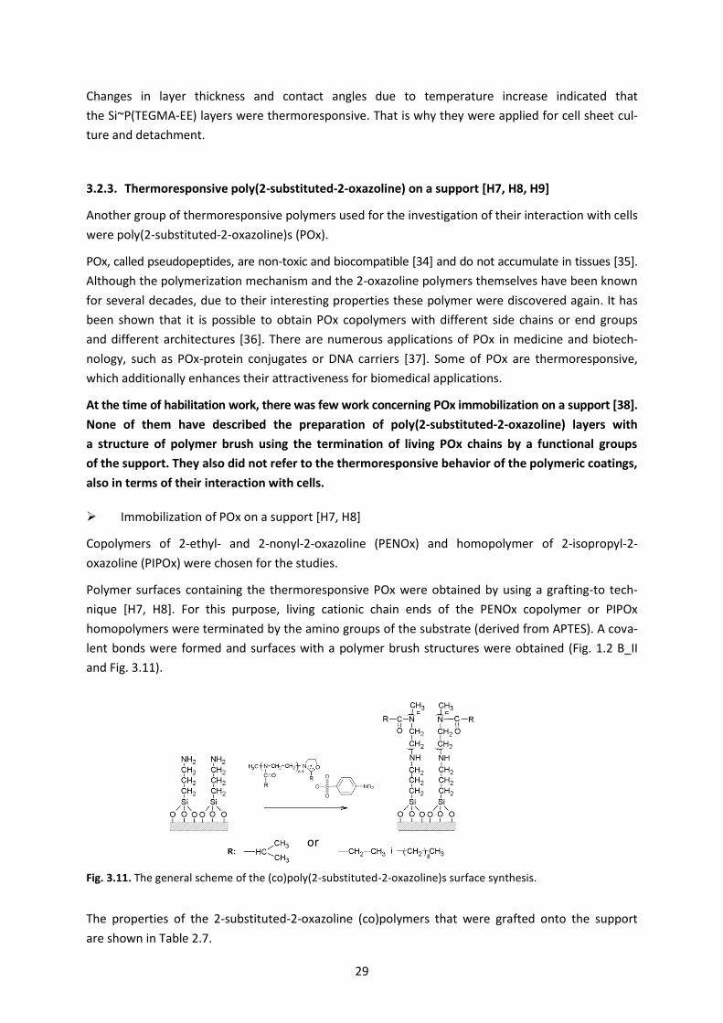
Immobilization of POx on a support [H7, H8]
Copolymers of 2-ethyl- and 2-nonyl-2-oxazoline (PENOx) and homopolymer of 2-isopropyl-2-
oxazoline (PIPOx) were chosen for the studies.
Polymer surfaces containing the thermoresponsive POx were obtained by using a grafting-to tech-
nique [H7, H8]. For this purpose, living cationic chain ends of the PENOx copolymer or PIPOx
homopolymers were terminated by the amino groups of the substrate (derived from APTES). A cova-
lent bonds were formed and surfaces with a polymer brush structures were obtained (Fig. 1.2 B_II
and Fig. 3.11).
Fig. 3.11. The general scheme of the (co)poly(2-substituted-2-oxazoline)s surface synthesis.
The properties of the 2-substituted-2-oxazoline (co)polymers that were grafted onto the support
are shown in Table 2.7.
or
30
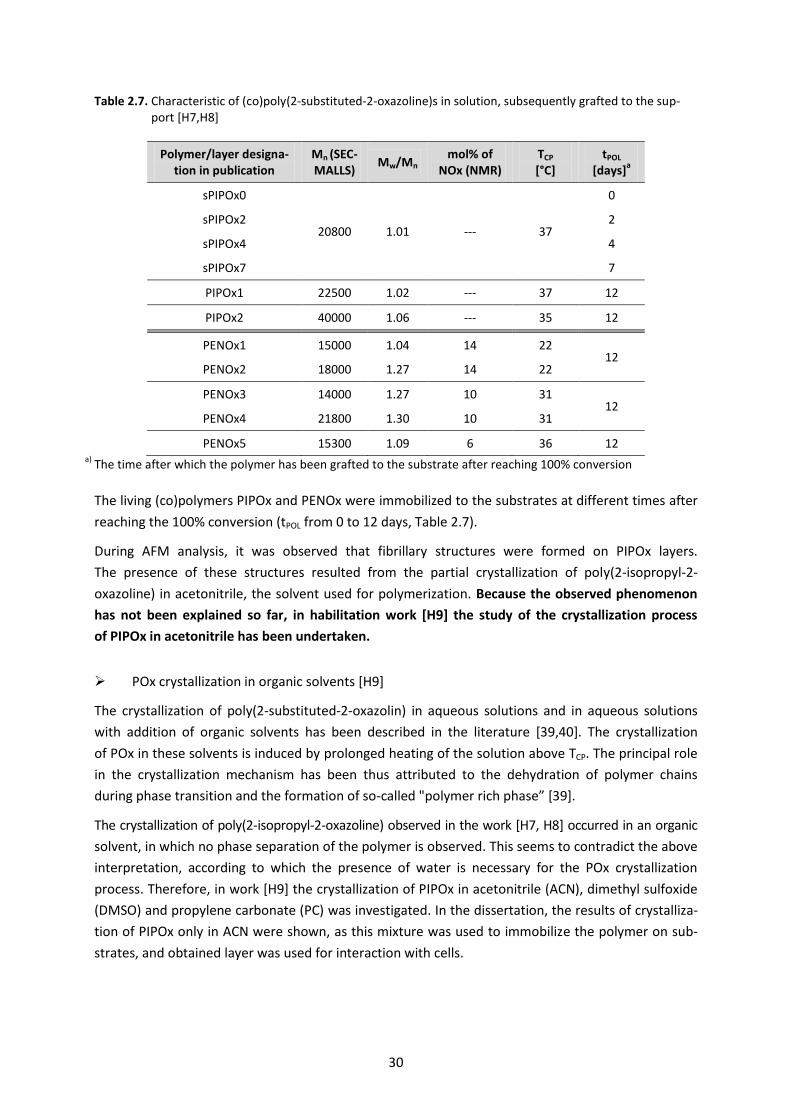
Table 2.7. Characteristic of (co)poly(2-substituted-2-oxazoline)s in solution, subsequently grafted to the sup-port [H7,H8]
Polymer/layer designa-tion in publication
Mn (SEC-MALLS)
Mw/Mn mol% of
NOx (NMR) TCP [°C]
tPOL [days]
a
sPIPOx0
20800 1.01 --- 37
0
sPIPOx2 2
sPIPOx4 4
sPIPOx7 7
PIPOx1 22500 1.02 --- 37 12
PIPOx2 40000 1.06 --- 35 12
PENOx1 15000 1.04 14 22 12
PENOx2 18000 1.27 14 22
PENOx3 14000 1.27 10 31 12
PENOx4 21800 1.30 10 31
PENOx5 15300 1.09 6 36 12 a) The time after which the polymer has been grafted to the substrate after reaching 100% conversion
The living (co)polymers PIPOx and PENOx were immobilized to the substrates at different times after
reaching the 100% conversion (tPOL from 0 to 12 days, Table 2.7).
During AFM analysis, it was observed that fibrillary structures were formed on PIPOx layers.
The presence of these structures resulted from the partial crystallization of poly(2-isopropyl-2-
oxazoline) in acetonitrile, the solvent used for polymerization. Because the observed phenomenon
has not been explained so far, in habilitation work [H9] the study of the crystallization process
of PIPOx in acetonitrile has been undertaken.
POx crystallization in organic solvents [H9]
The crystallization of poly(2-substituted-2-oxazolin) in aqueous solutions and in aqueous solutions
with addition of organic solvents has been described in the literature [39,40]. The crystallization
of POx in these solvents is induced by prolonged heating of the solution above TCP. The principal role
in the crystallization mechanism has been thus attributed to the dehydration of polymer chains
during phase transition and the formation of so-called "polymer rich phase” [39].
The crystallization of poly(2-isopropyl-2-oxazoline) observed in the work [H7, H8] occurred in an organic
solvent, in which no phase separation of the polymer is observed. This seems to contradict the above
interpretation, according to which the presence of water is necessary for the POx crystallization
process. Therefore, in work [H9] the crystallization of PIPOx in acetonitrile (ACN), dimethyl sulfoxide
(DMSO) and propylene carbonate (PC) was investigated. In the dissertation, the results of crystalliza-
tion of PIPOx only in ACN were shown, as this mixture was used to immobilize the polymer on sub-
strates, and obtained layer was used for interaction with cells.

31
PIPOx solutions (Mn = 20 800 g/mol, Mw/Mn = 1.01) in acetonitrile (5 %, 10 % and 30 %) were heated
at 50 C for 20 days. At this time, the mixture became cloudy and a precipitate was formed. The pre-
cipitate was analyzed by DSC and WAXS (Fig. 3.12).
Fig. 3.12. (A) DSC traces for PIPOx crystallized from ACN at different concentration (heating rate 10 °C/min) and (B) X-ray diffraction curves for precipitate derived from 5 % solution of PIPOx in ACN.
The DSC thermograms of precipitates formed in all PIPOx solutions showed the presence of an endo-
thermal peak indicating that the samples contained a crystalline fractions that melted. The enthalpy
of melting increased with increasing the concentration of the solution from which the precipitate
was formed (from 20 J/g for 5 % solution to 41 J/g for 30 % solution). WAXS analysis revealed two
principal diffraction peaks at 2 = 7.93 and 18.11 . Identical peak positions and intensities were
obtained for PIPOx crystallized in water [41]. This means that PIPOx crystallized in organic solvents
has the same elemental cell as PIPOx crystallized in water. The highest crystal fraction χc of 68 %
was obtained for PIPOx crystallized from a 30 % solution in ACN.
The morphology of PIPOx crystallized from organic solvent was analyzed by SEM (Fig. 3.13).
Fig. 3.13. SEM micrographs of PIPOx crystallized from ACN of 30 % (A) and 5% (B) (scale 5 µm).
In the SEM micrograph of PIPOx, a network-like structure is present. Within this network, separate
objects possessing a fibril-like morphology can be distinguished (Fig. 3.13). The length of the fibrils
ranged from a few to several microns, with a width of approximately 50 nm. It was shown that
the concentration of the solution from which PIPOx crystallized, had little influence on the morphol-
ogy of the resulting structures.
The studies demonstrated that PIPOx crystallize in an organic solvent (without water) and this pro-
cess is not related to the thermoresponsivity of the polymer.
A B
A B
2
\
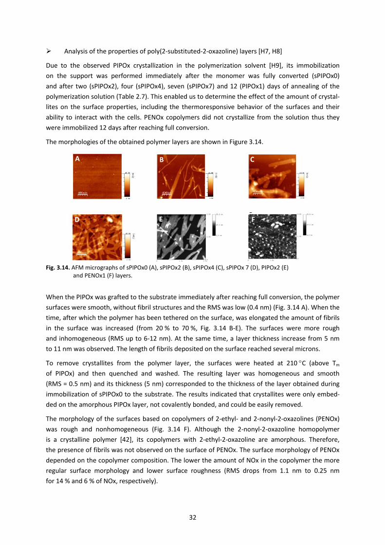
32
Analysis of the properties of poly(2-substituted-2-oxazoline) layers [H7, H8]
Due to the observed PIPOx crystallization in the polymerization solvent [H9], its immobilization
on the support was performed immediately after the monomer was fully converted (sPIPOx0)
and after two (sPIPOx2), four (sPIPOx4), seven (sPIPOx7) and 12 (PIPOx1) days of annealing of the
polymerization solution (Table 2.7). This enabled us to determine the effect of the amount of crystal-
lites on the surface properties, including the thermoresponsive behavior of the surfaces and their
ability to interact with the cells. PENOx copolymers did not crystallize from the solution thus they
were immobilized 12 days after reaching full conversion.
The morphologies of the obtained polymer layers are shown in Figure 3.14.
Fig. 3.14. AFM micrographs of sPIPOx0 (A), sPIPOx2 (B), sPIPOx4 (C), sPIPOx 7 (D), PIPOx2 (E) and PENOx1 (F) layers.
When the PIPOx was grafted to the substrate immediately after reaching full conversion, the polymer
surfaces were smooth, without fibril structures and the RMS was low (0.4 nm) (Fig. 3.14 A). When the
time, after which the polymer has been tethered on the surface, was elongated the amount of fibrils
in the surface was increased (from 20 % to 70 %, Fig. 3.14 B-E). The surfaces were more rough
and inhomogeneous (RMS up to 6-12 nm). At the same time, a layer thickness increase from 5 nm
to 11 nm was observed. The length of fibrils deposited on the surface reached several microns.
To remove crystallites from the polymer layer, the surfaces were heated at 210 C (above Tm
of PIPOx) and then quenched and washed. The resulting layer was homogeneous and smooth
(RMS = 0.5 nm) and its thickness (5 nm) corresponded to the thickness of the layer obtained during
immobilization of sPIPOx0 to the substrate. The results indicated that crystallites were only embed-
ded on the amorphous PIPOx layer, not covalently bonded, and could be easily removed.
The morphology of the surfaces based on copolymers of 2-ethyl- and 2-nonyl-2-oxazolines (PENOx)
was rough and nonhomogeneous (Fig. 3.14 F). Although the 2-nonyl-2-oxazoline homopolymer
is a crystalline polymer [42], its copolymers with 2-ethyl-2-oxazoline are amorphous. Therefore,
the presence of fibrils was not observed on the surface of PENOx. The surface morphology of PENOx
depended on the copolymer composition. The lower the amount of NOx in the copolymer the more
regular surface morphology and lower surface roughness (RMS drops from 1.1 nm to 0.25 nm
for 14 % and 6 % of NOx, respectively).
A B C
D E F
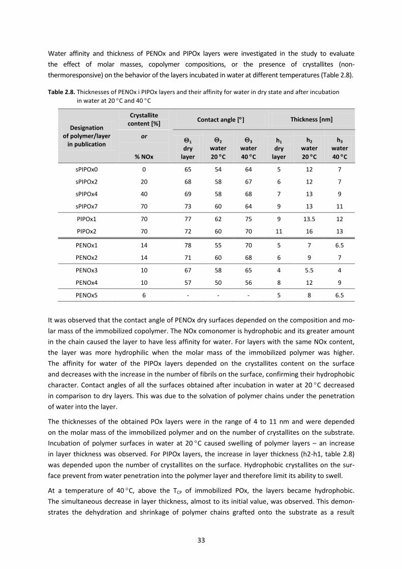
33
Water affinity and thickness of PENOx and PIPOx layers were investigated in the study to evaluate
the effect of molar masses, copolymer compositions, or the presence of crystallites (non-
thermoresponsive) on the behavior of the layers incubated in water at different temperatures (Table 2.8).
Table 2.8. Thicknesses of PENOx i PIPOx layers and their affinity for water in dry state and after incubation
in water at 20 C and 40 C
Designation of polymer/layer
in publication
Crystallite content [%]
Contact angle [] Thickness [nm]
or
% NOx
1 dry
layer
2 water
20 C
3 water
40 C
h1 dry
layer
h2 water
20 C
h3 water
40 C
sPIPOx0 0 65 54 64 5 12 7
sPIPOx2 20 68 58 67 6 12 7
sPIPOx4 40 69 58 68 7 13 9
sPIPOx7 70 73 60 64 9 13 11
PIPOx1 70 77 62 75 9 13.5 12
PIPOx2 70 72 60 70 11 16 13
PENOx1 14 78 55 70 5 7 6.5
PENOx2 14 71 60 68 6 9 7
PENOx3 10 67 58 65 4 5.5 4
PENOx4 10 57 50 56 8 12 9
PENOx5 6 - - - 5 8 6.5
It was observed that the contact angle of PENOx dry surfaces depended on the composition and mo-
lar mass of the immobilized copolymer. The NOx comonomer is hydrophobic and its greater amount
in the chain caused the layer to have less affinity for water. For layers with the same NOx content,
the layer was more hydrophilic when the molar mass of the immobilized polymer was higher.
The affinity for water of the PIPOx layers depended on the crystallites content on the surface
and decreases with the increase in the number of fibrils on the surface, confirming their hydrophobic
character. Contact angles of all the surfaces obtained after incubation in water at 20 C decreased
in comparison to dry layers. This was due to the solvation of polymer chains under the penetration
of water into the layer.
The thicknesses of the obtained POx layers were in the range of 4 to 11 nm and were depended
on the molar mass of the immobilized polymer and on the number of crystallites on the substrate.
Incubation of polymer surfaces in water at 20 C caused swelling of polymer layers – an increase
in layer thickness was observed. For PIPOx layers, the increase in layer thickness (h2-h1, table 2.8)
was depended upon the number of crystallites on the surface. Hydrophobic crystallites on the sur-
face prevent from water penetration into the polymer layer and therefore limit its ability to swell.
At a temperature of 40 C, above the TCP of immobilized POx, the layers became hydrophobic.
The simultaneous decrease in layer thickness, almost to its initial value, was observed. This demon-
strates the dehydration and shrinkage of polymer chains grafted onto the substrate as a result
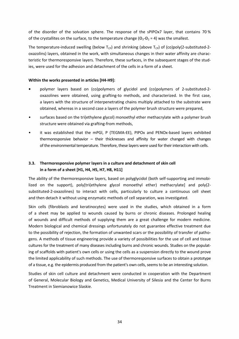
34
of the disorder of the solvation sphere. The response of the sPIPOx7 layer, that contains 70 %
of the crystallites on the surface, to the temperature change (Θ3-Θ2 = 4) was the smallest.
The temperature-induced swelling (below TCP) and shrinking (above TCP) of (co)poly(2-substituted-2-
oxazolins) layers, obtained in the work, with simultaneous changes in their water affinity are charac-
teristic for thermoresponsive layers. Therefore, these surfaces, in the subsequent stages of the stud-
ies, were used for the adhesion and detachment of the cells in a form of a sheet.
Within the works presented in articles [H4-H9]:
• polymer layers based on (co)polymers of glycidol and (co)polymers of 2-substituted-2-
oxazolines were obtained, using grafting-to methods, and characterized. In the first case,
a layers with the structure of interpenetrating chains multiply attached to the substrate were
obtained, whereas in a second case a layers of the polymer brush structure were prepared,
• surfaces based on the tri(ethylene glycol) monoethyl ether methacrylate with a polymer brush
structure were obtained via grafting-from methods,
• it was established that the mPGl, P (TEGMA-EE), PIPOx and PENOx-based layers exhibited
thermoresponsive behavior – their thicknesses and affinity for water changed with changes
of the environmental temperature. Therefore, these layers were used for their interaction with cells.
3.3. Thermoresponsive polymer layers in a culture and detachment of skin cell
in a form of a sheet [H1, H4, H5, H7, H8, H11]
The ability of the thermoresponsive layers, based on polyglycidol (both self-supporting and immobi-
lized on the support), poly[tri(ethylene glycol monoethyl ether) methacrylate] and poly(2-
substituted-2-oxazolines) to interact with cells, particularly to culture a continuous cell sheet
and then detach it without using enzymatic methods of cell separation, was investigated.
Skin cells (fibroblasts and keratinocytes) were used in the studies, which obtained in a form
of a sheet may be applied to wounds caused by burns or chronic diseases. Prolonged healing
of wounds and difficult methods of supplying them are a great challenge for modern medicine.
Modern biological and chemical dressings unfortunately do not guarantee effective treatment due
to the possibility of rejection, the formation of unwanted scars or the possibility of transfer of patho-
gens. A methods of tissue engineering provide a variety of possibilities for the use of cell and tissue
cultures for the treatment of many diseases including burns and chronic wounds. Studies on the populat-
ing of scaffolds with patient's own cells or using the cells as a suspension directly to the wound prove
the limited applicability of such methods. The use of thermoresponsive surfaces to obtain a prototype
of a tissue, e.g. the epidermis produced from the patient's own cells, seems to be an interesting solution.
Studies of skin cell culture and detachment were conducted in cooperation with the Department
of General, Molecular Biology and Genetics, Medical University of Silesia and the Center for Burns
Treatment in Siemianowice Slaskie.
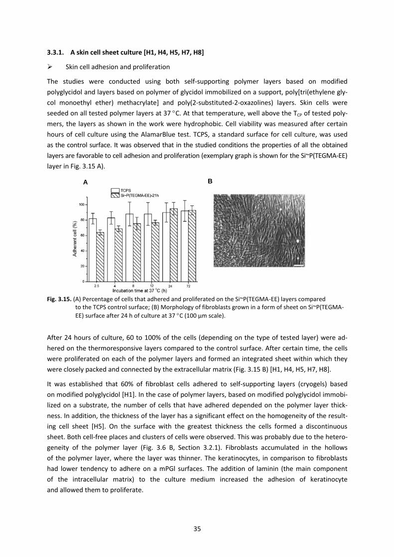
35
3.3.1. A skin cell sheet culture [H1, H4, H5, H7, H8]
Skin cell adhesion and proliferation
The studies were conducted using both self-supporting polymer layers based on modified
polyglycidol and layers based on polymer of glycidol immobilized on a support, poly[tri(ethylene gly-
col monoethyl ether) methacrylate] and poly(2-substituted-2-oxazolines) layers. Skin cells were
seeded on all tested polymer layers at 37 C. At that temperature, well above the TCP of tested poly-
mers, the layers as shown in the work were hydrophobic. Cell viability was measured after certain
hours of cell culture using the AlamarBlue test. TCPS, a standard surface for cell culture, was used
as the control surface. It was observed that in the studied conditions the properties of all the obtained
layers are favorable to cell adhesion and proliferation (exemplary graph is shown for the Si~P(TEGMA-EE)
layer in Fig. 3.15 A).
Fig. 3.15. (A) Percentage of cells that adhered and proliferated on the Si~P(TEGMA-EE) layers compared to the TCPS control surface; (B) Morphology of fibroblasts grown in a form of sheet on Si~P(TEGMA-
EE) surface after 24 h of culture at 37 C (100 μm scale).
After 24 hours of culture, 60 to 100% of the cells (depending on the type of tested layer) were ad-
hered on the thermoresponsive layers compared to the control surface. After certain time, the cells
were proliferated on each of the polymer layers and formed an integrated sheet within which they
were closely packed and connected by the extracellular matrix (Fig. 3.15 B) [H1, H4, H5, H7, H8].
It was established that 60% of fibroblast cells adhered to self-supporting layers (cryogels) based
on modified polyglycidol [H1]. In the case of polymer layers, based on modified polyglycidol immobi-
lized on a substrate, the number of cells that have adhered depended on the polymer layer thick-
ness. In addition, the thickness of the layer has a significant effect on the homogeneity of the result-
ing cell sheet [H5]. On the surface with the greatest thickness the cells formed a discontinuous
sheet. Both cell-free places and clusters of cells were observed. This was probably due to the hetero-
geneity of the polymer layer (Fig. 3.6 B, Section 3.2.1). Fibroblasts accumulated in the hollows
of the polymer layer, where the layer was thinner. The keratinocytes, in comparison to fibroblasts
had lower tendency to adhere on a mPGl surfaces. The addition of laminin (the main component
of the intracellular matrix) to the culture medium increased the adhesion of keratinocyte
and allowed them to proliferate.
A B
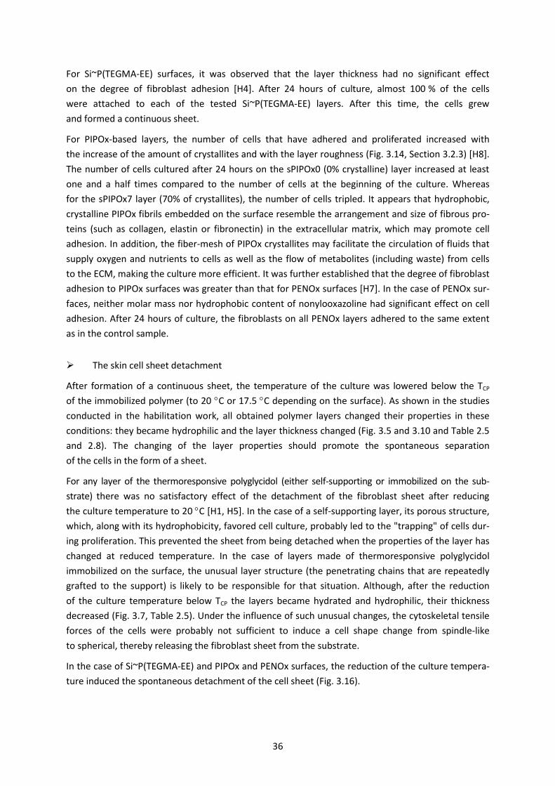
36
For Si~P(TEGMA-EE) surfaces, it was observed that the layer thickness had no significant effect
on the degree of fibroblast adhesion [H4]. After 24 hours of culture, almost 100 % of the cells
were attached to each of the tested Si~P(TEGMA-EE) layers. After this time, the cells grew
and formed a continuous sheet.
For PIPOx-based layers, the number of cells that have adhered and proliferated increased with
the increase of the amount of crystallites and with the layer roughness (Fig. 3.14, Section 3.2.3) [H8].
The number of cells cultured after 24 hours on the sPIPOx0 (0% crystalline) layer increased at least
one and a half times compared to the number of cells at the beginning of the culture. Whereas
for the sPIPOx7 layer (70% of crystallites), the number of cells tripled. It appears that hydrophobic,
crystalline PIPOx fibrils embedded on the surface resemble the arrangement and size of fibrous pro-
teins (such as collagen, elastin or fibronectin) in the extracellular matrix, which may promote cell
adhesion. In addition, the fiber-mesh of PIPOx crystallites may facilitate the circulation of fluids that
supply oxygen and nutrients to cells as well as the flow of metabolites (including waste) from cells
to the ECM, making the culture more efficient. It was further established that the degree of fibroblast
adhesion to PIPOx surfaces was greater than that for PENOx surfaces [H7]. In the case of PENOx sur-
faces, neither molar mass nor hydrophobic content of nonylooxazoline had significant effect on cell
adhesion. After 24 hours of culture, the fibroblasts on all PENOx layers adhered to the same extent
as in the control sample.
The skin cell sheet detachment
After formation of a continuous sheet, the temperature of the culture was lowered below the TCP
of the immobilized polymer (to 20 C or 17.5 C depending on the surface). As shown in the studies
conducted in the habilitation work, all obtained polymer layers changed their properties in these
conditions: they became hydrophilic and the layer thickness changed (Fig. 3.5 and 3.10 and Table 2.5
and 2.8). The changing of the layer properties should promote the spontaneous separation
of the cells in the form of a sheet.
For any layer of the thermoresponsive polyglycidol (either self-supporting or immobilized on the sub-
strate) there was no satisfactory effect of the detachment of the fibroblast sheet after reducing
the culture temperature to 20 C [H1, H5]. In the case of a self-supporting layer, its porous structure,
which, along with its hydrophobicity, favored cell culture, probably led to the "trapping" of cells dur-
ing proliferation. This prevented the sheet from being detached when the properties of the layer has
changed at reduced temperature. In the case of layers made of thermoresponsive polyglycidol
immobilized on the surface, the unusual layer structure (the penetrating chains that are repeatedly
grafted to the support) is likely to be responsible for that situation. Although, after the reduction
of the culture temperature below TCP the layers became hydrated and hydrophilic, their thickness
decreased (Fig. 3.7, Table 2.5). Under the influence of such unusual changes, the cytoskeletal tensile
forces of the cells were probably not sufficient to induce a cell shape change from spindle-like
to spherical, thereby releasing the fibroblast sheet from the substrate.
In the case of Si~P(TEGMA-EE) and PIPOx and PENOx surfaces, the reduction of the culture tempera-
ture induced the spontaneous detachment of the cell sheet (Fig. 3.16).
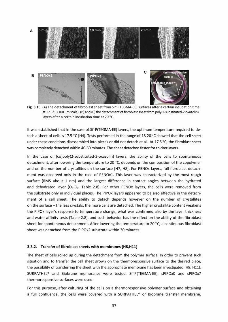
37
Fig. 3.16. (A) The detachment of fibroblast sheet from Si~P(TEGMA-EE) surfaces after a certain incubation time
at 17.5 C (100 μm scale); (B) and (C) the detachment of fibroblast sheet from poly(2-substituted-2-oxazolin)
layers after a certain incubation time at 20 C.
It was established that in the case of Si~P(TEGMA-EE) layers, the optimum temperature required to de-
tach a sheet of cells is 17.5 C [H4]. Tests performed in the range of 18-20 C showed that the cell sheet
under these conditions disassembled into pieces or did not detach at all. At 17.5 C, the fibroblast sheet
was completely detached within 40-60 minutes. The sheet detached faster for thicker layers.
In the case of (co)poly(2-substituted-2-oxazolin) layers, the ability of the cells to spontaneous
detachment, after lowering the temperature to 20 C, depends on the composition of the copolymer
and on the number of crystallites on the surface [H7, H8]. For PENOx layers, full fibroblast detach-
ment was observed only in the case of PENOx1. This layer was characterized by the most rough
surface (RMS about 1 nm) and the largest difference in contact angles between the hydrated
and dehydrated layer (Θ3-Θ2, Table 2.8). For other PENOx layers, the cells were removed from
the substrate only in individual places. The PIPOx layers appeared to be also effective in the detach-
ment of a cell sheet. The ability to detach depends however on the number of crystallites
on the surface – the less crystals, the more cells are detached. The higher crystallite content weakens
the PIPOx layer's response to temperature change, what was confirmed also by the layer thickness
and water affinity tests (Table 2.8), and such behavior has the effect on the ability of the fibroblast
sheet for spontaneous detachment. After lowering the temperature to 20 C, a continuous fibroblast
sheet was detached from the PIPOx2 substrate within 30 minutes.
3.3.2. Transfer of fibroblast sheets with membranes [H8,H11]
The sheet of cells rolled up during the detachment from the polymer surface. In order to prevent such
situation and to transfer the cell sheet grown on the thermoresponsive surface to the desired place,
the possibility of transferring the sheet with the appropriate membrane has been investigated [H8, H11].
SURPATHEL® and Biobrane membranes were tested. Si~P(TEGMA-EE), sPIPOx0 and sPIPOx7
thermoresponsive surfaces were used.
For this purpose, after culturing of the cells on a thermoresponsive polymer surface and obtaining
a full confluence, the cells were covered with a SURPATHEL® or Biobrane transfer membrane.
A 5 min 10 min 20 min
B PENOx1 PIPOx2 C
detachable sheet
thermoresponsive
surface

38
Cells adhered to the membrane. Subsequently, the culture temperature was reduced, which caused
the cells to spontaneously detach from the thermoresponsive surface and, together with the mem-
brane, were "raised" from the culture surface. Due to the fact that the cells were attached
to the membrane, it was possible to transfer the sheet without rolling it to the new culture surface
(Fig. 3.17 shows, for example, the transfer of cells from the sPIPOx0 surface).
Fig. 3.17. Detachment and transfer of the cell sheet from the sPIPOx0 thermoresponsive surface: (A) fibroblast sheet; (B) coverage of cells with SURPATHEL® membrane; (C) polymer layer after removal of the cell sheet (no visible cell sheet).
It was established, that the SURPATHEL® membrane, as compared to the Biobrane membrane, more
efficiently transferred the cell sheet. Almost 90 % of the cell sheet was transferred from the sPIPOx0
surface and 60 % from the sPIPOx7 surface. The sPIPOx7 surface was covered in 70 % of crystallites,
thus it was characterized by poor response of physicochemical properties to temperature change.
This resulted in lower cell transfer efficiency. In the case of Si~P(TEGMA-EE), cell sheet transfer effi-
ciency was 92 %. It has been shown that cells after transplantation into a new culture surface retain
their viability and are capable for further cell division and proliferation.
A similar experiment was conducted for cells grown on TCPS (standard, non-thermoresponsive
culture surface). In this case, the cells were not separated from the surface and it was not possible
to transfer the sheet. This confirmed the fact that the thermoresponsive properties of the obtained
within habilitation work surfaces are necessary detachment of the cell sheet in an intact form.
3.3.3. Biological characterization of skin cells after culture on thermoresponsive surfaces [H7]
Having in mind the usefulness of the obtained polymer surfaces in biomedicine, a researches consid-
ering the biological properties of the cells after culture on thermoresponsive surfaces were per-
formed. Cell phenotype, cell gene expression and a genotoxicity of the poly(2-substituted-2-
oxazoline) layer was monitored. The results showed that the cells did not change their phenotype
during the culture, no damage to the DNA of the cells (Fig. 3.18) or significant changes in their gene
expression were detected [H7].
A B C
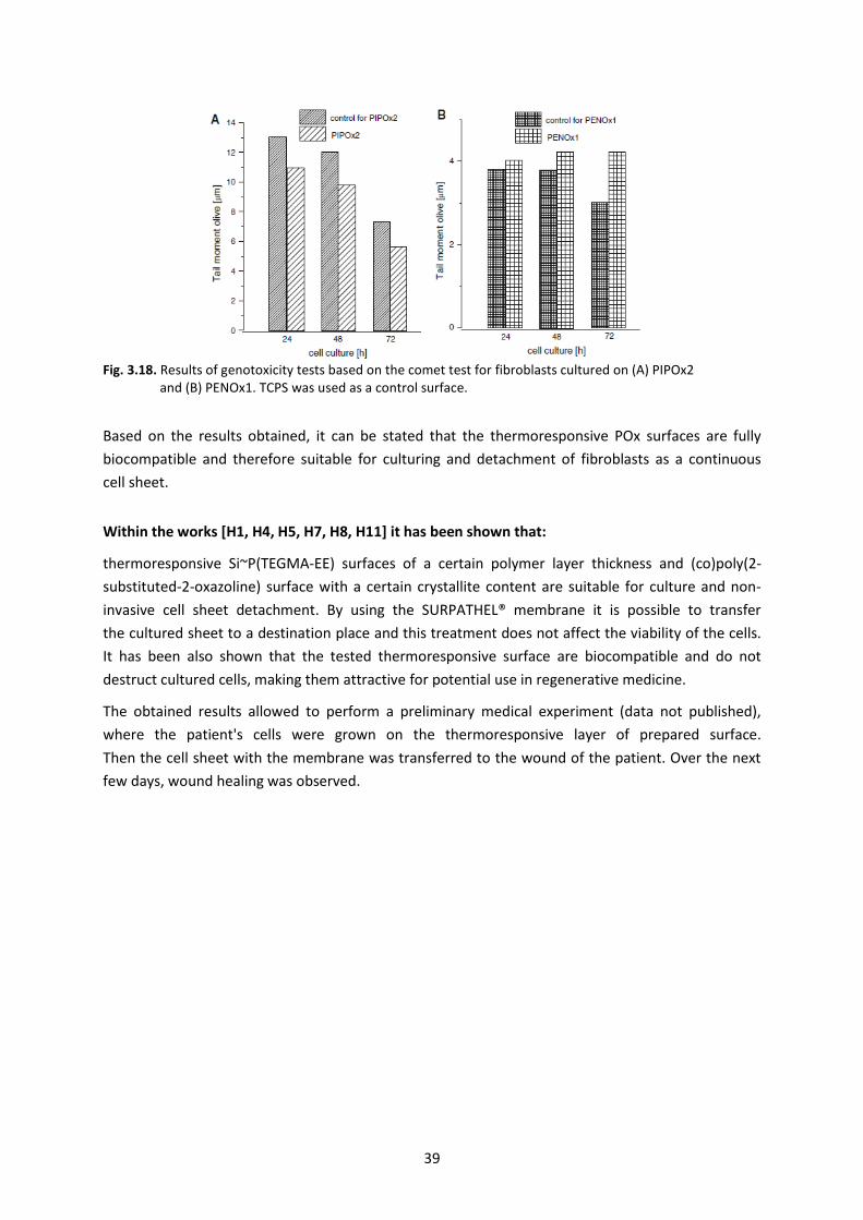
39
Fig. 3.18. Results of genotoxicity tests based on the comet test for fibroblasts cultured on (A) PIPOx2 and (B) PENOx1. TCPS was used as a control surface.
Based on the results obtained, it can be stated that the thermoresponsive POx surfaces are fully
biocompatible and therefore suitable for culturing and detachment of fibroblasts as a continuous
cell sheet.
Within the works [H1, H4, H5, H7, H8, H11] it has been shown that:
thermoresponsive Si~P(TEGMA-EE) surfaces of a certain polymer layer thickness and (co)poly(2-
substituted-2-oxazoline) surface with a certain crystallite content are suitable for culture and non-
invasive cell sheet detachment. By using the SURPATHEL® membrane it is possible to transfer
the cultured sheet to a destination place and this treatment does not affect the viability of the cells.
It has been also shown that the tested thermoresponsive surface are biocompatible and do not
destruct cultured cells, making them attractive for potential use in regenerative medicine.
The obtained results allowed to perform a preliminary medical experiment (data not published),
where the patient's cells were grown on the thermoresponsive layer of prepared surface.
Then the cell sheet with the membrane was transferred to the wound of the patient. Over the next
few days, wound healing was observed.
40
4. SUMMARY OF A SERIES OF PUBLICATIONS „BIOCOMPATIBLE POLYMER LAYERS
OF CONTROLLED AFFINITY FOR WATER. SYNTHESIS AND APPLICATION”
The main purpose of the work was the development and characterization of biocompatible polymer
layers for use in regenerative and reconstructive medicine.
A method for the preparation of hydrophilic polyglycidol-based layers with protein-repellant proper-
ties has been developed. Various types of layers with different composition and structures (interpen-
etrating chains multiply grafted with substrate or polymer brushes) were obtained. It has been
determined how the composition and structure of the polymer affect the properties of the resulting
polymer layers, mainly on the affinity for water, and thus on the reduction of protein adsorption.
Based on the obtained results, the possibility to use the obtained biocompatible hydrophilic
polyglycidol layers in reconstructive medicine has been presented.
In this work, methods for obtaining thermoresponsive polymer layers based on polymers of glycidol,
oligo(ethylene glycol) methacrylates and oxazolines, and their interaction with cells have been
developed. It was established how the composition and structure of the polymer affect the proper-
ties of the resulting polymeric layers, primarily on the affinity for water both at ambient temperature
and at elevated temperature. The layers are hydrophobic at a temperature above the transition
temperature of the immobilized polymer. These conditions promote skin cell culture. Lowering
the culture temperature below the transition temperature of the immobilized polymer alters
the properties of the substrate and thus allows the cells to be detached in form of a sheet. The prop-
erties of the obtained layers were correlated with their effectiveness on cell growth and sheet
formation, and thus the possibilities of using so obtained surfaces in tissue engineering were presented.
The gained knowledge allowed to present the possibility of using the obtained materials in biomedi-
cine: to reduce protein adsorption and to culture and detach cell sheets.
41
8 LITERATURA
1. red. M. Olszówka, K. Maciąg „Nowoczesne trendy w medycynie” Lublin 2015, wyd. fundacja na rzecz pro-mocji nauki i rozwoju TYGIEL.
2. red. B. Ratner, A. S. Hoffman, F. J. Schoen, J.E. Lemons “Biomaterials science” USA 1990, wyd. Academic Press.
3. V. K. Vendra, L. Wu, S. Krishan “Polymer thin films for biomedical application” w “Nanomaterials for the Life Sciences Vol.5: Nanostructured Thin Films and Surfaces” 2010, wyd. Challa S. S. R. Kumar WILEY-VCH.
4. J. R. Smith, D. A. Lamprou Transactions of the IMF The International Journal of Surface Engineering and Coatings 2014, 92(1), 9-19.
5. A. Dworak, B. Trzebicka, W. Wałach, Macromol. Chem. Phys. 1995, 196, 1963-1970.
6. W. Wałach, A. Kowalczuk, B. Trzebicka, A. Dworak, Macromol. Rapid Commun. 2001, 22, 1272-1277.
7. P.Y. Yeh, R. Kainthan, Y. Zou, M. Chiao, J. Kizhakkedathu, Langmuir 2008, 24, 4907-4916.
8. M. Wyszogrodzka, R. Haag, Langmuir 2009, 25, 5703-5712.
9. M. Wyszogrodzka, R. Haag, Biomacromolecules 2009, 10, 1043-1054.
10. M. Jamróz-Piegza, A. Utrata-Wesołek, B. Trzebicka, A. Dworak, Eur. Polymer J. 2006, 42, 2497-2506.
11. Z. Tang, Y. Akiyama, T. Okano, Polymers 2012, 4, 1478-1498.
12. M. Keerl, V. Simirnovas, R. Winter, W. Richtering, Angew. Chem. 2008, 120, 344-347.
13. B. D. Ratner, J. Dent. Educ. 2001, 65, 1340-1347.
14. S. R. Meyers, M. W. Grinstaff, Chem. Rev. 2012, 112, 1615-1632.
15. M. Krishnamoorthy, S. Hakobyan, M. Ramstedt, J. E. Gautrot, Chem. Rev. 2014, 114, 10976-11026.
16. Q. Wei, T. Becherer, S. Angioletti-Uberti, J. Dziubella, Ch. Wischke, A. T. Neffe, A. Lendlein, M. Ballauff, R. Haag, Angew. CHem. Int. Ed. 2014, 53, 8004-8031.
17. A. Dworak, I. Panchev, B. Trzebicka, W. Walach, Macromol. Symp. 2000, 153, 233-242.
18. W. Wałach, B. Trzebicka, J. Justyńska, A. Dworak, Polymer 2004, 45, 1755-1762.
19. A. Mendrek, S. Mendrek, B. Trzebicka, D. Kuckling, W. Wałach, H.-J. Adler, Macromol. Chem. Phys. 2005, 206, 2018-2026.
20. A. Thomas, S. S. Muller, H. Frey, Biomacromolecules 2014, 15, 1935-1954.
21. A. Dworak, B. Trzebicka, A. Utrata, W. Wałach, Polym. Bull. 2003, 50, 47-54.
22. M. Weinhart, T. Becherer, N. Schnurbusch, K. Schwibbert, H.-J. Kunte, R. Haag, Adv. Eng. Mater. 2011, 13, B501-B510.
23. M. Weinhart, I. Grunwald, M. Wyszogrodzka, L. Gaetjen, A. Hartwig, R. Haag, Chem. Asian J. 2010, 5, 1992-2000.
24. Ch. Xue, B.-Ch. Choi, S. Choi, P.V. Braun, D.E. Leckband, Adv. Funct. Mater. 2012, 22, 2394-2401.
25. X. Wang, R. Berger, J.I. Ramos, T. Wang, K. Koynov, G. Liu, H.-J. Butt, S. Wu, RSC Adv. 2014, 4, 45059-450564.
26. S. J. Sofia, V. Premnath, E.W. Merrill, Macromolecules 1998, 31, 5059-5070.
27. Y.-J. Kim, Y.T. Matsunaga, J. Mater. Chem. B 2017, 5, 4307-4321.
28. D. Roy, W. L. A. Brooks, B. S. Sumerlin, Chem. Soc. Rev. 2013, 42, 7214-7243.
29. A. Dworak, B. Trzebicka, A. Utrata, W. Wałach, Polymer Bulletin 2003, 50, 47-54.
30. Doycheva, M., E. Petrova, R. Stamenova, C. Tsvetanov, G. Riess, Macromol. Mater. Eng. 2004, 289, 676-680.
31. K. Nagase, J. Kobayashi, T. Okano, J. R. Soc. Interface 2009, 6(3), S293309.
32. Z. Hu, T. Cai, Ch. Chi, Soft Matter 2010, 6, 2115-2123.
42
33. Y. Tsujii, K. Ohno, S. Yamamoto, A. Goto, T. Fukuda, w Surface initiated polymerization I, Advances in pol-ymer science; J. Jordan, wyd. Springer: Berlin, Heidelberg, 2006; Vol. 197; p 1-45.
34. R. Hoogenboom, Angew. Chem. Int. Ed. 2009, 48, 7978-7994.
35. F. C. Gaertner, R. Luxenhofer, B. Blechert, R. Jordan, M. Essler, J. Controlled Release 2007, 119, 291-300.
36. B. Guillerm, S. Monge, V. Lapinte, J. J. Robin, Macromol. Rapid Comm. 2012, 33, 1600-1612.
37. R. Hoogenboom, H. Schlaad, Polymers 2011, 3, 467-488.
38. G. Morgese, E. M. Benetti, Eur. Polym. J. 2017, 88, 470485.
39. M. Meyer, M. Antonietti, H. Schlaad, Soft Matter 2007, 3, 430-431.
40. P.T. Guner, A. Miko, F.F. Schweinberger, A.L. Demirel, Polym. Chem. 2012, 3, 322-324.
41. A. L. Demirel, M. Meyer, H. Schlaad, Angew. Chem. Int. Ed. 2007, 46, 8622-8624.
42. C. Diehl, I. Dambrowsky, R. Hoogenboom, H. Schlaad, Macromol. Rapid Comm. 2011, 32, 1753-1758.


![[FDD 2016] Kamil Dworak - Przetwarzanie danych pomiarowych w tle z wykorzystaniem Azure](https://img.pdfslide.net/doc/110x75/58cedd081a28abd4098b6731/fdd-2016-kamil-dworak-przetwarzanie-danych-pomiarowych-w-tle-z-wykorzystaniem.jpg)