Embed Size (px)
Citation preview
PMA P100026: FDA Summary of Safety and Effectiveness Data Page 1
SUMMARY OF SAFETY AND EFFECTIVENESS DATA (SSED) I. GENERAL INFORMATION
Device Generic Name: Implanted brain stimulator
Device Trade Name: RNS® System Device Procode: PFN
Applicant’s Name and Address: NeuroPace, Inc.
1375 Shorebird Way Mountain View, California 94043
Date of Panel Recommendation: February 22, 2013
Premarket Approval Application (PMA) Number: P100026
Date of FDA Notice of Approval: November 14, 2013
Expedited: Not applicable
II. INDICATIONS FOR USE The RNS® System is an adjunctive therapy in reducing the frequency of seizures in individuals 18 years of age or older with partial onset seizures who have undergone diagnostic testing that localized no more than 2 epileptogenic foci, are refractory to two or more antiepileptic medications, and currently have frequent and disabling seizures (motor partial seizures, complex partial seizures and/ or secondarily generalized seizures). The RNS® System has demonstrated safety and effectiveness in patients who average 3 or more disabling seizures per month over the three most recent months (with no month with fewer than two seizures), and has not been evaluated in patients with less frequent seizures.
III. CONTRAINDICATIONS
The RNS® System is contraindicated for: • Patients at high risk for surgical complications such as active systemic infection,
coagulation disorders (such as the use of anti-thrombotic therapies) or platelet count below 50,000.
• Patients who have medical devices implanted that deliver electrical energy to the brain.
• Patients who are unable, or do not have the necessary assistance, to properly operate the NeuroPace® Remote Monitor or Magnet.

PMA P100026: FDA Summary of Safety and Effectiveness Data Page 2
• The following medical procedures are contraindicated for patients with an implanted RNS® System. Energy from these procedures can be sent through the implanted brain stimulation system and cause permanent brain damage which may cause severe injury, coma, or death. Brain damage can occur from any of the listed procedures even if the RNS® Neurostimulator is turned off or if the Leads are not connected to the Neurostimulator, and can occur even if the Neurostimulator has been removed, if any Leads (or any part of a Lead), or the cranial prosthesis remain.
- MR imaging is contraindicated for patients with an implanted RNS® System. Do not perform an MRI on a patient with any implanted RNS® Neurostimulator or Lead (or any portion of a Lead). Even if the Neurostimulator has been removed, the patient should not have an MRI if any part of a Lead or the Cranial Prosthesis is still implanted.
The RNS® System is MR Unsafe. Testing has not been performed to define conditions of use to ensure safety of the RNS® System in an MR environment.
- Diathermy procedures are contraindicated in patients implanted with an RNS® Neurostimulator and associated Leads. (Diathermy is any treatment that uses high-frequency electromagnetic radiation, electric currents, or ultrasonic waves to produce heat in body tissues.) Patients absolutely CANNOT be treated with any type of shortwave, microwave, or therapeutic ultrasound diathermy device whether or not it is used to produce heat. These treatments should not be applied anywhere on the body.
- Electroconvulsive Therapy (ECT) is contraindicated for patients with an
implanted RNS® System. - Transcranial Magnetic Stimulation (TMS) is contraindicated for patients with
an implanted RNS® System. IV. WARNINGS AND PRECAUTIONS
The warnings and precautions can be found in the RNS® System labeling. V. DEVICE DESCRIPTION
The NeuroPace RNS® System includes a cranially implantable programmable neurostimulator that senses and records brain electrical activity. In response to the detection of previously identified patterns the neurostimulator is designed to deliver electrical stimulation to the brain to interrupt those patterns before the patient experiences clinical seizures. It is not a seizure detection device. A. Implanted Components
The following are the implanted components of the RNS® System:

PMA P100026: FDA Summary of Safety and Effectiveness Data Page 3
• RNS® Neurostimulator (model RNS-300M) The RNS® Neurostimulator (model RNS-300M) contains electronic circuitry and a Lithium-carbon monofluoride/silver vanadium oxide (Li-CFx/SVO) battery that are hermetically sealed within a flat curved titanium enclosure. It is implanted within the cranium coplanar with the skull surface and is covered by the scalp. A Ferrule mechanically supports and secures the Neurostimulator in the skull. The Neurostimulator is connected to one or two Leads that are surgically placed in or near the epileptic seizure foci in the brain. The neurostimulator monitors electrocorticographic (ECoG) activity and can be programmed to detect abnormal electrical activity. Three programmable detection tools (area, line-length, and bandpass) are provided. The detection tools are highly configurable and can be adjusted by the physician to optimize the detection for each individual patient. Up to two independent detectors can be programmed for any two sensing channels. When detection criteria are met, the Neurostimulator delivers short trains of constant current, rectangular biphasic charge balanced pulses. Stimulation parameters can be programmed as follows:
Table 1: Stimulation Output Parameters
Maximum Current Amplitude @ 500 Ω 11.5 mA ± 10% Maximum Voltage Amplitude @ 500 Ω 6V ± 10% Pulse Width 40 – 1000 μs Frequency 1 to 333Hz Pulses Per Burst 1 to 1666 Maximum Charge Density 25 μC/cm2/phase Current Path Options Bipolar or Multipolar
• NeuroPace® Cortical and Depth Leads
The NeuroPace® Leads provide an interface through which electrical activity of the brain can be sensed and recorded by the RNS® Neurostimulator and through which electrical stimulation can be delivered. Cortical Strip Leads are placed on the surface of the brain near the epileptic foci and Depth Leads are stereotactically introduced into epileptic foci in the brain. The lead specifications are provided in Table 2 below.
Table 2: Cortical and Depth Lead Specifications
Cortical Strip Leads Depth Leads Lead Length 15 cm, 25 cm, and 35 cm 30 cm and 44 cm Lead Diameter 1.27 mm 1.27 mm Number of Electrodes 4 4 Electrode Arrangement 1 x 4 array 1 x 4 array Electrode Material Platinum/Iridium Platinum/Iridium Electrode Spacing 10 mm 3.5 mm and 10 mm Electrode Surface Area 0.079 cm2 0.079 cm2

PMA P100026: FDA Summary of Safety and Effectiveness Data Page 4
Cortical Strip Leads Depth Leads
Impedance 15 cm: 15 Ω (+/- 10%) 25 cm: 25 Ω (+/- 10%) 35 cm: 35 Ω (+/- 10%)
30 cm: 30 Ω (+/- 10%) 44 cm: 44 Ω (+/- 10%)
Lead Body Material Silicone Silicone Electrode Material Platinum/Iridium Platinum/Iridium
Figure 1: RNS® Neurostimulator and Cortical and Depth Lead
B. External Components The following are the external components of the RNS® System:
• NeuroPace® Programmer (model PGM-300)
The NeuroPace® Programmer is a laptop computer that runs proprietary NeuroPace® Programmer Application Software (model 3302 version 1.6.0.2) and utilizes a Wand (model W-02) to communicate with an RNS® Neurostimulator. The Programmer provides the clinician with a user interface to select and download detection and responsive stimulation settings to the neurostimulator, to view real-time ECoG signals, to test the RNS® System integrity, and to upload data and diagnostic information from the RNS® Neurostimulator for viewing.
• NeuroPace® Remote Monitor (model DTR-300)
The NeuroPace® Remote Monitor is a home-use monitoring device that utilizes Wand (model W-02) to communicate with an implanted RNS® Neurostimulator. The Remote Monitor is provided to a patient or caregiver to collect data from the implanted RNS® Neurostimulator and to upload these data using analog telephone lines by way of a secure connection to the Patient Data Management System (PDMS).
Neurostimulator
Cortical Lead Depth Lead

PMA P100026: FDA Summary of Safety and Effectiveness Data Page 5
• NeuroPace® Patient Data Management System (model 4340) The NeuroPace® Patient Data Management System (PDMS) is used for storage and access to historical Neurostimulator and patient data. During synchronization of the Programmer or Remote Monitor with the PDMS, Neurostimulator information regarding detections and stimulations, as well as stored ECoG recordings and Neurostimulator self-diagnostic information are uploaded automatically to the PDMS and combined with previously uploaded information.
C. Accessories The following accessories are provided with the RNS® System: • Cranial Prosthesis - Occupies a vacant Ferrule if the Neurostimulator has been
explanted and not replaced. • Connector Cover (Model CC-01) - Secures the proximal lead contacts to the
neurostimulator. • Connector Plug (Model CP-01) – Are used to fill all vacant ports in the Connector
Cover. • Craniectomy Template - May be used as a pattern to mark and delineate the shape
of the Ferrule on the skull prior to making a craniectomy. • Ferrule and Ferrule Clamp (Model F-01 and Model FC-01) - Installed in a
craniectomy to secure and mechanically support the RNS® Neurostimulator in the skull. The Ferrule Clamp is used to secure the neurostimulator to the ferrule.
• Lead Strain Relief (Model LSR-01) - Supports the proximal end(s) of the lead(s)
at their exit from the Neurostimulator Connector Cover, protecting the Lead from stress near the connector.
• Magnet (Model M-01) - When placed over the implanted RNS® Neurostimulator,
suppresses therapy as long as the Magnet is in position and, if the neurostimulator is programmed to do so, triggers ECoG storage.
• Torque Driver (Model TD-01) - Used to tighten the screw that secures the
Connector Cover to the Neurostimulator and to tighten the Ferrule Clamp that secures the Neurostimulator to the Ferrule.
• Tunneling Tool (Model TT-01), Tunneling Tool Tip (Model TTT-01), Tunneling
Straw (Model TTS-01) - Used to tunnel an implanted Lead from its cranial exit point, through a sub-galeal pathway, to the implanted RNS® Neurostimulator location.
PMA P100026: FDA Summary of Safety and Effectiveness Data Page 6
• Lead Cap (Model LC-01) - The Lead Cap protects the proximal end of a lead when it is not connected to the RNS® Neurostimulator.
• Stop Gauge (Model SG-01) - Placed on a Depth Lead prior to implantation to
indicate the appropriate depth of its insertion. • Suture Sleeve (Model SS-01) - Protects the lead body when sutures are used to
secure a lead.
VI. ALTERNATIVE PRACTICES AND PROCEDURES There are currently three alternative modalities available for the treatment of epilepsy: antiepileptic drugs (AEDs), vagus nerve stimulation, and resective epilepsy surgery. Antiepileptic medications are tried first, usually in monotherapy. If the first AED is not effective, alternative AEDs are tried alone or in polytherapy. In people with epilepsy for whom medications are not effective or who have unacceptable medication related side effects, vagus nerve stimulation or resective neurosurgery may be an option. Vagus nerve stimulation therapy is adjunctive to AED therapy. Neurosurgery for the treatment of epilepsy usually requires removal of some portion of the brain.
VII. MARKETING HISTORY The RNS® System has not been marketed in the United States or any foreign country.
VIII. POTENTIAL ADVERSE EFFECTS OF THE DEVICE ON HEALTH
Potential adverse effects associated with the RNS® System include those related to the implantation procedure, those related to performance of the Neurostimulator and Leads and those related to long-term patient tolerance of the implant. Adverse effects which may potentially occur, but were not reported in the clinical trials for the RNS® System, include the following: • Allergic reaction to the implanted material • Brain abscess For the specific adverse events that occurred in the clinical studies, please see Section X below.
IX. SUMMARY OF PRECLINICAL STUDIES
A. Laboratory Studies
1. RNS® Neurostimulator: The RNS® Neurostimulator underwent numerous testing for electrical safety, output characterization, dimensional verification, hermeticity, environmental conditions, mechanical verification, battery safety and validation, and x-ray interaction. Key testing on the neurostimulator is summarized in Table 3 below. Testing demonstrated the RNS® Neurostimulator operated according to specifications after exposure to the tested conditions (i.e., passed testing).

PMA P100026: FDA Summary of Safety and Effectiveness Data Page 7
Table 3: Summary of key testing performed and passed on the RNS® Neurostimulator Test Purpose Acceptance Criteria
Output Characterization
Verify proper output (amplitude, pulse width, frequency, etc.) and detection parameters of the IPG function are within specified tolerances
Device output is within specifications under expected temperature (35-40°C) and loads (500 to 1200 Ω).
DC Leakage Current Verify the leakage current is in an acceptable range
Per ISO 14708-1:2000, part 16 (modified, requirement limit is stricter than standard (<0.5 µA)
Integrated Circuits (IC)
Verify the proper functioning of the ICs including
ICs function per specifications.
Dimensional Verify that device meets dimensional requirements
Physical Inspection per ISO 14708-1:2000, 15.2 -Device meets geometric requirements for thickness and external features.
Helium leak
Verify feedthroughs remain hermetic after mechanical loading
Helium leak per Mil Std 202, Method 112, Condition C, Procedure 1
Verify neurostimulator hermetic seal
Neurostimulator hermetic seal has a leak rate no greater than 5.0 x 10- 9 cc-atm/s of helium per MIL-STD-883.
Environmental
Verify device conforms to functional requirements and is not damaged by temperature change and thermal shock
Testing per EN 45502-1: 1997, 26.2; IEC 60601-1; IEC 60068-2-14 test Nb, and ASTM D 4169-99, 15.2. Confirm devices continue to meet visual, hermeticity, fine leak and functional requirements after stress.
Verify device conforms to functional requirements and is not damaged by mechanical loads (shock, vibration and atmospheric pressure change)
Testing per ISO 14708-1:1997, 23.2 (Note that test was performed with lower frequency limit changed from 5Hz to 10Hz), CEI/IEC 60068-2-47 & 60068-2-64, ASTM D 3332-99 and ASTM D 4169-99. Confirm devices continue to meet visual, hermeticity fine leak and functional requirements after stress.
Drop Test Verify the device performance is not affected by being dropped
Expose to a mechanical shock equivalent to a 19.5” drop. Confirm devices continue to meet visual, hermeticity fine leak and functional requirements after stress.
Temperature rise limit during single
fault condition
Temperature rise should not cause burns during single fault conditions
Temperature rise is less than or equal to 2°C limit during single fault conditions per EN 45502-1: 1997 17.1.
Battery Battery Capacity Verification (Longevity).
Battery longevity testing using maximum and medium use parameters from clinical study to estimate battery longevity.

PMA P100026: FDA Summary of Safety and Effectiveness Data Page 8
Test Purpose Acceptance Criteria Electrical, Visual, Dimensional, Hermeticity, Short Circuit Testing, Environmental, and Forced Discharge Tests
Testing fulfills the requirements of UN Recommendations on Transport of Dangerous Goods, Manual of Tests and Criteria, 4th revised edition, section 38.3.
X-ray Imaging
Evaluate the safety and functionality after x-ray imaging and identification of radiographic markings.
Device remains functional after exposure to x-ray; radiographic marker is visible in x-ray; and minimal to no distortion of anatomical features adjacent to device.
Particulate Matter
Verify there is no unacceptable release of particulate matter when the device is used as intended
Per EN 45502-1, 14.2.
2. Depth and Cortical Leads: The depth and cortical leads underwent numerous testing for dimensional verification, electrical safety, environmental conditions, mechanical verification, and x-ray interaction. Key testing on the leads is summarized in Table 4 below. Testing demonstrated the depth and cortical leads operated according to specifications after exposure to the tested conditions (i.e., passed testing).
Table 4: Summary of key testing performed and passed on the depth and cortical leads
Test Purpose Acceptance Criteria
Dimensional
Verify that the lead geometry (diameter, length, distal lead configuration) meet design specifications
Depth lead geometry (diameter and length) meet design specifications
DC Resistance and Electrical Isolation
Verify protection due to electricity
Testing per ISO 14708-3:2008, 6.102 and 16.3
Environmental
Verify lead remains electrically functional after exposure to pressure changes.
Testing per ISO 14708-1:2000, section 25.1. Verify lead is electrically functional (measure continuity and leakage) and performs per specifications.
Verify lead remains electrically functional after exposure to temperature changes
Per ISO 14708-1:2000, section 26.2. Verify lead is electrically functional (measure continuity and leakage) and performs per specifications.
Lead Tensile Strength Lead remains electrically functional after exposure to tensile stressors
Testing per ISO 14708-1:2000, section 23.3. Verify lead is electrically functional (measure continuity and leakage) and performs per specifications.
Flexural Fatigue Lead conductors do not Testing per EN45502-1, clause 23.3 and 23.4.
PMA P100026: FDA Summary of Safety and Effectiveness Data Page 9
Test Purpose Acceptance Criteria Testing fatigue after flexural
stressors Verify lead is electrically functional (measure continuity and leakage) and performs per specifications.
Connector Assembly Lead
Insertion/Extraction Force Test
Confirm insertion/extraction forces meet specifications
Insertion and extraction forces before and after preconditioning shall not exceed limits as follows:
• connector insertion force ≤ 1N • extraction force ≤ 2N
Connector Assembly Lead Fixation Test
Connector lead fixation (retention) force
The assembly shall be subjected to straight separating pulls of 4 N +/- 0.5 N, for 60 sec without lead slippage. The assemblies are then tested to failure.
Connector Electrical Isolation
Verify protection due to electricity
Testing per ISO 14708-3:2008, 6.102 and 16.3
Particulate Matter
No unacceptable release of particulate matter when the lead is used as intended
Test per EN 45502-1, 14.2.
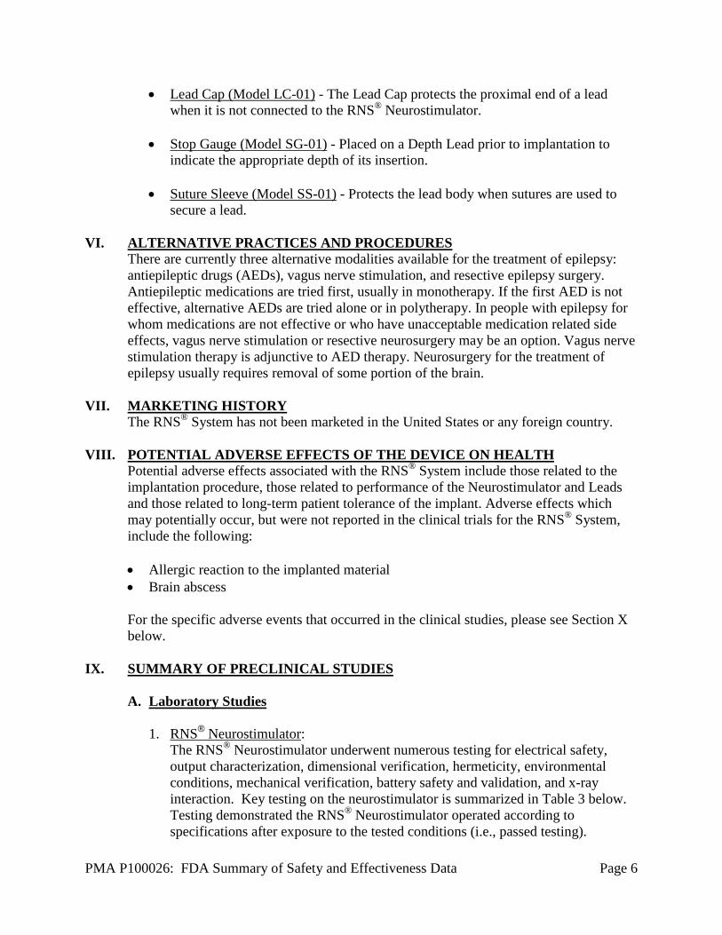
3. NeuroPace® Programmer (model PGM-300), NeuroPace® Remote Monitor
(model DTR-300), NeuroPace® Patient Data Management System (PDMS), and Programming Wand (W-02) The model PGM-300 Programmer, model DTR-300 Remote Monitor, and model 4340 Patient Data Management System (PDMS) underwent software development and verification testing in accordance with the Food and Drug Administration (FDA) guidance, entitled, “Guidance for the Content of Premarket Submissions for Software Contained in Medical Devices” (May 11, 2005) and all requirements were met. Electrical and mechanical verification of the Wand included leakage current and dielectric strength of insulation (per CEI/IEC 60601-1 Second Edition 1998-12), USB 2.0 protocol compliance, telemetry communication protocol compliance, temperature, humidity, liquid ingress and mechanical stresses (per CEI/IEC 60601-1 Second Edition 1998-12), and model W-02 drop test (drop from a height of ≈ 7 feet) and performed according to their specified requirements.
4. Electromagnetic Compatibility (EMC) and Wireless Technology
EMC and wireless technology testing was performed using appropriate essential performance criteria in accordance with the relevant clauses of the following standards and met specified acceptance criteria except for essential performance criteria associated with radio-frequency identification (RFID) and the electrocorticographic (ECoG) sensing feature of the device as discussed below:
PMA P100026: FDA Summary of Safety and Effectiveness Data Page 10
o IEC 60601-1-2: 2007, “Medical electrical equipment - Part 1-2: General requirements for basic safety and essential performance - Collateral standard: Electromagnetic compatibility - Requirements and tests”
o ISO 14708-3:2008(E): Implants for surgery – Active implantable medical
devices – Part 3: Implantable neurostimulators”, Part 27 o Wireless radio testing per United States FCC CFR Title 47 Part 2 and 15
The System performed as specified except for the electrocorticographic (ECoG) sensing feature of the device when in proximity to radio-frequency identification (RFID) devices. The ECoG recording feature of the System can be affected when exposed to RFID resulting in a sensing artifact that might deliver stimulation therapy to the patient based on the device setting. The stimulation will be in a safe range and will be the same as the output stimulation parameters that are programmed by the physician. Thus, a precaution was placed in the labeling advising the physician and patient of this risk. The conclusion of the testing was that the RNS® System met its essential performance criteria with the necessary contraindications, warnings, precautions, and additional information included in the labeling for the clinician and patient.
5. Sterility
The RNS® System components are terminally sterilized utilizing 100% ethylene oxide (EO) gas with heated aeration to allow for residual sterilant dissipation. The EO sterilization cycle has been validated according to ISO 11135-1: 2007. Sterilization of health care products – Ethylene oxide – Part 1: Requirements for development, validation, and routine control of a sterilization process for medical devices. The results obtained from the sterilization validation studies show that the sterilization process provides a Sterility Assurance Level (SAL) of I 0-6. The sterile device components were tested successfully to meet the most rigorous category (permanent contact) for allowable limits of EO and Ethylene Chlorohydrin (ECH) residuals, as specified in ISO l 09937: 2008. Biological Evaluation of Medical Devices – Part 7: Ethylene Oxide Sterilization Residuals. The amount of bacterial endotoxins on fully configured RNS kits and fully configured Leads kits was verified using Limulus Amebocyte Lysate (LAL) testing and found to be within the specification limit of < 0.06 EU/ml (or < 2.15 EU/device), as indicated for devices in contact with cerebrospinal fluid.
6. Packaging and Shelf-life
Packaging and shelf life validation tests for the sterile RNS® System products were successfully completed per AAMI / ANSI/ ISO 11607-1:2006. Packaging for terminally sterilized devices – Part 1: Requirements for materials, sterile barrier systems and packaging systems. Accelerated aging, transportation and handling studies met the requirements for sterile packaging, protection of components, and product functional testing. Shelf-life for the RNS® Neurostimulator has been established as 9 months from the date the battery is attached. All other sterile products including the Connector Cover Kit,
PMA P100026: FDA Summary of Safety and Effectiveness Data Page 11
Craniectomy Template Kit, Ferrule Kit, Cranial Prosthesis Kit, Lead and Lead Accessory Kits have a 3-year shelf life from the date of sterile packaging.
7. Biocompatibility
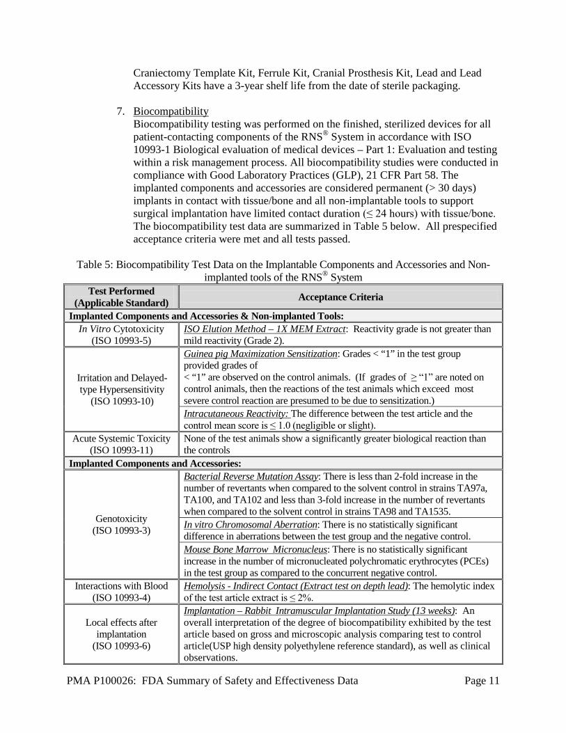
Biocompatibility testing was performed on the finished, sterilized devices for all patient-contacting components of the RNS® System in accordance with ISO 10993-1 Biological evaluation of medical devices – Part 1: Evaluation and testing within a risk management process. All biocompatibility studies were conducted in compliance with Good Laboratory Practices (GLP), 21 CFR Part 58. The implanted components and accessories are considered permanent (> 30 days) implants in contact with tissue/bone and all non-implantable tools to support surgical implantation have limited contact duration (≤ 24 hours) with tissue/bone. The biocompatibility test data are summarized in Table 5 below. All prespecified acceptance criteria were met and all tests passed.
Table 5: Biocompatibility Test Data on the Implantable Components and Accessories and Non-
implanted tools of the RNS® System Test Performed
(Applicable Standard) Acceptance Criteria
Implanted Components and Accessories & Non-implanted Tools: In Vitro Cytotoxicity
(ISO 10993-5) ISO Elution Method – 1X MEM Extract: Reactivity grade is not greater than mild reactivity (Grade 2).
Irritation and Delayed-type Hypersensitivity
(ISO 10993-10)
Guinea pig Maximization Sensitization: Grades < “1” in the test group provided grades of < “1” are observed on the control animals. (If grades of ≥ “1” are noted on control animals, then the reactions of the test animals which exceed most severe control reaction are presumed to be due to sensitization.) Intracutaneous Reactivity: The difference between the test article and the control mean score is ≤ 1.0 (negligible or slight).
Acute Systemic Toxicity (ISO 10993-11)
None of the test animals show a significantly greater biological reaction than the controls
Implanted Components and Accessories:
Genotoxicity (ISO 10993-3)
Bacterial Reverse Mutation Assay: There is less than 2-fold increase in the number of revertants when compared to the solvent control in strains TA97a, TA100, and TA102 and less than 3-fold increase in the number of revertants when compared to the solvent control in strains TA98 and TA1535. In vitro Chromosomal Aberration: There is no statistically significant difference in aberrations between the test group and the negative control. Mouse Bone Marrow Micronucleus: There is no statistically significant increase in the number of micronucleated polychromatic erythrocytes (PCEs) in the test group as compared to the concurrent negative control.
Interactions with Blood (ISO 10993-4)
Hemolysis - Indirect Contact (Extract test on depth lead): The hemolytic index of the test article extract is ≤ 2%.
Local effects after implantation
(ISO 10993-6)
Implantation – Rabbit Intramuscular Implantation Study (13 weeks): An overall interpretation of the degree of biocompatibility exhibited by the test article based on gross and microscopic analysis comparing test to control article(USP high density polyethylene reference standard), as well as clinical observations.

PMA P100026: FDA Summary of Safety and Effectiveness Data Page 12
Test Performed (Applicable Standard) Acceptance Criteria
Systemic Toxicity (ISO 10993-11)
Subchronic Toxicity: The correlation of all data for patterns of toxicity, including death of > 1 animal/group, mean body weight loss for each group, clinical signs of toxicity in > 1 animal/group, hematological and clinical chemistry values, and histopathology of tissues Material-mediated Pyrogenicity: No rabbit shows an individual rise in temperature of 0.5oC or more above the baseline temperature.
Combined implantation and systemic toxicity
(ISO 10993-6 and -11)
Combined Neuroimplantation/Chronic Toxicity Study in Rabbits: See section below for details.
Carcinogenicity An adequate carcinogenicity risk assessment was provided.
Combined Neuroimplantation/Chronic Toxicity Study in Rabbits: The objectives of this study (conducted in accordance with GLPs) were to assess potential neurotoxicity, acute and chronic local tissue responses as well as long-term systemic effects following implantation of the device in rabbits. The study was based on the testing recommendations in the International Organization for Standardization (ISO) 10993: Biological evaluation of medical devices, Part 6: Tests for local effects after implantation, ISO 10993 – Part 11: Tests for systemic toxicity, and ASTM F2901 – 12, Standard Guide for Selecting Tests to Evaluate Potential Neurotoxicity of Medical Devices. The study design is summarized in Table 6 below.
Table 6: Combined Neuroimplantation/Chronic Toxicity Study in Rabbits Study Design
Summary
Termination Interval
Number of Animals Study Objective Articles Implanted Control Test
Male Female Male Female
6 Days 4 4 4 4 Brain tissue reaction &
Neurotoxicity
Partial Depth Lead (T1), Partial Cortical Strip Lead (T2), Partial Lead body material section (T3), Ferrule (partial coupon) (T4)1
4 Weeks 5 5 5 5
Brain tissue reaction &
Neurotoxicity T1, T2, T3, and T4
Systemic toxicity & Local
subcutaneous tissue reaction
Neurostimulator (T5)2, Depth Lead (T6), Cortical Strip Lead (T7), Burr Hole Covers (T8)
26 Weeks 7 7 7 7
Brain tissue reaction &
Neurotoxicity T1, T2, T3, and T4
Systemic toxicity & Local T5, T6, T7, and T8

PMA P100026: FDA Summary of Safety and Effectiveness Data Page 13
Termination Interval
Number of Animals Study Objective Articles Implanted Control Test
Male Female Male Female subcutaneous tissue reaction
1 The adapted Ferrule (partial coupon) was a finished device modified for implantation using only manufacturing methods and tooling materials used in finished device manufacturing.
2 Neurostimulator includes the Upper Strain Relief, Connector Cover, Ferrule, Ferrule Clamp, and Connector Plugs.
For purposes of determining neurotoxicity and brain tissue reaction, the implant location of the control articles replicated the location of the test articles. Table 7 below provides a summary of the examinations performed and the study results.
Table 7: Results of Combined Neuroimplantation/Chronic Toxicity Study in Rabbits
Examination Timing Results
Clinical Signs of Disease or Abnormality
2x/day: for general health prior to surgery, weekly At termination: detailed
examinations
No clinically significant findings or signs of toxicity were noted
Neurological Days 1-5: daily; Days > 5:
weekly; and Prior to termination
No abnormal findings were noted
Body Weight
Prior to implantation, weekly for the first 4 weeks, every 4 weeks thereafter, and prior to
termination
Clinically acceptable following treatment at 6 days and 4 and 26 weeks No statistically significant differences w/ controls at 4 and 26 weeks and within group
Blood Hematology & Clinical Chemistry 4 and 26-week termination
Hematology: no biologically significant differences between the test and control groups and all mean values were within an acceptable range Clinical Chemistry: no biologically significant differences between the test and control groups for any of the clinical chemistry parameters
Necropsy 4 and 26-week termination No changes that could be attributed to test articles
Organ weights and organ/body weight ratios
4 and 26-week termination No biologically significant differences between the test and control groups
Macroscopic implant site evaluation
6-day, 4 and 26-week termination
No test device-related differences in macroscopic observations were detected between those that received the control HDPE implants in brain and subcutaneous locations and those that received various components of the test device in brain

PMA P100026: FDA Summary of Safety and Effectiveness Data Page 14
Examination Timing Results and/or subcutaneous locations.
Histopathology† 6-day, 4 and 26-week termination
Microscopically, no evidence for systemic toxicity, neurotoxicity, or local tissue reaction occurred beyond the expected effects due to surgical placement and the physical presence of the various implants in the brain, the calvarium, or the subcutaneous tissue.
† Hematoxylin and Eosin (H & E), Fluoro-Jade B (for evidence of neuronal degeneration), Anti-Glial Fibrillary Acidic Protein (GFAP) antibody (astroglial activation), and Mouse Monoclonal Anti-Rabbit Macrophage (RAM11) antibody (macrophages and activated microglia) were used for histopathological evaluation of the brain/dural implant sites. For histopathological evaluation of the subcutaneous implant sites and the designated tissues, H & E stain was used
B. Animal Studies
Lead Implantation Study in Sheep Testing on sheep was performed to examine the safety of the NeuroPace® Depth Leads and NeuroPace® Cortical Strip Leads in a simulated use condition. Two depth and 2 cortical leads were implanted into each sheep. One of each lead type was then stimulated for a specified amount of time at prescribed intervals over the course of the survival period. Longer continuous stimulation periods were used than would be experienced in human use with the RNS® System. The other of each lead type had lead impedance measurements taken at implantation and explantation.
The chronic implant duration of 11 sheep ranged from 33 days to 200 days, with a mean of 131 days. No histological analysis was performed on 4 sheep which were sacrificed early due to hardware problems relating to externalizing the leads for the scheduled stimulations and two animals that were inadvertently frozen. Five other animals experienced hardware-related complications but were survived until 18-24 weeks (135 - 200 days). Injury to the neuronal tissue immediately adjacent to the leads was as expected and the reaction did not appear to extend into the surrounding tissue. No significant neuronal disorganization or necrosis was observed. Tissue reactions included chronic inflammation, astrocytic gliosis, some foreign body giant cells, and a fibrous capsule around the implantation track. These are not unexpected. The cortical strip leads appeared to result in no detectable cytoarchitectural changes to underlying tissue. Thirty (30) ECoG recordings were examined and the magnitudes of the signals provided sufficient characterization of brain activity for monitoring purposes. No evidence of electrode-to-tissue sensor block was observed.
C. Additional Studies
The detection algorithm was not evaluated for its efficacy in accurately identifying specified ECoG activity and seizures. Verification and validation testing provided for the detection algorithm was determined by the FDA to be adequate for proceeding
PMA P100026: FDA Summary of Safety and Effectiveness Data Page 15
with the IDE feasibility and pivotal studies (i.e., the risks to the subjects for participating in the studies were outweighed by the anticipated benefits to the subjects and the importance of the knowledge to be gained). Testing of the detection algorithm included the following:
• Evaluation of the algorithm in detecting epileptiform activity using artificial
ECoG waveforms, using a MATLAB simulator. The detection tool met predefined success criteria.
• Evaluation using archived ECoG data from 11 consecutive patients admitted to
the Emory Epilepsy Monitoring Unit (between January 1997 and May 1999). The majority of seizures were of mesial or neocortical temporal origin, but seizures recorded in 3 of the subjects originated extratemporally. Detection tools were programmed individually for patients and training sets were used for tuning the detectors in some cases. Of 125 seizures marked by an epileptologist all but 2 were detected (98.4% sensitivity) and the average false positive rate was 0.013 (range: 0-0.051) per hour.
• As part of software verification testing simulated data were used to test the
minimum, maximum, and at least one intermediate value for each detection parameter of each detection tool by performing simulations using real-time ECoG data. The device met specifications.
• A multi-center feasibility IDE clinical investigation (G010288) was performed
using on an external model (eRNS) of the RNS® System that incorporated the same detection algorithm as the RNS® System. The study enrolled at 8 sites 125 subjects who were candidates for epilepsy surgery and were under-going video-EEG monitoring in an epilepsy monitoring unit. The study demonstrated that the eRNS could safely deliver electrical stimulation in response to detected ECoG activity. However, the study was not designed to determine specificity and sensitivity of the detection algorithm.
X. SUMMARY OF PRIMARY CLINICAL STUDIES
The sponsor performed a Feasibility study, a Pivotal study, and a Long-term Treatment (LTT) study (ongoing) as described below. Feasibility Study A Feasibility study was performed to evaluate preliminary safety and effectiveness and the results were used to inform the design of the Pivotal Study and to assess the integrity of the blind. Additionally, the safety data from the Feasibility study were included in the primary safety analysis for this PMA. It was a multi-center clinical investigation of individuals with medically intractable epilepsy. The first subject was enrolled on January 19, 2004 and the last subject transitioned to the Long Term Treatment (LTT) study (see below) on December 17, 2007. Eligible subjects were 18-65 years of age with medically intractable partial onset seizures and a minimum of 4 simple partial seizures (motor or sensory), complex partial seizures, and/or secondarily generalized seizures in each of the
PMA P100026: FDA Summary of Safety and Effectiveness Data Page 16
previous three months. Subjects were required to be on a stable AED regimen and to have previously undergone diagnostic testing that localized one or two epileptogenic region(s). Subjects with psychogenic or non-epileptic seizures, status epilepticus, active psychosis, severe depression, or suicidal ideation within the preceding year were excluded. Sixty-five subjects were implanted with the RNS® Neurostimulator and Leads in the Feasibility study. The first four subjects implanted with the RNS® Neurostimulator and Leads at a clinical site participated in an open label protocol (all subjects received responsive stimulation), and subsequent subjects at that site participated in a randomized, double-blind, concurrent sham-stimulation control protocol in which the Treatment group received stimulation and Sham group did not. Forty-two (42) subjects were in the open label protocol and 23 were in the blinded protocol. Following completion of the 16 week Evaluation Period, subjects transitioned to an Open Label Period, and all subjects were able to receive responsive stimulation. Pivotal Study A Pivotal study was performed to establish a reasonable assurance of safety and effectiveness of the RNS® System in reducing the frequency of seizures in individuals 18 years of age or older with partial onset seizures who have undergone diagnostic testing that localized no more than 2 epileptogenic foci, are refractory to two or more antiepileptic medications, currently have frequent and disabling seizures (motor partial seizures, complex partial seizures and/ or secondarily generalized seizures, and in patients who average 3 or more disabling seizures per month over the three most recent months (with no month with fewer than two seizures) in the US under IDE# G030126. Data from this clinical study combined with safety data from the Feasibility study were the basis for the PMA approval decision. Patients were enrolled in the Pivotal trial beginning on December 29, 2005 and the last subject finished the blinded evaluation period (BEP) and transitioned to the Open label period on October 16, 2009. A summary is presented below.
Long-term Treatment Study (LTT) The LTT is an ongoing open label, multi-center, prospective clinical investigation of two-hundred and thirty (230) subjects who consented to enroll once they completed the Feasibility or Pivotal study. Each subject participates for a maximum of 7 years. Adverse event and seizure data are collected at 6-month intervals, and data regarding quality of life are collected at yearly intervals. AED adjustments are permitted as needed. The first subject was enrolled on April 6, 2007 and the study is ongoing.
A. Study Design
The database for this PMA includes data from all 3 studies (Feasibility, Pivotal, and LTT) and reflects data collected through May 12, 2011 and includes 191 subjects in the Pivotal trial and 65 in the Feasibility study. Note that data for deaths, including Sudden Unexplained Death in Epilepsy (SUDEP), are current through October 24, 2012. There were 12 investigational sites in the Feasibility study, 28 investigational sites in the Pivotal study, and 29 investigational sites in the LTT study.

PMA P100026: FDA Summary of Safety and Effectiveness Data Page 17
The RNS® System Pivotal study was a randomized, double-blinded, multi-center, sham-controlled clinical study. The investigation had five periods: the Baseline Period (which includes the Pre-Implant Period defined in Section 2 below), Post-Operative Stabilization Period, Stimulation Optimization Period, Blinded Evaluation Period (BEP), and Open Label Period. Enrolled subjects were implanted with the RNS® Neurostimulator and Leads within 28 days following the date of qualification for implantation. Subjects were randomized 1:1 at the end of the Post-Operative Stabilization Period (4 weeks post-implant). To ensure equal representation in the two therapy groups, an adaptive randomization approach (minimization) was used to balance variables that might influence the clinical response to responsive stimulation. These variables (listed in order of priority) were:
1) Investigational site; 2) Seizure onset zone location (partial onset seizures of mesial temporal origin
versus partial onset seizures arising from any other region of the cortex); 3) Number of seizure foci (unifocal versus bifocal); and 4) Previous therapeutic epilepsy surgery (resection, subpial transection and/or
corpus callosotomy). Subjects randomized to the Treatment group received responsive stimulation during the Stimulation Optimization and BEP and subjects randomized to the Sham group did not receive responsive stimulation during these periods. Following completion of the BEP (20 weeks post-implant), subjects transitioned to the Open Label Evaluation Period and both Treatment and Sham group subjects received responsive stimulation. The Pivotal study was designed to have 80% power with an overall 2-sided Type 1 error of 0.05, assuming responder rates (i.e., subjects with a 50% or greater reduction in seizures from baseline) in the Treatment group and Sham groups of 40% and 20%, respectively. To meet these criteria, 180 subjects were required in the BEP. Assuming approximately 10% of subjects would not be compliant (including subjects who did not complete the BEP), approximately 200 subjects were to be randomized, 100 each into the Treatment and Sham groups. An independent Data Monitoring Committee (DMC) for the Feasibility, Pivotal, and LTT studies was established. The DMC was responsible for independently monitoring the safety of interventions during the investigation by reviewing data made available by NeuroPace acting in the capacity of the Coordinating Center. The DMC made recommendations to NeuroPace about safeguarding the interests of trial participants and about stopping, modifying or continuing the investigation. Information regarding all deaths that occurred during the investigation, the SUDEP Analysis Committee's classification with respect to SUDEP, as well as the data supporting that classification, was communicated to the Chair of the Data Monitoring Committee (DMC) by NeuroPace. The DMC reviewed composite safety and effectiveness data on a regular basis depending on subject enrollment, at a minimum of every six months.

PMA P100026: FDA Summary of Safety and Effectiveness Data Page 18
1. Clinical Inclusion and Exclusion Criteria The inclusion and exclusion criteria for the Feasibility and Pivotal studies were similar (the key inclusion and exclusion criteria for the studies are presented in Table 8 and Table 9).
Table 8: Key Inclusion Criteria
Inclusion Criteria Feasibility Study
Pivotal Study
Subject has simple partial motor seizures, complex partial seizures and/or secondarily generalized seizures Yes1 Yes
Seizure counts per month 4 or more 2 average of ≥ 33 Age 18-65 years 18-70 years Subject has seizures that are severe enough to cause injuries or significantly impair functional ability in domains including employment, psychosocial, education and mobility.
Yes Yes
Subject has seizures that are distinct, stereotypical events that can be reliably counted Yes Yes
Subject failed treatment with a minimum of two AEDs (used in appropriate doses) with adequate monitoring of compliance and the effects of treatment.
Yes Yes
Subject has remained on the same AED(s) over the preceding three (3) months Yes Yes
Subject has undergone diagnostic testing that has established the epileptiform activity onset region(s) Yes
Yes, ≤ 2 epileptogenic
regions 1 The Feasibility study also included simple partial sensory seizures. 2 Subject has a minimum of four (4) or more countable seizures every month over the last three (3) months. 3 Subject has an average of three or more disabling simple partial seizures, complex partial seizures, or
secondarily generalized seizures per month (28 days) over the three most recent months, with no month with less than two seizures.
Table 9: Key Exclusion Criteria
Exclusion Criteria Feasibility Study
Pivotal Study
Subject has been diagnosed with psychogenic or non-epileptic seizures in the preceding year. Yes Yes
Subject has been diagnosed with primarily generalized seizures. Yes Yes Subject has experienced unprovoked status epilepticus in the preceding year. Yes Yes
Subject has a clinically significant or unstable medical condition or a progressive central nervous system disease. Yes Yes
Subject is taking anticoagulants. Yes Yes Subject has been diagnosed with active psychosis, severe depression or suicidal ideation in the preceding year. Yes Yes
Subject has an implanted Vagus Nerve Stimulator (VNS). Yes1 Yes2

PMA P100026: FDA Summary of Safety and Effectiveness Data Page 19
Exclusion Criteria Feasibility Study
Pivotal Study
Subject has had therapeutic surgery to treat epilepsy in the preceding year
in the preceding 6 months
Subject is implanted with an electronic medical device that delivers electrical energy to the head. Yes Yes
Subject requires repeat MRIs Yes Yes3
Subject’s epileptiogenic region(s) is / are located caudal to the level of the thalamus. Yes Yes
Subject is pregnant. Yes Yes 1 A subject with an inactive VNS could be enrolled so long as the VNS was explanted prior to or at the same
time as the RNS® System implant. 2 A subject could be enrolled if the subject is willing to have the VNS explanted (excluding leads)
prior to or at the time of the RNS® System implant. (Subjects with VNS devices must have had VNS therapy discontinued for at least three months prior to enrollment.)
3 In which the head is exposed to the radio frequency field.
Subjects were eligible to enroll into the LTT study if they had completed either the Feasibility or Pivotal study, had the RNS® System implanted, had elected to continue to receive responsive stimulation, and were able to attend scheduled appointments for the study. They were not eligible if they had an active psychiatric or mental illness that made it inadvisable for the subject to continue to receive responsive stimulation or if the subject had been diagnosed with psychogenic or non-epileptic seizures, or primarily generalized seizures during the Feasibility or Pivotal studies.
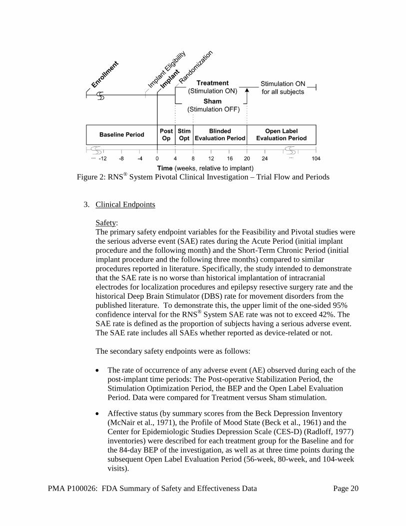
2. Follow-up Schedule A schematic of the study timeline is provided in Figure 2. The primary effectiveness analysis compares changes in seizure frequency in the Treatment group and in the Sham group during the 12-week BEP relative to the 12-week Pre-Implant Period. The Pre-Implant Period (not shown in Figure 2) is defined as the 12-weeks in the Baseline Period leading up to and including the date of qualification for implantation. Primary safety analyses include adverse event data over the first 12 weeks post-implantation. Secondary safety and effectiveness analyses include data from all periods of the study. Information regarding daily seizure counts, adverse events and subject well-being was collected at all visits by a physician investigator who was blinded to the subject’s randomization status and a second non-blinded physician investigator was responsible for Neurostimulator programming.
PMA P100026: FDA Summary of Safety and Effectiveness Data Page 20
Figure 2: RNS® System Pivotal Clinical Investigation – Trial Flow and Periods
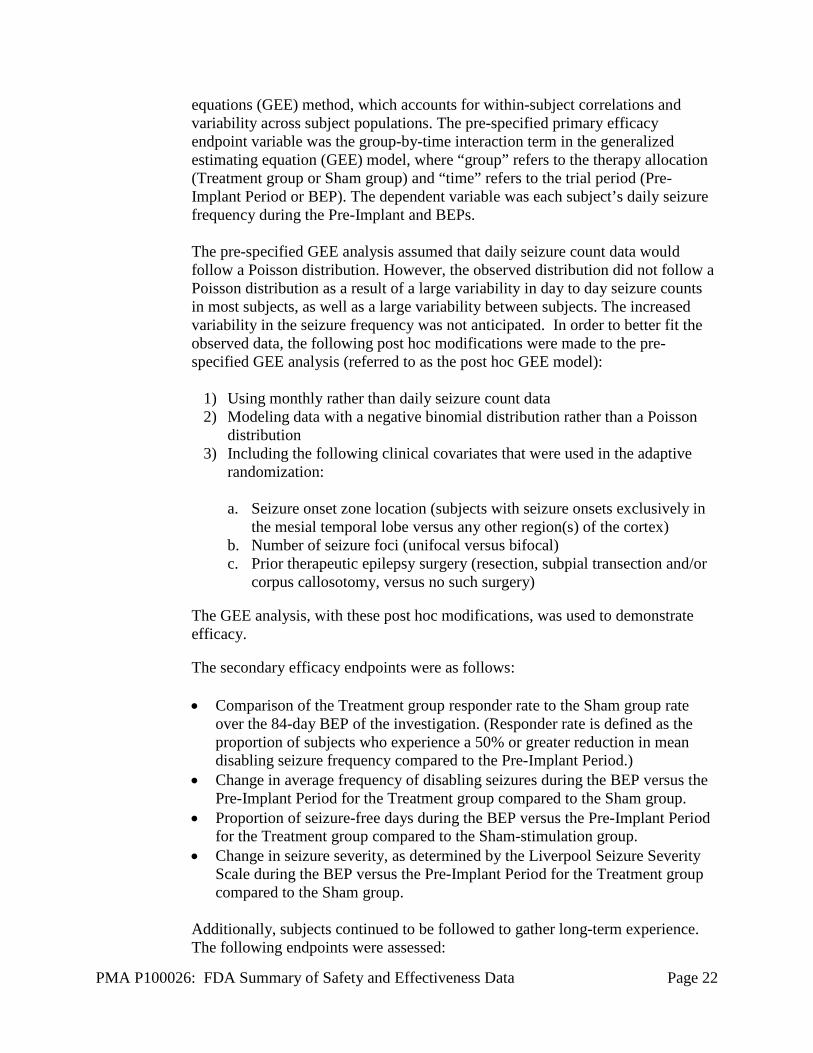
3. Clinical Endpoints
Safety: The primary safety endpoint variables for the Feasibility and Pivotal studies were the serious adverse event (SAE) rates during the Acute Period (initial implant procedure and the following month) and the Short-Term Chronic Period (initial implant procedure and the following three months) compared to similar procedures reported in literature. Specifically, the study intended to demonstrate that the SAE rate is no worse than historical implantation of intracranial electrodes for localization procedures and epilepsy resective surgery rate and the historical Deep Brain Stimulator (DBS) rate for movement disorders from the published literature. To demonstrate this, the upper limit of the one-sided 95% confidence interval for the RNS® System SAE rate was not to exceed 42%. The SAE rate is defined as the proportion of subjects having a serious adverse event. The SAE rate includes all SAEs whether reported as device-related or not.
The secondary safety endpoints were as follows:
• The rate of occurrence of any adverse event (AE) observed during each of the
post-implant time periods: The Post-operative Stabilization Period, the Stimulation Optimization Period, the BEP and the Open Label Evaluation Period. Data were compared for Treatment versus Sham stimulation.
• Affective status (by summary scores from the Beck Depression Inventory (McNair et al., 1971), the Profile of Mood State (Beck et al., 1961) and the Center for Epidemiologic Studies Depression Scale (CES-D) (Radloff, 1977) inventories) were described for each treatment group for the Baseline and for the 84-day BEP of the investigation, as well as at three time points during the subsequent Open Label Evaluation Period (56-week, 80-week, and 104-week visits).

PMA P100026: FDA Summary of Safety and Effectiveness Data Page 21
• Neuropsychological functioning as assessed by neuropsychological testing with validated, standardized inventories obtained pre-implant (within 28 days of the implant) and then at 20 weeks, 56 weeks and 104 weeks after implantation. The neuropsychological testing assessed visual and verbal memory, verbal fluency and naming, and cognitive flexibility.
• At the time of IDE approval, the sponsor prespecified in the clinical protocol that the SUDEP occurrence rate for the RNS® System would be no worse than 6.3/1000 patient-years. This rate was based on the reported incidence of SUDEP which ranges from 3.5 deaths per 1000 person years in a population-based cohort with epilepsy (Lhatoo et.al, 1991); 3.5/1000 patient-years in a well-defined cohort of 4,700 patients (5,747 patient-years of exposure) included in the worldwide clinical development database of the AED lamotrigine (Leetsma et.al., 1997); 4.5/1000 patient-years for the Cyberonics Vagus Nerve Stimulator; 6 /l000 patient-years in patients with medically refractory epilepsy followed in an epilepsy clinic (Sperling et.al., 1999); to 6.3 deaths/1000 person-years in a population based Swedish cohort with refractory epilepsy who were candidates but did not choose to undergo epilepsy surgery (Nilsson et.al., 2003). The protocol stated that data from the RNS® System Feasibility Clinical Investigation, as well as the 5 year Long-term Treatment Investigation, would be used to collect approximately1500 patient-years of data in order to confidently calculate the rate of SUDEP.
The number of patient-years necessary to determine that the 95% confidence
interval for SUDEP does not exceed 6.3/1000 patient-years can be calculated based on the number of patient deaths attributed to SUDEP that occur during the RNS® System Clinical Investigations and the number of patient-years of data. As seen in Table 10 below, if there are 4 patient deaths identified as possibly or probably related to SUDEP, then 1446 years of patient follow-up will permit NeuroPace to be 95% confident that the true rate of SUDEP does not exceed 6.3/1000 patient-years. After three SUDEP events occurred the sponsor, with FDA concurrence, increased the acceptable SUDEP rate to 9.3/1000 patient-years.
Table 10: Follow-Up Required to Establish SUDEP Rate
K1 1 2 3 4 5 6 M2 823 1016 1231 1446 1657 1864
1 K = number of deaths identified as possibly or probably related to SUDEP.
2 M = number of patient-years necessary to establish that the 95% confidence interval is < 6.3/1000 patient-years.
Effectiveness:
The primary effectiveness objective for the Pivotal study was to demonstrate a significantly greater reduction in the frequency of total disabling seizures in the Treatment group compared to the Sham group during the BEP relative to the Pre-Implant Period. Seizure frequency was modeled using the generalized estimating
PMA P100026: FDA Summary of Safety and Effectiveness Data Page 22
equations (GEE) method, which accounts for within-subject correlations and variability across subject populations. The pre-specified primary efficacy endpoint variable was the group-by-time interaction term in the generalized estimating equation (GEE) model, where “group” refers to the therapy allocation (Treatment group or Sham group) and “time” refers to the trial period (Pre-Implant Period or BEP). The dependent variable was each subject’s daily seizure frequency during the Pre-Implant and BEPs.
The pre-specified GEE analysis assumed that daily seizure count data would follow a Poisson distribution. However, the observed distribution did not follow a Poisson distribution as a result of a large variability in day to day seizure counts in most subjects, as well as a large variability between subjects. The increased variability in the seizure frequency was not anticipated. In order to better fit the observed data, the following post hoc modifications were made to the pre-specified GEE analysis (referred to as the post hoc GEE model):
1) Using monthly rather than daily seizure count data 2) Modeling data with a negative binomial distribution rather than a Poisson
distribution 3) Including the following clinical covariates that were used in the adaptive
randomization:
a. Seizure onset zone location (subjects with seizure onsets exclusively in the mesial temporal lobe versus any other region(s) of the cortex)
b. Number of seizure foci (unifocal versus bifocal) c. Prior therapeutic epilepsy surgery (resection, subpial transection and/or
corpus callosotomy, versus no such surgery)
The GEE analysis, with these post hoc modifications, was used to demonstrate efficacy.
The secondary efficacy endpoints were as follows:
• Comparison of the Treatment group responder rate to the Sham group rate
over the 84-day BEP of the investigation. (Responder rate is defined as the proportion of subjects who experience a 50% or greater reduction in mean disabling seizure frequency compared to the Pre-Implant Period.)
• Change in average frequency of disabling seizures during the BEP versus the Pre-Implant Period for the Treatment group compared to the Sham group.
• Proportion of seizure-free days during the BEP versus the Pre-Implant Period for the Treatment group compared to the Sham-stimulation group.
• Change in seizure severity, as determined by the Liverpool Seizure Severity Scale during the BEP versus the Pre-Implant Period for the Treatment group compared to the Sham group.
Additionally, subjects continued to be followed to gather long-term experience. The following endpoints were assessed:
PMA P100026: FDA Summary of Safety and Effectiveness Data Page 23
• Change in average frequency of disabling seizures in the group originally
randomized to the Sham-stimulation group (Therapy OFF) once therapy has been enabled in that group (Open Label Evaluation Period). Average seizure frequency during 84 days of the Open Label Evaluation Period was compared to the average seizure frequency during the 84-day BEP.
• Each subject’s responder status over the Open Label Evaluation Period. • Daily seizure frequency counts compared to baseline during the Open Label
Evaluation Period for both Sham and Treatment groups. • Quality of life in individual subjects as measured with the QOLIE-89
assessment inventory to provide a descriptive analysis for each treatment group for the Baseline, Blinded Evaluation, and Open Label Evaluation Periods.
B. Accountability of PMA Cohort
Subject participation in the Feasibility, Pivotal and LTT studies is presented in Figure 3 as of May 12, 2011. The safety and effectiveness analysis populations for the Pivotal study included all 191 subjects implanted and randomized; this is the intent-to-treat population. The combined safety analysis population includes the intent-to-treat safety population from the RNS® System Feasibility, Pivotal and Long-term Treatment (LTT) Clinical Investigations combined. This includes all 256 subjects implanted with the RNS® Neurostimulator and Leads.
Withdrawals and Discontinuations Forty-three (43) of the 256 enrolled subjects, discontinued treatment from all three studies. An additional 6 subjects did not transition In the Feasibility study, 6 of 65 subjects discontinued. In the Pivotal study, 16 of 191 subjects discontinued. Two hundred thirty subjects transitioned to the LTT including 2 subjects who discontinued the Pivotal study early and later enrolled in the LTT study. As of May 12, 2011, 21 of 230 subjects had discontinued the LTT study. An additional 2 subjects have died since May 12, 2011, bringing the total number of death to 11 and the total number of subjects who had discontinued treatment to 45. For the subjects who discontinued treatment, 7 subjects were explanted because of infection, 1 subject was explanted because of hemorrhage, 3 subjects were lost to follow-up, 9 subjects died and 23 subjects withdrew electively. The reasons given for elective withdrawal included: to pursue other treatments (13), because the reduction in seizures was not sufficient (4), and because the subject did not want to have the Neurostimulator replaced when the battery reached expected end of service (3). Another subject had a seizure-related fall that caused a scalp laceration that exposed the Neurostimulator: this subject chose not to have the laceration sutured and withdrew from the trial. Another subject was withdrawn because the physician felt that the subject was no longer a suitable candidate to participate because of psychiatric issues not related to treatment with the RNS® System. The reason for withdrawal for one subject was not specified.

PMA P100026: FDA Summary of Safety and Effectiveness Data Page 24
Figure 3: Patient Flow Diagram
* Two subjects withdrew early (discontinued) from the Pivotal study to undergo resective epilepsy surgery. Waivers were granted to allow enrollment into the LTT study so that the subjects could continue to receive responsive stimulation to treat seizures arising from the non-resected seizure focus.
C. Study Population Demographics and Baseline Parameters
Demographic information for subjects implanted in the Feasibility and Pivotal studies is presented in Table 11. All subjects participating in the LTT study originally enrolled in the Feasibility or Pivotal study. Table 12 provides information on epilepsy clinical characteristics and prior treatment for epilepsy by randomization group. Table 13 provides information on the number and types of leads implanted.
Table 11: Demographics
Characteristic All (N = 256)
By Study Feasibility (N = 65)
Pivotal (N = 191)
Gender (percent female) 49% (125/256) 52% (34/65) 48% (91/191)
Age in years1 (average, SD , range)
34.0 ± 11.4 (18 - 66)
30.9 ± 10.3 (18 - 56)
34.9 ± 11.6 (18 - 66)
Years with epilepsy (average, SD, range)
19.6 ± 11.4 (2 - 57)
17.0 ± 10.1 (2 - 42)
20.5 ± 11.6 (2 - 57)
Number of AEDs (average, SD, range)
2.9 ± 1.1 (0 - 8)
2.9 ± 1.0 (1 - 6)
2.8 ± 1.2 (0 - 8)
Seizures per month (average, SD, range, median)
50.7 ± 177.4 (0 – 2320)
median = 10.2
99.2 ± 332.8 (0 – 2320)
median = 11.3
34.2 ± 61.9 (3 – 338)
median = 9.7
PMA P100026: FDA Summary of Safety and Effectiveness Data Page 25
1 Due to hospital confidentiality requirements some institutions did not provide date of birth for subjects
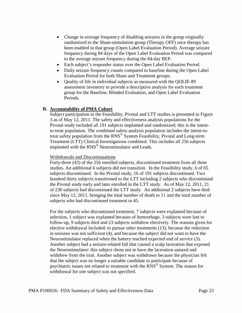
Table 12: Subset Populations of Interest (Implanted Subjects)
Characteristic All
Implanted (N = 191)
By Randomization Group
Treatment (N = 97)
Sham (N = 94)
p-value1
Seizure onset location - Mesial Temporal Lobe Only (v. other)2
50% (95/191)
49% (48/97)
50% (47/94) 0.943
Number of seizure foci - Bifocal (v. unifocal)2 55% (106/191)
49% (48/97)
62% (58/94) 0.089
Prior therapeutic surgery for epilepsy2 32% (62/191)
35% (34/97)
30% (28/94) 0.437
Prior EEG monitoring with intracranial electrodes
59% (113/191)
65% (63/97)
53% (50/94) 0.098
Prior VNS 34% (64/191)
31% (30/97)
36% (34/94) 0.443
Anatomical brain abnormality (by neuroimaging)
67% (128/191)
68% (66/97)
66% (62/94) 0.759
Benzodiazepine use (acute)3 36% (69/191)
31% (30/97)
41% (39/94) 0.129
1 p-value per chi-square 2 Characteristics used as strata in adaptive randomization algorithm 3 Subjects who used acute benzodiazepines as rescue medications for seizures at any time during the Pre-
Implant Period up until the implantation procedure. Does not include daily use of benzodiazepines
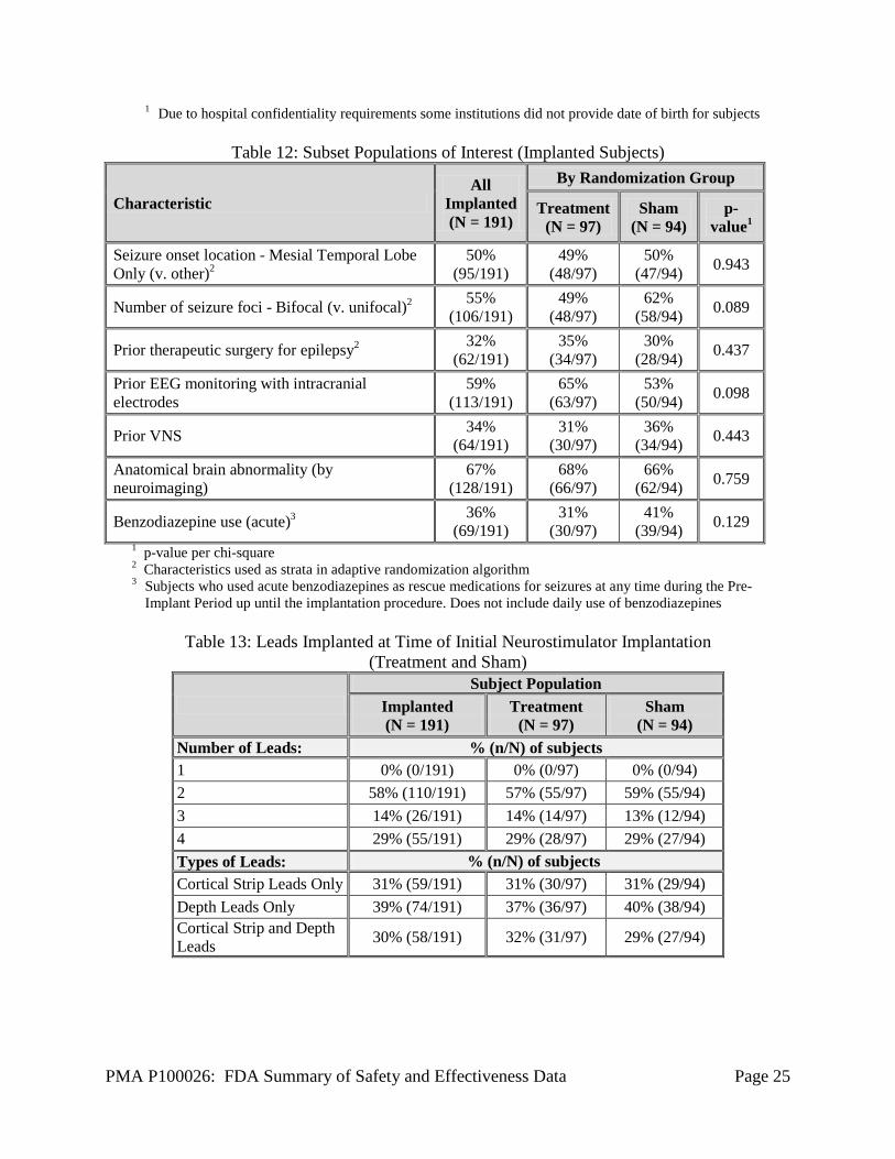
Table 13: Leads Implanted at Time of Initial Neurostimulator Implantation (Treatment and Sham)
Subject Population
Implanted (N = 191)
Treatment (N = 97)
Sham (N = 94)
Number of Leads: % (n/N) of subjects 1 0% (0/191) 0% (0/97) 0% (0/94) 2 58% (110/191) 57% (55/97) 59% (55/94) 3 14% (26/191) 14% (14/97) 13% (12/94) 4 29% (55/191) 29% (28/97) 29% (27/94) Types of Leads: % (n/N) of subjects Cortical Strip Leads Only 31% (59/191) 31% (30/97) 31% (29/94) Depth Leads Only 39% (74/191) 37% (36/97) 40% (38/94) Cortical Strip and Depth Leads 30% (58/191) 32% (31/97) 29% (27/94)
PMA P100026: FDA Summary of Safety and Effectiveness Data Page 26
D. Safety and Effectiveness Results
1. Safety Results The analysis of the primary safety endpoint was based on the cohort of 191 subjects available for the 12-week evaluation. Additional safety analyses were provided for the 256 implanted subjects in the Feasibility, Pivotal, and LTT as of May 12, 2011. The key safety outcomes for this study are presented below in Table 14 through Table 27. All adverse effects through 2 years are reported in Table 28. Adverse effects that occurred in the PMA clinical study: The RNS® System Feasibility, Pivotal and LTT studies evaluated the safety of the RNS® System for epilepsy in 256 implanted subjects over 903 patient-years of implant experience and 819 patient-years of responsive stimulation. All adverse event data are current as of May 12, 2011 with the exception of deaths and SUDEP analysis which are current through October 24, 2012. The investigator classified each adverse event as serious or non-serious and as device-related (which includes device-related and device-relation uncertain) or not device-related. Adverse events were considered serious if the event resulted in significant risks or consequences to the subject's acute or long-term health, serious injury or death, hospital admission, or if invasive medical intervention was required to alleviate the adverse event. Adverse events are presented using MedDRA Coding according to the Preferred Term (PT). During all study periods, 165/256 (64.5%) subjects experienced a serious adverse event and 254/256 (99.2%) subjects experienced a non-serious adverse event, including common and expected illnesses. The RNS® System Feasibility and Pivotal studies met the safety endpoints pre-specified in the investigational plans (see Table 14 below). Adverse events associated with neuropsychological function are shown in Table 22. Serious adverse events (SAEs) and Adverse Events occurring in ≥ 2.5% of subjects are reported by study period in Table 15 through Table 21 below. Adverse events of special interest are also discussed. A full listing of adverse events (AEs) in subjects by study period is presented in Table 28. There were no unanticipated device-related serious adverse events during the RNS® System studies. The primary safety endpoint was to compare similar procedures to the significant adverse events for the surgical procedure and following 28 days (acute) and to compare similar procedures to the surgical procedure and the following 84 days (short-term chronic). The primary safety endpoint was met. There was no difference between the Treatment and Sham groups in the overall percentage of subjects experiencing an adverse event, or any specific type of adverse event during the evaluation periods of the studies. The overall frequency of adverse events or of specific adverse events did not increase over time, whether or not the investigator considered the event as device-related or not device-related.

PMA P100026: FDA Summary of Safety and Effectiveness Data Page 27
Pivotal Study: Primary Safety Endpoint The primary safety endpoint was met. The rate of serious adverse events after implantation of the Neurostimulator and Leads was similar over the first 4 weeks (Acute Period) and in the first 12 weeks (Short-Term Chronic Period) compared to similar procedures, i.e., the combined risks of implantation of intracranial electrodes for purposes of an epilepsy surgery evaluation and epilepsy surgery, and the risks of deep brain stimulation for treatment of movement disorders. The results, presented in Table 14, demonstrate that the SAE rate over the first month and the first 3 months after implantation is comparable to the literature based historical controls.
Table 14: Pivotal Study – Primary Safety Endpoint
Period
SAE Rate1 Met primary safety
endpoint?3 RNS® System Comparator2
% (n/N) subjects [upper 95% CI]
Acute (Surgery – Week 4)
12.0% (23 /191) [16.5%]
15% [20%]
Yes (16.5% < 20%)
Short-Term Chronic (Surgery – Week 12)
18.3% (35 /191) [23.4%]
36% [42%]
Yes (23.4% < 42%)
1 Upper limit of the one-sided 95% confidence interval, estimated using the Score Interval (also known as the Wilson Interval). Upper limits for literature comparators were pre-specified in the protocol, estimated using the Score Interval based on a sample size of 180.
2 Protocol-specified Acute endpoint, based on literature: SAE rate associated with implantation of intracranial electrodes and epilepsy surgery (Tanriverdi et al., 2009; Wong et al., 2009; Fountas and Smith, 2007; Hamer et al., 2002; Behrens et al., 1997). Protocol-specified Short-Term Chronic endpoint, based on literature: SAE rate associated with deep brain stimulation for movement disorders (Oh et al., 2002; Summary of Safety and Effectiveness, Activa Tremor Control System P960009; Beric et al., 2001; Behrens et al., 1997; Hariz, 2002; Joint et al., 2002; Koller et al., 2001).
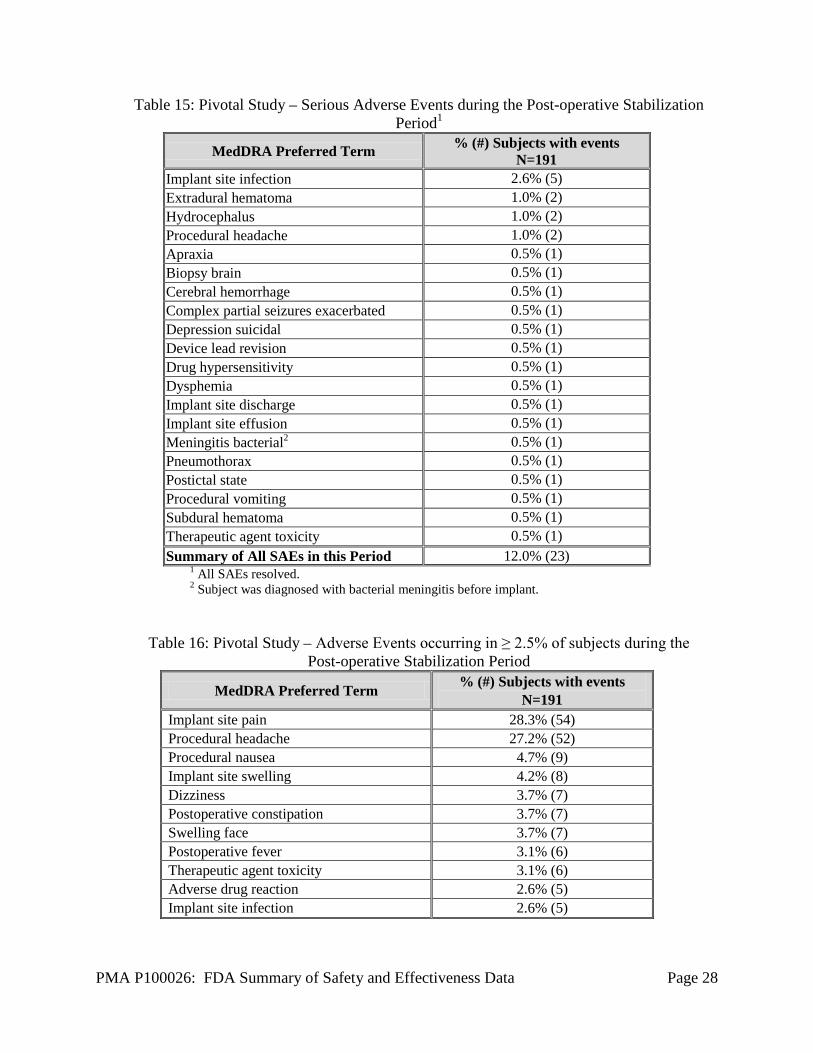
3 Upper limit for the RNS® System is less than that of the comparator. Pivotal Study: Post-operative Stabilization Period AEs Table 15 presents the SAEs that occurred in 23 subjects during the Post-operative Stabilization Period. Five (5) subjects had implant site infections, one of which required explant of the leads and stimulator. Two additional subjects had serious adverse events reported as effusion or discharge at the implant site. One subject was reported to have bacterial meningitis, which was diagnosed before the RNS® Neurostimulator and Leads were implanted. The infection was a chronic infection from a prior evaluation with intracranial electrodes. The investigator elected to implant the RNS® Neurostimulator and NeuroPace Leads despite the observed infection and treated the patient with antibiotics. Three subjects had intracranial hemorrhages. Additional information regarding all hemorrhages is provided in the section below entitled, “Combined Studies: Adverse Events of Particular Relevance”. Adverse events that occurred in ≥ 2.5% of subjects are presented in Table 16. A full listing of adverse events during the Post-operative Stabilization Period is presented in Table 28.
PMA P100026: FDA Summary of Safety and Effectiveness Data Page 28
Table 15: Pivotal Study – Serious Adverse Events during the Post-operative Stabilization Period1
MedDRA Preferred Term % (#) Subjects with events N=191
Implant site infection 2.6% (5) Extradural hematoma 1.0% (2) Hydrocephalus 1.0% (2) Procedural headache 1.0% (2) Apraxia 0.5% (1) Biopsy brain 0.5% (1) Cerebral hemorrhage 0.5% (1) Complex partial seizures exacerbated 0.5% (1) Depression suicidal 0.5% (1) Device lead revision 0.5% (1) Drug hypersensitivity 0.5% (1) Dysphemia 0.5% (1) Implant site discharge 0.5% (1) Implant site effusion 0.5% (1) Meningitis bacterial2 0.5% (1) Pneumothorax 0.5% (1) Postictal state 0.5% (1) Procedural vomiting 0.5% (1) Subdural hematoma 0.5% (1) Therapeutic agent toxicity 0.5% (1) Summary of All SAEs in this Period 12.0% (23)
1 All SAEs resolved. 2 Subject was diagnosed with bacterial meningitis before implant.
Table 16: Pivotal Study – Adverse Events occurring in ≥ 2.5% of subjects during the Post-operative Stabilization Period
MedDRA Preferred Term % (#) Subjects with events N=191
Implant site pain 28.3% (54) Procedural headache 27.2% (52) Procedural nausea 4.7% (9) Implant site swelling 4.2% (8) Dizziness 3.7% (7) Postoperative constipation 3.7% (7) Swelling face 3.7% (7) Postoperative fever 3.1% (6) Therapeutic agent toxicity 3.1% (6) Adverse drug reaction 2.6% (5) Implant site infection 2.6% (5)

PMA P100026: FDA Summary of Safety and Effectiveness Data Page 29
Pivotal Study: Stimulation Optimization Period AEs Table 17 presents the SAEs that occurred in 12 subjects (6 in the Treatment group and 6 in the Sham group) during the Stimulation Optimization Period. Serious adverse events of particular interest include implant site infection and subdural hematoma (due to a seizure). Adverse events that occurred in ≥ 2.5% of subjects are presented in Table 18.
Table 17: Pivotal Study – Serious Adverse Events during the Stimulation Optimization Period
(Treatment and Sham)
MedDRA Preferred Term Treatment
N=97 Sham N=94
% (#) Subjects with events Summary of All SAEs in this Period 6.2% (6) 6.4% (6) Device lead revision 1.0% (1) 1.1% (1) Medical device removal (VNS) 1.0% (1) 1.1% (1) Adverse drug reaction 1.0% (1) -- Arthritis -- 1.1% (1) Central venous catheterisation 1.0% (1) -- Death -- 1.1% (1) EEG monitoring -- 1.1% (1) Implant site infection (dts1) 1.0% (1) -- Meningioma benign 1.0% (1) -- Non-cardiac chest pain -- 1.1% (1) Psychotic disorder -- 1.1% (1) Skin laceration (dts) 1.0% (1) -- Subdural hematoma (dts) 1.0% (1) -- Syncope 1.0% (1) --
1 dts = due to seizure
PMA P100026: FDA Summary of Safety and Effectiveness Data Page 30
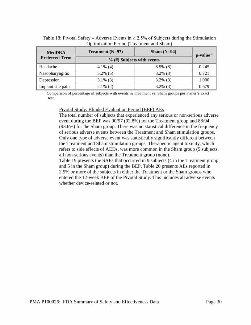
Table 18: Pivotal Safety – Adverse Events in ≥ 2.5% of Subjects during the Stimulation
Optimization Period (Treatment and Sham)
MedDRA Preferred Term
Treatment (N=97) Sham (N=94) p-value 1
% (#) Subjects with events Headache 4.1% (4) 8.5% (8) 0.245 Nasopharyngitis 5.2% (5) 3.2% (3) 0.721 Depression 3.1% (3) 3.2% (3) 1.000 Implant site pain 2.1% (2) 3.2% (3) 0.679
1 Comparison of percentage of subjects with events in Treatment vs. Sham groups per Fisher’s exact test.
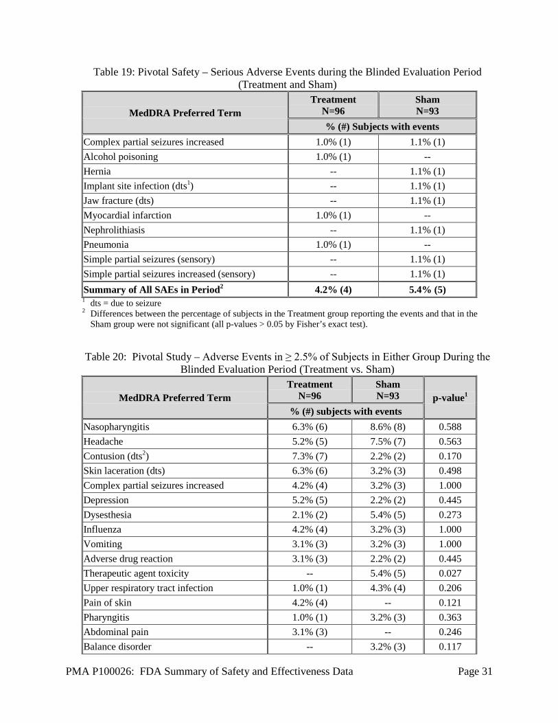
Pivotal Study: Blinded Evaluation Period (BEP) AEs The total number of subjects that experienced any serious or non-serious adverse event during the BEP was 90/97 (92.8%) for the Treatment group and 88/94 (93.6%) for the Sham group. There was no statistical difference in the frequency of serious adverse events between the Treatment and Sham stimulation groups. Only one type of adverse event was statistically significantly different between the Treatment and Sham stimulation groups. Therapeutic agent toxicity, which refers to side effects of AEDs, was more common in the Sham group (5 subjects, all non-serious events) than the Treatment group (none). Table 19 presents the SAEs that occurred in 9 subjects (4 in the Treatment group and 5 in the Sham group) during the BEP. Table 20 presents AEs reported in 2.5% or more of the subjects in either the Treatment or the Sham groups who entered the 12-week BEP of the Pivotal Study. This includes all adverse events whether device-related or not.
PMA P100026: FDA Summary of Safety and Effectiveness Data Page 31
Table 19: Pivotal Safety – Serious Adverse Events during the Blinded Evaluation Period (Treatment and Sham)
MedDRA Preferred Term Treatment
N=96 Sham N=93
% (#) Subjects with events Complex partial seizures increased 1.0% (1) 1.1% (1) Alcohol poisoning 1.0% (1) -- Hernia -- 1.1% (1) Implant site infection (dts1) -- 1.1% (1) Jaw fracture (dts) -- 1.1% (1) Myocardial infarction 1.0% (1) -- Nephrolithiasis -- 1.1% (1) Pneumonia 1.0% (1) -- Simple partial seizures (sensory) -- 1.1% (1) Simple partial seizures increased (sensory) -- 1.1% (1) Summary of All SAEs in Period2 4.2% (4) 5.4% (5) 1 dts = due to seizure 2 Differences between the percentage of subjects in the Treatment group reporting the events and that in the
Sham group were not significant (all p-values > 0.05 by Fisher’s exact test). Table 20: Pivotal Study – Adverse Events in ≥ 2.5% of Subjects in Either Group During the
Blinded Evaluation Period (Treatment vs. Sham)
MedDRA Preferred Term Treatment
N=96 Sham N=93 p-value1
% (#) subjects with events Nasopharyngitis 6.3% (6) 8.6% (8) 0.588 Headache 5.2% (5) 7.5% (7) 0.563 Contusion (dts2) 7.3% (7) 2.2% (2) 0.170 Skin laceration (dts) 6.3% (6) 3.2% (3) 0.498 Complex partial seizures increased 4.2% (4) 3.2% (3) 1.000 Depression 5.2% (5) 2.2% (2) 0.445 Dysesthesia 2.1% (2) 5.4% (5) 0.273 Influenza 4.2% (4) 3.2% (3) 1.000 Vomiting 3.1% (3) 3.2% (3) 1.000 Adverse drug reaction 3.1% (3) 2.2% (2) 0.445 Therapeutic agent toxicity -- 5.4% (5) 0.027 Upper respiratory tract infection 1.0% (1) 4.3% (4) 0.206 Pain of skin 4.2% (4) -- 0.121 Pharyngitis 1.0% (1) 3.2% (3) 0.363 Abdominal pain 3.1% (3) -- 0.246 Balance disorder -- 3.2% (3) 0.117
PMA P100026: FDA Summary of Safety and Effectiveness Data Page 32
MedDRA Preferred Term Treatment
N=96 Sham N=93 p-value1
% (#) subjects with events Head injury -- 3.2% (3) 0.117 1 Fisher’s exact test 2 dts = due to seizure
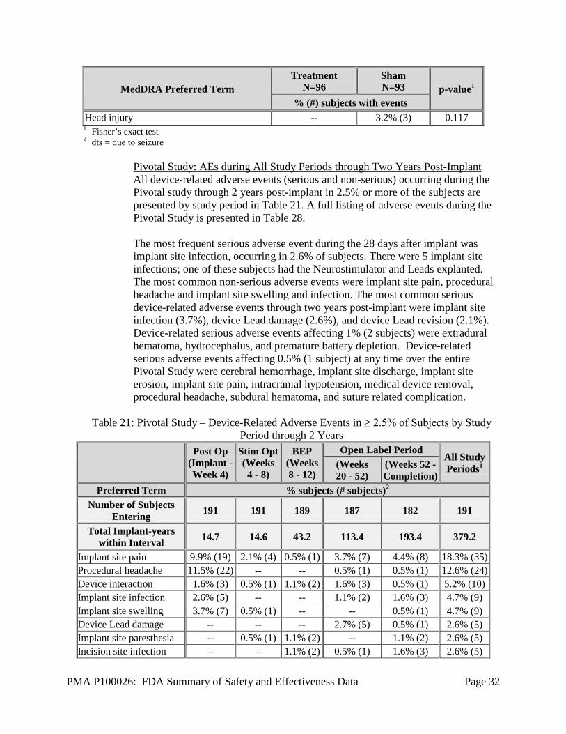
Pivotal Study: AEs during All Study Periods through Two Years Post-Implant All device-related adverse events (serious and non-serious) occurring during the Pivotal study through 2 years post-implant in 2.5% or more of the subjects are presented by study period in Table 21. A full listing of adverse events during the Pivotal Study is presented in Table 28. The most frequent serious adverse event during the 28 days after implant was implant site infection, occurring in 2.6% of subjects. There were 5 implant site infections; one of these subjects had the Neurostimulator and Leads explanted. The most common non-serious adverse events were implant site pain, procedural headache and implant site swelling and infection. The most common serious device-related adverse events through two years post-implant were implant site infection (3.7%), device Lead damage (2.6%), and device Lead revision (2.1%). Device-related serious adverse events affecting 1% (2 subjects) were extradural hematoma, hydrocephalus, and premature battery depletion. Device-related serious adverse events affecting 0.5% (1 subject) at any time over the entire Pivotal Study were cerebral hemorrhage, implant site discharge, implant site erosion, implant site pain, intracranial hypotension, medical device removal, procedural headache, subdural hematoma, and suture related complication.
Table 21: Pivotal Study – Device-Related Adverse Events in ≥ 2.5% of Subjects by Study Period through 2 Years
Post Op
(Implant - Week 4)
Stim Opt (Weeks 4 - 8)
BEP (Weeks 8 - 12)
Open Label Period All Study Periods1 (Weeks
20 - 52) (Weeks 52 - Completion)
Preferred Term % subjects (# subjects)2 Number of Subjects
Entering 191 191 189 187 182 191
Total Implant-years within Interval 14.7 14.6 43.2 113.4 193.4 379.2
Implant site pain 9.9% (19) 2.1% (4) 0.5% (1) 3.7% (7) 4.4% (8) 18.3% (35) Procedural headache 11.5% (22) -- -- 0.5% (1) 0.5% (1) 12.6% (24) Device interaction 1.6% (3) 0.5% (1) 1.1% (2) 1.6% (3) 0.5% (1) 5.2% (10) Implant site infection 2.6% (5) -- -- 1.1% (2) 1.6% (3) 4.7% (9) Implant site swelling 3.7% (7) 0.5% (1) -- -- 0.5% (1) 4.7% (9) Device Lead damage -- -- -- 2.7% (5) 0.5% (1) 2.6% (5) Implant site paresthesia -- 0.5% (1) 1.1% (2) -- 1.1% (2) 2.6% (5) Incision site infection -- -- 1.1% (2) 0.5% (1) 1.6% (3) 2.6% (5)

PMA P100026: FDA Summary of Safety and Effectiveness Data Page 33
Post Op
(Implant - Week 4)
Stim Opt (Weeks 4 - 8)
BEP (Weeks 8 - 12)
Open Label Period All Study Periods1 (Weeks
20 - 52) (Weeks 52 - Completion)
Preferred Term % subjects (# subjects)2 1 Row totals may not sum to totals in this column because some subjects may have had AEs in more than
one period 2 % subjects = # subjects with event / number of subjects entering interval
Pivotal Study: Neuropsychological Testing Neuropsychological function was a safety endpoint. The results of the neuropsychological testing are presented in the following tables. There was no difference between Treatment and Sham groups at the end of the BEP and no statistically significant difference in any of the 16 neuropsychological domains at the end of the BEP or at 1 and 2 years after implant.
Table 22: Pivotal Study: Neuropsychological Measures - Change in Summary Scores at End of Blinded Evaluation Period Relative to Baseline, Treatment versus Sham1
Test
Treatment Sham p-value2
N Mean ± SD N Mean ± SD Visual Motor Speed Trailmaking - Part A3 91 0.87 ± 14.61 86 -0.37 ± 14.97 0.578 Trailmaking - Part B3 90 -6.02 ± 52.72 85 -6.06 ± 33.49 0.996 Motor Speed / Dexterity Grooved Pegboard – Dominant3 89 -1.24 ± 14.95 79 -2.19 ± 22.80 0.746 Grooved Pegboard – Nondominant3 88 -1.89 ± 18.81 76 1.32 ± 27.31 0.378 Auditory Attention WAIS-III Digit Span 90 -0.21 ± 1.55 86 0.09 ± 1.48 0.185 General Verbal Ability WAIS-III Information 91 0.11 ± 1.14 83 0.12 ± 1.17 0.952 General Visuospatial Ability WAIS-III Block Design 90 -0.03 ± 2.06 84 0.31 ± 1.62 0.227 Verbal Memory RAVLT - I-V (Sum Across Trials) 86 -1.94 ± 9.20 84 -0.21 ± 10.01 0.243 RAVLT - VII (Delayed Recall) 86 -0.10 ± 2.75 84 0.01 ± 2.33 0.766 RAVLT - Recognition Memory 86 -0.21 ± 2.69 83 0.23 ± 3.12 0.329 Visuospatial Memory BVMT-R - Total Recall 90 1.94 ± 6.16 85 2.00 ± 5.95 0.952 BVMT-R - Delayed Recall 87 0.24 ± 2.75 85 0.32 ± 2.26 0.832 BVMT-R - Recognition Discrimination Index 88 0.14 ± 1.42 83 -0.07 ± 1.24 0.309

PMA P100026: FDA Summary of Safety and Effectiveness Data Page 34
Test
Treatment Sham p-value2
N Mean ± SD N Mean ± SD Language BNT - Spontaneous with semantic cue 90 0.70 ± 4.37 84 1.36 ± 3.89 0.297 D-KEFS Verbal Fluency - Condition 1: Letter Fluency 83 -0.06 ± 1.69 77 0.53 ± 2.32 0.065
Design Fluency D-KEFS Design Fluency - Total Composite 89 0.49 ± 2.32 80 0.40 ± 2.40 0.795
1 Analysis includes subjects (N) with assessments at both Baseline and end of BEP time points. WAIS-III = Wechsler Adult Intelligence Scale; RAVLT = Rey Auditory Verbal Learning Test; BVMT-R = Brief Visuospatial Memory Test-Revised; BNT = Boston Naming Test (60 item); D-KEFS = Delis-Kaplan Executive Function System
2 Statistical significance of the between-group difference in change in score (at 20 weeks relative to pre-implant) between Treatment and Sham groups per two-sample t-test.
3 Higher values mean improved performance with the exception of the footnoted tests where lower mean values indicate improved performance.
Table 23: Pivotal Study – Neuropsychological Measures: Change in Summary Scores at 1 and 2
years relative to Baseline1
Test Change at 1 year Change at 2 years
N Mean ± SD p-value2 N Mean ± SD p-
value2 Visual Motor Speed Trailmaking – Part A3 157 -1.59 ± 15.29 0.196 154 -1.47 ± 18.75 0.333 Trailmaking – Part B3 154 -7.28 ± 43.92 0.041 150 -5.95 ± 46.73 0.121 Motor Speed / Dexterity Grooved Pegboard – Dominant3 151 -2.47 ± 20.68 0.145 147 -2.00 ± 22.82 0.289 Grooved Pegboard – Nondominant3 145 -0.68 ± 28.72 0.776 143 -0.22 ± 30.44 0.932 Auditory Attention WAIS-III Digit Span 156 -0.10 ± 2.01 0.526 152 0.04 ± 1.74 0.781 General Verbal Ability WAIS-III Information 156 0.19 ± 1.27 0.069 153 0.31 ± 1.32 0.004 General Visuospatial Ability WAIS-III Block Design 156 0.44 ± 1.88 0.004 152 0.57 ± 2.02 0.001 Verbal Memory RAVLT - I-V (Sum Across Trials) 145 1.80 ± 7.99 0.008 145 0.95 ± 8.81 0.196 RAVLT - VII (Delayed Recall) 144 0.42 ± 2.54 0.047 147 0.21 ± 2.71 0.346 RAVLT - Recognition Memory 144 0.17 ± 2.44 0.413 145 0.22 ± 2.48 0.286 Visuospatial Memory BVMT-R - Total Recall 153 0.82 ± 6.06 0.095 149 0.90 ± 5.30 0.040 BVMT-R - Delayed Recall 151 0.06 ± 2.69 0.797 147 -0.07 ± 2.17 0.704
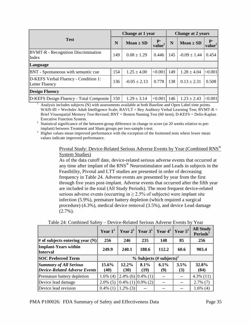
PMA P100026: FDA Summary of Safety and Effectiveness Data Page 35
Test Change at 1 year Change at 2 years
N Mean ± SD p-value2 N Mean ± SD p-
value2 BVMT-R - Recognition Discrimination Index 149 0.08 ± 1.29 0.446 145 -0.09 ± 1.44 0.454
Language BNT - Spontaneous with semantic cue 154 1.25 ± 4.00 <0.001 149 1.28 ± 4.04 <0.001 D-KEFS Verbal Fluency - Condition 1: Letter Fluency 136 -0.05 ± 2.13 0.778 138 0.13 ± 2.31 0.508
Design Fluency D-KEFS Design Fluency - Total Composite 150 1.29 ± 3.14 <0.001 146 1.23 ± 2.43 <0.001
1 Analysis includes subjects (N) with assessments available at both Baseline and Open Label time points. WAIS-III = Wechsler Adult Intelligence Scale; RAVLT = Rey Auditory Verbal Learning Test; BVMT-R = Brief Visuospatial Memory Test-Revised; BNT = Boston Naming Test (60 item); D-KEFS = Delis-Kaplan Executive Function System.
2 Statistical significance of the between-group difference in change in score (at 20 weeks relative to pre-implant) between Treatment and Sham groups per two-sample t-test.
3 Higher values mean improved performance with the exception of the footnoted tests where lower mean values indicate improved performance.
Pivotal Study: Device-Related Serious Adverse Events by Year (Combined RNS® System Studies) As of the data cutoff date, device-related serious adverse events that occurred at any time after implant of the RNS® Neurostimulator and Leads in subjects in the Feasibility, Pivotal and LTT studies are presented in order of decreasing frequency in Table 24. Adverse events are presented by year from the first through five years post-implant. Adverse events that occurred after the fifth year are included in the total (All Study Periods). The most frequent device-related serious adverse events (occurring in ≥ 2.5% of subjects) were implant site infection (5.9%), premature battery depletion (which required a surgical procedure) (4.3%), medical device removal (3.5%), and device Lead damage (2.7%).
Table 24: Combined Safety – Device-Related Serious Adverse Events by Year
Year 15 Year 25 Year 35 Year 45 Year 55 All Study Periods1
# of subjects entering year (N) 256 246 235 148 85 256 Implant-Years within Interval 249.9 240.1 188.6 112.2 60.6 903.4
SOC Preferred Term % Subjects (# subjects)2
Summary of All Serious Device-Related Adverse Events
15.6% (40)
12.2% (30)
8.1% (19)
6.1% (9)
3.5% (3)
32.8% (84)
Premature battery depletion 1.6% (4) 2.4% (6) 0.4% (1) -- -- 4.3% (11) Device lead damage 2.0% (5) 0.4% (1) 0.9% (2) -- -- 2.7% (7) Device lead revision 0.4% (1) 1.2% (3) -- -- -- 1.6% (4)
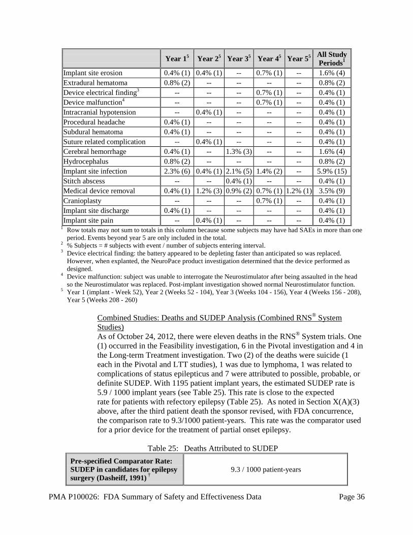
PMA P100026: FDA Summary of Safety and Effectiveness Data Page 36
Year 15 Year 25 Year 35 Year 45 Year 55 All Study Periods1
Implant site erosion 0.4% (1) 0.4% (1) -- 0.7% (1) -- 1.6% (4) Extradural hematoma 0.8% (2) -- -- -- -- 0.8% (2) Device electrical finding3 -- -- -- 0.7% (1) -- 0.4% (1) Device malfunction4 -- -- -- 0.7% (1) -- 0.4% (1) Intracranial hypotension -- 0.4% (1) -- -- -- 0.4% (1) Procedural headache 0.4% (1) -- -- -- -- 0.4% (1) Subdural hematoma 0.4% (1) -- -- -- -- 0.4% (1) Suture related complication -- 0.4% (1) -- -- -- 0.4% (1) Cerebral hemorrhage 0.4% (1) -- 1.3% (3) -- -- 1.6% (4) Hydrocephalus 0.8% (2) -- -- -- -- 0.8% (2) Implant site infection 2.3% (6) 0.4% (1) 2.1% (5) 1.4% (2) -- 5.9% (15) Stitch abscess -- -- 0.4% (1) -- -- 0.4% (1) Medical device removal 0.4% (1) 1.2% (3) 0.9% (2) 0.7% (1) 1.2% (1) 3.5% (9) Cranioplasty -- -- -- 0.7% (1) -- 0.4% (1) Implant site discharge 0.4% (1) -- -- -- -- 0.4% (1) Implant site pain -- 0.4% (1) -- -- -- 0.4% (1)
1 Row totals may not sum to totals in this column because some subjects may have had SAEs in more than one period. Events beyond year 5 are only included in the total.
2 % Subjects = # subjects with event / number of subjects entering interval. 3 Device electrical finding: the battery appeared to be depleting faster than anticipated so was replaced.
However, when explanted, the NeuroPace product investigation determined that the device performed as designed.
4 Device malfunction: subject was unable to interrogate the Neurostimulator after being assaulted in the head so the Neurostimulator was replaced. Post-implant investigation showed normal Neurostimulator function.
5 Year 1 (implant - Week 52), Year 2 (Weeks 52 - 104), Year 3 (Weeks 104 - 156), Year 4 (Weeks 156 - 208), Year 5 (Weeks 208 - 260)
Combined Studies: Deaths and SUDEP Analysis (Combined RNS® System Studies) As of October 24, 2012, there were eleven deaths in the RNS® System trials. One (1) occurred in the Feasibility investigation, 6 in the Pivotal investigation and 4 in the Long-term Treatment investigation. Two (2) of the deaths were suicide (1 each in the Pivotal and LTT studies), 1 was due to lymphoma, 1 was related to complications of status epilepticus and 7 were attributed to possible, probable, or definite SUDEP. With 1195 patient implant years, the estimated SUDEP rate is 5.9 / 1000 implant years (see Table 25). This rate is close to the expected rate for patients with refectory epilepsy (Table 25). As noted in Section X(A)(3) above, after the third patient death the sponsor revised, with FDA concurrence, the comparison rate to 9.3/1000 patient-years. This rate was the comparator used for a prior device for the treatment of partial onset epilepsy.
Table 25: Deaths Attributed to SUDEP
Pre-specified Comparator Rate: SUDEP in candidates for epilepsy surgery (Dasheiff, 1991) †
9.3 / 1000 patient-years
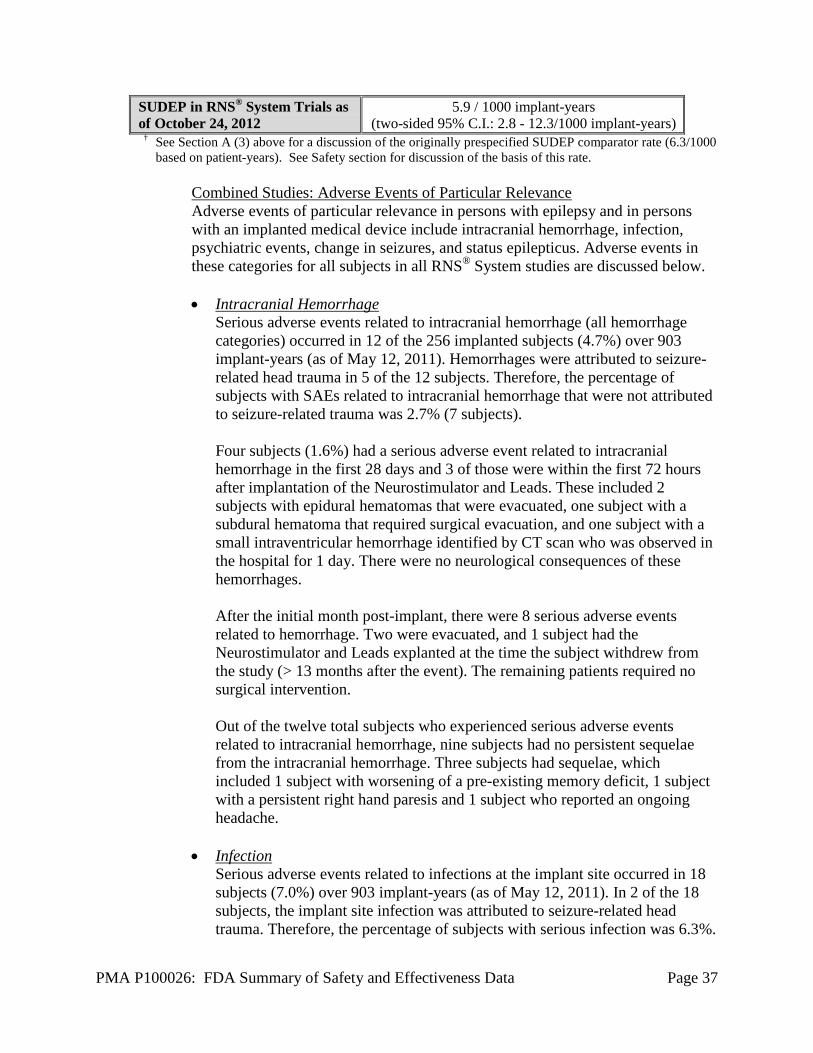
PMA P100026: FDA Summary of Safety and Effectiveness Data Page 37
SUDEP in RNS® System Trials as of October 24, 2012
5.9 / 1000 implant-years (two-sided 95% C.I.: 2.8 - 12.3/1000 implant-years)
† See Section A (3) above for a discussion of the originally prespecified SUDEP comparator rate (6.3/1000 based on patient-years). See Safety section for discussion of the basis of this rate.
Combined Studies: Adverse Events of Particular Relevance Adverse events of particular relevance in persons with epilepsy and in persons with an implanted medical device include intracranial hemorrhage, infection, psychiatric events, change in seizures, and status epilepticus. Adverse events in these categories for all subjects in all RNS® System studies are discussed below.
• Intracranial Hemorrhage
Serious adverse events related to intracranial hemorrhage (all hemorrhage categories) occurred in 12 of the 256 implanted subjects (4.7%) over 903 implant-years (as of May 12, 2011). Hemorrhages were attributed to seizure-related head trauma in 5 of the 12 subjects. Therefore, the percentage of subjects with SAEs related to intracranial hemorrhage that were not attributed to seizure-related trauma was 2.7% (7 subjects).
Four subjects (1.6%) had a serious adverse event related to intracranial hemorrhage in the first 28 days and 3 of those were within the first 72 hours after implantation of the Neurostimulator and Leads. These included 2 subjects with epidural hematomas that were evacuated, one subject with a subdural hematoma that required surgical evacuation, and one subject with a small intraventricular hemorrhage identified by CT scan who was observed in the hospital for 1 day. There were no neurological consequences of these hemorrhages. After the initial month post-implant, there were 8 serious adverse events related to hemorrhage. Two were evacuated, and 1 subject had the Neurostimulator and Leads explanted at the time the subject withdrew from the study (> 13 months after the event). The remaining patients required no surgical intervention. Out of the twelve total subjects who experienced serious adverse events related to intracranial hemorrhage, nine subjects had no persistent sequelae from the intracranial hemorrhage. Three subjects had sequelae, which included 1 subject with worsening of a pre-existing memory deficit, 1 subject with a persistent right hand paresis and 1 subject who reported an ongoing headache.
• Infection
Serious adverse events related to infections at the implant site occurred in 18 subjects (7.0%) over 903 implant-years (as of May 12, 2011). In 2 of the 18 subjects, the implant site infection was attributed to seizure-related head trauma. Therefore, the percentage of subjects with serious infection was 6.3%.
PMA P100026: FDA Summary of Safety and Effectiveness Data Page 38
One infection was diagnosed by a positive culture prior to implantation of the Neurostimulator and Leads; this was believed to be an incompletely treated infection that began with implantation of intracranial electrodes for video-EEG monitoring 3 years before. All infections were treated with antibiotics with or without drainage or debridement. Eleven (4.3%) subjects had the Neurostimulator and/or Leads explanted because of infection. One of the subjects was re-implanted after the infection resolved. There were no infections of the brain, no cases of sepsis, and no permanent neurological consequences related to infection.
• Psychiatric Adverse Events
Many subjects in these studies had a history of depression (49%) and/or suicidality (5.2%). According to responses on the Beck Depression Inventory (BDI-II) during the Baseline Period, 15.6% of subjects had moderate depression before implant and 9.2% endorsed suicidality. In order to fully capture any adverse event that could be representative of suicidality, suicidality was broadly defined to include the MedDRA preferred terms: suicide attempt, suicidal behavior, suicidal ideation, depression suicidal, self-injurious ideation, and suicide. In the combined studies over the 903 patient-years (as of May 12, 2011), psychiatric adverse events occurred in a total of 102 of 256 subjects (39.8%). Twenty-one of the 102 subjects had a total of 33 serious adverse events. Serious adverse events were related to depression (1 subject), suicidality (12 subjects; discussed below), acute psychosis (2 subjects, 3 events in 1 subject), chronic psychosis (3 subjects), post-ictal psychosis (1 subject) and conversion disorder (2 subjects). The remaining psychiatric serious adverse events affected 1 subject each and were emotional distress, affect lability, agitation, alcohol abuse and alcohol withdrawal, and an episode of a visual hallucination. There were 12 subjects with serious suicidality adverse events; some subjects had more than one event. The serious adverse events were: suicide (2), suicidal depression (6), suicide attempt (6), suicidal ideation (2) and suicidal behavior (2). Eleven of the 12 subjects with serious adverse events related to suicidality had a history of suicidality and/or depression or met depression criteria at baseline per BDI-II or CES-D. Fifty-six (56) subjects reported a depression adverse event. One subject had a serious adverse event of depression and 55 had non-serious adverse events. The one serious adverse event was a brief hospitalization because of depression. Sixteen (16) subjects had adverse events related to memory impairment, all of which were non-serious. Seven of these 16 subjects had a history of memory dysfunction and 11 of the 16 had memory deficits documented by neuropsychological testing obtained prior to implantation.

PMA P100026: FDA Summary of Safety and Effectiveness Data Page 39
• Adverse Events Related to Changes in Seizures In the Pivotal Study over the 379 patient implant-years of experience, 41 subjects had 70 adverse events related to changes in seizures that were considered serious. The majority of these adverse events were considered serious because the subject was admitted for video-EEG monitoring or hospitalized to receive AEDs. An increase in complex partial seizure frequency was seen in 6.3% of subjects, 5.9% of subjects experienced an increase in generalized tonic-clonic seizures, and 1.6% of subjects experienced an increase in simple partial motor seizures. A serious exacerbation of complex partial seizures was seen in 2.0% of subjects, 4.3% of subjects experienced an exacerbation of generalized tonic-clonic seizures, and 0.4% of subjects experienced an exacerbation in simple partial motor seizures. A serious adverse event of a new seizure type occurred in 3 subjects.
• Status Epilepticus
There were 10 subjects (3.9%) with serious and non-serious adverse events of status epilepticus. There were 16 serious adverse events related to status epilepticus in 8 subjects (3.1%) implanted with the Neurostimulator and Leads. One additional subject had convulsive status epilepticus after the RNS System was explanted but before the subject had withdrawn from the study; the status occurred when the patient had AEDs tapered during an EEG monitoring procedure with intracranial electrodes.
Of the subjects implanted with the Neurostimulator and Leads, 6 episodes were convulsive and 10 were non-convulsive (note that if the type of status was not known, it was coded as convulsive). Seven subjects had 1 episode of status epilepticus and 1 subject had 9 episodes. In addition, there was one subject with a single episode of nonconvulsive status that was considered non-serious. None of the events occurred acutely at the time responsive stimulation was enabled (all events occurred during the open period at least 1 month after enabling responsive stimulation).
• Seizure-Related Injury
There was no difference between Treatment and Sham groups during the BEP. In the combined studies over the 903 patient implant-years (as of May 12, 2011), there were 402 non-serious and serious events of injury related to a seizure occurring in 126 subjects (49.2%). Thirty-two serious events of injury related to a seizure occurred in 23 subjects (9.0%). Head injury due to a seizure occurred 31 times in 27 subjects (10.5%). Two of the events were considered serious. There were 4 serious intracranial hemorrhages, 3 subdural and one traumatic intracranial hemorrhage. Sixteen (16) subjects sustained skeletal bone fractures due to a seizure, 7 events were considered serious.
Device Failures and Replacements
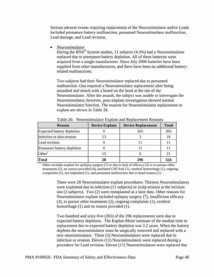
PMA P100026: FDA Summary of Safety and Effectiveness Data Page 40
Serious adverse events requiring replacement of the Neurostimulator and/or Leads included premature battery malfunction, presumed Neurostimulator malfunction, Lead damage, and Lead revision. • Neurostimulator
During the RNS® System studies, 11 subjects (4.3%) had a Neurostimulator replaced due to premature battery depletion. All of these batteries were acquired from a single manufacturer. Since July 2006 batteries have been supplied from other manufacturers, and there have been no additional battery-related malfunctions. Two subjects had their Neurostimulator replaced due to presumed malfunction. One required a Neurostimulator replacement after being assaulted and struck with a board on the head at the site of the Neurostimulator. After the assault, the subject was unable to interrogate the Neurostimulator; however, post-implant investigation showed normal Neurostimulator function. The reasons for Neurostimulator replacement or explant are shown in Table 26.
Table 26: Neurostimulator Explant and Replacement Reasons Reason Device Explant Device Replacement Total
Expected battery depletion 0 265 265 Infection or skin erosion 13 3 16 Lead revision 0 11 11 Premature battery depletion 0 11 11 Other1 15 6 21 Total 28 296 324 1 Other includes explant for epilepsy surgery (7) or due to lack of efficacy (3) or to pursue other
treatments (2), no reason provided (4), unrelated CSF leak (1), cerebral hemorrhage (1), ongoing complaints (1), not implanted (1), and presumed malfunction due to head trauma (1).
There were 28 Neurostimulator explant procedures. Thirteen Neurostimulators were explanted due to infection (11 subjects) or scalp erosion at the incision site (2 subjects). Two (2) were reimplanted at a later date. Other reasons for Neurostimulator explant included epilepsy surgery (7), insufficient efficacy (3), to pursue other treatments (2), ongoing complaints (1), cerebral hemorrhage (1) and no reason provided (1). Two hundred and sixty-five (265) of the 296 replacements were due to expected battery depletion. The Kaplan-Meier estimate of the median time to replacement due to expected battery depletion was 2.2 years. When the battery depletes the neurostimulator must be surgically removed and replaced with a new neurostimulator. Three (3) Neurostimulators were replaced due to infection or erosion. Eleven (11) Neurostimulators were replaced during a procedure for Lead revision. Eleven (11) Neurostimulators were replaced due
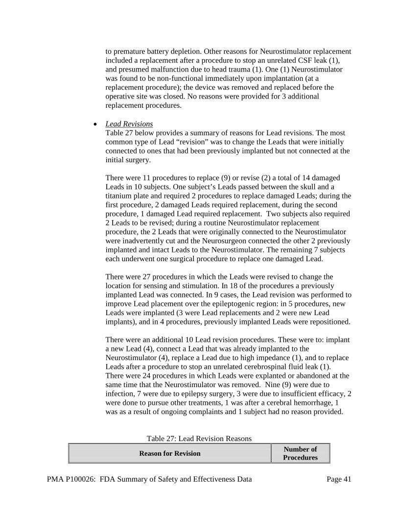
PMA P100026: FDA Summary of Safety and Effectiveness Data Page 41
to premature battery depletion. Other reasons for Neurostimulator replacement included a replacement after a procedure to stop an unrelated CSF leak (1), and presumed malfunction due to head trauma (1). One (1) Neurostimulator was found to be non-functional immediately upon implantation (at a replacement procedure); the device was removed and replaced before the operative site was closed. No reasons were provided for 3 additional replacement procedures.
• Lead Revisions
Table 27 below provides a summary of reasons for Lead revisions. The most common type of Lead “revision” was to change the Leads that were initially connected to ones that had been previously implanted but not connected at the initial surgery. There were 11 procedures to replace (9) or revise (2) a total of 14 damaged Leads in 10 subjects. One subject’s Leads passed between the skull and a titanium plate and required 2 procedures to replace damaged Leads; during the first procedure, 2 damaged Leads required replacement, during the second procedure, 1 damaged Lead required replacement. Two subjects also required 2 Leads to be revised; during a routine Neurostimulator replacement procedure, the 2 Leads that were originally connected to the Neurostimulator were inadvertently cut and the Neurosurgeon connected the other 2 previously implanted and intact Leads to the Neurostimulator. The remaining 7 subjects each underwent one surgical procedure to replace one damaged Lead. There were 27 procedures in which the Leads were revised to change the location for sensing and stimulation. In 18 of the procedures a previously implanted Lead was connected. In 9 cases, the Lead revision was performed to improve Lead placement over the epileptogenic region: in 5 procedures, new Leads were implanted (3 were Lead replacements and 2 were new Lead implants), and in 4 procedures, previously implanted Leads were repositioned. There were an additional 10 Lead revision procedures. These were to: implant a new Lead (4), connect a Lead that was already implanted to the Neurostimulator (4), replace a Lead due to high impedance (1), and to replace Leads after a procedure to stop an unrelated cerebrospinal fluid leak (1). There were 24 procedures in which Leads were explanted or abandoned at the same time that the Neurostimulator was removed. Nine (9) were due to infection, 7 were due to epilepsy surgery, 3 were due to insufficient efficacy, 2 were done to pursue other treatments, 1 was after a cerebral hemorrhage, 1 was as a result of ongoing complaints and 1 subject had no reason provided.
Table 27: Lead Revision Reasons
Reason for Revision Number of Procedures
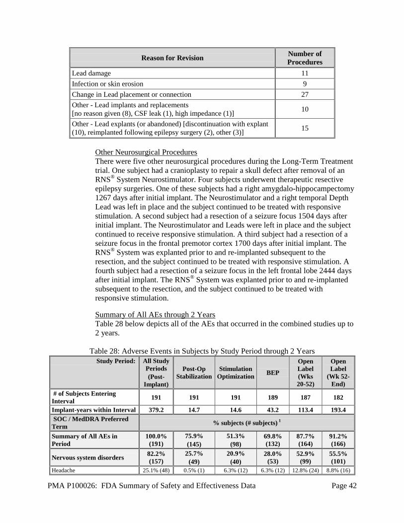
PMA P100026: FDA Summary of Safety and Effectiveness Data Page 42
Reason for Revision Number of Procedures
Lead damage 11 Infection or skin erosion 9 Change in Lead placement or connection 27 Other - Lead implants and replacements [no reason given (8), CSF leak (1), high impedance (1)] 10
Other - Lead explants (or abandoned) [discontinuation with explant (10), reimplanted following epilepsy surgery (2), other (3)] 15
Other Neurosurgical Procedures There were five other neurosurgical procedures during the Long-Term Treatment trial. One subject had a cranioplasty to repair a skull defect after removal of an RNS® System Neurostimulator. Four subjects underwent therapeutic resective epilepsy surgeries. One of these subjects had a right amygdalo-hippocampectomy 1267 days after initial implant. The Neurostimulator and a right temporal Depth Lead was left in place and the subject continued to be treated with responsive stimulation. A second subject had a resection of a seizure focus 1504 days after initial implant. The Neurostimulator and Leads were left in place and the subject continued to receive responsive stimulation. A third subject had a resection of a seizure focus in the frontal premotor cortex 1700 days after initial implant. The RNS® System was explanted prior to and re-implanted subsequent to the resection, and the subject continued to be treated with responsive stimulation. A fourth subject had a resection of a seizure focus in the left frontal lobe 2444 days after initial implant. The RNS® System was explanted prior to and re-implanted subsequent to the resection, and the subject continued to be treated with responsive stimulation.
Summary of All AEs through 2 Years Table 28 below depicts all of the AEs that occurred in the combined studies up to 2 years.
Table 28: Adverse Events in Subjects by Study Period through 2 Years
Study Period: All Study Periods (Post-
Implant)
Post-Op Stabilization
Stimulation Optimization BEP
Open Label (Wks 20-52)
Open Label
(Wk 52- End)
# of Subjects Entering Interval 191 191 191 189 187 182
Implant-years within Interval 379.2 14.7 14.6 43.2 113.4 193.4 SOC / MedDRA Preferred Term % subjects (# subjects) 1
Summary of All AEs in Period
100.0% (191)
75.9% (145)
51.3% (98)
69.8% (132)
87.7% (164)
91.2% (166)
Nervous system disorders 82.2% (157)
25.7% (49)
20.9% (40)
28.0% (53)
52.9% (99)
55.5% (101)
Headache 25.1% (48) 0.5% (1) 6.3% (12) 6.3% (12) 12.8% (24) 8.8% (16)

PMA P100026: FDA Summary of Safety and Effectiveness Data Page 43
Study Period: All Study Periods (Post-
Implant)
Post-Op Stabilization
Stimulation Optimization BEP
Open Label (Wks 20-52)
Open Label
(Wk 52- End)
Complex partial seizures increased 15.7% (30) 1.0% (2) 0.5% (1) 3.7% (7) 8.6% (16) 6.6% (12) Dizziness 13.1% (25) 3.7% (7) 1.6% (3) 0.5% (1) 3.2% (6) 4.9% (9) Complex partial seizures 12.6% (24) 0.5% (1) 2.1% (4) 1.6% (3) 5.3% (10) 6.0% (11) Dysaesthesia 12.6% (24) 0.5% (1) 2.1% (4) 3.7% (7) 5.3% (10) 3.8% (7) Simple partial seizures (sensory) 11.0% (21) 1.6% (3) 1.6% (3) 1.1% (2) 5.3% (10) 3.3% (6) Complex partial seizures exacerbated 9.9% (19) 0.5% (1) -- 1.6% (3) 4.8% (9) 4.4% (8)
Tonic-clonic seizures exacerbated 9.9% (19) -- 1.0% (2) -- 6.4% (12) 3.3% (6) Tremor 9.9% (19) 1.0% (2) 1.6% (3) 0.5% (1) 4.8% (9) 3.8% (7) Tonic-clonic seizures increased 9.4% (18) -- 0.5% (1) 0.5% (1) 5.9% (11) 4.4% (8) Insomnia 8.4% (16) 2.1% (4) 1.6% (3) 0.5% (1) 4.3% (8) 1.6% (3) Memory impairment 8.4% (16) 1.0% (2) 0.5% (1) 0.5% (1) 4.3% (8) 2.7% (5) Paraesthesia 6.8% (13) 1.0% (2) -- 1.6% (3) 2.7% (5) 2.7% (5) Photopsia 6.3% (12) -- 1.0% (2) -- 4.3% (8) 1.6% (3) Simple partial seizures (motor) 6.3% (12) 2.1% (4) 1.0% (2) 1.6% (3) 2.1% (4) 2.2% (4) Nystagmus 5.2% (10) 0.5% (1) 1.0% (2) 1.1% (2) -- 2.7% (5) Confusional state 4.7% (9) 0.5% (1) -- -- 2.7% (5) 2.2% (4) Somnolence 4.7% (9) -- 0.5% (1) 0.5% (1) -- 3.8% (7) Balance disorder 4.2% (8) 1.0% (2) 0.5% (1) 1.6% (3) 1.6% (3) 0.5% (1) Postictal state 4.2% (8) 1.0% (2) 1.0% (2) -- 1.6% (3) 1.1% (2) Migraine 3.7% (7) -- 0.5% (1) 1.1% (2) 1.1% (2) 1.1% (2) Simple partial seizures increased (motor) 3.7% (7) -- -- 0.5% (1) 2.1% (4) 1.1% (2)
Aphasia 3.1% (6) 1.6% (3) 0.5% (1) -- -- 1.1% (2) Dizziness postural 3.1% (6) 1.0% (2) -- -- 1.1% (2) 1.1% (2) Hypoaesthesia 3.1% (6) 0.5% (1) 0.5% (1) 0.5% (1) 0.5% (1) 1.1% (2) Simple partial seizures exacerbated (motor) 3.1% (6) -- -- 0.5% (1) 1.6% (3) 1.1% (2)
Simple partial seizures increased (sensory) 3.1% (6) 0.5% (1) -- 0.5% (1) 1.6% (3) 0.5% (1)
Dyskinesia 2.6% (5) 0.5% (1) -- -- 1.6% (3) 0.5% (1) Monoparesis 2.6% (5) 1.0% (2) -- -- 1.1% (2) 0.5% (1) Sciatica 2.6% (5) 0.5% (1) -- 1.1% (2) 0.5% (1) 0.5% (1) Simple partial seizures exacerbated (sensory) 2.6% (5) 0.5% (1) 0.5% (1) -- 1.1% (2) 1.6% (3)
Syncope 2.6% (5) 0.5% (1) 0.5% (1) -- -- 1.6% (3) Ataxia 2.1% (4) 0.5% (1) 0.5% (1) -- -- 1.1% (2) Cerebellar syndrome 2.1% (4) 0.5% (1) -- 1.1% (2) 0.5% (1) -- Dysarthria 2.1% (4) -- -- -- 1.1% (2) 1.1% (2) Peripheral nerve injury 2.1% (4) -- 0.5% (1) 1.6% (3) -- 0.5% (1) Visual field defect 2.1% (4) 0.5% (1) 0.5% (1) -- 0.5% (1) 0.5% (1) Atonic seizures increased 1.6% (3) -- -- -- -- 1.6% (3) Coordination abnormal 1.6% (3) 0.5% (1) 0.5% (1) -- -- 0.5% (1) Disturbance in attention 1.6% (3) -- 0.5% (1) -- 0.5% (1) 0.5% (1) Hydrocephalus 1.6% (3) 1.6% (3) -- -- -- -- Lethargy 1.6% (3) 0.5% (1) -- -- 0.5% (1) 0.5% (1) Photophobia 1.6% (3) 1.6% (3) -- -- -- -- Tonic-clonic seizures 1.6% (3) -- -- -- 1.1% (2) 0.5% (1) Acquired epileptic aphasia 1.0% (2) 0.5% (1) -- -- 0.5% (1) -- Atonic seizures 1.0% (2) -- -- 1.1% (2) -- --

PMA P100026: FDA Summary of Safety and Effectiveness Data Page 44
Study Period: All Study Periods (Post-
Implant)
Post-Op Stabilization
Stimulation Optimization BEP
Open Label (Wks 20-52)
Open Label
(Wk 52- End)
Atonic seizures exacerbated 1.0% (2) -- -- -- -- 1.1% (2) Blindness transient 1.0% (2) -- -- 0.5% (1) 0.5% (1) -- Cerebral haemorrhage 1.0% (2) 1.0% (2) -- -- -- -- Convulsive status epilepticus 1.0% (2) -- -- -- 0.5% (1) 0.5% (1) Dysgeusia 1.0% (2) -- -- -- 1.1% (2) -- Dysphasia 1.0% (2) 0.5% (1) -- 0.5% (1) -- -- Nonconvulsive status epilepticus 1.0% (2) -- -- -- 1.1% (2) -- Sleep apnea 1.0% (2) -- -- -- 1.1% (2) -- Abnormal dreams 0.5% (1) -- 0.5% (1) -- -- -- Alexia 0.5% (1) -- -- 0.5% (1) 0.5% (1) -- Altered visual depth perception 0.5% (1) 0.5% (1) -- -- -- -- Anosmia 0.5% (1) -- -- -- 0.5% (1) -- Apraxia 0.5% (1) 0.5% (1) -- -- -- -- Aura 0.5% (1) -- -- -- -- 0.5% (1) Bradyphrenia 0.5% (1) -- -- -- 0.5% (1) -- Brain oedema 0.5% (1) 0.5% (1) -- -- -- -- Carpal tunnel syndrome 0.5% (1) -- -- -- -- 0.5% (1) Disorientation 0.5% (1) -- -- 0.5% (1) -- -- Dysphemia 0.5% (1) 0.5% (1) -- -- -- -- Dystonia 0.5% (1) -- -- -- -- 0.5% (1) Essential tremor 0.5% (1) -- -- -- -- 0.5% (1) Eyelid ptosis 0.5% (1) -- 0.5% (1) -- -- -- Facial paresis 0.5% (1) 0.5% (1) -- -- -- -- Head titubation 0.5% (1) -- -- -- -- 0.5% (1) Headache (dts2) 0.5% (1) -- -- -- -- 0.5% (1) Hemiparesis 0.5% (1) -- -- -- 0.5% (1) -- Masked Facies 0.5% (1) -- -- -- 0.5% (1) -- Mononeuropathy 0.5% (1) -- -- 0.5% (1) -- -- Myoclonus 0.5% (1) -- -- -- -- 0.5% (1) Neuropathy peripheral 0.5% (1) -- -- -- -- 0.5% (1) Scintillating scotoma 0.5% (1) -- -- -- -- 0.5% (1) Tonic seizures 0.5% (1) -- -- 0.5% (1) -- -- Tonic seizures exacerbated 0.5% (1) -- -- 0.5% (1) 0.5% (1) -- Toxic encephalopathy 0.5% (1) -- -- -- 0.5% (1) -- Injury, poisoning and procedural complications
81.7% (156)
42.4% (81)
15.2% (29)
22.8% (43)
42.8% (80)
50.5% (92)
Procedural headache 28.8% (55) 27.2% (52) -- -- 1.6% (3) 1.6% (3) Therapeutic agent toxicity 22.5% (43) 3.1% (6) 0.5% (1) 2.6% (5) 8.6% (16) 11.5% (21) Contusion (dts) 16.8% (32) 1.6% (3) 1.6% (3) 4.8% (9) 5.9% (11) 6.6% (12) Skin laceration (dts) 16.2% (31) 2.1% (4) 2.1% (4) 4.8% (9) 5.3% (10) 9.3% (17) Head injury (dts) 8.9% (17) -- 1.0% (2) -- 1.6% (3) 6.6% (12) Excoriation (dts) 7.3% (14) -- 0.5% (1) 1.1% (2) 4.8% (9) 2.7% (5) Head injury 7.3% (14) 0.5% (1) 1.0% (2) 1.6% (3) 2.1% (4) 2.7% (5) Contusion 6.3% (12) 0.5% (1) 0.5% (1) 1.6% (3) 2.1% (4) 1.6% (3) Joint injury (dts) 6.3% (12) -- 1.6% (3) -- 2.1% (4) 2.7% (5) Procedural nausea 6.3% (12) 4.7% (9) -- -- 0.5% (1) 1.1% (2) Implant site swelling 5.8% (11) 4.2% (8) 0.5% (1) -- 0.5% (1) 0.5% (1) Joint injury 5.2% (10) -- -- 0.5% (1) 1.6% (3) 3.8% (7) Multiple injuries (dts) 4.7% (9) 0.5% (1) -- 0.5% (1) 2.1% (4) 3.3% (6)

PMA P100026: FDA Summary of Safety and Effectiveness Data Page 45
Study Period: All Study Periods (Post-
Implant)
Post-Op Stabilization
Stimulation Optimization BEP
Open Label (Wks 20-52)
Open Label
(Wk 52- End)
Skin laceration 4.7% (9) -- 0.5% (1) 1.1% (2) 1.6% (3) 1.6% (3) Device lead revision 3.7% (7) 0.5% (1) 1.0% (2) -- 0.5% (1) 1.6% (3) Postoperative constipation 3.7% (7) 3.7% (7) -- -- -- -- Postoperative fever 3.7% (7) 3.1% (6) -- -- 0.5% (1) -- Thermal burn 3.7% (7) -- -- -- 2.1% (4) 1.6% (3) Limb injury 3.1% (6) -- 0.5% (1) 1.1% (2) 1.1% (2) 0.5% (1) Multiple injuries 3.1% (6) -- -- 1.1% (2) 0.5% (1) 1.6% (3) Thermal burn (dts) 3.1% (6) -- -- 0.5% (1) 1.1% (2) 1.6% (3) Back injury 2.6% (5) -- -- -- 1.1% (2) 1.6% (3) Back injury (dts) 2.6% (5) 0.5% (1) -- -- 1.1% (2) 1.1% (2) Device lead damage 2.6% (5) -- -- -- 2.7% (5) 0.5% (1) Excoriation 2.6% (5) -- 0.5% (1) 0.5% (1) 1.1% (2) 0.5% (1) Laceration (dts) 2.6% (5) -- 1.0% (2) 1.1% (2) 1.1% (2) -- Limb injury (dts) 2.6% (5) -- -- -- 2.1% (4) 0.5% (1) Procedural vomiting 2.6% (5) 2.1% (4) -- -- -- 0.5% (1) Tongue injury (dts) 2.6% (5) -- 0.5% (1) -- 1.1% (2) 1.1% (2) Foot fracture 2.1% (4) -- -- -- 1.1% (2) 1.1% (2) Muscle strain 2.1% (4) -- -- -- 1.1% (2) 1.1% (2) Road traffic accident 2.1% (4) -- -- 0.5% (1) 1.1% (2) 0.5% (1) Extradural haematoma 1.6% (3) 1.6% (3) -- -- -- -- Fall (dts) 1.6% (3) -- -- 0.5% (1) 1.1% (2) -- Lower limb injury (dts) 1.6% (3) -- -- 0.5% (1) 0.5% (1) 0.5% (1) Skeletal injury (dts) 1.6% (3) -- -- 0.5% (1) -- 1.1% (2) Subdural haematoma (dts) 1.6% (3) -- 0.5% (1) -- 0.5% (1) 0.5% (1) Tooth injury 1.6% (3) 0.5% (1) 0.5% (1) -- -- 0.5% (1) Chest injury (dts) 1.0% (2) -- -- -- 1.1% (2) -- Face injury (dts) 1.0% (2) -- -- -- 0.5% (1) 0.5% (1) Hand fracture (dts) 1.0% (2) -- -- -- 0.5% (1) 0.5% (1) Incision site haemorrhage (dts) 1.0% (2) 0.5% (1) -- -- -- 0.5% (1) Mouth injury (dts) 1.0% (2) -- 0.5% (1) -- -- 0.5% (1) Muscle injury (dts) 1.0% (2) -- 0.5% (1) -- 0.5% (1) -- Post procedural diarrhoea 1.0% (2) 0.5% (1) -- -- -- 0.5% (1) Premature battery depletion 1.0% (2) -- -- -- 1.1% (2) -- Procedural dizziness 1.0% (2) 0.5% (1) -- -- -- 0.5% (1) Vertebral injury (dts) 1.0% (2) -- 0.5% (1) -- 0.5% (1) -- Wrist fracture (dts) 1.0% (2) 0.5% (1) -- -- -- 0.5% (1) Agitation postoperative 0.5% (1) 0.5% (1) -- -- -- -- Alcohol poisoning 0.5% (1) -- -- 0.5% (1) -- -- Animal bite 0.5% (1) -- -- -- -- 0.5% (1) Ankle fracture 0.5% (1) -- -- -- -- 0.5% (1) Ankle fracture (dts) 0.5% (1) -- -- 0.5% (1) -- -- Application site excoriation 0.5% (1) 0.5% (1) -- -- -- -- Arthropod bite 0.5% (1) -- -- -- -- 0.5% (1) Barotitis media 0.5% (1) -- -- 0.5% (1) -- -- Catheter site rash 0.5% (1) 0.5% (1) -- -- -- -- Chest injury 0.5% (1) -- -- -- -- 0.5% (1) Corneal abrasion 0.5% (1) -- -- -- -- 0.5% (1) Corneal perforation (dts) 0.5% (1) -- -- -- -- 0.5% (1) Ear canal abrasion 0.5% (1) -- -- 0.5% (1) -- -- Ear injury (dts) 0.5% (1) -- -- -- -- 0.5% (1)
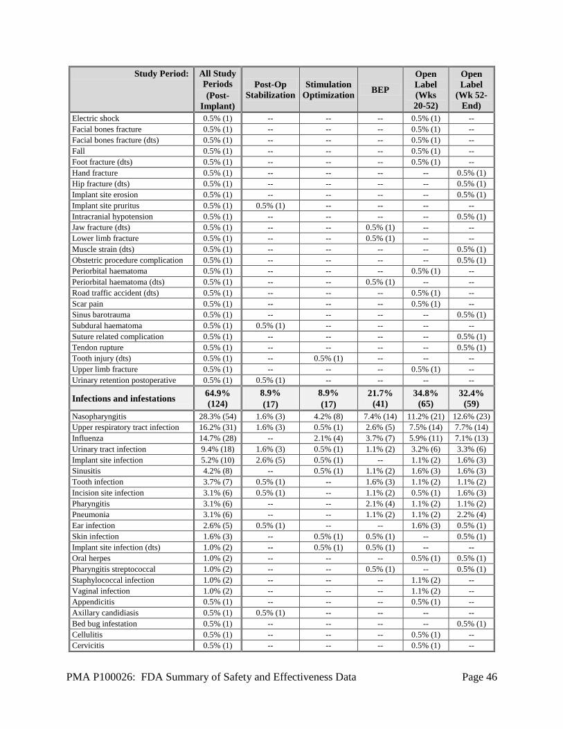
PMA P100026: FDA Summary of Safety and Effectiveness Data Page 46
Study Period: All Study Periods (Post-
Implant)
Post-Op Stabilization
Stimulation Optimization BEP
Open Label (Wks 20-52)
Open Label
(Wk 52- End)
Electric shock 0.5% (1) -- -- -- 0.5% (1) -- Facial bones fracture 0.5% (1) -- -- -- 0.5% (1) -- Facial bones fracture (dts) 0.5% (1) -- -- -- 0.5% (1) -- Fall 0.5% (1) -- -- -- 0.5% (1) -- Foot fracture (dts) 0.5% (1) -- -- -- 0.5% (1) -- Hand fracture 0.5% (1) -- -- -- -- 0.5% (1) Hip fracture (dts) 0.5% (1) -- -- -- -- 0.5% (1) Implant site erosion 0.5% (1) -- -- -- -- 0.5% (1) Implant site pruritus 0.5% (1) 0.5% (1) -- -- -- -- Intracranial hypotension 0.5% (1) -- -- -- -- 0.5% (1) Jaw fracture (dts) 0.5% (1) -- -- 0.5% (1) -- -- Lower limb fracture 0.5% (1) -- -- 0.5% (1) -- -- Muscle strain (dts) 0.5% (1) -- -- -- -- 0.5% (1) Obstetric procedure complication 0.5% (1) -- -- -- -- 0.5% (1) Periorbital haematoma 0.5% (1) -- -- -- 0.5% (1) -- Periorbital haematoma (dts) 0.5% (1) -- -- 0.5% (1) -- -- Road traffic accident (dts) 0.5% (1) -- -- -- 0.5% (1) -- Scar pain 0.5% (1) -- -- -- 0.5% (1) -- Sinus barotrauma 0.5% (1) -- -- -- -- 0.5% (1) Subdural haematoma 0.5% (1) 0.5% (1) -- -- -- -- Suture related complication 0.5% (1) -- -- -- -- 0.5% (1) Tendon rupture 0.5% (1) -- -- -- -- 0.5% (1) Tooth injury (dts) 0.5% (1) -- 0.5% (1) -- -- -- Upper limb fracture 0.5% (1) -- -- -- 0.5% (1) -- Urinary retention postoperative 0.5% (1) 0.5% (1) -- -- -- --
Infections and infestations 64.9% (124)
8.9% (17)
8.9% (17)
21.7% (41)
34.8% (65)
32.4% (59)
Nasopharyngitis 28.3% (54) 1.6% (3) 4.2% (8) 7.4% (14) 11.2% (21) 12.6% (23) Upper respiratory tract infection 16.2% (31) 1.6% (3) 0.5% (1) 2.6% (5) 7.5% (14) 7.7% (14) Influenza 14.7% (28) -- 2.1% (4) 3.7% (7) 5.9% (11) 7.1% (13) Urinary tract infection 9.4% (18) 1.6% (3) 0.5% (1) 1.1% (2) 3.2% (6) 3.3% (6) Implant site infection 5.2% (10) 2.6% (5) 0.5% (1) -- 1.1% (2) 1.6% (3) Sinusitis 4.2% (8) -- 0.5% (1) 1.1% (2) 1.6% (3) 1.6% (3) Tooth infection 3.7% (7) 0.5% (1) -- 1.6% (3) 1.1% (2) 1.1% (2) Incision site infection 3.1% (6) 0.5% (1) -- 1.1% (2) 0.5% (1) 1.6% (3) Pharyngitis 3.1% (6) -- -- 2.1% (4) 1.1% (2) 1.1% (2) Pneumonia 3.1% (6) -- -- 1.1% (2) 1.1% (2) 2.2% (4) Ear infection 2.6% (5) 0.5% (1) -- -- 1.6% (3) 0.5% (1) Skin infection 1.6% (3) -- 0.5% (1) 0.5% (1) -- 0.5% (1) Implant site infection (dts) 1.0% (2) -- 0.5% (1) 0.5% (1) -- -- Oral herpes 1.0% (2) -- -- -- 0.5% (1) 0.5% (1) Pharyngitis streptococcal 1.0% (2) -- -- 0.5% (1) -- 0.5% (1) Staphylococcal infection 1.0% (2) -- -- -- 1.1% (2) -- Vaginal infection 1.0% (2) -- -- -- 1.1% (2) -- Appendicitis 0.5% (1) -- -- -- 0.5% (1) -- Axillary candidiasis 0.5% (1) 0.5% (1) -- -- -- -- Bed bug infestation 0.5% (1) -- -- -- -- 0.5% (1) Cellulitis 0.5% (1) -- -- -- 0.5% (1) -- Cervicitis 0.5% (1) -- -- -- 0.5% (1) --

PMA P100026: FDA Summary of Safety and Effectiveness Data Page 47
Study Period: All Study Periods (Post-
Implant)
Post-Op Stabilization
Stimulation Optimization BEP
Open Label (Wks 20-52)
Open Label
(Wk 52- End)
Hordeolum 0.5% (1) -- -- -- 0.5% (1) -- Infected sebaceous cyst 0.5% (1) -- -- -- -- 0.5% (1) Infection 0.5% (1) -- -- -- 0.5% (1) -- Meningitis bacterial 0.5% (1) 0.5% (1) -- -- -- -- Onychomycosis 0.5% (1) -- -- 0.5% (1) -- -- Oral candidiasis 0.5% (1) -- -- -- -- 0.5% (1) Peritoneal infection 0.5% (1) -- -- -- 0.5% (1) -- Prostate infection 0.5% (1) -- -- 0.5% (1) -- -- Stitch abscess 0.5% (1) -- -- -- 0.5% (1) -- General disorders and administration site conditions
63.9% (122)
36.1% (69)
6.8% (13)
11.6% (22)
19.3% (36)
29.1% (53)
Implant site pain 37.7% (72) 28.3% (54) 2.6% (5) 2.1% (4) 4.3% (8) 9.3% (17) Adverse drug reaction 16.8% (32) 2.6% (5) 1.0% (2) 2.6% (5) 5.9% (11) 8.8% (16) Fatigue 8.9% (17) 1.6% (3) -- 0.5% (1) 2.7% (5) 4.9% (9) Device interaction 5.8% (11) 2.1% (4) 0.5% (1) 1.1% (2) 2.1% (4) 0.5% (1) Implant site paraesthesia 4.7% (9) 1.0% (2) 1.0% (2) 1.6% (3) -- 1.1% (2) Death 3.1% (6) -- 0.5% (1) -- 1.6% (3) 1.1% (2) Pyrexia 2.6% (5) -- -- 0.5% (1) 1.1% (2) 1.1% (2) Gait disturbance 2.1% (4) 0.5% (1) -- -- 1.1% (2) 0.5% (1) Implant site scar 2.1% (4) -- 0.5% (1) 0.5% (1) -- 1.1% (2) Non-cardiac chest pain 2.1% (4) -- 0.5% (1) -- 0.5% (1) 1.1% (2) Impaired healing 1.6% (3) 1.6% (3) -- -- -- -- Implant site discharge 1.6% (3) 1.0% (2) -- 0.5% (1) 0.5% (1) -- Oedema peripheral 1.6% (3) 0.5% (1) -- -- -- 1.1% (2) Catheter site pain 1.0% (2) 0.5% (1) -- -- -- 0.5% (1) Chest pain 1.0% (2) -- -- -- 0.5% (1) 0.5% (1) Drug withdrawal syndrome 1.0% (2) 0.5% (1) -- 0.5% (1) -- -- Facial pain 1.0% (2) 1.0% (2) -- -- -- -- Generalised oedema 1.0% (2) 0.5% (1) -- -- 0.5% (1) -- Hernia 1.0% (2) -- -- 0.5% (1) 0.5% (1) -- Night sweats 1.0% (2) 0.5% (1) -- -- 0.5% (1) -- Procedural pain 1.0% (2) -- -- 0.5% (1) -- 0.5% (1) Alcoholic hangover 0.5% (1) -- -- 0.5% (1) -- -- Facial swelling 0.5% (1) -- -- 0.5% (1) -- -- Feeling abnormal 0.5% (1) 0.5% (1) -- -- -- -- Flushing 0.5% (1) -- -- -- 0.5% (1) -- Hyperhidrosis 0.5% (1) 0.5% (1) -- -- -- -- Implant site atrophy 0.5% (1) -- -- -- 0.5% (1) -- Implant site effusion 0.5% (1) 0.5% (1) -- -- -- -- Local swelling 0.5% (1) -- -- -- -- 0.5% (1) Thirst 0.5% (1) -- -- -- 0.5% (1) -- Musculoskeletal and connective tissue disorders
31.9% (61)
8.9% (17)
3.7% (7)
6.3% (12)
10.7% (20)
15.4% (28)
Pain in extremity 6.8% (13) 1.6% (3) 0.5% (1) 1.6% (3) 1.1% (2) 2.2% (4) Muscle twitching 6.3% (12) 1.6% (3) 1.6% (3) 0.5% (1) 2.7% (5) 1.1% (2) Arthralgia 5.2% (10) 0.5% (1) -- 0.5% (1) 1.6% (3) 3.8% (7) Back pain 5.2% (10) -- -- 0.5% (1) 2.7% (5) 2.2% (4) Pain in jaw 4.2% (8) 2.1% (4) -- 1.1% (2) -- 1.6% (3) Neck pain 3.7% (7) 2.1% (4) -- -- 0.5% (1) 1.1% (2)

PMA P100026: FDA Summary of Safety and Effectiveness Data Page 48
Study Period: All Study Periods (Post-
Implant)
Post-Op Stabilization
Stimulation Optimization BEP
Open Label (Wks 20-52)
Open Label
(Wk 52- End)
Myalgia 2.6% (5) 0.5% (1) 0.5% (1) 0.5% (1) -- 1.1% (2) Osteoporosis 2.1% (4) -- -- 0.5% (1) 0.5% (1) 1.1% (2) Musculoskeletal pain 1.6% (3) 0.5% (1) 0.5% (1) -- -- 0.5% (1) Musculoskeletal stiffness 1.6% (3) 0.5% (1) -- 0.5% (1) 0.5% (1) -- Muscle injury 1.0% (2) -- -- -- 0.5% (1) 0.5% (1) Musculoskeletal pain (dts) 1.0% (2) -- -- -- -- 1.1% (2) Pain in extremity (dts) 1.0% (2) -- -- -- 0.5% (1) 0.5% (1) Arthritis 0.5% (1) -- 0.5% (1) -- 0.5% (1) -- Bursitis 0.5% (1) -- -- 0.5% (1) -- -- Intervertebral disc disorder 0.5% (1) -- -- 0.5% (1) -- -- Intervertebral disc protrusion 0.5% (1) -- -- -- 0.5% (1) -- Muscle spasms 0.5% (1) -- -- -- 0.5% (1) -- Musculoskeletal chest pain 0.5% (1) -- -- -- -- 0.5% (1) Plantar fasciitis 0.5% (1) -- -- -- -- 0.5% (1) Rotator cuff syndrome 0.5% (1) -- -- -- 0.5% (1) -- Temporomandibular joint syndrome 0.5% (1) -- -- -- -- 0.5% (1)
Temporomandibular joint syndrome (dts) 0.5% (1) -- -- -- 0.5% (1) --
Tendonitis 0.5% (1) -- -- -- -- 0.5% (1)
Psychiatric disorders 31.4% (60)
3.1% (6)
5.8% (11)
6.9% (13)
13.9% (26)
13.7% (25)
Depression 14.1% (27) 0.5% (1) 3.1% (6) 3.7% (7) 4.3% (8) 4.9% (9) Anxiety 7.3% (14) 1.6% (3) 1.0% (2) -- 3.2% (6) 3.3% (6) Panic attack 3.7% (7) 0.5% (1) 0.5% (1) 1.1% (2) 1.6% (3) 0.5% (1) Depression suicidal 3.1% (6) 0.5% (1) -- 0.5% (1) 1.6% (3) 0.5% (1) Suicidal ideation 2.1% (4) -- 0.5% (1) -- -- 1.6% (3) Acute psychosis 1.0% (2) -- -- -- 0.5% (1) 0.5% (1) Affect lability 1.0% (2) -- -- -- -- 1.1% (2) Aggression 1.0% (2) -- -- 1.1% (2) -- -- Conversion disorders 1.0% (2) -- -- -- 0.5% (1) 0.5% (1) Hallucination, auditory 1.0% (2) -- -- -- 1.1% (2) -- Hallucination, visual 1.0% (2) -- -- -- 0.5% (1) 0.5% (1) Sleep disorder 1.0% (2) -- -- -- -- 1.1% (2) Suicide attempt 1.0% (2) -- -- -- 0.5% (1) 0.5% (1) Anger 0.5% (1) -- -- -- 0.5% (1) -- Bipolar disorder 0.5% (1) -- -- 0.5% (1) -- -- Dissociative disorder 0.5% (1) -- -- -- 0.5% (1) -- Epileptic psychosis 0.5% (1) -- -- -- -- 0.5% (1) Frustration 0.5% (1) -- -- 0.5% (1) -- -- Paranoia 0.5% (1) -- -- 0.5% (1) -- -- Personality change 0.5% (1) -- -- -- 0.5% (1) -- Psychotic disorder 0.5% (1) -- 0.5% (1) -- -- -- Self-injurious ideation 0.5% (1) -- -- -- -- 0.5% (1) Sleep paralysis 0.5% (1) -- -- -- 0.5% (1) -- Social phobia 0.5% (1) -- -- -- -- 0.5% (1)
Gastrointestinal disorders 26.7% (51)
2.1% (4)
2.1% (4)
5.8% (11)
9.1% (17)
13.2% (24)
Vomiting 6.8% (13) 0.5% (1) -- 3.2% (6) 1.1% (2) 2.7% (5)
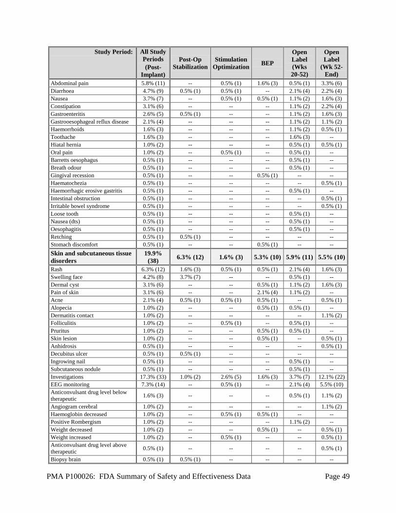
PMA P100026: FDA Summary of Safety and Effectiveness Data Page 49
Study Period: All Study Periods (Post-
Implant)
Post-Op Stabilization
Stimulation Optimization BEP
Open Label (Wks 20-52)
Open Label
(Wk 52- End)
Abdominal pain 5.8% (11) -- 0.5% (1) 1.6% (3) 0.5% (1) 3.3% (6) Diarrhoea 4.7% (9) 0.5% (1) 0.5% (1) -- 2.1% (4) 2.2% (4) Nausea 3.7% (7) -- 0.5% (1) 0.5% (1) 1.1% (2) 1.6% (3) Constipation 3.1% (6) -- -- -- 1.1% (2) 2.2% (4) Gastroenteritis 2.6% (5) 0.5% (1) -- -- 1.1% (2) 1.6% (3) Gastrooesophageal reflux disease 2.1% (4) -- -- -- 1.1% (2) 1.1% (2) Haemorrhoids 1.6% (3) -- -- -- 1.1% (2) 0.5% (1) Toothache 1.6% (3) -- -- -- 1.6% (3) -- Hiatal hernia 1.0% (2) -- -- -- 0.5% (1) 0.5% (1) Oral pain 1.0% (2) -- 0.5% (1) -- 0.5% (1) -- Barretts oesophagus 0.5% (1) -- -- -- 0.5% (1) -- Breath odour 0.5% (1) -- -- -- 0.5% (1) -- Gingival recession 0.5% (1) -- -- 0.5% (1) -- -- Haematochezia 0.5% (1) -- -- -- -- 0.5% (1) Haemorrhagic erosive gastritis 0.5% (1) -- -- -- 0.5% (1) -- Intestinal obstruction 0.5% (1) -- -- -- -- 0.5% (1) Irritable bowel syndrome 0.5% (1) -- -- -- -- 0.5% (1) Loose tooth 0.5% (1) -- -- -- 0.5% (1) -- Nausea (dts) 0.5% (1) -- -- -- 0.5% (1) -- Oesophagitis 0.5% (1) -- -- -- 0.5% (1) -- Retching 0.5% (1) 0.5% (1) -- -- -- -- Stomach discomfort 0.5% (1) -- -- 0.5% (1) -- -- Skin and subcutaneous tissue disorders
19.9% (38) 6.3% (12) 1.6% (3) 5.3% (10) 5.9% (11) 5.5% (10)
Rash 6.3% (12) 1.6% (3) 0.5% (1) 0.5% (1) 2.1% (4) 1.6% (3) Swelling face 4.2% (8) 3.7% (7) -- -- 0.5% (1) -- Dermal cyst 3.1% (6) -- -- 0.5% (1) 1.1% (2) 1.6% (3) Pain of skin 3.1% (6) -- -- 2.1% (4) 1.1% (2) -- Acne 2.1% (4) 0.5% (1) 0.5% (1) 0.5% (1) -- 0.5% (1) Alopecia 1.0% (2) -- -- 0.5% (1) 0.5% (1) -- Dermatitis contact 1.0% (2) -- -- -- -- 1.1% (2) Folliculitis 1.0% (2) -- 0.5% (1) -- 0.5% (1) -- Pruritus 1.0% (2) -- -- 0.5% (1) 0.5% (1) -- Skin lesion 1.0% (2) -- -- 0.5% (1) -- 0.5% (1) Anhidrosis 0.5% (1) -- -- -- -- 0.5% (1) Decubitus ulcer 0.5% (1) 0.5% (1) -- -- -- -- Ingrowing nail 0.5% (1) -- -- -- 0.5% (1) -- Subcutaneous nodule 0.5% (1) -- -- -- 0.5% (1) -- Investigations 17.3% (33) 1.0% (2) 2.6% (5) 1.6% (3) 3.7% (7) 12.1% (22) EEG monitoring 7.3% (14) -- 0.5% (1) -- 2.1% (4) 5.5% (10) Anticonvulsant drug level below therapeutic 1.6% (3) -- -- -- 0.5% (1) 1.1% (2)
Angiogram cerebral 1.0% (2) -- -- -- -- 1.1% (2) Haemoglobin decreased 1.0% (2) -- 0.5% (1) 0.5% (1) -- -- Positive Rombergism 1.0% (2) -- -- -- 1.1% (2) -- Weight decreased 1.0% (2) -- -- 0.5% (1) -- 0.5% (1) Weight increased 1.0% (2) -- 0.5% (1) -- -- 0.5% (1) Anticonvulsant drug level above therapeutic 0.5% (1) -- -- -- -- 0.5% (1)
Biopsy brain 0.5% (1) 0.5% (1) -- -- -- --
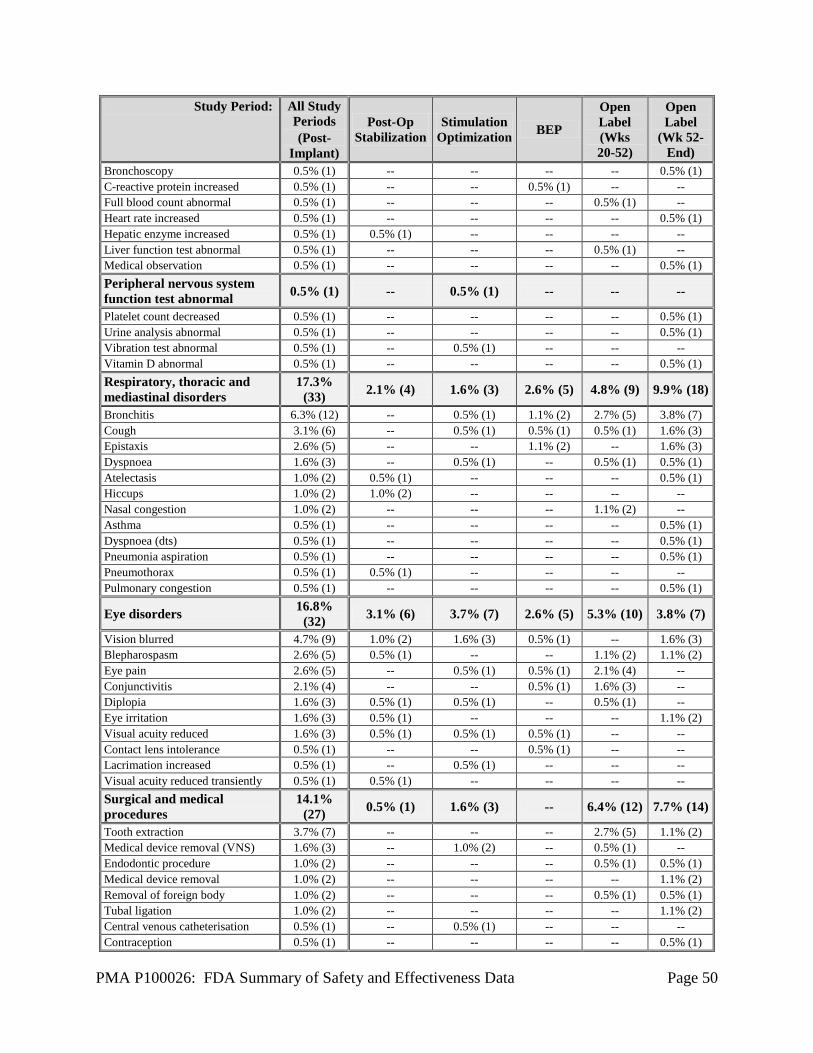
PMA P100026: FDA Summary of Safety and Effectiveness Data Page 50
Study Period: All Study Periods (Post-
Implant)
Post-Op Stabilization
Stimulation Optimization BEP
Open Label (Wks 20-52)
Open Label
(Wk 52- End)
Bronchoscopy 0.5% (1) -- -- -- -- 0.5% (1) C-reactive protein increased 0.5% (1) -- -- 0.5% (1) -- -- Full blood count abnormal 0.5% (1) -- -- -- 0.5% (1) -- Heart rate increased 0.5% (1) -- -- -- -- 0.5% (1) Hepatic enzyme increased 0.5% (1) 0.5% (1) -- -- -- -- Liver function test abnormal 0.5% (1) -- -- -- 0.5% (1) -- Medical observation 0.5% (1) -- -- -- -- 0.5% (1) Peripheral nervous system function test abnormal 0.5% (1) -- 0.5% (1) -- -- --
Platelet count decreased 0.5% (1) -- -- -- -- 0.5% (1) Urine analysis abnormal 0.5% (1) -- -- -- -- 0.5% (1) Vibration test abnormal 0.5% (1) -- 0.5% (1) -- -- -- Vitamin D abnormal 0.5% (1) -- -- -- -- 0.5% (1) Respiratory, thoracic and mediastinal disorders
17.3% (33) 2.1% (4) 1.6% (3) 2.6% (5) 4.8% (9) 9.9% (18)
Bronchitis 6.3% (12) -- 0.5% (1) 1.1% (2) 2.7% (5) 3.8% (7) Cough 3.1% (6) -- 0.5% (1) 0.5% (1) 0.5% (1) 1.6% (3) Epistaxis 2.6% (5) -- -- 1.1% (2) -- 1.6% (3) Dyspnoea 1.6% (3) -- 0.5% (1) -- 0.5% (1) 0.5% (1) Atelectasis 1.0% (2) 0.5% (1) -- -- -- 0.5% (1) Hiccups 1.0% (2) 1.0% (2) -- -- -- -- Nasal congestion 1.0% (2) -- -- -- 1.1% (2) -- Asthma 0.5% (1) -- -- -- -- 0.5% (1) Dyspnoea (dts) 0.5% (1) -- -- -- -- 0.5% (1) Pneumonia aspiration 0.5% (1) -- -- -- -- 0.5% (1) Pneumothorax 0.5% (1) 0.5% (1) -- -- -- -- Pulmonary congestion 0.5% (1) -- -- -- -- 0.5% (1)
Eye disorders 16.8% (32) 3.1% (6) 3.7% (7) 2.6% (5) 5.3% (10) 3.8% (7)
Vision blurred 4.7% (9) 1.0% (2) 1.6% (3) 0.5% (1) -- 1.6% (3) Blepharospasm 2.6% (5) 0.5% (1) -- -- 1.1% (2) 1.1% (2) Eye pain 2.6% (5) -- 0.5% (1) 0.5% (1) 2.1% (4) -- Conjunctivitis 2.1% (4) -- -- 0.5% (1) 1.6% (3) -- Diplopia 1.6% (3) 0.5% (1) 0.5% (1) -- 0.5% (1) -- Eye irritation 1.6% (3) 0.5% (1) -- -- -- 1.1% (2) Visual acuity reduced 1.6% (3) 0.5% (1) 0.5% (1) 0.5% (1) -- -- Contact lens intolerance 0.5% (1) -- -- 0.5% (1) -- -- Lacrimation increased 0.5% (1) -- 0.5% (1) -- -- -- Visual acuity reduced transiently 0.5% (1) 0.5% (1) -- -- -- -- Surgical and medical procedures
14.1% (27) 0.5% (1) 1.6% (3) -- 6.4% (12) 7.7% (14)
Tooth extraction 3.7% (7) -- -- -- 2.7% (5) 1.1% (2) Medical device removal (VNS) 1.6% (3) -- 1.0% (2) -- 0.5% (1) -- Endodontic procedure 1.0% (2) -- -- -- 0.5% (1) 0.5% (1) Medical device removal 1.0% (2) -- -- -- -- 1.1% (2) Removal of foreign body 1.0% (2) -- -- -- 0.5% (1) 0.5% (1) Tubal ligation 1.0% (2) -- -- -- -- 1.1% (2) Central venous catheterisation 0.5% (1) -- 0.5% (1) -- -- -- Contraception 0.5% (1) -- -- -- -- 0.5% (1)

PMA P100026: FDA Summary of Safety and Effectiveness Data Page 51
Study Period: All Study Periods (Post-
Implant)
Post-Op Stabilization
Stimulation Optimization BEP
Open Label (Wks 20-52)
Open Label
(Wk 52- End)
Dental implantation 0.5% (1) -- -- -- -- 0.5% (1) Lesion excision 0.5% (1) -- -- -- 0.5% (1) -- Removal of foreign body from external ear 0.5% (1) 0.5% (1) -- -- -- --
Scar excisions 0.5% (1) -- -- -- -- 0.5% (1) Shoulder operation 0.5% (1) -- -- -- 0.5% (1) -- Skin cosmetic procedure 0.5% (1) -- -- -- -- 0.5% (1) Skin neoplasm excision 0.5% (1) -- -- -- 0.5% (1) -- Stapes mobilisation 0.5% (1) -- -- -- -- 0.5% (1) Tendon transfer 0.5% (1) -- -- -- -- 0.5% (1) Vasectomy 0.5% (1) -- -- -- 0.5% (1) --
Ear and labyrinth disorders 11.5% (22) 4.7% (9) 2.1% (4) 0.5% (1) 3.7% (7) 2.2% (4)
Hypoacusis 2.6% (5) 2.1% (4) -- -- 0.5% (1) -- Tinnitus 2.6% (5) 1.0% (2) 0.5% (1) -- 2.1% (4) -- Vertigo 2.6% (5) -- 0.5% (1) -- 0.5% (1) 1.6% (3) Cerumen impaction 1.6% (3) 0.5% (1) -- 0.5% (1) 0.5% (1) -- Ear pain 1.6% (3) -- -- -- 0.5% (1) 1.1% (2) Ear discomfort 1.0% (2) 0.5% (1) 0.5% (1) -- -- -- Hearing impaired 0.5% (1) 0.5% (1) 0.5% (1) -- -- --
Immune system disorders 10.5% (20) 1.0% (2) 1.6% (3) 0.5% (1) 3.7% (7) 3.8% (7)
Seasonal allergy 5.8% (11) 0.5% (1) 1.0% (2) 0.5% (1) 1.6% (3) 2.2% (4) Drug hypersensitivity 4.7% (9) 0.5% (1) 0.5% (1) -- 2.7% (5) 1.1% (2) Erythema multiforme 0.5% (1) -- -- -- -- 0.5% (1) Renal and urinary disorders 8.9% (17) 1.6% (3) -- 2.6% (5) 2.1% (4) 3.8% (7) Nephrolithiasis 3.1% (6) 1.0% (2) -- 0.5% (1) 0.5% (1) 1.1% (2) Polyuria 2.6% (5) 0.5% (1) -- 0.5% (1) 0.5% (1) 1.1% (2) Dysuria 1.0% (2) -- -- 1.1% (2) -- -- Bladder spasm 0.5% (1) -- -- -- -- 0.5% (1) Micturition urgency 0.5% (1) -- -- -- -- 0.5% (1) Renal cyst 0.5% (1) -- -- -- 0.5% (1) -- Urinary incontinence 0.5% (1) -- -- -- 0.5% (1) -- Urinary retention 0.5% (1) -- -- -- -- 0.5% (1) Urine flow decreased 0.5% (1) -- -- 0.5% (1) -- -- Vascular disorders 7.3% (14) 1.0% (2) -- 0.5% (1) 2.1% (4) 3.8% (7) Hypertension 4.2% (8) -- -- 0.5% (1) 0.5% (1) 3.3% (6) Deep vein thrombosis 1.0% (2) 0.5% (1) -- -- 0.5% (1) -- Hypotension 1.0% (2) -- -- -- 0.5% (1) 0.5% (1) Orthostatic hypotension 0.5% (1) -- -- -- -- 0.5% (1) Procedural hypertension 0.5% (1) 0.5% (1) -- -- -- -- Raynauds phenomenon 0.5% (1) -- -- -- 0.5% (1) -- Metabolism and nutritional disorders 6.8% (13) 2.1% (4) 1.0% (2) -- 2.1% (4) 1.6% (3)
Decreased appetite 3.1% (6) 2.1% (4) 0.5% (1) -- 0.5% (1) -- Hyponatraemia 1.6% (3) -- -- -- 0.5% (1) 1.1% (2) Hypokalaemia 1.0% (2) -- 0.5% (1) -- -- 0.5% (1) Hypoglycaemia 0.5% (1) -- -- -- 0.5% (1) -- Vitamin B12 deficiency 0.5% (1) -- -- -- 0.5% (1) --
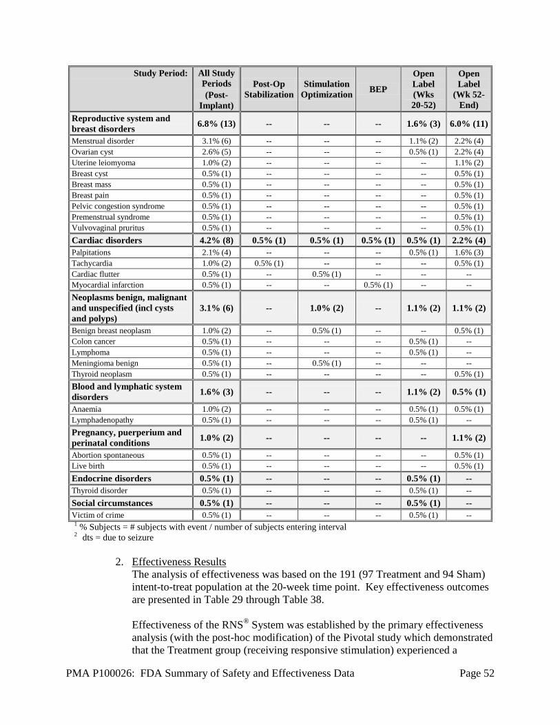
PMA P100026: FDA Summary of Safety and Effectiveness Data Page 52
Study Period: All Study Periods (Post-
Implant)
Post-Op Stabilization
Stimulation Optimization BEP
Open Label (Wks 20-52)
Open Label
(Wk 52- End)
Reproductive system and breast disorders 6.8% (13) -- -- -- 1.6% (3) 6.0% (11)
Menstrual disorder 3.1% (6) -- -- -- 1.1% (2) 2.2% (4) Ovarian cyst 2.6% (5) -- -- -- 0.5% (1) 2.2% (4) Uterine leiomyoma 1.0% (2) -- -- -- -- 1.1% (2) Breast cyst 0.5% (1) -- -- -- -- 0.5% (1) Breast mass 0.5% (1) -- -- -- -- 0.5% (1) Breast pain 0.5% (1) -- -- -- -- 0.5% (1) Pelvic congestion syndrome 0.5% (1) -- -- -- -- 0.5% (1) Premenstrual syndrome 0.5% (1) -- -- -- -- 0.5% (1) Vulvovaginal pruritus 0.5% (1) -- -- -- -- 0.5% (1) Cardiac disorders 4.2% (8) 0.5% (1) 0.5% (1) 0.5% (1) 0.5% (1) 2.2% (4) Palpitations 2.1% (4) -- -- -- 0.5% (1) 1.6% (3) Tachycardia 1.0% (2) 0.5% (1) -- -- -- 0.5% (1) Cardiac flutter 0.5% (1) -- 0.5% (1) -- -- -- Myocardial infarction 0.5% (1) -- -- 0.5% (1) -- -- Neoplasms benign, malignant and unspecified (incl cysts and polyps)
3.1% (6) -- 1.0% (2) -- 1.1% (2) 1.1% (2)
Benign breast neoplasm 1.0% (2) -- 0.5% (1) -- -- 0.5% (1) Colon cancer 0.5% (1) -- -- -- 0.5% (1) -- Lymphoma 0.5% (1) -- -- -- 0.5% (1) -- Meningioma benign 0.5% (1) -- 0.5% (1) -- -- -- Thyroid neoplasm 0.5% (1) -- -- -- -- 0.5% (1) Blood and lymphatic system disorders 1.6% (3) -- -- -- 1.1% (2) 0.5% (1)
Anaemia 1.0% (2) -- -- -- 0.5% (1) 0.5% (1) Lymphadenopathy 0.5% (1) -- -- -- 0.5% (1) -- Pregnancy, puerperium and perinatal conditions 1.0% (2) -- -- -- -- 1.1% (2)
Abortion spontaneous 0.5% (1) -- -- -- -- 0.5% (1) Live birth 0.5% (1) -- -- -- -- 0.5% (1) Endocrine disorders 0.5% (1) -- -- -- 0.5% (1) -- Thyroid disorder 0.5% (1) -- -- -- 0.5% (1) -- Social circumstances 0.5% (1) -- -- -- 0.5% (1) -- Victim of crime 0.5% (1) -- -- -- 0.5% (1) -- 1 % Subjects = # subjects with event / number of subjects entering interval 2 dts = due to seizure
2. Effectiveness Results The analysis of effectiveness was based on the 191 (97 Treatment and 94 Sham) intent-to-treat population at the 20-week time point. Key effectiveness outcomes are presented in Table 29 through Table 38.
Effectiveness of the RNS® System was established by the primary effectiveness analysis (with the post-hoc modification) of the Pivotal study which demonstrated that the Treatment group (receiving responsive stimulation) experienced a

PMA P100026: FDA Summary of Safety and Effectiveness Data Page 53
statistically significant greater reduction in total disabling seizures compared to the Sham group (implanted, but not receiving stimulation) during the BEP compared to the Pre-Implant Period of the investigation. None of the differences between Treatment and Sham in the secondary endpoints were statistically significant, however, only the responder rate analysis was powered to show a difference. During the Open Label Period of the Pivotal study, there was a reduction in the frequency of disabling seizures. Another measure of effectiveness was quality of life. A clinically important improvement is defined as a 5-point improvement. There was no difference between the Treatment and Sham group subjects in the percent of subjects who achieved a clinically important improvement in QOL (36.6% and 39.1% respectively) at the end of the BEP. At 1 and 2 years post-implant, 38% and 44% of subjects (respectively) experienced a significant clinical improvement on the QOLIE (an epilepsy-specific quality of life assessment).
Primary Effectiveness Endpoint
Observed Data A total of 97 and 94 patients entered the Pre-Implant Period in the Treatment and Sham groups, respectively. The mean pre-implant seizure frequency per month in the Treatment group was 33.5 (with a range of 3 – 295) and 34.9 (with a range of 3 – 338) in the Sham group. Over the entire Blinded Evaluation Period, the mean seizure frequency per month in the Treatment group was 22.4 (with a range of 0 – 227) and was 29.9 (with a range of 0 – 447) in the Sham group. Table 29 presents the mean, median and range of seizure frequencies and the mean percent change and median percent change, for the Treatment and Sham groups for the Pre-Implant and BEP by individual month and overall.
Table 29: Mean, median and range of seizure frequencies and the mean percent change and median percent change, for the Treatment and Sham groups for the Pre-Implant and BEP by
individual month and overall
Treatment Sham
N Mean Median (range)
Change
N Mean Median (range)
Change
Mean %
Median %
Mean %
Median %
Pre-
Impl
ant
Mon
ths
0-1 97 34.5 9.3 (2.0 - 305.0) 94 28.7 10.0
(1.0 - 283.0)
1-2 97 34.3 9.0 (2.0 - 294.0) 94 34.5 12.0
(2.0 - 342.0)
2-3 97 31.7 9.0 (0.0 - 350.0) 94 41.5 11.5
(2.0 - 634.0)
Pre-Implant Period 97 33.5 8.7
(3.0 - 294.7) 94 34.9 11.6 (3.0 - 338.0)

PMA P100026: FDA Summary of Safety and Effectiveness Data Page 54
Treatment Sham
N Mean Median (range)
Change
N Mean Median (range)
Change
Mean %
Median %
Mean %
Median %
Post
-Op
Mon
ths 0-1 96 23.8 6.0
(0.0-258.1) -24.7% -33.3% 94 24.2 7.0 (0.0-286.0) -19.8% -30.2%
1-2 96 25.0 6.4 (0,0-247.0) -25.3% -28.4% 93 27.4 9.0
(0.0-323.0) -13.5% -21.9%
Blin
ded
E
valu
atio
n Pe
riod
M
onth
s
2-3 96 22.9 6.5 (0.0 - 226.0) -19.9% -27.2% 93 27.1 8.3
(0.0 - 369.4) -19.5% -31.9%
3-4 95 22.8 6.0 (0.0 - 266.0) -30.8% -36.4% 90 28.9 8.3
(0.0 - 336.0) -14.1% -25.0%
4-5 95 21.4 6.0 (0.0 - 226.0) -28.0% -34.0% 91 35.4 7.0
(0.0 - 799.0) -13.7% -18.9%
Blinded Evaluation Period
96 22.4 5.8 (0.0 - 226.8) -24.1% -28.0% 93 29.8 7.6
(0.3 - 446.6) -17.3% -19.2%
Figure 4 depicts the mean seizure frequency per month for Treatment and Sham groups. Following implantation of the RNS® Neurostimulator and Leads and prior to enabling stimulation in either the Treatment or Sham group, both groups experienced a mean percent reduction in the observed number of seizures (25% Treatment and 20% Sham) and a median percent reduction in the observed number of seizures (33% Treatment and 30% Sham). Whether this is an effect of the surgical procedure and / or anesthesia, an effect of Lead implantation, regression to the mean or placebo effect is not known.
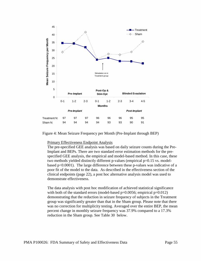
PMA P100026: FDA Summary of Safety and Effectiveness Data Page 55
Treatment N: 97 97 97 96 96 96 95 95Sham N: 94 94 94 94 93 93 90 91
0-1 1-2 2-3 0-1 1-2 2-3 3-4 4-5
0
5
10
15
20
25
30
35
40
45
0-1 1-2 2-3 0-1 1-2 2-3 3-4 4-5
Months
Mea
n Se
izur
e Fr
eque
ncy
per M
onth
TreatmentSham
Pre-Implant Blinded EvaulationPost-Op &Stim Opt
Pre-Implant Post-Implant
Stimulation on in Treatment group
Figure 4: Mean Seizure Frequency per Month (Pre-Implant through BEP)
Primary Effectiveness Endpoint Analysis The pre-specified GEE analysis was based on daily seizure counts during the Pre-Implant and BEPs. There are two standard error estimation methods for the pre-specified GEE analysis, the empirical and model-based method. In this case, these two methods yielded distinctly different p-values (empirical p=0.15 vs. model-based p<0.0001). The large difference between these p-values was indicative of a poor fit of the model to the data. As described in the effectiveness section of the clinical endpoints (page 22), a post hoc alternative analysis model was used to demonstrate effectiveness. The data analysis with post hoc modification of achieved statistical significance with both of the standard errors (model-based p=0.0056; empirical p=0.012) demonstrating that the reduction in seizure frequency of subjects in the Treatment group was significantly greater than that in the Sham group. Please note that there was no correction for multiplicity testing. Averaged over the entire BEP, the mean percent change in monthly seizure frequency was 37.9% compared to a 17.3% reduction in the Sham group. See Table 30 below.

PMA P100026: FDA Summary of Safety and Effectiveness Data Page 56
Table 30: Estimates of seizure frequency percent change from the modified GEE model over the entire Blinded Evaluation Period
Parameter Estimate
(log scale)1
Ratio of Seizure Frequency (BEP vs. Pre-Implant Period)2
Percent Change from Pre-Implant Period3
[95% confidence interval]
Treatment -0.4771 0.621 -37.9% [-46.7%, -27.7%]
Sham -0.1898 0.827 -17.3% [-29.9%, -2.3%]
1 The parameter estimate (β) for the Sham group is the coefficient for Time (β1). The parameter estimate for the Treatment group is the coefficient for Time + the coefficient for Group-by-Time (β1 + β2).
2 The ratio of seizure frequency (natural scale) is given by e β. 3 The percent change is given by (eβ -1)*100%.
To put the model-predicted mean percent change from baseline into perspective, the model predicts a subject in the Treatment group with 30 seizures at baseline would experience a reduction of 11 seizures per month in the BEP. Similarly, the model predicts that a subject in the Sham group with 30 seizures at baseline would experience a reduction of 5 seizures per month in the BEP. The difference between Treatment and Sham groups as predicted by the model is 6 seizures per month in the BEP. The Forest Plot in Figure 5 shows models that explore combinations of the 3 modifications: distribution, covariate inclusion, and time. The pre-specified model is the bottom-most model while the modified post-hoc model is the top-most model in Figure 5. Ninety-five percent (95%) confidence intervals are presented as horizontal lines for model-based (solid) and empirical (dashed). The scale parameter is denoted as Φ. A model that fits the data well has similar model-based and empirical confidence intervals and has a scale parameter that is close to 1, which is not the case for the pre-specified or many of the additional exploratory post-hoc models. The relative risk reduction corresponds to the additional reduction in seizure frequency attributable to active stimulation relative to Sham stimulation; a value of 1 would suggest there is no additional benefit from active stimulation, while a value less than 1 or greater than 1 would indicate that active stimulation reduces or increases seizures relative to Sham stimulation.

PMA P100026: FDA Summary of Safety and Effectiveness Data Page 57
Figure 5: “Forest plot” Comparison of Alternative GEE Models
The post-hoc negative binomial model with monthly seizure counts (employed by the sponsor to support a reasonable assurance of effectiveness) was favored by the advisory panel's statisticians and the FDA statistical reviewer; it yielded consistent results with model-based and empirically based distributions. Alternative post hoc models were less appropriate because they used either an over-dispersed Poisson distribution, more varied daily seizure counts, and/or did not use the prespecified randomization covariates.
Individual Subject Success Seventy-six percent (76%) of subjects in the Treatment group and 70% in the Sham group reported a decrease in seizures during the BEP. Figure 6 below depicts the distribution of subject results.
Figure 6: Percent change* in seizure frequency in Blinded Evaluation Period
compared to pre-implant baseline (Treatment and Sham) * Two patients had an increase > 100%; Sham (n=1, 115%); Treatment (n=1, 281%)

PMA P100026: FDA Summary of Safety and Effectiveness Data Page 58
Secondary Effectiveness Endpoints The secondary effectiveness analyses were intended to support the primary effectiveness endpoint. Pre-specified secondary effectiveness endpoints were the responder rate, change in mean seizure frequency, proportion of seizure-free days, and self-reported seizure severity according to the Liverpool Seizure Severity Scale inventory. The results of the four secondary effectiveness analyses are presented in Table 31. None of the secondary endpoints achieved statistical significance. Note that with the exception of the responder rate comparison, which was used to determine the trial’s sample size, the protocol did not evaluate the trial’s power to detect a statistically significant difference for the secondary outcomes.
Table 31: Pivotal Study – Secondary Effectiveness Endpoints Averaged Monthly over the Entire
BEP Effectiveness Endpoint Treatment Sham p-value1
50% Responder Rate2 29% 27% 0.7273 Change in Mean Seizure Frequency -11.4 -5.3 0.2384 % Change in Days with Seizures -19% -18% 0.95 Liverpool Seizure Severity Change -4.7 -5.9 0.5746
1 p-values represent across group evaluations. 2 used to power sample size. 3 z-statistic 4 two-sample t-test. 5 paired t-test. 6 two-sample t-test Open Label Period Subjects entered the open label phase of the study at 5 months (20 weeks) post-implant. At the 20-Week visit, subjects in the Sham group were able to receive responsive stimulation for the first time. When subjects in the Sham group first received responsive stimulation in the Open Label Period, there was an immediate reduction in seizure frequency. The mean change in seizures in the Sham group compared to the pre-implant baseline and compared to the Blinded Evaluation Period is presented in Table 32.
Table 32: Mean Change in Seizure Frequency in Sham Group
Time Period
Seizure Frequency1 (mean ± SD, seizures / month)
Open Label to Pre-Implant Comparison
(N = 91)
Open Label to Blinded Evaluation Comparison
(N=91)
Pre-implant 35.725 ± 68.037 -- Blinded Evaluation (Months 2-5) -- 30.442 ± 67.082 Open Label (Months 6-9) 27.964 ± 2.014 27.964 ± 62.014 Change -7.763 ± 35.498 -2.478 ± 27.33
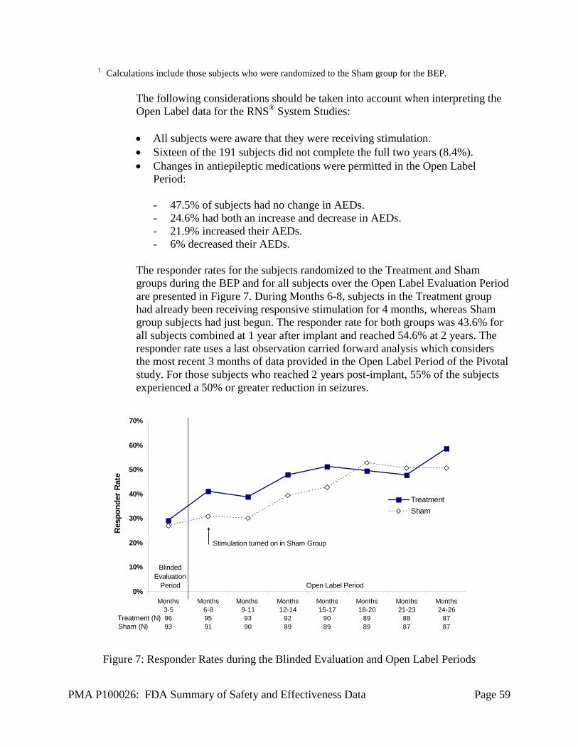
PMA P100026: FDA Summary of Safety and Effectiveness Data Page 59
1 Calculations include those subjects who were randomized to the Sham group for the BEP.
The following considerations should be taken into account when interpreting the Open Label data for the RNS® System Studies: • All subjects were aware that they were receiving stimulation. • Sixteen of the 191 subjects did not complete the full two years (8.4%). • Changes in antiepileptic medications were permitted in the Open Label
Period: - 47.5% of subjects had no change in AEDs. - 24.6% had both an increase and decrease in AEDs. - 21.9% increased their AEDs. - 6% decreased their AEDs.
The responder rates for the subjects randomized to the Treatment and Sham groups during the BEP and for all subjects over the Open Label Evaluation Period are presented in Figure 7. During Months 6-8, subjects in the Treatment group had already been receiving responsive stimulation for 4 months, whereas Sham group subjects had just begun. The responder rate for both groups was 43.6% for all subjects combined at 1 year after implant and reached 54.6% at 2 years. The responder rate uses a last observation carried forward analysis which considers the most recent 3 months of data provided in the Open Label Period of the Pivotal study. For those subjects who reached 2 years post-implant, 55% of the subjects experienced a 50% or greater reduction in seizures.
0%
10%
20%
30%
40%
50%
60%
70%
Months3-59693
Months6-89591
Months 9-119390
Months12-14
9289
Months15-17
9089
Months18-20
8989
Months21-23
8887
Months24-26
8787
Res
pond
er R
ate
TreatmentSham
Stimulation turned on in Sham Group
Treatment (N)Sham (N)
Blinded Evaluation
Period Open Label Period
Figure 7: Responder Rates during the Blinded Evaluation and Open Label Periods
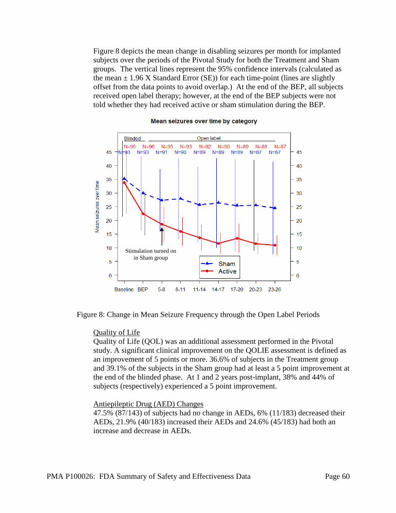
PMA P100026: FDA Summary of Safety and Effectiveness Data Page 60
Figure 8 depicts the mean change in disabling seizures per month for implanted subjects over the periods of the Pivotal Study for both the Treatment and Sham groups. The vertical lines represent the 95% confidence intervals (calculated as the mean ± 1.96 X Standard Error (SE)) for each time-point (lines are slightly offset from the data points to avoid overlap.) At the end of the BEP, all subjects received open label therapy; however, at the end of the BEP subjects were not told whether they had received active or sham stimulation during the BEP.
Figure 8: Change in Mean Seizure Frequency through the Open Label Periods
Quality of Life Quality of Life (QOL) was an additional assessment performed in the Pivotal study. A significant clinical improvement on the QOLIE assessment is defined as an improvement of 5 points or more. 36.6% of subjects in the Treatment group and 39.1% of the subjects in the Sham group had at least a 5 point improvement at the end of the blinded phase. At 1 and 2 years post-implant, 38% and 44% of subjects (respectively) experienced a 5 point improvement. Antiepileptic Drug (AED) Changes 47.5% (87/143) of subjects had no change in AEDs, 6% (11/183) decreased their AEDs, 21.9% (40/183) increased their AEDs and 24.6% (45/183) had both an increase and decrease in AEDs.
Stimulation turned on in Sham group
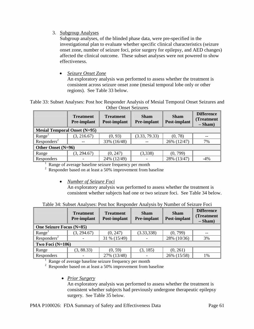
PMA P100026: FDA Summary of Safety and Effectiveness Data Page 61
3. Subgroup Analyses Subgroup analyses, of the blinded phase data, were pre-specified in the investigational plan to evaluate whether specific clinical characteristics (seizure onset zone, number of seizure foci, prior surgery for epilepsy, and AED changes) affected the clinical outcome. These subset analyses were not powered to show effectiveness.
• Seizure Onset Zone
An exploratory analysis was performed to assess whether the treatment is consistent across seizure onset zone (mesial temporal lobe only or other regions). See Table 33 below.
Table 33: Subset Analyses: Post hoc Responder Analysis of Mesial Temporal Onset Seizures and
Other Onset Seizures Treatment
Pre-implant Treatment
Post-implant Sham
Pre-implant Sham
Post-implant
Difference (Treatment
– Sham) Mesial Temporal Onset (N=95) Range1 (3, 216.67) (0, 93) (3.33, 79.33) (0, 78) -- Responders2 -- 33% (16/48) -- 26% (12/47) 7% Other Onset (N=96) Range (3, 294.67) (0, 247) (3,338) (0, 799) Responders - 24% (12/49) - 28% (13/47) -4%
1 Range of average baseline seizure frequency per month 2 Responder based on at least a 50% improvement from baseline
• Number of Seizure Foci
An exploratory analysis was performed to assess whether the treatment is consistent whether subjects had one or two seizure foci. See Table 34 below.
Table 34: Subset Analyses: Post hoc Responder Analysis by Number of Seizure Foci
Treatment Pre-implant
Treatment Post-implant
Sham Pre-implant
Sham Post-implant
Difference (Treatment
– Sham) One Seizure Focus (N=85) Range1 (3, 294.67) (0, 247) (3.33,338) (0, 799) -- Responders2 - 31 % (15/49) - 28% (10/36) 3% Two Foci (N=106) Range (3, 88.33) (0, 59) (3, 185) (0, 261) Responders - 27% (13/48) - 26% (15/58) 1%
1 Range of average baseline seizure frequency per month 2 Responder based on at least a 50% improvement from baseline
• Prior Surgery
An exploratory analysis was performed to assess whether the treatment is consistent whether subjects had previously undergone therapeutic epilepsy surgery. See Table 35 below.

PMA P100026: FDA Summary of Safety and Effectiveness Data Page 62
Table 35: Subset Analyses: Post hoc Responder Analysis by Resection and No Prior Resection Treatment
Pre-implant Treatment
Post-implant Sham
Pre-implant Sham
Post-implant
Difference (Treatment
– Sham) Prior Resection (N= 62) Range (3, 294.67) (0, 226) (3, 338) (0, 799) -- Responders - 24% (8/34) - 29% (8/28) -5% No Prior Resection (N= 129) Range (3, 266) (0, 247) (3.33, 207) (0, 261) Responders - 32% (20/63) - 26% (17/66) 6%
1 Range of average baseline seizure frequency per month 2 Responder based on at least a 50% improvement from baseline
• Changes in Antiepileptic Drugs
There were only 6 subjects who had changes in their AED treatment regimen, 3 subjects had changes in the Pre-Implant Period and 3 subjects had changes in AEDs in the BEP. The 6 subjects who had AED changes were excluded from analyses of the Per-Protocol Population. Results of the Per-Protocol analysis of the post hoc GEE model indicate that the Treatment Effect remains significant with the subjects who had significant protocol deviations (including AED changes) removed from the analysis (p = 0.027).
4. Stimulation Parameters and Detection Algorithm
The initial recommended stimulation settings were a frequency of 200 Hz, a pulse duration of 160 µs, and a 100 ms burst duration. Subsequent changes in stimulation parameters were not specified by the protocol. Stimulation settings could be modified by the investigator based on subject status, subject perception of stimulation (for the Treatment group) and the presence of afterdischarges. With the exception of 1 Hz frequency and 1000 μs pulse duration, subjects used the full range of available stimulation parameters. The range of stimulation parameters that were used in the Pivotal study are provided in Table 36.
During the Pivotal study, 76% of subjects’ Neurostimulators were initially programmed to the default stimulation settings (frequency = 200 Hz, pulse width = 160 µs, burst duration = 100 ms). The physician varied current amplitude as necessary. Stimulation programming was changed in all but 4 subjects over the 2 post-implant-years. Burst duration was adjusted in 55%, frequency in 52%, and pulse width in 21% of subjects.
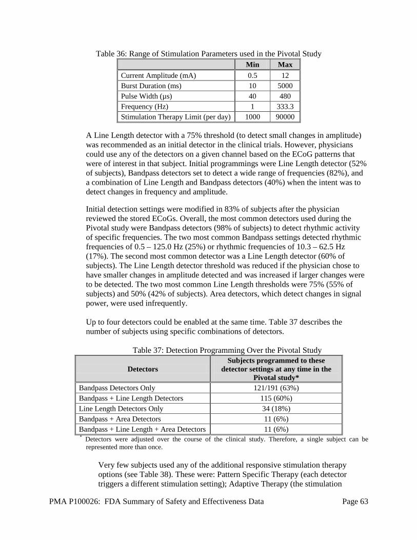
PMA P100026: FDA Summary of Safety and Effectiveness Data Page 63
Table 36: Range of Stimulation Parameters used in the Pivotal Study Min Max
Current Amplitude (mA) 0.5 12 Burst Duration (ms) 10 5000 Pulse Width (µs) 40 480 Frequency (Hz) 1 333.3 Stimulation Therapy Limit (per day) 1000 90000
A Line Length detector with a 75% threshold (to detect small changes in amplitude) was recommended as an initial detector in the clinical trials. However, physicians could use any of the detectors on a given channel based on the ECoG patterns that were of interest in that subject. Initial programmings were Line Length detector (52% of subjects), Bandpass detectors set to detect a wide range of frequencies (82%), and a combination of Line Length and Bandpass detectors (40%) when the intent was to detect changes in frequency and amplitude.
Initial detection settings were modified in 83% of subjects after the physician reviewed the stored ECoGs. Overall, the most common detectors used during the Pivotal study were Bandpass detectors (98% of subjects) to detect rhythmic activity of specific frequencies. The two most common Bandpass settings detected rhythmic frequencies of 0.5 – 125.0 Hz (25%) or rhythmic frequencies of 10.3 – 62.5 Hz (17%). The second most common detector was a Line Length detector (60% of subjects). The Line Length detector threshold was reduced if the physician chose to have smaller changes in amplitude detected and was increased if larger changes were to be detected. The two most common Line Length thresholds were 75% (55% of subjects) and 50% (42% of subjects). Area detectors, which detect changes in signal power, were used infrequently. Up to four detectors could be enabled at the same time. Table 37 describes the number of subjects using specific combinations of detectors.
Table 37: Detection Programming Over the Pivotal Study
Detectors Subjects programmed to these
detector settings at any time in the Pivotal study*
Bandpass Detectors Only 121/191 (63%) Bandpass + Line Length Detectors 115 (60%) Line Length Detectors Only 34 (18%) Bandpass + Area Detectors 11 (6%) Bandpass + Line Length + Area Detectors 11 (6%) * Detectors were adjusted over the course of the clinical study. Therefore, a single subject can be
represented more than once. Very few subjects used any of the additional responsive stimulation therapy options (see Table 38). These were: Pattern Specific Therapy (each detector triggers a different stimulation setting); Adaptive Therapy (the stimulation

PMA P100026: FDA Summary of Safety and Effectiveness Data Page 64
frequency adjusts with the ECoG frequency); Synchronized Stimulation (stimulation is delivered into a specific part of the ECoG waveform); and Post Episode Monitoring/Post Episode Monitoring Interval (responsive therapies are disabled for a specified period of time after detecting the end of the episode). Table 38 provides the number and percentage of subjects who were treated with any of the additional stimulation therapy options.
Table 38: Additional Responsive Stimulation Therapy Options
Number and (%) of Subjects Programmed
Pattern Specific Therapy 29 (15%) Adaptive Therapy 4 (2%) Synchronized Stimulation 3 (2%) Post-Episode Monitoring Interval 4 (2%)
The investigator determined which electrocorticographic records would be stored in the Neurostimulator by selecting one of four possible storage categories. These included time of day, duration of detection, sustained high amplitude and when the magnet was swiped over the Neurostimulator by the patient. Analysis of the electrocorticograms and the method by which they were stored was not an intent of the study.
E. Financial Disclosure
The Financial Disclosure by Clinical Investigators regulation (21 CFR 54) requires applicants who submit a marketing application to include certain information concerning the compensation to, and financial interests and arrangement of, any clinical investigator conducting clinical studies covered by the regulation. The pivotal clinical study included 219 investigators. None of the clinical investigators had disclosable financial interests/arrangements as defined in sections 54.2(a), (b), (c), and (f). The information provided does not raise any questions about the reliability of the data. The Feasibility study included 82 of investigators and the LTT included 135 investigators of which one of investigator had disclosable financial interests/arrangements as defined in 21 CFR 54.2(a), (b), (c) and (f) and described below: • Compensation to the investigator for conducting the study where the value could
be influenced by the outcome of the study: none of investigators • Significant payment of other sorts: one of the investigators [NeuroPace provided
financial support for research conducted at the investigator’s institution] • Proprietary interest in the product tested held by the investigator: none of
investigators • Significant equity interest held by investigator in sponsor of covered study: none
of investigators

PMA P100026: FDA Summary of Safety and Effectiveness Data Page 65
The applicant has adequately disclosed the financial interest/arrangements with clinical investigators. Two subjects that enrolled and were implanted in the Feasibility study at the institution were transitioned to the LTT investigation. The investigator’s role as a clinical investigator in the LTT investigation was as the neurosurgeon responsible for the surgical procedures involving the RNS® System. The investigator’s involvement did not extend past that delegated responsibility. The investigator had no involvement in the collection of any data relating to the primary or secondary objectives of the LTT investigation. Analyses were conducted by FDA to determine whether the financial interests/arrangements had any impact on the clinical study outcome. The information provided does not raise any questions about the reliability of the data.
XI. PANEL MEETING RECOMMENDATION AND FDA’S POST-PANEL ACTION
A. Panel Meeting Recommendation
At an advisory meeting held on February 22, 2013, the Neurological Devices Panel voted 13-0 that there is reasonable assurance the device is safe, 12-0 with 1 abstention that there is reasonable assurance that the device is effective, and 11-0 with 2 abstentions that the benefits of the device do outweigh the risks in patients who meet the criteria specified in the proposed indication. The “Summary of the Neurological Devices Panel Meeting, February 22, 2013” can be found at: www.fda.gov/downloads/AdvisoryCommittees/CommitteesMeetingMaterials/MedicalDevices/MedicalDevicesAdvisoryCommittee/NeurologicalDevicesPanel/UCM341125.pdf. No specific Conditions of Approval were recommended by the Panel but the Panel did suggest the following in their deliberations: • That there should be a comprehensive training program for device use. • That there should be some restrictions on which doctors or centers utilize the
device.
• A Post Approval Study should be performed to evaluate long-term safety of the device.
B. FDA’s Post-Panel Action
CDRH agreed with the Panel’s suggestion that there is a comprehensive training program for device use and NeuroPace, Inc. will require training by NeuroPace, Inc. prior to physician use. CDRH agreed with the Panel’s suggestions that there are some restrictions on which doctors or centers can utilize the device and NeuroPace, Inc. has agreed to limit use of the RNS® System to the following:
PMA P100026: FDA Summary of Safety and Effectiveness Data Page 66
• Neurosurgeons with adequate experience in the implantation of subdural and stereotactic implantation of intraparenchymal electrodes and in the surgical treatment of intractable epilepsy.
• Neurologists or neurosurgeons with adequate experience in the management of intractable epilepsy and in the localization of epileptic foci, including the use of scalp and intracranial electrodes.
• Neurologists and neurosurgeons using the RNS® System must have completed the NeuroPace® RNS® System training program.
• To qualify to manage patients with the RNS® System, physicians must demonstrate specific expertise related to epilepsy, video-EEG monitoring, interpretation of ECoGs, the pharmacology of AEDs and selection of patients for epilepsy surgery.
• Implantation of the RNS® System should be performed only by physicians with expertise in neurosurgical techniques at centers capable of providing comprehensive epilepsy care, i.e. “Comprehensive Epilepsy Centers”. These centers should have the expertise to provide diagnostic services that include video-EEG monitoring with scalp and intracranial electrodes and neuroimaging, and are experts in the treatment of epilepsy with AEDs, epilepsy surgery and devices.
CDRH agreed with the Panel’s suggestion that Post Approval Study (PAS) should be performed to evaluate long-term safety of the device and the NeuroPace, Inc. has agreed to perform a PAS study.
XII. CONCLUSIONS DRAWN FROM PRECLINICAL AND CLINICAL STUDIES
A. Effectiveness Conclusions
Statistically significant seizure reduction was achieved in the Treatment group as compared to the Sham group. As estimated using the GEE model with post hoc modifications, over the entire BEP, the Treatment group experienced a monthly reduction in seizure frequency averaging 37.9% compared to a 17.3% reduction in the Sham group; this difference is statistically significant (p = 0.012). There was no significant difference between the Treatment and the Sham groups on the secondary endpoints, i.e. responder rate, change in mean seizure frequency, proportion of seizure-free days or seizure severity as assessed using the Liverpool Seizure Severity Scale. For those subjects who reached 2 years post-implant, 55% of the subjects experienced a 50% or greater reduction in seizures. However, without a comparative group and with changes in AEDs the open label data are inconclusive evidence for continued effectiveness. During the Open Label Period, 7.6% of subjects (14/183) decreased their AEDs, 21.9% of subjects (40/183) increased their AEDs and 16.4% of subjects (30/183) had both an increase and decrease in AEDs. Without a comparative group and with changes in AEDs the open label data are inconclusive evidence for continued effectiveness.
PMA P100026: FDA Summary of Safety and Effectiveness Data Page 67
B. Safety Conclusions The risks of the device are based on nonclinical laboratory and animal studies as well as data collected in a clinical studies conducted to support PMA approval as described above. The primary safety endpoint was met. The SAEs that occurred in the Feasibility and Pivotal studies as compared with historical controls that included procedures related to epilepsy surgery (implantation of intracranial electrodes for purposes of localizing the seizure focus and the epilepsy surgery procedure) and implantation of DBS systems for treatment of movement disorders showed that the upper limit of the one-sided 95% CI for the RNS® System is less than that of the comparator. Additionally, during the evaluation periods, there was no difference between the Treatment and Sham groups in the overall percentage of subjects experiencing a serious adverse event, or any specific type of serious adverse event. The total number of subjects that experienced any serious or non-serious adverse event during the Blinded Evaluation Period were 90/97 (92.8%) for the Treatment group and 88/94 (93.6%) for the Sham group. There were 55 serious adverse events (38 subjects) from the time of implant through the end of the BEP (20 weeks post-implantation). Over the entire RNS® Studies experience in 256 subjects with over 903 patient-years of implant experience and 819 patient-years of responsive stimulation(as of May 12, 2011), there were no serious unanticipated device-related adverse events. The device-related serious adverse events reported with the greatest frequency were implant site infection (5.9%; 3.1% requiring explant), premature battery depletion (4.3%), and medical device removal (3.5%). Over the combined RNS System clinical studies, as of October 24, 2012, there were 11 deaths. Two deaths were by suicide, one was due to status epilepticus, one was due to lymphoma, and seven were attributed by an independent SUDEP adjudication committee to be possible, probable, or definite SUDEP.
C. Benefit-Risk Conclusions
The probable benefits of the device are based on data collected in a clinical study conducted to support PMA approval as described above. The Pivotal study data showed that the RNS® System reduces the frequency of disabling seizures in the studied population. An analysis of safety data combined from the Feasibility, Pivotal and LTT Clinical Investigations suggests that the safety of the RNS® System is equivalent to comparable procedures: implantation of intracranial electrodes for localization of the seizure focus, epilepsy surgery and DBS for movement disorders. In conclusion, given the available information above, the data support that for the following indications for use the probable benefits outweigh the probable risks:
The RNS® System is an adjunctive therapy in reducing the frequency of seizures in individuals 18 years of age or older with partial onset seizures who have undergone diagnostic testing that localized no more than 2 epileptogenic foci, are refractory to two or more antiepileptic medications, and currently have frequent and disabling seizures (motor partial seizures, complex partial seizures and/ or secondarily generalized seizures). The RNS® System has demonstrated safety and effectiveness
PMA P100026: FDA Summary of Safety and Effectiveness Data Page 68
in patients who average 3 or more disabling seizures per month over the three most recent months (with no month with fewer than two seizures), and has not been evaluated in patients with less frequent seizures.
D. Overall Conclusions
The data in this application as described in this summary support the reasonable assurance of safety and effectiveness of this device when used in accordance with the indications for use.
XIII. CDRH DECISION
CDRH issued an approval order on November 14 2013. The final conditions of approval cited in the approval order are described below. The following five post-approval studies (PAS) will be performed:
1. Continued Follow-up PAS 1 - Long Term Treatment (LTT) Study: This study must be
conducted as per protocol (IDE G030126). The objectives of this study are to describe the long-term safety and effectiveness associated with use of the NeuroPace device through 7 years.
Safety will be evaluated using adverse event rates (serious and non-serious) and sudden unexplained death in epilepsy (SUDEP) rate through 7 years. Effectiveness will be measured as the average decrease across 7 years in disabling seizure frequency from pre-implant baseline, responder rates, and quality of life. Responder rates will be the proportion of subjects with a sustained ≥50% reduction in total disabling seizures compared to pre-implant baseline. Quality of life will be measured using QOLIE-89. The surviving patients in the premarket Long-term Treatment Investigation Study will be followed annually up to 7 years.
2. New Enrollment PAS 2 – All Comers: The study will be a prospective observational
study of newly-enrolled patients treated with the RNS System.
The primary safety objective is to characterize the annual serious adverse event (SAE) rate over 5 years. SAEs include adverse events related to intra-cranial hemorrhages, adverse events related to seizure-associated injuries requiring medical attention, and all-cause mortality (including adjudicated SUDEP and suicidality). The secondary safety objective is to demonstrate that there is not a worsening in seizures over time in patients treated with the RNS System beginning at 6 to 12 months post-implant and extending to 3 years. This will be to demonstrate that there is not a 20% increase in disabling seizures at 3 years compared to 6 to 12 months post-implant. The secondary safety endpoints will include seizure frequency, new seizure types (disabling and non-disabling), and seizure foci and lead location.

PMA P100026: FDA Summary of Safety and Effectiveness Data Page 69
The primary effectiveness endpoint will estimate the median percent reduction in seizures from baseline (3 months pre-implant) to 3 years in the PAS, and evaluate if this reduction is comparable to the median percent reduction in seizures from baseline to 3 years in the controlled clinical study (51.3%). The secondary objective will be to characterize observed battery longevity overall and in relation to total stimulation time and stimulation programming at 5 years. Longevity will be defined as the time to replacement and/or end of service, whichever is first. Additional objectives will be to describe the demographic and clinical characteristics of treated patients, additional procedures including explants and re-implants (reasons, associated complications, and resolution), responder rate, antiepileptic drug (AED) use, discontinuations, and autopsy data. Patients will be followed out to 5 years post implant. Based on a linear regression t-test of the slope with a one-sided alpha of 0.05 (standard deviation=0.25), and 20% overall attrition, 300 enrolled patients will provide over 80% power for the ability to detect a 20% cumulative increase in seizures over the period from 6 months to 3 years. Data will also be collected on seizure frequency from >3 to 5 years and descriptive statistics will be provided. Approximately 30 sites are to enroll no more than 15 patients per site. NeuroPace will access the National Death Index annually and search for patients who have withdrawn, are lost to follow-up, or have died.
3. New Enrollment PAS 3 – Performance & Programming: This will be a sub-study of
the PAS 2 – All Comers study detailed above. This is a two-part observational study of patients who are treated with the RNS System at NeuroPace qualified Comprehensive Epilepsy Centers. Part 1 will study the overall performance of the device and Part 2 will assess the impact of physician and center experience and programming configuration on device performance.
Part 1: Performance The primary safety objective is to characterize the annual serious adverse event (SAE) rate over 5 years. SAEs include adverse events related to intra-cranial hemorrhages, adverse events related to seizure-associated injuries requiring medical attention, and all-cause mortality (including adjudicated SUDEP and suicidality). The secondary safety objective is to demonstrate that there is not a worsening in seizures over time in patients treated with the RNS System beginning at 6 to 12 months post-implant and extending to 3 years. This will be to demonstrate that there is not a 20% increase in disabling seizures at 3 years compared to 6 to 12 months post-implant. The secondary safety endpoints will include seizure frequency, new seizure types (disabling and non-disabling), and seizure foci and lead location. The secondary safety endpoints will include seizure frequency, new seizure types (disabling and non-disabling), and seizure foci and lead location.
PMA P100026: FDA Summary of Safety and Effectiveness Data Page 70
The primary effectiveness endpoint will estimate the median percent reduction in seizures from baseline (3 months pre-implant) to 3 years in the PAS, and evaluate if this reduction is comparable to the median percent reduction in seizures from baseline to 3 years in the controlled clinical study (51.3%). Additional objectives will be to describe the demographic and clinical characteristics of treated patients, additional procedures including explants and revisions (reasons, associated complications, and resolution), responder rate, AED use, battery life and replacement, discontinuations, and autopsy data. The study endpoints will be examined overall, by electrode location (mesial temporal vs. neocortical by lobe), and within patients with explanted leads. Part 2: Programming The primary safety objective is to demonstrate that there is no difference in safety 1 year post-implant based on the experience of NeuroPace qualified and trained treating physicians and Comprehensive Epilepsy Centers. The safety endpoints examined will include intra-cranial hemorrhages, seizure-associated injuries requiring medical attention, and all-cause mortality (including adjudicated SUDEP and suicidality). The primary safety objective will be evaluated by examining all serious and non-serious adverse events in the perioperative period (implant through 6 weeks after implant) by neurosurgeon experience with the RNS System, and all serious adverse events at 1 year by the experience of the Comprehensive Epilepsy Centers. Descriptive data will be provided for SAEs for each subsequent year. The secondary safety objective is to characterize the effects of various baseline stimulation programming parameters on the overall 5-year rate of SAEs and product-related adverse events. SAEs include adverse events related to intra-cranial hemorrhages, adverse events related to seizure-associated injuries requiring medical attention, and all-cause mortality (including adjudicated SUDEP and suicidality). The primary effectiveness objective is to demonstrate that the stimulation programming classes have similar effects on the overall seizure frequency. Stimulation programming parameters include stimulation frequency, charge densities, and total stimulation time per 24 hours. All endpoints will be analyzed by stimulation frequency classes (75 Hz < frequency ≤ 133 Hz and 133 Hz < frequency ≤ 250 Hz), and for tertiles of charge density, considering epochs of at least 70 days in which stimulation settings are held constant. Additional objectives will be to characterize observed battery longevity overall and in relation to total stimulation time and stimulation programming at 5 years. Longevity will be defined as the time to replacement and/or end of service, whichever is first.
PMA P100026: FDA Summary of Safety and Effectiveness Data Page 71
Based on a linear regression t-test of the slope with a one-sided alpha of 0.05 (standard deviation=0.25), and 20% overall attrition, a minimum of 250 enrolled patients will provide over 80% power for the ability to detect a 20% cumulative increase in seizures over the period from 6 months to 3 years. Approximately 30 sites are to enroll no more than 15 patients each. NeuroPace will access the National Death Index annually and search for patients who have withdrawn, are lost to follow-up, or have died.
4. PAS 4: Lead Extraction Study – This will be a prospective, non-randomized,
controlled registry study of patients with chronic extraction of RNS system leads.
The primary objective is to characterize serious and non-serious adverse events related to the surgical procedures related to the implant, explant, or revision of the RNS neurostimulator and leads. The primary study endpoints will also include reason for explanation, resolutions of complications, and analysis of devices returned to NeuroPace (i.e. no anomalies found, conductor wire damage, etc.). Additional data to be collected will be patient study identifier information, type and location of lead(s), and date of initial implant, explanation, and reimplantation. The NeuroPace customer service and product monitoring systems will: • Collect product registration data (implanted, explanted and revised) • Review data on field events • Document and evaluate product related complaints • Perform inspection and functional analysis of all returned products • Determine reportable events, including device malfunction and adverse device
effects • Perform tracking and trending complaints and reportable events
Explant data will be collected and compiled from the PAS and LTT studies and from the NeuroPace customer service for study subjects and commercial patients, respectively. Of note, PAS and LTT study subjects will remain as active participants for 6 weeks following explant. Data for a minimum of 20 extraction attempts of the leads will be collected. There is no study hypothesis and data analysis will be descriptive.
5. PAS 5: Autopsy Study –The study will be a non-randomized, open-label,
observational study of all patients implanted with the RNS System who die and in whom it is possible to obtain an autopsy.
The primary objective is to characterize autopsy data for deaths which have occurred in patients treated with the RNS System. Each investigator will be asked to make a diligent attempt to have an autopsy performed to include complete removal of the NeuroPace RNS System with the leads still implanted (explants), and, if possible, remove and preserve the contralateral tissue for histological analysis. Tissue extracted

PMA P100026: FDA Summary of Safety and Effectiveness Data Page 72
from the areas abutting the lead implant location and contralateral tissue should be subjected to standard pathological and histopathological examinations and the findings from these reports should be provided. The secondary objective is to describe 1) demographics and characteristics, including length of implant and lead type and location; 2) date and cause of death (including relatedness as Related, Not Related, or Unknown); and 3) device condition. Additional data collection will include date of original NeuroPace RNS System implant, date of last evaluation/clinic visit and whether or not the device was still functioning, NeuroPace RNS System Programming history for the past 6 to 12 months, and copies of recent laboratory reports of findings considered relevant to the cause or presumed cause of death. The autopsies can be conducted on any patients implanted with the RNS system regardless of participation in a PAS. Of note, patients in the Newly-enrolled PAS and the Commercial Study will be followed out to 5-years post implant and patients in the LTT study will be followed 7-years post-implant. A minimum of 15 autopsies must be conducted. There is no study hypothesis and data analysis will be descriptive.
The applicant’s manufacturing facilities have been inspected and found to be in compliance with the device Quality System (QS) regulation (21 CFR 820).
XIV. APPROVAL SPECIFICATIONS
Directions for use: See device labeling. Hazards to Health from Use of the Device: See Indications, Contraindications, Warnings, Precautions, and Adverse Events in the device labeling. Post-approval Requirements and Restrictions: See approval order.
XV. REFERENCES
Beck, A. T., Ward, C. H., Mendelson, M., Mock, J., & Erbaugh, J. (1961). An inventory for measuring depression. Arch Gen Psychiatry, 4(6), 561-71.
Beric, A., Kelly, P. J., Rezai, A., & al., e. (2001). Complications of deep brain stimulation surgery. Sterotact Funct Neurosurg, 77, 73-78.
Behrens, E., Schramm, J., Zentner, J., & Konig, R. (1997). Surgical and neurological complications in a. Neurosurgery, 41, 1-9.
Dashieff, R. M. (1991). Sudden unexpected death in epilepsy: a series from an epilepsy. J Clin Neurophysiol, 8(2), 216-222.

PMA P100026: FDA Summary of Safety and Effectiveness Data Page 73
Fountas, K. N., & Smith, J. R. (2007). Subdural electrode-associated complications: a 20-year. Stereotact Funct Neurosurg, 85, 264-272.
Hamer, H. M., Morris, H. H., Mascha, E. J., & al., e. (2002). Complications of invasive video-EEG monitoring. Neurology, 58, 97-103.
Hariz, M. I. (2002). Complications of deep brain stimulation surgery. Mov Disord, 17(Suppl 3), S162-S166.
Joint, C., Nandi, D., Parkins, S., Gregory, R., & Aziz, T. (2002). Hardware-related problems of deep brain. Mov Disord, 17(Suppl 3), S175-S180.
Koller, W. C., Lyons, K. E., Wilkinson, S. B., Troster, A. I., & Pahwa, R. (2001). Long-term safety and efficacy of. Mov Disord, 16, 464-468.
Leestma, J. E., Annegers, J. F., Brodie, M. J., & et al. (1997). Sudden unexplained death in epilepsy: Observations from a large clinical development program. Epilepsia, 38, 47-55.
Lhatoo, S. D., Langan, Y., & Sander, J. (1999). Sudden unexpected death in epilepsy. Postgrad Med J, 75, 705-709.
McNair, D., Lorr, M., & Droppleman, L. (1971). Manual for the Profile of Mood States. San Diego, CA: Educational and Industrial Testing Service.
Nilsson, L., Ahlbom, A., Farahmand, B., & Tomson, T. (2003). Mortality in a Population-based Cohort of Epilepsy Surgery Patients Epilepsia. Epilepsia, 44, 575-581.
Oh, M. Y., Abosch, A., Kim, S. H., Lang, A. E., & Lozano, A. M. (2002). Long tern hardware-related complications of deep brain stimulation. Neurosurgery, 50, 1268-1276.
Radloff, L. S. (1977). The CES-D scale: A self-report depression scale for research in the general population. Applied Psychological Measurement, 1, 385-401.
Sperling, M. R., Feldman, H., Kinman, J., Liporace, J. D., & O'Connor, M. J. (1999). Seizure control and mortality in epilepsy. Ann Neurol, 46, 45-50.
Tanriverdi, T., Ajlan, A., Poulin, N., & Olivier, A. (2009). Morbidity in epilepsy surgery: an experience. J Neurosurg, 110, 1111-1123.
Wong, C. H., Birkett, J., Byth, K., & al., e. (2009). Risk factors for complications during intracranial electrode. Acta Neurochir (Wien), 151, 37-50.