Embed Size (px)
Citation preview
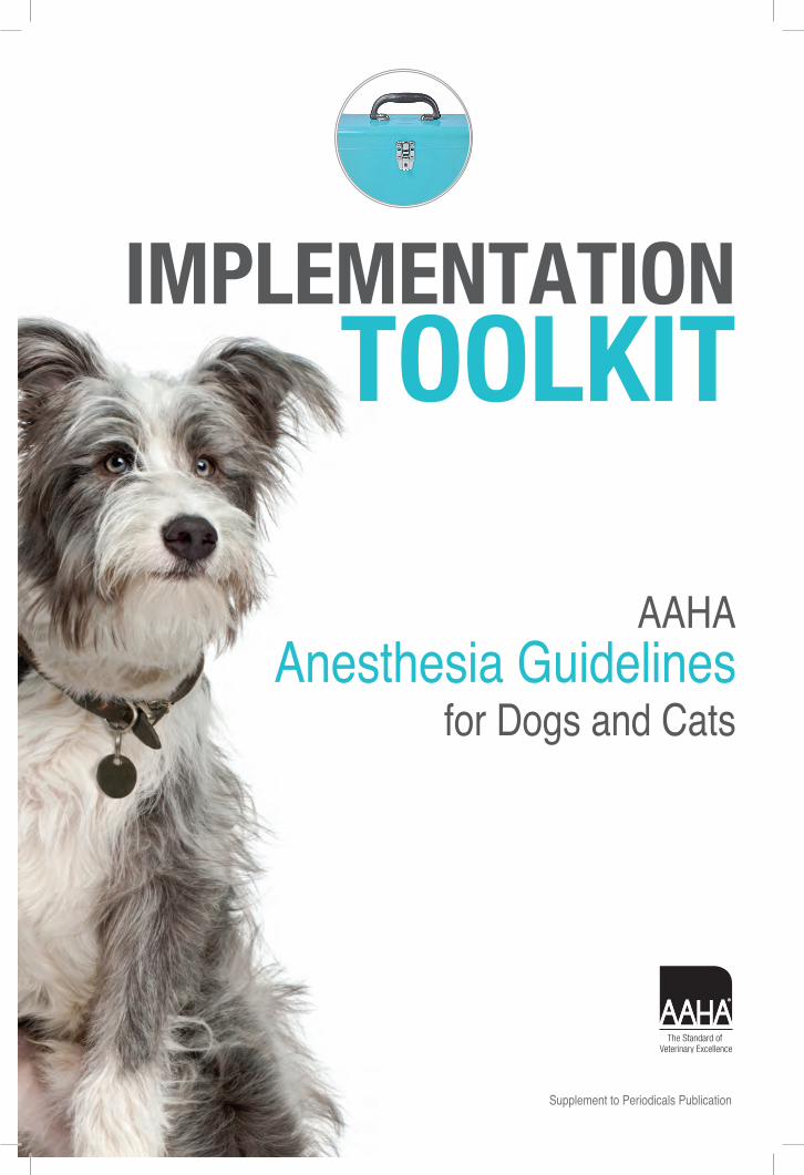
Supplement to Periodicals Publication

Inside This Toolkit
© 2012 American Animal Hospital Association (aahanet.org). All rights reserved.
Why Guidelines Matter 1
At-a-Glance Summary 2
AAHA Anesthesia Guidelines for Dogs and Cats 3
Anesthesia: What to Expect (Client Handout) 18
Financial Facts 20
Anesthesia Resources 21
This implementation toolkit is sponsored by a generous educational grant from Abbott Animal Health.
1
Why GuIdelInes MaTTer
Veterinary practice guidelines, like the recent AAHA Anesthesia Guidelines for Dogs and Cats, help ensure that pets get the best possible care. From medical director to veterinary assistant, guidelines keep your hospital staff on the cutting edge of veterinary medicine.
Completed in 2011, the AAHA Anesthesia Guidelines for Dogs and Cats is the most complete and medically sound compilation of updates, insights, advice, and recommendations ever de-veloped for ensuring the safe and effective use of anesthesia.
AAHA Guidelines review the latest information to help staff address central issues and perform essential tasks to improve the health of the pet. In addition, Guidelines define the role of each staff member, so everyone on the healthcare team can work together to offer the best-quality medical care.
Guidelines are just that—a guide—established by experts in a particular area of veterinary medicine. Guidelines do not outweigh the veterinarian’s clinical judgment; instead, they help veterinarians develop treatment plans that meet each patient’s needs and circumstances.
Aligning your practice protocols with Guideline recommendations is a key step in ensuring that your practice continues to deliver best-quality care.
To support your dedicated efforts, AAHA is pleased to offer this toolkit. Here you’ll find check-lists, tips, a client handout, a poster, and other tools you can use every day to implement the recommendations of the AAHA Anesthesia Guidelines for Dogs and Cats.
Thank you for helping to advance our shared mission to deliver the best in companion animal medical care. Together, we can make a difference!
Michael T. Cavanaugh, DVM, DABVPAAHA Executive Director and CEO
Why Guidelines Matter 1
At-a-Glance Summary 2
AAHA Anesthesia Guidelines for Dogs and Cats 3
Anesthesia: What to Expect (Client Handout) 18
Financial Facts 20
Anesthesia Resources 21
2
aT-a-Glance suMMary
create a specific or customized plan for each patient based on
the animal’s physical status (based on history, physical exam, and laboratory exam),
temperament, and the procedure to be performed.
a person trained and skilled in anesthesia should be present during
the perianesthetic period to deliver and monitor anesthesia.
Multimodal anesthesia is optimal and recommended. This involves
concurrent use of sedatives or tranquilizers, opioids, and both intravenous and inhalant
anesthetic drugs.
adjunctive procedures, such as local anesthetic nerve blocks, epidural
analgesia, and analgesic drug infusions, in conjunction with general anesthesia, can
improve analgesia, muscle relaxation, and hemodynamic status.
Opioid drugs provide excellent analgesia and should be
administered during painful procedures.
have emergency drugs, including anticholinergics and those for
CPCR, readily available. Know the appropriate dosage for each drug for each patient
anesthetized.
ensure airway patency by inserting a suitably sized, cuffed endotracheal
tube.
regularly monitor and record anesthetic depth, oxygenation,
ventilation, and cardiovascular function. Monitor these variables throughout the entire
anesthetic period, including recovery.
Insert an IV catheter preoperatively and have perioperative IV fluids available for
infusion.
Monitor each animal throughout recovery for adequate analgesia and an
appropriate level of sedation.
Successful anesthesia is more than absence of perianesthetic morbidity and mortality. It encompasses physiologic stability as well as adequate hypnosis and analgesia.

3
From the Veterinary Medical Center, The Ohio State University, Columbus, OH (R.B.); Veterinary Specialist Services PC, Conifer, CO (K.G.); Depart-ment of Small Animal Clinical Sciences, University of Tennessee College of Veterinary Medicine, Knox-ville, TN (R.H.); Southwest Veterinary Anesthesiol-ogy, Southern Arizona Veterinary Specialists, Tuc-son, AZ (V.L.); Phoenix, AZ (W.S.P.); Front Range Veterinary Clinic, Lakewood, CO (B.S.); and Peak Performance Veterinary Group, Colorado Springs, CO (K.S.).
AAHA American Animal Hospital Association; ACVA American College of Veterinary Anesthesiologists; ASA American Society of Anesthesiologists; AVMA Ameri-can Veterinary Medical Association; ET endotracheal; PLIT Professional Liability Insurance Trust.
*This report was prepared by a task force of experts con-vened by the American Animal Hospital Association for the express purpose of producing this article. This report was sponsored by an educational grant from Abbott Ani-mal Health, and was subjected to the same external review process as are all of Journal of American Animal Hospital Association articles.
Correspondence:[email protected] (R.B.)
Richard Bednarski, MS, DVM, DACVA (Chair), Kurt Grimm, DVM, MS, PhD, DACVA, DACVCP, Ralph Harvey, DVM, MS, DACVA, Victoria M. Lukasik, DVM, DACVA, W. Sean Penn, DVM, DABVP (Canine/Feline), Brett Sargent, DVM, DABVP (Canine/Feline), Kim Spelts, CVT, VTS, CCRP (Anesthesia)
*
Safe and effective anesthesia of dogs and cats relies on preanesthetic patient assessment and preparation. Patients should be premedicated with drugs that provide sedation and analgesia prior to anesthetic induction with drugs that allow endotracheal intubation. Maintenance is typically with a volatile anesthetic such as isoflurane or sevoflurane de-livered via an endotracheal tube. In addition, local anesthetic nerve blocks; epidural ad-ministration of opioids; and constant-rate infusions of lidocaine, ketamine, and opioids are useful to enhance analgesia. Cardiovascular, respiratory, and central nervous system functions are continuously monitored so that anesthetic depth can be modified as needed. Emergency drugs and equipment, as well as an action plan for their use, should be avail-able throughout the perianesthetic period. Additionally, intravenous access and crystal-loid or colloids are administered to maintain circulating blood volume. Someone trained in the detection of recovery abnormalities should monitor patients throughout recovery. Postoperatively, attention is given to body temperature, level of sedation, and appropriate analgesia. (J Am Anim Hosp Assoc 2011; 47:377–385. DOI 10.5326/JAAHA-MS-5846)
aBsTracT
Reprinted from Journal of the American Animal Hospital Association (Nov/Dec2011). © 2011 American Animal Hospital Association. All rights reserved.
4
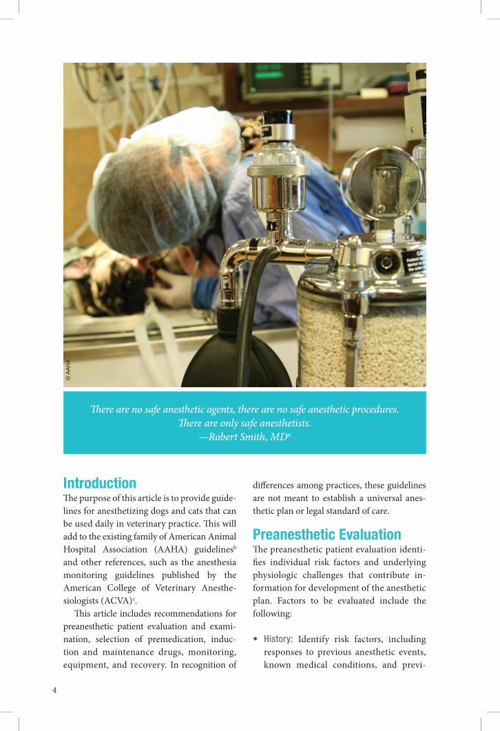
There are no safe anesthetic agents, there are no safe anesthetic procedures. There are only safe anesthetists.
—Robert Smith, MDa
IntroductionThe purpose of this article is to provide guide-lines for anesthetizing dogs and cats that can be used daily in veterinary practice. This will add to the existing family of American Animal Hospital Association (AAHA) guidelinesb and other references, such as the anesthesia monitoring guidelines published by the American College of Veterinary Anesthe-siologists (ACVA)c.
This article includes recommendations for preanesthetic patient evaluation and exami-nation, selection of premedication, induc-tion and maintenance drugs, monitoring, equipment, and recovery. In recognition of
differences among practices, these guidelines are not meant to establish a universal anes-thetic plan or legal standard of care.
Preanesthetic evaluationThe preanesthetic patient evaluation identi-fies individual risk factors and underlying physiologic challenges that contribute in-formation for development of the anesthetic plan. Factors to be evaluated include the following:
• History: Identify risk factors, including responses to previous anesthetic events, known medical conditions, and previ-
© A
AH
A
5
ous adverse drug responses. Identify all prescribed and over-the-counter medica-tions (including aspirin) and supplements to avoid adverse drug interactions.1
• Physical examination: A thorough physi-cal examination may reveal risk factors, such as heart murmur and/or arrhythmia or abnormal lung sounds.
• Age: Advanced age can increase anesthetic risk because of changes in cardiovascular and respiratory function. Disease processes occur more commonly in aged patients. Very young patients can be at increased risk from hypoglycemia, hypothermia, and decreased drug metabolism.
• Breed: Few breed-specific anesthesia is-sues are documented. Brachycephalic dogs and cats are more prone to upper airway obstruction. Greyhounds have longer sleep times after receiving some anesthetics such as propofol and thiopentald. Some breeds of dogs (e.g., Cavalier King Charles span-iel) and cats (e.g., Maine coon) may be pre-disposed to cardiac disease as they age.2
• Temperament: An aggressive or fractious temperament may pose a danger to staff and can limit the preanesthetic evaluation or make examination impossible. The selection of an alternative preanesthetic drug or drug combination may be re-quired for the aggressive or overly fearful animal due to the need for higher-than-usual drug doses. Conversely, a quiet or depressed animal may benefit from lower doses for sedation or anesthesia.
• Type of procedure: Evaluate the proce-dure’s level of invasiveness, anticipated pain, risk of hemorrhage, and/or pre-disposition to hypothermia. Some pro-cedures may limit physical access to the patient for monitoring.
• Usingheavysedationversusgeneralanes-thesia: This choice depends on the proce-
dure, patient temperament, and the need for monitoring and support. In general, sedation may be appropriate for shorter (<30 min) and less invasive procedures (e.g., diagnostic procedures, joint injec-tions, suture removal, and wound man-agement). Sedated patients, like those under general anesthesia, require appro-priate monitoring and supportive care. They may require airway management and/or O2 supplementation. Be prepared to intubate if necessary.
• Experienceandqualificationsofpersonnel:Previous training in local and regional anesthesia techniques will facilitate their perioperative use. Also, a more experi-enced surgeon may be faster and cause less tissue trauma to a patient than a less experienced one.Risk factors and individual patients’ needs
provide a framework for developing individu-alized patient plans and may indicate the need for additional diagnostic testing or stabiliza-tion before anesthesia.
Individual practice procedures may in-clude a minimum database of laboratory analysis, electrocardiogram, and diagnostic imaging for different patient groups. There is no evidence to indicate the minimum time frame before anesthesia within which labora-tory analysis should be performed. However, the timing should be reasonable to detect changes that impact anesthetic risk. The type and timing of such testing are determined by the veterinarian based on the previously men-tioned factors, as well as any change in patient status or the presence of concurrent disease.
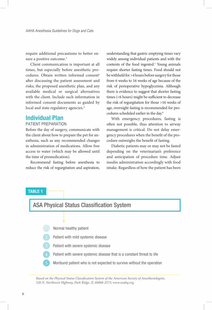
Categorization of patients using the American Society of Anesthesiologists (ASA) Physical Status Classification System pro-vides a framework for evaluation (Table 1). Patients with a higher ASA status are at greater risk for anesthetic complications and
PreaneSthetic evaluation
6
require additional precautions to better en-sure a positive outcome.3
Client communication is important at all times, but especially before anesthetic pro-cedures. Obtain written informed consente after discussing the patient assessment and risks, the proposed anesthetic plan, and any available medical or surgical alternatives with the client. Include such information in informed consent documents as guided by local and state regulatory agencies.4
Individual PlanPatient PreParationBefore the day of surgery, communicate with the client about how to prepare the pet for an-esthesia, such as any recommended changes in administration of medications. Allow free access to water (which may be allowed until the time of premedication).
Recommend fasting before anesthesia to reduce the risk of regurgitation and aspiration,
understanding that gastric emptying times vary widely among individual patients and with the contents of the food ingested.5 Young animals require shorter fasting times. Food should not be withheld for >4 hours before surgery for those from 6 weeks to 16 weeks of age because of the risk of perioperative hypoglycemia. Although there is evidence to suggest that shorter fasting times (<6 hours) might be sufficient to decrease the risk of regurgitation for those >16 weeks of age, overnight fasting is recommended for pro-cedures scheduled earlier in the day.6
With emergency procedures, fasting is often not possible, thus attention to airway management is critical. Do not delay emer-gency procedures when the benefit of the pro-cedure outweighs the benefit of fasting.
Diabetic patients may or may not be fasted depending on the veterinarian’s preference and anticipation of procedure time. Adjust insulin administration accordingly with food intake. Regardless of how the patient has been
23
45
Patient with mild systemic disease
Patient with severe systemic disease
Patient with severe systemic disease that is a constant threat to life
Moribund patient who is not expected to survive without the operation
Normal healthy patient
Based on the Physical Status Classi cation System of the American Society of Anesthesiologists, 520 N. Northwest Highway, Park Ridge, IL 60068-2573; www.asahq.org.
TABLE 1
ASA Physical Status Classification System
aaha anesthesia Guidelines for Dogs and cats
7
fasted, manage the airway of every patient as if its stomach were full.
aneSthetic PlanCreate an individualized plan for patient management based on the anesthetic risks identified in the preanesthetic evaluation, understanding that no single plan is appro-priate for all patients. Resources such as staff-ing, equipment, and drug availability also influence plan development. A complete an-esthetic plan addresses perioperative analge-sia, pre- and postanesthetic sedation and/or tranquilization, induction and maintenance drugs, ongoing physiologic support, moni-toring parameters, and responses to adverse events. The plan should be flexible to allow for dynamic patient responses during anesthesia.
PreaneSthetic MeDicationThe advantages of preoperative sedation and analgesia include lowered patient and staff stress, ease of handling, and reduction of in-duction and inhalant anesthetic doses, most of which have dose-dependent adverse effects.
There can be disadvantages to the admin-istration of preanesthetic medications, such as dysphoria related to benzodiazepines, bradycardia related to α-2 agonists and opi-oids, and hypotension related to aceproma-zine. These disadvantages can be mitigated by appropriate dosing and selecting the right combination of drugs for the individual. Patients in critical condition may not require any premedication.
Pain ManaGeMentChoose drugs and techniques that provide both intraoperative and postoperative an-algesia. Because there is a high variability in patient response to sedation and analgesia, individually tailor the medication type, dose, and frequency based on the anticipated in-
tensity and duration of pain. In addition to opioid premedication, perioperative anal-gesic techniques include nonsteroidal anti- inflammatory drugs, local and regional nerve blocks, as well as IV infusions of opioids, N-methyl-d-aspartate receptor antagonists (e.g., ketamine), and/or lidocaine. Multiple analgesic techniques should be considered for more painful procedures. Frequently reassess patient comfort and adjust pain management as needed. The AAHA Pain Management Guidelines and many other sources provide descriptions of and suggestions for pain man-agement f.7–9
aneSthetic ManaGeMent of PatientS with coMorbiDitieSCertain conditions require modification of the anesthetic protocol. Extensive discus-sion of the anesthetic management of the diseased patient is beyond the scope of these guidelines. However, brief mention of dia-betes, renal, cardiac, and hepatic disease is warranted.
DiabetesPerform periodic blood glucose measure-ments at sufficient intervals throughout the perianesthetic period to detect hypoglycemia or hyperglycemia before it becomes severe. Ideally, diabetic patients should be well regu-lated before anesthesia induction unless the procedure cannot be delayed.
Renal DiseaseNo one anesthetic drug or drug combination is better for renal disease; most important is to maintain blood pressure and adequate renal perfusion. Diuresis of moderately or severely azotemic patients before anesthetic induction may be warranted. Base the specific fluid types and rates on patient condition and response, but generally 1.5–2 times maintenance crystal-
renal DiSeaSe
8
loid administration for the 12–24 hours before anesthesia will reduce the magnitude of the azotemia. Continue fluids into the postopera-tive period as patient needs dictate. Fluid rates up to 20–30 mL/kg/hr during anesthesia have been recommended in patients with renal dysfunction.10,11
Patients with renal insufficiency may benefit from mannitol-induced diuresis and the associated increased renal medul-lary perfusion.12,13 To be effective, low-dose mannitol must be given before the ischemic episode; at higher doses it can cause renal vasoconstriction.
Vasopressors and inotropes have been rec-ommended, but strictly to maintain cardiac output. It has not been concluded that they contribute to increased renal perfusion or renal protection.
Cardiac DiseaseIn patients with severe cardiac disease, care-fully titrate IV fluids to avoid inducing con-gestive heart failure from fluid overload. Patients will vary in how much fluid and at what rate they can tolerate. Guide fluid ad-ministration by monitoring any of the fol-lowing: systemic blood pressure, central ve-nous pressure, oxygenation, or auscultation of lung sounds.
Preoperatively evaluate cardiac arrhyth-mias for consideration of perianesthetic treatment. Cardiac medications should be administered normally the day of surgery. Some medications may potentiate hypoten-sion (e.g., angiotensin-converting enzyme inhibitors and β blockers). Be prepared to administer inotropes or other supportive measures if needed.14
Opioid analgesics are useful during anes-thesia of the patient with cardiovascular com-promise. Certain anesthetic medications may be less appropriate in some types of cardiac
disease (e.g., at higher doses, ketamine may increase heart rate, which could be a problem in patients with hypertrophic cardiomyopathy; avoid α-2 agonists in dogs with mitral valve disease).15 A multimodal approach using drugs from multiple pharmacologic categories is pre-ferred to minimize extreme cardiovascular ef-fects of any one drug.16
Liver DiseaseTrue liver dysfunction also warrants special attention; however, increases in the liver en-zymes of an otherwise healthy patient are not an absolute reason to avoid anesthesia. In pa-tients with liver dysfunction, hypoglycemia can be a concern due to insufficient glyco-gen storage and impaired gluconeogenesis. Dextrose supplementation may be necessary. If hypoproteinemia is present, the administra-tion of fresh frozen plasma may be warranted. In general, delayed anesthetic recovery can be expected with the use of any anesthetic agent metabolized by the liver. Therefore, inhalants and drugs with specific antagonists such as opioids and α-2 agonists can be useful.
areaS of controverSyThe authors recognize that opinions vary re-garding the administration of certain peri-anesthetic drugs. Some of these are briefly outlined here.
There are misconceptions about the ef-fects of acepromazine in patients with seizure history. There is no evidence to show that acepromazine increases the risk of seizures in epileptic patients or patients with other sei-zure disorders.17,18
Indiscriminate use of anticholinergic drugs such as atropine and glycopyrrolate as part of a premedication protocol is contro-versial. Some think they should not be used routinely because the action will be short, and they may cause tachycardia, which increases
aaha anesthesia Guidelines for Dogs and cats

9
myocardial O2 consumption and the potential for myocardial hypoxemia.
In contrast, the preemptive use of anticho-linergics may be indicated for procedures with an increased risk of vagal bradycardia (e.g., oc-ular surgery) as well as in conjunction with opi-oid administration, to offset the potential bra-dycardic effects of the opioid. Anticholinergics may also be indicated in dogs with brachyce-phalic syndrome, which is associated with air-way obstruction and higher resting vagal tone, making these dogs more prone to developing bradycardia than are other breeds.19
The simultaneous use of anticholinergics with α-2 agonists has been debated. Some practitioners prefer to administer anticho-linergics to reduce the magnitude of brady-cardia and associated drop in cardiac output. However, the combination creates the poten-tial for myocardial hypoxemia to develop as a result of increased myocardial work. Use of anticholinergics should be based on individual patient risk factors and monitored parameters such as heart rate and blood pressure.20,21
Anesthesia PreparationEnsure that all equipment and medications deemed necessary for the procedure to be per-formed are readily accessible and in working order before induction of anesthesia. Regularly ensure proper maintenance and function of all anesthetic equipment. Table 2 provides a convenient maintenance checklist. Have emer-gency supplies and protocols available before any anesthetic procedure (e.g., tracheal suc-tion; emergency lighting in the event of power failure). Conspicuously post a chart of emer-gency drug doses or preemptively calculate such doses for each patient. Familiarize your-self with the most current recommendations for cardiopulmonary cerebral resuscitation and stock appropriate drugs. Useful emergency drug dose charts are available in many texts
and also from the Veterinary Emergency and Critical Care Societyg.
Prepare a written anesthetic record for each patient, beginning with preparation for the anesthetic event and continuing through the recovery period. Record preanesthetic patient status and all perianesthetic events, including drugs and dosages administered, routes of administration, patient vital signs, events, and interventions. Record resuscita-tion orders in the anesthetic record at the time consent is obtained. Regularly record patient parameters at 5–10 minute intervals, or more frequently if sudden changes in physiologic status occur. An anesthetic record template is available from AAHAh.
Patient PreParationPreparing a patient for anesthesia may in-clude some or all of the following:• Inserting an IV catheter and administering
IV fluids. This helps to avoid perivascular administration of induction drugs. It facili-tates intravascular volume support, which may correct hypovolemia resulting from vasodilation and blood loss that can occur during surgery. It also allows for rapid ad-ministration of emergency medications.
• Connecting monitoring equipment appro-priate for the disease condition present and
aneStheSia PreParation
© A
AH
A

10
TABLE 2
Anesthetic Equipment Check List
CO2 absorbent
Oxygen
Breathingsystem
Inhalant
Endotrachealtubes and
masks
Electronicmonitoringequipment
Waste-scavengingequipment
� Change the CO2 absorbent regularly based on individual anesthesia machine manufacturer recommendations.
� The useful lifespan of absorbent varies with the patient size and fresh gas flow rate.
� Color change is not an accurate indicator of remaining absorption capacity.
� Ensure supply lines are attached.
� Ensure the flowmeter is functioning.
� Ensure the supply tank and at least one spare tank are sufficiently full.
To calculate the estimated remaining tank volume, follow this example:
An E-cylinder contains 660 L, and has a full pressure of 2,200 psi.Pressure drop is proportional to remaining O2 volume.A tank with 500 psihas 150 L.When used at a flow rate of 1 L/min, it will last approximately 2 ½ hr.22
� Have access to various sizes of masks and endotracheal tubes.
� Provide a light source such as a laryngoscope.
� Check cuff integrity and amount of air needed to properly inflate the cuff.
� Refer to anesthesia machine’s documentation for proper leak-checking procedures.
� Conduct a check before every procedure.
� Select the appropriate size and type of reservoir bag and breathing circuit.23
� Non-rebreathing systems are generally used in patients weighing less than 5−7 kg or when the work of breathing associated with the circle system might not be easily sustainable by an individual patient.24
� Ensure the vaporizer is sufficiently full.
� Verify a functioning scavenging system.
� If using a charcoal absorbent canister, ensure there is sufficient capacity remaining for the duration of the procedure.
� Observe all regulations concerning the dispersion of waste anesthesia gases.25,26
� Ensure devices are operational and either are connected to a power source or have adequate battery reserve.
� Check alarms for limits and activation.
aaha anesthesia Guidelines for Dogs and cats

11
that the patient will tolerate before induc-tion (Table 3).
• Stabilizing hemodynamically unstable patients, including but not limited to:{{ Administering IV fluid boluses.
Hypovolemic patients may require iso-tonic crystalloids, colloids, and/or hy-pertonic saline to improve vascular fill-ing, cardiac output, and tissue perfusion.
{{ Managing cardiac arrhythmias.{{ Providing blood products. Hypoprotei-
nemia, anemia, or coagulation disorders can aggravate the decreased delivery of O2 to the tissues that normally oc-curs as a result of hypoventilation and recumbency.
{{ Preoxygenation reduces the risk of hemoglobin desaturation and hypox-emia during the induction process. Preoxygenation is especially beneficial if a prolonged or difficult intubation is expected or if the patient is already de-pendent on supplemental oxygenation. However, preoxygenation may be con-traindicated if it agitates the patient. Removing the rubber diaphragm from the face mask may increase patient tol-erance of the mask.29
Once the patient is as stable as possible, pro-ceed according to the individual patient plan.
aneSthetic inDuctionAnesthetic induction is best achieved using rapid-acting IV drugs, although this may not always be a reasonable option for fractious pa-tients.30 IV induction allows for rapid airway control and allows for titration of the induc-tion drug to effect within the given dosage range. Sick, debilitated, or depressed patients will require less drug than healthy, alert pa-tients. A patient’s response to preanesthetic drugs can influence the amount and type of induction drug needed.
Mask or chamber inductions can cause stress, delayed airway control, and environ-mental contamination.31 Adequate room ventilation must be present to minimize exposure to personnel. Reserve these tech-niques for situations where other alternatives are not suitable.
Ensure endotracheal (ET) tubes and in-tubation aids (e.g., stylets, laryngoscope) are readily available. Establish and maintain a patent airway using an ET tube as soon as possible. Use the largest-diameter ET tube that will easily fit through the arytenoid cartilages without damaging them; this will minimize resistance and the work of breath-ing. Insert the ET tube such that the distal tip of the tube lies midway between the larynx and the thoracic inlet. Applying a light coat-ing of sterile lubricating jelly improves the cuff ’s ability to seal the airway against fluid migration.32
Inflate the cuff sufficiently to create a seal for adequate positive pressure ventilation, being aware that overinflation may cause tracheal damage.33 When changing the patient’s posi-tion after intubation, take care not to rotate the ET tube within the trachea. This might in-duce tracheal tears, especially if the cuff is rela-tively overinflated. The American Veterinary Medical Association (AVMA) Professional Liability Insurance Trust (PLIT) has indicated that tracheal tears are a significant issue in anesthetized intubated catsi.34 However, tra-cheal intubation, when properly performed and maintained, is an essential part of main-taining an open and protected airway.
Apply corneal lubricant postinduction to protect the eyes from corneal ulceration.
Maintenance and MonitoringAnesthesia is typically maintained using in-halant anesthetics, although maintenance can also be achieved with continuous infusions
Maintenance anD MonitorinG
12
or intermittent doses of injectable agents, or a combination of injectable and inhalant drugs. An O2-enriched gas mixture is necessary for the safe and effective administration of inhal-ant anesthesia.23,29
O2 flow rates depend on the breathing circuit used. For a circle rebreathing system, use a relatively high flow rate when rapid changes in anesthetic depth are needed, such as during the transition from injectables to inhalants (induction), or when turning the vaporizer off at the end of the procedure. During the maintenance phase, total O2 flow rate should typically be between 200 and 500 mL. The system must be leak-free for these
flow rates to be effective. These are, perhaps, lower O2 flow rates than many are accus-tomed to. The benefits of lower flow rates include decreased environmental contami-nation and the economy of decreased con-sumption of O2 and volatile anesthetic gases. Lower flow rates also conserve moisture and heat. Disadvantages of lower flow rates in-clude increased time to change anesthetic depth. Administer an O2 flow of approxi-mately 200 mL/kg/min to patients connected to a non-rebreathing circuit.22
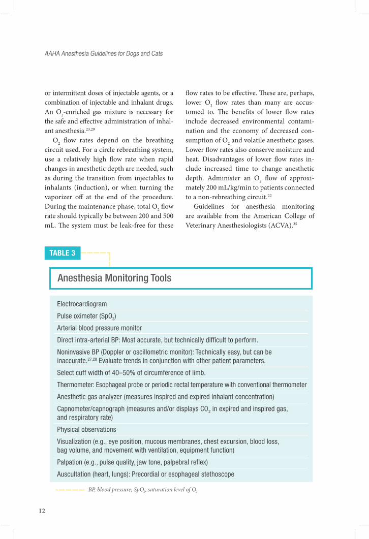
Guidelines for anesthesia monitoring are available from the American College of Veterinary Anesthesiologists (ACVA).35
aaha anesthesia Guidelines for Dogs and cats
TABLE 3
Anesthesia Monitoring Tools
Electrocardiogram
Pulse oximeter (SpO2)
Arterial blood pressure monitor
Direct intra-arterial BP: Most accurate, but technically difficult to perform.
Noninvasive BP (Doppler or oscillometric monitor): Technically easy, but can beinaccurate.27,28 Evaluate trends in conjunction with other patient parameters.
Select cuff width of 40–50% of circumference of limb.
Thermometer: Esophageal probe or periodic rectal temperature with conventional thermometer
Anesthetic gas analyzer (measures inspired and expired inhalant concentration)
Capnometer/capnograph (measures and/or displays CO2 in expired and inspired gas,and respiratory rate)
Physical observations
Visualization (e.g., eye position, mucous membranes, chest excursion, blood loss, bag volume, and movement with ventilation, equipment function)
Palpation (e.g., pulse quality, jaw tone, palpebral reflex)
Auscultation (heart, lungs): Precordial or esophageal stethoscope
BP, blood pressure; SpO2, saturation level of O2.
13
Continue the cardiovascular monitoring and physiologic support measures that began in the patient preparation and/or induction periods. Monitoring includes evaluation of oxygenation, ventilation, cardiac rate and rhythm, adequacy of anesthetic depth, muscle relaxation, body temperature, and analgesia. Blood pressure, heart rate and rhythm, mu-cous membrane color, and pulse oximetry provide the best indexes of cardiovascular function. Multiparameter electronic monitors are available and serve as tools to assess physi-ologic parameters during the perianesthetic period (Table 3). One must always evaluate the data the monitor is conveying in light of all other parameters and make treatment de-cisions based on the whole picture. Vigilant monitoring, interpretation, and responding to patient physiologic status by well-trained and attentive staff are critical.
Provide thermal support and monitor body temperature throughout the perianesthetic period. Supplemental heat may include warm IV fluids, use of a fluid line warmer, insulation on the patient’s feet (e.g., bubble wrap), circu-lating warm-water blankets, and/or warm air circulation systems. Do not use supplemental heat sources that are not designed specifically for anesthetized patients, as they can cause se-vere thermal injury.36
troubleShootinG aneSthetic coMPlicationSRecognize and then quickly and effectively respond to complications as they develop. Anesthesia-related complications are respon-sible for a significant number of AVMA PLIT insurance claimsj.
Hypoventilation is an expected effect of general anesthesia and can be estimated by observing respiratory rate and depth, but can be quantified using capnometry.
Observation of respiratory tidal volume is subjective, and it can be difficult to dis-tinguish a normal from an abnormal tidal volume. Normal end-tidal CO2 is approxi-mately 35–40 mm Hg in awake patients and approximately 40–50 mm Hg in patients in a light surgical plane of anesthesia. With in-creasing CO2, identify causes such as exces-sive anesthetic depth, provide initial patient support by positive pressure ventilation, and adjust anesthetic management as indicated. Hypotension is a common complication dur-ing anesthesia. Diagnose hypotension through blood pressure monitoring and evaluation of other physiologic parameters. Therapies for hypotension include decreasing the depth of anesthesia, administering crystalloid and/or colloid boluses, and/or administering vaso-pressors and inotropes.
Monitor for arrhythmias via auscultation, electrocardiography, or observing pulse–heart rate discongruity when using Doppler ultrasound. Common perioperative arrhyth-mias include bradycardia and ventricular ar-rhythmias. The decision of whether to treat a given arrhythmia should be based on the severity, the effect on other hemodynamic pa-rameters (e.g., blood pressure), and the like-lihood of deterioration to a more significant arrhythmia.
There are limited data to provide insight into the causes of anesthetic and perianesthetic deaths in dogs and cats.37 Many complica-tions and deaths occur during recovery. Most anesthetic deaths are unexplained because of insufficient information regarding the event. Increased monitoring and early diagnosis of physiologic changes and earlier intervention may reduce the risk of anesthetic death.
After an anesthetic death, offer clients the option of having a necropsy performed. Necropsy may detect preexisting disease
Maintenance anD MonitorinG

14
that contributed to anesthetic death that was not detectable with preoperative evaluation. Empathetic communication may help clients deal with loss, anger, and the grief process.
recoveryRecovery is a critical phase of anesthesia that includes a continuation of patient support, monitoring, and record keeping. It begins when the anesthetic gas is turned off. It does not end at the time of extubation.
Patients recovering from anesthesia re-quire monitoring by someone trained in the recognition of complications. Although many complications occur throughout an-esthesia, most anesthesia-associated deaths occur during recovery, especially in the first 3 hours. Forty-seven percent of canine anesthetic mortalities and 60% of feline an-esthetic mortalities have been reported to occur in the postoperative period.38
Continue regular monitoring of param-eters until they return to near baseline. Pulse oximetry, blood pressure monitoring, and periodic auscultation are valuable for detecting life-threatening complications. Continue to monitor the electrocardiogram and blood pressure in those patients at sig-nificant risk of life-threatening hypotension or dysrhythmias.
Respiratory depression persists during the early recovery from anesthesia. Continue sup-plemental oxygen until SpO2 measurements are acceptable when breathing room air.
Extubate when the patient can adequately protect its airway by vigorously swallowing. Deflate the cuff immediately before remov-ing the ET tube. With patients that have un-dergone a dental procedure or oral surgery, it is beneficial to position the nose slightly lower than the back of the head and leave the ET tube cuff slightly inflated during extuba-tion. This will help clear blood clots and de-
bris from the trachea and deposits any fluid or debris into the pharyngeal region, where it can drain from the mouth or be swallowed, thereby reducing the risk of aspiration.
Recovery from anesthesia can be pro-longed in hypothermic patients, resulting in increased morbidity.39 Provide adequate thermal support until the patient’s tempera-ture is consistently rising and approaching normal.
Reapply eye ointment during the recovery period, especially if an anticholinergic was administered, until an adequate blink reflex is present. Express the bladder if distended to minimize any distention-related discomfort.
Reassess the patient’s pain level and, if necessary, adjust the plan for postoperative pain management. Adequate analgesia and a quiet environment encourage smooth re-covery. Evaluate patient for dysphoria, emer-gence delirium, and pain. Treat if necessary.7
Discharge of a patient having undergone anesthesia should occur only after the pa-tient is awake, aware, warm, and comfort-able. Evaluate the animal for its responses and its ability to interact safely with own-ers and maintain physiologic homeostasis. Provide written instructions for owners, out-lining the dose and potential side effects of analgesics and other medications to be given to the patient at home.
summary/conclusionAnesthesia includes more than the selection of anesthetic drugs. A comprehensive individ-ualized anesthetic plan will minimize periop-erative morbidity and optimize perioperative conditions. Monitoring, the ability to discern normal from abnormal, and expedient inter-vention are critical to ensure that potentially reversible problems do not become irrevers-ible. Vigilance and patient support must be maintained during the recovery period.
aaha anesthesia Guidelines for Dogs and cats
15
Successful anesthetic management re-quires trained, observant team members who understand the clinical pharmacology and physiologic adaptations of the patient under-going anesthetic procedures, as well as the use of anesthetic and monitoring equipment. Staff must be able to assess abnormal patient responses quickly and respond efficiently, by being familiar with the expected responses seen with different anesthetic drugs and with the changes seen in the phases and/or depth of general anesthesia. Provide training and review procedures with staff upon hiring,
at regular intervals, and after adverse events occur, as part of routine morbidity and mor-tality discussions.
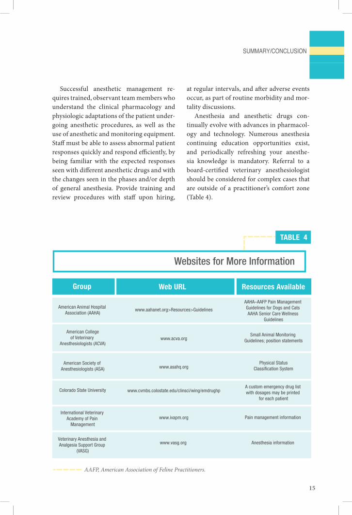
Anesthesia and anesthetic drugs con-tinually evolve with advances in pharmacol-ogy and technology. Numerous anesthesia continuing education opportunities exist, and periodically refreshing your anesthe-sia knowledge is mandatory. Referral to a board-certified veterinary anesthesiologist should be considered for complex cases that are outside of a practitioner’s comfort zone (Table 4).
TABLE 4
Websites for More Information
American Animal Hospital Association (AAHA)
www.aahanet.org>Resources>Guidelines
AAHA–AAFP Pain Management Guidelines for Dogs and Cats AAHA Senior Care Wellness
Guidelines
Group Web URL Resources Available
American Collegeof Veterinary
Anesthesiologists (ACVA)www.acva.org
Small Animal Monitoring Guidelines; position statements
American Society of Anesthesiologists (ASA) www.asahq.org
Physical StatusClassification System
Colorado State University www.cvmbs.colostate.edu/clinsci/wing/emdrughpA custom emergency drug list with dosages may be printed
for each patient
International Veterinary Academy of Pain
Managementwww.ivapm.org Pain management information
Veterinary Anesthesia and Analgesia Support Group
(VASG)
www.vasg.org Anesthesia information
AAFP, American Association of Feline Practitioners.
SuMMary/concluSion
16
FootnotesaThis quote appears as an introduction to Chapter 1 of: Muir W, Hubbell J, Bednarski R. Introduction to anesthesia. In: Muir WW, Hubbell JAE, Bednarski RM, Skarda RT, eds. Handbook of veterinary anesthesia. 4th ed. St. Louis: Elsevier, 2007;1. However, the original source of the quote is not referenced. bSee www.aahanet.org resources. cSee www.acva.org dAt the time of this publication, thiopental is not available in the United States eA standard consent form may be found at www.avma.org/issues/policy/consent_form.asp fVeterinary Anesthesia & Analgesia Support Group, www.vasg.org; International Veterinary Academy of Pain Management, www. ivapm.org gSee www.veccs.org hSee www.aahanet.org > AAHA store > Books and products >Anesthesia record iPersonal communication, March 2011, AVMA PLIT jPersonal communication, March 2011, AVMA PLIT.
references1. Seahorn J, Robertson S. Concurrent medications and their impact on anesthetic management. Vet Forum 2002;119:50–67.
2. Gough A, Thomas A. Breed predispositions to disease in dogs and cats. Oxford: Blackwell Publishing Ltd., 2004;44, 170.
3. Muir WW. Considerations for general anesthesia. In: Tranquilli WJ, Thurmon JC, Grimm KG, eds. Lumb and Jones’ veteri-nary anesthesia and analgesia. 4th ed. Ames: Blackwell; 2007:17–30.
4. Flemming DD, Scott JF. The informed consent doctrine: what veterinarians should tell their clients. J Am Vet Med Assoc 2004;224(9):1436–9.
5. Bednarski RM. Dogs and cats. In: Tranquilli WJ, Thurmon JC, Grimm KA, eds. Lumb and Jones’ veterinary anesthesia and analgesia. 4th ed. Ames: Blackwell; 2007:705–17.
6. Looney AL, Bohling MW, Bushby PA. The Association of Shelter Veterinarians veterinary medical care guidelines for spay-neuter programs. Association of Shelter Veterinarians1 Spay-Neuter Task Force. J Am Vet Med Assoc 2008;233:1,74–86.
7. Hellyer P, Rodan I, Brunt J, et al; American Animal Hospital Association; American Association of Feline Practitioners; AAHA/AAFP Pain Management Guidelines Task Force Members. AAHA/AAFP pain management guidelines for dogs & cats. J Am Anim Hosp Assoc 2007;43(5):235–48.
8. Gaynor J, Muir W. Handbook of veterinary pain management. 2nd ed. St. Louis: Mosby, Inc.; 2009.
9. Greene S. Veterinary anesthesia and pain management secrets. Philadelphia: Hanley & Belfus; 2001.
10. Brezis M, Rosen S. Hypoxia of the renal medulla—its implications for disease. N Engl J Med 1995;332(10):647–55.
11. Heyman SN, Fuchs S, Brezis M. The role of medullary ischemia in acute renal failure. N Horizons 1995;3:597–607.
12. Behnia R, Koushanpour E, Brunner EA. Effects of hyperosmotic mannitol infusion on hemodynamics of dog kidney. Anesth Analg 1996;82(5):902–8.
13. Fisher AR, Jones P, Barlow P, et al. The influence of mannitol on renal function during and after open-heart surgery. Perfusion 1998;13(3):181–6.
14. Evans AT, Wilson DV. Anesthetic emergencies and procedures. In: Tranquilli WJ, Thurmon JC, Grimm KG, eds. Lumb and Jones’ veterinary anesthesia and analgesia. 4th ed. Ames: Blackwell; 2007:1033–48.
15. Jakobsen CJ, Torp P, Vester AE, et al. Ketamine reduce left ventricular systolic and diastolic function in patients with ischaemic heart disease. Acta Anaesthesiol Scand 2010;54(9):1137–44.
16. Harvey RC, Ettinger SJ. Cardiovascular disease. In: Tranquilli WJ, Thurman JC, Grimm KA, eds. Lumb and Jones veteri-nary anesthesia and analgesia. 4th ed. Ames, IA: Blackwell Publishing; 2007:891–8.
17. Tobias KM, Marioni-Henry K, Wagner R. A retrospective study on the use of acepromazine maleate in dogs with sei-zures. J Am Anim Hosp Assoc 2006;42(4):283–9.
18. McConnell J, Kirby R, Rudloff E. Administration of acepromazine maleate to 31 dogs with a history of seizures. J Vet Emerg Crit Care 2007;17(3):262–7.
19. Doxey S, Boswood A. Differences between breeds of dog in a measure of heart rate variability. Vet Rec 2004;154(23):713–7.
20. Alvaides RK, Neto FJ, Aguiar AJ, et al. Sedative and cardiorespiratory effects of acepromazine or atropine given before dexmedetomidine in dogs. Vet Rec 2008;162(26):852–6.
aaha anesthesia Guidelines for Dogs and cats
17
21. Ko JC, Fox SM, Mandsager RE. Effects of preemptive atropine administration on incidence of medetomidine-induced bra-dycardia in dogs. J Am Vet Med Assoc 2001;218(1):52–8.
22. Hartsfield SM. Anesthetic machines and breathing systems. In: Tranquilli WJ, Thurmon JC, Grimm KA, eds. Lumb and Jones’ veterinary anesthesia and analgesia. 4th ed. Ames, IA: Blackwell; 2007:481–2.
23. Lerche P, Muir WW III, Bednarski RM. Rebreathing anesthetic systems in small animal practice. J Am Vet Med Assoc 2000;217(4):485–92.
24. Hodgson DS. The case for non-rebreathing circuits for very small animals. Vet Clin N Am Sm Anim Pract 1992;2:397–9.
25. US Dept of Labor, Occupational Safety and Health Administration. Anesthetic Gases: Guidelines for Workplace Exposures. Available at www.osha.gov/dts/osta/anestheticgases/index.html. Accessed September 23, 2011.
26. ACVA. Control of Waste Anesthetic Gases in the Workplace. Position statements. Available at www.AVCA.org. Accessed September 23, 2011.
27. Bosiack AP, Mann FA, Dodam JR, et al. Comparison of ultrasonic Doppler flow monitor, oscillometric, and direct arterial blood pressure measurements in ill dogs. J Vet Emerg Crit Care (San Antonio) 2010;20(2):207–15.
28. Shih A, Robertson S, Vigani A, et al. Evaluation of an indirect oscillometric blood pressure monitor in normotensive and hypotensive anesthetized dogs. J Vet Emerg Crit Care (San Antonio) 2010;20(3):313–8.
29. McNally EM, Robertson SA, Pablo LS. Comparison of time to desaturation between preoxygenated and nonpreoxygenated dogs following sedation with acepromazine maleate and morphine and induction of anesthesia with propofol. Am J Vet Res 2009;70(11):1333–8.
30. Psatha E, Alibhai HI, Jimenez-Lozano A, et al. Clinical efficacy and cardiorespiratory effects of alfaxalone, or diazepam/fen-tanyl for induction of anaesthesia in dogs that are a poor anaesthetic risk. Vet Anaesth Analg 2011;38(1):24–36.
31. Tzannes S, Govendir M, Zaki S, et al. The use of sevoflurane in a 2:1 mixture of nitrous oxide and oxygen for rapid mask induction of anaesthesia in the cat. J Feline Med Surg 2002:2:83–90.
32. Dave MH, Koepfer N, Madjdpour C, et al. Tracheal fluid leakage in benchtop trials: comparison of static versus dynamic ventilation model with and without lubrication. J Anesth 2010;24(2):247–52.
33. Hardie EM, Spodnick GJ, Gilson SD, et al. Tracheal rupture in cats: 16 cases (1983–1998). J Am Vet Med Assoc 1999;214(4):508–12.
34. Mitchell SL, McCarthy R, Rudloff E, Pernell RT. Tracheal rupture associated with intubation in cats: 20 cases (1996–1998). J Am Vet Med Assoc 2000;216:1592–5.
35. ACVA. Small animal monitoring guidelines. Available at www.acva.org. Accessed September 23, 2011.
36. Swaim SF, Lee AH, Hughes KS. Heating pads and thermal burns in small animals. J Am Anim Hosp Assoc 1989;25:156–62.
37. Brodbelt DC, Pfeiffer DU, Young LE, et al. Results of the confidential enquiry into perioperative small animal fatalities re-garding risk factors for anesthetic-related death in dogs. J Am Vet Med Assoc 2008;233(7):1096–1104.
38. Brodbelt DC, Blissitt KJ, Hammond RA, et al. The risk of death: the confidential enquiry into perioperative small animal fatalities. Vet Anaesth Analg 2008;35(5):365–73.
39. Pottie RG, Dart CM, Perkins NR, et al. Effect of hypothermia on recovery from general anaesthesia in the dog. Aust Vet J 2007;85(4):158–62.
© A
AH
A
18
anesThesIa: WhaT TO exPecT
Beforethedayoftheprocedure
• Follow the veterinarian’s directions.
• You might be asked to change the medications you give your pet. You could be asked to skip a dose or to give a different medication.
• You will be asked to withhold food for a certain time to reduce the risk of regurgitation and aspiration—breathing in the contents of the stomach and gastric juices into the lungs. You may also be instructed to withhold water from your pet, depending on the veterinarian.
• Older animals must fast longer than younger animals do for three reasons: (1) older pets’ metabolism is slower, (2) it often takes them longer to digest their food, and (3) they usually have greater energy reserves than younger animals.
• If your pet has diabetes, your veterinarian might not require fasting or might instruct you to adjust your pet’s insulin.
Your veterinarian will perform certain tasks before the procedure (often the same day), including a thorough evaluation of your pet. This evaluation should include a blood test to make sure your pet is healthy enough to undergo anesthesia. (If the situation is an emergency, the veterinarian might run additional tests and perform measures to stabilize your pet before the procedure to better prepare your pet for anesthesia.)
The evaluation also will include:
History Physical examination Review the age, breed, and temperament Evaluate the procedure’s level of invasiveness, anticipated pain, risk of hemorrhage (bleeding) or hypo-thermia (decreased body temperature) Consider the best type of anesthesia and medica-tion Make sure the team assisting the veterinarian is well trained Create an individual anesthesia plan for your pet
Whether the patient is a person or a pet, undergoing anesthesia carries some risk of complica-tions. If the situation is not an emergency, your veterinarian will examine your pet and might run some tests, such as blood work, to help identify those risks. Your veterinarian wants to make sure the animal is healthy enough to undergo anesthesia.
Your veterinarian or veterinary technician will explain the procedure to you and discuss the patient assessment and risks, the proposed anesthetic plan, and any medical or surgical alterna-tives before obtaining informed consent to anesthetize your pet and perform the procedure.
To help reduce the risk of complications, it is very important that you follow the directions of the veterinarian, especially regarding patient preparation.
note! Whenyourpetisunconscious,thegagreflexissuppressed.Yourpetcouldinhalestomachcontents,causingseriousinjury,evendeath.So, you must be very strict about withholding food, and maybe water, for the specified time, if instructed to do so.

19
Onthedayoftheprocedure
BEfOrEThEPrOcEdUrEAs the veterinary team prepares your pet for the procedure, your veterinarian will:• Make sure equipment is working and medication is close by.
• Prepare your pet for anesthesia.
• Begin to implement your pet’s individual anesthesia plan.
• Make sure your pet is monitored throughout the procedure and during recovery.
• Recognize and quickly respond to any complications if they develop.
• Assess and manage your pet’s potential pain level before, during, and after the procedure.
AfTErThEPrOcEdUrEWhen your pet is awake, aware, warm, and comfortable, he or she will be discharged. But first, the veterinarian or veterinary staff will:• Review the procedure and how it went.
• Explain follow-up care, including when your pet can begin to eat and drink.
• Tell you when to resume current medications.
• Tell you how to give new medications, if needed.
• Explain how to recognize signs of complications in your pet. It is important that you call the veterinarian’s office immediately if your pet has a complication.
• Tell you when to bring your pet back for a re-check.
• In addition to telling you the instructions, your veterinarian or veterinary staff should give you a written copy of the aftercare instructions.
To see a full list of articles about anesthesia on Healthypet.com, go to ow.ly/7BWaM or scan this code using your phone.
Guidelines and Standards created by the American Animal Hospital Association (healthypet.org) provide advice and recommendations that help your veterinary team provide the best medical care possible for your pet. AAHA is the only organization in the U.S. and Canada that accredits companion animal hospitals based on standards that go above and beyond state regulations.
This client handout is sponsored by a generous educational grant from Abbott Animal Health.
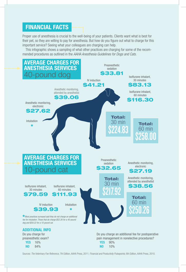
FInancIal FacTsProper use of anesthesia is crucial to the well-being of your patients. Clients want what is best for their pet, so they are willing to pay for anesthesia. But how do you figure out what to charge for this important service? Seeing what your colleagues are charging can help.
This infographic shows a sampling of what other practices are charging for some of the recom-mended procedures as outlined in the AAHA Anesthesia Guidelines for Dogs and Cats.
aVeraGe charGes FOr anesThesIa serVIces 40-pound dog
aVeraGe charGes FOr anesThesIa serVIces 10-pound cat
IV induction
$41.21
IV induction
$39.93
Intubation
*
Intubation
*
Total:30 min
$224.83
Total:30 min
$217.92
Total:60 min
$258.00
Total:60 min
$250.26
Preanesthetic sedation
$33.81
Preanesthetic sedation
$32.65
Isoflurane inhalant,30 minutes
$83.13
Isoflurane inhalant,30 minutes
$79.59
Isoflurane inhalant,60 minutes
$116.30
Isoflurane inhalant,60 minutes
$111.93
Anesthetic monitoring, attended by anesthetist
$39.06
Anesthetic monitoring, attended by anesthetist
$38.56
Anesthetic monitoring, electronic
$27.62
Anesthetic monitoring, electronic
$27.19
Sources: The Veterinary Fee Reference, 7th Edition, AAHA Press, 2011; Financial and Productivity Pulsepoints, 6th Edition, AAHA Press, 2010.
addITIOnal InFO Do you charge for preanesthetic exam?
yes 16%nO 84%
Do you charge an additional fee for postoperative pain management in nonelective procedures?
yes 90%nO 10%
*Most practices surveyed said they do not charge an additional fee for intubation. Those that do charge $52.26 for a 40-pound dog and $59.22 for a 10-pound cat.

21
anesThesIa resOurces
From aaha Press
Please check ✓
❒ Anesthesia machine
❒ Oxygen supply and flow
❒ Soda lime
❒ Pop-off valve
❒ Scavenging system
❒ Endotracheal tube cuff
❒ ET tube used ______ mm
❒ Grounding systems (e.g., cautery)
❒ Anesthetic agents
❒ Lubrication for eyes
Anesthesia (AM/PM)
Procedure (AM/PM)
Anesthesia (AM/PM)
Procedure (AM/PM)
Patient Identification
Veterinarian
Procedure(s)
Anesthesia Record
PREP
ROCE
DURE
CHEC
KLIS
TSPA
TIEN
T ID
© 2004 American Animal Hospital Association
STAR
T TI
ME
DRU
GS/
FLUID
S/AG
ENTS
USE
D
STOP
TIM
E
Please check ✓
❒ Pulse oximeter
❒ End tidal CO2
❒ Blood pressure
❒ Continuous EKG
❒ Dedicated continuous observation
❒ Esophageal stethoscope
❒ Respiratory monitor
❒ Temperature
Please check ✓
❒ Patient identified
❒ ID confirmed
❒ NPO_______time
Monitor’s Signature/Date
SIGNAT
URE
Dose/ Time GivenRate Initials (AM/PM)
Preanesthetic Drugs
Induction Drugs
Maintenance Drugs
Reversal Drug
Fluids
Pain Medications
Antibiotics
Other Drugs/Fluids/Agents
Comments
Postanesthetic Care
Patient Safety Checklist
Monitors Utilized Checklist
Preprocedure Checklist
Diagnosis
IV IM SQ
IV IM SQ
IV IM SQ
IV IM SQ
IV Mask Chamber
IV Mask Chamber
IV Epidural Intubation
IV Epidural Intubation
BE SAFE! BE SAFE!
Workbook on Veterinary Safety Training for the Whole Practice Team
Philip J. Seibert, Jr., CVT
Philip J. Seibert, Jr., CVT
© 2007 American Animal
Hos
pita
l Ass
ocia
tion
!
Minor Surgical/Anesthetic Procedure
R T
TSize
G
I II III IV V
For more information, visit press.aahanet.org.
anesthesia assessment and Plan Form Allows for convenient docu-mentation of the preanesthetic patient evaluation and helps ensure ac-curate medical record keeping and becomes part of the patient’s medi-cal record. Complies with the AAHA standard of accreditation related to documentation of a preanesthetic evaluation.
Veterinary anesthesia update: Practical Guidelines and Protocols for small animal anesthesia, second edition This step-by-step manual bridges the gap between new scientific developments and their applica-tion in clinical practice, and covers equipment, general guidelines, an-esthesia, protocols, troubleshooting, and pain management.
anesthesia record This form becomes part of the patient’s medical record and allows the technician to document vital signs during an an-esthetic procedure. Can also be used to record drugs and fluids given, safety procedures completed before surgery, and comments on pain management and postanesthesia care.
Minor surgical/anesthetic Procedure sticker Save time by quickly and easily summarizing minor surgical or anesthetic procedures on these stickers. Available in packs of 50 and rolls of 500.
Be safe! Manager’s Guide to radiation and Waste anesthetic GasesKeep your team safe from radiation and waste anesthetic gases, two of the most worrisome issues in veterinary safety. Find out what OSHA’s regulations are in these areas.
Be safe! Veterinary safety Training for Medical and Technical staff dVd Blend safety, quality patient care, and productivity while raising awareness of safety and OSHA issues for your medical and technical staff.
The Veterinary Fee reference, seventh edition This bestseller includes fees for more than 450 services, including anesthesia services. Nearly 700 tables contain data comparing fees by type and size of practice, with benchmarks and advice on how to establish the right value for your ser-vices and products.
aBOuT aahaThe American Animal Hospital Association is an international organization of nearly 6,000 veterinary care teams comprising more than 48,000 veterinary professionals committed to excellence in companion animal care. Established in 1933, AAHA is recognized for its leadership in the profession, its high standards for pet healthcare, and most important, its accreditation of companion animal practices. For more information about AAHA, visit aahanet.org.
Photo credits:Front cover (top): © iStockphoto/gabyjalbertFront cover (bottom): © iStockphoto/Eric IsseléeBack cover: © iStockphoto/Pavel Sazonov
This implementation toolkit was developed by the American Animal Hospital Association (AAHA) to provide information for practitioners regarding the safe and effective anesthesia of dogs and cats. The information contained in this toolkit should not be construed as dictating an exclusive protocol, course of treatment, or procedure, nor is it intended to be an AAHA standard of care. AAHA hopes that you find this toolkit useful.