Embed Size (px)
Citation preview
Supplemental Data
Nicastrin Functions as a
γ-Secretase-Substrate Receptor Sanjiv Shah, Sheu-Fen Lee, Katsuhiko Tabuchi, Yi-Heng Hao, Cong Yu, Quincey LaPlant, Haydn Ball, Charles E. Dann III, Thomas Südhof, and Gang Yu Supplemental Experimental Procedures cDNAs and Antibodies Human cDNAs were used in this study unless indicated otherwise. Wild-type and mutant Nct-His, PS1, APH-1aL-His, HA-PEN-2, Flag-PEN-2, C99-Flag, nFlag-C99, N100-Flag/His, and His-Syt1 cDNAs were cloned into pFASTBac and baculoviruses were generated using the Bac-to-Bac Baculovirus Expression System (Invitrogen). pFASTBac/C99-Flag encodes C-terminally Flag-tagged C99 with a Met followed immediately by the β-secretase cleaved APP C-terminal fragment. pFASTBac/nFlag-C99 encodes a C99-derived polypeptide in which the seven residues of C99 after Met were replaced by the Flag epitope (DYKDDDDK). pFASTBac/N100-Flag/His encodes Val1711-Glu1809 of mouse Notch-1 and a C-terminal Flag/His tag. The bacterial expression construct pET21b/C99-Flag was a gift from Drs. M.J. LaVoie, D.J. Selkoe, and Y.M. Li (Esler et al., 2002; Li et al., 2000). To generate the construct for Cys-C99 (pHV-Cys), the His-tag and tobacco etch virus (TEV) recognition site sequence from pHis-parallel was amplified and mutated via PCR to generate an NdeI/BamHI product with an internal NsiI site, changing the final Gly of the TEV recognition site to Cys. This fragment was then subcloned using the same sites into pET21b followed by insertion of C99-Flag as an NsiI/XhoI fragment. pCMV-IgG fusion constructs for mammalian cells were generated to contain the following sequences of various proteins fused to the Fc fragment of human IgG: nicastrin residues 1-50, nicastrin residues 1-669, full-length α2δ1, and APP residues 1-205. pSGHV0-Nct(ECD) encodes nicastrin residues 34-669 in frame with the human growth hormone (hGH), an octahistidine tag (H8), and a cleavage site for TEV protease (Dann et al., 2001; Leahy et al., 2000). Wild-type and mutant nicastrin cDNAs in pcDNA6-V5/His and a cDNA for the APP signal peptide-fused C99 (SpC99) in pcDNA3 were generated as previously described (Lee et al., 2004; Luo et al., 2003; Yu et al., 2000). Antibodies used in this study include anti-sera for APH-1aL, PEN-2, PS1, and Nct (Lee et al., 2002; Lee et al., 2004; Luo et al., 2003), anti-cleaved Notch-1 (Val1744) antibody (Cell Signaling), anti-APP cytoplasmic domain (anti-APP-CTD), anti-biotin, anti-human IgG, anti-Flag (Sigma), anti-His (Qiagen), anti-HA (Santa Cruz), and anti-fluorescein/Oregon Green (Molecular Probes) antibodies. Protein Purification Membranes from Sf9 cells infected with baculoviruses were solubilized in 1% Triton X-100 or 0.5% CHAPSO using a procedure similar to that for mammalian cells (Yu et al., 1998; Lee et al., 2004). The active recombinant γ-secretase and its subunits were purified from the membrane extracts by Ni-NTA affinity chromatography, glycerol gradient fractionation, anti-Flag affinity chromatography, and HiTrap Q ion-exchange chromatography on an ÄKTA FPLC system (Amersham). γ-Secretase substrates N100-Flag/His, C99-Flag, and nFlag-C99 were purified using Ni-NTA or anti-Flag affinity chromatography. IgG-fusion proteins were purified on protein A resins from conditioned media of HEK293 cells stably expressing the IgG-fusion cDNAs. The untagged nicastrin ectodomain was expressed in Chinese hamster ovary (CHO) cells as a fusion protein with the human growth hormone, an octahistidine tag, and a TEV

protease recognition site (Fig. 2A). hGH-Nct(ECD) was initially purified by Ni-NTA immobilized metal affinity chromatography from conditioned media of the CHO cells. The fusion protein was then cleaved by TEV protease, and untagged nicastrin ectodomain was further purified by Mono Q anion exchange chromatography and gel filtration to near homogeneity (Leahy et al., 2000). Biochemical Analyses Edman protein sequencing and mass-spectrometry (MS) services were provided by the Protein Chemistry Technology Center at the University of Texas Southwestern Medical Center. The first five amino acids from five cycles of N-terminal protein sequencing are: C99-Flag from E. coli, MDAEF; Cys-C99 from E. coli, CMDAE; N100-Flag/His from Sf9 cells, VKSEP; NICD’-Flag/His generated in vitro, VLLSR; Nct-His from Sf9 cells, NSVER; Nct(ECD)-IgG from HEK293 media, NSVER. Immunoprecipitation and protein A and Ni-NTA affinity pull-down experiments were performed as previously described (Lee et al., 2004; Yu et al., 2000) except that 1% Triton X-100 and higher salt concentration were used for investigating interactions of γ-secretase substrates and nicastrin. For the Flag-peptide pre-absorption or elution experiments, saturating amounts of Flag peptide were used. For in vitro γ-secretase assays, addition of lipids (e.g. phosphatidylcholine, phosphatidylethanolamine, or phosphatidylinositol) is absolutely necessary for purified recombinant γ-secretase from Sf9 cells but is not required for cell-free γ-secretase from crude HeLa or fibroblast cell membranes. γ-Secretase activity can be reconstituted in proteoliposomes when CHAPSO is removed from a mixture of cholesterol and phosphatidylcholine by dialysis and ultracentrifugation. Native Chemical Ligation Materials 2-(1H-Benzotriazol-1-yl)-1,1,3,3-tetramethyluronium hexafluorophosphate (HBTU), hydroxybenzotriazole (HOBt) and tert-butoxycarbonyl (t-Boc) –protected amino acids were obtained from Novabiochem (San Diego). S-Tritylmercaptopropionic acid was obtained from Peptides International (Louisville, KY). N,N-diisopropylethylamine (DIEA), diisopropylcarbodiimide (DIC), 5,6-carboxyfluorescein and triisopropylsilane were purchased from Aldrich. Methylbenzhydrylamine (MBHA) resin and d-biotin-amidocaproic acid (biotin-LC) were obtained from Anaspec (San Jose, CA). N-methylpyrrolidone (NMP), trifluoroacetic acid (TFA), HPLC grade acetonitrile and dichloromethane (DCM) were obtained from Fisher. Hydrogen fluoride (HF) was purchased from Matheson Gas (Cucamonga, CA). Synthesis of Fluorescein-Gly-Gly-Thioester and Biotin-LC-Gly-Gly-Thioester Derivatives t-Boc Leu (1 mmol) was activated with 0.9 mmol of HBTU/HOBt in the presence of 2 mmol of DIEA and coupled for 1 hr to 0.3 mmol of MBHA resin. The Boc group was removed following a 30 min treatment with 50% TFA in DCM. The TFA salt was neutralized by washing with 5% DIEA in DCM for 5 mins. Next, 1 mmol of S-trityl mercaptopropionic acid was activated with 0.9 mmol HBTU/HOBt and 2 mmol of DIEA. The coupling was allowed to continue for 3 hrs. The resulting trityl-associated mercaptopropionic acid-leucine resin was ready for manual solid phase peptide synthesis (SPPS) after the S-trityl group is removed with a 1 x 1 min and 1 x 10 min treatment with 50% TFA in DCM and 1% triisopropylsilane. After SPPS the N-terminal t-Boc protecting group was removed with a 30 min treatment using 50% TFA in DCM. A three-fold excess 5,6-carboxyfluorescein or biotin-LC was dissolved in NMP and activated with DIC and HOBt and added to the Gly-Gly-thioester resin. Coupling was monitored using the quantitative ninhydrin test (Sarin et al., 1981). The derivatized or unmodified thioester peptide was cleaved from the resin support with anhydrous hydrogen fluoride to yield a peptide that can be used for native chemical ligation.

HPLC and MS Analytical reversed-phase HPLC was performed on a Waters 717 system using either a C4 or C18 Vydac column (5 µm, 150 x 4.6 mm). Semi-preparative HPLC was conducted on a Waters 600 system using a C18 Vydac column (10 µm, 250 x 10 mm). Buffer A was water/0.045% TFA and buffer B was acetonitrile/0.036% TFA. A linear gradient was applied from 0% B to 100% B in either 30 mins at a flow rate of 1 ml/min (analytical) or 120 mins at a flow rate of 3 ml/min (semi-preparative). Detection was at 220 nm. Matrix-assisted laser-desorption ionization (MALDI) mass spectrometry (MS) was performed on a Micromass L/R reflectron instrument (Waters Corp., Milford MA) by using α-cyano-4-hydroxycinnamic acid (Agilent) as a matrix. Ligation Reaction The ligation of synthesized C-terminal thioesters with N-terminal Cys-C99 was performed by mixing 1 mM synthesized C-terminal thioester and 0.5 mM Cys-C99 in 50 mM Tris-HCl, 100 mM glycine, pH 8.5, 0.2% Triton X-100, and a suitable thiol additive [i.e., 20 mM 2-mercaptoethanesulfonic acid or 2% (vol/vol) thiophenol]. The ligation reaction was performed at 4°C and was vortexed periodically. Efficient ligation reaction was also observed when performed in the presence of 3 M guanidine hydrochloride or 10% DMSO. The ligation products were dialyzed against 50 mM Tris-HCl, pH 7.4, 0.2% Triton X-100 before being used in subsequent γ-secretase assays. Production of fluorescein-Gly-Gly-Cys-C99 was confirmed under UV light and also with Western blotting using anti-fluorescein antibody. Production of biotin-LC-Gly-Gly-Cys-C99 was confirmed with Western blotting using HRP-Streptavidin and also anti-biotin antibody. Ligation reactions are highly chemoselective; the reaction occurred exclusively with Cys-C99 and failed in mock chemical ligation experiments with C99-Flag (which has N-terminal methionine as the first residue). Supplemental References
Dann, C. E., Hsieh, J. C., Rattner, A., Sharma, D., Nathans, J., and Leahy, D. J. (2001). Insights into Wnt binding and signalling from the structures of two Frizzled cysteine-rich domains. Nature 412, 86-90. Esler, W. P., Kimberly, W. T., Ostaszewski, B. L., Ye, W., Diehl, T. S., Selkoe, D. J., and Wolfe, M. S. (2002). Activity-dependent isolation of the presenilin- gamma -secretase complex reveals nicastrin and a gamma substrate. Proc Natl Acad Sci U S A 99, 2720-2725. Leahy, D. J., Dann, C. E., 3rd, Longo, P., Perman, B., and Ramyar, K. X. (2000). A mammalian expression vector for expression and purification of secreted proteins for structural studies. Protein Expr Purif 20, 500-506. Li, Y. M., Lai, M. T., Xu, M., Huang, Q., DiMuzio-Mower, J., Sardana, M. K., Shi, X. P., Yin, K. C., Shafer, J. A., and Gardell, S. J. (2000). Presenilin 1 is linked with gamma-secretase activity in the detergent solubilized state. Proc Natl Acad Sci U S A 97, 6138-6143. Luo, W. J., Wang, H., Li, H., Kim, B. S., Shah, S., Lee, H. J., Thinakaran, G., Kim, T. W., Yu, G., and Xu, H. (2003). PEN-2 and APH-1 coordinately regulate proteolytic processing of presenilin 1. J Biol Chem 278, 7850-7854. Muir, T. W. (2003). Semisynthesis of proteins by expressed protein ligation. Annu Rev Biochem 72, 249-289. Sarin, V. K., Kent, S. B., Tam, J. P., and Merrifield, R. B. (1981). Quantitative monitoring of solid-phase peptide synthesis by the ninhydrin reaction. Anal Biochem 117, 147-157.
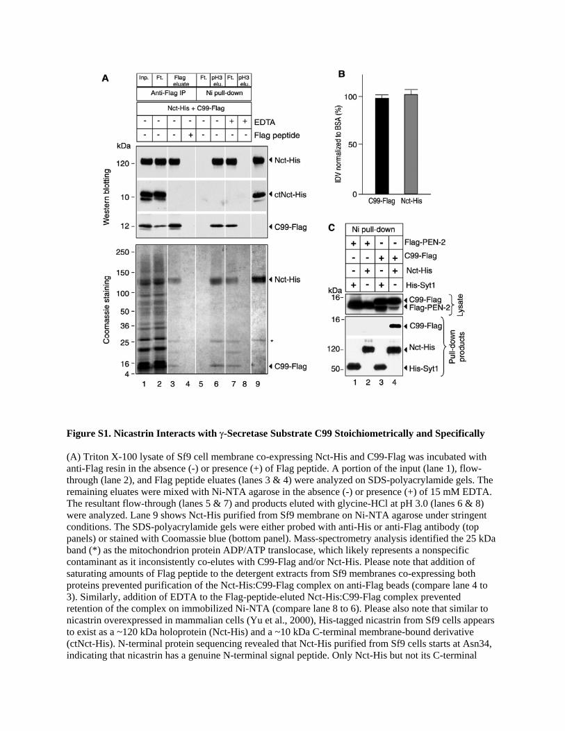
Figure S1. Nicastrin Interacts with γ-Secretase Substrate C99 Stoichiometrically and Specifically (A) Triton X-100 lysate of Sf9 cell membrane co-expressing Nct-His and C99-Flag was incubated with anti-Flag resin in the absence (-) or presence (+) of Flag peptide. A portion of the input (lane 1), flow-through (lane 2), and Flag peptide eluates (lanes 3 & 4) were analyzed on SDS-polyacrylamide gels. The remaining eluates were mixed with Ni-NTA agarose in the absence (-) or presence (+) of 15 mM EDTA. The resultant flow-through (lanes 5 & 7) and products eluted with glycine-HCl at pH 3.0 (lanes 6 & 8) were analyzed. Lane 9 shows Nct-His purified from Sf9 membrane on Ni-NTA agarose under stringent conditions. The SDS-polyacrylamide gels were either probed with anti-His or anti-Flag antibody (top panels) or stained with Coomassie blue (bottom panel). Mass-spectrometry analysis identified the 25 kDa band (*) as the mitochondrion protein ADP/ATP translocase, which likely represents a nonspecific contaminant as it inconsistently co-elutes with C99-Flag and/or Nct-His. Please note that addition of saturating amounts of Flag peptide to the detergent extracts from Sf9 membranes co-expressing both proteins prevented purification of the Nct-His:C99-Flag complex on anti-Flag beads (compare lane 4 to 3). Similarly, addition of EDTA to the Flag-peptide-eluted Nct-His:C99-Flag complex prevented retention of the complex on immobilized Ni-NTA (compare lane 8 to 6). Please also note that similar to nicastrin overexpressed in mammalian cells (Yu et al., 2000), His-tagged nicastrin from Sf9 cells appears to exist as a ~120 kDa holoprotein (Nct-His) and a ~10 kDa C-terminal membrane-bound derivative (ctNct-His). N-terminal protein sequencing revealed that Nct-His purified from Sf9 cells starts at Asn34, indicating that nicastrin has a genuine N-terminal signal peptide. Only Nct-His but not its C-terminal
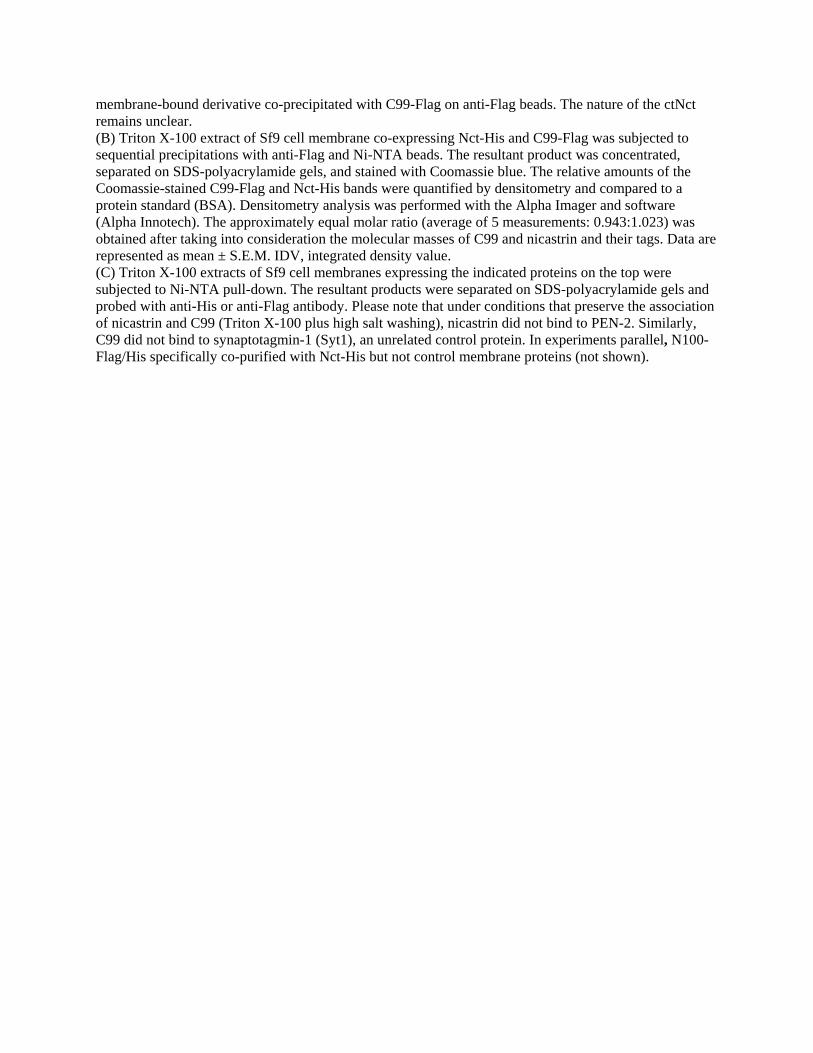
membrane-bound derivative co-precipitated with C99-Flag on anti-Flag beads. The nature of the ctNct remains unclear. (B) Triton X-100 extract of Sf9 cell membrane co-expressing Nct-His and C99-Flag was subjected to sequential precipitations with anti-Flag and Ni-NTA beads. The resultant product was concentrated, separated on SDS-polyacrylamide gels, and stained with Coomassie blue. The relative amounts of the Coomassie-stained C99-Flag and Nct-His bands were quantified by densitometry and compared to a protein standard (BSA). Densitometry analysis was performed with the Alpha Imager and software (Alpha Innotech). The approximately equal molar ratio (average of 5 measurements: 0.943:1.023) was obtained after taking into consideration the molecular masses of C99 and nicastrin and their tags. Data are represented as mean ± S.E.M. IDV, integrated density value. (C) Triton X-100 extracts of Sf9 cell membranes expressing the indicated proteins on the top were subjected to Ni-NTA pull-down. The resultant products were separated on SDS-polyacrylamide gels and probed with anti-His or anti-Flag antibody. Please note that under conditions that preserve the association of nicastrin and C99 (Triton X-100 plus high salt washing), nicastrin did not bind to PEN-2. Similarly, C99 did not bind to synaptotagmin-1 (Syt1), an unrelated control protein. In experiments parallel, N100-Flag/His specifically co-purified with Nct-His but not control membrane proteins (not shown).
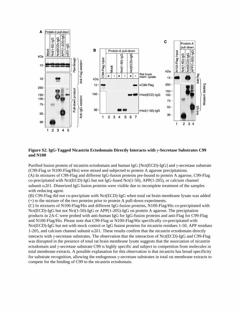
Figure S2. IgG-Tagged Nicastrin Ectodomain Directly Interacts with γ-Secretase Substrates C99 and N100 Purified fusion protein of nicastrin ectodomain and human IgG [Nct(ECD)-IgG] and γ-secretase substrate (C99-Flag or N100-Flag/His) were mixed and subjected to protein A agarose precipitations. (A) In mixtures of C99-Flag and different IgG-fusion proteins pre-bound to protein A agarose, C99-Flag co-precipitated with Nct(ECD)-IgG but not IgG-fused Nct(1-50), APP(1-205), or calcium channel subunit α2δ1. Dimerized IgG fusion proteins were visible due to incomplete treatment of the samples with reducing agent. (B) C99-Flag did not co-precipitate with Nct(ECD)-IgG when total rat brain membrane lysate was added (+) to the mixture of the two proteins prior to protein A pull-down experiments. (C) In mixtures of N100-Flag/His and different IgG-fusion proteins, N100-Flag/His co-precipitated with Nct(ECD)-IgG but not Nct(1-50)-IgG or APP(1-205)-IgG on protein A agarose. The precipitation products in 2A-C were probed with anti-human IgG for IgG-fusion proteins and anti-Flag for C99-Flag and N100-Flag/His. Please note that C99-Flag or N100-Flag/His specifically co-precipitated with Nct(ECD)-IgG but not with mock control or IgG fusion proteins for nicastrin residues 1-50, APP residues 1-205, and calcium channel subunit α2δ1. These results confirm that the nicastrin ectodomain directly interacts with γ-secretase substrates. The observation that the interaction of Nct(ECD)-IgG and C99-Flag was disrupted in the presence of total rat brain membrane lysate suggests that the association of nicastrin ectodomain and γ-secretase substrate C99 is highly specific and subject to competition from molecules in total membrane extracts. A possible explanation for this observation is that nicastrin has broad specificity for substrate recognition, allowing the endogenous γ-secretase substrates in total rat membrane extracts to compete for the binding of C99 to the nicastrin ectodomain.
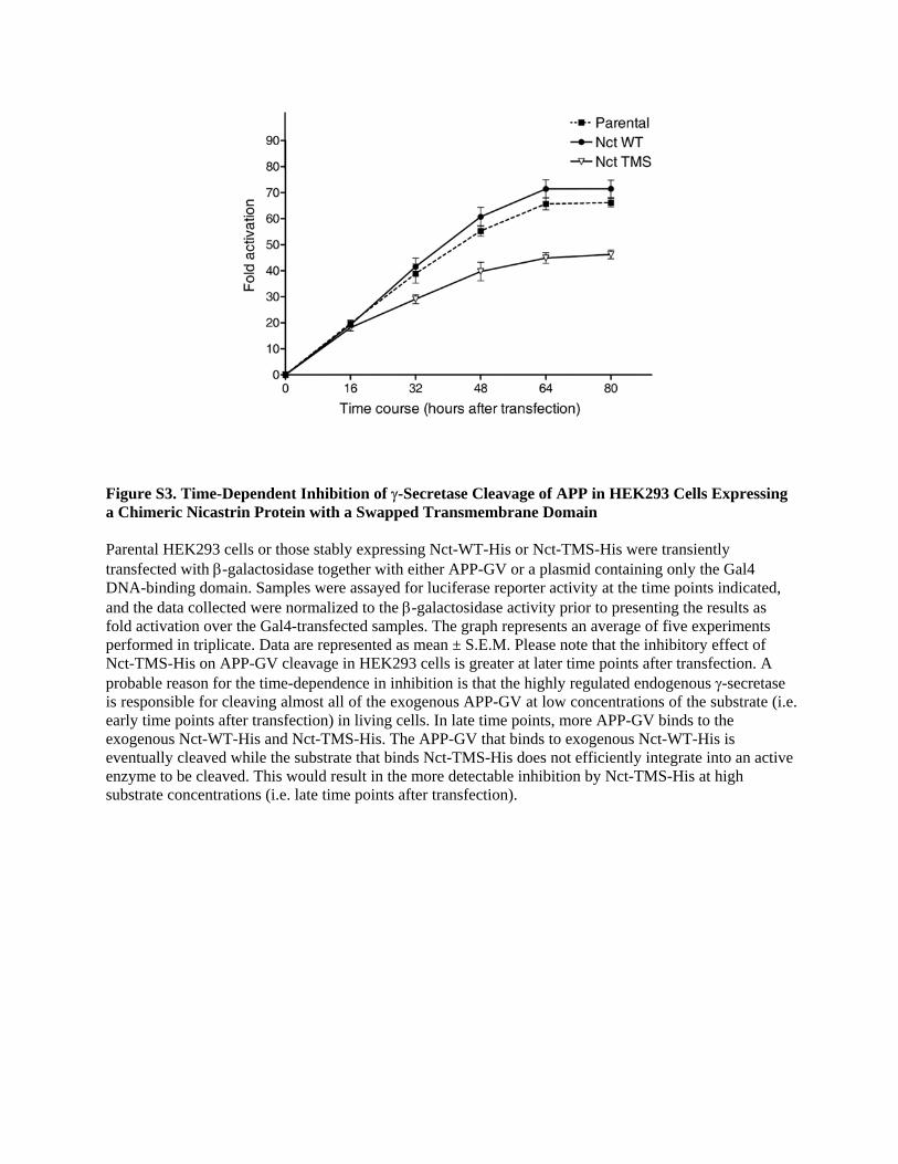
Figure S3. Time-Dependent Inhibition of γ-Secretase Cleavage of APP in HEK293 Cells Expressing a Chimeric Nicastrin Protein with a Swapped Transmembrane Domain Parental HEK293 cells or those stably expressing Nct-WT-His or Nct-TMS-His were transiently transfected with β-galactosidase together with either APP-GV or a plasmid containing only the Gal4 DNA-binding domain. Samples were assayed for luciferase reporter activity at the time points indicated, and the data collected were normalized to the β-galactosidase activity prior to presenting the results as fold activation over the Gal4-transfected samples. The graph represents an average of five experiments performed in triplicate. Data are represented as mean ± S.E.M. Please note that the inhibitory effect of Nct-TMS-His on APP-GV cleavage in HEK293 cells is greater at later time points after transfection. A probable reason for the time-dependence in inhibition is that the highly regulated endogenous γ-secretase is responsible for cleaving almost all of the exogenous APP-GV at low concentrations of the substrate (i.e. early time points after transfection) in living cells. In late time points, more APP-GV binds to the exogenous Nct-WT-His and Nct-TMS-His. The APP-GV that binds to exogenous Nct-WT-His is eventually cleaved while the substrate that binds Nct-TMS-His does not efficiently integrate into an active enzyme to be cleaved. This would result in the more detectable inhibition by Nct-TMS-His at high substrate concentrations (i.e. late time points after transfection).

Figure S4. APP- and Notch-Derived γ-Secretase Substrates N100 and C99 Are Not Cleaved in the Cell-free Extracts of Nct−/− Fibroblasts CHAPSO-solubilized cell membranes from wild-type (Nct+/+) and nicastrin knockout (Nct-/-) fibroblasts were subjected to incubation with Flag-tagged N100 or C99 for 0 and 3 hours at 37 oC in the absence (-) or presence (+) of γ-secretase inhibitor DAPT. Please note that there is no γ-secretase activity in the Nct-/- cell extracts, indicating that the Nct-/- fibroblasts are bona fide knockout cells.

Figure S5. Reconstitution and Characterization of γ-Secretase Activity in Sf9 Cells and in Lipids and Liposomes (A) Sf9 cells were co-infected with baculoviruses harboring different combinations of PS1, PEN-2, APH-1aL, and/or Nct as indicated. Equal amounts of membrane proteins extracted with CHAPSO were probed with appropriate antibodies (top four panels). The membrane extracts were subjected to incubation with N100-Flag/His at 37 °C in a reaction buffer containing 0.25% CHAPSO for 0 or 6 hours and investigated with anti-Flag (bottom two panels). Please note that PS1 exists only as a ~ 50 kDa holoprotein when expressed alone or co-expressed with any one or two γ-secretase components (lanes 2-4 & not shown). In membranes from Sf9 cells infected with baculoviruses co-expressing all four γ-secretase components, PS1 undergoes endoproteolysis to generate the endoproteolytic fragments (lane 5). N100-Flag/His could be efficiently cleaved to generate NICD’-Flag/His in CHAPSO-solubilized membrane extracts of Sf9 cells expressing all four proteins (lane 5). Endogenous proteins in Sf9 cells do not appear to cleave N100-Flag/His intramembranously. Also, APH-1aL runs as a doublet under certain SDS-PAGE conditions (top third panel & Fig. 5B). (B) Purified γ-secretase from Sf9 cells co-expressing all four γ-secretase subunits (please refer to Fig. 5B) was subjected to incubation with N100-Flag/His in the presence of the indicated amounts of phosphatidylcholine (PC) at 37 °C for 0 or 4 hours. Resultant samples were probed with anti-Flag antibody. (C) Purified γ-secretase was reconstituted in a mixture of cholesterol and phosphatidylcholine at the indicated molar ratio after CHAPSO was removed by dialysis and ultracentrifugation. The resultant proteoliposomes were incubated with N100-Flag/His at 37 °C for 0 hours or overnight and investigated with anti-Flag antibody. The purified γ-secretase was enzymatically inactive unless reconstituted with lipids or liposomes, and was sensitive to γ-secretase inhibitors such as L685, 458 and DAPT. The in vitro generated NICD’-Flag/His reacted with the anti-cleaved Notch-1 (Val1744) antibody that specifically recognizes the N-terminus of Notch intracellular domain. Five cycles of N-terminal protein sequencing confirmed that the first five amino acids of the resultant NICD’-Flag/His are Val Leu Leu Ser Arg. Thus, the purified recombinant γ-secretase preparation cleaved N100-Flag/His intramembranously at the physiological S3 site of Notch to generate a Notch intracellular fragment starting at Val1744.
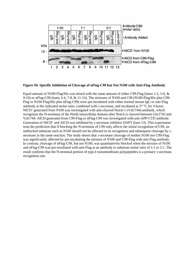
Figure S6. Specific Inhibition of Cleavage of nFlag-C99 but Not N100 with Anti-Flag Antibody Equal amount of N100-Flag/His was mixed with the same amount of either C99-Flag (lanes 1-2, 5-6, & 9-10) or nFlag-C99 (lanes 3-4, 7-8, & 11-13). The mixtures of N100 and C99 (N100-Flag/His plus C99-Flag or N100-Flag/His plus nFlag-C99) were pre-incubated with either normal mouse IgG or anti-Flag antibody at the indicated molar ratio, combined with γ-secretase, and incubated at 37 oC for 4 hours. NICD’ generated from N100 was investigated with anti-cleaved Notch-1 (Val1744) antibody, which recognizes the N-terminus of the North intracellular domain after Notch is cleaved between Gly1743 and Val1744. AICD generated from C99-Flag or nFlag-C99 was investigated with anti-APP-CTD antibody. Generation of NICD’ and AICD was inhibited by γ-secretase inhibitor DAPT (lane 13). This experiment tests the prediction that if blocking the N-terminus of C99 only affects the initial recognition of C99, an unblocked substrate such as N100 should not be affected in its recognition and subsequent cleavage by γ-secretase in the same reaction. The study shows that γ-secretase cleavage of neither N100 nor C99-Flag was significantly affected by pre-incubating the mixture of N100 and C99-Flag with anti-Flag antibody. In contrast, cleavage of nFlag-C99, but not N100, was quantitatively blocked when the mixture of N100 and nFlag-C99 was pre-incubated with anti-Flag at an antibody to substrate molar ratio of 1:1 or 2:1. The result confirms that the N-terminal portion of type-I transmembrane polypeptides is a primary γ-secretase recognition site.
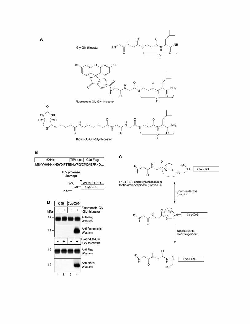
Figure S7. Strategy of Native Chemical Ligation (A) Structures of synthesized reagents (Gly-Gly dipeptide; fluorescein-Gly-Gly; and biotin-LC-Gly-Gly) containing a C-terminal thioester group. (B) Proteolytic cleavage approach to generate Cys-C99, which starts C99 with an N-terminal cysteine residue. (C) Native chemical ligation reaction takes place as follows. First, a reversible transthioesterification reaction between the synthesized C-terminal thioester group and the sulfhydryl group from the N-terminal cysteine residue from Cys-C99 gives rise to a ligated thioester cyclic intermediate. Second, the thioester intermediate undergoes an irreversible spontaneous S→N acyl rearrangement to form a stable ligated product (modified from Dawson and Kent, 2000 and Muir, 2003). (D) Top two panels: C-terminally Flag-tagged C99 and Cys-C99 were subjected to native chemical ligation in the presence (+) or absence (-) of fluorescein-Gly-Gly-thioester. The resultant products were probed on Western blots using anti-Flag antibody or anti-fluorescein antibody. Bottom two panels: C-terminally Flag-tagged C99 and Cys-C99 were subjected to native chemical ligation in the presence (+) or absence (-) of biotin-LC-Gly-Gly-thioester. The resultant products were probed with anti-Flag or anti-biotin antibody. Please note that ligation products were generated specifically in reactions containing Cys-C99 (but not C99) and thioester derivatives (compare lane 4 to lanes 1-3).