Embed Size (px)
Citation preview
1
Molecular Cell, Volume 30
Supplemental Data
Structural and Functional Studies
of Nup107/Nup133 Interaction and Its Implications
for the Architecture of the Nuclear Pore Complex Thomas Boehmer, Sandra Jeudy, Ian C. Berke, and Thomas U. Schwartz Supplemental Experimental Procedures Plasmid construction and site-directed mutagenesis
For protein binding assays, the coding region of human Nup107 for residues 1-925 (full-length), 1-644 and 658-925, respectively, were cloned between the BamHI and NotI sites of pGEX-4T2 (GE Healthcare). The coding region of human Nup133 for residues 1-1156 (full-length), 517-1156, 517-820, 830-1156, 880-1156, 934-1156 and 987-1156, respectively, were cloned between the NdeI and NotI sites of pET28a (Novagen) to generate N-terminally His-tagged proteins.
For immunofluorescence microscopy, hNup107 coding sequences for residues 1-925, 1-669, and 663-925, respectively, were cloned between the BglII and SalI sites of pEGFP-N2 (Clontech), full-length hNup133 was cloned between the XhoI and SalI sites of the same vector.
For crystallographic studies, coding sequences for hNup107658-925 and hNup133934-1156 were cloned into a bi-cistronic bacterial expression vector as described (Boehmer and Schwartz, 2007).
Site-directed mutagenesis of Nup107 (A878E and L901E) and Nup133 (L973E and L976E) was performed with the QuikChange site-directed mutagenesis kit (Stratagene).
For the crystal construct lacking the protruding helix α6 of Nup107, called hNup107658-
925dF, replacement of residues 772-801 in hNup107658-925 with a flexible linker of sequence GGSGGS was achieved through standard PCR methods. hNup107658-925dF and hNup133934-1156 were then cloned into the first and second multiple cloning sites of pET-Duet1 (Novagen), resulting in His-tagged hNup107658-925dF and untagged hNup133934-1156. Protein purification
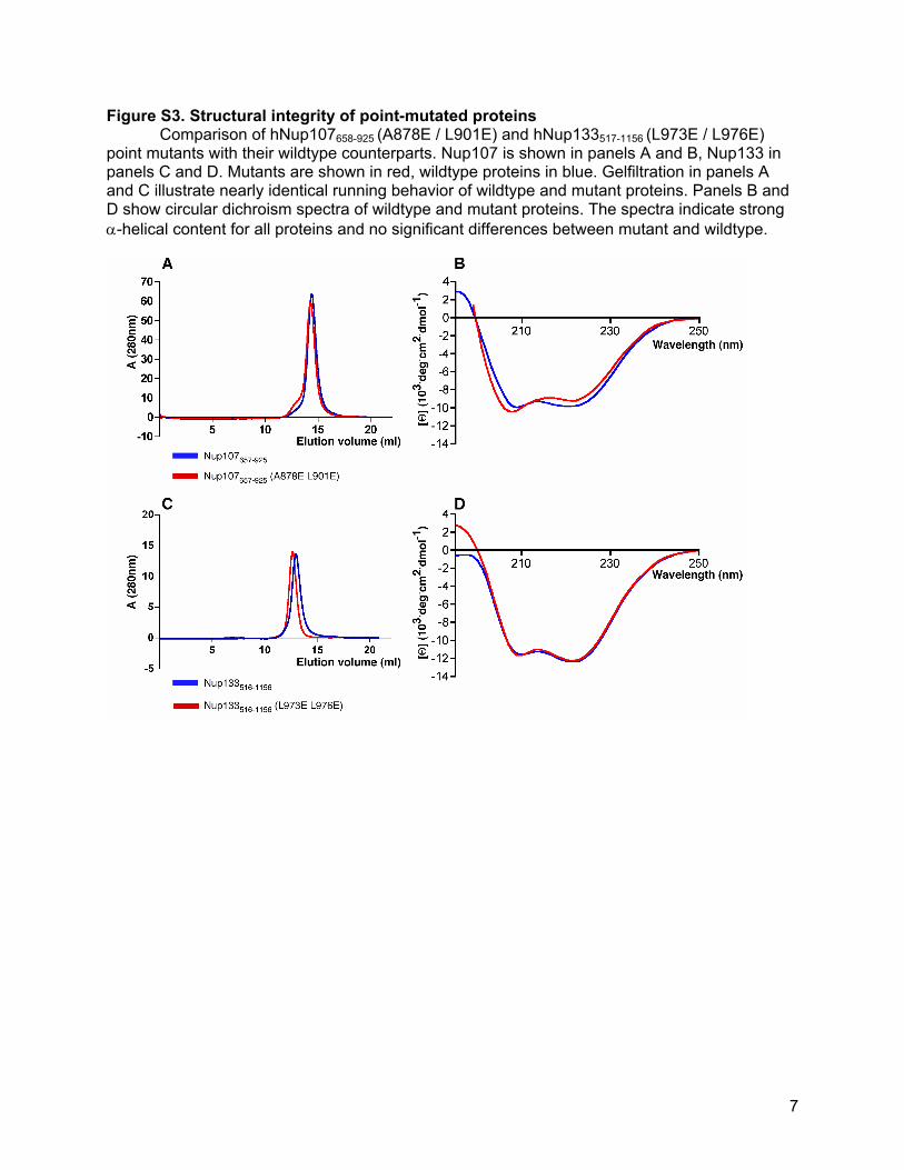
Proteins used in binding assays were purified according to standard chromatographic protocols. The complex of hNup107658-925 and hNup133934-1156 used for crystallization, as well as a seleno-methionine derivative, were purified as described (Boehmer and Schwartz, 2007). The hNup107658-925dF/hNup133934-1156 complex was co-purified via metal-affinity chromatography, followed by anion exchange and size-exclusion chromatography. To test structural integrity of the mutants hNup107658-925 (A878E / L901E) and hNup133517-1156 (L973E / L976E) the proteins were purified uncomplexed and were compared to the respective wildtype proteins. Gelfiltration shown in Figure S3 were carried out using a Superdex 200 HR10/300 (GE Healthcare) column in 10 mM potassium phosphate pH 8.0, 250 mM NaCl, 0.1mM EDTA, 1mM DTT.
2
Circular Dichroism The experiments were carried out at 25 ºC using an Aviv Model 202 spectrometer. Spectra were recorded at 1 mM peptide concentration in a cuvette with 1 mm optical pathlength. Spectra were recorded in 1nm steps, averaged for 8 sec, and corrected for buffer baseline. Crystallization, Data Collection and Structure Refinement
hNup107658-925/hNup133934-1156 was crystallized as described (Boehmer and Schwartz, 2007). hNup107658-925dF/hNup133934-1156 was crystallized by hanging drop vapor diffusion. Total protein concentration was 10-15mg/ml. The reservoir solution contained 25-27% (w/v) PEG 2000MME, 500 mM KSCN, 100 mM Hepes pH 7.4. Crystals grew in 3-7 days at 17°C were cryo-protected by washing in reservoir solution supplemented with 9% (v/v) glycerol and were flash-frozen in LN2. The structure of hNup107658-925/hNup133934-1156 was solved with the single anomalous dispersion (SAD) technique using the SeMet derivative. The positions of 13 (out of 15 possible) were found with the program SHELXD and were used for phasing with SHARP. The resulting solvent-flattened electron density map was used to build the model with Coot. Refinement was carried out using the PHENIX program suite. The current model of hNup107658-925/hNup133934-
1156 includes residues 668-924 of hNup107, and residues 934-1156 of hNup133. Several side chains and loops within the C-terminal Nup133 domain have been omitted due to limited clarity of the electrondensity map in that area. The hNup107658-925dF/hNup133934-1156 structure was solved by molecular replacement with the hNup107658-925/hNup133934-1156 structure using Phaser. Refinement was carried out with PHENIX, the model was built with Coot.
3
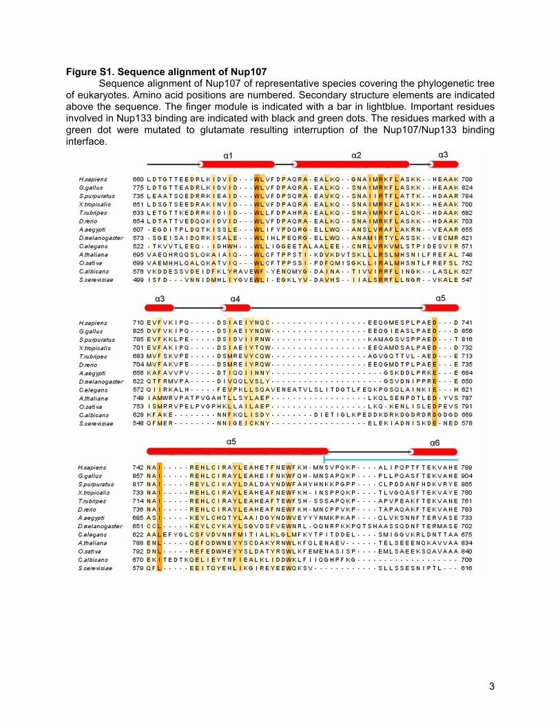
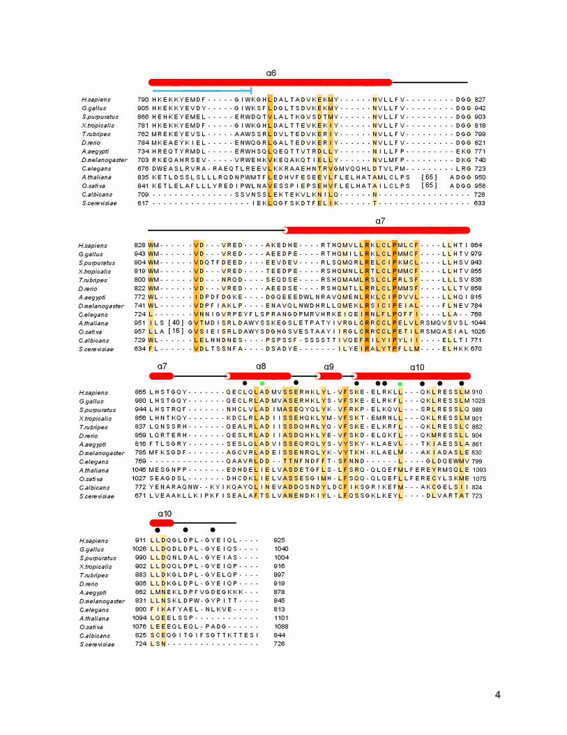
Figure S1. Sequence alignment of Nup107 Sequence alignment of Nup107 of representative species covering the phylogenetic tree
of eukaryotes. Amino acid positions are numbered. Secondary structure elements are indicated above the sequence. The finger module is indicated with a bar in lightblue. Important residues involved in Nup133 binding are indicated with black and green dots. The residues marked with a green dot were mutated to glutamate resulting interruption of the Nup107/Nup133 binding interface.
4
5
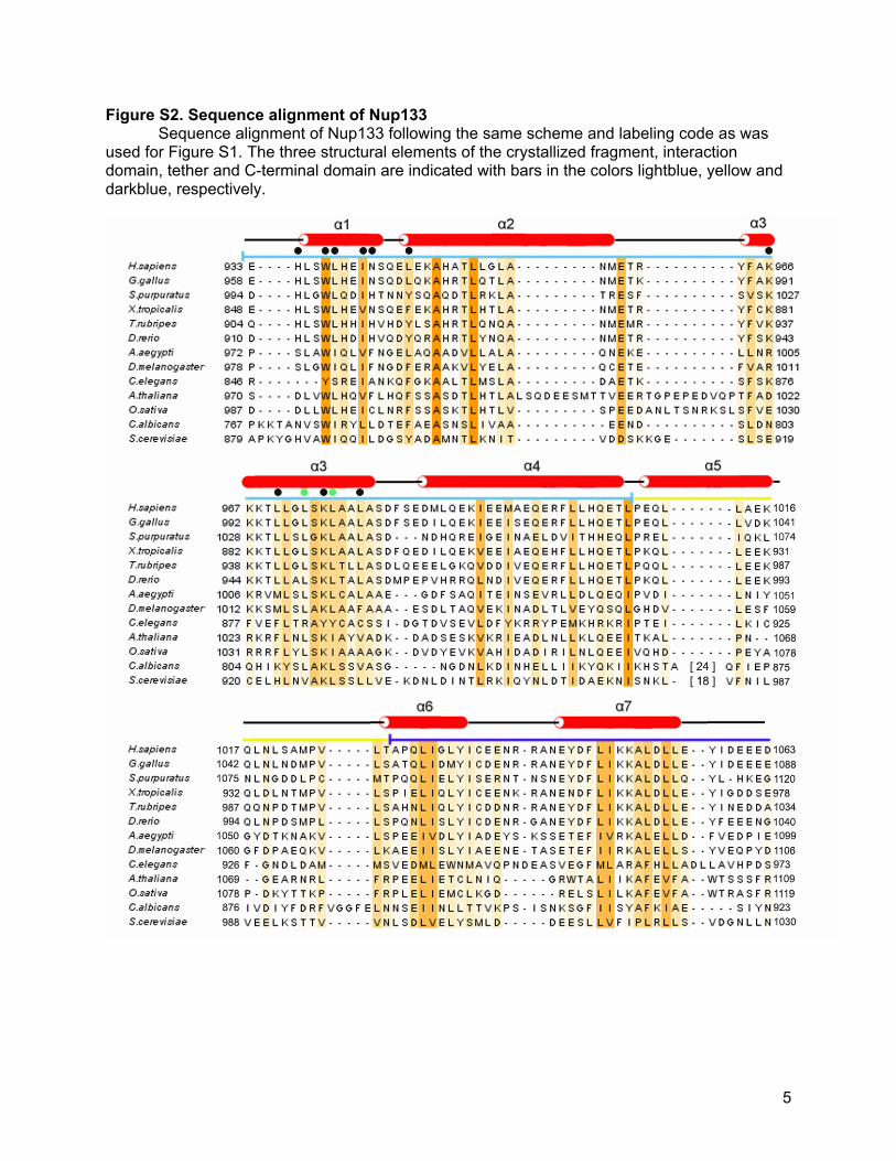
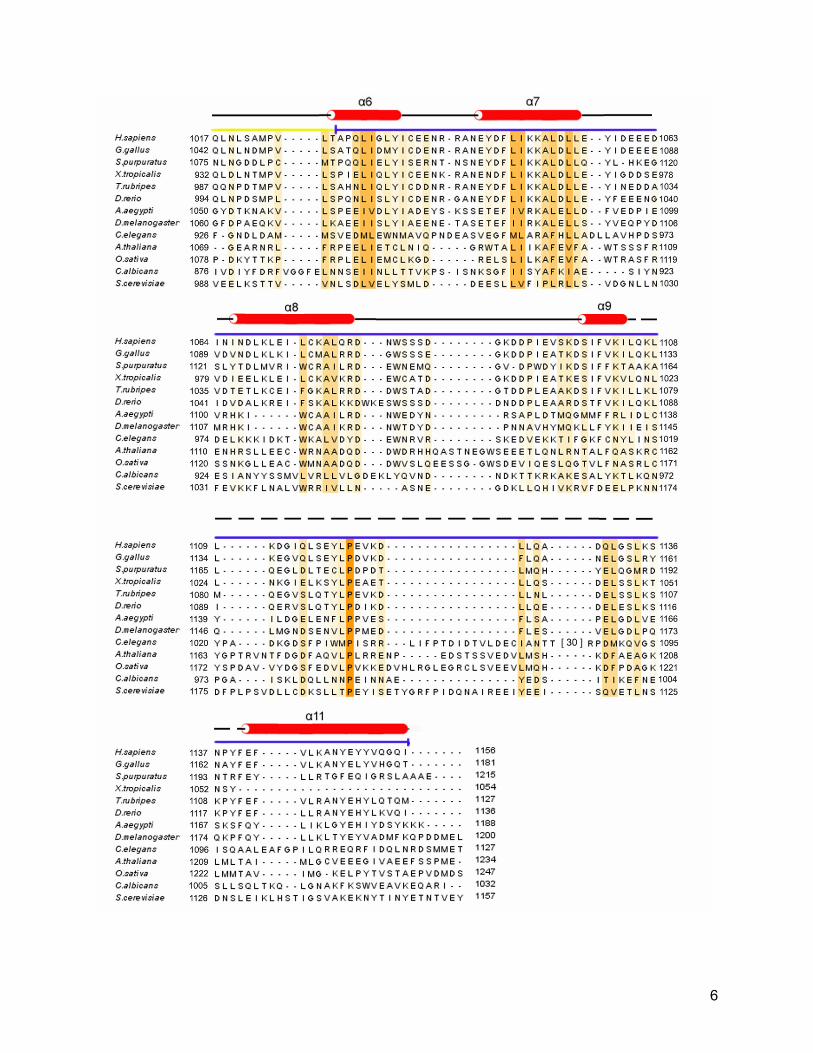
Figure S2. Sequence alignment of Nup133 Sequence alignment of Nup133 following the same scheme and labeling code as was
used for Figure S1. The three structural elements of the crystallized fragment, interaction domain, tether and C-terminal domain are indicated with bars in the colors lightblue, yellow and darkblue, respectively.
6
7
Figure S3. Structural integrity of point-mutated proteins Comparison of hNup107658-925 (A878E / L901E) and hNup133517-1156 (L973E / L976E) point mutants with their wildtype counterparts. Nup107 is shown in panels A and B, Nup133 in panels C and D. Mutants are shown in red, wildtype proteins in blue. Gelfiltration in panels A and C illustrate nearly identical running behavior of wildtype and mutant proteins. Panels B and D show circular dichroism spectra of wildtype and mutant proteins. The spectra indicate strong α-helical content for all proteins and no significant differences between mutant and wildtype.


![Electronic Supplementary Information interactions in the ...Electronic Supplementary Information Magnetic and structural correlations in [Fe(nsal2trien)] salts: The role of cation-anion](https://img.pdfslide.net/doc/110x75/603efa159819b50d960552c8/electronic-supplementary-information-interactions-in-the-electronic-supplementary.jpg)