Embed Size (px)
Citation preview

1
Molecular Cell, Volume 38
Supplemental Information
Systematic Analysis of Essential Genes Reveals New Regulators of G Protein Signaling Steven D. Cappell, Rachael Baker, Dorota Skowyra, and Henrik G. Dohlman
SUPPLEMENTAL INFORMATION INVENTORY
Supplemental Figures: Figure S1, related to Figure 2 Genes identified in our screen and listed in Figure 2 were
also analyzed using Osprey to identify any known
interactions either among the hits or between hits and known
pheromone components. Figure S1 shows the interactions
that were identified.
Table S1, related to Figure 2 We identified 92 essential genes required for pheromone
signaling and listed these genes in Figure 2. However, we
excluded an additional 97 genes due to their known role in
cellular processes that would result in false-positives in our
transcriptional reporter assay. These 97 genes are listed in
Table S1.
Figure S2, related to Table 1 The high confidence hits from our screen listed in Table 1
were validated using a dual-reporter system. Figure S2
shows the immunoblots from this experiment.
Figure S3, related to Figure 3 We chose 6 essential genes to further characterize using
transcriptional reporter assays and measurement of
activation of the MAP kinases as shown in Figure 3. Figure
S3 shows the measurement of total levels of the MAPK Fus3

2
and quantification of bands to demonstrate that
phosphorylated Fus3 changes relative to total Fus3.
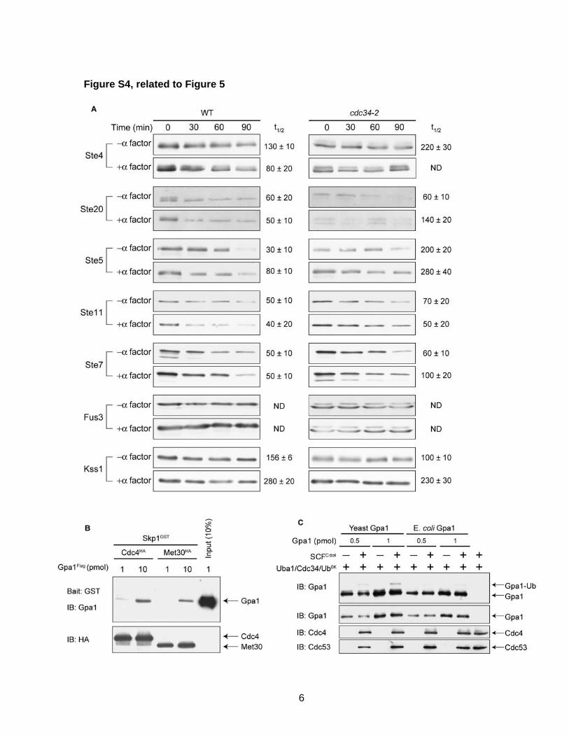
Figure S4, related to Figure 5 We tested the stability of two pheromone pathway
components in an SCF-mutant strain as shown in Figure 5.
As a control, we also tested the stability of 7 other pathway
components and display the results in Figure S4. We show
that SCFMet30 cannot ubiquitinate Gpa1 in vitro (Figure 5). As
a control, we show that Met30 can bind weakly to Gpa1,
demonstrating it is functional. We also show that Gpa1
expressed in E. coli cannot be ubiquitinated by SCFCdc4 in
vitro.
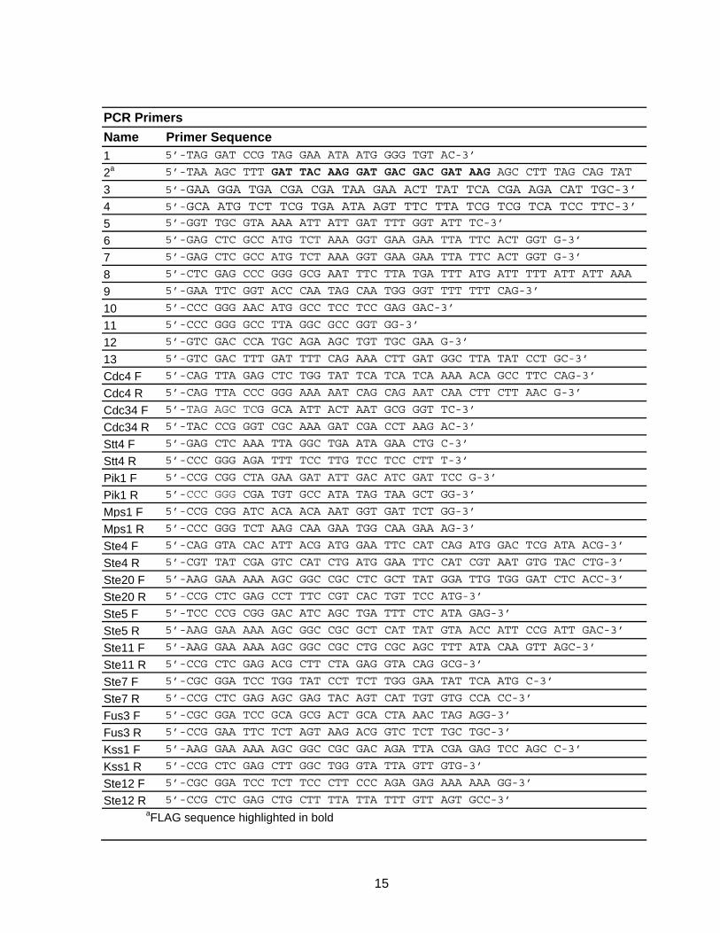
Supplemental Experimental Procedures Table of PCR Primers Supplemental References

3
Figure S1, related to Figure 2
Figure S1. Many of the hits from the essential-gene screen have known
interactions with pheromone pathway components, related to Figure 2.
The Osprey Network Visualization System software was used to generate an interaction
network based on known genetic and physical interactions from BioGRID.

4
Figure S2, related to Table 1
Figure S2. Hits from the essential-gene screen regulate G protein signaling
upstream of translation, related to Table 1.
(A-L) The indicated TetO7 strains expressing the pRS315 AR/FG dual reporter plasmid
were treated with 10μg/mL doxycycline for 15hrs and 3μM α factor for 30min. Cell
lysates were probed with GFP and RFP antibodies. Five additional strains, TetO7-RIO2,
TetO7-KIC1, TetO7-SSY1, TetO7-PMA1 and TetO7-SLN1, were also tested. Strains
representing RIO2, KIC1, and SSY1 exhibited changes in RFP expression. Strains
representing PMA1 and SLN1 showed no changes in RFP expression but were not
considered further.

5
Figure S3, related to Figure 3
Figure S3. Knock-down of essential genes affects Fus3 activation.
(A-G) The indicated TetO7 strains were treated with 10μg/mL doxycycline for 15hrs and
3μM α factor for 30min. Cell lysates were probed with phospho-p42/44 (P-Fus3, P-Kss1)
and Fus3 antibodies. Bands were quantified by densitometry and the ratio of P-
Fus3:Fus3 is shown below each immunoblot. Results are the mean ± S.E. (n=3).
6
Figure S4, related to Figure 5

7
Figure S4. The SCF does not destabilize any positive regulators of G protein
signaling and primarily functions to ubiquitinate Gpa1, related to Figure 5.
(A) The indicated TAP-fusion genes were integrated into wild-type and cdc34-2 ts
strains. Cycloheximide was administered at time zero and protein levels measured by
immunoblotting with protein A antibodies.
(B) GST-Skp1/Cdc4-HA or Met30-HA complexes were immobilized on Glutathione
SepharoseTM resin and incubated with purified Gpa1-Flag, followed by washing and
analysis of the bound proteins with Gpa1-antibodies to detect co-purification of Gpa1.
(C) In vitro ubiquitination of Gpa1 expressed in yeast and E coli, which lacks most post-
translational modifications.

8
Table S1: Essential Gene Hits Involved in Global Gene Transcription and Translation, related to Figure 2 Systematic Name
Standard Name Go Process
Systematic Name
Standard Name Go Process
YAL043C PTA1 RNA Processing YKR062W TFA2 TranscriptionYBL014C RRN6 Transcription YKR086W PRP16 RNA ProcessingYBL035C POL12 DNA Replication YLL008W DRS1 RNA ProcessingYBL076C ILS1 Protein Biosynthesis YLL035W GRC3 RNA ProcessingYBR079C RPG1 Protein Biosynthesis YLR009W RLP24 Protein BiosynthesisYBR088C POL30 DNA Replication YLR033W RSC58 TranscriptionYBR193C MED8 Transcription YLR060W FRS1 Protein BiosynthesisYBR253W SRB6 Transcription YLR129W DIP2 RNA ProcessingYCR042C TAF2 Transcription YLR141W RRN5 TranscriptionYCR052W RSC6 Transcription YLR249W YEF3 Protein BiosynthesisYDL060W TSR1 RNA Processing YLR275W SMD2 RNA ProcessingYDL140C RPO21 Transcription YLR276C DBP9 RNA ProcessingYDL150W RPC53 Transcription YLR291C GCD7 Protein BiosynthesisYDL153C SAS10 RNA Processing YML046W PRP39 RNA ProcessingYDR045C RPC11 Transcription YML069W POB3 DNA ReplicationYDR091C RLI1 Protein Biosynthesis YML114C TAF65 TranscriptionYDR145W TAF12 Transcription YML127W RSC9 TranscriptionYDR167W TAF10 Transcription YMR005W TAF4 TranscriptionYDR228C PCF11 RNA Processing YMR061W RNA14 RNA ProcessingYDR341C YDR341C Protein Biosynthesis YMR093W YMR093W RNA ProcessingYDR398W UTP5 RNA Processing YMR235C RNA1 RNA ProcessingYDR460W TFB3 Transcription YMR236W TAF9 TranscriptionYDR489W YDR489W DNA Replication YNL039W TFC5 TranscriptionYEL034W YEL034W Protein Biosynthesis YNL113W RPC19 TranscriptionYER029C SMB1 RNA processing YNL151C RPC31 TranscriptionYER171W RAD3 Transcription YNL216W RAP1 TranscriptionYER172C BRR2 RNA Processing YNL221C YNL221C RNA ProcessingYFR037C RSC8 Transcription YNL247W YNL247W Protein BiosynthesisYGL122C NAB2 RNA Processing YOL005C RPB11 TranscriptionYGL207W SPT16 Transcription YOL021C DIS3 RNA ProcessingYGR005C TFG2 Transcription YOL077c YOL077C Protein BiosynthesisYGR030C POP6 RNA Processing YOL139C CDC33 Protein BiosynthesisYGR094W VAS1 Protein Biosynthesis YOR116C RPO31 TranscriptionYGR158C MTR3 RNA Processing YOR145c YOR145C RNA ProcessingYHR019C DED81 Protein Biosynthesis YOR151C RPB2 TranscriptionYHR062C RPP1 RNA Processing YOR159C SME1 RNA ProcessingYHR069C RRP4 RNA Processing YOR194C TOA1 TranscriptionYHR122W YHR122W Transcription YOR204W DED1 RNA ProcessingYHR164C DNA2 DNA replication YOR224C RPB8 TranscriptionYHR170W NMD3 Protein Biosynthesis YOR340C RPA43 TranscriptionYJL011C RPC17 Transcription YPL016W SWI1 TranscriptionYJL069C UTP18 RNA Processing YPL043W NOP4 RNA processingYJL081C ARP4 Transcription YPL093W NOG1 RNA ProcessingYKL059C YKL059C RNA Processing YPL126W NAN1 RNA ProcessingYKL125W RRN3 Transcription YPL228W CET1 RNA ProcessingYKL180W RPL17 Protein Biosynthesis YPL266W DIM1 RNA ProcessingYKL186C MTR2 Ribosome Biogenesis YPR016C YPR016C Protein BiosynthesisYKR008W RSC4 Transcription YPR187W RPO26 Transcription

9
SUPPLEMENTAL EXPERIMENTAL PROCEDURES
Strains and Plasmids
Standard procedures for the growth, maintenance, and transformation of yeast
and bacteria and for the manipulation of DNA were used throughout. Yeast
Saccharomyces cerevisiae strains used in this study were BY4741 (MATa leu2Δ met15Δ
his3-1 ura3Δ), MTY235 (MATa ade2-1 his3-11,15 leu2-3,112 trp1-1 ura3-1 can1-100),
MTY670 (MTY235 cdc34-2), and MTY668 (MTY235 cdc4-1) (provided by Mike Tyers,
Samuel Lunenfield Research Institute) (Tang et al., 2005), BY4741-derived strains
containing a C-terminal tandem affinity purification (TAP)-tag (Yeast TAP-Fusion Library,
Open Biosystems), and the BY4741-derived strain R1158 (MATa URA3::CMV-tTA leu2Δ
met15Δ his3-1 ura3Δ) (Hughes et al., 2000). The tetracycline-repressible strains were
purchased as the yeast Tet-promoter Hughes Collection (yTHC, Open Biosystems)
(Mnaimneh et al., 2004).
Yeast shuttle plasmids used were pRS315 (CEN, ampR, LEU2), and pRS316
(CEN, ampR, URA3). Expression plasmids described previously were pRS423-FUS1-
lacZ (Hoffman et al., 2002), pRS316-ADH1, pRS316-ADH1-GPA1 (Song et al., 1996),
pFAGa-mRFP1-KanMX6 (Huh et al., 2003), pUG35 (provided by Johannes Hegemann,
Heinrich-Heine-Universität), YCp50-STE11-4 (from George Sprague, University of
Oregon) (Stevenson et al., 1992), and pRS315-GAL-STE4 (Dohlman et al., 1995).
Plasmid pRS316-ADH-GPA1-Flag was constructed by PCR amplification of a 384 bp
fragment of GPA1 from pRS316-ADH-GPA1 using primers 1 and 2 (see Table of PCR
primers below). Plasmid pRS316-ADH-GPA1Δ128-236-Flag was constructed by
QuikChange (Stratagene) using primers 3 and 4 to remove the 324 bp fragment
corresponding to amino acids 128-236. Plasmid pRS315-STE11 was constructed by
PCR amplification of STE11 from BY4741 genomic DNA, followed by SacI and XmaI

10
digestion and ligation into the corresponding sites of pRS315. Plasmid pRS315-STE11-4
was constructed by engineering the single point mutation Thr-596-Ile (Stevenson et al.,
1992) into pRS315-STE11 using QuikChange and primer 5 and its complement. Rescue
plasmids for CDC4, CDC34, and STT4 were constructed by PCR amplification of each
gene from BY4741 genomic DNA, followed by SacI and XmaI digestion and ligation into
the corresponding sites of pRS315. Rescue plasmids for PIK1 and MPS1 were made in
a similar manner except that SacII was used in place of SacI.
The pRS316-ADH1-RFP/FUS1-GFP (AR/FG) dual reporter was constructed
using the steps outlined below. The plasmid pRS316-ADH1-GFP was constructed by
PCR amplification of GFP from the plasmid pUG35 using primers 6 and 7 including SacI
sites, followed by SacI digestion and insertion into the corresponding site in pRS316-
ADH1. The ADH1 terminator sequence (ADH1t) (from the stop codon to 600bp
downstream) was PCR amplified from genomic DNA with primers 8 and 9 including
XmaI and SalI sites. The resulting PCR product was digested with XmaI and SalI and
inserted into the corresponding sites in pRS316-ADH1-GFP, resulting in pRS316-
ADH1p-ADH1t-GFP. RFP was PCR amplified from pFA6a-mRFP1-KanMX6 using
primers 10 and 11 with XmaI sites. This fragment was ligated into the corresponding
XmaI sites of pRS316-ADH1p-ADH1t-GFP, resulting in the plasmid pRS316-ADH1p-RFP-
ADH1t-GFP. The FUS1 promoter (FUS1p) (600bp upstream of the start codon of FUS1)
was PCR amplified from genomic DNA using primers 12 and 13 containing SalI sites.
The resulting fragment was digested with SalI and inserted into the corresponding sites
of pRS316-ADH1p-RFP-ADH1t-GFP resulting in the plasmid pRS316-ADH1p-RFP-
ADH1t-FUS1-GFP (designated pRS316-AR/FG). pRS315-AR/FG was constructed by
digestion of pRS316-AR/FG with PvuI and ligation into the corresponding sites in
pRS315.

11
Bioinformatics
Physical and genetic interactions among the genes identified in the screen were
analyzed using Osprey Network Visualization System (Breitkreutz et al., 2002) which
incorporates published data from the Biological General Repository for Interaction
Datasets (BioGRID) (Stark et al., 2006). Functional categories were assigned based on
Gene Ontology annotations using Functional Clustering in Osprey. Genes involved in
multiple GO processes were assigned a single GO term based on Osprey’s hierarchical
GO process order. Hierarchical clustering of TetO7-Strain phenotypes was conducted
with the open source software Cluster 3.0 (de Hoon et al., 2004) using uncentered
correlation and centroid linkage. The generated clustering data was visualized with the
open source software Java TreeView (v1.1.3) (Saldanha, 2004).
TAP-Fusion protein turnover screen
TAP-fusion genes were PCR amplified and integrated into MTY235 and cdc34-2
cells. Cells were grown at room temperature to A600nm ~0.25, shifted to 37oC for 3hrs
treated with 3 μM α factor for 1hr, and treated with cycloheximide for up to 90min.
Protein extracts were resolved by 7.5% SDS-PAGE and immunoblotting with protein A
(P3775, Sigma-Aldrich) antibodies at 1:50,000. Experiments were performed in triplicate,
and bands were quantified by densitometry.
Cell-Extract Preparation and Immuno-Blot Analysis
The yeast TetO7 strains were grown in selective medium to A600nm~0.8 and re-
inoculated at 1:80 into medium containing doxycycline at a final concentration of 10μg/ml
and grown to A600nm~0.8. Cell cultures were then divided in half, and either treated with
3μM α factor pheromone or left untreated at 30oC for 30min. Protein extracts were
produced by glass bead lysis in trichloroacetic acid (TCA) as previously described (Lee

12
and Dohlman, 2008). Protein extracts were resolved by 12% SDS-PAGE and
immunoblotting with Phospho-p44/42 MAPK antibodies (9101L, Cell Signaling
Technology) at 1:500, Fus3 antibodies (sc-6773, Santa Cruz Biotechnology, inc.) at
1:500, GFP antibodies (632375, BD Biosciences) at 1:500, dSRed antibodies (632496,
Clontech) at 1:1000, and G6PDH antibodies (A9521, Sigma-Aldrich) at 1:100,000.
Immunoreactive species were visualized by chemiluminescent detection (PerkinElmer
Life Sciences LAS) of horseradish peroxidase-conjugated antibodies (170-5047 and
170-5046, Bio-Rad). Protein concentration was determined by Dc protein assay (Bio-
Rad Laboratories). Where indicated, TetO7 cells were transformed with pRS315-GAL-
STE4, pRS315-STE11-4, pRS315-ADH-RFP/FUS1-GFP, or empty vector, and grown in
selective medium containing 2% (w/v) dextrose or galactose to induce STE4 expression.
Co-Immunoprecipitation Assay
Insect cell lysates containing GST-Skp1 and either Cdc4-HA or Met30-HA where
mixed with Glutathione SepharoseTM 4 Fast Flow resin (GE Healthcare) for 1hr rotating
at 4oC. The beads were then washed 3x with 50x bead-bed volume of binding buffer
(50mM Tris-HCl, pH 7.5, 150mM KCl, 0.5% NP-40, 0.2mM dithiothreitol, 2mM MgCl2,
20μM GDP, 10mM NaF, 10mM β-glycerol phosphate, 1mM sodium orthovanadate, and
proteinase inhibitor tablets). Gpa1-Flag purified from yeast was added to the beads and
incubated for 2hrs at 4oC with rotating. The beads were washed 3x with 50x bead-bed
volume of binding buffer for 5min each with rocking and 3x quick washes with 50x bead-
volume of binding buffer. Bound protein was then eluted in 2x bead-bed volume of
binding buffer supplemented with 20mM glutathione. Protein samples were resolved by
10% SDS-Page and immunoblotting with Gpa1 antibodies at 1:1000 and HA antibodies
(3F10, Roche Applied Sciences) at 1:2000.

13
Preparation and Purification of Recombinant Proteins
BY4741 yeast cells were transformed with pRS316-ADH1-GPA1-Flag and
pRS316-ADH1-GPA1Δ128-236-Flag and grown to early log phase (A600nm ~1.0) before
harvesting by centrifugation. The cell pellet was frozen in liquid nitrogen and lysed by
grinding cells blast-frozen in a 1:0.7 ratio of lysis buffer (50mM Tris-HCl, pH 7.5, 400mM
KCl, 0.1% Triton, 0.2mM dithiothreitol) supplemented with 20μM GDP, 10mM NaF,
10mM β-glycerol phosphate, 1mM sodium orthovanadate, 1mM phenylmethylsulfonyl
fluoride and 1 proteinase inhibitor tablet per 50mL (11873580001, Roche Applied
Science). The cell lysate was thawed on ice, and centrifuged at 15,000xg for 30min at
4oC. The supernatant was transferred and incubated with EZview anti-Flag M2 beads
(Sigma-Aldrich) for 2hrs rotating at 4oC. Beads were harvested by centrifugation and
washed 3 times with 100x bead-bed volume of ubiquitination buffer (50mM Tris-HCl, pH
7.5, 50mM KCl, 0.2mM dithiothreitol, 2mM MgCl2, 20μM GDP) supplemented with 5%
glycerol, followed by elution with 2x bead-bed volume of the supplemented ubiquitination
buffer containing 0.25mg/mL 3XFlag peptide (Sigma-Aldrich). Protein aliquots were
frozen and stored at -80oC.
Yeast HisCdc4 E2 was purified from E. coli, yeast HisUba1 E1 was purified from
yeast, and yeast SCF E3 Complexes were purified from insect cells infected with the
baculoviruses expressing yeast Flag-Skp1, Cdc53, Myc-Rbx1, and HA-Cdc4 or HA-
Met30 for 40hrs as described previously (Skowyra et al., 1997). Cells were disrupted in
NETN buffer (50mM Tris-HCl, pH7.5, 150mM KCl, 0.5% Nonidet P-40, 0.2mM
dithiothreitol, 10mM NaF, 10mM β-glycerophosphate, 1mM phenylmethylsulfonyl
fluoride) supplemented with proteinase inhibitor tablets and cleared by centrifugation at
15,000xg for 30min at 4oC. Typically 3ml of NETN buffer was used per 0.5 x 10⁸ cells.
For immunopurification 300μL of cell lysate was incubated with 10μL EZview anti-Flag

14
M2 beads with rotating for 1hr at 4oC. Beads were washed 3 times with 500μL of NETN
buffer for 5min each with rocking and 3x quick washes with 500μL of ubiquitination
buffer. Bound protein was eluted from the beads with 2x 10μL of ubiquitination buffer
supplemented 0.25mg/mL 3XFlag peptide for 10min each. Eluted protein was added
directly to ubiquitination reactions.
6xHIS-Gpa1 expression plasmid was described previously (Apanovitch et al.,
1998) and transformed into BL21 (DE3) E. coli. Cells were grown from a single colony
overnight at 37oC in Luria Broth (LB) supplemented with 100μg/mL carbenicillin and then
diluted 1:100 into fresh media. Once cells grew to A600nm ~0.7, 6xHIS-Gpa1 expression
was induced by addition of 0.2mM isopropyl β-D-1-thiogalactopyranoside and incubation
at room temperature for 5hr with shaking. Cells were harvested by centrifugation,
resuspended in Buffer A (20mM Tris pH 8.0, 200mM NaCl, 5% glycerol, 20μM GDP,
2mM MgCl2, 1mM DTT) supplemented with protease inhibitor tablets (Roche), and
homogenized with an Emulsiflex-C5 Homogenizer (Avestin). Lysates were clarified by
centrifugation at 12,000xg for 30min, and the resulting supernatent was mixed with
Buffer A-equilibrated Ni-SepharoseTM6 Fast Flow resin (GE Healthcare) for 2hrs rotating
at 4oC. Resin was collected by centrifugation at 500xg for 5min and washed 3 times with
Buffer A followed by elution with Buffer A supplemented with 250mM imidazole. The
elution was mixed with His-tagged tobacco etch virus protease (to remove the N-terminal
6xHIS from Gpa1) and dializyed in 1L of Buffer B (20mM Tris, pH 8.0, 100mM NaCl,, 5%
glycerol, 20μM GDP, 2mM MgCl2, 1mM DTT) overnight. Sample was incubated with Ni-
Sepharose resin for 1hr to remove tobacco etch virus protease and cleavage products.
Flow-through from the Ni-Sepharose was concentrated using Vivaspin concentrators
(Vivascience AG).
15
PCR Primers Name Primer Sequence 1 5’-TAG GAT CCG TAG GAA ATA ATG GGG TGT AC-3’
2a 5’-TAA AGC TTT GAT TAC AAG GAT GAC GAC GAT AAG AGC CTT TAG CAG TAT
3 5’-GAA GGA TGA CGA CGA TAA GAA ACT TAT TCA CGA AGA CAT TGC-3’
4 5’-GCA ATG TCT TCG TGA ATA AGT TTC TTA TCG TCG TCA TCC TTC-3’
5 5’-GGT TGC GTA AAA ATT ATT GAT TTT GGT ATT TC-3’
6 5’-GAG CTC GCC ATG TCT AAA GGT GAA GAA TTA TTC ACT GGT G-3’
7 5’-GAG CTC GCC ATG TCT AAA GGT GAA GAA TTA TTC ACT GGT G-3’
8 5’-CTC GAG CCC GGG GCG AAT TTC TTA TGA TTT ATG ATT TTT ATT ATT AAA
9 5’-GAA TTC GGT ACC CAA TAG CAA TGG GGT TTT TTT CAG-3’
10 5’-CCC GGG AAC ATG GCC TCC TCC GAG GAC-3’
11 5’-CCC GGG GCC TTA GGC GCC GGT GG-3’
12 5’-GTC GAC CCA TGC AGA AGC TGT TGC GAA G-3’
13 5’-GTC GAC TTT GAT TTT CAG AAA CTT GAT GGC TTA TAT CCT GC-3’
Cdc4 F 5’-CAG TTA GAG CTC TGG TAT TCA TCA TCA AAA ACA GCC TTC CAG-3’
Cdc4 R 5’-CAG TTA CCC GGG AAA AAT CAG CAG AAT CAA CTT CTT AAC G-3’
Cdc34 F 5’-TAG AGC TCG GCA ATT ACT AAT GCG GGT TC-3’
Cdc34 R 5’-TAC CCG GGT CGC AAA GAT CGA CCT AAG AC-3’
Stt4 F 5’-GAG CTC AAA TTA GGC TGA ATA GAA CTG C-3’
Stt4 R 5’-CCC GGG AGA TTT TCC TTG TCC TCC CTT T-3’
Pik1 F 5’-CCG CGG CTA GAA GAT ATT GAC ATC GAT TCC G-3’
Pik1 R 5’-CCC GGG CGA TGT GCC ATA TAG TAA GCT GG-3’
Mps1 F 5’-CCG CGG ATC ACA ACA AAT GGT GAT TCT GG-3’
Mps1 R 5’-CCC GGG TCT AAG CAA GAA TGG CAA GAA AG-3’
Ste4 F 5’-CAG GTA CAC ATT ACG ATG GAA TTC CAT CAG ATG GAC TCG ATA ACG-3’
Ste4 R 5’-CGT TAT CGA GTC CAT CTG ATG GAA TTC CAT CGT AAT GTG TAC CTG-3’
Ste20 F 5’-AAG GAA AAA AGC GGC CGC CTC GCT TAT GGA TTG TGG GAT CTC ACC-3’
Ste20 R 5’-CCG CTC GAG CCT TTC CGT CAC TGT TCC ATG-3’
Ste5 F 5’-TCC CCG CGG GAC ATC AGC TGA TTT CTC ATA GAG-3’
Ste5 R 5’-AAG GAA AAA AGC GGC CGC GCT CAT TAT GTA ACC ATT CCG ATT GAC-3’
Ste11 F 5’-AAG GAA AAA AGC GGC CGC CTG CGC AGC TTT ATA CAA GTT AGC-3’
Ste11 R 5’-CCG CTC GAG ACG CTT CTA GAG GTA CAG GCG-3’
Ste7 F 5’-CGC GGA TCC TGG TAT CCT TCT TGG GAA TAT TCA ATG C-3’
Ste7 R 5’-CCG CTC GAG AGC GAG TAC AGT CAT TGT GTG CCA CC-3’
Fus3 F 5’-CGC GGA TCC GCA GCG ACT GCA CTA AAC TAG AGG-3’
Fus3 R 5’-CCG GAA TTC TCT AGT AAG ACG GTC TCT TGC TGC-3’
Kss1 F 5’-AAG GAA AAA AGC GGC CGC GAC AGA TTA CGA GAG TCC AGC C-3’
Kss1 R 5’-CCG CTC GAG CTT GGC TGG GTA TTA GTT GTG-3’
Ste12 F 5’-CGC GGA TCC TCT TCC CTT CCC AGA GAG AAA AAA GG-3’
Ste12 R 5’-CCG CTC GAG CTG CTT TTA TTA TTT GTT AGT GCC-3’ aFLAG sequence highlighted in bold

16
SUPPLEMENTAL REFERENCES
Apanovitch, D. M., Slep, K. C., Sigler, P. B., and Dohlman, H. G. (1998). Sst2 is a GTPase-activating protein for Gpa1: purification and characterization of a cognate RGS-Galpha protein pair in yeast. Biochemistry 37, 4815-4822.
Breitkreutz, B. J., Stark, C., and Tyers, M. (2002). Osprey: a network visualization system. Genome Biol 3, PREPRINT0012.
de Hoon, M. J., Imoto, S., Nolan, J., and Miyano, S. (2004). Open source clustering software. Bioinformatics 20, 1453-1454.
Dohlman, H. G., Apaniesk, D., Chen, Y., Song, J., and Nusskern, D. (1995). Inhibition of G-protein signaling by dominant gain-of-function mutations in Sst2p, a pheromone desensitization factor in Saccharomyces cerevisiae. Mol Cell Biol 15, 3635-3643.
Hoffman, G. A., Garrison, T. R., and Dohlman, H. G. (2002). Analysis of RGS proteins in Saccharomyces cerevisiae. Methods Enzymol 344, 617-631.
Hughes, T. R., Marton, M. J., Jones, A. R., Roberts, C. J., Stoughton, R., Armour, C. D., Bennett, H. A., Coffey, E., Dai, H., He, Y. D., et al. (2000). Functional discovery via a compendium of expression profiles. Cell 102, 109-126.
Huh, W. K., Falvo, J. V., Gerke, L. C., Carroll, A. S., Howson, R. W., Weissman, J. S., and O'Shea, E. K. (2003). Global analysis of protein localization in budding yeast. Nature 425, 686-691.
Lee, M. J., and Dohlman, H. G. (2008). Coactivation of G protein signaling by cell-surface receptors and an intracellular exchange factor. Curr Biol 18, 211-215.
Mnaimneh, S., Davierwala, A. P., Haynes, J., Moffat, J., Peng, W. T., Zhang, W., Yang, X., Pootoolal, J., Chua, G., Lopez, A., et al. (2004). Exploration of essential gene functions via titratable promoter alleles. Cell 118, 31-44.
Saldanha, A. J. (2004). Java Treeview--extensible visualization of microarray data. Bioinformatics 20, 3246-3248.
Skowyra, D., Craig, K. L., Tyers, M., Elledge, S. J., and Harper, J. W. (1997). F-box proteins are receptors that recruit phosphorylated substrates to the SCF ubiquitin-ligase complex. Cell 91, 209-219.
Song, J., Hirschman, J., Gunn, K., and Dohlman, H. G. (1996). Regulation of membrane and subunit interactions by N-myristoylation of a G protein alpha subunit in yeast. J Biol Chem 271, 20273-20283.
Stark, C., Breitkreutz, B. J., Reguly, T., Boucher, L., Breitkreutz, A., and Tyers, M. (2006). BioGRID: a general repository for interaction datasets. Nucleic Acids Res 34, D535-539.
Stevenson, B. J., Rhodes, N., Errede, B., and Sprague, G. F., Jr. (1992). Constitutive

17
mutants of the protein kinase STE11 activate the yeast pheromone response pathway in the absence of the G protein. Genes Dev 6, 1293-1304.
Tang, X., Orlicky, S., Liu, Q., Willems, A., Sicheri, F., and Tyers, M. (2005). Genome-wide surveys for phosphorylation-dependent substrates of SCF ubiquitin ligases. Methods Enzymol 399, 433-458.