Embed Size (px)
Citation preview
LOCOMOTION AND SUPPORT
S6B BIOLOGY 2020 Page 1 of 33
SUPPORT IN TERRESTRIAL PLANTS
Importance of support in terrestrial plants
1. Enables holding leaves to receive maximum sunlight for photosynthesis
2. Enables exposing flowers in the most suitable position for pollination
3. Allows holding fruits and seeds in the possible favourable position for dispersal
4. Maintains plant shape.
SUPPORT MECHANISMS IN DICOTYLEDONOUS PLANTS
1. Turgidity of cells
Turgor pressure: outward pressure from the inside of a fully turgid cell.
When fully turgid, the close packing of parenchyma cells in cortex and pith of the stem causes them
to press against one another to keep herbaceous plants and young woody plants erect. Absence /
insufficient water reduces turgor pressure causing loss of support due to wilts.
2. Mechanical tissues
(a) Collenchyma cells have uneven thickened cellulose cell walls, and are alive.
(i) Collenchyma tissue provide flexible support (a mechanical function) to stems and leaves,
enabling withstanding the lateral force of the wind.
(ii) The walls of collenchyma cells can be deformed by pressure or tension and retain the new shape
even if the pressure or tension ceases.
Location: in young plants, herbaceous plants and some organs such as leaves
(b) Sclerenchyma fibres and sclereids have lignified cell walls and are dead when mature.
(i) The tough and elastic cell wall of elongated fibres allow the cell to be deformed but can snap
back to their original size and shape when the pressure or tension is released.
(ii) Provides great tensile or compressional strength in plants parts, such as in the vascular tissues of
stems and roots and the bundle sheath of leaves
(iii) Support the tree while the elasticity allows the trunk and the branches to sway in the wind
without breaking.
Location: found in small groups in cortex, pith, phloem and shells of coconuts.
3. Distribution of vascular tissues (xylem vessels and tracheids)
The distribution is related to the resistance of the various forces acting upon them, e.g. in land plants
the stem is mainly exposed to bending stresses due to the action of wind while roots experience
pulling stress.
(i) Xylem vessels and tracheids are dead, the cell walls are lignified and thickened which
provides great mechanical strength to resist bending in the stem, reinforce against pulling
in the root and are the most important supporting cells in the veins of leaves.
LOCOMOTION AND SUPPORT
S6B BIOLOGY 2020 Page 2 of 33
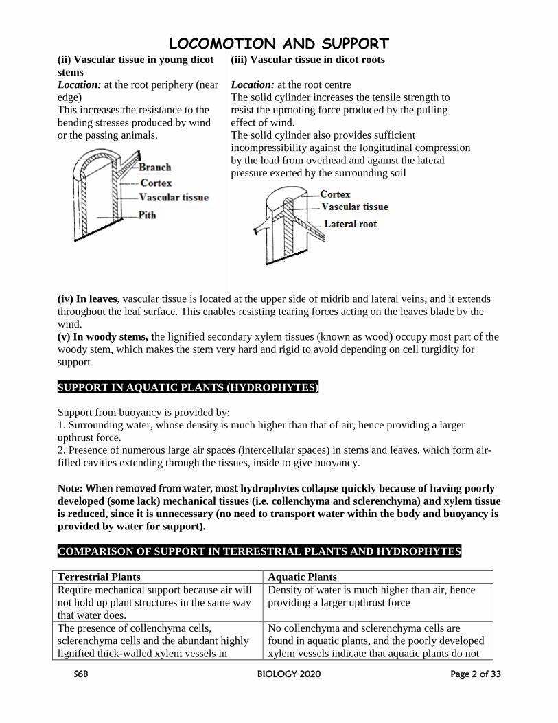
(ii) Vascular tissue in young dicot
stems
(iii) Vascular tissue in dicot roots
Location: at the root periphery (near
edge)
This increases the resistance to the
bending stresses produced by wind
or the passing animals.
Location: at the root centre
The solid cylinder increases the tensile strength to
resist the uprooting force produced by the pulling
effect of wind.
The solid cylinder also provides sufficient
incompressibility against the longitudinal compression
by the load from overhead and against the lateral
pressure exerted by the surrounding soil
(iv) In leaves, vascular tissue is located at the upper side of midrib and lateral veins, and it extends
throughout the leaf surface. This enables resisting tearing forces acting on the leaves blade by the
wind.
(v) In woody stems, the lignified secondary xylem tissues (known as wood) occupy most part of the
woody stem, which makes the stem very hard and rigid to avoid depending on cell turgidity for
support
SUPPORT IN AQUATIC PLANTS (HYDROPHYTES)
Support from buoyancy is provided by:
1. Surrounding water, whose density is much higher than that of air, hence providing a larger
upthrust force.
2. Presence of numerous large air spaces (intercellular spaces) in stems and leaves, which form air-
filled cavities extending through the tissues, inside to give buoyancy.
Note: When removed from water, most hydrophytes collapse quickly because of having poorly
developed (some lack) mechanical tissues (i.e. collenchyma and sclerenchyma) and xylem tissue
is reduced, since it is unnecessary (no need to transport water within the body and buoyancy is
provided by water for support).
COMPARISON OF SUPPORT IN TERRESTRIAL PLANTS AND HYDROPHYTES
Terrestrial Plants Aquatic Plants
Require mechanical support because air will
not hold up plant structures in the same way
that water does.
Density of water is much higher than air, hence
providing a larger upthrust force
The presence of collenchyma cells,
sclerenchyma cells and the abundant highly
lignified thick-walled xylem vessels in
No collenchyma and sclerenchyma cells are
found in aquatic plants, and the poorly developed
xylem vessels indicate that aquatic plants do not
LOCOMOTION AND SUPPORT
S6B BIOLOGY 2020 Page 3 of 33
terrestrial plants implies that support depends
on these specialized thick-walled cells.
depend on these cells for mechanical support.
Small air spaces in stem since air with low
density only provides limited support to
plants.
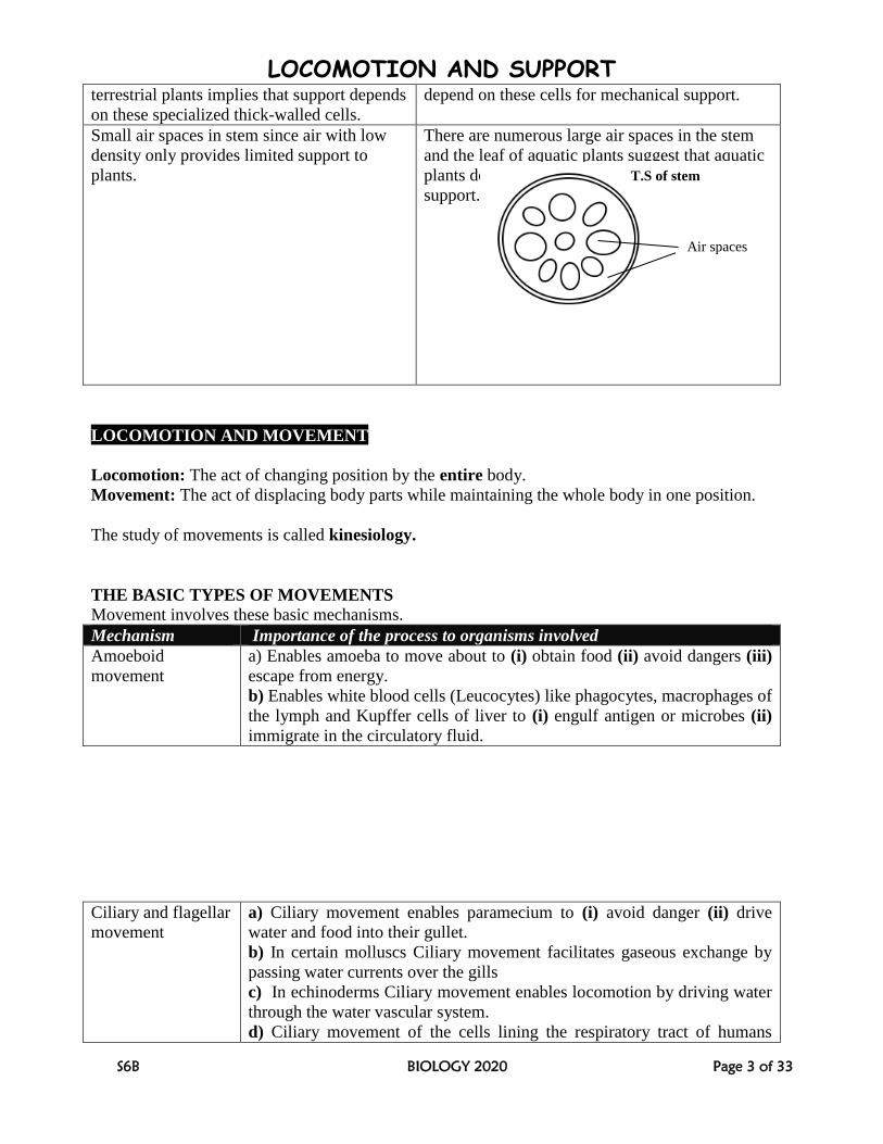
There are numerous large air spaces in the stem
and the leaf of aquatic plants suggest that aquatic
plants depend on the buoyancy of water for
support.
LOCOMOTION AND MOVEMENT
Locomotion: The act of changing position by the entire body.
Movement: The act of displacing body parts while maintaining the whole body in one position.
The study of movements is called kinesiology.
THE BASIC TYPES OF MOVEMENTS
Movement involves these basic mechanisms.
Mechanism Importance of the process to organisms involved
Amoeboid
movement
a) Enables amoeba to move about to (i) obtain food (ii) avoid dangers (iii)
escape from energy.
b) Enables white blood cells (Leucocytes) like phagocytes, macrophages of
the lymph and Kupffer cells of liver to (i) engulf antigen or microbes (ii)
immigrate in the circulatory fluid.
Ciliary and flagellar
movement
a) Ciliary movement enables paramecium to (i) avoid danger (ii) drive
water and food into their gullet.
b) In certain molluscs Ciliary movement facilitates gaseous exchange by
passing water currents over the gills
c) In echinoderms Ciliary movement enables locomotion by driving water
through the water vascular system.
d) Ciliary movement of the cells lining the respiratory tract of humans
Air spaces
T.S of stem
LOCOMOTION AND SUPPORT
S6B BIOLOGY 2020 Page 4 of 33
drives away the microbes and dust particles towards the nose or mouth.
f) Ciliary movement in the oviduct or fallopian tubes of human female
moves ova towards the uterus.
g) Ciliary movement in nephridia of annelids e.g. earthworms moves
wastes
h) Flagellum of sperms enables their swimming movement.
i) Flagellum enables the movement in certain protozoans like euglena
Muscular
movement
Muscular movements enable (i) animals to find food, mate up, avoid
predators and unsuitable environmental conditions (ii) flow of contents in
the gut and arteries (iii) positioning of eyes and external ears for effective
functioning in some animals
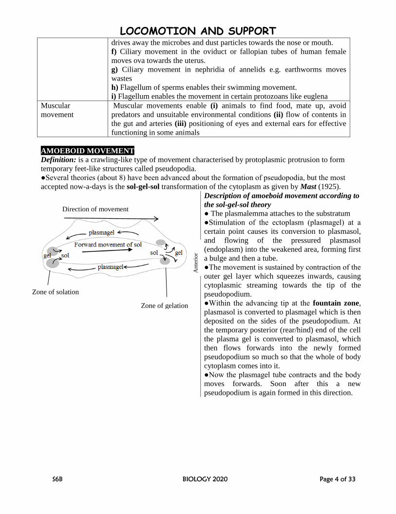
AMOEBOID MOVEMENT
Definition: is a crawling-like type of movement characterised by protoplasmic protrusion to form
temporary feet-like structures called pseudopodia.
●Several theories (about 8) have been advanced about the formation of pseudopodia, but the most
accepted now-a-days is the sol-gel-sol transformation of the cytoplasm as given by Mast (1925).
Description of amoeboid movement according to
the sol-gel-sol theory
● The plasmalemma attaches to the substratum
●Stimulation of the ectoplasm (plasmagel) at a
certain point causes its conversion to plasmasol,
and flowing of the pressured plasmasol
(endoplasm) into the weakened area, forming first
a bulge and then a tube.
●The movement is sustained by contraction of the
outer gel layer which squeezes inwards, causing
cytoplasmic streaming towards the tip of the
pseudopodium.
●Within the advancing tip at the fountain zone,
plasmasol is converted to plasmagel which is then
deposited on the sides of the pseudopodium. At
the temporary posterior (rear/hind) end of the cell
the plasma gel is converted to plasmasol, which
then flows forwards into the newly formed
pseudopodium so much so that the whole of body
cytoplasm comes into it.
●Now the plasmagel tube contracts and the body
moves forwards. Soon after this a new
pseudopodium is again formed in this direction.
Direction of movement
Zone of solation
Zone of gelation
An
teri
or
LOCOMOTION AND SUPPORT
S6B BIOLOGY 2020 Page 5 of 33
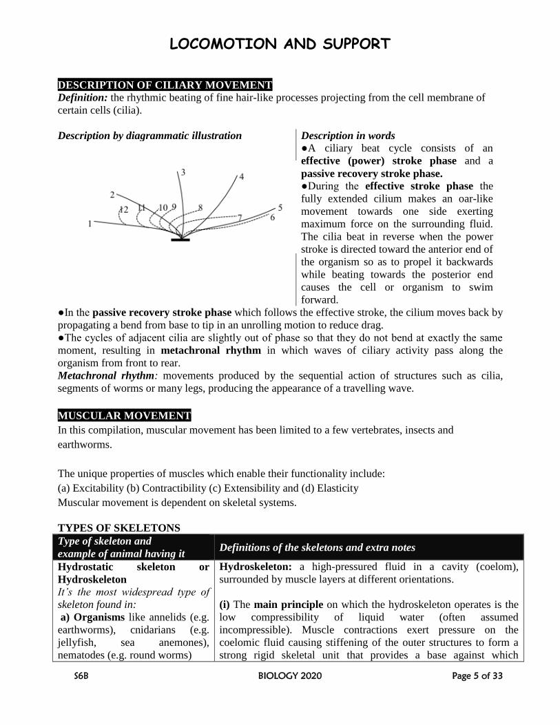
DESCRIPTION OF CILIARY MOVEMENT
Definition: the rhythmic beating of fine hair-like processes projecting from the cell membrane of
certain cells (cilia).
Description by diagrammatic illustration Description in words
●A ciliary beat cycle consists of an
effective (power) stroke phase and a
passive recovery stroke phase.
●During the effective stroke phase the
fully extended cilium makes an oar-like
movement towards one side exerting
maximum force on the surrounding fluid.
The cilia beat in reverse when the power
stroke is directed toward the anterior end of
the organism so as to propel it backwards
while beating towards the posterior end
causes the cell or organism to swim
forward.
●In the passive recovery stroke phase which follows the effective stroke, the cilium moves back by
propagating a bend from base to tip in an unrolling motion to reduce drag.
●The cycles of adjacent cilia are slightly out of phase so that they do not bend at exactly the same
moment, resulting in metachronal rhythm in which waves of ciliary activity pass along the
organism from front to rear.
Metachronal rhythm: movements produced by the sequential action of structures such as cilia,
segments of worms or many legs, producing the appearance of a travelling wave.
MUSCULAR MOVEMENT
In this compilation, muscular movement has been limited to a few vertebrates, insects and
earthworms.
The unique properties of muscles which enable their functionality include:
(a) Excitability (b) Contractibility (c) Extensibility and (d) Elasticity
Muscular movement is dependent on skeletal systems.
TYPES OF SKELETONS
Type of skeleton and
example of animal having it Definitions of the skeletons and extra notes
Hydrostatic skeleton or
Hydroskeleton
It’s the most widespread type of
skeleton found in:
a) Organisms like annelids (e.g.
earthworms), cnidarians (e.g.
jellyfish, sea anemones),
nematodes (e.g. round worms)
Hydroskeleton: a high-pressured fluid in a cavity (coelom),
surrounded by muscle layers at different orientations.
(i) The main principle on which the hydroskeleton operates is the
low compressibility of liquid water (often assumed
incompressible). Muscle contractions exert pressure on the
coelomic fluid causing stiffening of the outer structures to form a
strong rigid skeletal unit that provides a base against which
LOCOMOTION AND SUPPORT
S6B BIOLOGY 2020 Page 6 of 33
b) Structures like mammalian
eyes (the aqueous and vitreous
humour), spinal cord
(cerebrospinal fluid), extra
embryonic membranes (amniotic
fluid), hearts (move blood), and
intestines (move food).
movements can occur.
(ii) The optimal volume of fluid for a particular system must
remain constant for effective contraction and expansion of the
antagonistic muscles. Too much loss of fluid causes limpness of
tissues and pressure loss, and too much gain causes over swelling,
both of which fail muscle stretching and hence movement fails.
This explains why snails and earthworms are restricted in their
activity to moist conditions.
Type of skeleton and
example of animal having it Definitions of the skeletons and extra notes
Exoskeleton
●Chitinous exoskeleton is in:
arthropods like insects, arachnids
(e.g. spiders) crustaceans (e.g.
crabs, lobsters), some fungi and
bacteria
●Calcified exoskeleton is in:
shelled mollusks (e.g. snails,
clams), some polychaetes like
lugworms.
●Silicated exoskeleton is in
diatoms.
●Bone, cartilage, or dentine make
up the exoskeletons of turtles and
primitive fish
Exoskeleton: a non living external body structure that supports
and protects an organism.
(i) Exoskeletons are secreted by ectoderm
(ii) Chitinous exoskeleton has complex muscular system which
enables insects to lift or pull an object 20 or more times heavier
than their body weights! Grasshoppers have about 900 muscles,
caterpillars up to 4,000 yet human beings have fewer than 700
muscles (Source: The World Book Encyclopedia).
(iii) Exoskeletons do not grow with the body so in arthropods they
must be periodically shed to allow growth; mollusks e.g. snails
continually enlarge their shells as they grow.
(iv) In insects and spiders the epicuticle is waterproof.
Endoskeleton
a) Chordates: birds, mammals,
reptiles.
b) Echinoderms: starfish, brittle
stars, sea urchins, sea cucumbers
c) Poriferans: sponges
d) Molluscs (class Cephalopoda)
e.g. cuttlefish
Note: Some animals, such as the
tortoise, have both an endoskeleton
and an exoskeleton.
Endoskeleton: a living internal support structure of an animal,
usually composed of mineralized tissue which develops within the
skin or in the deeper body tissues.
(i) The vertebrate endoskeleton is made up of bone and cartilage
tissues.
(ii) In sponges, the endoskeleton is purely for support, but in
vertebrates and echinoderms it’s also for attachment of muscle and
locomotion.
(iii) Echinoderms and chordates have a true endoskeleton derived
from mesodermal tissue
LOCOMOTION AND SUPPORT
S6B BIOLOGY 2020 Page 7 of 33
ADVANTAGES / FUNCTIONS AND LIMITATIONS OF THE SKELETONS
Advantages / Functions Limitations / Disadvantages
Hydroskeleto
n
●Hydroskeleton is elastic and
can bend accordingly when a
muscle contracts enabling
fitting in narrow burrows.
● Coelenterates that use a hydroskeleton regularly face
a loss of pressure because their skeleton is also their
gut.
● Due to lack of a strong supportive system, majority
of the invertebrates are small
●The slow motion due to lack of effective ways to
support a large body compromises the animals’ escape
response from predators.
●The organisms are limited to moist habitats because
of the need to minimise water loss by evaporation
Advantages / Functions Limitations / Disadvantages
Exoskeleto
n
● Exoskeletons contain rigid and resistant
components that offer protection against
predators, bacterial attack and desiccation while
on land.
●Exoskeletons contain rigid components that
offer support enabling maintaining body shape.
●Exoskeleton of arthropods contains rigid
framework of ingrowths known as apodemes
which serve as attachment sites for muscles.
● In arthropods the exoskeleton is modified into
appendages which offer more rapid locomotion
than the hydroskeleton
● The arthropod exoskeleton contains various
folds, flaps and parts modified for feeding and
structures for respiration.
● Exoskeletons are often highly coloured for
camouflage from predators, recognition by
mates, and warning to scare off predators.
●The arthropod exoskeleton is jointed enabling
flexibility in locomotion.
● Since exoskeletons are rigid and do
not grow with the body, in arthropods
they disrupt smooth and steady growth
and so must be periodically shed to
allow growth, which makes the animal
temporarily vulnerable for predation and
water loss by evaporation until
hardening.
NB: Snails and many other mollusks
solve that problem by continually
enlarging their shells as they grow.
● An exoskeleton cannot support large
sized animals because of their large
volume and body mass in proportion to
the cube of their linear dimensions,
necessitating an impossibly heavy and
thick exoskeleton.
●It requires modifications in movement.
Many individual muscles are attached to
the outer shell in order to create
movement. In the appendages, these
muscles are set up within multiple hinge
joints, as these allow a wide range of
motions.
Advantages / Functions Limitations /
Disadvantages
Endoskeleto
n
●Vertebrates have a versatile support system and as a
result, they develop faster and bigger bodies than
invertebrates.
● It’s jointed for flexibility to allow diverse range of
locomotory patterns: swimming, digging, running,
●Endoskeletons are
enclosed in other tissues do
not offer much protection
from predators in some
animals.
LOCOMOTION AND SUPPORT
S6B BIOLOGY 2020 Page 8 of 33
climbing, and flying, feeding (jaws).
●Endoskeleton does not limit space available for internal
organs and can support greater weight.
●Bone are hard for protecting delicate parts like the brain,
lungs, heart, spinal cord, etc.
●Bone tissue is mineralized and hence acts as mineral
reserve for the body’s’ physiological processes.
●Mammalian bones manufacture the defensive
leucocytes
● Endoskeletons do not
contribute to minimizing
water loss from the body by
evaporation
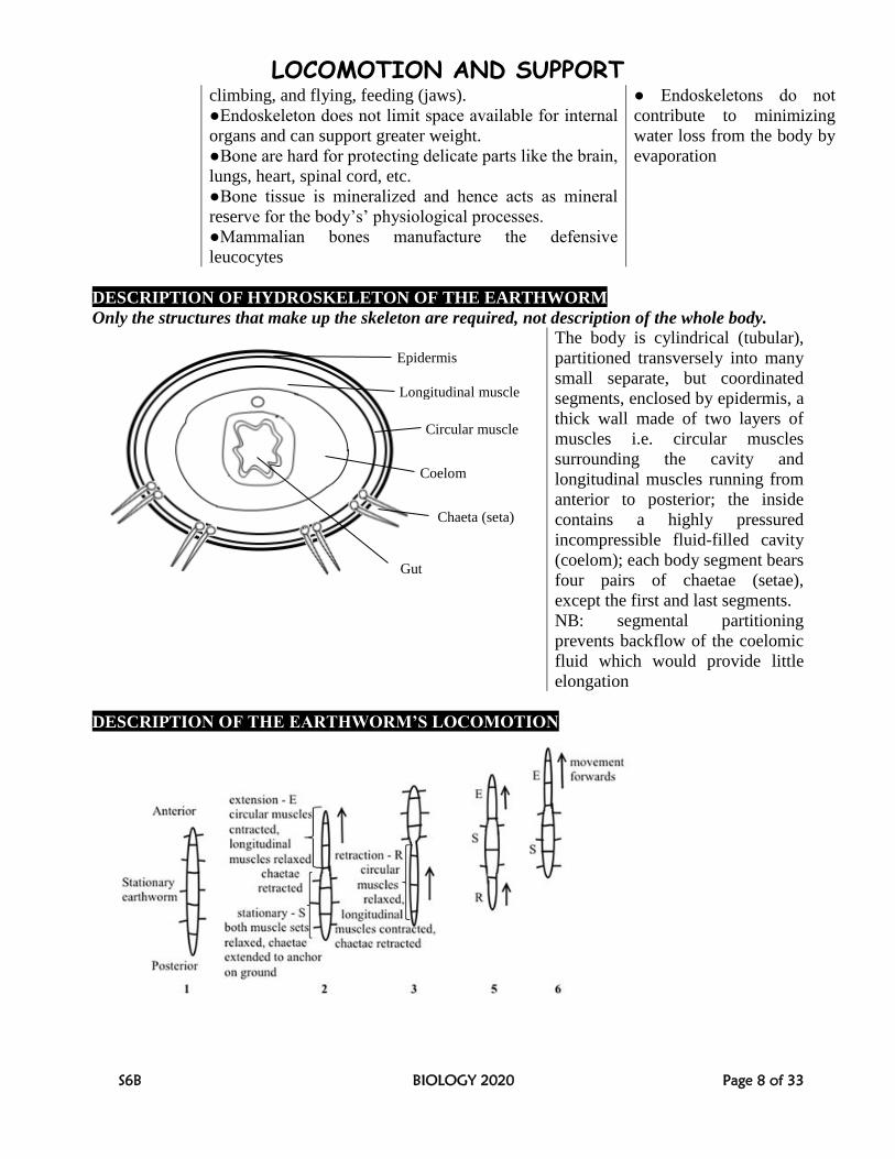
DESCRIPTION OF HYDROSKELETON OF THE EARTHWORM
Only the structures that make up the skeleton are required, not description of the whole body.
The body is cylindrical (tubular),
partitioned transversely into many
small separate, but coordinated
segments, enclosed by epidermis, a
thick wall made of two layers of
muscles i.e. circular muscles
surrounding the cavity and
longitudinal muscles running from
anterior to posterior; the inside
contains a highly pressured
incompressible fluid-filled cavity
(coelom); each body segment bears
four pairs of chaetae (setae),
except the first and last segments.
NB: segmental partitioning
prevents backflow of the coelomic
fluid which would provide little
elongation
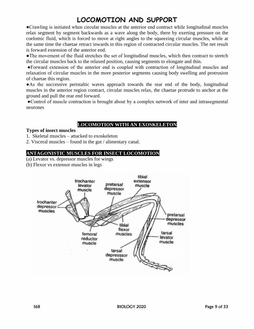
DESCRIPTION OF THE EARTHWORM’S LOCOMOTION
Epidermis
Longitudinal muscle
muscle
Circular muscle
Coelom
Chaeta (seta)
Gut
LOCOMOTION AND SUPPORT
S6B BIOLOGY 2020 Page 9 of 33
●Crawling is initiated when circular muscles at the anterior end contract while longitudinal muscles
relax segment by segment backwards as a wave along the body, there by exerting pressure on the
coelomic fluid, which is forced to move at right angles to the squeezing circular muscles, while at
the same time the chaetae retract inwards in this region of contracted circular muscles. The net result
is forward extension of the anterior end.
●The movement of the fluid stretches the set of longitudinal muscles, which then contract to stretch
the circular muscles back to the relaxed position, causing segments to elongate and thin.
●Forward extension of the anterior end is coupled with contraction of longitudinal muscles and
relaxation of circular muscles in the more posterior segments causing body swelling and protrusion
of chaetae this region.
●As the successive peristaltic waves approach towards the rear end of the body, longitudinal
muscles in the anterior region contract, circular muscles relax, the chaetae protrude to anchor at the
ground and pull the rear end forward.
●Control of muscle contraction is brought about by a complex network of inter and intrasegmental
neurones
LOCOMOTION WITH AN EXOSKELETON
Types of insect muscles
1. Skeletal muscles – attacked to exoskeleton
2. Visceral muscles – found in the gut / alimentary canal.
ANTAGONISTIC MUSCLES FOR INSECT LOCOMOTION
(a) Levator vs. depressor muscles for wings
(b) Flexor vs extensor muscles in legs
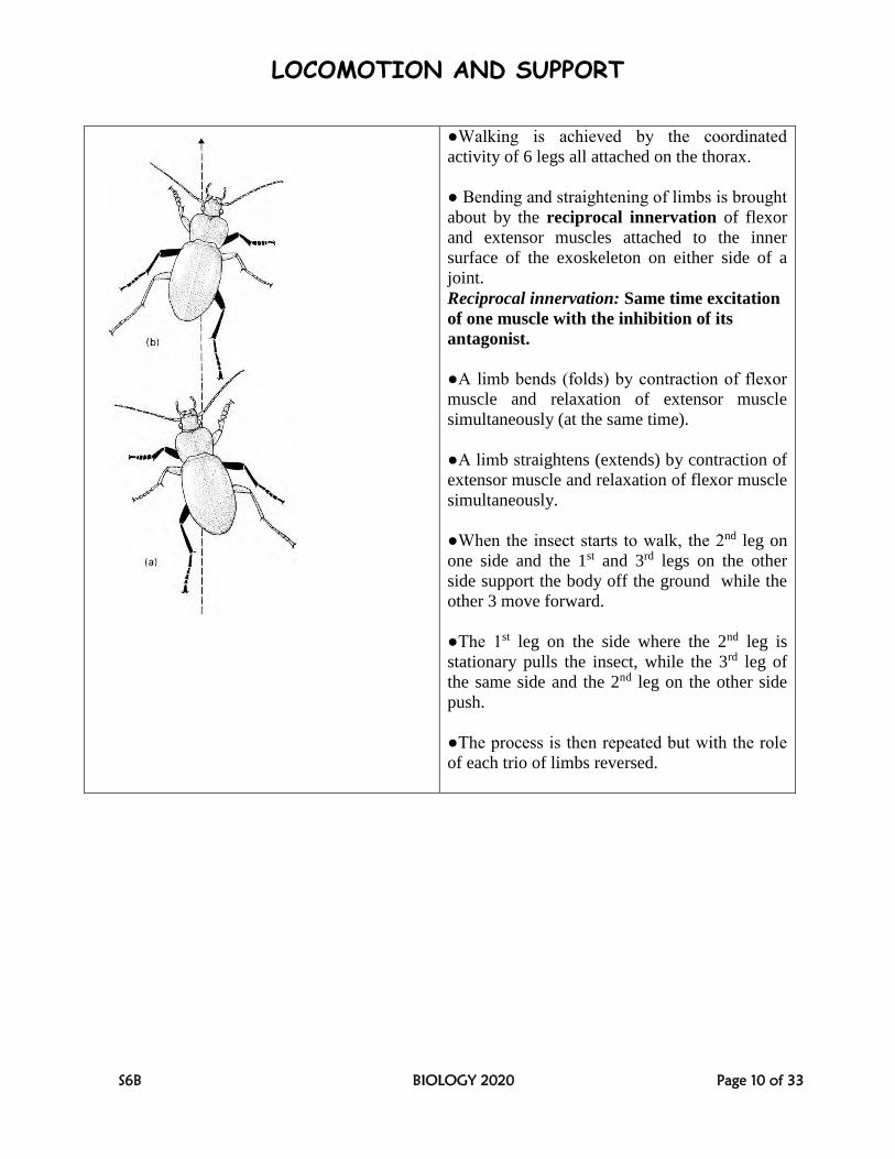
A. DESCRIPTION OF WALKING IN INSECTS
LOCOMOTION AND SUPPORT
S6B BIOLOGY 2020 Page 10 of 33
●Walking is achieved by the coordinated
activity of 6 legs all attached on the thorax.
● Bending and straightening of limbs is brought
about by the reciprocal innervation of flexor
and extensor muscles attached to the inner
surface of the exoskeleton on either side of a
joint.
Reciprocal innervation: Same time excitation
of one muscle with the inhibition of its
antagonist.
●A limb bends (folds) by contraction of flexor
muscle and relaxation of extensor muscle
simultaneously (at the same time).
●A limb straightens (extends) by contraction of
extensor muscle and relaxation of flexor muscle
simultaneously.
●When the insect starts to walk, the 2nd leg on
one side and the 1st and 3rd legs on the other
side support the body off the ground while the
other 3 move forward.
●The 1st leg on the side where the 2nd leg is
stationary pulls the insect, while the 3rd leg of
the same side and the 2nd leg on the other side
push.
●The process is then repeated but with the role
of each trio of limbs reversed.
LOCOMOTION AND SUPPORT
S6B BIOLOGY 2020 Page 11 of 33
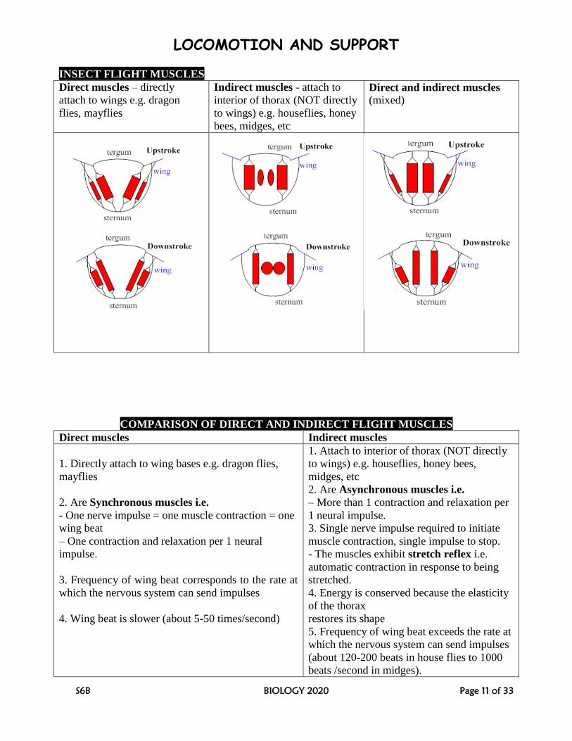
INSECT FLIGHT MUSCLES
Direct muscles – directly
attach to wings e.g. dragon
flies, mayflies
Indirect muscles - attach to
interior of thorax (NOT directly
to wings) e.g. houseflies, honey
bees, midges, etc
Direct and indirect muscles (mixed)
COMPARISON OF DIRECT AND INDIRECT FLIGHT MUSCLES
Direct muscles Indirect muscles
1. Directly attach to wing bases e.g. dragon flies,
mayflies
2. Are Synchronous muscles i.e.
- One nerve impulse = one muscle contraction = one
wing beat
– One contraction and relaxation per 1 neural
impulse.
3. Frequency of wing beat corresponds to the rate at
which the nervous system can send impulses
4. Wing beat is slower (about 5-50 times/second)
1. Attach to interior of thorax (NOT directly
to wings) e.g. houseflies, honey bees,
midges, etc
2. Are Asynchronous muscles i.e.
– More than 1 contraction and relaxation per
1 neural impulse.
3. Single nerve impulse required to initiate
muscle contraction, single impulse to stop.
- The muscles exhibit stretch reflex i.e.
automatic contraction in response to being
stretched.
4. Energy is conserved because the elasticity
of the thorax
restores its shape
5. Frequency of wing beat exceeds the rate at
which the nervous system can send impulses
(about 120-200 beats in house flies to 1000
beats /second in midges).
LOCOMOTION AND SUPPORT
S6B BIOLOGY 2020 Page 12 of 33
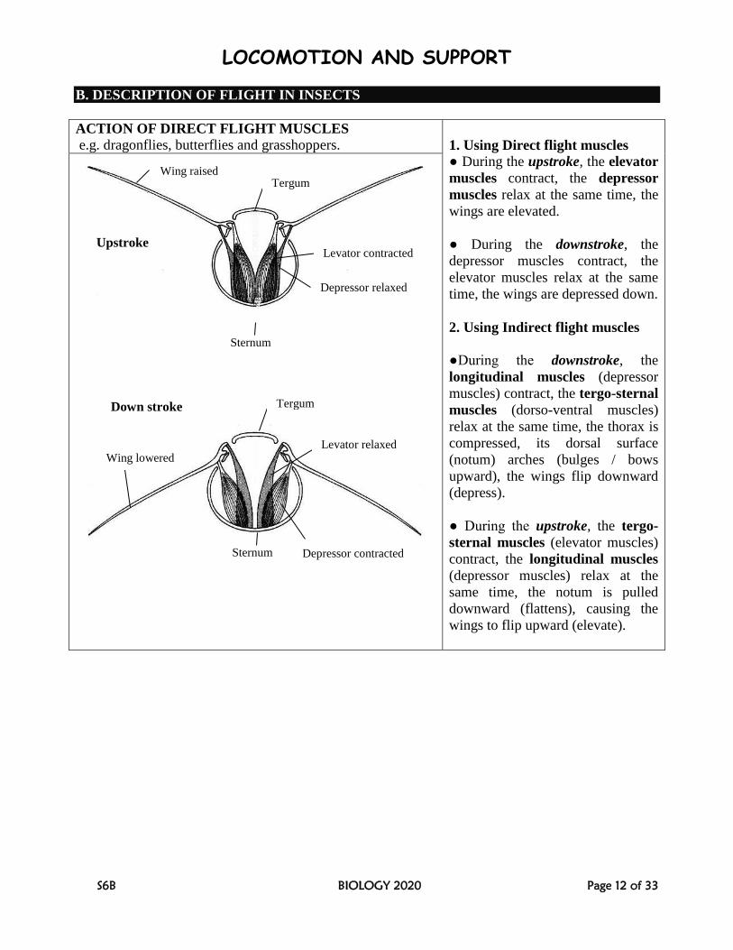
B. DESCRIPTION OF FLIGHT IN INSECTS
ACTION OF DIRECT FLIGHT MUSCLES
e.g. dragonflies, butterflies and grasshoppers.
1. Using Direct flight muscles
● During the upstroke, the elevator
muscles contract, the depressor
muscles relax at the same time, the
wings are elevated.
● During the downstroke, the
depressor muscles contract, the
elevator muscles relax at the same
time, the wings are depressed down.
2. Using Indirect flight muscles
●During the downstroke, the
longitudinal muscles (depressor
muscles) contract, the tergo-sternal
muscles (dorso-ventral muscles)
relax at the same time, the thorax is
compressed, its dorsal surface
(notum) arches (bulges / bows
upward), the wings flip downward
(depress).
● During the upstroke, the tergo-
sternal muscles (elevator muscles)
contract, the longitudinal muscles
(depressor muscles) relax at the
same time, the notum is pulled
downward (flattens), causing the
wings to flip upward (elevate).
Wing raised Tergum
Levator contracted
Depressor relaxed
Sternum
Tergum
Sternum
Wing lowered Levator relaxed
Depressor contracted
Upstroke
Down stroke
LOCOMOTION AND SUPPORT
S6B BIOLOGY 2020 Page 13 of 33
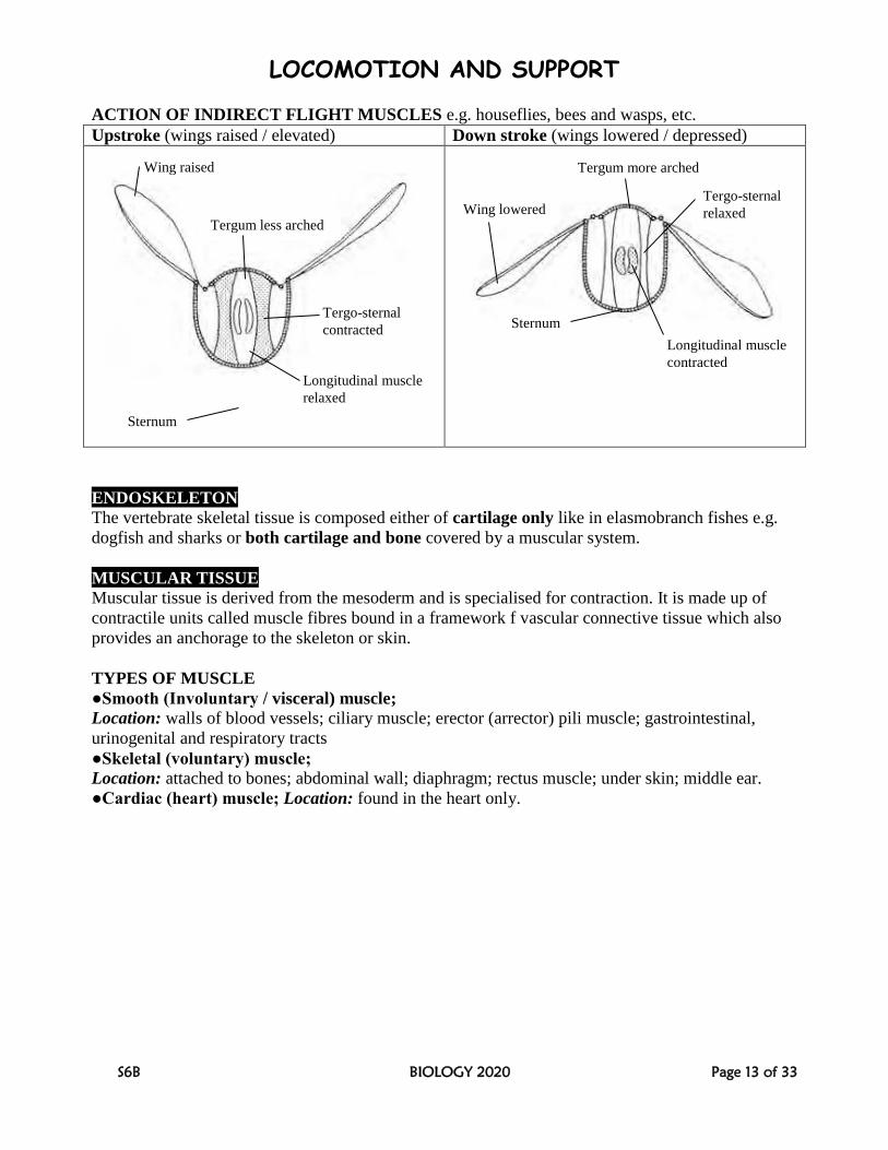
ACTION OF INDIRECT FLIGHT MUSCLES e.g. houseflies, bees and wasps, etc.
Upstroke (wings raised / elevated) Down stroke (wings lowered / depressed)
ENDOSKELETON
The vertebrate skeletal tissue is composed either of cartilage only like in elasmobranch fishes e.g.
dogfish and sharks or both cartilage and bone covered by a muscular system.
MUSCULAR TISSUE
Muscular tissue is derived from the mesoderm and is specialised for contraction. It is made up of
contractile units called muscle fibres bound in a framework f vascular connective tissue which also
provides an anchorage to the skeleton or skin.
TYPES OF MUSCLE
●Smooth (Involuntary / visceral) muscle;
Location: walls of blood vessels; ciliary muscle; erector (arrector) pili muscle; gastrointestinal,
urinogenital and respiratory tracts
●Skeletal (voluntary) muscle;
Location: attached to bones; abdominal wall; diaphragm; rectus muscle; under skin; middle ear.
●Cardiac (heart) muscle; Location: found in the heart only.
Sternum
Wing raised
Tergum less arched
Tergo-sternal
contracted
Longitudinal muscle
relaxed
Sternum
Tergum more arched
Wing lowered Tergo-sternal
relaxed
Longitudinal muscle
contracted
LOCOMOTION AND SUPPORT
S6B BIOLOGY 2020 Page 14 of 33
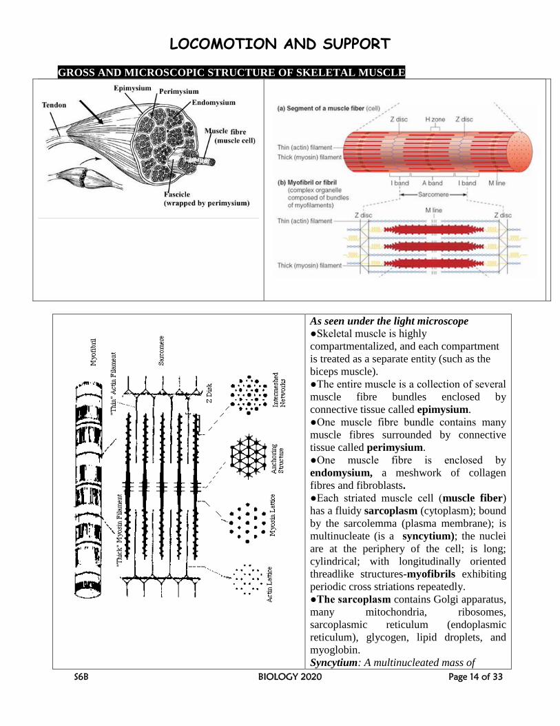
GROSS AND MICROSCOPIC STRUCTURE OF SKELETAL MUSCLE
As seen under the light microscope
●Skeletal muscle is highly
compartmentalized, and each compartment
is treated as a separate entity (such as the
biceps muscle).
●The entire muscle is a collection of several
muscle fibre bundles enclosed by
connective tissue called epimysium.
●One muscle fibre bundle contains many
muscle fibres surrounded by connective
tissue called perimysium.
●One muscle fibre is enclosed by
endomysium, a meshwork of collagen
fibres and fibroblasts.
●Each striated muscle cell (muscle fiber)
has a fluidy sarcoplasm (cytoplasm); bound
by the sarcolemma (plasma membrane); is
multinucleate (is a syncytium); the nuclei
are at the periphery of the cell; is long;
cylindrical; with longitudinally oriented
threadlike structures-myofibrils exhibiting
periodic cross striations repeatedly.
●The sarcoplasm contains Golgi apparatus,
many mitochondria, ribosomes,
sarcoplasmic reticulum (endoplasmic
reticulum), glycogen, lipid droplets, and
myoglobin.
Syncytium: A multinucleated mass of
LOCOMOTION AND SUPPORT
S6B BIOLOGY 2020 Page 15 of 33
cytoplasm that is not separated into
individual cells.
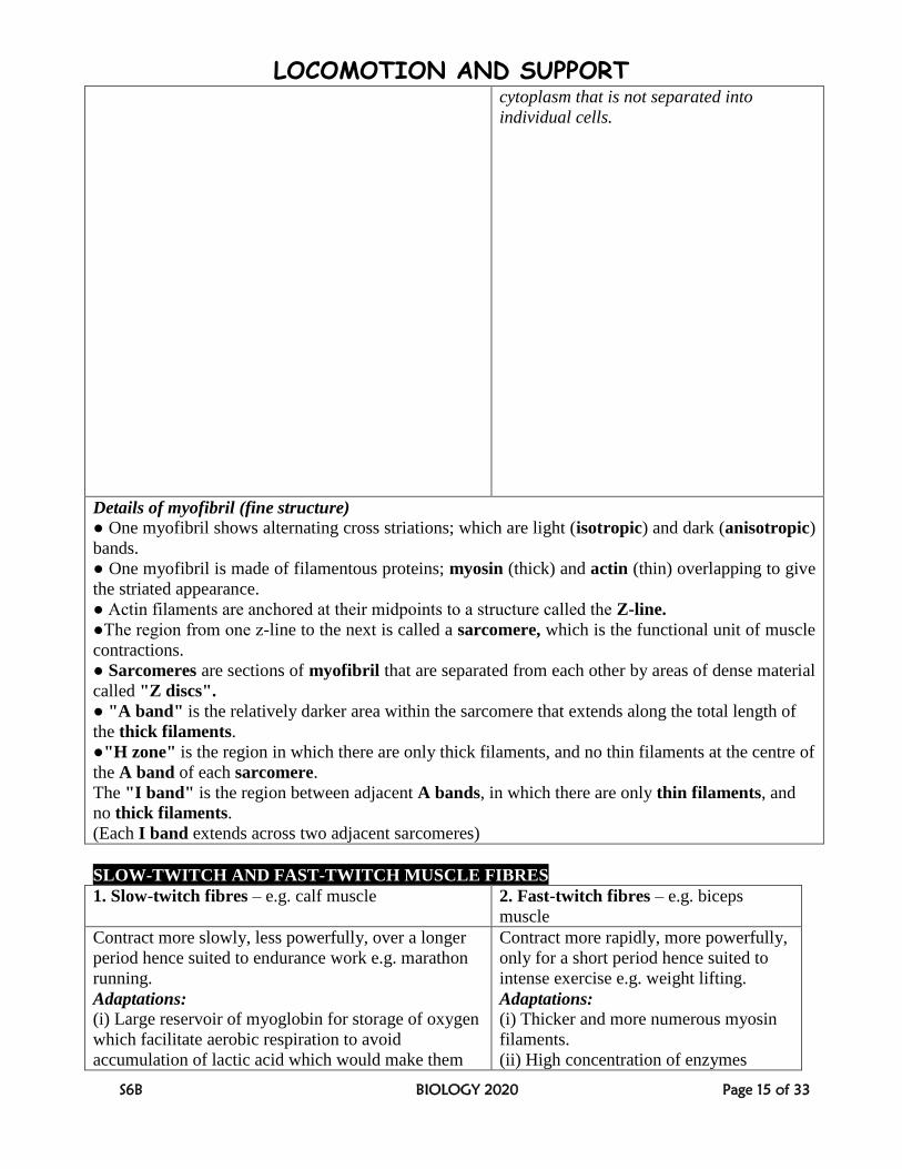
Details of myofibril (fine structure)
● One myofibril shows alternating cross striations; which are light (isotropic) and dark (anisotropic)
bands.
● One myofibril is made of filamentous proteins; myosin (thick) and actin (thin) overlapping to give
the striated appearance.
● Actin filaments are anchored at their midpoints to a structure called the Z-line.
●The region from one z-line to the next is called a sarcomere, which is the functional unit of muscle
contractions.
● Sarcomeres are sections of myofibril that are separated from each other by areas of dense material
called "Z discs".
● "A band" is the relatively darker area within the sarcomere that extends along the total length of
the thick filaments.
●"H zone" is the region in which there are only thick filaments, and no thin filaments at the centre of
the A band of each sarcomere.
The "I band" is the region between adjacent A bands, in which there are only thin filaments, and
no thick filaments.
(Each I band extends across two adjacent sarcomeres)
SLOW-TWITCH AND FAST-TWITCH MUSCLE FIBRES
1. Slow-twitch fibres – e.g. calf muscle 2. Fast-twitch fibres – e.g. biceps
muscle
Contract more slowly, less powerfully, over a longer
period hence suited to endurance work e.g. marathon
running.
Adaptations:
(i) Large reservoir of myoglobin for storage of oxygen
which facilitate aerobic respiration to avoid
accumulation of lactic acid which would make them
Contract more rapidly, more powerfully,
only for a short period hence suited to
intense exercise e.g. weight lifting.
Adaptations:
(i) Thicker and more numerous myosin
filaments.
(ii) High concentration of enzymes
LOCOMOTION AND SUPPORT
S6B BIOLOGY 2020 Page 16 of 33
less effective.
(ii) Much glycogen to provide a source of metabolic
energy.
(iii) A rich supply of blood vessels to deliver oxygen
and glucose needed in aerobic respiration to provide
ATP.
(iv) Numerous mitochondria to produce ATP that
maintains muscle contraction.
involved in anaerobic respiration.
(iii) Store of phosphocreatine, a molecule
that can rapidly generate ATP from ADP
in anaerobic conditions and so provide
energy for muscle contraction.
HOW SKELETAL MUSCLE STRUCTURE RELATES TO FUNCTIONING
●Each muscle cell is long to allow considerable contractile effect.
●The fibres are parallel to each other so that contractile effect is transmitted along same axis.
●Muscle fibres taper at both ends for interweaving to improve muscle strength.
●Muscle fibres have very many mitochondria to provide much ATP needed in muscle contraction.
●Cross bridges enable actin and myosin to fit into each other to allow sliding during muscle
contraction.
●There is a rich supply of blood vessels to supply nutrients to and drain wastes away from cells.
●There is much myoglobin for storage of oxygen needed very much in aerobic respiration during
exercising.
●There are motor end plates to allow innervation that result in contraction.
●There is a dense network of internal membrane system (including transverse tubules) for calcium
ion storage which is very much needed in muscle contraction.
●Reciprocal innervation ensures antagonistic muscular contraction to bring about realistic movement
LOCOMOTION AND SUPPORT
S6B BIOLOGY 2020 Page 17 of 33
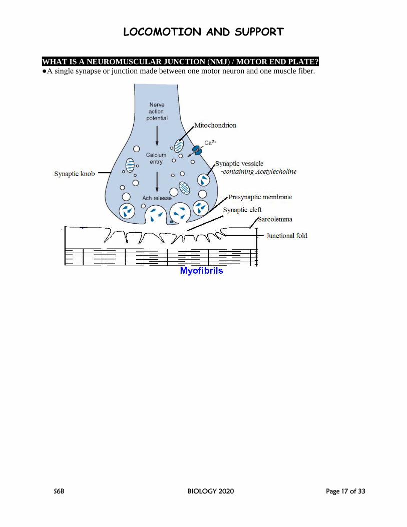
WHAT IS A NEUROMUSCULAR JUNCTION (NMJ) / MOTOR END PLATE?
●A single synapse or junction made between one motor neuron and one muscle fiber.
LOCOMOTION AND SUPPORT
S6B BIOLOGY 2020 Page 18 of 33
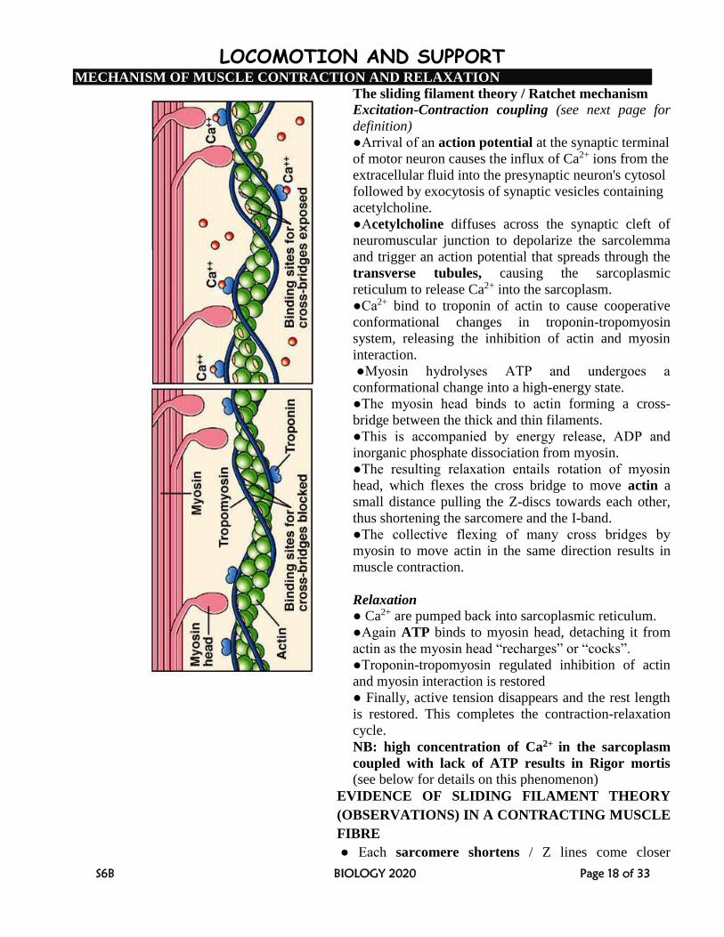
MECHANISM OF MUSCLE CONTRACTION AND RELAXATION
The sliding filament theory / Ratchet mechanism
Excitation-Contraction coupling (see next page for
definition)
●Arrival of an action potential at the synaptic terminal
of motor neuron causes the influx of Ca2+ ions from the
extracellular fluid into the presynaptic neuron's cytosol
followed by exocytosis of synaptic vesicles containing
acetylcholine.
●Acetylcholine diffuses across the synaptic cleft of
neuromuscular junction to depolarize the sarcolemma
and trigger an action potential that spreads through the
transverse tubules, causing the sarcoplasmic
reticulum to release Ca2+ into the sarcoplasm.
●Ca2+ bind to troponin of actin to cause cooperative
conformational changes in troponin-tropomyosin
system, releasing the inhibition of actin and myosin
interaction.
●Myosin hydrolyses ATP and undergoes a
conformational change into a high-energy state.
●The myosin head binds to actin forming a cross-
bridge between the thick and thin filaments.
●This is accompanied by energy release, ADP and
inorganic phosphate dissociation from myosin.
●The resulting relaxation entails rotation of myosin
head, which flexes the cross bridge to move actin a
small distance pulling the Z-discs towards each other,
thus shortening the sarcomere and the I-band.
●The collective flexing of many cross bridges by
myosin to move actin in the same direction results in
muscle contraction.
Relaxation
● Ca2+ are pumped back into sarcoplasmic reticulum.
●Again ATP binds to myosin head, detaching it from
actin as the myosin head “recharges” or “cocks”.
●Troponin-tropomyosin regulated inhibition of actin
and myosin interaction is restored
● Finally, active tension disappears and the rest length
is restored. This completes the contraction-relaxation
cycle.
NB: high concentration of Ca2+ in the sarcoplasm
coupled with lack of ATP results in Rigor mortis
(see below for details on this phenomenon)
EVIDENCE OF SLIDING FILAMENT THEORY
(OBSERVATIONS) IN A CONTRACTING MUSCLE
FIBRE
● Each sarcomere shortens / Z lines come closer
LOCOMOTION AND SUPPORT
S6B BIOLOGY 2020 Page 19 of 33
●I Band shortens
●H zone shortens greatly (usually disappears).
●A Band remains unchanged in length during
contraction or relaxation
●Cross bridges are visible between actin and myosin in
photomicrographs.
CHANGES DURING MUSCLE PASSIVE
STRETCHING
●Sarcomere lengthens
● I Band elongates.
WHAT IS RIGOR MORTIS?
●The progressive stiffening of the body that occurs several hours after death as a result of failure of
contracted muscles to relax.
WHAT CAUSES RIGOR MORTIS?
●Upon death, there’s increased permeability of sarcoplasmic membrane to Ca2+, allowing Ca2+
influx into the sarcoplasm hence promoting the cross-bridge formation between actin and myosin
(muscle contraction).
●However efflux of Ca2+ from the sarcoplasm into the sarcoplasmic reticulum fails because of lack
of ATP since respiration would have ceased. This causes the muscle to remain contracted, relaxing
only when decomposition starts.
NB: Interestingly, meat is generally considered to be tenderer if it is consumed after expiry of rigor
mortis.
WHAT IS EXCITATION-CONTRACTION COUPLING?
●The sequence of events by which an action potential in the plasma membrane of the muscle fibre
leads to force production via an increase in intracellular calcium and cross bridge formation and
turn-over (see previous page for explanation)
ATP PRODUCTION DURING MUSCLE CONTRACTION 1. Phosphorylation of ADP by creatine phosphate provides a very rapid means of forming ATP at
the onset of contractile activity. Phosphocreatine + ADP ATP + creatine
In a resting muscle fiber, the concentration of ATP is always greater than ADP leading to the
reformation of creatine phosphate.
2. Oxidative phosphorylation of ADP in mitochondria during aerobic respiration (need
myoglobin for oxygen transfer)
3. Substrate phosphorylation of ADP in glycolysis during anaerobic respiration to form lactic
acid in the process. The accumulation of lactic acid is associated with muscle fatigue, which is
broken down later in the liver using oxygen to constitute what is called oxygen debt.
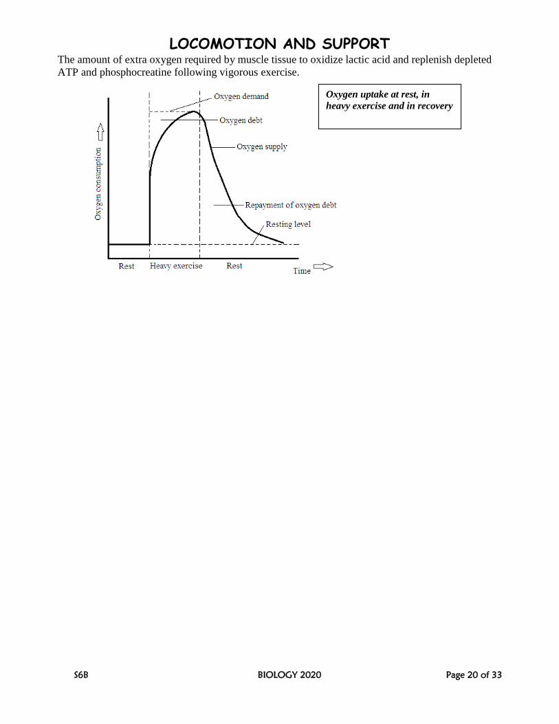
WHAT IS THE OXYGEN DEBT?
LOCOMOTION AND SUPPORT
S6B BIOLOGY 2020 Page 20 of 33
The amount of extra oxygen required by muscle tissue to oxidize lactic acid and replenish depleted
ATP and phosphocreatine following vigorous exercise.
Oxygen uptake at rest, in
heavy exercise and in recovery
LOCOMOTION AND SUPPORT
S6B BIOLOGY 2020 Page 21 of 33
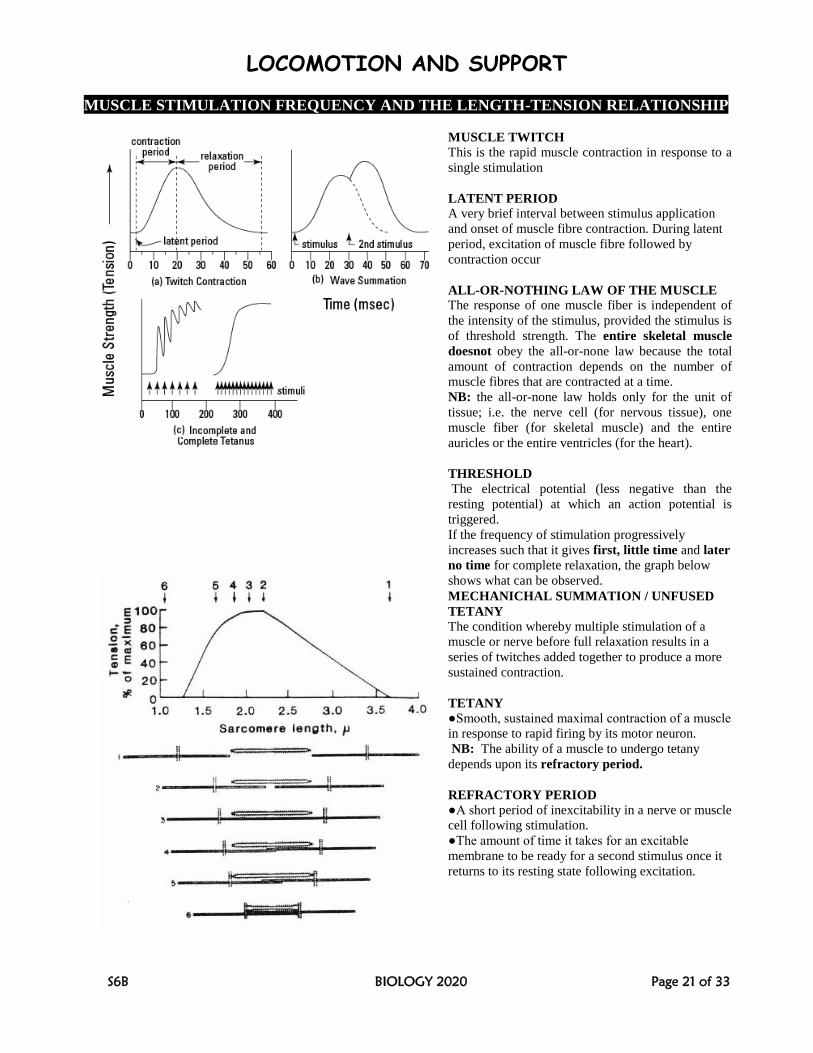
MUSCLE STIMULATION FREQUENCY AND THE LENGTH-TENSION RELATIONSHIP
MUSCLE TWITCH
This is the rapid muscle contraction in response to a
single stimulation
LATENT PERIOD
A very brief interval between stimulus application
and onset of muscle fibre contraction. During latent
period, excitation of muscle fibre followed by
contraction occur
ALL-OR-NOTHING LAW OF THE MUSCLE
The response of one muscle fiber is independent of
the intensity of the stimulus, provided the stimulus is
of threshold strength. The entire skeletal muscle
doesnot obey the all-or-none law because the total
amount of contraction depends on the number of
muscle fibres that are contracted at a time.
NB: the all-or-none law holds only for the unit of
tissue; i.e. the nerve cell (for nervous tissue), one
muscle fiber (for skeletal muscle) and the entire
auricles or the entire ventricles (for the heart).
THRESHOLD
The electrical potential (less negative than the
resting potential) at which an action potential is
triggered.
If the frequency of stimulation progressively
increases such that it gives first, little time and later
no time for complete relaxation, the graph below
shows what can be observed.
MECHANICHAL SUMMATION / UNFUSED
TETANY
The condition whereby multiple stimulation of a
muscle or nerve before full relaxation results in a
series of twitches added together to produce a more
sustained contraction.
TETANY ●Smooth, sustained maximal contraction of a muscle
in response to rapid firing by its motor neuron.
NB: The ability of a muscle to undergo tetany
depends upon its refractory period.
REFRACTORY PERIOD
●A short period of inexcitability in a nerve or muscle
cell following stimulation.
●The amount of time it takes for an excitable
membrane to be ready for a second stimulus once it
returns to its resting state following excitation.
LOCOMOTION AND SUPPORT
S6B BIOLOGY 2020 Page 22 of 33
MUSCLE FATIGUE
●A condition of the muscle in which its capacity to produce maximum voluntary action, or to
perform a series of repetitive actions, is reduced.
Muscle fatigue results when there is tissue oxygen deprivation, glycogen or Phosphocreatine
depletion, and increased level of blood and muscle lactic acid in an exercised muscle.
LOCOMOTION IN FISH
Most fish have a line of muscle blocks, called myomeres (myotomes), along each side of the
vertebral column. To swim, they alternately contract one side and relax the other side in a
progression which goes from the head to the tail. In this way, an undulatory locomotion results, first
bending the body one way in a wave which travels down the body, and then back the other way, with
the contracting and relaxing muscles switching roles. They use their fins to propel themselves
through the water in this swimming motion.
LOCOMOTION AND SUPPORT
S6B BIOLOGY 2020 Page 23 of 33
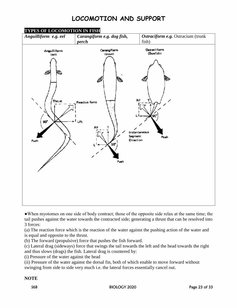
TYPES OF LOCOMOTION IN FISH
Anguilliform e.g. eel Carangiform e.g. dog fish,
perch
Ostraciform e.g. Ostracium (trunk
fish)
●When myotomes on one side of body contract; those of the opposite side relax at the same time; the
tail pushes against the water towards the contracted side; generating a thrust that can be resolved into
3 forces:
(a) The reaction force which is the reaction of the water against the pushing action of the water and
is equal and opposite to the thrust.
(b) The forward (propulsive) force that pushes the fish forward.
(c) Lateral drag (sideways) force that swings the tail towards the left and the head towards the right
and thus slows (drags) the fish. Lateral drag is countered by:
(i) Pressure of the water against the head
(ii) Pressure of the water against the dorsal fin, both of which enable to move forward without
swinging from side to side very much i.e. the lateral forces essentially cancel out.
NOTE
LOCOMOTION AND SUPPORT
S6B BIOLOGY 2020 Page 24 of 33
Many invertebrates like round worms and some flagellated cells including spermatozoa exhibit the
principles involved in propulsion as in the above fishes.
SUPPORT (BUOYANCY) IN FISH
Elasmobranchs like dog fish sharks, skates and
rays. Teleosts like perch
●Support is provided by constant swimming
using fins.
●These fish’s density is slightly greater than that
of water and must swim continuously to avoid
sinking.
●How they are adapted to this: (1) possession of
large pectoral and pelvic fins which direct
swimming upwards (2) possession of
heterocercal tail i.e. a tail with smaller upper
and larger lower lobes for generating much lift
and forward motion
●During forward motion the pectoral and pelvic
fins are all held at a slight angle to the body,
generating a force which can be resolved into
upward and backward components.
●The upward component force lifts the anterior
end up in the water while the backward
component force called backward drag being
small only slightly impedes motion and is easily
suppressed.
●Support is provided by adjusting air in the
swim bladder which may be (1) a closed swim
bladder filled with gaseous mixture of oxygen,
nitrogen and carbondioxide – all secreted from
blood vessels in its wall. The closed type is the
most common in bony fish. (2) an open swim
bladder having a duct connection to the pharynx
and operates as follows:
●Expulsion of air from the swim bladder
increases the fish’s relative density and it sinks.
If it’s to stay afloat, the fish first swims to the
water surface then gulps air into the swim
bladder to decrease the specific gravity so that
the body weight equals the weight of water
displaced.
NB: Unlike in elasmobranchs, the teleost’s
pectoral fins are only moved at will e.g. during
braking and steering but do not act as main
support structures.
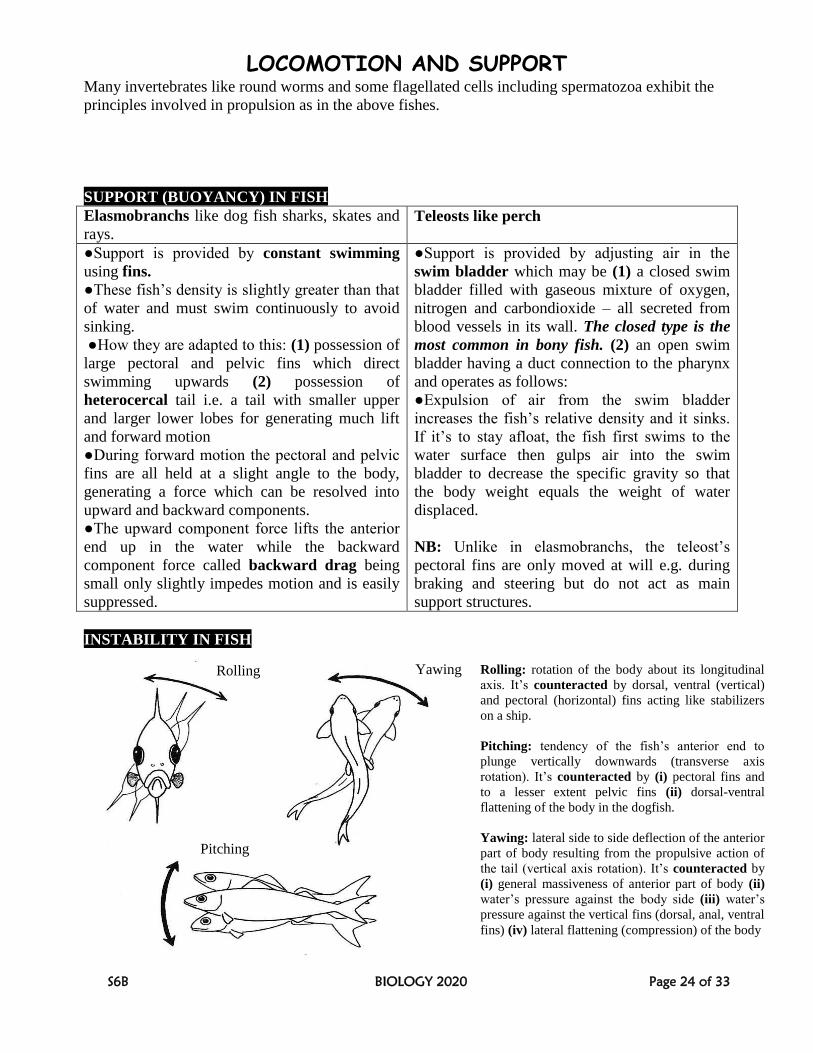
INSTABILITY IN FISH
Pitching
Rolling Yawing Rolling: rotation of the body about its longitudinal
axis. It’s counteracted by dorsal, ventral (vertical)
and pectoral (horizontal) fins acting like stabilizers
on a ship.
Pitching: tendency of the fish’s anterior end to
plunge vertically downwards (transverse axis
rotation). It’s counteracted by (i) pectoral fins and
to a lesser extent pelvic fins (ii) dorsal-ventral
flattening of the body in the dogfish.
Yawing: lateral side to side deflection of the anterior
part of body resulting from the propulsive action of
the tail (vertical axis rotation). It’s counteracted by
(i) general massiveness of anterior part of body (ii)
water’s pressure against the body side (iii) water’s
pressure against the vertical fins (dorsal, anal, ventral
fins) (iv) lateral flattening (compression) of the body
LOCOMOTION AND SUPPORT
S6B BIOLOGY 2020 Page 25 of 33
ADAPTATIONS FOR LOCOMOTION IN FISH
●Fish’s body is fusiform-shaped (spindle shaped) and laterally compressed to reduce water
resistance during swimming
●The slippery layer of mucus on the skin reduces water resistance during swimming
●The presence of many rayed-fins enables the fish to swim and also maintain its balance / stability
in water
●The lateral line enables sensitivity of fish and also functions as an echo location process for the fish
to identify its surroundings while in water
●Scales are arranged in a head to tail direction to reduce water resistance during swimming
●The swim bladder in bony fish maintains buoyancy
●Extensive blood vascular system supplies oxygen and nutrients to the muscle tissues for
contraction and drain away wastes
●Body is highly muscular to generate great propulsive force against water resistance
●The neuromuscular activity is highly coordinated resulting in reciprocal innervation.
LOCOMOTION IN BIRDS
EXTERNAL AND INTERNAL FEATURES THAT ADAPT BIRDS TO LOCOMOTION
●Birds’ bodies are streamlined (spindle shaped) during flight for overcoming air-resistance during
flight.
●The endoskeleton is hollow (pneumatized) to reduce weight, and many unnecessary bones are
fused into a single structure e.g. some vertebrae, pelvic girdle, finger and leg bones.
●Many unnecessary parts like urinary bladder and pinna are totally eliminated while reproductive
organs (testes, ovaries and oviducts) are kept tiny during non-breeding seasons to reduce weight.
●The sternum bone is extended into a large keel, for the attachment of large powerful flight muscles.
●The vanes of the feathers have hooklets called barbules that zip them together, giving the feathers
the strength needed to hold the airfoil.
●The major wing bones have internal strut-like reinforcements to prevent buckling during stress.
●The respiratory system is extensive and very efficient in supplying muscles with oxygen to
facilitate much energy release needed in muscle contraction during respiration
●Their efficient circulatory system powered by a four-chambered heart enables fast supply of
oxygen and food to the body tissues and carry away wastes.
●The large brains that are connected to eyes coupled with high-speed nerve transmission enable
quick decision making especially when landing.
●The large size of eyes in relation to their body size, coupled with eye keenness enable high visual
acuity without crashing into objects.
●The flight muscles of most birds contain oxygen-carrying compounds, (myoglobin and
cytochrome) for storing much oxygen which facilitates the release of much energy needed in muscle
contraction.
●The forelimbs have become modified into wings which act as aerofoil, generating lift when passed
into air.
LOCOMOTION AND SUPPORT
S6B BIOLOGY 2020 Page 26 of 33
●They have high body temperature which maintains the high metabolic rate for generating much
energy.
FLIGHT BIOMECHANICS (FLIGHT BIOPHYSICS)
THE BIRD’S WING AS AN AEROFOIL / AIRFOIL
Aerofoil / Airfoil: A structure whose shape and orientation provides lift, propulsion, stability, or
directional control in a flying object.
An airfoil-shaped body moved through a fluid (air or liquid) produces an aerodynamic force, which
is the resultant forces exerted on a body by the air in which the body is immersed, and is due to the
relative motion between the body and the fluid.
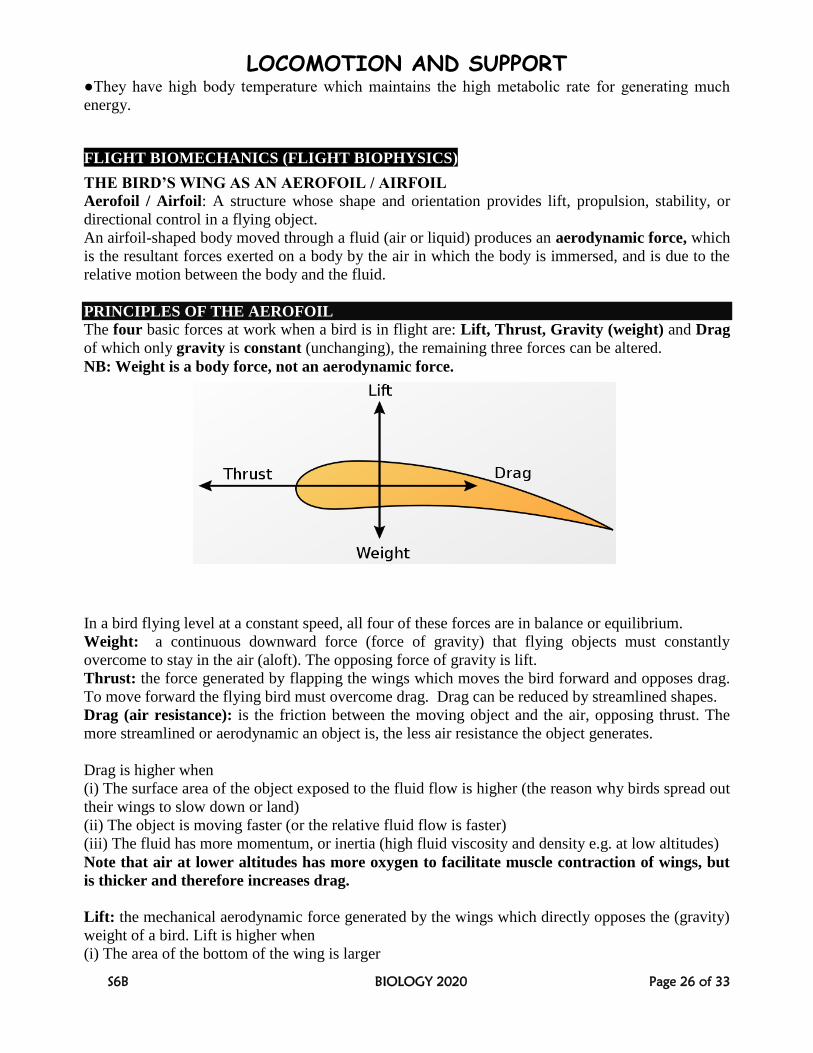
PRINCIPLES OF THE AEROFOIL
The four basic forces at work when a bird is in flight are: Lift, Thrust, Gravity (weight) and Drag
of which only gravity is constant (unchanging), the remaining three forces can be altered.
NB: Weight is a body force, not an aerodynamic force.
In a bird flying level at a constant speed, all four of these forces are in balance or equilibrium.
Weight: a continuous downward force (force of gravity) that flying objects must constantly
overcome to stay in the air (aloft). The opposing force of gravity is lift.
Thrust: the force generated by flapping the wings which moves the bird forward and opposes drag.
To move forward the flying bird must overcome drag. Drag can be reduced by streamlined shapes.
Drag (air resistance): is the friction between the moving object and the air, opposing thrust. The
more streamlined or aerodynamic an object is, the less air resistance the object generates.
Drag is higher when
(i) The surface area of the object exposed to the fluid flow is higher (the reason why birds spread out
their wings to slow down or land)
(ii) The object is moving faster (or the relative fluid flow is faster)
(iii) The fluid has more momentum, or inertia (high fluid viscosity and density e.g. at low altitudes)
Note that air at lower altitudes has more oxygen to facilitate muscle contraction of wings, but
is thicker and therefore increases drag.
Lift: the mechanical aerodynamic force generated by the wings which directly opposes the (gravity)
weight of a bird. Lift is higher when
(i) The area of the bottom of the wing is larger
LOCOMOTION AND SUPPORT
S6B BIOLOGY 2020 Page 27 of 33
(ii) The animal is moving faster
(iii) Fluid viscosity and density are higher.
Thicker air increases thrust by supplying the wings with more mass to move.
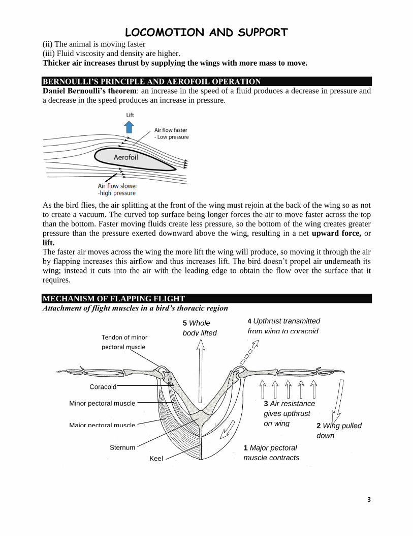
BERNOULLI’S PRINCIPLE AND AEROFOIL OPERATION
Daniel Bernoulli’s theorem: an increase in the speed of a fluid produces a decrease in pressure and
a decrease in the speed produces an increase in pressure.
As the bird flies, the air splitting at the front of the wing must rejoin at the back of the wing so as not
to create a vacuum. The curved top surface being longer forces the air to move faster across the top
than the bottom. Faster moving fluids create less pressure, so the bottom of the wing creates greater
pressure than the pressure exerted downward above the wing, resulting in a net upward force, or
lift. The faster air moves across the wing the more lift the wing will produce, so moving it through the air
by flapping increases this airflow and thus increases lift. The bird doesn’t propel air underneath its
wing; instead it cuts into the air with the leading edge to obtain the flow over the surface that it
requires.
MECHANISM OF FLAPPING FLIGHT
Attachment of flight muscles in a bird’s thoracic region
5 Whole
body lifted
4 Upthrust transmitted
from wing to coracoid
3 Air resistance
gives upthrust
on wing 2 Wing pulled
down
1 Major pectoral
muscle contracts
Tendon of minor
pectoral muscle
Coracoid
Minor pectoral muscle
Major pectoral muscle
Sternum
Keel
LOCOMOTION AND SUPPORT
S6B BIOLOGY 2020 Page 28 of 33
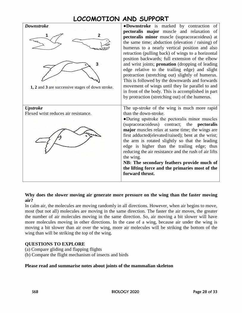
Downstroke
●Downstroke is marked by contraction of
pectoralis major muscle and relaxation of
pectoralis minor muscle (supracoracoideus) at
the same time; abduction (elevation / raising) of
humerus to a nearly vertical position and also
retraction (pulling back) of wings to a horizontal
position backwards; full extension of the elbow
and wrist joints; pronation (dropping of leading
edge relative to the trailing edge) and slight
protraction (stretching out) slightly of humerus.
This is followed by the downwards and forwards
movement of wings until they lie parallel to and
in front of the body. This is accomplished in part
by protraction (stretching out) of the humerus.
Upstroke
Flexed wrist reduces air resistance.
The up-stroke of the wing is much more rapid
than the down-stroke.
●During upstroke the pectoralis minor muscles
(supracoracoideus) contract; the pectoralis
major muscles relax at same time; the wings are
first adducted(elevated/raised); bent at the wrist;
the arm is rotated slightly so that the leading
edge is higher than the trailing edge; thus
reducing the air resistance and the rush of air lifts
the wing.
NB: The secondary feathers provide much of
the lifting force and the primaries most of the
forward thrust.
Why does the slower moving air generate more pressure on the wing than the faster moving
air? In calm air, the molecules are moving randomly in all directions. However, when air begins to move,
most (but not all) molecules are moving in the same direction. The faster the air moves, the greater
the number of air molecules moving in the same direction. So, air moving a bit slower will have
more molecules moving in other directions. In the case of a wing, because air under the wing is
moving a bit slower than air over the wing, more air molecules will be striking the bottom of the
wing than will be striking the top of the wing.
QUESTIONS TO EXPLORE
(a) Compare gliding and flapping flights
(b) Compare the flight mechanism of insects and birds
Please read and summarise notes about joints of the mammalian skeleton
1, 2 and 3 are successive stages of down stroke.
1 2
3
LOCOMOTION AND SUPPORT
S6B BIOLOGY 2020 Page 29 of 33
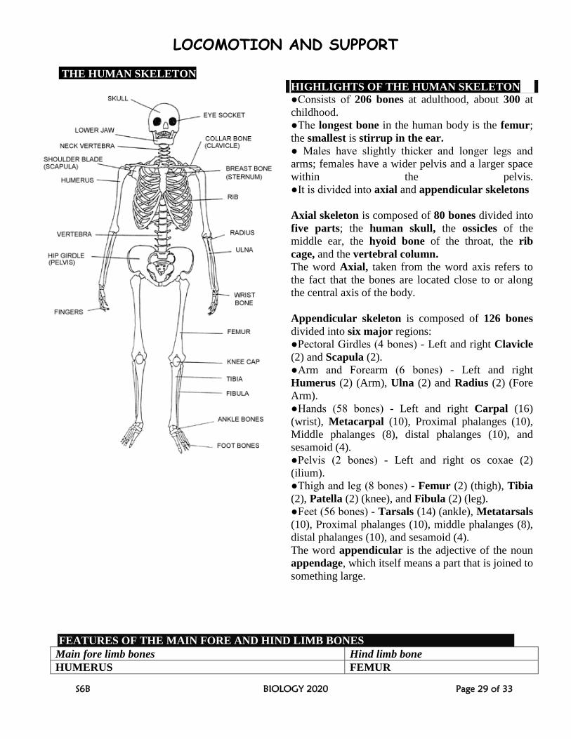
THE HUMAN SKELETON
HIGHLIGHTS OF THE HUMAN SKELETON ●Consists of 206 bones at adulthood, about 300 at
childhood.
●The longest bone in the human body is the femur;
the smallest is stirrup in the ear.
● Males have slightly thicker and longer legs and
arms; females have a wider pelvis and a larger space
within the pelvis.
●It is divided into axial and appendicular skeletons
Axial skeleton is composed of 80 bones divided into
five parts; the human skull, the ossicles of the
middle ear, the hyoid bone of the throat, the rib
cage, and the vertebral column.
The word Axial, taken from the word axis refers to
the fact that the bones are located close to or along
the central axis of the body.
Appendicular skeleton is composed of 126 bones
divided into six major regions:
●Pectoral Girdles (4 bones) - Left and right Clavicle
(2) and Scapula (2).
●Arm and Forearm (6 bones) - Left and right
Humerus (2) (Arm), Ulna (2) and Radius (2) (Fore
Arm).
●Hands (58 bones) - Left and right Carpal (16)
(wrist), Metacarpal (10), Proximal phalanges (10),
Middle phalanges (8), distal phalanges (10), and
sesamoid (4).
●Pelvis (2 bones) - Left and right os coxae (2)
(ilium).
●Thigh and leg (8 bones) - Femur (2) (thigh), Tibia
(2), Patella (2) (knee), and Fibula (2) (leg).
●Feet (56 bones) - Tarsals (14) (ankle), Metatarsals
(10), Proximal phalanges (10), middle phalanges (8),
distal phalanges (10), and sesamoid (4).
The word appendicular is the adjective of the noun
appendage, which itself means a part that is joined to
something large.
FEATURES OF THE MAIN FORE AND HIND LIMB BONES
Main fore limb bones Hind limb bone
HUMERUS FEMUR
LOCOMOTION AND SUPPORT
S6B BIOLOGY 2020 Page 30 of 33
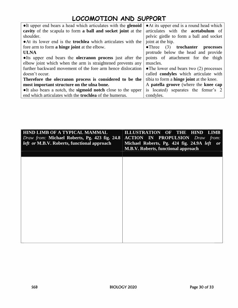
●It upper end bears a head which articulates with the glenoid
cavity of the scapula to form a ball and socket joint at the
shoulder.
●At its lower end is the trochlea which articulates with the
fore arm to form a hinge joint at the elbow.
ULNA ●Its upper end bears the olecranon process just after the
elbow joint which when the arm is straightened prevents any
further backward movement of the fore arm hence dislocation
doesn’t occur.
Therefore the olecranon process is considered to be the
most important structure on the ulna bone.
●It also bears a notch, the sigmoid notch close to the upper
end which articulates with the trochlea of the humerus.
●At its upper end is a round head which
articulates with the acetabulum of
pelvic girdle to form a ball and socket
joint at the hip.
●Three (3) trochanter processes
protrude below the head and provide
points of attachment for the thigh
muscles.
●The lower end bears two (2) processes
called condyles which articulate with
tibia to form a hinge joint at the knee.
A patella groove (where the knee cap
is located) separates the femur’s 2
condyles.
HIND LIMB OF A TYPICAL MAMMAL
Draw from: Michael Roberts, Pg. 423 fig. 24.8
left or M.B.V. Roberts, functional approach
ILLUSTRATION OF THE HIND LIMB
ACTION IN PROPULSION Draw from:
Michael Roberts, Pg. 424 fig. 24.9A left or
M.B.V. Roberts, functional approach
LOCOMOTION AND SUPPORT
S6B BIOLOGY 2020 Page 31 of 33
BIPEDALISM [PROPULSION USING 2 REAR LEGS]
Bipedal locomotion is walking, running, and standing on two rear limbs.
●During walking, the calf muscle of the right limb contracts to raise the right heel; causing the ball
of foot to exert a contact force on the ground; generating the ground reaction force (GRF) which
thrusts the body forward and slightly upwards.
●The weight of the body shifts to the left foot which is still in contact with the ground to provide
support.
●Extension of the right limb results in its heel touching the ground first to bear the body weight
transferred to it from the left side.
●Further forward movement of the body exerts backward pressure against the ground through the
right big toe.
●As the right leg bears the body weight, the left heel is raised and the whole sequence repeats.
●This sequence in which the right leg alternates with the left, heel-and-toe action continues until
walking ceases.
●The GRF is composed of the lift force which thrusts the body off the ground and the forward force
that propels the body forward - the magnitude of which depends on the angle between the ground
and the main axis of the limb.
●A large angle between the ground and the main axis of the limb (e.g. 900) results in large lift force
which thrusts the body vertically upwards with no forward force, a small angle causes a relatively
bigger forward force and small upward lift.
NB: The ball of the foot is where the toes join with the rest of the foot.
WHY MAN STANDS ON SOLES BUT GENERALLY SPRINTS ON TOES
●Standing on soles increases the surface area for supporting the body weight in a balanced posture.
●Sprinting on toes increases the effective length of limbs; enabling taking longer strides that propel
the body forward over a greater distance and at a faster pace even if the speed of limb movement
remains the same.
WHY SPRINTERS CROUCH (BEND DOWN) BEFORE TAKEOFF
Crouching creates a small angle between the ground and the main axis of the limb; resulting in
maximum forward thrust rather than upward lift; hence propelling the body a greater distance
forward.
QUADRUPEDALISM [PROPULSION USING FOUR LEGS]
Quadruped: an animal especially a mammal,
having four limbs all specialized for walking,
except humans and the birds.
Tetrapod: a vertebrate animal having four limbs
e.g. amphibians, reptiles, birds and mammals.
NB: A Tetrapod may use only two limbs for
walking
●Contraction of extensor muscle causes each limb to act as a lever by extending and exerting a
backward force that presses the foot against the ground thus thrusting the animal forward and
slightly upwards; because an equal and opposite force called reaction force is transmitted along the
length of the limb against the body while contraction of flexor muscle pulls the limb forward and
lifts it off the ground.
LOCOMOTION AND SUPPORT
S6B BIOLOGY 2020 Page 32 of 33
●During walking, only one limb is raised at a time; the other three remain anchored to the ground to
provide tripod support / stability in a sequence of leg movement as follows: left forelimb; right
hindlimb; right forelimb; left hindlimb [N.P.O. Green; etal, Biol Sc] or LH; LF; RH; RF
[Michael Roberts Pg 426& FA]
●During slow running, tripod support is lost because the two forelimbs are moved together
followed by the two hind limbs in the sequence of: left forelimb; right forelimb; right hindlimb;
left hindlimb.
● During maximum speed running, a dog uses its back to attain speed. All the four legs may be
lifted off the ground at the same time, with alternate upward arching of the back coupled with rear
feet extension in front of the front feet and the front feet extension behind the rear feet, and full
extension of the vertebral column coupled with full extension of front legs forward and rear legs
rearward to increase stride length.
SUCCESSIVE STAGES IN THE DIAGONAL
LOCOMOTORY PATTERN OF A WALKING
TETRAPOD
Draw from: Michael Roberts, etal Page 426 fig. 24.11B
(walking) or M.B.V. Roberts, functional approach
TASK
Summarize the importance of centre of
gravity Check: Michael Roberts, etal
Page 426
PLANTIGRADE, DIGITIGRADE AND UNGULIGRADE LOCOMOTION
●Plantigrade locomotion: walking with the podials and metatarsals flat on the ground e.g. humans,
raccoons, opossums, bears, rabbits, kangaroo, mice, pandas, rats and hedgehogs.
●Digitigrade locomotion: walking on the toes with the heel and wrist permanently raised e.g. birds,
wolf, dog, coyote, cat, lion, elephant (semi-digitigrade)
●Unguligrade locomotion: walking on the nail or nails of the toes (the hoof) with the heel/wrist and
the digits permanently raised. Ungulates include horse, zebra, donkey, cattle, bison, rhinoceros,
camel, hippopotamus, goat, pig, sheep, giraffe, okapi, moose, deer, antelope, and gazelle.
Advantage of a plantigrade foot: because of a large surface area, it offers stability and ability to
bear much weight.
Disadvantage of a plantigrade foot: locomotion is of slow speed because of many bones and joints
in the foot making the leg heavier at the far end.
Advantage of digitigrades: They are generally faster and quieter than other types of animals
LOCOMOTION AND SUPPORT
S6B BIOLOGY 2020 Page 33 of 33
QUESTION
Explain why in terrestrial tetrapods it is advantageous to have limbs below and parallel to the
sides of the body e.g. in mammals rather than lateral to the body e.g. in amphibians
Consult with: Clegg and Mackean, Adv. Biol Princ. & Applic. Page 495