Embed Size (px)
Citation preview

28
Supported Biomimetic Membranes for Pressure-Driven Water Purification
Yair Kaufman and Viatcheslav Freger Ben-Gurion University of the Negev
Israel
1. Introduction
The desalination technology of choice today is reverse osmosis (RO). In this process saline water is forced using mechanical pressure across a membrane that can selectively pass water and reject almost all ions and most neutral molecules. Reverse osmosis proved to be robust and most energy efficient technology that can be exploited for a wide range of water sources. Nevertheless water desalination is still an energy intensive process. Theoretical calculations show that the minimum required energy for seawater desalination is about 1 kW hr m-3, whereas the most energy optimized plants require 2-4 kWh m-3, suggesting that there is much room for improving the membranes performance and energy efficiency (Shannon et al. 2008). Despite the numerous improvements the RO membranes have gone through over the last 5 decades, they are still inferior to cell membranes both in terms of permeability and, especially, selectivity. The fast and selective water transport in biological membranes is achieved by means of aquaporins, specialized trance-membrane proteins. The osmotic water permeability of aquaporins was shown to be in the range of 6×10-14 - 11×10-14 cm3 sec-1 channel-1 for AQP1 (Saparov et al. 2001) and their ion rejection exceeds by far the ion rejection of the most advanced commercial membranes. Estimates show that a biomimetic lipid bilayer with incorporated aquaporins with a lipids to protein ratio (LPR) of 50, would yield a membrane with a hydraulic permeability of ~9 – 16.5 L m-2 hr-1 bar-1 whereas the permeability of seawater RO membrane does not exceed 2 L m-2 hr-1 bar-1 (Kaufman, Berman & Freger 2010). Biological membranes are also known to reject small molecules, such as urea or boric acid, which are poorly removed by commercial membranes (Borgnia et al. 1999). The combination of ultra-selectivity with extremely high water permeability makes biomimetic membranes highly attractive for water purification applications. It is important to emphasize that water transport through biological membranes, usually existing as microscopic free-standing self-supporting structures, is usually driven by electrochemical potential gradients, i.e. osmotic pressure or electric field (Tanaka, Sackmann 2005). However, for practical membrane applications, such as water purification, the use of hydraulic pressure as a driving force and planar membranes of macroscopic dimensions, the common configuration in membrane technology, will be far more preferable. However, the preparation of such biomimetic membranes poses a number of challenging questions, such as: how can one prepare a large and defect-free biomimetic planar membrane? can they be made to withstand hydraulic pressures? how can aquaporins be incorporated in such membranes? will aquaporins keep their activity under a hydraulic pressure gradient?
www.intechopen.com
On Biomimetics
588
This chapter will describe the on-going effort to address these challenges and build a viable biomimetic membrane using biological and non-biological building blocks. Emerging concepts for building biomimetic membranes for water purification will be presented with emphasis on the approach recently proposed and explored by the authors. The common element of different proposed approaches is that the biomimetic membrane has to be assembled on some sort of a water-permeable mechanical support that, on one hand, allows the membrane to withstand the applied hydraulic pressure and, on the other hand, passes the purified water. Two aspects are pivotal: first, what kind of support would best suit this purpose and, second, how to assemble a robust functioning biomimetic membrane on such a support. Within this discussion, it is expedient to briefly review relevant building blocks, preparation methods and interactions involved in assembly of biological membranes and their biomimetic analogues.
2. Building blocks of biological and biomimetic membranes
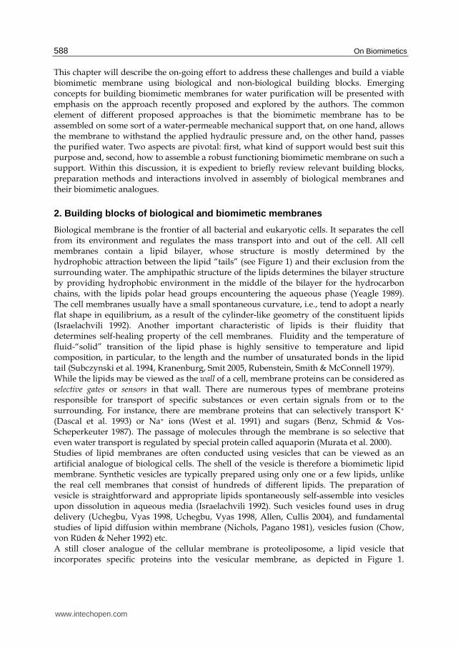
Biological membrane is the frontier of all bacterial and eukaryotic cells. It separates the cell from its environment and regulates the mass transport into and out of the cell. All cell membranes contain a lipid bilayer, whose structure is mostly determined by the hydrophobic attraction between the lipid “tails” (see Figure 1) and their exclusion from the surrounding water. The amphipathic structure of the lipids determines the bilayer structure by providing hydrophobic environment in the middle of the bilayer for the hydrocarbon chains, with the lipids polar head groups encountering the aqueous phase (Yeagle 1989). The cell membranes usually have a small spontaneous curvature, i.e., tend to adopt a nearly flat shape in equilibrium, as a result of the cylinder-like geometry of the constituent lipids (Israelachvili 1992). Another important characteristic of lipids is their fluidity that determines self-healing property of the cell membranes. Fluidity and the temperature of fluid-“solid” transition of the lipid phase is highly sensitive to temperature and lipid composition, in particular, to the length and the number of unsaturated bonds in the lipid tail (Subczynski et al. 1994, Kranenburg, Smit 2005, Rubenstein, Smith & McConnell 1979). While the lipids may be viewed as the wall of a cell, membrane proteins can be considered as selective gates or sensors in that wall. There are numerous types of membrane proteins responsible for transport of specific substances or even certain signals from or to the surrounding. For instance, there are membrane proteins that can selectively transport K+ (Dascal et al. 1993) or Na+ ions (West et al. 1991) and sugars (Benz, Schmid & Vos-Scheperkeuter 1987). The passage of molecules through the membrane is so selective that even water transport is regulated by special protein called aquaporin (Murata et al. 2000). Studies of lipid membranes are often conducted using vesicles that can be viewed as an artificial analogue of biological cells. The shell of the vesicle is therefore a biomimetic lipid membrane. Synthetic vesicles are typically prepared using only one or a few lipids, unlike the real cell membranes that consist of hundreds of different lipids. The preparation of vesicle is straightforward and appropriate lipids spontaneously self-assemble into vesicles upon dissolution in aqueous media (Israelachvili 1992). Such vesicles found uses in drug delivery (Uchegbu, Vyas 1998, Uchegbu, Vyas 1998, Allen, Cullis 2004), and fundamental studies of lipid diffusion within membrane (Nichols, Pagano 1981), vesicles fusion (Chow, von Rüden & Neher 1992) etc. A still closer analogue of the cellular membrane is proteoliposome, a lipid vesicle that incorporates specific proteins into the vesicular membrane, as depicted in Figure 1.
www.intechopen.com
Supported Biomimetic Membranes for Pressure-Driven Water Purification
589
Proteoliposomes have long been used to study the functionality of specific membrane proteins. Such studies of relevance to plant transport proteins were reviewed by Emes et al. (Emes et al. 1999).
Fig. 1. Schematic illustration of the structure of a lipid vesicle with incorporated membrane proteins (a proteoliposome).
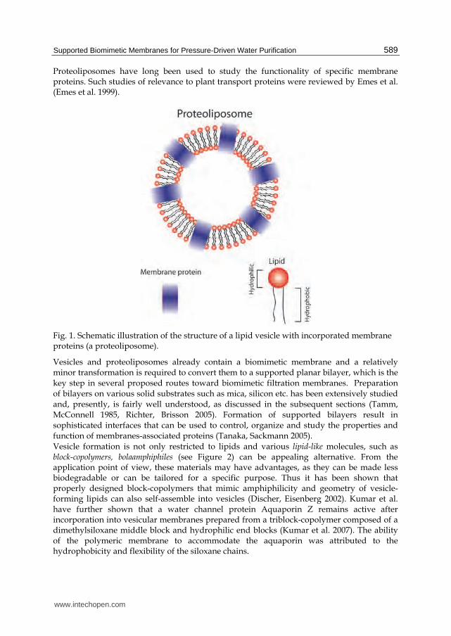
Vesicles and proteoliposomes already contain a biomimetic membrane and a relatively minor transformation is required to convert them to a supported planar bilayer, which is the key step in several proposed routes toward biomimetic filtration membranes. Preparation of bilayers on various solid substrates such as mica, silicon etc. has been extensively studied and, presently, is fairly well understood, as discussed in the subsequent sections (Tamm, McConnell 1985, Richter, Brisson 2005). Formation of supported bilayers result in sophisticated interfaces that can be used to control, organize and study the properties and function of membranes-associated proteins (Tanaka, Sackmann 2005). Vesicle formation is not only restricted to lipids and various lipid-like molecules, such as block-copolymers, bolaamphiphiles (see Figure 2) can be appealing alternative. From the application point of view, these materials may have advantages, as they can be made less biodegradable or can be tailored for a specific purpose. Thus it has been shown that properly designed block-copolymers that mimic amphiphilicity and geometry of vesicle-forming lipids can also self-assemble into vesicles (Discher, Eisenberg 2002). Kumar et al. have further shown that a water channel protein Aquaporin Z remains active after incorporation into vesicular membranes prepared from a triblock-copolymer composed of a dimethylsiloxane middle block and hydrophilic end blocks (Kumar et al. 2007). The ability of the polymeric membrane to accommodate the aquaporin was attributed to the hydrophobicity and flexibility of the siloxane chains.
www.intechopen.com

On Biomimetics
590
Fig. 2. Schematic structure of the lipids or lipid analogues discussed along this chapter (left to right): 1,2-dimyristoyl-3-trimethylammonium-propane (DMTAP, a lipid with a positively charged head), 1,2-dimyristoyl-sn-glycero-3-phosphocholine (DMPC, a phospholipid with a zwitter-ionic head), , and 1,2-dimyristoyl-sn-glycero-phosphoethanolamine-N-Lissamine-Rhodamine B Sulfonyl (Rh-PE, a fluorescent lipid), a bolaamphiphile, an ABA triblock-copolymer. The tails are hydrocarbon chains except for block-copolymers that may have any hydophobic middle block, e.g., hydrocarbon, polyimethylsiloxane etc.
3. Preparation methods for supported lipid membranes
Several techniques have been proposed for preparation of supported lipid bilayer
membranes. All of them basically rely on the fact that lipids tend to self-assemble into
bilayer and in some cases, as discussed below, supported bilayer configuration can be made
thermodynamically more favourable as compared to free vesicles. The main techniques are
as follows:
1. The so-called Langmuir-Blodgett technique, as depicted in Figure 3a, was introduced in the late 30’s of the 20’th century (Blodgett 1940). In brief, (i) a motorised stage is used to move the substrate between an aqueous (green background) and gas phase (white background). A lipid monolayer is held at defined tension at the interface, which maximizes and controls the packing density. Using a hydrophilic substrate and starting in the solution, a lipid monolayer can be deposited onto the moving substrate (Simon et al. 2007). (ii) Vertical reinsertion of the lipid monolayer formed in (i) through the interface deposits a second monolayer on top, which results in a supported lipid bilayer. (iii) If the substrate is instead taken through the interface horizontally the same result is achieved, which is referred to as the Langmuir-Schäfer deposition (Reimhult, Kumar 2008).
2. The method of detergent dialysis is depicted in Figure 3b, scheme (i). By forming micelles of lipids mixed with detergents lipid material can deposited from aqueous solution at the solid interface. The detergent is continuously removed from the micelles by dialysis leading to destabilization and decomposition of the micelles and the formation of a planar lipid bilayer at the interface (Giess et al. 2004).
3. The technique depicted in Figure 3b, scheme (ii) is called painting and solvent extraction. A drop of organic solvent containing dissolved lipids is added to a surface in aqueous phase. The amphiphilic lipids align at the solvent interface and then the solvent is extracted forcing the lipids to form a bilayer at the provided substrate-aqueous solution interface (Han et al. 2007).
www.intechopen.com

Supported Biomimetic Membranes for Pressure-Driven Water Purification
591
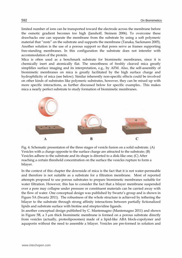
4. Vesicle fusion, as depicted in Figure 4, is probably the most common technique to prepare supported biomimetic membranes, since it is applied in the most straightforward manner. The process is mostly governed by electrostatic (electric double layer), van der Waals (vdW) and mechanical forces (see next section). It is emphasized that vesicles, even if composed of overall neutral lipids, may bear a significant surface charge in solution, as discussed below. The vesicle fusion is initiated once the surface is exposed to a vesicle solution. For vesicles having a surface charge opposite to the substrate charge, the interacting of electric double layers results in thermodynamically favorable release of counter-ions leading to attraction and adhesion to substrate by electrostatic force (Garcia-Manyes, Oncins & Sanz 2006). In absence of electrostatic forces VDW attraction can also favor bilayer deposition (Cremer, Boxer 1999). Once vesicles adhere, they are flattened against the surface, presumably by electrostatic and vdW interactions, and adopt a disk-like shape (Leonenko, Carnini & Cramb 2000). The last step of the vesicle fusion occurs after the vesicle concentration on the surface reaches a certain threshold (Leonenko, Carnini & Cramb 2000) and vesicles begin to rupture and assemble into a bilayer.
Fig. 3. Formation of a supported bilayer on hidrophilic substrates by the methods of (a) Langmuir-Bludgett and (b) detergent dialysis and painting. Adopted from. Reproduced from (Reimhult, Kumar 2008) with permission by the publisher.
Choosing the appropriate support is important and depends on the specific application. For
instance, for electrochemical measurements, e.g., for studying transport of charged species
across a bilayer or embedded channel proteins, the electrical conductivity of the substrate
and its high surface area may be essential (Martinez et al. 2009). Another factor that may
have to be considered is the fact that the supported bilayer is usually situated very close to
the substrate, no more than a few nanometers away, leaving only an ultra-thin layer of
water between the substrate and the membrane. This arrangement imposes two major
drawbacks: 1) the proximity of the membrane to the solid substrate can interfere with
accommodating membrane proteins such as cell adhesion receptors that protrude by several
tens of nanometers (Tanaka, Sackmann 2005). 2) the limited aqueous compartment between
the electrode and the membrane also restricts electrochemical measurements because only a
www.intechopen.com
On Biomimetics
592
limited number of ions can be transported toward the electrode across the membrane before
the osmotic gradient becomes too high (Janshoff, Steinem 2006). To overcome these
drawbacks one can separate the membrane from the substrate by using a soft polymeric
material that “rests” on the substrate and supports the membrane (Tanaka, Sackmann 2005).
Another solution is the use of a porous support so that pores serve as frames supporting
free-standing membranes. In this configuration the substrate does not interefer with
accommodation of the protein.
Mica is often used as a benchmark substrate for biomimetic membranes, since it is chemically inert and atomically flat. The smoothness of freshly cleaved mica greatly simplifies surface imaging and its interpretation, e.g., by AFM. Also, the self-assembly of biomimetic membranes on mica is greatly facilitated by the high surface charge and hydrophilicity of mica (see below). Similar inherently non-specific effects could be involved on other kinds of substrates like polymeric substrates, however, they can be mixed up with more specific interactions, as further discussed below for specific examples.. This makes mica a nearly perfect substrate to study formation of biomimetic membranes.
Fig. 4. Schematic presentaton of the three stages of vesicle fusion on a solid substrate. (A) Vesicles with a charge opposite to the surface charge are attracted to the substrate; (B) Vesicles adhere to the substrate and its shape is ditorted to a disk-like one; (C) After reaching a certain threshold concentration on the surface the vesicles rupture to form a bilayer.
In the context of this chapter the downside of mica is the fact that it is not water-permeable
and therefore is not suitable as a substrate for a filtration membrane. Most of reported
attempts proposed to use porous substrates to prepare biomimetic membranes suitable for
water filtration. However, this has to consider the fact that a bilayer membrane suspended
over a pore may collapse under pressure or constituent materials can be carried away with
the flow of water. One conceptual design was published by Swartz’s group and is shown in
Figure 5A (Swartz 2011). The robustness of the whole structure is achieved by tethering the
bilayer to the substrate through strong affinity interactions between partially fictionalized
lipids and substrate surface with biotine and streptavidine ligands.
In another conceptual design published by C. Mantemagno (Mantemagno 2011) and shown
in Figure 5B, a 3 m thick biomimetic membrane is formed on a porous substrate directly from vesicles (actually, proteoliposomes) made of a lipid-like ABA block-copolymer and aquaporin without the need to assemble a bilayer. Vesicles are pre-formed in solution and
www.intechopen.com

Supported Biomimetic Membranes for Pressure-Driven Water Purification
593
concentrated/deposited onto a porous support as a bed of densely packed vesicles. The filtration may then be carried out along a tortuous path across many vesicle shells rather than across a single planar membrane. The robustness could be achieved by using UV-cross-linkable blocks and cross-linking the whole structure by UV irradiation once the membrane is formed. Unfortunately, the success of two above approaches is unclear, as the results have not yet been published.
Fig. 5. Two conceptual designs of biomimetic membranes for water purification by filtration: (A) a lipid bilayer/aquaporin membrane tethered to a porous substrate proposed by the Swartz group, after (Swartz 2011); (B) a membrane assembled from proteoliposomes on a porous substrate proposed by C. Mantemagno (Montemagno 2011). Image B reproduced with permission by the author.
A third approach uses a hydrophobic porous support to suspend lipid membranes within
pores rather than on top of the support. Two monolayers formed the two sides of the
hydrophobic support merge within the pores to form a bilayer. A small pore size is crucial
for stability of the membrane and its ability to withstand pressures. The large area required
for filtration is achieved by using an array of multiple pores or holes formed within a thin
film made of a rigid hydrophobic polymer material. Formation of such membrane arrays
using a combination of painting and Langmuir-Blodgett techniques was successfully
demonstrated, however, the pore size was apparently too large to withstand any pressure
gradient under filtration conditions (Vogel et al. 2009).
It appears then that, although it was possible to prepare supported biomimetic membranes on porous substrates, this has not led so far to successful filtration experiments. As a potentially more viable alternative, it has been proposed to prepare supported biomimetic membrane on a dense water-permeable substrate. In the present context “dense” means that the substrate have a pore size smaller than the molecular size of all constituents of the biomimetic membrane. This should prevent their washout or, at least, commensurate with the thickness of the biomimetic membrane, i.e. <5 nm, thereby the bilayer will not collapse under applied pressure. Commercial nano-filtration (NF) membranes appear to be an appropriate choice. Their pore size is smaller than 1 nm and they are robust enough to endure pressures over 20 bar. The water permeability of available NF membranes is commensurable with biological membranes and may be much higher for state-of-the-art membranes; hence the permeability of the biomimetic membrane is not expected to be dramatically reduced due to the substrate.
www.intechopen.com

On Biomimetics
594
NF membranes represent a new class of substrates that bear certain similarity with benchmark solid substrates such as mica or Si, yet they show clear differences in terms of surface characteristics and relevant interactions as well as lipid dynamics within the bilayer. These differences and associated challenges are discussed in the subsequent chapters. They are revealed by applying appropriate characterization techniques that produce the surface structure and chemical composition from the molecular level up to the macroscopic scale.
3.1 Control of interactions during vesicle fusion and membrane formation on a substrate An efficient way to control vesicle fusion on a substrate and the characteristics of the supported bilayer is to regulate the vesicles charge versus the substrate charge. This can be carried out by varying pH, changing the ionic strength of the solution or adding certain charge-regulating ions such as Mg2+. DMPC (see Figure 2) is often used to mimic the phospholipids in biological cells. Figure 6 shows as an example the effect of the solution pH on the morphology of DMPC bilayer on mica. The main effect of pH in this case is apparently through protonation or deprotonation of the zwitterionic heads of DMPC and,
correspondingly, variation of the surface charge and –potential of the DMPC vesicles. Mica is negatively charged in a wide range of pH (Egawa, Furusawa 1999) therefore vesicle fusion was enhanced and surfaced coverage increased at low pH when the vesicles acquired a positive charge (Garcia-Manyes, Oncins & Sanz 2006). Interaction of the double electric layers in this case is attractive due to entropically favorable release of counter-ions (Safran 2005, Meier-Koll, Fleck & Gruenberg 2004). On the other hand, AFM images show that the surface coverage was poorer and less regular at high pH when the vesicles were negatively charged, thereby the double electric layers of vesicle and substrate repel each other.
Fig. 6. (a) Variation of ζ-potential of DMPC unilamellar liposomes in solution versus pH and its effect on the morephology and surface coverage of DMPC bilayer formed on mica as a substrate. AFM images of bilayers deposited on mica corresponds to pH 2 (b), 4 (c), 7 (d), 10 (e), and 12 (f). The images were obtained in contact mode.The ionic strength was kept constant at 10 mM for all solutions. (c.1) shows a cross-section profile indicating the bilayer thickness of about 4 nm. Reproduced from (Garcia-Manyes, Oncins & Sanz 2006) with permission by the publisher.
www.intechopen.com

Supported Biomimetic Membranes for Pressure-Driven Water Purification
595
The effect of ionic strength on the vesicle ζ-potential and the surface coverage of mica by a DMPC bilayer at pH 7 is depicted in Figure 7. DMPC vesicles are negatively charged at pH 7, which is unfavorable for vesicle fusion thereby the surface coverage at low ionic strength is poor (Figure 7b and c). However, increasing ionic strength results in compression of the
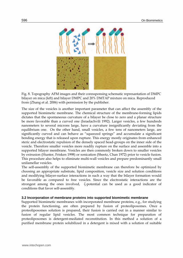
diffuse double layer, thereby -potential decreases and vanishes at about 100 mM NaCl, apparently giving way to van der Waals interactions that favor vesicle fusion thus surface coverage increases (Figure 7d). The surface coverage is further improved upon addition of Mg2+ ions, whose binding to the negatively charged vesicle surface causes ion-bridging or charge reversal, which enhances attraction between vesicles and mica (Figure 7e). A different and highly efficient approach to controlling electrostatics within DMPC bilayers on mica was proposed by Zhang et al. (Zhang et al. 2006). These authors assumed that the charged groups of the zwitterionic phosphocholine head-group are not only involved in interaction with the substrate during vesicle fusion, but also affect the stability and integrity of the lipid membrane itself, as schematically depicted in Figure 8. The parallel alignment of the head-groups dipoles within the bilayer promotes repulsion and favors formation of defects (holes) in the bilayer resulting in incomplete coverage even at optimal pH and ionic strength (Figure 6 and Figure 7). A proposed elegant way to overcome head repulsion dubbed “electrostatic stitching” is addition of a fraction of a lipid or lipids that can insert an opposite charge between similarly charged groups of zwitterionic heads. The resulting attraction then “stitches” the holes and greatly stabilizes the bilayer, as can be seen in Figure 8.
Fig. 7. (a) Varition of ξ-potential value of DMPC unilamellar liposomes in solution versus ionic strength at pH 7. Contact mode AFM images show a DMPC bilayer formed on mica in 0 mM NaCl (b), 50 mM NaCl (c), 100 mM NaCl (d), and 150 mM NaCl + 20 mM MgCl2 (e). Reproduced from (Garcia-Manyes, Oncins & Sanz 2006) with permission by the publisher.
www.intechopen.com
On Biomimetics
596
Fig. 8. Topography AFM images and their coressponsing schematic represantation of DMPC bilayer on mica (left) and bilayer DMPC and 20% DMTAP mixture on mica. Reproduced from (Zhang et al. 2006) with permission by the publisher.
The size of the vesicles is another important parameter that can affect the assembly of the supported biomimetic membrane. The chemical structure of the membrane-forming lipids dictates that the spontaneous curvature of a bilayer be close to zero and a planar structure be more favorable than a curved one (Israelachvili 1992). Larger vesicles, a few hundreds nanometers to several microns large, have a curvature insignificantly deviating from the equilibrium one. On the other hand, small vesicles, a few tens of nanometers large, are significantly curved and can behave as “squeezed springs” and accumulate a significant bending energy that is released upon rupture. This energy mostly originates from enhanced steric and electrostatic repulsion of the densely spaced head-groups on the inner side of the vesicle. Therefore smaller vesicles more readily rupture on the surface and assemble into a supported bilayer membrane. Vesicles are then commonly broken down to smaller vesicles by extrusion (Hunter, Frisken 1998) or sonication (Sheetz, Chan 1972) prior to vesicle fusion. This procedure also helps to eliminate multi-wall vesicles and prepare predominantly small unilamellar vesicles. The self-assembly of the supported biomimetic membrane can therefore be optimized by choosing an appropriate substrate, lipid composition, vesicle size and solution conditions and modifying bilayer-surface interactions in such a way that the bilayer formation would be favorable as compared to free vesicles. Since the electrostatic interactions are the strongest among the ones involved, ζ-potential can be used as a good indicator of conditions that favor self-assembly.
3.2 Incorporation of membrane proteins into supported biomimetic membrane Supported biomimetic membranes with incorporated membrane proteins, e.g., for studying the protein functioning, are often prepared by fusion of proteoliposomes. Once a proteoliposomes solution is prepared, their fusion is carried out in a manner similar to fusion of regular lipid vesicles. The most common technique for preparation of proteoliposomes is detergent-mediated reconstitution. In this method a solution of a purified membrane protein solubilized in a detergent is mixed with a solution of suitable
www.intechopen.com
Supported Biomimetic Membranes for Pressure-Driven Water Purification
597
lipid(s). Subsequently, the detergent that is significantly smaller than the lipids and protein is selectively removed from the solution by dialysis through a low molecular cutoff membrane or adsorption on a hydrophobic resin with small pores (e.g., BioBeads). As a result the protein gets progressively self-inserted into the lipid shell of vesicles forming proteoliposomes. For successful reconstitution many parameters, such as lipids composition, detergent concentration, detergent removal rate and etc., have to be optimized. More detail may be found in available reviews (Rigaud et al. 1998, Angrand et al. 1997). Figure 9A and B show examples of two supported lipid bilayers, DMPC and mixed
DMPC/DMTAP, both containing a SoPIP 1,2 aquaporin protein prepared on mica by fusion
of proteoliposomes (Kaufman, Berman & Freger 2010). Proteoliposome solutions were
obtained by mixing a solution of respective lipids with a solution of aquaporin solubilized
with a detergent 1-n-octyl-β-D-glucopyranoside (OG)-bolaamphiphile followed by removal
of OG by dialysis and reducing the proteoliposome size by extrusion through a
polycarbonate membrane with 100 nm pores. The depth of the holes in Figure 9A was ~5
nm, which closely matches the bilayer thickness and indicates that holes are defects in the
bilayer. On the other hand, these defects indicate that the mica surface was covered by a
lipid bilayer. The bilayer integrity is markedly improved and holes disappear for the mixed
DMPC/DMTAP bilayer (Figure 9B), apparently due to the “electrostatic stitching” effect.
The lateral dimensions of the smallest protruding features magnified in the inset C closely
match the ~8 nm diameter of the aquaporin tetramer (Fotiadis et al. 2001). The height of
protrusions, about 2 nm above the surface of the DMPC bilayer, is also in good agreement
with the known height of aquaporins. Much larger objects, a few tens nanometers large, and
protruding from the bilayer by more than 2 nm are also observed in Figure 9B and C, are
likely to be aggregated aquaporins. The propensity of membrane proteins to aggregation is
well known (Bruinsma, Pincus 1996) and is often utilized for preparation of two-or three
dimensional protein crystals (Durbin, Feher 1996, Garavito, Picot & Loll 1996). Aggregated
aquaporins within supported layers often preserve their native configuration and are quite
densely packed; therefore it is unclear whether protein aggregation is an obstacle to their
functioning as selective water filters.
3.3 Preparation and characterization of biomimetic membrane on water-permeable polymeric substrates During the preparation of biomimetic filtration membranes on NF membranes as substrates
one faces 2 challenges:
1. integrity of the lipid bilayer or its analogues – the impermeable matrix for aquaporins -
has to be verified to ensure minimal rate of non-selective transport of water and solutes
through imperfections;
2. membrane proteins have to be incorporated into the supported bilayer in such way that
their activity is preserved.
Both aspects are to be realized simultaneously during fusion of proteoliposomes on the
surface, but it is expedient to separate them and begin with preparation and fusion of lipid
vesicles without a protein. The present study focuses on the first aspect. A recent paper by
Kaufman et al. reported preparation of lipid bilayers supported on two commercial NF
membranes : NF-270 (Dow-Filmtec) and NTR-7450 (Hydranautics/Nitto Denko). The top
layer on the NF-270 is composed of polyamide.
www.intechopen.com

On Biomimetics
598
Fig. 9. (A) AFM topography image of (A) a DMPC bilayer and (B) a mixed DMPC/DMTAP bilayer with incorporated aquaporins on mica. (C) A higher resolution scan of the marked region in (B), and (D) a suggested structure of the mixed DMPC/DMTAP bilayer shown in (B) and (C). AFM images were aquired in tapping mode under water. The vesicles fusion was carried out on freshly cleaved mica for 30 minutes followed by rinsing with DI water. Reproduced from (Kaufman, Berman & Freger 2010) with the permission by the publisher.
(Mänttäri, Pihlajamäki & Nyström 2006, Boussu et al. 2005) and that of the NTR-7450 is
composed of sulfonated polyethersulfone (Mänttäri, Pihlajamäki et al. 2006, Boussu, Van der
Bruggen et al. 2005). The polyamide layer of the NF-270 contains a significant amount of
weakly acidic carboxylic groups that render the surface negatively charged in aqueous
solution at above pH about 3.5 - 4; as the pH increases the negative surface charge increases
as well (Mänttäri, Pekuri & Nyström 2004). In constrast, the toplayer of NTR-7450 is made of
sulfonated polysulfone that contains strong sulfonic groups thereby its surface is negatively
charged in a wide pH range down to pH ~ 2 (Schaep, Vandecasteele 2001)
The preparation of a lipid bilayer on these polymeric substrates followed essentially the same procedure as on mica. However, this study shows the visualization and characterization of supported bilayer on these polymeric substrates is somewhat more challenging in comparison to ideal surfaces such as mica or silicon wafers. Their relatively rough and irregular topography reduces the resolution of AFM images and makes interpretation difficult. As a matter of fact, no single characterization method can
www.intechopen.com

Supported Biomimetic Membranes for Pressure-Driven Water Purification
599
unequivocally verify formation and morphology of bilayer on such substrates. Consequently, the need for several complimentary characterization techniques becomes essential. By using different characterization techniques one can also gain a structural picture at different length scales spanning several order of magnitudes, starting from the nanometer scale up to the macro-scale (mm and even cm). At the millimeter scale an efficient method to assess the coverage in a semiquantitative way is fluorescence microscopy. A small fraction of a lipid with a fluorescent head-group such as Rh-PE (Figure 2) is added and gets randomly mixed with other lipids present in the system, which makes the lipid bilayer fluoresce. The images presented in Figure 10, which were acquired using fluorescence microscopy, compare coverage of the two NF membranes after fusion of vesicles composed of 1.5 mM DMPC with 20% (mol) DMTAP and 0.5% (mol) Rh-PE at different pH without added electrolyte. The results clearly show the dominant role of electrostatic attraction between vesicles and substrate in formation of a bilayer on the surface. Indeed, maximal coverage is obtained for NTR7450 at pH 2, which are conditions, at which the vesicles and the membrane are oppositely surface charge (cf. Figure 6). The low ionic strength maximizes the entropic gain and attraction forces between the surfaces due to counterion release. A somewhat smaller coverage is obtained at neutral pH, when the vesicles have a smaller yet still negative surface charge. Possible, a substantial coverage of NTR7450 at this pH could be aided by van der Waals interactions as well. On the other hand, since the isoelectric points of NF270 and vesicles are quite similar, around pH 4, at all pH this membrane and vesicle have a surface charge of the same sign and experience attraction, which is also true for NTR7450 at pH 10.
Fig. 10. Fluorescence micrsocopy images of the surface of NTR7450 and NF279 nanofiltration membranes after vesicles fusion using a solution of 1.5 mM DMPC with 20% (mol) DMTAP at different pH. The lipid bilayer is made flurescent through addition of 0.5% (mol) Rh-PE. Scale bar is 100 μm. Reproduced from (Kaufman, Berman & Freger 2010) with the permission by the publisher.
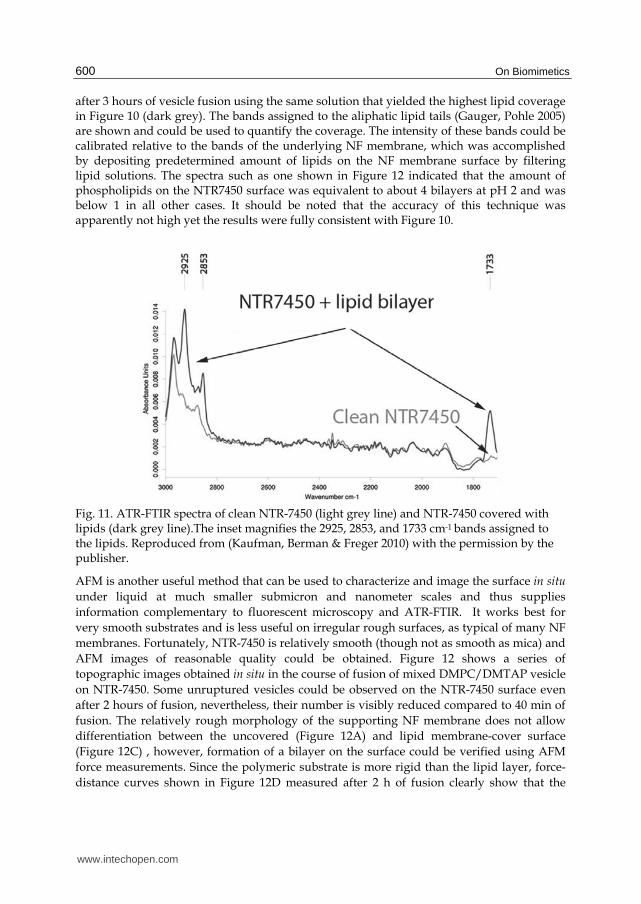
The coverage on the millimeter scale could also be more quantitatively accessed using ATR-FTIR. Figure 11 shows the ATR-FTIR spectra of clean NTR-7450 (light grey) and NTR-7450
www.intechopen.com
On Biomimetics
600
after 3 hours of vesicle fusion using the same solution that yielded the highest lipid coverage in Figure 10 (dark grey). The bands assigned to the aliphatic lipid tails (Gauger, Pohle 2005) are shown and could be used to quantify the coverage. The intensity of these bands could be calibrated relative to the bands of the underlying NF membrane, which was accomplished by depositing predetermined amount of lipids on the NF membrane surface by filtering lipid solutions. The spectra such as one shown in Figure 12 indicated that the amount of phospholipids on the NTR7450 surface was equivalent to about 4 bilayers at pH 2 and was below 1 in all other cases. It should be noted that the accuracy of this technique was apparently not high yet the results were fully consistent with Figure 10.
Fig. 11. ATR-FTIR spectra of clean NTR-7450 (light grey line) and NTR-7450 covered with lipids (dark grey line).The inset magnifies the 2925, 2853, and 1733 cm-1 bands assigned to the lipids. Reproduced from (Kaufman, Berman & Freger 2010) with the permission by the publisher.
AFM is another useful method that can be used to characterize and image the surface in situ
under liquid at much smaller submicron and nanometer scales and thus supplies
information complementary to fluorescent microscopy and ATR-FTIR. It works best for
very smooth substrates and is less useful on irregular rough surfaces, as typical of many NF
membranes. Fortunately, NTR-7450 is relatively smooth (though not as smooth as mica) and
AFM images of reasonable quality could be obtained. Figure 12 shows a series of
topographic images obtained in situ in the course of fusion of mixed DMPC/DMTAP vesicle
on NTR-7450. Some unruptured vesicles could be observed on the NTR-7450 surface even
after 2 hours of fusion, nevertheless, their number is visibly reduced compared to 40 min of
fusion. The relatively rough morphology of the supporting NF membrane does not allow
differentiation between the uncovered (Figure 12A) and lipid membrane-cover surface
(Figure 12C) , however, formation of a bilayer on the surface could be verified using AFM
force measurements. Since the polymeric substrate is more rigid than the lipid layer, force-
distance curves shown in Figure 12D measured after 2 h of fusion clearly show that the
www.intechopen.com
Supported Biomimetic Membranes for Pressure-Driven Water Purification
601
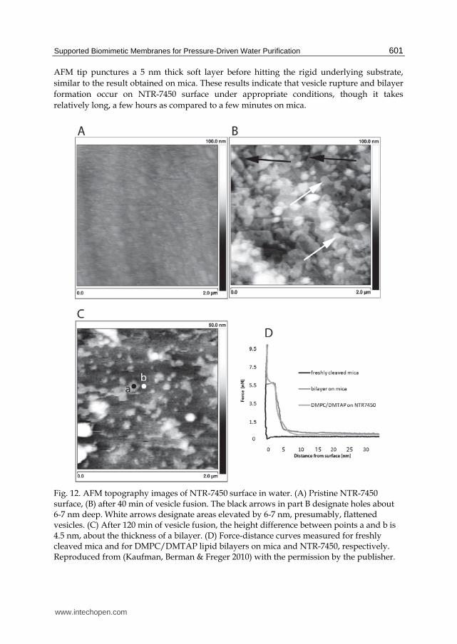
AFM tip punctures a 5 nm thick soft layer before hitting the rigid underlying substrate,
similar to the result obtained on mica. These results indicate that vesicle rupture and bilayer
formation occur on NTR-7450 surface under appropriate conditions, though it takes
relatively long, a few hours as compared to a few minutes on mica.
Fig. 12. AFM topography images of NTR-7450 surface in water. (A) Pristine NTR-7450 surface, (B) after 40 min of vesicle fusion. The black arrows in part B designate holes about 6-7 nm deep. White arrows designate areas elevated by 6-7 nm, presumably, flattened vesicles. (C) After 120 min of vesicle fusion, the height difference between points a and b is 4.5 nm, about the thickness of a bilayer. (D) Force-distance curves measured for freshly cleaved mica and for DMPC/DMTAP lipid bilayers on mica and NTR-7450, respectively. Reproduced from (Kaufman, Berman & Freger 2010) with the permission by the publisher.
www.intechopen.com

On Biomimetics
602
The slow dynamics of lipid re-arrangement resulting in slow fusion could be clarified using fluorescence recovery after photobleaching (FRAP). FRAP is fluorescence based technique that enables measurements of the lateral diffusivity of lipids on the surface. The rate of fluorescence recovery of a laser-bleached area may be related to diffusivity and provide useful insights into the state of lipid aggregation at the surface, e.g., whether it is a lipids bilayer, intact vesicles or mixture of both. Lipids in a bilayer retain lateral mobility like molecules in a liquid and undergo free lateral diffusion. This behavior is part of the self-healing ability of the biological membranes and its mimetic versions. On the other hand, in the case of lipids in a vesicle or similar aggregates of limited size, the fast diffusion of the individual lipids is possible only within the vesicle, while at larger scales diffusion may only occur as slow diffusion of entire vesicles. Figure 13 shows fluorescence recovery of lipid layers self-assembled on modified gold substrates with incremental changes of surface charge from 100% to 0% positively charged thiol SAM (Cha, Guo & Zhu 2006). FRAP showed that on highly positively charged surfaces vesicles ruptured and self-assembled to a bilayer, whereas on low charged surfaces vesicles tended not to rupture. In the case of moderately charged surfaces a mixture of vesicles and bilayer was assembled on the surface.
Fig. 13. FRAP images obtained after absorption of egg phosphatidylcholine vesicles on surfaces of mixed NH3+/OH terminated thiol SAMs. The series of images taken at 1-min time intervals after photobleaching images are labeled with mole percentages of NH3+ in the SAMs.. The size of each photobleached area is 16×16 μm, and the size of each image is 160×160 μm. Cartoons on the right-hand side illustrate the suggested state of aggregation of the supported phospholipids bilayers, continuou bilyer for 100–80% NH3+, adsorbed vesicles for 60-0% NH3+ and mixted aggregation at 75%. Reproduced from (Cha, Guo & Zhu 2006) with the permission by the publisher.
www.intechopen.com

Supported Biomimetic Membranes for Pressure-Driven Water Purification
603
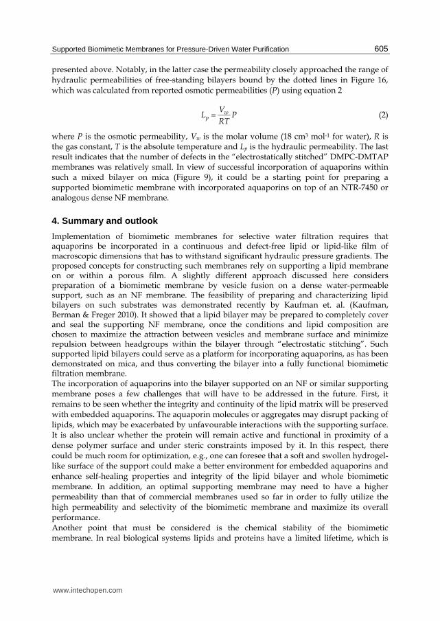
Figure 14 makes a similar comparison between mica and NTR-7450 surfaces showing the kinetics of the fluorescence recovery on each surface after vesicle fusion. Fitting the kinetic curves to a well-known model (Soumpasis 1983) yields diffusion coefficients differing by 2 orders of magnitude for the 2 substrates. The much smaller diffusion coefficient on NTR7450 seems to imply co-existence of vesicles and an extended bilayer on the surface of the NTR-7450, as schematically depicted in Figure 13 for 75% surface. Still, one cannot rule the possibility that the lipid diffusion – as well as vesicle fusion - was slowed down on NTR-7450 by van der Waal interactions between the lipids and relatively hydrophobic NTR-7450 surface. The above characterization methods supply a good picture of lipid aggregation, surface coverage and morphology of the biomimtic membranes self-assembled on the surface of NF membranes. They are unable however to verify absence or low rate of imperfections, through which non-selective transport may occur by-passing aquaporins. Such an ultimate test for fairly large membrane areas may be conducted taking advantage of water permeability of the supporting NF membrane and measuring water permeation or hydraulic permeability of the lipid membrane in a pressurized filtration cell schematically shown in Figure 15. Since the water permeability of lipid bilayers is low in comparison with the permeability of NF membranes, one would expect a dramatic drop in the permeability after self-assembly of a biomimetic lipid membrane (without aquaporin) on top.
Fig. 14. FRAP results for bilaye assembled by vesicle fusion from a DMPC solution with 0.5% Rh-PE on mica and NTR-7450. Inserts A and B show CLSM fluorescence images of mica and NTR-7450 surfaces immediately after bleaching. Scale bar 20 μm, the diameter of bleached area was 308 μm2. The fitted lateral diffusion coefficients were 2.20×10⁻⁸ cm2 s-1 on mica and 1.60×10⁻¹⁰ cm2 s-1 on NTR-7450. Reproduced from (Kaufman, Berman & Freger 2010) with the permission by the publisher.
The water permeability of DMPC/DMTAP bilayer itself was evaluated by measuring the permeability of the NTR-7450 support with and without bilayer and using the relation for resistances-in-series (Mulder 1996)
1 2
1 1 1
p p pL L L (1)
www.intechopen.com
On Biomimetics
604
Where Lp, Lp1, and Lp2 are the permeabilities of the whole supported lipid membrane, pristine NTR-7450, and the biomimetic lipid membrane, respectively.
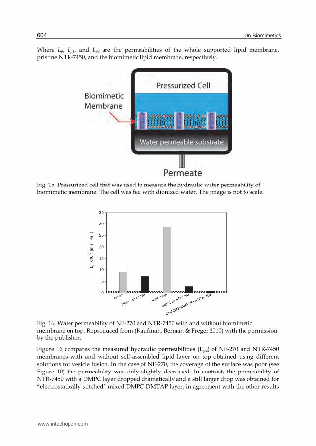
Fig. 15. Pressurized cell that was used to measure the hydraulic water permeability of biomimetic membrane. The cell was fed with dionized water. The image is not to scale.
Fig. 16. Water permeability of NF-270 and NTR-7450 with and without biomimetic membrane on top. Reproduced from (Kaufman, Berman & Freger 2010) with the permission by the publisher.
Figure 16 compares the measured hydraulic permeabilities (Lp2) of NF-270 and NTR-7450
membranes with and without self-assembled lipid layer on top obtained using different
solutions for vesicle fusion. In the case of NF-270, the coverage of the surface was poor (see
Figure 10) the permeability was only slightly decreased. In contrast, the permeability of
NTR-7450 with a DMPC layer dropped dramatically and a still larger drop was obtained for
“electrostatically stitched” mixed DMPC-DMTAP layer, in agreement with the other results
www.intechopen.com
Supported Biomimetic Membranes for Pressure-Driven Water Purification
605
presented above. Notably, in the latter case the permeability closely approached the range of
hydraulic permeabilities of free-standing bilayers bound by the dotted lines in Figure 16,
which was calculated from reported osmotic permeabilities (P) using equation 2
wp
VL P
RT (2)
where P is the osmotic permeability, Vw is the molar volume (18 cm3 mol-1 for water), R is the gas constant, T is the absolute temperature and Lp is the hydraulic permeability. The last result indicates that the number of defects in the “electrostatically stitched” DMPC-DMTAP membranes was relatively small. In view of successful incorporation of aquaporins within such a mixed bilayer on mica (Figure 9), it could be a starting point for preparing a supported biomimetic membrane with incorporated aquaporins on top of an NTR-7450 or analogous dense NF membrane.
4. Summary and outlook
Implementation of biomimetic membranes for selective water filtration requires that aquaporins be incorporated in a continuous and defect-free lipid or lipid-like film of macroscopic dimensions that has to withstand significant hydraulic pressure gradients. The proposed concepts for constructing such membranes rely on supporting a lipid membrane on or within a porous film. A slightly different approach discussed here considers preparation of a biomimetic membrane by vesicle fusion on a dense water-permeable support, such as an NF membrane. The feasibility of preparing and characterizing lipid bilayers on such substrates was demonstrated recently by Kaufman et. al. (Kaufman, Berman & Freger 2010). It showed that a lipid bilayer may be prepared to completely cover and seal the supporting NF membrane, once the conditions and lipid composition are chosen to maximize the attraction between vesicles and membrane surface and minimize repulsion between headgroups within the bilayer through “electrostatic stitching”. Such supported lipid bilayers could serve as a platform for incorporating aquaporins, as has been demonstrated on mica, and thus converting the bilayer into a fully functional biomimetic filtration membrane. The incorporation of aquaporins into the bilayer supported on an NF or similar supporting membrane poses a few challenges that will have to be addressed in the future. First, it remains to be seen whether the integrity and continuity of the lipid matrix will be preserved with embedded aquaporins. The aquaporin molecules or aggregates may disrupt packing of lipids, which may be exacerbated by unfavourable interactions with the supporting surface. It is also unclear whether the protein will remain active and functional in proximity of a dense polymer surface and under steric constraints imposed by it. In this respect, there could be much room for optimization, e.g., one can foresee that a soft and swollen hydrogel-like surface of the support could make a better environment for embedded aquaporins and enhance self-healing properties and integrity of the lipid bilayer and whole biomimetic membrane. In addition, an optimal supporting membrane may need to have a higher permeability than that of commercial membranes used so far in order to fully utilize the high permeability and selectivity of the biomimetic membrane and maximize its overall performance. Another point that must be considered is the chemical stability of the biomimetic membrane. In real biological systems lipids and proteins have a limited lifetime, which is
www.intechopen.com

On Biomimetics
606
overcome by constantly replacing the degraded biomaterials in the cell. In order to prepare biomimetic membrane that will be functional for a long time, a substantial advantage is offered by synthetic biomimetic analogues, e.g., suitable amphiphilic block-copolymers or bolaamphiphiles etc. could beneficially replace degradable lipids. Moreover, recent advances in supramolecular chemistry suggest that preparation of synthetic analogues of aquaporins through self-assembly of carefully designed synthetic building blocks could be possible as well. For instance, Barboiu and co-workers have shown that ureido crown ethers can self-assemble into columnar superamolecular structures can function as selective ion channels (Cazacu et al. 2006). Carbon nanotubes are another material that could potentially be used for creating synthetic nano-pores or nano-channels for selective transport of water. A highly attractive feature of such nanopores is exceptionally fast flow of water due to very smooth inner surface of such nano-channels resulting in a single-file-like collective motion of water molecules predicted by similations and observed experimentally (Whitby, Quirke 2007, Noy et al. 2007).
5. Acknowledgments
This research conducted by the authors was supported by the MEMBAQ project funded by European Union (contract NMP4-CT-006-33234). Prof. Per Kjellbom and Dr. Urban Johanson of Lund University, Sweden, are greatfully acknowledged for supplying aquaporins for this study. The authors also thank Dow-Filmtec and Hydranautics for supplying NF-270 and NTR-7450 membranes.
6. References
Allen, T.M. & Cullis, P.R. 2004, Drug delivery systems: entering the mainstream, Science, vol. 303, no. 5665, pp. 1818.
Angrand, M., Briolay, A., Ronzon, F. & Roux, B. 1997, Detergent-Mediated Reconstitution of a Glycosyl-Phosphatidylinositol-Protein into Liposomes, European Journal of Biochemistry, vol. 250, no. 1, pp. 168-176.
Benz, R., Schmid, A. & Vos-Scheperkeuter, G.H. 1987, Mechanism of sugar transport through the sugar-specific LamB channel ofEscherichia coli outer membrane, Journal of Membrane Biology, vol. 100, no. 1, pp. 21-29.
Blodgett, K.B. 1940, FILM STRUCTURE AND METHOD OF, . Borgnia, M.J., Kozono, D., Calamita, G., Maloney, P.C. & Agre, P. 1999, Functional
reconstitution and characterization of AqpZ, the E. coli water channel protein1, Journal of Molecular Biology, vol. 291, no. 5, pp. 1169-1179.
Boussu, K., Van der Bruggen, B., Volodin, A., Snauwaert, J., Van Haesendonck, C. & Vandecasteele, C. 2005, Roughness and hydrophobicity studies of nanofiltration membranes using different modes of AFM, Journal of colloid and interface science, vol. 286, no. 2, pp. 632-638.
Bruinsma, R. & Pincus, P. 1996, Protein aggregation in membranes, Current Opinion in Solid State & Materials Science, vol. 1, no. 3, pp. 401-406.
Cazacu, A., Tong, C., van der Lee, A., Fyles, T.M. & Barboiu, M. 2006, Columnar self-assembled ureido crown ethers: an example of ion-channel organization in lipid bilayers, Journal of the American Chemical Society, vol. 128, no. 29, pp. 9541- 9548.
www.intechopen.com

Supported Biomimetic Membranes for Pressure-Driven Water Purification
607
Cha, T.W., Guo, A. & Zhu, X.Y. 2006, Formation of supported phospholipid bilayers on
molecular surfaces: role of surface charge density and electrostatic interaction,
Biophysical journal, vol. 90, no. 4, pp. 1270-1274.
Chow, R.H., von Rüden, L. & Neher, E. 1992, Delay in vesicle fusion revealed by
electrochemical monitoring of single secretory events in adrenal chromaffin cells, .
Cremer, P.S. & Boxer, S.G. 1999, Formation and spreading of lipid bilayers on planar glass
supports, J.Phys.Chem.B, vol. 103, no. 13, pp. 2554-2559.
Dascal, N., Schreibmayer, W., Lim, N.F., Wang, W., Chavkin, C., DiMagno, L., Labarca, C.,
Kieffer, B.L., Gaveriaux-Ruff, C. & Trollinger, D. 1993, Atrial G protein-activated K
channel: expression cloning and molecular properties, Proceedings of the National
Academy of Sciences of the United States of America, vol. 90, no. 21, pp. 10235.
Discher, D.E. & Eisenberg, A. 2002, Polymer vesicles, Science, vol. 297, no. 5583, pp.
967.
Durbin, S. & Feher, G. 1996, Protein crystallization, Annual Review of Physical Chemistry, vol.
47, no. 1, pp. 171-204.
Egawa, H. & Furusawa, K. 1999, Liposome adhesion on mica surface studied by atomic
force microscopy, Langmuir, vol. 15, no. 5, pp. 1660-1666.
Emes, M., Hanke, G., Bowsher, C., Jones, M. & Tetlow, I. 1999, Review article.
Proteoliposomes and plant transport proteins, Journal of experimental botany, vol. 50,
no. 341, pp. 1715-1726.
Fotiadis, D., Jeno, P., Mini, T., Wirtz, S., Muller, S.A., Fraysse, L., Kjellbom, P. & Engel, A.
2001, Structural characterization of two aquaporins isolated from native spinach
leaf plasma membranes, Journal of Biological Chemistry, vol. 276, no. 3, pp. 1707-
1714.
Garavito, R.M., Picot, D. & Loll, P.J. 1996, Strategies for crystallizing membrane proteins,
Journal of Bioenergetics and Biomembranes, vol. 28, no. 1, pp. 13-27.
Garcia-Manyes, S., Oncins, G. & Sanz, F. 2006, Effect of pH and ionic strength on
phospholipid nanomechanics and on deposition process onto hydrophilic surfaces
measured by AFM, Electrochimica Acta, vol. 51, no. 24, pp. 5029-5036.
Gauger, D.R. & Pohle, W. 2005, FT-IR spectroscopy for exposing the CH vibrational bands
from the polar parts of phospholipids, Journal of Molecular Structure, vol. 744, pp.
211-215.
Giess, F., Friedrich, M.G., Heberle, J., Naumann, R.L. & Knoll, W. 2004, The protein-tethered
lipid bilayer: a novel mimic of the biological membrane, Biophysical journal, vol. 87,
no. 5, pp. 3213-3220.
Han, X., Studer, A., Sehr, H., Geissbühler, I., Di Berardino, M., Winkler, F.K. & Tiefenauer,
L.X. 2007, Nanopore Arrays for Stable and Functional Free-Standing Lipid Bilayers,
Advanced Materials, vol. 19, no. 24, pp. 4466-4470.
Hunter, D. & Frisken, B. 1998, Effect of extrusion pressure and lipid properties on the size
and polydispersity of lipid vesicles, Biophysical journal, vol. 74, no. 6, pp. 2996-
3002.
Israelachvili, J.N. 1992, Intermolecular and surface forces, .
www.intechopen.com

On Biomimetics
608
Janshoff, A. & Steinem, C. 2006, Transport across artificial membranes–an
analytical perspective, Analytical and bioanalytical chemistry, vol. 385, no. 3, pp. 433-
451.
Kaufman, Y., Berman, A. & Freger, V. 2010, Supported Lipid Bilayer Membranes for Water
Purification by Reverse Osmosis, Langmuir, vol. in press.
Kranenburg, M. & Smit, B. 2005, Phase Behavior of Model Lipid Bilayers†, J.Phys.Chem.B,
vol. 109, no. 14, pp. 6553-6563.
Kumar, M., Grzelakowski, M., Zilles, J., Clark, M. & Meier, W. 2007, Highly permeable
polymeric membranes based on the incorporation of the functional water channel
protein Aquaporin Z, Proceedings of the National Academy of Sciences, vol. 104, no. 52,
pp. 20719.
Leonenko, Z.V., Carnini, A. & Cramb, D.T. 2000, Supported planar bilayer formation by
vesicle fusion: the interaction of phospholipid vesicles with surfaces and the effect
of gramicidin on bilayer properties using atomic force microscopy, BBA-
Biomembranes, vol. 1509, no. 1-2, pp. 131-147.
Mänttäri, M., Pekuri, T. & Nyström, M. 2004, NF270, a new membrane having promising
characteristics and being suitable for treatment of dilute effluents from the paper
industry, Journal of Membrane Science, vol. 242, no. 1-2, pp. 107-116.
Mänttäri, M., Pihlajamäki, A. & Nyström, M. 2006, Effect of pH on hydrophilicity and
charge and their effect on the filtration efficiency of NF membranes at different pH,
Journal of Membrane Science, vol. 280, no. 1-2, pp. 311-320.
Martinez, J.A., Misra, N., Wang, Y., Stroeve, P., Grigoropoulos, C.P. & Noy, A. 2009, Highly
efficient biocompatible single silicon nanowire electrodes with functional biological
pore channels, Nano letters, vol. 9, no. 3, pp. 1121-1126.
Meier-Koll, A., Fleck, C. & Gruenberg, H.H. 2004, The counterion-release interaction, Journal
of Physics: Condensed Matter, vol. 16, pp. 6041.
Montemagno, C., http://www.cnsi.ucla.edu/arr/paper?paper_id=195905, accessed May 9,
2011.
Mulder, M. 1996, Basic principles of membrane technology, Springer.
Murata, K., Mitsuoka, K., Hirai, T., Walz, T., Agre, P., Heymann, J.B., Engel, A. & Fujiyoshi,
Y. 2000, Structural determinants of water permeation through aquaporin-1, Nature,
vol. 407, pp. 599-605.
Nichols, J.W. & Pagano, R.E. 1981, Kinetics of soluble lipid monomer diffusion between
vesicles, Biochemistry, vol. 20, no. 10, pp. 2783-2789.
Noy, A., Park, H.G., Fornasiero, F., Holt, J.K., Grigoropoulos, C.P. & Bakajin, O. 2007,
Nanofluidics in carbon nanotubes, Nano Today, vol. 2, no. 6, pp. 22-29.
Reimhult, E. & Kumar, K. 2008, Membrane biosensor platforms using nano-and
microporous supports, Trends in biotechnology, vol. 26, no. 2, pp. 82-89.
Richter, R.P. & Brisson, A.R. 2005, Following the formation of supported lipid bilayers on
mica: a study combining AFM, QCM-D, and ellipsometry, Biophysical journal, vol.
88, no. 5, pp. 3422-3433.
www.intechopen.com

Supported Biomimetic Membranes for Pressure-Driven Water Purification
609
Rigaud, J.L., Levy, D., Mosser, G. & Lambert, O. 1998, Detergent removal by non-
polar polystyrene beads, European Biophysics Journal, vol. 27, no. 4, pp. 305-
319.
Rubenstein, J.L.R., Smith, B.A. & McConnell, H.M. 1979, Lateral diffusion in binary mixtures
of cholesterol and phosphatidylcholines, Proceedings of the National Academy of
Sciences, vol. 76, no. 1, pp. 15-18.
Safran, S. 2005, Scaling relations for counterion release and attraction of oppositely charged
surfaces, EPL (Europhysics Letters), vol. 69, pp. 826.
Saparov, S.M., Kozono, D., Rothe, U., Agre, P. & Pohl, P. 2001, Water and ion permeation of
aquaporin-1 in planar lipid bilayers, Journal of Biological Chemistry, vol. 276, no. 34,
pp. 31515.
Schaep, J. & Vandecasteele, C. 2001, Evaluating the charge of nanofiltration membranes,
Journal of Membrane Science, vol. 188, no. 1, pp. 129-136.
Shannon, M.A., Bohn, P.W., Elimelech, M., Georgiadis, J.G., Mariñas, B.J. & Mayes, A.M.
2008, Science and technology for water purification in the coming decades, Nature,
vol. 452, no. 7185, pp. 301-310.
Sheetz, M.P. & Chan, S.I. 1972, Effect of sonication on the structure of lecithin bilayers,
Biochemistry, vol. 11, no. 24, pp. 4573-4581.
Simon, A., Girard-Egrot, A., Sauter, F., Pudda, C. & Picollet, D.H. 2007, Formation and
stability of a suspended biomimetic lipid bilayer on silicon submicrometer-sized
pores, Journal of colloid and interface science, vol. 308, no. 2, pp. 337-343.
Soumpasis, D.M. 1983, Theoretical analysis of fluorescence photobleaching recovery
experiments, Biophysical journal, vol. 41, no. 1, pp. 95-97.
Subczynski, W.K., Wisniewska, A., Yin, J.J., Hyde, J.S. & Kusumi, A. 1994, Hydrophobic
barriers of lipid bilayer membranes formed by reduction of water penetration by
alkyl chain unsaturation and cholesterol, Biochemistry, vol. 33, no. 24, pp. 7670-
7681.
Swartz, J. http://www.stanford.edu/group/swartzlab/index.htm, accessed May 9, 2011
Tamm, L.K. & McConnell, H.M. 1985, Supported phospholipid bilayers, Biophysical journal,
vol. 47, no. 1, pp. 105-113.
Tanaka, M. & Sackmann, E. 2005, Polymer-supported membranes as models of the cell
surface, Nature, vol. 437, no. 7059, pp. 656-663.
Uchegbu, I.F. & Vyas, S.P. 1998, Non-ionic surfactant based vesicles (niosomes) in drug
delivery, International journal of pharmaceutics, vol. 172, no. 1-2, pp. 33-70.
Vogel, J., Perry, M., Hansen, J.S., Bolinger, P., Nielsen, C. & Geschke, O. 2009, A support
structure for biomimetic applications, Journal of Micromechanics and
Microengineering, vol. 19, pp. 025026.
West, J.W., Numann, R., Murphy, B.J., Scheuer, T. & Catterall, W.A. 1991, A
phosphorylation site in the Na channel required for modulation by protein kinase
C, Science, vol. 254, no. 5033, pp. 866.
Whitby, M. & Quirke, N. 2007, Fluid flow in carbon nanotubes and nanopipes, Nature
Nanotechnology, vol. 2, no. 2, pp. 87-94.
www.intechopen.com

On Biomimetics
610
Yeagle, P.L. 1989, Lipid regulation of cell membrane structure and function, The FASEB
journal, vol. 3, no. 7, pp. 1833.
Zhang, L., Spurlin, T.A., Gewirth, A.A. & Granick, S. 2006, Electrostatic stitching in gel-
phase supported phospholipid bilayers, Journal of Physical Chemistry B-Condensed
Phase, vol. 110, no. 1, pp. 33-35.
www.intechopen.com

On BiomimeticsEdited by Dr. Lilyana Pramatarova
ISBN 978-953-307-271-5Hard cover, 642 pagesPublisher InTechPublished online 29, August, 2011Published in print edition August, 2011
InTech EuropeUniversity Campus STeP Ri Slavka Krautzeka 83/A 51000 Rijeka, Croatia Phone: +385 (51) 770 447 Fax: +385 (51) 686 166www.intechopen.com
InTech ChinaUnit 405, Office Block, Hotel Equatorial Shanghai No.65, Yan An Road (West), Shanghai, 200040, China
Phone: +86-21-62489820 Fax: +86-21-62489821
Bio-mimicry is fundamental idea ‘How to mimic the Nature’ by various methodologies as well as newideas or suggestions on the creation of novel materials and functions. This book comprises seven sections onvarious perspectives of bio-mimicry in our life; Section 1 gives an overview of modeling of biomimeticmaterials; Section 2 presents a processing and design of biomaterials; Section 3 presents various aspects ofdesign and application of biomimetic polymers and composites are discussed; Section 4 presents a generalcharacterization of biomaterials; Section 5 proposes new examples for biomimetic systems; Section 6summarizes chapters, concerning cells behavior through mimicry; Section 7 presents various applications ofbiomimetic materials are presented. Aimed at physicists, chemists and biologists interested inbiomineralization, biochemistry, kinetics, solution chemistry. This book is also relevant to engineers anddoctors interested in research and construction of biomimetic systems.
How to referenceIn order to correctly reference this scholarly work, feel free to copy and paste the following:
Yair Kaufman and Viatcheslav Freger (2011). Supported Biomimetic Membranes for Pressure-Driven WaterPurification, On Biomimetics, Dr. Lilyana Pramatarova (Ed.), ISBN: 978-953-307-271-5, InTech, Available from:http://www.intechopen.com/books/on-biomimetics/supported-biomimetic-membranes-for-pressure-driven-water-purification