Embed Size (px)
Citation preview
Supporting InformationSchonthaler et al. 10.1073/pnas.0907550106SI Material and MethodsMouse Strains and Treatment. Mice carrying JunB and c-Jun allelesflanked by loxP sites and the K5-CreERT allele were used in thisstudy (1). To induce the deletion of the 2 genes in the epidermis,8-week-old mice and their littermate controls were injected i.p.for 5 consecutive days with 1 mg tamoxifen (Sigma). Miceshowing the first signs of the psoriasis-like phenotype (10–11days after the last tamoxifen injection) were injected with 4 dosesevery 48 h of the monoclonal antibody G6–31 (2) (n � 12,anti-VEGF-A mAb, 50 �g; gift of Germaine Fuh, Genentech) orof a human control IgG (n � 8), 50 �g, (Southern Biotech). Thehuman variant of the anti-VEGF-A antibody and the corre-sponding human IgG as control were used as described by Lianget al. (2). The human variant of the anti-VEGF-A antibody hadbeen used previously in mice (3) and showed no increase ininflammation. The treatment regimen is based on a previouslypublished study of human tumor xenograft using the G6–31antibody by Germaine Fuh (2). The skin phenotype was analyzed2 days after the last antibody injection. All mouse experimentswere performed in accordance with local and institutionalregulations/licenses.
Immunohistochemistry and Immunofluorescence. On day 23/24 ofthe study, mice were killed, and their ears were collected and cutin half. One half of each ear was processed for paraffin sections,and the other half was embedded in OCT compound (SakkuraFinetek) and was snap-frozen on liquid nitrogen; then 7-�mcryostat sections were cut. Immunohistological analyses wereperformed as described (4, 5), using the following antibodies:anti-mouse LYVE-1 (Angiobio), anti-mouse CD31, biotinylatedMECA32 or anti-mouse CD11b, and CD4 (all from BD Bio-sciences), anti-mouse keratin 6, keratin 10, and loricrin (all fromCovance Research Products). Alexa488- or Alexa594-coupledsecondary antibodies (Invitrogen) or the Vectastain ABC EliteKit (Vector Laboratories) were used to detect the immunore-active product. Cy3-conjugated streptavidin (Rockland) wasused to detect the biotinylated MECA32 antibody. Hoechst33342 (Invitrogen) was used for nuclear stains.
For paraffin embedding, tissues were fixed in 3.7% parafor-maldehyde in PBS (pH 7.2) at 4 °C overnight. Before standarddehydration and paraffin infiltration, the tissue was washed inPBS. Three-micrometer sections of formalin-fixed samples werestained by immunohistochemistry with antibodies against Pax5(sc-1974; Santa Cruz Biotechnology), Gr-1 (550291; BD PharM-ingen), S100A4 (27957; Abcam), phospho-H3-S10 (UpstateTechnology), Ki67 (Novocastra), and F4/80 (Santa Cruz Bio-
technology) as previously described (1, 6) or using the LSAB�System-HRP kit (DakoCytomation) according to the manufac-turer’s instructions.
Computer-Assisted Morphometric Analyses. Double immunofluores-cence stains of ear sections for CD31 and LYVE-1 were analyzedusing the IP-LAB software (Scanalytics), as previously described(7). The average number of lymphatic vessels or blood vessels permm epidermis and the average size of CD31�/LYVE-1� lymphaticvessels and of CD31�/LYVE-1� blood vessels were determined inthe area between cartilage and stratum corneum of the inner sideof the ears. The average total ear thickness and epidermal thicknesswere assessed using H&E-stained sections, with 5 measurementsper section. Ear immunofluorescence stains of ear sections forCD31 and LYVE-1 were examined on an Axioskop 2 mot plusmicroscope, equipped with an AxioCam MRc camera and aPlan-Apochromat 10�/0.45 objective (Carl Zeiss. Images of 3individual fields of view were acquired per section.
Statistical Analysis. Data are shown as mean � SD and wereanalyzed with a 2-sided, unpaired Student’s t-test. Differences wereconsidered statistically significant when P � 0.05.
Quantitative RT-PCR. Total RNA was isolated from the tail epider-mis using TRIzol (Invitrogen) according to the manufacturer’sinstructions. cDNA was synthesized from total RNA using the‘‘Ready-To-Go You-Prime It First-Strand-Beads’’ (AmershamPharmacia Biotech) and random primers (Invitrogen) as describedin the manufacturer’s protocol. PCR products were quantified byRT-PCR analysis using Ep-Realplex (Eppendorf) and the 2���CT
method. Primer sequences are provided in Table S1.For the analysis of IL-1 and CXCL2 mRNA levels, total
cellular RNA also was isolated from mouse ears using a Tis-sueLyser, stainless steel beads and the RNeasy Mini Kit (allQiagen) and was treated with RQ1 RNase-free-DNase (Pro-mega). cDNA was synthesized using the High-Capacity cDNAReverse Transcription Kit (Applied Biosystems). The expressionof mouse IL-1� and CXCL2 mRNA was investigated by TaqManreal-time RT-PCR using the AB 7900 HT Fast Real-Time PCRSystem (Applied Biosystems) and quantified using the 2���CT
method. The probes and primers for IL-1� (Mm99999061�mH;Applied Biosystems) and CXCL2 (Mm00436450�m1, AppliedBiosystems) were predesigned. Each reaction was multiplexedwith �-actin (probe, 5�-JOE-CAGCTTCACCACCACGGC-CGAG-TAMRA-3�) as an internal control, and all data werenormalized based on the expression levels of �-actin.
1. Zenz R, et al. (2005) Psoriasis-like skin disease and arthritis caused by inducibleepidermal deletion of Jun proteins. Nature 437(7057):369–375.
2. Liang WC, et al. (2006) Cross-species vascular endothelial growth factor (VEGF)-blocking antibodies completely inhibit the growth of human tumor xenografts andmeasure the contribution of stromal VEGF. J Biol Chem 281(2):951–961.
3. Halin C, Tobler NE, Vigl B, Brown LF, Detmar M (2007) VEGF-A produced by chronicallyinflamed tissue induces lymphangiogenesis in draining lymph nodes. Blood110(9):3158–3167.
4. Kunstfeld R, et al. (2004) Induction of cutaneous delayed-type hypersensitivity reac-tions in VEGF-A transgenic mice results in chronic skin inflammation associated withpersistent lymphatic hyperplasia. Blood 104(4):1048–1057.
5. Traxler P, et al. (2004) AEE788: A dual family epidermal growth factor receptor/ErbB2and vascular endothelial growth factor receptor tyrosine. Cancer Res 64:4931–4941.
6. Zibert JR, Skov L, Thyssen JP, Jacobsen GK, Grigorian M (July 30, 2009) Significance ofthe S100A4 protein in psoriasis. J Invest Dermatol, 10.1038/jid.2009.206.
7. Kunstfeld R, et al. (2004) Induction of cutaneous delayed-type hypersensitivity reac-tions in VEGF-A transgenic mice results in chronic skin inflammation associated withpersistent lymphatic hyperplasia. Blood 104(4):1048–1057.
Schonthaler et al. www.pnas.org/cgi/content/short/0907550106 1 of 4
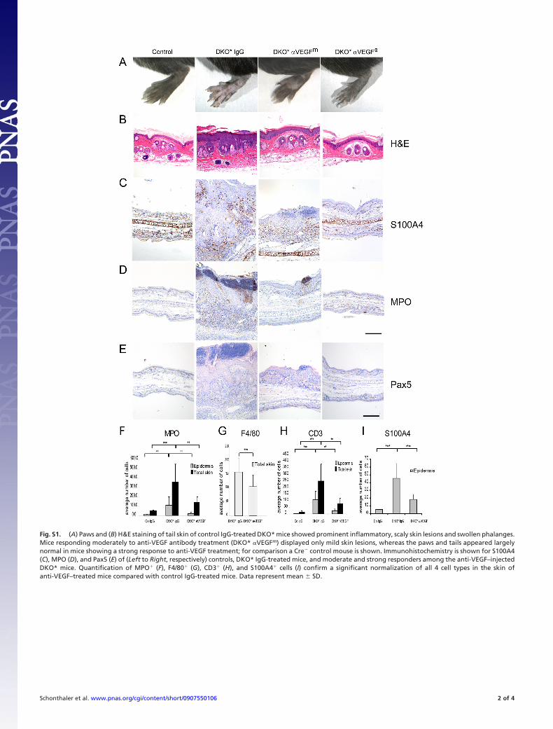
Fig. S1. (A) Paws and (B) H&E staining of tail skin of control IgG-treated DKO* mice showed prominent inflammatory, scaly skin lesions and swollen phalanges.Mice responding moderately to anti-VEGF antibody treatment (DKO* �VEGFm) displayed only mild skin lesions, whereas the paws and tails appeared largelynormal in mice showing a strong response to anti-VEGF treatment; for comparison a Cre� control mouse is shown. Immunohistochemistry is shown for S100A4(C), MPO (D), and Pax5 (E) of (Left to Right, respectively) controls, DKO* IgG-treated mice, and moderate and strong responders among the anti-VEGF–injectedDKO* mice. Quantification of MPO� (F), F4/80� (G), CD3� (H), and S100A4� cells (I) confirm a significant normalization of all 4 cell types in the skin ofanti-VEGF–treated mice compared with control IgG-treated mice. Data represent mean � SD.
Schonthaler et al. www.pnas.org/cgi/content/short/0907550106 2 of 4
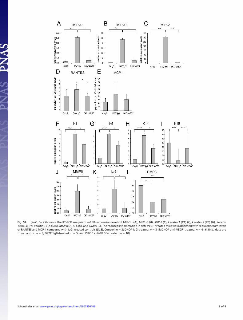
Fig. S2. (A–C, F–L) Shown is the RT-PCR analysis of mRNA expression levels of MIP-1� (A), MIP1-� (B), MIP-2 (C), keratin 1 (K1) (F), keratin 5 (K5) (G), keratin14 (K14) (H), keratin 15 (K15) (I), MMP8 (J), IL-6 (K), and TIMP3 (L). The reduced inflammation in anti-VEGF–treated mice was associated with reduced serum levelsof RANTES and MCP-1 compared with IgG- treated controls (D, E). Control: n � 3; DKO* IgG-treated: n � 3–5; DKO* anti-VEGF–treated: n � 4–6. (In L, data arefrom control: n � 3; DKO* IgG-treated: n � 5; and DKO* anti-VEGF–treated: n � 10).
Schonthaler et al. www.pnas.org/cgi/content/short/0907550106 3 of 4
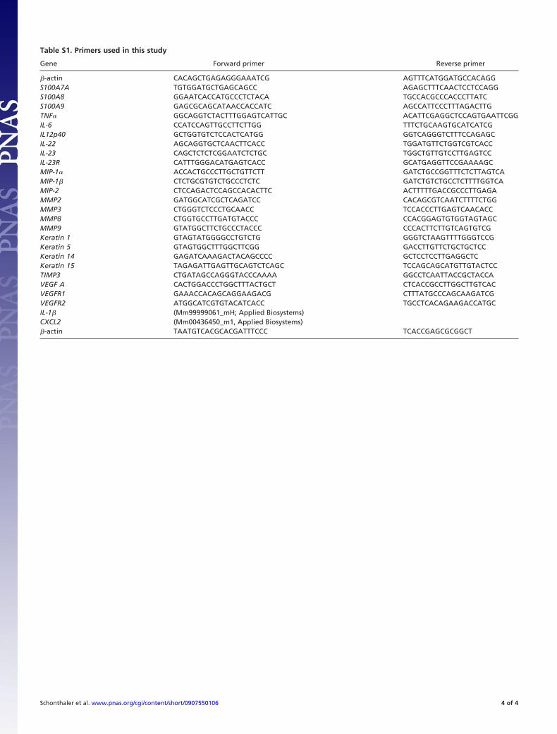
Table S1. Primers used in this study
Gene Forward primer Reverse primer
�-actin CACAGCTGAGAGGGAAATCG AGTTTCATGGATGCCACAGGS100A7A TGTGGATGCTGAGCAGCC AGAGCTTTCAACTCCTCCAGGS100A8 GGAATCACCATGCCCTCTACA TGCCACGCCCACCCTTATCS100A9 GAGCGCAGCATAACCACCATC AGCCATTCCCTTTAGACTTGTNF� GGCAGGTCTACTTTGGAGTCATTGC ACATTCGAGGCTCCAGTGAATTCGGIL-6 CCATCCAGTTGCCTTCTTGG TTTCTGCAAGTGCATCATCGIL12p40 GCTGGTGTCTCCACTCATGG GGTCAGGGTCTTTCCAGAGCIL-22 AGCAGGTGCTCAACTTCACC TGGATGTTCTGGTCGTCACCIL-23 CAGCTCTCTCGGAATCTCTGC TGGCTGTTGTCCTTGAGTCCIL-23R CATTTGGGACATGAGTCACC GCATGAGGTTCCGAAAAGCMIP-1� ACCACTGCCCTTGCTGTTCTT GATCTGCCGGTTTCTCTTAGTCAMIP-1� CTCTGCGTGTCTGCCCTCTC GATCTGTCTGCCTCTTTTGGTCAMIP-2 CTCCAGACTCCAGCCACACTTC ACTTTTTGACCGCCCTTGAGAMMP2 GATGGCATCGCTCAGATCC CACAGCGTCAATCTTTTCTGGMMP3 CTGGGTCTCCCTGCAACC TCCACCCTTGAGTCAACACCMMP8 CTGGTGCCTTGATGTACCC CCACGGAGTGTGGTAGTAGCMMP9 GTATGGCTTCTGCCCTACCC CCCACTTCTTGTCAGTGTCGKeratin 1 GTAGTATGGGGCCTGTCTG GGGTCTAAGTTTTGGGTCCGKeratin 5 GTAGTGGCTTTGGCTTCGG GACCTTGTTCTGCTGCTCCKeratin 14 GAGATCAAAGACTACAGCCCC GCTCCTCCTTGAGGCTCKeratin 15 TAGAGATTGAGTTGCAGTCTCAGC TCCAGCAGCATGTTGTACTCCTIMP3 CTGATAGCCAGGGTACCCAAAA GGCCTCAATTACCGCTACCAVEGF A CACTGGACCCTGGCTTTACTGCT CTCACCGCCTTGGCTTGTCACVEGFR1 GAAACCACAGCAGGAAGACG CTTTATGCCCAGCAAGATCGVEGFR2 ATGGCATCGTGTACATCACC TGCCTCACAGAAGACCATGCIL-1� (Mm99999061_mH; Applied Biosystems)CXCL2 (Mm00436450_m1, Applied Biosystems)�-actin TAATGTCACGCACGATTTCCC TCACCGAGCGCGGCT
Schonthaler et al. www.pnas.org/cgi/content/short/0907550106 4 of 4