Embed Size (px)
Citation preview
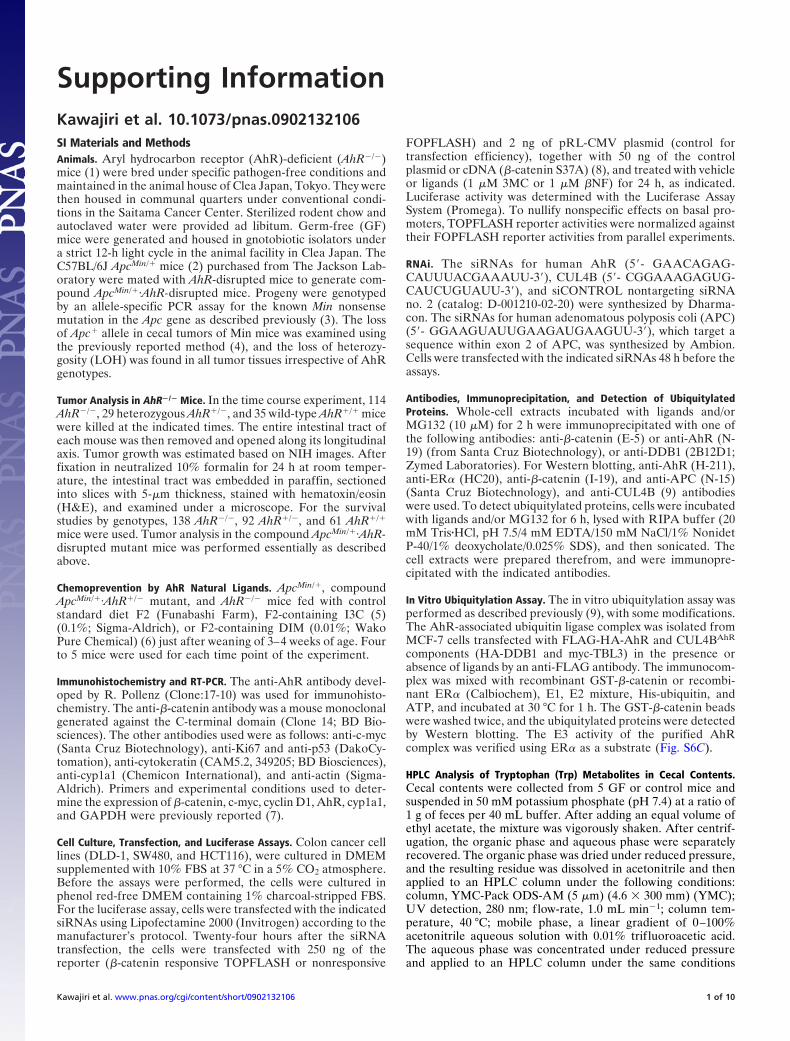
Supporting InformationKawajiri et al. 10.1073/pnas.0902132106SI Materials and MethodsAnimals. Aryl hydrocarbon receptor (AhR)-deficient (AhR�/�)mice (1) were bred under specific pathogen-free conditions andmaintained in the animal house of Clea Japan, Tokyo. They werethen housed in communal quarters under conventional condi-tions in the Saitama Cancer Center. Sterilized rodent chow andautoclaved water were provided ad libitum. Germ-free (GF)mice were generated and housed in gnotobiotic isolators undera strict 12-h light cycle in the animal facility in Clea Japan. TheC57BL/6J ApcMin/� mice (2) purchased from The Jackson Lab-oratory were mated with AhR-disrupted mice to generate com-pound ApcMin/�·AhR-disrupted mice. Progeny were genotypedby an allele-specific PCR assay for the known Min nonsensemutation in the Apc gene as described previously (3). The lossof Apc� allele in cecal tumors of Min mice was examined usingthe previously reported method (4), and the loss of heterozy-gosity (LOH) was found in all tumor tissues irrespective of AhRgenotypes.
Tumor Analysis in AhR�/� Mice. In the time course experiment, 114AhR�/�, 29 heterozygous AhR�/�, and 35 wild-type AhR�/� micewere killed at the indicated times. The entire intestinal tract ofeach mouse was then removed and opened along its longitudinalaxis. Tumor growth was estimated based on NIH images. Afterfixation in neutralized 10% formalin for 24 h at room temper-ature, the intestinal tract was embedded in paraffin, sectionedinto slices with 5-�m thickness, stained with hematoxin/eosin(H&E), and examined under a microscope. For the survivalstudies by genotypes, 138 AhR�/�, 92 AhR�/�, and 61 AhR�/�
mice were used. Tumor analysis in the compound ApcMin/�·AhR-disrupted mutant mice was performed essentially as describedabove.
Chemoprevention by AhR Natural Ligands. ApcMin/�, compoundApcMin/�·AhR�/� mutant, and AhR�/� mice fed with controlstandard diet F2 (Funabashi Farm), F2-containing I3C (5)(0.1%; Sigma-Aldrich), or F2-containing DIM (0.01%; WakoPure Chemical) (6) just after weaning of 3–4 weeks of age. Fourto 5 mice were used for each time point of the experiment.
Immunohistochemistry and RT-PCR. The anti-AhR antibody devel-oped by R. Pollenz (Clone:17-10) was used for immunohisto-chemistry. The anti-�-catenin antibody was a mouse monoclonalgenerated against the C-terminal domain (Clone 14; BD Bio-sciences). The other antibodies used were as follows: anti-c-myc(Santa Cruz Biotechnology), anti-Ki67 and anti-p53 (DakoCy-tomation), anti-cytokeratin (CAM5.2, 349205; BD Biosciences),anti-cyp1a1 (Chemicon International), and anti-actin (Sigma-Aldrich). Primers and experimental conditions used to deter-mine the expression of �-catenin, c-myc, cyclin D1, AhR, cyp1a1,and GAPDH were previously reported (7).
Cell Culture, Transfection, and Luciferase Assays. Colon cancer celllines (DLD-1, SW480, and HCT116), were cultured in DMEMsupplemented with 10% FBS at 37 °C in a 5% CO2 atmosphere.Before the assays were performed, the cells were cultured inphenol red-free DMEM containing 1% charcoal-stripped FBS.For the luciferase assay, cells were transfected with the indicatedsiRNAs using Lipofectamine 2000 (Invitrogen) according to themanufacturer’s protocol. Twenty-four hours after the siRNAtransfection, the cells were transfected with 250 ng of thereporter (�-catenin responsive TOPFLASH or nonresponsive
FOPFLASH) and 2 ng of pRL-CMV plasmid (control fortransfection efficiency), together with 50 ng of the controlplasmid or cDNA (�-catenin S37A) (8), and treated with vehicleor ligands (1 �M 3MC or 1 �M �NF) for 24 h, as indicated.Luciferase activity was determined with the Luciferase AssaySystem (Promega). To nullify nonspecific effects on basal pro-moters, TOPFLASH reporter activities were normalized againsttheir FOPFLASH reporter activities from parallel experiments.
RNAi. The siRNAs for human AhR (5�- GAACAGAG-CAUUUACGAAAUU-3�), CUL4B (5�- CGGAAAGAGUG-CAUCUGUAUU-3�), and siCONTROL nontargeting siRNAno. 2 (catalog: D-001210-02-20) were synthesized by Dharma-con. The siRNAs for human adenomatous polyposis coli (APC)(5�- GGAAGUAUUGAAGAUGAAGUU-3�), which target asequence within exon 2 of APC, was synthesized by Ambion.Cells were transfected with the indicated siRNAs 48 h before theassays.
Antibodies, Immunoprecipitation, and Detection of UbiquitylatedProteins. Whole-cell extracts incubated with ligands and/orMG132 (10 �M) for 2 h were immunoprecipitated with one ofthe following antibodies: anti-�-catenin (E-5) or anti-AhR (N-19) (from Santa Cruz Biotechnology), or anti-DDB1 (2B12D1;Zymed Laboratories). For Western blotting, anti-AhR (H-211),anti-ER� (HC20), anti-�-catenin (I-19), and anti-APC (N-15)(Santa Cruz Biotechnology), and anti-CUL4B (9) antibodieswere used. To detect ubiquitylated proteins, cells were incubatedwith ligands and/or MG132 for 6 h, lysed with RIPA buffer (20mM Tris�HCl, pH 7.5/4 mM EDTA/150 mM NaCl/1% NonidetP-40/1% deoxycholate/0.025% SDS), and then sonicated. Thecell extracts were prepared therefrom, and were immunopre-cipitated with the indicated antibodies.
In Vitro Ubiquitylation Assay. The in vitro ubiquitylation assay wasperformed as described previously (9), with some modifications.The AhR-associated ubiquitin ligase complex was isolated fromMCF-7 cells transfected with FLAG-HA-AhR and CUL4BAhR
components (HA-DDB1 and myc-TBL3) in the presence orabsence of ligands by an anti-FLAG antibody. The immunocom-plex was mixed with recombinant GST-�-catenin or recombi-nant ER� (Calbiochem), E1, E2 mixture, His-ubiquitin, andATP, and incubated at 30 °C for 1 h. The GST-�-catenin beadswere washed twice, and the ubiquitylated proteins were detectedby Western blotting. The E3 activity of the purified AhRcomplex was verified using ER� as a substrate (Fig. S6C).
HPLC Analysis of Tryptophan (Trp) Metabolites in Cecal Contents.Cecal contents were collected from 5 GF or control mice andsuspended in 50 mM potassium phosphate (pH 7.4) at a ratio of1 g of feces per 40 mL buffer. After adding an equal volume ofethyl acetate, the mixture was vigorously shaken. After centrif-ugation, the organic phase and aqueous phase were separatelyrecovered. The organic phase was dried under reduced pressure,and the resulting residue was dissolved in acetonitrile and thenapplied to an HPLC column under the following conditions:column, YMC-Pack ODS-AM (5 �m) (4.6 � 300 mm) (YMC);UV detection, 280 nm; flow-rate, 1.0 mL min�1; column tem-perature, 40 °C; mobile phase, a linear gradient of 0–100%acetonitrile aqueous solution with 0.01% trif luoroacetic acid.The aqueous phase was concentrated under reduced pressureand applied to an HPLC column under the same conditions
Kawajiri et al. www.pnas.org/cgi/content/short/0902132106 1 of 10

except for the mobile phase, which consisted of 20 mM potas-sium phosphate (pH 6.0) (solution A) and a 60% methanolaqueous solution (solution B). Samples were eluted with solution
A for 5 min followed by a linear gradient of 0–50% solution Bfor 20 min.
1. Mimura J, et al. (1997) Loss of teratogenic response to 2,3,7,8-tetrachlorodibenzo-p-dioxin (TCDD) in mice lacking the Ah (dioxin) receptor. Genes Cells 2:645–654.
2. Moser AR, Pitot HC, Dove WF (1990) A dominant mutation that predisposes to multipleintestinal neoplasia in the mouse. Science 247:322–324.
3. Jacoby RF, et al. (1996) Chemoprevention of spontaneous intestinal adenomas in theApc Min mouse model by the nonsteroidal anti-inflammatory drug piroxicam. CancerRes 56:710–714.
4. Luongo C, Moser AR, Gledhill S, Dove WF (1994) Loss of Apc� in intestinal adenomasfrom Min mice. Cancer Res 54:5947–5952.
5. Xu M, et al. (1996) Protection by green tea, black tea, and indole-3-carbinol against2-amino-3-methylimidazo[4,5-f]quinoline-induced DNA adducts and colonic aberrantcrypts in the F344 rat. Carcinogenesis 17:1429–1434.
6. Chen I, McDougal A, Wang F, Safe S (1998) Aryl hydrocarbon receptor-mediatedantiestrogenic and antitumorigenic activity of diindolylmethane. Carcinogenesis19:1631–1639.
7. Hayashi S-I, Okabe-Kado J, Honma Y, Kawajiri K (1995) Expression of Ah receptor(TCDD receptor) during human monocytic differentiation. Carcinogenesis 16:1403–1409.
8. Liu C, et al. (1999) �-Trcp couples �-catenin phosphorylation-degradation and regu-lates Xenopus axis formation. Proc Natl Acad Sci USA 96:6273–6278.
9. Ohtake F, et al. (2007) Dioxin receptor is a ligand-dependent E3 ubiquitin ligase. Nature446:562–566.
10. Dharmasiri N, Dharmasiri S, Estelle M (2005) The F-box protein TIR1 is an auxin receptor.Nature 435:441–445.
11. Kepinski S, Leyser O (2005) The Arabidopsis F-box protein TIR1 is an auxin receptor.Nature 435:446–451.
12. Goryo K, et al. (2007) Identification of amino acid residues in the Ah receptor involvedin ligand binding. Biochem Biophys Res Commun 354:396–402.
13. Tan X, et al. (2007) Mechanism of auxin perception by the TIR1 ubiquitin ligase. Nature446:640–645.
Kawajiri et al. www.pnas.org/cgi/content/short/0902132106 2 of 10
10
5
0
Age in weeks
Mic
e w
ith
re
ctal
pro
lap
se
5040302010
A
0 10 20 30 40 50 60 70
1.0
0.8
0.6
0.4
0.2
0
Su
rviv
al r
atio
AhR-/-:n=138
AhR+/-:n=92
AhR+/+:n=61
Age in weeks
(Log rank test: P = 4.4x10⁻⁹; df=2)
80
C
Tu
mo
r-b
eari
ng
(+)
(-)
B
11 12 15 16 21 25 40 5510986 >70Age in weeks
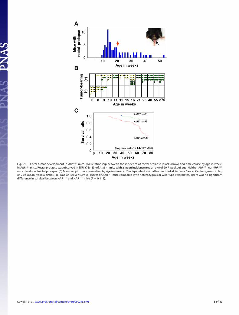
Fig. S1. Cecal tumor development in AhR�/� mice. (A) Relationship between the incidence of rectal prolapse (black arrow) and time course by age in weeksin AhR�/� mice. Rectal prolapse was observed in 55% (73/133) of AhR�/� mice with a mean incidence (red arrow) of 20.7 weeks of age. Neither AhR�/� nor AhR�/�
mice developed rectal prolapse. (B) Macroscopic tumor formation by age in weeks at 2 independent animal houses bred at Saitama Cancer Center (green circles)or Clea Japan (yellow circles). (C) Kaplan-Meyer survival curves of AhR�/� mice compared with heterozygous or wild-type littermates. There was no significantdifference in survival between AhR�/� and AhR�/� mice (P � 0.115).
Kawajiri et al. www.pnas.org/cgi/content/short/0902132106 3 of 10
A B C
D E F G
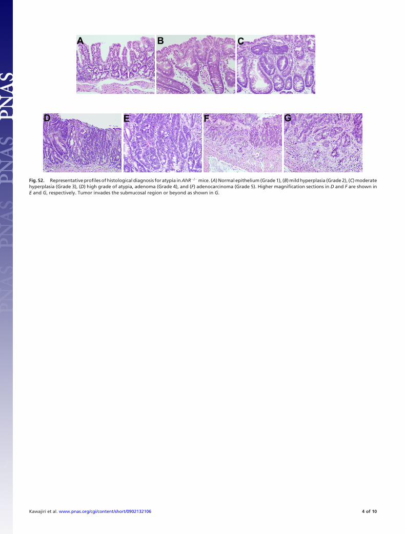
Fig. S2. Representative profiles of histological diagnosis for atypia in AhR�/� mice. (A) Normal epithelium (Grade 1), (B) mild hyperplasia (Grade 2), (C) moderatehyperplasia (Grade 3), (D) high grade of atypia, adenoma (Grade 4), and (F) adenocarcinoma (Grade 5). Higher magnification sections in D and F are shown inE and G, respectively. Tumor invades the submucosal region or beyond as shown in G.
Kawajiri et al. www.pnas.org/cgi/content/short/0902132106 4 of 10
fE
B C
D
G H I
β-catenin
Fc-myc
cytokeratin
PAS PAS
HE
HE HE
HE
A
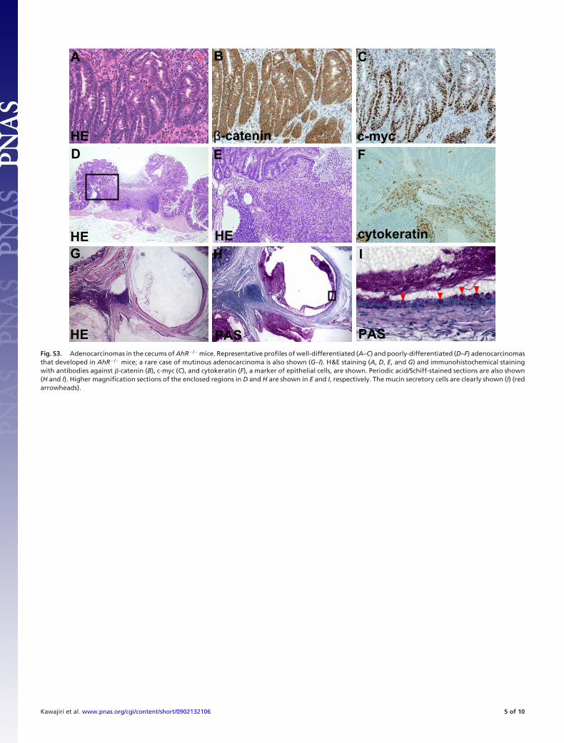
Fig. S3. Adenocarcinomas in the cecums of AhR�/� mice. Representative profiles of well-differentiated (A–C) and poorly-differentiated (D–F) adenocarcinomasthat developed in AhR�/� mice; a rare case of mutinous adenocarcinoma is also shown (G–I). H&E staining (A, D, E, and G) and immunohistochemical stainingwith antibodies against �-catenin (B), c-myc (C), and cytokeratin (F), a marker of epithelial cells, are shown. Periodic acid/Schiff-stained sections are also shown(H and I). Higher magnification sections of the enclosed regions in D and H are shown in E and I, respectively. The mucin secretory cells are clearly shown (I) (redarrowheads).
Kawajiri et al. www.pnas.org/cgi/content/short/0902132106 5 of 10
Ki67 p53
ββ-catenin
C
AhR
A
c-myc
B
FECecumD
T
Cecal Cancers AhR expression in normal
AhRDownregulation
β-Catenin*Overexpression Ileum Colon
12/12 12/12C>N : 4C=N : 4C<N : 4
12/12 12/127/12
p53**Overexpression
G
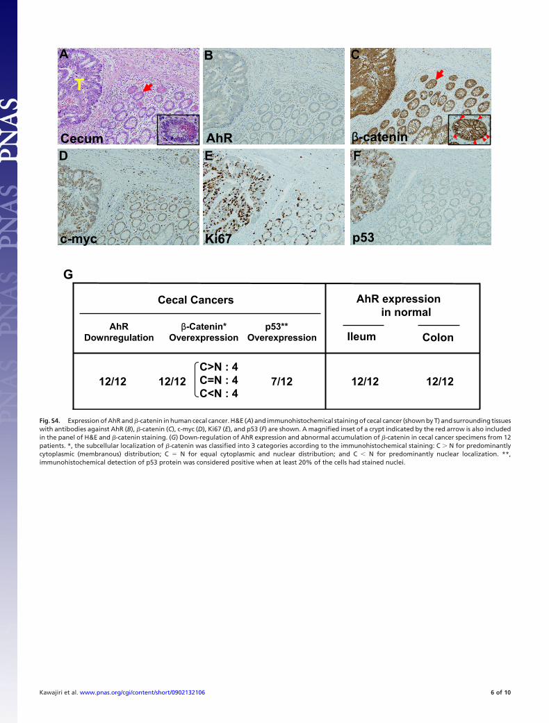
Fig. S4. Expression of AhR and �-catenin in human cecal cancer. H&E (A) and immunohistochemical staining of cecal cancer (shown by T) and surrounding tissueswith antibodies against AhR (B), �-catenin (C), c-myc (D), Ki67 (E), and p53 (F) are shown. A magnified inset of a crypt indicated by the red arrow is also includedin the panel of H&E and �-catenin staining. (G) Down-regulation of AhR expression and abnormal accumulation of �-catenin in cecal cancer specimens from 12patients. *, the subcellular localization of �-catenin was classified into 3 categories according to the immunohistochemical staining: C � N for predominantlycytoplasmic (membranous) distribution; C � N for equal cytoplasmic and nuclear distribution; and C � N for predominantly nuclear localization. **,immunohistochemical detection of p53 protein was considered positive when at least 20% of the cells had stained nuclei.
Kawajiri et al. www.pnas.org/cgi/content/short/0902132106 6 of 10
AhRIleum
DC
Paneth cells Paneth cells
Paneth cells Paneth cells
A B
Cecum AhR
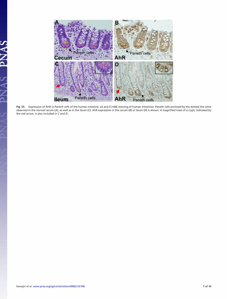
Fig. S5. Expression of AhR in Paneth cells of the human intestine. (A and C) H&E staining of human intestines. Paneth cells enclosed by the dotted line wereobserved in the normal cecum (A), as well as in the ileum (C). AhR expression in the cecum (B) or ileum (D) is shown. A magnified inset of a crypt, indicated bythe red arrow, is also included in C and D.
Kawajiri et al. www.pnas.org/cgi/content/short/0902132106 7 of 10
ERαα
IgG IP
AhR complex
3MC
Ub, E1, E2, ATP
MW (kD)
B
FTOPFLASH/FOPFLASH
(DLD-1 cells)
R.L
.U.
1
-
2
+
3
-
4
+3MC
RNAi Control CUL4B
Anti-AhR
Anti-β-actin
Anti-CUL4B
Anti-β-actin
Anti-APC
Anti-β-actin
RNAi Con
trol
AhR
Con
trol
CU
L4B
Con
trol
APC
RNAi
RNAi
E
250
150
100
1
+
+
-
-
+
2
+
-
+
-
+
3
+
-
+
+
+
(Dark exposure)
(Light exposure) ERα
ERα
-Ub
n
A
Control AhR
IAARNAi
1-
2+
3-
4+
Anti-β-catenin
Anti-β-actinIB:
(DLD-1)
IB: Anti-ERα
G
Anti-α-actin
Anti-β-catenin
C 3MC
AhR-/-
Time (3h)
cyp1a1
β-catenin
c-myc
GAPDH
3MCCTime (3h)
I
H
MW (kD)
D
250
150
100
50
GST-β-Catenin
(CBB staining)
Aquaous phaseTrp
Control
GF
Indole
3-MI
Organic phase
GF
ControlIndirubinIndigo
TAIAA
C
Anti-β-catenin
Anti-β-actinIB:
3MC(h)MG132
--
3-
Membrane
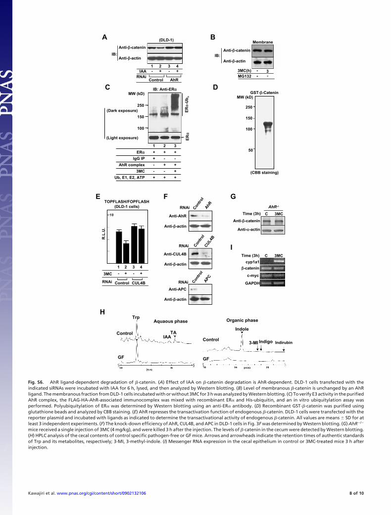
Fig. S6. AhR ligand-dependent degradation of �-catenin. (A) Effect of IAA on �-catenin degradation is AhR-dependent. DLD-1 cells transfected with theindicated siRNAs were incubated with IAA for 6 h, lysed, and then analyzed by Western blotting. (B) Level of membranous �-catenin is unchanged by an AhRligand. The membranous fraction from DLD-1 cells incubated with or without 3MC for 3 h was analyzed by Western blotting. (C) To verify E3 activity in the purifiedAhR complex, the FLAG-HA-AhR-associated immunocomplex was mixed with recombinant ER� and His-ubiquitin, and an in vitro ubiquitylation assay wasperformed. Polyubiquitylation of ER� was determined by Western blotting using an anti-ER� antibody. (D) Recombinant GST-�-catenin was purified usingglutathione beads and analyzed by CBB staining. (E) AhR represses the transactivation function of endogenous �-catenin. DLD-1 cells were transfected with thereporter plasmid and incubated with ligands as indicated to determine the transactivational activity of endogenous �-catenin. All values are means � SD for atleast 3 independent experiments. (F) The knock-down efficiency of AhR, CUL4B, and APC in DLD-1 cells in Fig. 3F was determined by Western blotting. (G) AhR�/�
mice received a single injection of 3MC (4 mg/kg), and were killed 3 h after the injection. The levels of �-catenin in the cecum were detected by Western blotting.(H) HPLC analysis of the cecal contents of control specific pathogen-free or GF mice. Arrows and arrowheads indicate the retention times of authentic standardsof Trp and its metabolites, respectively; 3-MI, 3-methyl-indole. (I) Messenger RNA expression in the cecal epithelium in control or 3MC-treated mice 3 h afterinjection.
Kawajiri et al. www.pnas.org/cgi/content/short/0902132106 8 of 10
C
D
Tu
mo
r-b
eari
ng
(%
)Age in weeks
0 10 20 25155
Control+I3C+DIM
100
80
60
40
20
0
I3C
Cecum Small intestine
I3C
ApcMin/+・AhR+/-
ControlControl
DIMDIM DIM
I3C
Control
ApcMin/+ ApcMin/+・AhR+/-Colon
DIM
I3C
Control
DIM
A
AhR
Apc
GAPDH
Wt AhR-/-
ApcMin/+
FE
ApcMin/+・AhR+/+W51W8W8
ApcMin/+・AhR-/- ApcMin/+・AhR+/+
BMin/+
+/+ +/- -/-AhR
Anti-β-cateninAnti-α-actin
+/+Apc
+/+ +/- -/-
cyclin D1c-myc
GAPDH
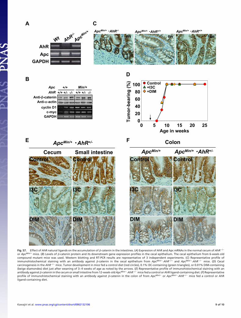
Fig. S7. Effect of AhR natural ligands on the accumulation of �-catenin in the intestines. (A) Expression of AhR and Apc mRNAs in the normal cecum of AhR�/�
or ApcMin/� mice. (B) Levels of �-catenin protein and its downstream gene expression profiles in the cecal epithelium. The cecal epithelium from 6-week-oldcompound mutant mice was used. Western blotting and RT-PCR results are representative of 3 independent experiments. (C) Representative profile ofimmunohistochemical staining with an antibody against �-catenin in the cecal epithelium from ApcMin/�·AhR�/� and ApcMin/�·AhR�/� mice. (D) Cecalcarcinogenesis in the AhR�/� mice. Tumor development in mice fed a control diet (red circles), 0.1% I3C-containing (green triangles), or 0.01% DIM-containing(beige diamondes) diet just after weaning of 3–4 weeks of age as noted by the arrows. (E) Representative profile of immunohistochemical staining with anantibody against �-catenin in the cecum or small intestine from 12-week-old ApcMin/�·AhR�/� mice fed a control or AhR ligand-containing diet. (F) Representativeprofile of immunohistochemical staining with an antibody against �-catenin in the colon of from ApcMin/� or ApcMin/�·AhR�/� mice fed a control or AhRligand-containing diet.
Kawajiri et al. www.pnas.org/cgi/content/short/0902132106 9 of 10
In plants
B C D
A
In intestine
Tryptophan IAA (auxin) SCFTIR1 Degradation of IAA repressors
AhR
IAA (I3C, DIM)
CUL4B
AhRRoc1
E2
TBL3
DDB1
Arnt Ubβ-cateninUb
AhR pathway β-catenin
E3
Degradation
UbUb
Fbw1
Skp1
CUL1E3
Roc1
E2Ub
APC pathway
APC
P
β-cateninPP Ub
UbUbUbUbUb
Degradation
GSK-3β
CKI
Axin
β-catenin
P P
CytoplasmNucleus
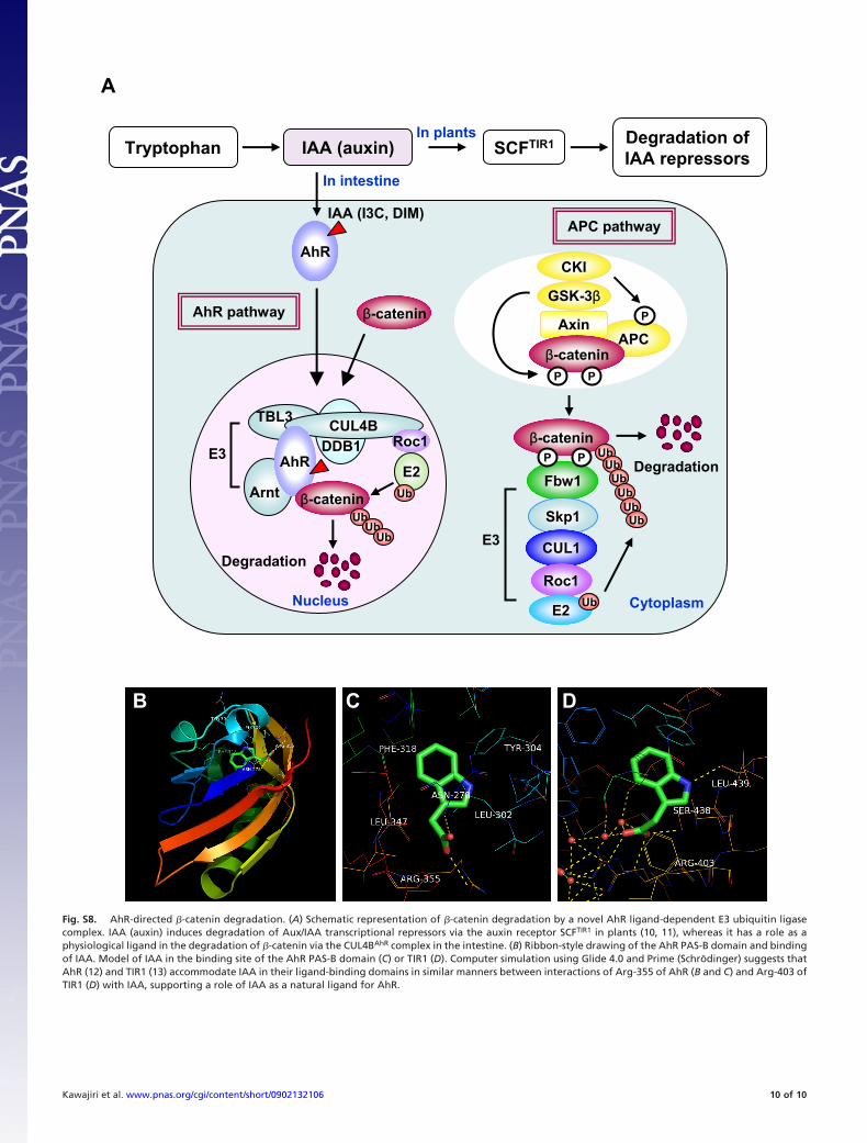
Fig. S8. AhR-directed �-catenin degradation. (A) Schematic representation of �-catenin degradation by a novel AhR ligand-dependent E3 ubiquitin ligasecomplex. IAA (auxin) induces degradation of Aux/IAA transcriptional repressors via the auxin receptor SCFTIR1 in plants (10, 11), whereas it has a role as aphysiological ligand in the degradation of �-catenin via the CUL4BAhR complex in the intestine. (B) Ribbon-style drawing of the AhR PAS-B domain and bindingof IAA. Model of IAA in the binding site of the AhR PAS-B domain (C) or TIR1 (D). Computer simulation using Glide 4.0 and Prime (Schrodinger) suggests thatAhR (12) and TIR1 (13) accommodate IAA in their ligand-binding domains in similar manners between interactions of Arg-355 of AhR (B and C) and Arg-403 ofTIR1 (D) with IAA, supporting a role of IAA as a natural ligand for AhR.
Kawajiri et al. www.pnas.org/cgi/content/short/0902132106 10 of 10