Embed Size (px)
Citation preview

Supporting Information for
Artificial Metabolism-Inspired Photoelectrochemical Probing of Biomolecules
and Cells
Jing Tang,a Yongcheng Wang,a Yuhang Wang,a Jun Li,b Biao Kong,a Min Jiang,c and Gengfeng
Zheng a,*
a Laboratory of Advanced Materials, Department of Chemistry, Fudan University, Shanghai 200433, P. R.
China
b School of Pharmacy, Fudan University, Shanghai 201203, China
c Institutes of Brain Science and State Key Laboratory of Medical Neurobiology, Fudan University,
Shanghai 200032, P. R. China
*Corresponding authors: Gengfeng Zheng
Email addresses: [email protected] (G.Z.)
Electronic Supplementary Material (ESI) for Journal of Materials Chemistry A.This journal is © The Royal Society of Chemistry 2014

Supporting Figures
Figure S1. (a, d) Top-view and (b, c) side-view SEM images of TiO2-Co3O4 NW arrays on FTO-
coated glass substrates.

Figure S2. Elemental mapping of a representative TiO2-Co3O4 NW for Ti, O and Co distribution
over the NW.
Figure S3. EDX of a representative TiO2-Co3O4 NW.

Figure S4. XRD patterns of the pristine TiO2 NWs (blue curve) and TiO2-Co3O4 NWs (red curve)
grown on FTO substrates. XRD pattern of a blank FTO substrate is also displayed for
comparison. The peaks of FTO are indicated by asterisks.

Figure S5. Energy bands of Co3O4 and TiO2, and the electron transfer process of Co3O4/TiO2
interface under a simulated sunlight illumination. All the energy levels are referenced to NHE
scale. CB and VB are conduction band and valence band, respectively.

Figure S6. (a, b, c) Photocurrent versus time data of the TiO2-Co3O4-ATPase NWs for cell
extracts (1 × 10⁵ cells/mL after 1:100 dilution) from solutions of HUVECs, HeLa and H1299
cells. The sizes of the active sample area were 0.02-0.06 cm2.

Figure S7. Stability test of a TiO2-Co3O4-ATPase NW biosensor for ATP detection over a
month. The sizes of the active sensor surfaces were around 0.02−0.06 cm2.
Figure S8. Schematic of the TiO2-Co3O4-ChOx NW-based PEC sensor for cholesterol detection.
Under sun light illumination, H2O or H2O2isoxidized by the photogenerated holes over the TiO2
NW anode to generate O2, which conducts as an efficient electron acceptor of FAD/FADH2.
During this cycle, cholesterol is oxidized to cholesterol-4-ene-3-one. On the cathode (Pt), the
photogenerated electrons reduce water to produce H2. The sensing signal represents the current
flowing through the circuit.

Figure S9. Stability test of a TiO2-Co3O4-ChOx NW biosensor over a month. The sizes of the
active sensor surfaces were around 0.02-0.06 cm2.

Table S1. Analytical performance of the present integrated TiO2-Co3O4-ATPase NW-based
photoelectrochemical detection of ATP, compared to previously reported literatures.
Methods Materials Linear RangeDetection
LimitReference
The present method TiO2-Co3O4-ATPase NWs 0.14nM-10 μM 0.14 nMThe present
work
Fluorometry
Ribonucleopeptide
Oligonucleotide
DNA-Ag nanoclusters
10-7-10-3 M
0~2×10⁻⁶ M
0-1000 μM
﹤10-7
25 nM
0.2 μM
S1
S2
S3
Colorimetry DNAzyme-Aptamer 100 μM 10-6 M S4
Odor-Based Sensor Tryptophanase ELISA-like - S5
Optical ATP Biosensor Enzyme 10−3 mM to 1.5 mM 1μM S6
Logic Gates SensorsThree-Dimensional DNA
Nanostructures0-600 nM 20 nM S7
FET Sensor Aptamer 1-1000 μM 1 μM S8
Chromogenic Sensor Zn(II)-Cyclam 0~2×10⁻⁴ M < 0 M S9
Photoelectrochemical
BiosensorBio-Barcode Amplification 0-100×10-8 M 3.2×10⁻⁹ M S10
LOD = limit of detection; LR = linear ranges;

Table S2. Analytical performance of the present integrated TiO2-Co3O4-ChOx -based
photoelectrochemical detection of cholesterol, compared to previously reported literatures.
Methods Materials Linear Range Detection Limit Reference
The present method TiO2-Co3O4-ChOx NWs 0.5 nM-50 μM 0.2 nMThe present
work
Electrogenerated
ChemiluminescenceHemin-Graphene Nanosheets 0.17 mM to 1.12 mM 0.06 mM S11
Fluorimetric Biosensors Alginate-Silica Microspheres 1.25 to 10 mM less than 1.25 mM S12
Amperometric Biosensor
Carbon Nanotubes
Chitosan/silica-MWCNT
ZnO
Pt nanoparticles
2.5-10 mM
8.0×10⁻⁶ mM~0.4 mM
1.0 to 700.0 nM
Up to 4.0 mM
less than 5 mM
1×10⁻⁶ mM
0.37nM
0.2μM
S13
S14
S15
S16
ElectrochemistryNickel Oxide-Chitosan Film
Cuprous Oxide/Chitosan
10-400 mg/dL
10-450 mg/dL
43.4 mg/dL
15.9 mg/dLcm-2
S17
S18
Microfluidics NanobiochipnNiO and MWCNTs
CNT
nNiO
0.25-12.93 mM
1.25-10.0 mM
1.5-10.3 mM
0.03 mM
25 mg/dL
0.16 mM
S19
S20
S21
Electrochemiluminescence Luminol S22

Solution-Gated
Feld Effect TransistorZnONanorods 0.001-45 mM 0.05 mM S23

References:
S1. S. Nakano, M. Fukuda, T. Tamura, R. Sakaguchi, E. Nakata, T. Morii, J. Am. Chem. Soc.,
2013, 135, 3465-3473.
S2. F. Li, Z. Du, L. Yang, B. Tang, Biosens. Bioelectron., 2013, 41, 907-910.
S3. Z. Zhou, Y. Du, S. Dong, Biosens. Bioelectron., 2011, 28, 33-37.
S4. F. Liu, J. Zhang, R. Chen, L. Chen, L. Deng, Chem. Biodivers., 2011, 8, 311-316.
S5. Y. Xu, Z. Zhang, M. M. Ali, J. Sauder, X. Deng, K. Giang, S. D. Aguirre, R. Pelton, Y.
Li, C. D. Filipe, Angew. Chem. Int. Ed., 2014, 53, 2620-2622.
S6. C. Wang, C. Y. C. Huang, W. C. Lin, Biosens. Bioelectron., 2013, 43, 355-361.
S7. H. Pei, L. Liang, G. Yao, J. Li, Q. Huang, C. Fan, Angew. Chem. Int. Ed., 2012, 124,
9154-9158.
S8. J. Das, K. B. Cederquist, A. A. Zaragoza, P. E. Lee, E. H. Sargent, S. O. Kelley, Nat.
Chem., 2012, 4, 642-648.
S9. P. Mahato, A. Ghosh, S. K. Mishra, A. Shrivastav, S. Mishra, A. Das, Inorg. Chem., 2011,
50, 4162-4170.
S10. X. Zhang, Y. Zhao, S. Li, S. Zhang, Chem. Commun., 2010, 46, 9173-9175.
S11. M. Zhang, R. Yuan, Y. Chai, S. Chen, X. Zhong, H. Zhong, C. Wang, RSC Adv., 2012, 2,
4639-4641.
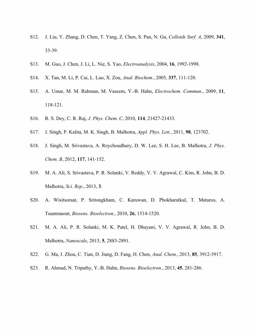
S12. J. Liu, Y. Zhang, D. Chen, T. Yang, Z. Chen, S. Pan, N. Gu, Colloids Surf. A, 2009, 341,
33-39.
S13. M. Guo, J. Chen, J. Li, L. Nie, S. Yao, Electroanalysis, 2004, 16, 1992-1998.
S14. X. Tan, M. Li, P. Cai, L. Luo, X. Zou, Anal. Biochem., 2005, 337, 111-120.
S15. A. Umar, M. M. Rahman, M. Vaseem, Y.-B. Hahn, Electrochem. Commun., 2009, 11,
118-121.
S16. R. S. Dey, C. R. Raj, J. Phys. Chem. C, 2010, 114, 21427-21433.
S17. J. Singh, P. Kalita, M. K. Singh, B. Malhotra, Appl. Phys. Lett., 2011, 98, 123702.
S18. J. Singh, M. Srivastava, A. Roychoudhury, D. W. Lee, S. H. Lee, B. Malhotra, J. Phys.
Chem. B, 2012, 117, 141-152.
S19. M. A. Ali, S. Srivastava, P. R. Solanki, V. Reddy, V. V. Agrawal, C. Kim, R. John, B. D.
Malhotra, Sci. Rep., 2013, 3.
S20. A. Wisitsoraat, P. Sritongkham, C. Karuwan, D. Phokharatkul, T. Maturos, A.
Tuantranont, Biosens. Bioelectron., 2010, 26, 1514-1520.
S21. M. A. Ali, P. R. Solanki, M. K. Patel, H. Dhayani, V. V. Agrawal, R. John, B. D.
Malhotra, Nanoscale, 2013, 5, 2883-2891.
S22. G. Ma, J. Zhou, C. Tian, D. Jiang, D. Fang, H. Chen, Anal. Chem., 2013, 85, 3912-3917.
S23. R. Ahmad, N. Tripathy, Y.-B. Hahn, Biosens. Bioelectron., 2013, 45, 281-286.


![OPTIMAL DECISION SUPPORT MIXTURE MODEL WITH WEIBULL … · 2019-09-28 · Tripathy and Sahoo [18], Tripathy and Sukla [19] developed improved inventory model in fuzzy sense using](https://img.pdfslide.net/doc/110x75/5e5692c04be7281bfb55fd41/optimal-decision-support-mixture-model-with-weibull-2019-09-28-tripathy-and-sahoo.jpg)