Embed Size (px)
Citation preview
Self-AssemblyDOI: 10.1002/anie.200903897
Supramolecular Amphiphiles Based on a Water-Soluble Charge-Transfer Complex: Fabrication of Ultralong Nanofibers with TunableStraightness**Chao Wang, Yinsheng Guo, Yapei Wang, Huaping Xu, Ruji Wang, and Xi Zhang*
Charge-transfer complexes are formed by the weak associa-tion of two molecules or molecular subgroups, one of whichacts as an electron donor and the other as an electronacceptor.[1] The formation of charge-transfer complexes isalways driven by a combination of charge-transfer interac-tions and other noncovalent interactions, such as host–guestinteractions and hydrophobic interactions.[2] One character-istic of charge-transfer complexes is their high charge-carrierdensities, which lead to high conductivity. From this point ofview, one-dimensional nanostructures that facilitate thedirectional movement of charge carriers are extremelyimportant for the promising application of charge-transfercomplexes as organic nanowires in electronic and optoelec-tronic nanodevices.[3] Typically, charge-transfer complexes areprepared by solid-phase reactions or self-assembly in non-polar solvents (but rarely in water) because of the generallylow solubility of the charge-transfer components.[4] Examplesof water-soluble organic charge-transfer complexes based onviologen and pyrene derivatives have been reported forapplications such as glucose sensors and guanosine triphos-phate (GTP) detectors.[5] Inspired by their easy preparationand the use of water as an environmentally friendly solvent,we wondered if we could directly obtain nanoscale materialsbased on one-dimensional charge-transfer complexes by self-assembly in aqueous solution. However, this concept is verychallenging, since the accurate stoichiometry and detailedsupramolecular structure of the charge-transfer complexwould not be fully revealed. Moreover, the complex forma-tion is simultaneously affected by Coulombic attractions andcharge-transfer interactions, but the dominant driving force isunclear, thus strongly hindering further application in supra-molecular soft materials.
Supramolecular amphiphiles are amphiphilic buildingblocks that are assembled by noncovalent interactions.[6]
Herein, we attempt to extend the concept of “supramolecular
amphiphiles” for the fabrication of one-dimensional ultralongnanofibers on the basis of the water-soluble charge-transfercomplex formation between viologen derivatives and the 8-hydroxypyrene-1,3,6-trisulfonic acid trisodium salt (PYR).Notably, the straightness of the nanofiber can be tuned bychanging the pH of the reaction solution. Furthermore, thedetailed supramolecular structure is revealed, and the dom-inant driving force of the viologen–PYR complex is clarified.As shown in Scheme 1, a viologen-containing amphiphile
(RV) was designed and synthesized. PYR is a water-solublepyrene derivative, and is a strong electron donor. PYR andRV are expected to form a supramolecular amphiphile drivenby the charge-transfer-complex formation between the PYRand the viologen. One advantage is that the supramolecularPYR–RV complex can be readily prepared by the directmixing of PYR and RV in aqueous solution.
When dissolved in aqueous solution, RV itself self-assembles into an aggregate because of its amphiphilicnature. The aggregation of RV at a concentration of 1.0 �10�4
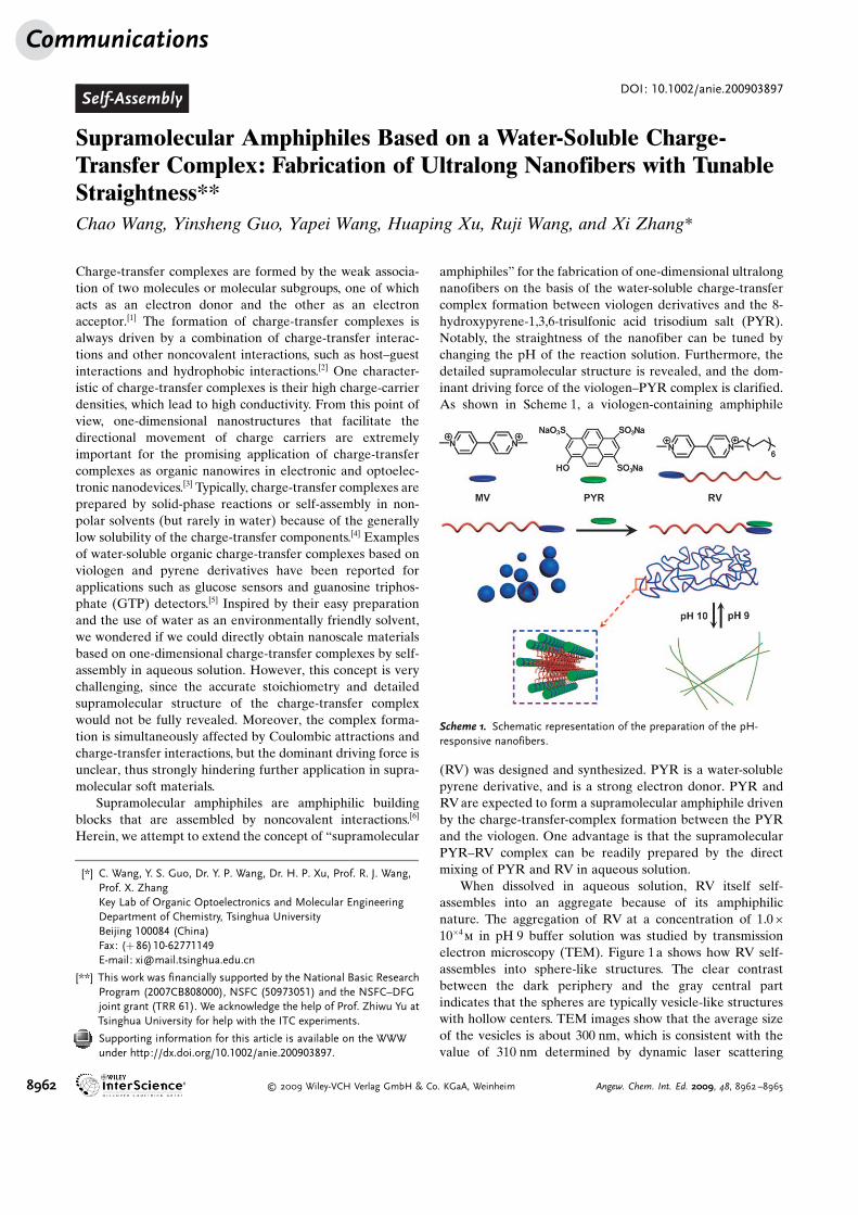
m in pH 9 buffer solution was studied by transmissionelectron microscopy (TEM). Figure 1a shows how RV self-assembles into sphere-like structures. The clear contrastbetween the dark periphery and the gray central partindicates that the spheres are typically vesicle-like structureswith hollow centers. TEM images show that the average sizeof the vesicles is about 300 nm, which is consistent with thevalue of 310 nm determined by dynamic laser scattering
Scheme 1. Schematic representation of the preparation of the pH-responsive nanofibers.
[*] C. Wang, Y. S. Guo, Dr. Y. P. Wang, Dr. H. P. Xu, Prof. R. J. Wang,Prof. X. ZhangKey Lab of Organic Optoelectronics and Molecular EngineeringDepartment of Chemistry, Tsinghua UniversityBeijing 100084 (China)Fax: (+ 86)10-62771149E-mail: [email protected]
[**] This work was financially supported by the National Basic ResearchProgram (2007CB808000), NSFC (50973051) and the NSFC–DFGjoint grant (TRR 61). We acknowledge the help of Prof. Zhiwu Yu atTsinghua University for help with the ITC experiments.
Supporting information for this article is available on the WWWunder http://dx.doi.org/10.1002/anie.200903897.
Communications
8962 � 2009 Wiley-VCH Verlag GmbH & Co. KGaA, Weinheim Angew. Chem. Int. Ed. 2009, 48, 8962 –8965
(DLS) experiments (Figure S1 in the Supporting Informa-tion).
It should be noted that the vesicles transformed into one-dimensional nanofibers when an equivalent amount of PYRwas added into the RV solution. As shown in Figure 1 b, thePYR–RV supramolecular amphiphile self-assembles into aone-dimensional wormlike structure. From the magnifiedimage of this structure, no clear contrast between the edgeand central part is observed, thus indicating that the wormlikestructures are solid nanofibers. Moreover, the nanofibershave a large length/diameter ratio: they exhibit a uniformdiameter of about 14 nm, and their length reaches tens ofmicrometers.
To understand the mechanism of the transformation fromvesicles to nanofibers, a series of complexes with differentPYR/RV ratios were prepared. When the PYR/RV ratio was1:10, the vesicles broke into pieces of membranes (Figure S2in the Supporting Information). As the PYR componentincreased, the membranes became longer and narrower. At aPYR/RV ratio of 2:3, wormlike nanowires appeared, whichcoexisted with the membranes. When the PYR/RV ratioreached 1:1, uniform wormlike nanostructures were observed.It should be pointed out that further increases in the PYR/RVratio did not bring any significant changes to the nanofibers.A plausible explanation is that when the PYR/RV molar ratiois larger than 1:1, all the RV molecules were complexed andtherefore no further structural changes could be generated.
Since PYR is itself pH-responsive, we wondered if thisproperty could be brought to the supramolecular amphiphile,and a 1.0 � 10�4
m solution of the PYR–RV complex in pH 10buffer was prepared. Figure 1d shows the self-assembledstructures of the PYR–RV complex. Notably, the curlywormlike nanofibers were straightened. Contrary to therandom wormlike nanofibers, the straight nanofibers preferto hierarchically aggregate into bundles (Figure S3 in theSupporting Information). The diameter of the nanofibers isstill about 14 nm, which is the same as that of the curlynanofibers, and suggests that the molecular packing frame-work should remain unchanged. When the pH value waschanged to 9, the straight nanofibers became curly again, thusindicating that the process is reversible. At pH 9 and pH 10,
the lengths of the nanofiber are nearly the same, ranging fromabout several micrometers to about 20 micrometers.
The formation of nanofibers relies on appropriate molec-ular packing and specific directional noncovalent interactions,such as p–p interactions and hydrogen bonding.[7] We useddifferent characterization techniques to help elucidate howthe nanofibers are formed. We specifically wished to identifywhether Coulombic attractions and charge-transfer interac-tions were the dominant driving force in the formation of thecomplex between viologen groups and PYR molecules. Wealso wanted to examine the viologen–PYR packing motif thatfavors the one-dimensional nanostructures. Considering thelow solubility of the PYR–RV complex, we synthesizedmethyl viologen (MV) as a model compound to investigatethe complex formation between the viologen group and thePYR molecule. MV (an electron acceptor) and PYR (anelectron donor) were mixed in water in a 1:1 ratio, and theresulting spectroscopic changes were observed. Both thefluorescence quenching in the emission spectra and the redshifts of the peaks in the UV/Vis spectra are indicative ofcharge-transfer complex formation (Figure S4 in the Support-ing Information). Furthermore, mass spectroscopy supportsthe formation of charge-transfer complex between MV andPYR. A peak at m/z 641.02 is observed, which corresponds tothe PYR3�–MV2+ complex (Figure S5 in the SupportingInformation), and indicates that PYR and MV indeed forma charge-transfer complex with 1:1 stoichiometry, thussupporting the previously reported assumption.[6]
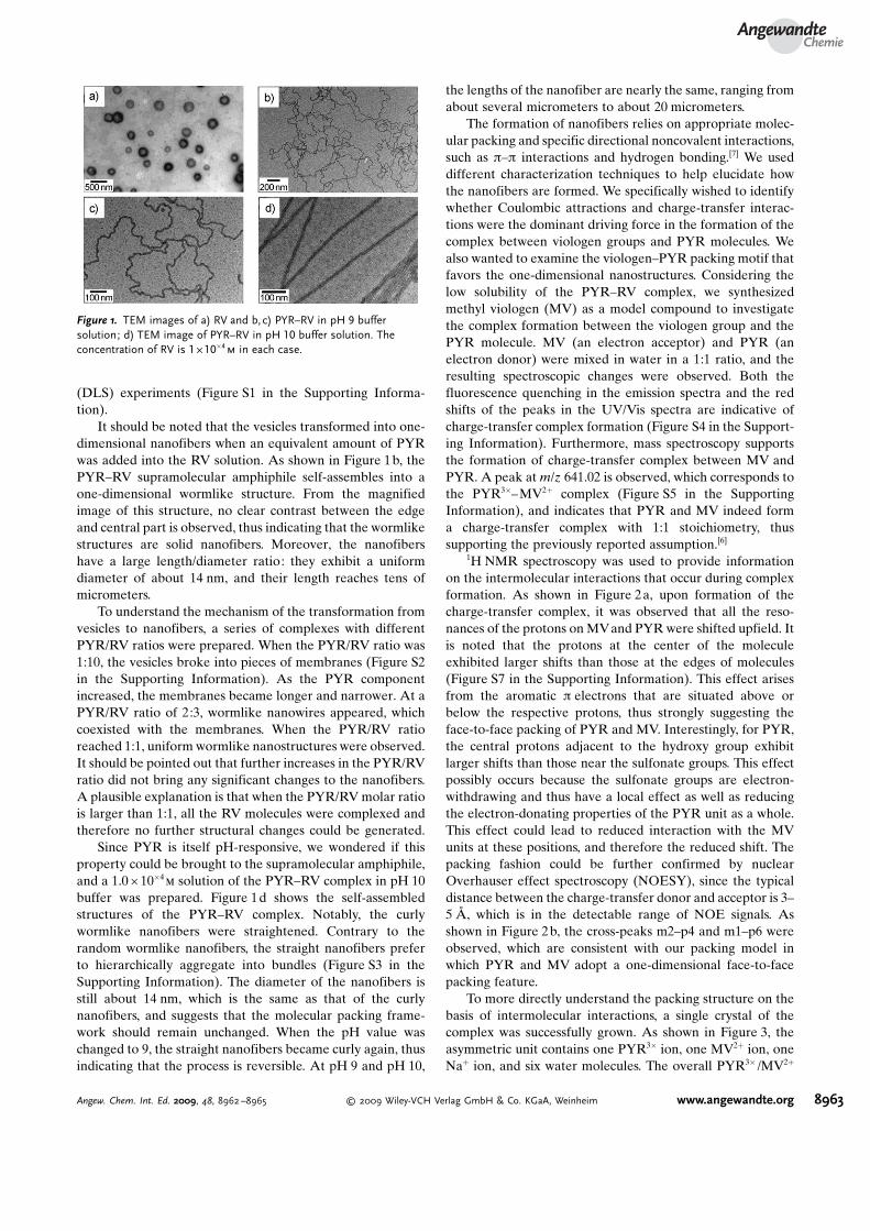
1H NMR spectroscopy was used to provide informationon the intermolecular interactions that occur during complexformation. As shown in Figure 2a, upon formation of thecharge-transfer complex, it was observed that all the reso-nances of the protons on MVand PYR were shifted upfield. Itis noted that the protons at the center of the moleculeexhibited larger shifts than those at the edges of molecules(Figure S7 in the Supporting Information). This effect arisesfrom the aromatic p electrons that are situated above orbelow the respective protons, thus strongly suggesting theface-to-face packing of PYR and MV. Interestingly, for PYR,the central protons adjacent to the hydroxy group exhibitlarger shifts than those near the sulfonate groups. This effectpossibly occurs because the sulfonate groups are electron-withdrawing and thus have a local effect as well as reducingthe electron-donating properties of the PYR unit as a whole.This effect could lead to reduced interaction with the MVunits at these positions, and therefore the reduced shift. Thepacking fashion could be further confirmed by nuclearOverhauser effect spectroscopy (NOESY), since the typicaldistance between the charge-transfer donor and acceptor is 3–5 �, which is in the detectable range of NOE signals. Asshown in Figure 2b, the cross-peaks m2–p4 and m1–p6 wereobserved, which are consistent with our packing model inwhich PYR and MV adopt a one-dimensional face-to-facepacking feature.
To more directly understand the packing structure on thebasis of intermolecular interactions, a single crystal of thecomplex was successfully grown. As shown in Figure 3, theasymmetric unit contains one PYR3� ion, one MV2+ ion, oneNa+ ion, and six water molecules. The overall PYR3� /MV2+
Figure 1. TEM images of a) RV and b,c) PYR–RV in pH 9 buffersolution; d) TEM image of PYR–RV in pH 10 buffer solution. Theconcentration of RV is 1 � 10�4
m in each case.
AngewandteChemie
8963Angew. Chem. Int. Ed. 2009, 48, 8962 –8965 � 2009 Wiley-VCH Verlag GmbH & Co. KGaA, Weinheim www.angewandte.org
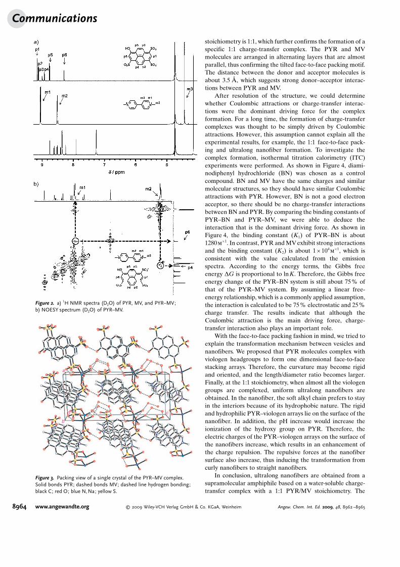
stoichiometry is 1:1, which further confirms the formation of aspecific 1:1 charge-transfer complex. The PYR and MVmolecules are arranged in alternating layers that are almostparallel, thus confirming the tilted face-to-face packing motif.The distance between the donor and acceptor molecules isabout 3.5 �, which suggests strong donor–acceptor interac-tions between PYR and MV.
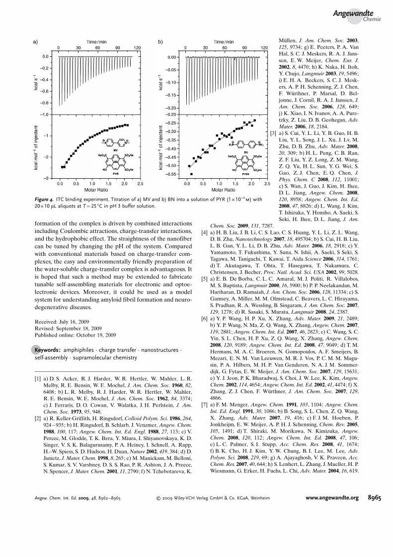
After resolution of the structure, we could determinewhether Coulombic attractions or charge-transfer interac-tions were the dominant driving force for the complexformation. For a long time, the formation of charge-transfercomplexes was thought to be simply driven by Coulombicattractions. However, this assumption cannot explain all theexperimental results, for example, the 1:1 face-to-face pack-ing and ultralong nanofiber formation. To investigate thecomplex formation, isothermal titration calorimetry (ITC)experiments were performed. As shown in Figure 4, diami-nodiphenyl hydrochloride (BN) was chosen as a controlcompound. BN and MV have the same charges and similarmolecular structures, so they should have similar Coulombicattractions with PYR. However, BN is not a good electronacceptor, so there should be no charge-transfer interactionsbetween BN and PYR. By comparing the binding constants ofPYR–BN and PYR–MV, we were able to deduce theinteraction that is the dominant driving force. As shown inFigure 4, the binding constant (K1) of PYR–BN is about1280m�1. In contrast, PYR and MV exhibit strong interactionsand the binding constant (K2) is about 1 � 104
m�1, which is
consistent with the value calculated from the emissionspectra. According to the energy terms, the Gibbs freeenergy DG is proportional to lnK. Therefore, the Gibbs freeenergy change of the PYR–BN system is still about 75% ofthat of the PYR–MV system. By assuming a linear free-energy relationship, which is a commonly applied assumption,the interaction is calculated to be 75 % electrostatic and 25%charge transfer. The results indicate that although theCoulombic attraction is the main driving force, charge-transfer interaction also plays an important role.
With the face-to-face packing fashion in mind, we tried toexplain the transformation mechanism between vesicles andnanofibers. We proposed that PYR molecules complex withviologen headgroups to form one dimensional face-to-facestacking arrays. Therefore, the curvature may become rigidand oriented, and the length/diameter ratio becomes larger.Finally, at the 1:1 stoichiometry, when almost all the viologengroups are complexed, uniform ultralong nanofibers areobtained. In the nanofiber, the soft alkyl chain prefers to stayin the interiors because of its hydrophobic nature. The rigidand hydrophilic PYR–viologen arrays lie on the surface of thenanofiber. In addition, the pH increase would increase theionization of the hydroxy group on PYR. Therefore, theelectric charges of the PYR–viologen arrays on the surface ofthe nanofibers increase, which results in an enhancement ofthe charge repulsion. The repulsive forces at the nanofibersurface also increase, thus inducing the transformation fromcurly nanofibers to straight nanofibers.
In conclusion, ultralong nanofibers are obtained from asupramolecular amphiphile based on a water-soluble charge-transfer complex with a 1:1 PYR/MV stoichiometry. The
Figure 3. Packing view of a single crystal of the PYR–MV complex.Solid bonds PYR; dashed bonds MV; dashed line hydrogen bonding;black C; red O; blue N,Na; yellow S.
Figure 2. a) 1H NMR spectra (D2O) of PYR, MV, and PYR–MV;b) NOESY spectrum (D2O) of PYR–MV.
Communications
8964 www.angewandte.org � 2009 Wiley-VCH Verlag GmbH & Co. KGaA, Weinheim Angew. Chem. Int. Ed. 2009, 48, 8962 –8965
formation of the complex is driven by combined interactionsincluding Coulombic attractions, charge-transfer interactions,and the hydrophobic effect. The straightness of the nanofibercan be tuned by changing the pH of the system. Comparedwith conventional materials based on charge-transfer com-plexes, the easy and environmentally friendly preparation ofthe water-soluble charge-transfer complex is advantageous. Itis hoped that such a method may be extended to fabricatetunable self-assembling materials for electronic and optoe-lectronic devices. Moreover, it could be used as a modelsystem for understanding amyloid fibril formation and neuro-degenerative diseases.
Received: July 16, 2009Revised: September 18, 2009Published online: October 19, 2009
.Keywords: amphiphiles · charge transfer · nanostructures ·self-assembly · supramolecular chemistry
[1] a) D. S. Acker, R. J. Harder, W. R. Hertler, W. Mahler, L. R.Melby, R. E. Bensin, W. E. Mochel, J. Am. Chem. Soc. 1960, 82,6408; b) L. R. Melby, R. J. Harder, W. R. Hertler, W. Mahler,R. E. Bensin, W. E. Mochel, J. Am. Chem. Soc. 1962, 84, 3374;c) J. Ferraris, D. O. Cowan, V. Walatka, J. H. Perlstein, J. Am.Chem. Soc. 1973, 95, 948.
[2] a) R. Keller-Griffith, H. Ringsdorf, Colloid Polym. Sci. 1986, 264,924 – 935; b) H. Ringsdorf, B. Schlarb, J. Venzmer, Angew. Chem.1988, 100, 117; Angew. Chem. Int. Ed. Engl. 1988, 27, 113; c) V.Percec, M. Glodde, T. K. Bera, Y. Miura, I. Shiyanovskaya, K. D.Singer, V. S. K. Balagurusamy, P. A. Heiney, I. Schnell, A. Rapp,H.–W. Spiess, S. D. Hudson, H. Duan, Nature 2002, 419, 384; d) D.Janietz, J. Mater. Chem. 1998, 8, 265; e) M. Manickam, M. Belloni,S. Kumar, S. V. Varshney, D. S. S. Rao, P. R. Ashton, J. A. Preece,N. Spencer, J. Mater. Chem. 2001, 11, 2790; f) N. Tchebotareva, K.
M�llen, J. Am. Chem. Soc. 2003,125, 9734; g) E. Peeters, P. A. VanHal, S. C. J. Meskers, R. A. J. Jans-sen, E. W. Meijer, Chem. Eur. J.2002, 8, 4470; h) K. Naka, H. Itoh,Y. Chujo, Langmuir 2003, 19, 5496;i) E. H. A. Beckers, S. C. J. Mesk-ers, A. P. H. Schenning, Z. J. Chen,F. W�rthner, P. Marsal, D. Bel-jonne, J. Cornil, R. A. J. Janssen, J.Am. Chem. Soc. 2006, 128, 649;j) K. Xiao, I. N. Ivanov, A. A. Pure-tzky, Z. Liu, D. B. Geohegan, Adv.Mater. 2006, 18, 2184.
[3] a) S. Cui, Y. L. Li, Y. B. Guo, H. B.Liu, Y. L. Song, J. L. Xu, J. Lv, M.Zhu, D. B. Zhu, Adv. Mater. 2008,20, 309; b) H. L. Peng, C. B. Ran,Z. F. Liu, Y. Z. Long, Z. M. Wang,Z. Q. Yu, H. L. Sun, Y. G. Wei, S.Gao, Z. J. Chen, E. Q. Chen, J.Phys. Chem. C 2008, 112, 11001;c) S. Wan, J. Guo, J. Kim, H. Ihee,D. L. Jiang, Angew. Chem. 2008,120, 8958; Angew. Chem. Int. Ed.2008, 47, 8826; d) L. Wang, J. Kim,T. Ishizuka, Y. Honsho, A. Saeki, S.Seki, H. Ihee, D. L. Jiang, J. Am.
Chem. Soc. 2009, 131, 7287.[4] a) H. B. Liu, J. B. Li, C. S. Lao, C. S. Huang, Y. L. Li, Z. L. Wang,
D. B. Zhu, Nanotechnology 2007, 18, 495704; b) S. Cui, H. B. Liu,L. B. Gan, Y. L. Li, D. B. Zhu, Adv. Mater. 2006, 18, 2918; c) Y.Yamamoto, T. Fukushima, Y. Suna, N. Ishii, A. Saeki, S Seki, S.Tagawa, M. Taniguchi, T. Kawai, T. Aida Science 2006, 314, 1761;d) T. Akutagawa, T. Ohta, T. Hasegawa, T. Nakamura, C.Christensen, J. Becher, Proc. Natl. Acad. Sci. USA 2002, 99, 5028.
[5] a) E. B. De Borba, C. L. C. Amaral, M. J. Politi, R. Villalobos,M. S. Baptista, Langmuir 2000, 16, 5900; b) P. P. Neelakandan, M.Hariharan, D. Ramaiah, J. Am. Chem. Soc. 2006, 128, 11334; c) S.Gamsey, A. Miller, M. M. Olmstead, C. Beavers, L. C. Hirayama,S. Pradhan, R. A. Wessling, B. Singaram, J. Am. Chem. Soc. 2007,129, 1278; d) R. Sasaki, S. Murata, Langmuir 2008, 24, 2387.
[6] a) Y. P. Wang, H. P. Xu, X. Zhang, Adv. Mater. 2009, 21, 2489;b) Y. P. Wang, N. Ma, Z. Q. Wang, X. Zhang, Angew. Chem. 2007,119, 2881; Angew. Chem. Int. Ed. 2007, 46, 2823; c) C. Wang, S. C.Yin, S. L. Chen, H. P. Xu, Z. Q. Wang, X. Zhang, Angew. Chem.2008, 120, 9189; Angew. Chem. Int. Ed. 2008, 47, 9049; d) T. M.Hermans, M. A. C. Broeren, N. Gomopoulos, A. F. Smeijers, B.Mezari, E. N. M. Van Leeuwen, M. R. J. Vos, P. C. M. M. Magu-sin, P. A. Hilbers, M. H. P. Van Genderen, N. A. J. M. Sommer-dijk, G. Fytas, E. W. Meijer, J. Am. Chem. Soc. 2007, 129, 15631;e) Y. J. Jeon, P. K. Bharadwaj, S. Choi, J. W. Lee, K. Kim, Angew.Chem. 2002, 114, 4654; Angew. Chem. Int. Ed. 2002, 41, 4474; f) X.Zhang, Z. J. Chen, F. W�rthner, J. Am. Chem. Soc. 2007, 129,4866.
[7] a) F. M. Menger, Angew. Chem. 1991, 103, 1104; Angew. Chem.Int. Ed. Engl. 1991, 30, 1086; b) B. Song, S. L. Chen, Z. Q. Wang,X. Zhang, Adv. Mater. 2007, 19, 416; c) F. J. M. Hoeben, P.Jonkheijm, E. W. Meijer, A. P. H. J. Schenning, Chem. Rev. 2005,105, 1491; d) T. Shiraki, M. Morikawa, N. Kimizuka, Angew.Chem. 2008, 120, 112; Angew. Chem. Int. Ed. 2008, 47, 106;e) L. C. Palmer, S. I. Stupp, Acc. Chem. Res. 2008, 41, 1674;f) B. K. Cho, H. J. Kim, Y. W. Chung, B. I. Lee, M. Lee, Adv.Polym. Sci. 2008, 219, 69; g) A. Ajayaghosh, V. K. Praveen, Acc.Chem. Res. 2007, 40, 644; h) S. Lenhert, L. Zhang, J. Mueller, H. P.Wiesmann, G. Erker, H. Fuchs, L. Chi, Adv. Mater. 2004, 16, 619.
Figure 4. ITC binding experiment. Titration of a) MV and b) BN into a solution of PYR (1 � 10�4m) with
20 � 10 mL aliquots at T = 25 8C in pH 3 buffer solution.
AngewandteChemie
8965Angew. Chem. Int. Ed. 2009, 48, 8962 –8965 � 2009 Wiley-VCH Verlag GmbH & Co. KGaA, Weinheim www.angewandte.org