Embed Size (px)
Citation preview
Surface-Induced Dissociation: An Effective Method forCharacterization of Protein Quaternary StructureAlyssa Q. Stiving,† Zachary L. VanAernum,† Florian Busch,†,‡ Sophie R. Harvey,†,‡
Samantha H. Sarni,†,§,⊥ and Vicki H. Wysocki*,†,‡,⊥
†Department of Chemistry and Biochemistry and Resource for Native Mass Spectrometry Guided Structural Biology, ‡CampusChemical Instrument Center, §Ohio State Biochemistry Program, and ⊥The Center for RNA Biology, The Ohio State University,Columbus, Ohio 43210, United States
■ CONTENTS
Introduction to Surface-Induced Dissociation 190Comparison of SID with Other DissociationMethods 191
Collision-Induced Dissociation 191Effect of Precursor Charge in CID vs SID 192
Photodissociation 193Electron-Based Dissociation Techniques 195
SID Instrumentation: Developments and Applica-tions over the Past 4 Years 195
SID in Ion Mobility Q-TOFs 195SID in FTICR Mass Spectrometers 196SID in Orbitrap Platforms 197
Structural Biology Applications: SID Coupled toNative MS 198
SID as Tool to Distinguish Different Gas-PhaseStructures 198SID as Tool to Determine Different In-SolutionStructures 199SID as a Tool to Probe the Quaternary Structureof RNA−Protein Complexes 201SID of Membrane Proteins 201
SID Applications Outside of Structural Biology 202Use of SID for the Study of Lipid Structure 202Combining Simulations and SID 202SID for the Characterization of Metal−OrganicCluster Ions 202
Emerging Complementary Technologies 202Nondenaturing Rapid Online Desalting Coupledto Native Mass Spectrometry 203Toward Automation and Simplified Tuning ofSID-MS 203Coupling with Computational Structure Predic-tion 203
Future Outlook 204Author Information 204
Corresponding Author 204ORCID 204Notes 204Biographies 204
Acknowledgments 204References 205
■ INTRODUCTION TO SURFACE-INDUCEDDISSOCIATION
Many mass spectrometry applications make use of tandem massspectrometry, where two stages of m/z analysis are coupled. Inbetween the two stages ofm/z analysis, an activation or reactionstep is carried out to cause either structurally informativefragmentation or structurally characteristic reaction of theprecursor ion of interest. This review focuses on the use ofcollisions with a surface (surface-induced dissociation, SID) asthe activation method in tandem mass spectrometry. Becausethis is the first review of SID in this Analytical Chemistry specialissue series, an emphasis on SID papers published over the past 4years, rather than only 2 years, is included. SID is described andcompared with other activation methods. The major applicationfocused on in this review is the structural characterization ofnative protein complexes, complexes kinetically trapped thatretain native-like solution structures upon transfer to the gas-phase and throughout the relatively short time frame of the massspectrometry experiment. Other SID applications currentlyunder investigation are also briefly described. Pioneering workon SID has been summarized previously and thus will not bediscussed in detail here.1−4
Surface-induced dissociation was developed in the laboratoryof Graham Cooks, with many studies later carried out in thelaboratories of Cooks, Russell, Wysocki, Whetten, Beck, Futrell,Laskin, Hanley, Gaskell, and Turecek, among others.5−14 SIDhas been used for fragmentation of many different types of ions.Initially, SID was used to fragment lower mass, singly chargedprojectiles because the ionization methods and mass analyzersneeded to form, transmit, and characterize high m/z, multiplycharged ions had not yet been developed to the point where highm/z ions could be conveniently studied. In the early days of SID,it was determined that self-assembled monolayer surfaces(SAMs) of CF3(CF2)10CH2CH2S- on gold serve as effectivecollision targets for surface-induced dissociation (SID) in atandem mass spectrometer.7,15 The use of these easy-to-preparesurfaces has persisted, although several other surface types havebeen utilized.16−24 These SAMs provide a large effective massfor collision of projectile ions; the fluorocarbon chains arerelatively rigid so that they do not severely dampen the energy ofthe colliding projectile, and the fluorocarbon resists facileelectron transfer from the metal surface to the incoming ions.
Special Issue: Fundamental and Applied Reviews in AnalyticalChemistry 2019
Published: November 9, 2018
Review
pubs.acs.org/acCite This: Anal. Chem. 2019, 91, 190−209
© 2018 American Chemical Society 190 DOI: 10.1021/acs.analchem.8b05071Anal. Chem. 2019, 91, 190−209
Dow
nloa
ded
via
OH
IO S
TA
TE
UN
IV o
n Ja
nuar
y 17
, 201
9 at
20:
45:4
5 (U
TC
).
See
http
s://p
ubs.
acs.
org/
shar
ingg
uide
lines
for
opt
ions
on
how
to le
gitim
atel
y sh
are
publ
ishe
d ar
ticle
s.

Surface targets, in contrast to the typical gaseous targets such asAr that are used for the more common activation methodcollision-induced dissociation (CID), have proven to beexceptionally useful for the characterization of protein complexquaternary structure. The ability of SID to producesubcomplexes that remain compact and provide connectivityinformation on the original native complex structure is currentlyunmatched by other dissociation techniques. While eachdissociation method can provide unique information that iscomplementary to other gas- and solution-phase techniques, nosingular mass spectrometry method currently exists in which theentire protein complex structure, including topology, relativeinterfacial strengths, and ligand binding details, can bedetermined and thus, complementary techniques are appealingto gain a more thorough understanding of structural details.Native or native-like protein complexes are produced by
electrospray or nanoelectrospray ionization in electrolytes suchas ammonium acetate at approximately physiological ionicstrength and pH. SID of these multiply charged proteincomplexes generally produces compact, native-like fragmentsthat retain a symmetrically distributed proportion of charge.25,26
This results in unique, easy-to-distinguish spectra in which thesubcomplex products center around a narrowerm/z distributionwhen compared with other tandem MS activation methods.Additionally, SID products and the energy at which they beginto form are typically reflective of relative interfacial strengths andtopology of the protein complex.26 For example, a D2-symmetric homotetramer (dimer of dimers) is anticipated todissociate into dimers at lower SID energies, with the dimersfurther dissociating into monomers at higher SID energies.26 Incontrast, a C4-symmetric tetramer (ring with equal protein−protein interactions between each subunit) will dissociate intomonomer, dimer, and trimer at low SID energy.25 Thesecharacteristic dissociation patterns help to assess the quaternarystructure of the intact protein complex. These two character-istics in particular, approximately symmetric charge partitioningand dissociation dependent on interfacial strengths of proteincomplexes, are unique to SID and make an SID spectrumdistinctive from that obtained by other dissociation techniques.
■ COMPARISONOF SIDWITHOTHERDISSOCIATIONMETHODS
Herein we describe alternative dissociation methods commonlyemployed within mass spectrometry with an emphasis on theoutcomes of probing native protein complexes using these
techniques. The various techniques described, in addition toSID, are shown as a cartoon in Figure 1.
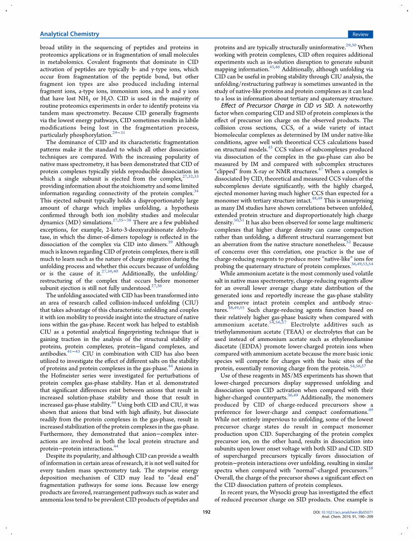
Collision-Induced Dissociation. Collision-induced disso-ciation (CID, or collisionally activated dissociation (CAD)) is atechnique incorporated in essentially every commercial tandemmass spectrometer due to its robust nature and simpleintegration and operation within an instrument.27 CID isaccomplished by accelerating precursor ions into a neutralbackground gas, resulting in multiple, low-energy collisions.During these collisions, a portion of the kinetic energy of the ionis converted into vibrational internal energy, resulting in astepwise buildup of internal energy within the precursor ion.28
As the internal energy builds up, dissociation of the ion canoccur. Because it typically takes many small steps of energyconversion in order to result in dissociation, CID in trappinganalyzers is often referred to as a “slow heating” process when itoccurs in a trapping instrument, and the products are oftenreflective of the lowest energy dissociation pathways (oftenrearrangements) as illustrated in Figure 2.25 CID has found
Figure 1.Cartoon illustration representing the major products of various common dissociation methods in the study of protein complexes. Small bluefragments correspond to covalent cleavage of an individual protein chain.
Figure 2. Simplified representation of the pathways a protein complexmay take when undergoing CID (left) or SID (right). CID involvesmultiple low-energy collisions (top left) that generally result indissociation via the lowest energy pathway (bottom left). SID involvesa single-step, high energy deposition via collision with a surface (topright) and typically results in dissociation via faster, alternativedissociation pathways and dissociation products form that are oftencompact and reflective of the native structure (bottom right).Reproduced from Zhou, M.; Wysocki, V. H., Surface induceddissociation: Dissecting noncovalent protein complexes in the gas-phase. Acc. Chem. Res., 47 (4), 1010−1018 (ref 3). Copyright 2014American Chemical Society.
Analytical Chemistry Review
DOI: 10.1021/acs.analchem.8b05071Anal. Chem. 2019, 91, 190−209
191
broad utility in the sequencing of peptides and proteins inproteomics applications or in fragmentation of small moleculesin metabolomics. Covalent fragments that dominate in CIDactivation of peptides are typically b- and y-type ions, whichoccur from fragmentation of the peptide bond, but otherfragment ion types are also produced including internalfragment ions, a-type ions, immonium ions, and b and y ionsthat have lost NH3 or H2O. CID is used in the majority ofroutine proteomics experiments in order to identify proteins viatandem mass spectrometry. Because CID generally fragmentsvia the lowest energy pathways, CID sometimes results in labilemodifications being lost in the fragmentation process,particularly phosphorylation.29−31
The dominance of CID and its characteristic fragmentationpatterns make it the standard to which all other dissociationtechniques are compared. With the increasing popularity ofnative mass spectrometry, it has been demonstrated that CID ofprotein complexes typically yields reproducible dissociation inwhich a single subunit is ejected from the complex,27,32,33
providing information about the stoichiometry and some limitedinformation regarding connectivity of the protein complex.34
This ejected subunit typically holds a disproportionately largeamount of charge which implies unfolding, a hypothesisconfirmed through both ion mobility studies and moleculardynamics (MD) simulations.27,35−38 There are a few publishedexceptions, for example, 2-keto-3-deoxyarabinonate dehydra-tase, in which the dimer-of-dimers topology is reflected in thedissociation of the complex via CID into dimers.39 Althoughmuch is known regarding CID of protein complexes, there is stillmuch to learn such as the nature of charge migration during theunfolding process and whether this occurs because of unfoldingor is the cause of it.27,36,40 Additionally, the unfolding/restructuring of the complex that occurs before monomersubunit ejection is still not fully understood.27,36
The unfolding associated with CID has been transformed intoan area of research called collision-induced unfolding (CIU)that takes advantage of this characteristic unfolding and couplesit with ion mobility to provide insight into the structure of nativeions within the gas-phase. Recent work has helped to establishCIU as a potential analytical fingerprinting technique that isgaining traction in the analysis of the structural stability ofproteins, protein complexes, protein−ligand complexes, andantibodies.41−43 CIU in combination with CID has also beenutilized to investigate the effect of different salts on the stabilityof proteins and protein complexes in the gas-phase.44 Anions inthe Hofmeister series were investigated for perturbations ofprotein complex gas-phase stability. Han et al. demonstratedthat significant differences exist between anions that result inincreased solution-phase stability and those that result inincreased gas-phase stability.44 Using both CID and CIU, it wasshown that anions that bind with high affinity, but dissociatereadily from the protein complexes in the gas-phase, result inincreased stabilization of the protein complexes in the gas-phase.Furthermore, they demonstrated that anion−complex inter-actions are involved in both the local protein structure andprotein−protein interactions.44
Despite its popularity, and although CID can provide a wealthof information in certain areas of research, it is not well suited forevery tandem mass spectrometry task. The stepwise energydeposition mechanism of CID may lead to “dead end”fragmentation pathways for some ions. Because low energyproducts are favored, rearrangement pathways such as water andammonia loss tend to be prevalent CID products of peptides and
proteins and are typically structurally uninformative.29,30 Whenworking with protein complexes, CID often requires additionalexperiments such as in-solution disruption to generate subunitmapping information.45,46 Additionally, although unfolding viaCID can be useful in probing stability through CIU analysis, theunfolding/restructuring pathway is sometimes unwanted in thestudy of native-like proteins and protein complexes as it can leadto a loss in information about tertiary and quaternary structure.
Effect of Precursor Charge in CID vs SID. A noteworthyfactor when comparing CID and SID of protein complexes is theeffect of precursor ion charge on the observed products. Thecollision cross sections, CCS, of a wide variety of intactbiomolecular complexes as determined by IM under native-likeconditions, agree well with theoretical CCS calculations basedon structural models.45 CCS values of subcomplexes producedvia dissociation of the complex in the gas-phase can also bemeasured by IM and compared with subcomplex structures“clipped” from X-ray or NMR structures.47 When a complex isdissociated by CID, theoretical and measured CCS values of thesubcomplexes deviate significantly, with the highly charged,ejected monomer having much higher CCS than expected for amonomer with tertiary structure intact.48,49 This is unsurprisingas many IM studies have shown correlations between unfolded,extended protein structure and disproportionately high chargedensity.50,51 It has also been observed for some large multimericcomplexes that higher charge density can cause compactionrather than unfolding, a different structural rearrangement butan aberration from the native structure nonetheless.52 Becauseof concerns over this correlation, one practice is the use ofcharge-reducing reagents to produce more “native-like” ions forprobing the quaternary structure of protein complexes.36,49,53,54
While ammonium acetate is the most commonly used volatilesalt in native mass spectrometry, charge-reducing reagents allowfor an overall lower average charge state distribution of thegenerated ions and reportedly increase the gas-phase stabilityand preserve intact protein complex and antibody struc-tures.48,49,55 Such charge-reducing agents function based ontheir relatively higher gas-phase basicity when compared withammonium acetate.54,56,57 Electrolyte additives such astriethylammonium acetate (TEAA) or electrolytes that can beused instead of ammonium acetate such as ethylenediaminediacetate (EDDA) promote lower-charged protein ions whencompared with ammonium acetate because the more basic ionicspecies will compete for charges with the basic sites of theprotein, essentially removing charge from the protein.54,56,57
Use of these reagents in MS/MS experiments has shown thatlower-charged precursors display suppressed unfolding anddissociation upon CID activation when compared with theirhigher-charged counterparts.36,49 Additionally, the monomersproduced by CID of charge-reduced precursors show apreference for lower-charge and compact conformations.49
While not entirely impervious to unfolding, some of the lowestprecursor charge states do result in compact monomerproduction upon CID. Supercharging of the protein complexprecursor ion, on the other hand, results in dissociation intosubunits upon lower onset voltage with both SID and CID. SIDof supercharged precursors typically favors dissociation ofprotein−protein interactions over unfolding, resulting in similarspectra when compared with “normal”-charged precursors.58
Overall, the charge of the precursor shows a significant effect onthe CID dissociation pattern of protein complexes.In recent years, the Wysocki group has investigated the effect
of reduced precursor charge on SID products. One example is
Analytical Chemistry Review
DOI: 10.1021/acs.analchem.8b05071Anal. Chem. 2019, 91, 190−209
192
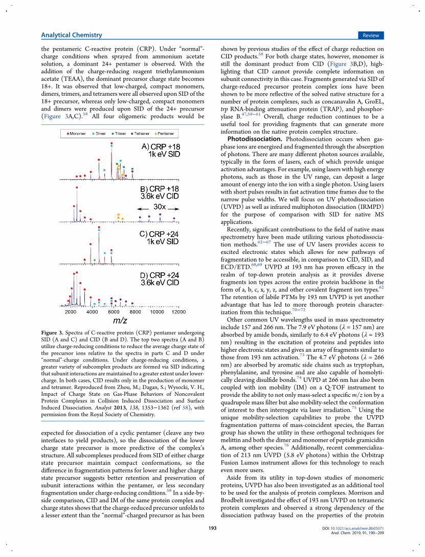
the pentameric C-reactive protein (CRP). Under “normal”-charge conditions when sprayed from ammonium acetatesolution, a dominant 24+ pentamer is observed. With theaddition of the charge-reducing reagent triethylammoniumacetate (TEAA), the dominant precursor charge state becomes18+. It was observed that low-charged, compact monomers,dimers, trimers, and tetramers were all observed upon SID of the18+ precursor, whereas only low-charged, compact monomersand dimers were produced upon SID of the 24+ precursor(Figure 3A,C).58 All four oligomeric products would be
expected for dissociation of a cyclic pentamer (cleave any twointerfaces to yield products), so the dissociation of the lowercharge state precursor is more predictive of the complex’sstructure. All subcomplexes produced from SID of either chargestate precursor maintain compact conformations, so thedifference in fragmentation patterns for lower and higher chargestate precursor suggests better retention and preservation ofsubunit interactions within the pentamer, or less secondaryfragmentation under charge-reducing conditions.58 In a side-by-side comparison, CID and IM of the same protein complex andcharge states shows that the charge-reduced precursor unfolds toa lesser extent than the “normal”-charged precursor as has been
shown by previous studies of the effect of charge reduction onCID products.58 For both charge states, however, monomer isstill the dominant product from CID (Figure 3B,D), high-lighting that CID cannot provide complete information onsubunit connectivity in this case. Fragments generated via SID ofcharge-reduced precursor protein complex ions have beenshown to be more reflective of the solved native structure for anumber of protein complexes, such as concanavalin A, GroEL,trp RNA-binding attenuation protein (TRAP), and phosphor-ylase B.47,59−61 Overall, charge reduction continues to be auseful tool for providing fragments that can generate moreinformation on the native protein complex structure.
Photodissociation. Photodissociation occurs when gas-phase ions are energized and fragmented through the absorptionof photons. There are many different photon sources available,typically in the form of lasers, each of which provide uniqueactivation advantages. For example, using lasers with high energyphotons, such as those in the UV range, can deposit a largeamount of energy into the ion with a single photon. Using laserswith short pulses results in fast activation time frames due to thenarrow pulse widths. We will focus on UV photodissociation(UVPD) as well as infrared multiphoton dissociation (IRMPD)for the purpose of comparison with SID for native MSapplications.Recently, significant contributions to the field of native mass
spectrometry have been made utilizing various photodissocia-tion methods.62−67 The use of UV lasers provides access toexcited electronic states which allows for new pathways offragmentation to be accessible, in comparison to CID, SID, andECD/ETD.68,69 UVPD at 193 nm has proven efficacy in therealm of top-down protein analysis as it provides diversefragments ion types across the entire protein backbone in theform of a, b, c, x, y, z, and other covalent fragment ion types.62
The retention of labile PTMs by 193 nm UVPD is yet anotheradvantage that has led to more thorough protein character-ization from this technique.70−72
Other common UV wavelengths used in mass spectrometryinclude 157 and 266 nm. The 7.9 eV photons (λ = 157 nm) areabsorbed by amide bonds, similarly to 6.4 eV photons (λ = 193nm) resulting in the excitation of proteins and peptides intohigher electronic states and gives an array of fragments similar tothose from 193 nm activation.73 The 4.7 eV photons (λ = 266nm) are absorbed by aromatic side chains such as tryptophan,phenylalanine, and tyrosine and are also capable of homolyti-cally cleaving disulfide bonds.74 UVPD at 266 nm has also beencoupled with ion mobility (IM) on a Q-TOF instrument toprovide the ability to not only mass-select a specificm/z ion by aquadrupole mass filter but also mobility-select the conformationof interest to then interrogate via laser irradiation.75 Using theunique mobility-selection capabilities to probe the UVPDfragmentation patterns of mass-coincident species, the Barrangroup has shown the utility in these orthogonal techniques formelittin and both the dimer and monomer of peptide gramicidinA, among other species.76 Additionally, recent commercializa-tion of 213 nm UVPD (5.8 eV photons) within the OrbitrapFusion Lumos instrument allows for this technology to reacheven more users.Aside from its utility in top-down studies of monomeric
proteins, UVPD has also been investigated as an additional toolto be used for the analysis of protein complexes. Morrison andBrodbelt investigated the effect of 193 nm UVPD on tetramericprotein complexes and observed a strong dependency of thedissociation pathway based on the properties of the protein
Figure 3. Spectra of C-reactive protein (CRP) pentamer undergoingSID (A and C) and CID (B and D). The top two spectra (A and B)utilize charge-reducing conditions to reduce the average charge state ofthe precursor ions relative to the spectra in parts C and D under“normal”-charge conditions. Under charge-reducing conditions, agreater variety of subcomplex products are formed via SID indicatingthat subunit interactions are maintained to a greater extent under lower-charge. In both cases, CID results only in the production of monomerand tetramer. Reproduced from Zhou, M.; Dagan, S.; Wysocki, V. H.,Impact of Charge State on Gas-Phase Behaviors of NoncovalentProtein Complexes in Collision Induced Dissociation and SurfaceInduced Dissociation. Analyst 2013, 138, 1353−1362 (ref 58), withpermission from the Royal Society of Chemistry.
Analytical Chemistry Review
DOI: 10.1021/acs.analchem.8b05071Anal. Chem. 2019, 91, 190−209
193
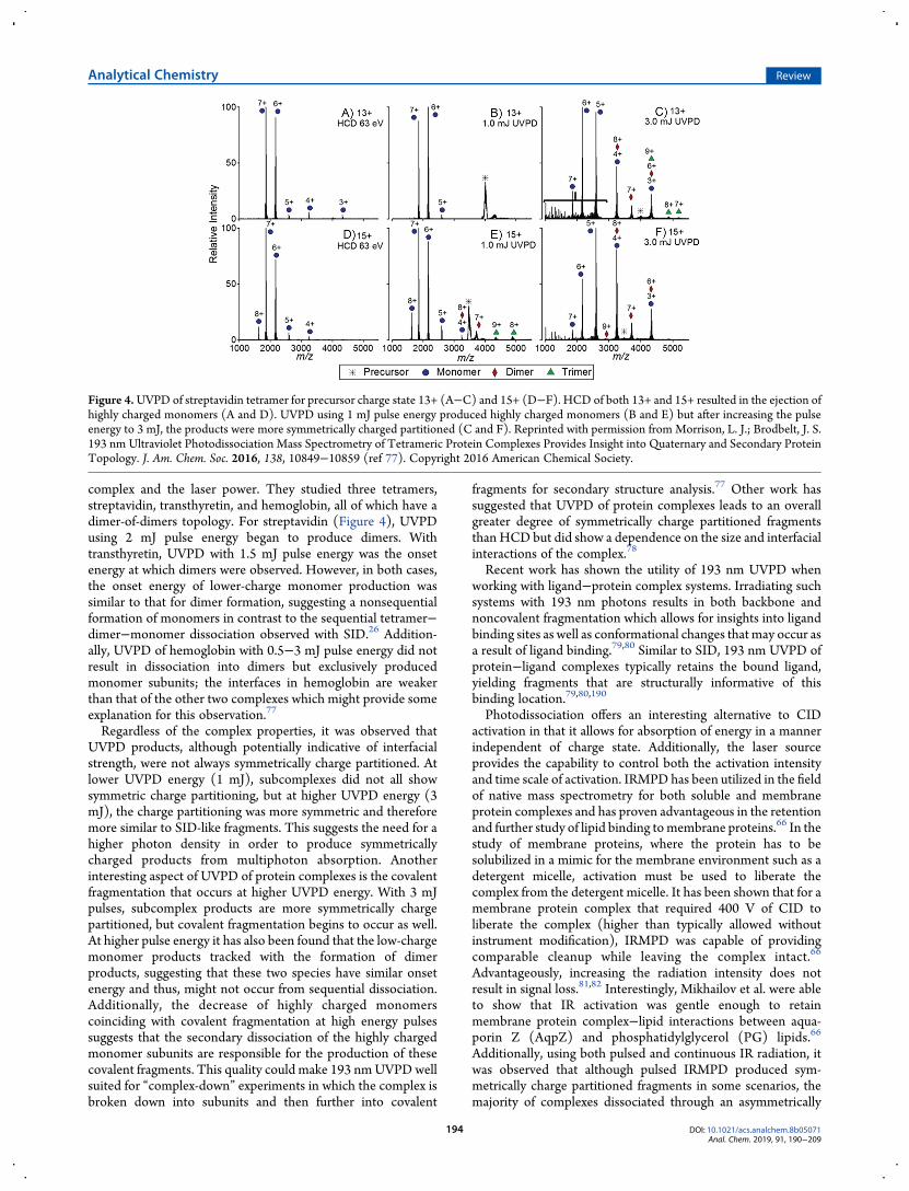
complex and the laser power. They studied three tetramers,streptavidin, transthyretin, and hemoglobin, all of which have adimer-of-dimers topology. For streptavidin (Figure 4), UVPDusing 2 mJ pulse energy began to produce dimers. Withtransthyretin, UVPD with 1.5 mJ pulse energy was the onsetenergy at which dimers were observed. However, in both cases,the onset energy of lower-charge monomer production wassimilar to that for dimer formation, suggesting a nonsequentialformation of monomers in contrast to the sequential tetramer−dimer−monomer dissociation observed with SID.26 Addition-ally, UVPD of hemoglobin with 0.5−3 mJ pulse energy did notresult in dissociation into dimers but exclusively producedmonomer subunits; the interfaces in hemoglobin are weakerthan that of the other two complexes which might provide someexplanation for this observation.77
Regardless of the complex properties, it was observed thatUVPD products, although potentially indicative of interfacialstrength, were not always symmetrically charge partitioned. Atlower UVPD energy (1 mJ), subcomplexes did not all showsymmetric charge partitioning, but at higher UVPD energy (3mJ), the charge partitioning was more symmetric and thereforemore similar to SID-like fragments. This suggests the need for ahigher photon density in order to produce symmetricallycharged products from multiphoton absorption. Anotherinteresting aspect of UVPD of protein complexes is the covalentfragmentation that occurs at higher UVPD energy. With 3 mJpulses, subcomplex products are more symmetrically chargepartitioned, but covalent fragmentation begins to occur as well.At higher pulse energy it has also been found that the low-chargemonomer products tracked with the formation of dimerproducts, suggesting that these two species have similar onsetenergy and thus, might not occur from sequential dissociation.Additionally, the decrease of highly charged monomerscoinciding with covalent fragmentation at high energy pulsessuggests that the secondary dissociation of the highly chargedmonomer subunits are responsible for the production of thesecovalent fragments. This quality could make 193 nmUVPDwellsuited for “complex-down” experiments in which the complex isbroken down into subunits and then further into covalent
fragments for secondary structure analysis.77 Other work hassuggested that UVPD of protein complexes leads to an overallgreater degree of symmetrically charge partitioned fragmentsthan HCD but did show a dependence on the size and interfacialinteractions of the complex.78
Recent work has shown the utility of 193 nm UVPD whenworking with ligand−protein complex systems. Irradiating suchsystems with 193 nm photons results in both backbone andnoncovalent fragmentation which allows for insights into ligandbinding sites as well as conformational changes that may occur asa result of ligand binding.79,80 Similar to SID, 193 nm UVPD ofprotein−ligand complexes typically retains the bound ligand,yielding fragments that are structurally informative of thisbinding location.79,80,190
Photodissociation offers an interesting alternative to CIDactivation in that it allows for absorption of energy in a mannerindependent of charge state. Additionally, the laser sourceprovides the capability to control both the activation intensityand time scale of activation. IRMPD has been utilized in the fieldof native mass spectrometry for both soluble and membraneprotein complexes and has proven advantageous in the retentionand further study of lipid binding tomembrane proteins.66 In thestudy of membrane proteins, where the protein has to besolubilized in a mimic for the membrane environment such as adetergent micelle, activation must be used to liberate thecomplex from the detergent micelle. It has been shown that for amembrane protein complex that required 400 V of CID toliberate the complex (higher than typically allowed withoutinstrument modification), IRMPD was capable of providingcomparable cleanup while leaving the complex intact.66
Advantageously, increasing the radiation intensity does notresult in signal loss.81,82 Interestingly, Mikhailov et al. were ableto show that IR activation was gentle enough to retainmembrane protein complex−lipid interactions between aqua-porin Z (AqpZ) and phosphatidylglycerol (PG) lipids.66
Additionally, using both pulsed and continuous IR radiation, itwas observed that although pulsed IRMPD produced sym-metrically charge partitioned fragments in some scenarios, themajority of complexes dissociated through an asymmetrically
Figure 4.UVPD of streptavidin tetramer for precursor charge state 13+ (A−C) and 15+ (D−F). HCD of both 13+ and 15+ resulted in the ejection ofhighly charged monomers (A and D). UVPD using 1 mJ pulse energy produced highly charged monomers (B and E) but after increasing the pulseenergy to 3 mJ, the products were more symmetrically charged partitioned (C and F). Reprinted with permission from Morrison, L. J.; Brodbelt, J. S.193 nm Ultraviolet Photodissociation Mass Spectrometry of Tetrameric Protein Complexes Provides Insight into Quaternary and Secondary ProteinTopology. J. Am. Chem. Soc. 2016, 138, 10849−10859 (ref 77). Copyright 2016 American Chemical Society.
Analytical Chemistry Review
DOI: 10.1021/acs.analchem.8b05071Anal. Chem. 2019, 91, 190−209
194
charge partitioned pathway despite pulsed IRMPD occurring ona faster time scale than collisional cooling.66 Mapping IRMPDfragments of glutamate dehydrogenase (GDH), a homohex-amer, on a 3D structure showed that the covalent fragments areproduced from the outer regions of the structure while leavingthe trimer−trimer interface intact.67 This pattern of preferentialIRMPD fragmentation on the outer region has been observedwith other complexes and could prove useful in interrogating thequaternary structure of complexes.67
Electron-Based Dissociation Techniques. Electrontransfer and electron capture dissociation (ETD and ECD,respectively) are two additional activation methods used inanalyzing peptides, proteins, and protein complex systems. ECDinvolves an interaction between low energy electrons andmultiply charged analyte cations in which the electrons arecaptured by the analyte cation. This exothermic reaction leads tosubsequent charge reduction, energy transfer, and fragmenta-tion.83,84 ECD is most commonly performed within Fouriertransform ion cyclotron resonance (FTICR) instruments inwhich both the electrons and analyte cations are simultaneouslytrapped within the magnetic field. Performing such experimentsin quadrupole ion traps proved to be challenging because rfvoltages are not sufficient for trapping the thermal electrons.85
To combat this issue, ETD was developed as an alternative, butsimilar, technique more applicable for instruments that utilize rftrapping. In ETD, multiply charged analyte cations interact withreagent radical anions resulting in the transfer of an electron.86,87
Similar to ECD, this process involves an exothermic reactionthat causes backbone cleavage via migration of a hydrogenradical.88 Both ECD and ETD are believed to proceed viapathways that involve very little vibrational energy redistributionprior to backbone cleavage, which allows for the retention oflabile modifications and for their use in characterizinghydrogen−deuterium exchange products without scrambling.89
Electron capture dissociation, perhaps best known for itssuccess within the analysis of intact proteins, has also beenutilized in the study of protein complexes. When undergoingECD, complexes predominantly fragment into c/z-type ionswhile retaining some noncovalent interactions. This has allowedfor mapping of protein−ligand contacts.90−92 ECD tends topreferentially cleave in backbone regions that are more flexibleboth within proteins and protein complexes, allowing forcorrelation between fragment efficiency via ECD and B-factorsof the protein of interest.90,92 Because covalent fragmentationtends to dominate, information regarding the overall assembly ofthe macromolecule is usually lost. While backbone fragmenta-tion is themost common outcome of ECD, at least one study hasshown protein−protein interface dissociation being favored overcovalent fragmentation.93
Amajor pitfall of electron-basedmethods is the observation offewer fragments at lower charge, due to both a reduction offragmentation efficiency and decreased fragment separationbecause of the more compact conformation of lower chargestates. While this is known to be true for monomeric proteinsand can be addressed in many ways when secondary structuralinformation is desired, the correlation between charge and ECDfragmentation is even more drastic for native-like proteinsexhibiting low charge states. Because lower-charge has beenshown to be demonstrative of compact, folded, native-likestructure, this is often a desirable regime in which to performnative mass spectrometry experiments, particularly within thestudy of larger protein complexes. However, this makes electron-based methods significantly more challenging to operate under
such conditions. ETD operates in a similar manner, encounter-ing many of the same challenges when working with native-likeions. In one example, a study that compared dissociation ofinsulin hexamer with both 193 nm UVPD and ETD reportedthat ETD mainly charge reduced the protein complex.80
To help combat the challenges facing ECD and ETD whenstudying native protein complexes (such as charge statedependency and electron capture efficiency), electron-induceddissociation (EID) utilizes >20 eV electrons to excite proteins tocreate electronically excited oxidized radical species andsubsequently fragment the radical ions producing a, b, c, x, y,and z product ion types.94 By oxidizing the analyte cation duringEID, alternative fragmentation pathways are accessed that allowfor greater sequence coverage than other electron-basedmethods. While still in the early stages for utility in studyingprotein complexes, EID has been shown to provide comple-mentary information to ECD such as providing interfacialfragments when ECDprovided no fragmentation for the Cu−Znsuperoxide dismutase (SOD1) enzyme.95
■ SID INSTRUMENTATION: DEVELOPMENTS ANDAPPLICATIONS OVER THE PAST 4 YEARS
SID in Ion Mobility Q-TOFs. The combination of ionmobility spectrometry (IMS) and mass spectrometry is gainingimportance due to its ability to separate and interrogateindividual conformations of ions, to distinguish betweendifferent classes of molecules,96 and to resolve overlappingspecies, for example, different oligomeric states of proteinswhich are present at the samem/z ratio.97−99 In IM-MS, ions areseparated based on their mass, charge, size, and shape. Ionmobility allows an ion’s rotationally averaged collision crosssection (CCS), which depends on its size and shape, to bedetermined. An ion’s CCS provides coarse-grained informationon the conformations adopted in the gas-phase and can becompared to solution coordinates obtained from molecularmodeling or structures solved by NMR, X-ray crystallography,and cryoEM.100−104 Ion mobility-mass spectrometry has beenused in a wide range of applications, from peptideanalysis,105−107 proteomics,108−110 metabolomics,111,112 smallmolecule and isomer identification,113−115 and native massspectrometry. Within native MS and structural biology, ionmobility has been used to study the conformations of proteinsand protein complexes,52,116,117 to study ligand binding,118
protein unfolding,42,119 and conformationally dynamic andintrinsically disordered proteins.120−122
Ion mobility has been coupled with many different types ofmass analyzers, details of which can be found elsewhere.123 Formany years, IM-MS experiments were limited to certainlaboratories with home-built instruments.124−129 In 2006,Waters introduced the first commercially available integratedIM-MS instrument, the Synapt HDMS.130,131 In recent years,several additional ion mobility instruments have come onto themarket including a linear drift tube Q-TOF instrument fromAgilent, a field asymmetric ion mobility interface that can becoupled with Thermo instruments,132,133 and a trapped ionmobility Q-TOF instrument from Bruker.134,135
To date, the Synapt G2 and G2-S instruments are the onlycommercial IM-MS instruments that have been modified ininvestigator laboratories to include SID, although home-builtion mobility instruments have previously been coupled withSID.136,137 The Wysocki group has previously reported thateither the trap cell (the collision cell before the IM) or thetransfer cell (the collision cell after the IM) can be truncated
Analytical Chemistry Review
DOI: 10.1021/acs.analchem.8b05071Anal. Chem. 2019, 91, 190−209
195
allowing incorporation of an SID device at either location,depending on the desired experiment.48,138 When SID is placedbefore the IM, CCS can be determined for the precursor and forthe SID products, giving additional structural information onsubcomplexes,26,48 whereas when SID is placed after the IMregion, complexes can be separated based on their arrival timesbefore dissociation and structural information can be obtainedon different conformations of precursors and the conformation-ally different precursors can each be fragmented independ-ently.47,138
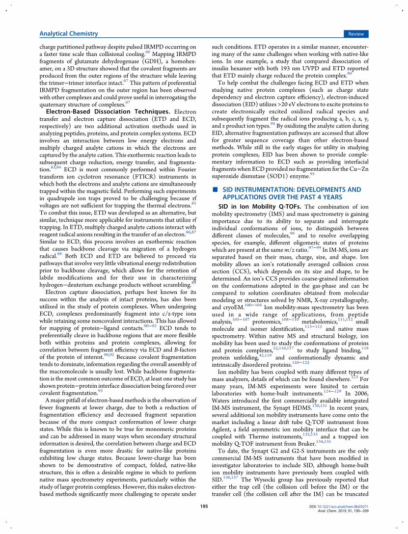
In 2015, Wysocki and co-workers demonstrated that two SIDdevices can be coupled, one before and one after the IM, to allowtwo stages of dissociation for protein complex ions, as shown inFigure 5A.139 In this proof-of-concept study, they demonstratedapplication of SID-IM-SID to study the disassembly of severalstandard protein complexes, which can provide information onthe assembly of the complex. One of the model systems studiedwas tryptophan synthase (TS), a heterotetramer with a linearαββα subunit arrangement. The strength of each interface wasanalyzed using PISA,140 which calculates the interfacial area andnumber and nature of interactions from Protein Data Bank(PDB) files. PISA interfacial analysis for TS showed that the β/βinterface is larger (1624 Å2) than the α/β interface (1363 Å2). InSID-IM-SID experiments, a single charge state of the intactcomplex (19+ TS) was selected by the quadrupole and collidedwith the surface in the first SID device. In the first stage ofdissociation, at low energy, the weakest (smallest) interfaces arebroken first, producing a ββα trimer (Figure 5B). By increasingthe SID energy, additional interfaces can be broken producingββ dimer in addition to the trimer (Figure 5C). The productionof a ββ dimer as opposed to a βα dimer is consistent with the ββinterface being the strongest. As complex dissociation is favoredover subunit unfolding in SID, further information on thedisassembly and hence assembly can be obtained by performinga second stage of SID. In the first stage of SID (SID-IM),products are generated before the IM cell, separated by IM, andappear in separate TOF pulses. However, the fragmentsproduced from the second stage of SID are formed after theIM region, and therefore appear in TOF pulses along with theproducts from which they are generated. By taking horizontalslices of the mobiligram plots, one can extract the MS/IM/MSspectra and successfully identify the fragments produced fromthe dissociation of the ββα trimer (Figure 5D) and ββ dimerindividually (Figure 5E). Dissociation of the ββα trimerproduced primarily ββ dimer and α monomer, which is againconsistent with the solved structure of TS, and suggests SID-IM-SID is a useful tool to study disassembly pathways of proteincomplexes, which can provide information on the relativestrengths of various interfaces within the protein complex. Thisapproach was also applied to study a protein complex ofunknown structure to aid in an MS-based structural determi-nation.59
SID in FTICR Mass Spectrometers. FTICR massspectrometers are capable of providing mass measurementswith ultrahigh resolution and highmass accuracy. These featuresmake FTICR mass spectrometers suitable for a wide range ofdifferent applications including complex mixture analysis,141,142
analysis of petroleum products,143,144 and proteomics andmetabolomics studies.145−148 In addition to the high massaccuracy and resolution, an advantage of FTICR instruments isthat they can be used to perform multiple types of dissociationexperiments,149 including collision-induced dissociation (CID),electron-transfer dissociation (ETD),150 electron-capture dis-
sociation (ECD),151,152 electron ionization dissociation(EID),94 ultraviolet photodissociation (UVPD),153−155 andinfrared multiphoton dissociation (IRPMD).156 In addition,surface-induced dissociation has previously been implementedin FTICR instruments, particularly for the study of fundamen-
Figure 5. (A) Traveling wave (T-wave) region of the modified WatersSynapt G2-S instrument showing the SID-IM-SID experiments. (B)Low energy SID-IM spectrum for 19+ tryptophan synthaseheterotetramer (comprised of αββα subunits arranged in a linearfashion) at a collision energy of 570 eV. Inset is the dominantdissociation pathway illustrated with the crystal structure (PDB 1WBJ).(C) High energy SID-IM spectrum for 19+ tryptophan synthaseheterotetramer at a collision energy of 1330 eV. The interfaces andcorresponding interfacial areas broken are highlighted in the inset. (D)SID-IM-SID of the 12+ αβ2-trimer and (E) 8+ β2-dimer, producedfrom SID-IM of the tetramer (collision energy of 1330 eV) with thesecond stage of SID performed at 2280 eV. Insets show the dissociationpathways. Reproduced from Quintyn, R. S., Harvey, S. H., Wysocki, V.H. Illustration of SID-IM-SID (surface-induced dissociation-ionmobility-SID) mass spectrometry: homo and hetero model proteincomplexes. Analyst, 2015 Oct 21; 140 (20): 7012−9 (ref 139) withpermission from the Royal Society of Chemistry.
Analytical Chemistry Review
DOI: 10.1021/acs.analchem.8b05071Anal. Chem. 2019, 91, 190−209
196
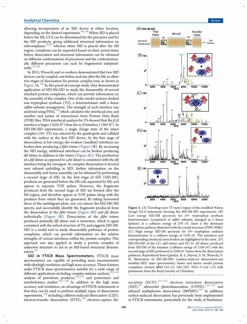
tals of peptide fragmentation.10,24,157−161 SID has been appliedto study the energetics and kinetics of gas-phase fragmentationwithin an ICR instrument.161 In this case, after ions aretransferred into the ICR cell, they are directed toward andcollide against a surface located at the rear trapping plate of theICR cell. The acceleration of the ions for collision with thesurface is controlled via the potential applied to the ICR cellelectrodes. This device design allows SID spectra to be acquiredas a function of the ion kinetic energy and the time between theion-surface collision and the analysis. Furthermore, utilizingresonant ejection of fragment ions, the kinetics of fragment ionformation can be further probed by varying the delay timebetween the surface collision and ejection pulse. This approachhas been used to better understand the gas-phase fragmentationof protonated peptides, odd-electron peptide ions, noncovalentligand−peptide complexes, and ligated metal clusters. Theseapplications are described briefly below.161
FTICR instruments have more recently also been used innative mass spectrometry and structural biology applica-tions,162−164 including structural characterization,95,165,166 top-down dissociation,67,92 ligand binding,167,168 and membraneprotein studies.169,170 In 2017, Yan et al. presented the designand application of a surface-induced dissociation device to studymultimeric protein complexes on a hybrid 15 T FTICR massspectrometer.171 In this design, an SID device with a trappingregion was designed to fit in place of the standard CID cell in theBruker SolariX XR cart. The SID device measures 2.75 cm longand is comprised of 10 dc electrodes (Figure 6A). The SIDdevice can be tuned either to allow ions to pass through withoutcollision with the surface for intact mass measurements (Figure6B) or the ion beam can be directed up toward the surface fordissociation studies (Figure 6C). After passing through the SIDdevice, the ions enter a rectilinear quadrupole, with fourasymptotic electrodes, that acts as the trapping region. Thistrapping region is enclosed to allow for higher pressure and isfilled with argon for collisional cooling. The rf and dc voltagesare applied to the rectilinear quadrupole for trapping ions, anddc voltages are applied to the asymptotic electrodes for trappingand pulsing ions into the ICR cell. Yan et al. demonstrated theapplication of this device with several model protein complexes.As described above, SID is advantageous in such studies as itproduces compact products and products that are consistentwith the known structure of the complex, cleaving the weakestinterfaces in the complex first.26 Previous reports using Q-TOFinstruments, however, have required the use of ion mobility todistinguish between overlapping oligomers; however, the 15 TFTICR was able to isotopically resolve overlapping oligomers(Figure 6D,E) as well as metal cations and ligands. The use ofthis device was further demonstrated by Zhou et al.,172 forcharacterization of a heterooligomeric protein complex MnXfrom Bacillus sp. PL-12. Here the authors used SID to dissociatethe 211 kDa complex into smaller subunits, which could then beisotopically resolved on the 15 T FTICR although the high m/zproducts were not well-resolved. As SID dissociated the complexto produce compact subunits, copper bound to two differentsubunits was retained, which is important for a betterunderstanding of structure and function for this complex, forwhich a high-resolution structure does not exist.172
SID in Orbitrap Platforms. Although the Orbitrap massanalyzer, introduced commercially in 2005, is one of the newestmass analyzers inmass spectrometry, its contributions have beenwidespread.173,174 The Orbitrap analyzer’s combination ofresolution, speed, and sensitivity have become an indispensable
tool in life science research, particularly in the fields ofproteomics175−177 and metabolomics.178,179 However, theOrbitrap would not be such a powerful tool for proteomicanalysis were it not for the many activation techniques such asbeam type CID (HCD),30 electron transfer dissociation(ETD),180 infrared and ultraviolet photodissociation(IRMPD),181 (UVPD),62 electron capture dissociation(ECD),182 and combinations thereof (EThcD, AI-ETD)183,184
that have been implemented on the Orbitrap platform. Eachactivation method serves as an important, and oftencomplementary, tool in bottom-up and top-down proteomicsresearch.Traditionally, the Orbitrap platform has been utilized for the
analysis of digested or denatured proteins, with mass/chargeranges not exceeding 6 000 m/z. Recently, however, Orbitrapinstruments specifically suited for the study of largebiomolecular complexes have been introduced to the market.The Orbitrap Exactive Plus EMR and Q-Exactive UHMR massspectrometers have made it possible to study large viral particles,membrane protein complexes, and soluble protein complexes athigh mass-resolution, with unrivaled sensitivity.185−188 Thehigh-resolution, high mass-accuracy, and (in the case of the
Figure 6. (A) SID device including SID region and rectilinearquadrupole with four asymptotic electrodes, schematic of transmissionmode (left) and SID mode (right). (B) 4 μM CTB pentamer in 100mM EDDA, acquired by averaging 30 scans with a 4.6 s transient. (C)SID of 13+ CTB pentamer at an SID acceleration voltage of 35 V. Thespectrum was acquired by averaging 181 scans with a 9.2 s transient.(D) Zoomed-in region showing the peak highlighted in blue in part B.The experimental data are shown in black, and the simulated isotopicdistributions from different species are shown in purple (8+ tetramer),green (6+ trimer), red (4+ dimer), blue (2+monomer), and gray (sumof all species). (E) Further zoom in of m/z around 5803.7. Adaptedwith permission from Yan, J.; Zhou, M.; Gilbert, J. D.; Wolff, J. J.;Somogyi, A.; Pedder, R. E.; Quintyn, R. S.; Morrison, L. J.; Easterling,M. L.; Pasa-Tolic, L.; Wysocki, V. H. Surface-Induced Dissociation ofProtein Complexes in a Hybrid Fourier Transform Ion CyclotronResonance Mass Spectrometer, Anal. Chem. 2017, 89, 895 (ref 171).Copyright 2018 American Chemical Society.
Analytical Chemistry Review
DOI: 10.1021/acs.analchem.8b05071Anal. Chem. 2019, 91, 190−209
197
experimental EMR instruments modified with a selectionquadrupole, and the Q-Exactive UHMR) high m/z precursorselection capability have made it possible to study small ligandbinding to large noncovalent complexes.189,190
With an increasing interest in studying the proteome at thenoncovalent multiprotein complex level,191 additional activationmethods are necessary to elucidate this higher-order structure.As discussed above, surface-induced dissociation has beenshown to access dissociation pathways for noncovalentcomplexes that are often inaccessible by other activationmethods, and it produces subcomplexes reflective of the overallnative structure.3 With the Orbitrap increasingly being used forthe study of large biomolecular complexes, it became clear thatthe combination of the high-resolution capabilities of theOrbitrap analyzer and structural information provided by SIDmay extend the current capabilities of native MS analysis.VanAernum et al. recently implemented surface-induced
dissociation on an Exactive Plus EMR Orbitrap platform thathad previously been modified to include a selection quadru-pole.192 The SID device was designed to fit in place of the smalltransfer multipole between the quadrupole mass filter and the C-trap. This design choice means that the same SID design can beimplemented in the newer Q-Exactive UHMR platform withoutany additional modifications. The performance of the SID-modified Exactive EMR was characterized by the dissociation ofa range of previously studied noncovalent protein complexes.The dissociation patterns and subcomplexes produced fromstreptavidin tetramer (53 kDa) and glutamate dehydrogenasehexamer (334 kDa) showed the distinctive symmetric chargepartitioning that was previously reported on time-of-flightinstruments (and in the case of streptavidin tetramer, also on anICR instrument) and reflected the dissociation pathways thatwould be expected based on the native structures.26,60,171
Furthermore, it was shown that subcomplexes that overlap inm/z space (e.g., 3+monomer and 6+ dimer) could be distinguishedwith the high resolving power of the instrument by directlyresolving the overlapping isotope distributions or by differ-entiating the number of nonvolatile salt adducts. The authors
also demonstrated that the streptavidin−biotin interactioncould remain intact through the SID process, and the relativeamount of biotin on streptavidin subcomplexes could be easilyquantified with the high-resolution capabilities of the Orbitrapinstrument.In another study, Busch et al. used SID on the Orbitrap EMR
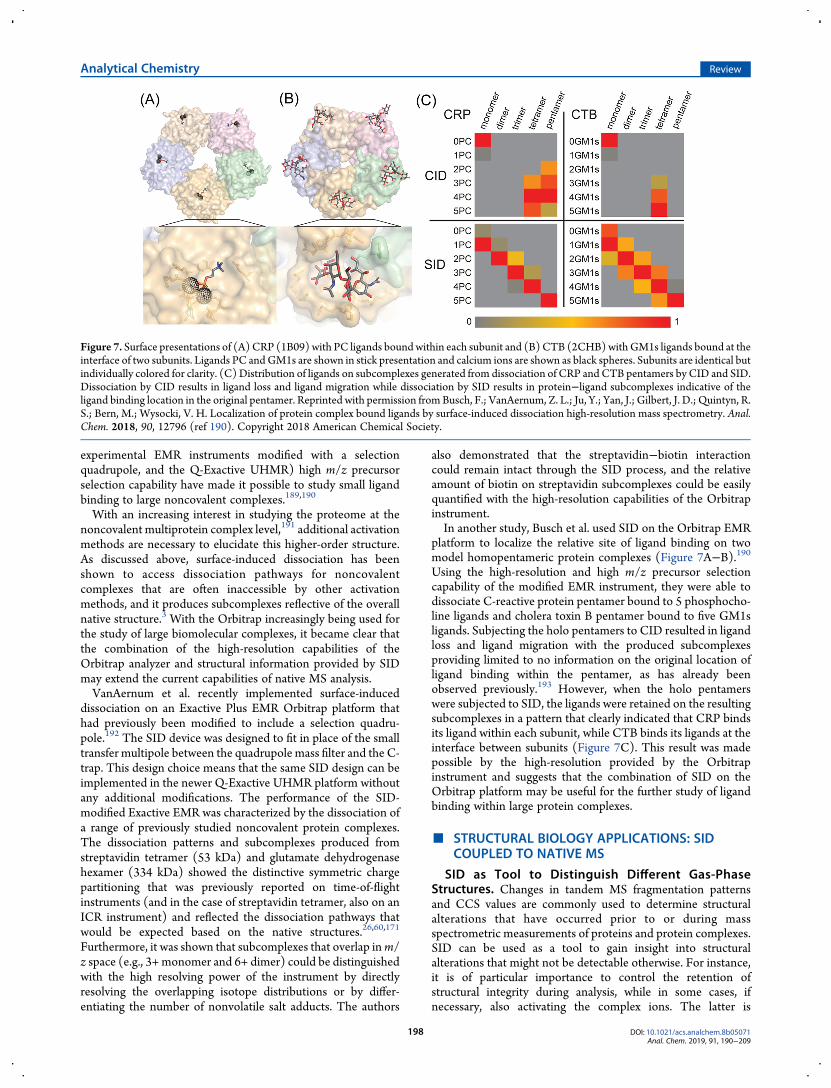
platform to localize the relative site of ligand binding on twomodel homopentameric protein complexes (Figure 7A−B).190Using the high-resolution and high m/z precursor selectioncapability of the modified EMR instrument, they were able todissociate C-reactive protein pentamer bound to 5 phosphocho-line ligands and cholera toxin B pentamer bound to five GM1sligands. Subjecting the holo pentamers to CID resulted in ligandloss and ligand migration with the produced subcomplexesproviding limited to no information on the original location ofligand binding within the pentamer, as has already beenobserved previously.193 However, when the holo pentamerswere subjected to SID, the ligands were retained on the resultingsubcomplexes in a pattern that clearly indicated that CRP bindsits ligand within each subunit, while CTB binds its ligands at theinterface between subunits (Figure 7C). This result was madepossible by the high-resolution provided by the Orbitrapinstrument and suggests that the combination of SID on theOrbitrap platform may be useful for the further study of ligandbinding within large protein complexes.
■ STRUCTURAL BIOLOGY APPLICATIONS: SIDCOUPLED TO NATIVE MS
SID as Tool to Distinguish Different Gas-PhaseStructures. Changes in tandem MS fragmentation patternsand CCS values are commonly used to determine structuralalterations that have occurred prior to or during massspectrometric measurements of proteins and protein complexes.SID can be used as a tool to gain insight into structuralalterations that might not be detectable otherwise. For instance,it is of particular importance to control the retention ofstructural integrity during analysis, while in some cases, ifnecessary, also activating the complex ions. The latter is
Figure 7. Surface presentations of (A)CRP (1B09) with PC ligands boundwithin each subunit and (B)CTB (2CHB)withGM1s ligands bound at theinterface of two subunits. Ligands PC andGM1s are shown in stick presentation and calcium ions are shown as black spheres. Subunits are identical butindividually colored for clarity. (C)Distribution of ligands on subcomplexes generated from dissociation of CRP andCTB pentamers by CID and SID.Dissociation by CID results in ligand loss and ligand migration while dissociation by SID results in protein−ligand subcomplexes indicative of theligand binding location in the original pentamer. Reprinted with permission fromBusch, F.; VanAernum, Z. L.; Ju, Y.; Yan, J.; Gilbert, J. D.; Quintyn, R.S.; Bern, M.; Wysocki, V. H. Localization of protein complex bound ligands by surface-induced dissociation high-resolution mass spectrometry. Anal.Chem. 2018, 90, 12796 (ref 190). Copyright 2018 American Chemical Society.
Analytical Chemistry Review
DOI: 10.1021/acs.analchem.8b05071Anal. Chem. 2019, 91, 190−209
198
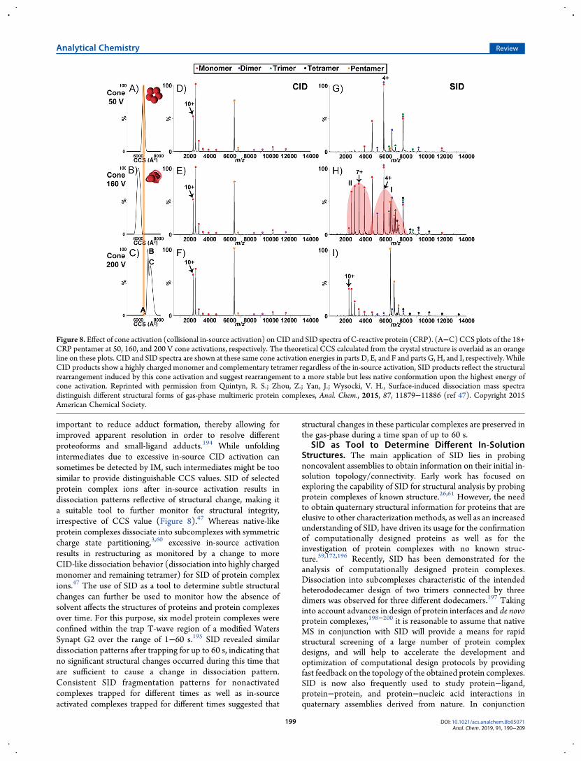
important to reduce adduct formation, thereby allowing forimproved apparent resolution in order to resolve differentproteoforms and small-ligand adducts.194 While unfoldingintermediates due to excessive in-source CID activation cansometimes be detected by IM, such intermediates might be toosimilar to provide distinguishable CCS values. SID of selectedprotein complex ions after in-source activation results indissociation patterns reflective of structural change, making ita suitable tool to further monitor for structural integrity,irrespective of CCS value (Figure 8).47 Whereas native-likeprotein complexes dissociate into subcomplexes with symmetriccharge state partitioning,3,60 excessive in-source activationresults in restructuring as monitored by a change to moreCID-like dissociation behavior (dissociation into highly chargedmonomer and remaining tetramer) for SID of protein complexions.47 The use of SID as a tool to determine subtle structuralchanges can further be used to monitor how the absence ofsolvent affects the structures of proteins and protein complexesover time. For this purpose, six model protein complexes wereconfined within the trap T-wave region of a modified WatersSynapt G2 over the range of 1−60 s.195 SID revealed similardissociation patterns after trapping for up to 60 s, indicating thatno significant structural changes occurred during this time thatare sufficient to cause a change in dissociation pattern.Consistent SID fragmentation patterns for nonactivatedcomplexes trapped for different times as well as in-sourceactivated complexes trapped for different times suggested that
structural changes in these particular complexes are preserved inthe gas-phase during a time span of up to 60 s.
SID as Tool to Determine Different In-SolutionStructures. The main application of SID lies in probingnoncovalent assemblies to obtain information on their initial in-solution topology/connectivity. Early work has focused onexploring the capability of SID for structural analysis by probingprotein complexes of known structure.26,61 However, the needto obtain quaternary structural information for proteins that areelusive to other characterizationmethods, as well as an increasedunderstanding of SID, have driven its usage for the confirmationof computationally designed proteins as well as for theinvestigation of protein complexes with no known struc-ture.59,172,196 Recently, SID has been demonstrated for theanalysis of computationally designed protein complexes.Dissociation into subcomplexes characteristic of the intendedheterododecamer design of two trimers connected by threedimers was observed for three different dodecamers.197 Takinginto account advances in design of protein interfaces and de novoprotein complexes,198−200 it is reasonable to assume that nativeMS in conjunction with SID will provide a means for rapidstructural screening of a large number of protein complexdesigns, and will help to accelerate the development andoptimization of computational design protocols by providingfast feedback on the topology of the obtained protein complexes.SID is now also frequently used to study protein−ligand,protein−protein, and protein−nucleic acid interactions inquaternary assemblies derived from nature. In conjunction
Figure 8. Effect of cone activation (collisional in-source activation) on CID and SID spectra of C-reactive protein (CRP). (A−C)CCS plots of the 18+CRP pentamer at 50, 160, and 200 V cone activations, respectively. The theoretical CCS calculated from the crystal structure is overlaid as an orangeline on these plots. CID and SID spectra are shown at these same cone activation energies in parts D, E, and F and parts G, H, and I, respectively. WhileCID products show a highly charged monomer and complementary tetramer regardless of the in-source activation, SID products reflect the structuralrearrangement induced by this cone activation and suggest rearrangement to a more stable but less native conformation upon the highest energy ofcone activation. Reprinted with permission from Quintyn, R. S.; Zhou, Z.; Yan, J.; Wysocki, V. H., Surface-induced dissociation mass spectradistinguish different structural forms of gas-phase multimeric protein complexes, Anal. Chem., 2015, 87, 11879−11886 (ref 47). Copyright 2015American Chemical Society.
Analytical Chemistry Review
DOI: 10.1021/acs.analchem.8b05071Anal. Chem. 2019, 91, 190−209
199
with IM measurements, SID provided insights into Cu(I)binding to the homotetrameric copper-sensitive operonrepressor (CsoR) from Bacillus subtilis.201 Whereas the apoprotein predominantly dissociated into monomers, Cu(I)-bound CsoR preferentially dissociates into dimers, requiringmuch higher SID energy in line with the coordination of Cu(I)between two subunits.201 Combined with other techniques, SIDhas recently been employed to investigate the quaternarystructure of the two hetero-oligomeric complexes manganeseoxidase (Mnx) from Bacillus sp. PL-12 and toyocamycin nitrile
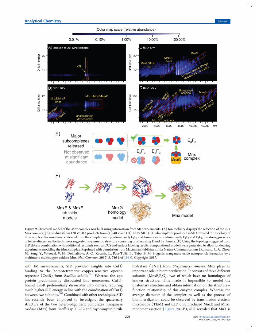
hydratase (TNH) from Streptomyces rimosus. Mnx plays animportant role in biomineralization. It consists of three differentsubunits (MnxE,F,G), two of which have no homologue ofknown structure. This made it impossible to model thequaternary structure and obtain information on the structure−function relationship of this enzyme complex. Whereas theaverage diameter of the complex as well as the process ofbiomineralization could be observed by transmission electronmicroscopy (TEM) and CID only produced MnxE and MnxFmonomer ejection (Figure 9A−B), SID revealed that MnX is
Figure 9. Structural model of the Mnx complex was built using information from SID experiments. (A) Ion mobility displays the selection of the 29+Mnx complex, (B) products from 120VCID, products from (C) 40 V and (D) 120 V SID. (E) Subcomplexes produced in SID revealed the topology ofthis complex. Because dimers released from the complex were predominantly E1F1 and trimers were predominantly E1F2 and E2F1, the strong presenceof heterodimers and heterotrimers suggested a symmetric structure consisting of alternating E and F subunits. (F) Using the topology suggested fromSID data in combination with additional restraints such as CCS and surface labeling results, computational models were generated to allow for dockingexperiments modeling theMnx complex. Reprinted with permission fromMacmillan Publishers Ltd.: Nature Communications (Romano, C. A., Zhou,M., Song, Y., Wysocki, V. H., Dohnalkova, A. C., Kovarik, L., Pasa-Tolic, L., Tebo, B. M. Biogenic manganese oxide nanoparticle formation by amultimeric multicopper oxidase Mnx. Nat. Commun. 2017, 8, 746 (ref 196)), Copyright 2017.
Analytical Chemistry Review
DOI: 10.1021/acs.analchem.8b05071Anal. Chem. 2019, 91, 190−209
200
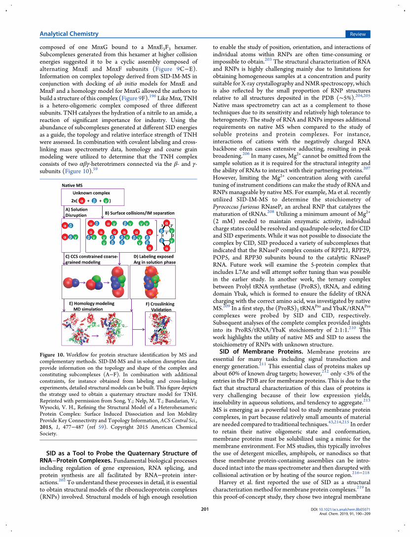
composed of one MnxG bound to a MnxE3F3 hexamer.Subcomplexes generated from this hexamer at higher collisionenergies suggested it to be a cyclic assembly composed ofalternating MnxE and MnxF subunits (Figure 9C−E).Information on complex topology derived from SID-IM-MS inconjunction with docking of ab initio models for MnxE andMnxF and a homology model for MnxG allowed the authors tobuild a structure of this complex (Figure 9F).196 LikeMnx, TNHis a hetero-oligomeric complex composed of three differentsubunits. TNH catalyzes the hydration of a nitrile to an amide, areaction of significant importance for industry. Using theabundance of subcomplexes generated at different SID energiesas a guide, the topology and relative interface strength of TNHwere assessed. In combination with covalent labeling and cross-linking mass spectrometry data, homology and coarse grainmodeling were utilized to determine that the TNH complexconsists of two αβγ-heterotrimers connected via the β- and γ-subunits (Figure 10).59
SID as a Tool to Probe the Quaternary Structure ofRNA−Protein Complexes. Fundamental biological processesincluding regulation of gene expression, RNA splicing, andprotein synthesis are all facilitated by RNA−protein inter-actions.202 To understand these processes in detail, it is essentialto obtain structural models of the ribonucleoprotein complexes(RNPs) involved. Structural models of high enough resolution
to enable the study of position, orientation, and interactions ofindividual atoms within RNPs are often time-consuming orimpossible to obtain.203 The structural characterization of RNAand RNPs is highly challenging mainly due to limitations forobtaining homogeneous samples at a concentration and puritysuitable for X-ray crystallography andNMR spectroscopy, whichis also reflected by the small proportion of RNP structuresrelative to all structures deposited in the PDB (∼5%).204,205Native mass spectrometry can act as a complement to thosetechniques due to its sensitivity and relatively high tolerance toheterogeneity. The study of RNA and RNPs imposes additionalrequirements on native MS when compared to the study ofsoluble proteins and protein complexes. For instance,interactions of cations with the negatively charged RNAbackbone often causes extensive adducting, resulting in peakbroadening.206 In many cases, Mg2+ cannot be omitted from thesample solution as it is required for the structural integrity andthe ability of RNAs to interact with their partnering proteins.207
However, limiting the Mg2+ concentration along with carefultuning of instrument conditions can make the study of RNA andRNPs manageable by native MS. For example, Ma et al. recentlyutilized SID-IM-MS to determine the stoichiometry ofPyrococcus furiosus RNaseP, an archeal RNP that catalyzes thematuration of tRNAs.208 Utilizing a minimum amount of Mg2+
(2 mM) needed to maintain enzymatic activity, individualcharge states could be resolved and quadrupole-selected for CIDand SID experiments. While it was not possible to dissociate thecomplex by CID, SID produced a variety of subcomplexes thatindicated that the RNaseP complex consists of RPP21, RPP29,POP5, and RPP30 subunits bound to the catalytic RNasePRNA. Future work will examine the 5-protein complex thatincludes L7Ae and will attempt softer tuning than was possiblein the earlier study. In another work, the ternary complexbetween Prolyl tRNA synthetase (ProRS), tRNA, and editingdomain Ybak, which is formed to ensure the fidelity of tRNAcharging with the correct amino acid, was investigated by nativeMS.209 In a first step, the (ProRS)2 tRNA
Pro and YbaK/tRNAPro
complexes were probed by SID and CID, respectively.Subsequent analyses of the complete complex provided insightsinto its ProRS/tRNA/YbaK stoichiometry of 2:1:1.210 Thiswork highlights the utility of native MS and SID to assess thestoichiometry of RNPs with unknown structure.
SID of Membrane Proteins. Membrane proteins areessential for many tasks including signal transduction andenergy generation.211 This essential class of proteins makes upabout 60% of known drug targets; however,212 only <3% of theentries in the PDB are for membrane proteins. This is due to thefact that structural characterization of this class of proteins isvery challenging because of their low expression yields,insolubility in aqueous solutions, and tendency to aggregate.213
MS is emerging as a powerful tool to study membrane proteincomplexes, in part because relatively small amounts of materialare needed compared to traditional techniques.43,214,215 In orderto retain their native oligomeric state and conformation,membrane proteins must be solubilized using a mimic for themembrane environment. For MS studies, this typically involvesthe use of detergent micelles, amphipols, or nanodiscs so thatthese membrane protein-containing assemblies can be intro-duced intact into the mass spectrometer and then disrupted withcollisional activation or by heating of the source region.216−218
Harvey et al. first reported the use of SID as a structuralcharacterizationmethod for membrane protein complexes.219 Inthis proof-of-concept study, they chose two integral membrane
Figure 10. Workflow for protein structure identification by MS andcomplementary methods. SID-IM-MS and in solution disruption dataprovide information on the topology and shape of the complex andconstituting subcomplexes (A−F). In combination with additionalconstraints, for instance obtained from labeling and cross-linkingexperiments, detailed structural models can be built. This figure depictsthe strategy used to obtain a quaternary structure model for TNH.Reprinted with permission from Song, Y.; Nelp, M. T.; Bandarian, V.;Wysocki, V. H., Refining the Structural Model of a HeterohexamericProtein Complex: Surface Induced Dissociation and Ion MobilityProvide Key Connectivity and Topology Information, ACS Central Sci.,2015, 1, 477−487 (ref 59). Copyright 2015 American ChemicalSociety.
Analytical Chemistry Review
DOI: 10.1021/acs.analchem.8b05071Anal. Chem. 2019, 91, 190−209
201
proteins, trimeric AmtB, and tetrameric AqpZ from Escherichiacoli. Both complexes have solved crystal structures and had beenstudied previously with native MS using the charge-reducingdetergent tetraethylene glycol monooctyl ether (C8E4).220
When AmtB and AqpZ were sprayed from C8E4 in a modifiedWaters Synapt G2, CID resulted in limited dissociation, withhighly charged, unfolded monomer being the major productobserved. SID of trimeric AmtB, with the SID device placedbetween the trap and IM cell of the Synapt G2,138 resulted in theproduction of compact monomers and dimers, consistent withthe ring-like structure of this complex which has equal interfacesbetween all subunits. Similar results were observed fortetrameric AqpZ, with compact monomers, dimers, and trimersbeing observed in agreement with the solved structure (cleavageat two interfaces is required to produce free fragment ions,monomer/trimer and dimer/dimer). This study highlightedthat SID of membrane protein complexes produces subcom-plexes consistent with their solved structure and therefore haspotential in the study of membrane protein complexes withoutsolved structures.The Robinson lab has recently demonstrated the use of SID as
a method to probe membrane protein lipid binding andselectivity. In these studies, semiSWEET, a dimeric bacterialsugar transporter, was studied using a modified Synapt G2-Siwith a longer TOF for enhanced resolution, SID before the IM,and a segmented quadrupole in place of the transfer ionguide.221 Previous studies have demonstrated that semiSWEETis stabilized through binding of cardiolipin and shows selectivelytoward longer chain lengths.222−224 In their SID studies,Robinson and co-workers observed greater stabilization ofdimers with longer chain lengths of cardiolipin, consistent withthe previous studies showing selectivity toward longer chainlengths. This study highlighted that SID can be useful tool tostudy membrane protein lipid interactions and that CID did notallow the distinction that was possible by SID.
■ SID APPLICATIONS OUTSIDE OF STRUCTURALBIOLOGY
In addition to its utility for structural biology studies, SID hasalso been used for other applications. We present here a briefsummary of other studies that have taken advantage of SID.Use of SID for the Study of Lipid Structure. In the
McLean lab at Vanderbilt University, SID has recently beenapplied to the study of lipids as a potentially advantageousmethod of observing greater fragmentation than possible byCID. In the study of phosphatidylcholines, it has been shownthat SID results in greater fragmentation than CID whencomparing equivalent lab frame energies. Head group loss andsubsequent headgroup breakdown shows a similar trendbetween the two techniques. While the types of fragmentsproduced between the two techniques are the same, SID does soat lower lab frame energies, providing higher-energy fragmentsthat would not be accessible by CID within instrumentacceleration voltage limits. While this work is still ongoing, itshows promise for production of fragments that typically appearin lower abundance via CID and that can be used in indicatingfatty acyl composition in the lipids.225
Combining Simulations and SID. Another area in whichSID has proven useful is the study of fragmentation energeticsand mechanisms. The relationship between simulations,mechanisms, and SID fragmentation patterns are important inunderstanding the experimental outcome of ion-surfacecollisions.226 Combining quantum mechanical/molecular me-
chanics (QM/MM) simulations and RRKM modeling, Laskinand co-workers have been able to explain fragmentation kineticsof singly protonated peptides that have enough vibrationalenergy to fragment yet do not show fragmentation during theexperimental time period.227 Additionally, by utilizing acombination of time- and collision energy-resolved SIDexperiments and resonant ejection experiments, dissociationpathways and dissociation onset energies for basic residues havealso been investigated. Specific peptides were used to representdissociation via salt-bridge and canonical pathways. Usingresonant ejection in the dissociation of these peptides allowedfor both fast and slow kinetics to be studied, in themillisecond tosecond time frame of the ICR.228
Hase and co-workers have contributed significantly to theunderstanding of SID by utilizing chemical dynamicssimulations to investigate energy transfer upon collision with asurface in addition to mechanisms of soft-landing and reactive-landing. Utilizing a QM approach for the intramolecularpotential of the protonated peptide and an MM approach forthe surface as well as interaction between the surface andprotonated peptide, comparisons between experimental resultsand simulations can be made for protonated peptidefragmentation to provide a better understanding of the SIDmechanism and dynamics involved upon such colli-sions.226,229,230
SID for the Characterization of Metal−Organic ClusterIons.Within recent years SID has also been utilized in the studyof ultrasmall gold cluster ions consisting of 7−9 Au atoms ligatedwith phosphines. Such ultrasmall gold clusters can be useful incatalysis and energy production. However, the complexity (withgreater than 500 vibrational degrees of freedom) and size ofthese clusters make it particularly challenging to obtaininformation needed for scalability such as thermodynamic andkinetic parameters for clusters with a specific number of Auatoms and charges. In one study, SID was used to investigate thesize-dependent stability toward fragmentation as well as ligandbinding energies because no other direct experimental measure-ments exist. The ability of SID to remain sensitive to smallvariations in threshold energies as well as activation entropiesmakes SID a valuable choice in probing the dissociation of theseions.231 This allowed observation of dissociation pathways thatcould then be related back to RRKM-based modeling. SID hasalso been utilized with monolayer-protected silver clusters toidentify gas-phase structural isomers, perhaps pointing todifferent cluster configurations. SID provided differentfragmentation of these isomers than CID at similar energy,producing a wide range of fragments and resulting in chargestripping that had not yet been observed with similar gold orsilver clusters.232
■ EMERGING COMPLEMENTARY TECHNOLOGIESNativeMS coupled to SID and/or other activationmethods, andto ion mobility, can be used throughout the biochemical/biological study of a complex, allowing midcourse adjustmentsand guiding higher resolution (X-ray crystallography, NMR,cryoEM) studies that are too time and resource intensive to beused in routine work. Many groups have come to recognize,however, that extensive protein purification followed by manualdesalting with spin columns, or dialysis, and static (nano)-electrospray approaches is too slow for routine application tostructural biology throughout a study. Direct introduction of celllysates using static nanospray for detection of overexpressedprotein complexes has been reported, which simplifies the
Analytical Chemistry Review
DOI: 10.1021/acs.analchem.8b05071Anal. Chem. 2019, 91, 190−209
202
protein purification steps.233,234 The long-term goal for suchstudies would be to study single cells. An alternative approach tothis is online desalting.235 Online desalting offers the advantageof higher-throughput and reduced sample handling compared toconventional desalting approaches and the potential toautomate the use of native MS for routine screening of proteincomplexes. We describe below the efforts in this field, includingadditional approaches to automate native MS acquisition andSID experiments.Nondenaturing Rapid Online Desalting Coupled to
Native Mass Spectrometry. One of the critical steps in thenative mass spectrometry workflow is sample preparation toremove nonvolatile salts and additives. The presence ofnonvolatile salts, buffers, or additives in the sample will sacrificesensitivity, mass accuracy, and mass resolution and will oftenlead to increased down time for cleaning of the massspectrometer. To avoid these problems, the structures andfunctions of proteins and protein complexes are typically studiedin volatile buffers. For many years, this has been accomplishedby buffer exchanging the biological sample into a volatileelectrolyte such as ammonium acetate prior toMS analysis. Thismethod works very well; however, the use of gel filtration spincolumns or ultrafiltration devices to perform the buffer exchangecan become time-consuming and cumbersome when analyzinglarge numbers of samples. Furthermore, it is often not obvioushow extended storage in MS-friendly buffers might affect theprotein integrity. Consequently, several alternatives to themanual buffer exchange process have been developed. It has alsobeen shown that the presence of additives such as electro-lytes,236,237 amino acids,162 or supercharging agents238 can helpto mitigate some of the adverse effects of nonvolatile salts. Whilethese results are impressive, they were not shown to be useful forthe high concentrations of nonvolatile salts typically used duringprotein purification. More recently, Williams and co-workershave shown that the use of submicrometer nanospray emittersprovides the ability to ionize proteins directly from highconcentrations of nonvolatile buffers while still achievingnarrow, well-resolved protein signals. The simplicity of using anarrow diameter nanospray emitter to spray samples directlyfrom biological buffers is appealing, and this approach is beingcharacterized in the native MS community. However, resultsindicate that the signal from nonvolatile salts in the low m/zregion still dominates the protein signal even when sprayingfrom narrow emitters, whichmay cause interference with speciesin this m/z region. Furthermore, narrow emitters tend to clogmore readily than larger nanospray emitters, making it moredifficult to automate this process.An alternative approach is to physically separate nonvolatile
salts from the analyte of interest, such as by manual bufferexchange, but doing so in a rapid, high-throughput andautomatable manner. To the best of our knowledge, Shen etal. were the first to couple size exclusion chromatography onlinewith mass spectrometry for the detection of noncovalent proteincomplexes.239 This method was further improved to providerobust removal of nonvolatile salt in a rapid and efficient mannerusing self-packed gel filtration columns and automated usingtraditional HPLC equipment.235,240 Additionally, online desalt-ing methods based on diffusion,241 dialysis,242,243 and electro-phoresis244 have also been demonstrated.Size exclusion-based methods benefit from the wide variety of
commercially available SEC columns and resins currentlyavailable and the ease of automation with basic HPLCequipment. Building off the work by Cavanagh et al. and
Waitt et al.,235,240 we recently showed that online desalting canbe implemented easily in any native MS lab for the routinescreening of native protein complexes. VanAernum et al. showedthat a wide range of proteins and protein complexes can beseparated from different biological buffers including phosphate,Tris, andHEPES-based buffers as well as different additives suchas glycerol, imidazole, and DMSO (manuscript in preparation).Analysis time, including flushing salt from the columns, isapproximately 3 min per sample. Furthermore, it wasdemonstrated that the desalting could be automated as an MS,MS/MS, and/or MS-IM-MS experiment, extending the rapidscreening approach to subunit dissociation studies in addition tothe intact complex measurement. Using this approach, we wereable to screen >100 heterodimers in an effort to provide rapidfeedback on successful protein complex designs.245 Thisapproach continues to be useful and widely utilized in theWysocki lab and other labs for general screening of largenumbers of protein complexes.233,246
Toward Automation and Simplified Tuning of SID-MS.The work described in this review demonstrates the utility ofSID as a structural biology tool; however, further disseminationinto the structural biology community will require technologicaladvances to improve the usability of SID by nonexperts.Ongoing work in the Wysocki lab involves redesigning SIDdevices for increased ion transmission, product ion collectionefficiency, decreased mass- and energy-dependent bias, andincreased ease of tuning (unpublished data). Furthermore,progress is being made in automation of SID experiments toproduce energy resolved mass spectra over a wide range of SIDenergies without user intervention or manual data collection.Future work will focus on developing data dependent SIDcapabilities to allow dissociation of protein complexes by SID ona chromatographic time scale. Online SID MS/MS capabilitieswill become even more beneficial as the application ofnondenaturing separation techniques continues to advance.247
As native mass spectrometry becomes an increasinglyimportant tool in structural biology, it will be beneficial tohave an automated workflow to screen samples for molecularweight, subunit connectivity, and topology. To this end, weenvision that the incorporation of automated gel filtration-basedonline desalting and automated (SID/CID/UVPD) MS/MSmethods along with data analysis packages capable of automatedprocessing of MS and IM data will further cement native MS inthe structural biology toolbox.
Coupling with Computational Structure Prediction.The power of mass spectrometry as a structural biology tool isgreatly increased when coupled with computational methods,which can provide a greater depth of information. Computa-tional methods are routinely used with IM data to determinetheoretical CCS from solved structures or computationalmodels, which can be compared with the experimentalCCS.102,248,249 In addition, computational tools to assist instructure prediction exist for complementary techniques such ascross-linking and HDX.250−252 A previous report has coupledSID, IM, and covalent labeling data with coarse-grainedcomputational approaches to predict a structure for aheterohexamer whose structure was unknown;59 however, thecoupling of SID with computational structure prediction is notyet routinely done.We expect that SID data could provide usefulstructural restraints which would assist in the structureprediction process and research in this area is currentlyunderway.253,254
Analytical Chemistry Review
DOI: 10.1021/acs.analchem.8b05071Anal. Chem. 2019, 91, 190−209
203
■ FUTURE OUTLOOK
Native MS coupled to SID, along with current commerciallyavailable activation methods and ion mobility, is a useful tool forcharacterization of protein and nucleoprotein complexes and iscomplementary to other structural biology tools. As massspectrometers designed to characterize higher-mass complexassemblies continue to improve, effective dissociation methodssuch as SID will be needed to fragment those large complexes.Furthermore, as cryoEM studies continue to rapidly expand ourdetailed working knowledge of protein structure and function,we predict that native MS coupled to surface-induceddissociation and applied to structural biology questions willbecome increasingly important for oligomeric state determi-nation, sample preparation optimization, and for providingconnectivity information for 3D structural refinement. SID isalready playing a role in characterization of designed proteinsand protein complexes, and we expect that to continue andexpand. Several challenges remain before SID becomes amainstream commercial activation method. Improvements tothe SID devices are needed so that they will work optimally forthe MS instrument types that will play a strong role in futurestructural biology studies. Coupling of SID to other activationmethods, to allow fragmentation of complex to subcomplex andsubcomplex to covalent fragments, needs to be developed forcomplex-down characterization of protein complexes. Couplingof SID to improved resolution ion mobility will allow improvedstructural characterization so IM improvements are desiredalong with SID improvements. As progress on these technologydevelopments is made, e.g., within the NIH-funded BiomedicalTechnology Research Resource that supports SID and IMdevelopment for structural biology studies, SID will initially beoffered to early adopters via third party vendors. As use of theseearly disseminated SID devices increases, instrument vendorswill make decisions on whether to fully commercialize andsupport SID as an activation method in mass spectrometersspecifically designed for structural biology applications.
■ AUTHOR INFORMATION
Corresponding Author*E-mail: [email protected].
ORCIDAlyssa Q. Stiving: 0000-0002-9334-1212Florian Busch: 0000-0002-4324-6065NotesThe authors declare no competing financial interest.Biographies
Alyssa Q. Stiving is a chemistry Ph.D. candidate in the research group ofProfessor Vicki Wysocki. She received her B.S. in Chemistry from TheOhio State University in 2015 and is currently working towards herPh.D. in Analytical Chemistry in the Department of Chemistry andBiochemistry at The Ohio State University. Her research focuses on thedevelopment of surface-induced dissociation technology for Q-TOFplatforms and the use of ion mobility-MS for structural biologyapplications.
Zachary L. VanAernum is a chemistry Ph.D. candidate in the researchgroup of Professor Vicki Wysocki at The Ohio State University. Hecompleted his B.S. degree in Chemistry with a concentration inBiochemistry at St. John Fisher College in 2014. He is currentlyworking toward his Ph.D. in Analytical Chemistry from The Ohio StateUniversity. His research focuses on development of surface-induced
dissociation on high-resolution mass spectrometers and its applicationsto structural biology.
Florian Busch received his Ph.D. from the University of Regensburgworking under the supervision of Professor Reinhard Sterner, usingspectroscopic methods for the biophysical characterization of proteinsand protein complexes. He did his postdoctoral research in the lab ofProfessor Vicki Wysocki at The Ohio State University (OSU) studyingthe structure of protein complexes using MS and IM-MS. Currently, heworks at the OSU Mass Spectrometry & Proteomics Facility and theResource for Native Mass Spectrometry Guided Structural Biology.
Sophie R. Harvey received her Ph.D. from the University of Edinburghworking under the supervision of Professor Perdita Barran, using MSand ion mobility-MS to study conformationally dynamic proteins andtheir interactions with small drug molecules or biomolecules. She didher postdoctoral research in the lab of Professor Vicki Wysocki at TheOhio State University (OSU) studying the structure of proteincomplexes and membrane protein complexes using MS and IM-MS.Currently, she works at the OSU Mass Spectrometry & ProteomicsFacility and the Resource for Native Mass Spectrometry GuidedStructural Biology.
Samantha H. Sarni received her B.S. in Chemistry from SouthernOregon University in 2015. She is currently working towards her Ph.D.under the supervision of Professor Vicki Wysocki in the Ohio StateBiochemistry Program within the Department of Chemistry andBiochemistry at The Ohio State University. Her doctoral thesis isfocused on the structural characterization of RNA and RNA−proteincomplexes using a combination of native and covalent labeling massspectrometry.
Vicki H.Wysocki received her B.S. in chemistry fromWestern KentuckyUniversity and her Ph.D. from Purdue University in 1987, under thedirection of Professor Graham Cooks. Following a National ResearchCouncil postdoctoral appointment at the U.S. Naval ResearchLaboratory, she became an Assistant Professor at Virginia Common-wealth University. She joined the University of Arizona in 1996 andeventually served as Chair of the Department of Chemistry andBiochemistry. In 2012 she moved to The Ohio State University whereshe is an Ohio Eminent Scholar and Director of the Campus ChemicalInstrument Center. Her awards include the 2009 DistinguishedContribution to Mass Spectrometry Award from the American Societyfor Mass Spectrometry, jointly with Professor Simon Gaskell, and the2017 ACS Field and Franklin Award for Outstanding Contributions toMass Spectrometry.
■ ACKNOWLEDGMENTS
We gratefully acknowledge all former and current Wysockigroup members and all other research groups who havecontributed to the development and understanding of surface-induced dissociation. Because this review is supposed to focuson recent years only (typically past 2 years), we could notinclude all the excellent work that has contributed to the currentstate of knowledge of SID. We would like to acknowledge NSFGrants DBI1455654 and DBI0923551 for SID instrumentdevelopment. We also acknowledge NIH Grant R01GM113658for development of an understanding of SID as a structuralbiology tool for protein and nucleoprotein complexes and NIHGrant P41GM128577 for additional technical development ofan integrated MS-based structural biology workflow anddissemination of SID. Finally, we would like to acknowledgeNIHGrant S10OD018507 for supporting the purchase of the 15T FTICR. We also express our gratitude to the many instrument
Analytical Chemistry Review
DOI: 10.1021/acs.analchem.8b05071Anal. Chem. 2019, 91, 190−209
204
vendors who have provided assistance and technologicalinformation to enhance our SID and separations developments.
■ REFERENCES(1) Cooks, R. G.; Ast, T.; Pradeep, T.; Wysocki, V. Acc. Chem. Res.1994, 27, 316−323.(2) Dongre, A. R.; Somogyi, A.;Wysocki, V. H. J. Mass Spectrom. 1996,31, 339−350.(3) Zhou, M.; Wysocki, V. H. Acc. Chem. Res. 2014, 47, 1010−1018.(4) Wysocki, V. H.; Joyce, K. E.; Jones, C. M.; Beardsley, R. L. J. Am.Soc. Mass Spectrom. 2008, 19, 190−208.(5) Cooks, R. G.; Terwilliger, D. T.; Ast, T.; Beynon, J. H.; Keough, T.J. Am. Chem. Soc. 1975, 97, 1583−1585.(6) Stone, E.; Gillig, K. J.; Ruotolo, B.; Fuhrer, K.; Gonin, M.; Schultz,A.; Russell, D. H. Anal. Chem. 2001, 73, 2233−2238.(7) Wysocki, V. H.; Jones, J. L.; Ding, J. M. J. Am. Chem. Soc. 1991,113, 8969−8970.(8) St. John, P. M.; Beck, R. D.; Whetten, R. L. Z. Phys. D: At., Mol.Clusters 1993, 26, 226−228.(9) Beck, R. D.; St. John, P.; Homer, M. L.; Whetten, R. L. Science1991, 253, 879−883.(10) Laskin, J.; Futrell, J. H. J. Am. Soc. Mass Spectrom. 2003, 14,1340−1347.(11) Schultz, D. G.; Hanley, L. J. Chem. Phys. 1998, 109, 10976−10983.(12) Volny, M.; Elam, W. T.; Ratner, B. D.; Turecek, F. Anal. Chem.2005, 77, 4846−4853.(13) Castoro, J. A.; Nuwaysir, L. M.; Ijames, C. F.; Wilkins, C. L. Anal.Chem. 1992, 64, 2238−2243.(14)Mohammed, S.; Chalmers, M. J.; Gielbert, J.; Ferro, M.; Gora, L.;Smith, D. C.; Gaskell, S. J. J. Mass Spectrom. 2001, 36, 1260−1268.(15)Winger, B. E.; Julian, R. K.; Cooks, R. G.; Chidsey, C. E. D. J. Am.Chem. Soc. 1991, 113, 8967−8969.(16) Jo, S. C.; Cooks, R. G. Eur. J. Mass Spectrom. 2003, 9, 237−234.(17) Gologan, B.; Green, J. R.; Alvarez, J.; Laskin, J.; Cooks, R. G. Phys.Chem. Chem. Phys. 2005, 7, 1490−1500.(18) Alvarez, J.; Cooks, R. G.; Barlow, S. E.; Gaspar, D. J.; Futrell, J. H.;Laskin, J. Anal. Chem. 2005, 77, 3452−3460.(19) Callahan, J. H.; Somogyi, A.; Wysocki, V. H. Rapid Commun.Mass Spectrom. 1993, 7, 693−699.(20) Somogyi, A.; Kane, T. E.; Ding, J. M.;Wysocki, V. H. J. Am. Chem.Soc. 1993, 115, 5275−5283.(21)Hayward,M. J.; Park, F. D. S.; Manzella, L.M.; Bernasek, S. L. Int.J. Mass Spectrom. Ion Processes 1995, 148, 25−30.(22) Cole, R. B.; Lemeillour, S.; Tabet, J. C. Anal. Chem. 1992, 64,365−371.(23) Rahaman, A.; Collins, O.; Scott, C.; Wang, J.; Hase, W. L. J. Phys.Chem. A 2006, 110, 8418−8422.(24) Fernandez, F. M.; Wysocki, V. H.; Futrell, J. H.; Laskin, J. J. Am.Soc. Mass Spectrom. 2006, 17, 700−709.(25) Beardsley, R. L.; Jones, C. M.; Galhena, A. S.; Wysocki, V. H.Anal. Chem. 2009, 81, 1347−1356.(26)Quintyn, R. S.; Yan, J.;Wysocki, V. H.Chem. Biol. 2015, 22, 583−592.(27) Benesch, J. L. J. Am. Soc. Mass Spectrom. 2009, 20, 341−348.(28) McLuckey, S. A. J. Am. Soc. Mass Spectrom. 1992, 3, 599−614.(29) Paizs, B.; Suhai, S. Mass Spectrom. Rev. 2005, 24, 508−548.(30) Olsen, J. V.; Macek, B.; Lange, O.; Makarov, A.; Horning, S.;Mann, M. Nat. Methods 2007, 4, 709−712.(31) Kim, M. S.; Zhong, J.; Kandasamy, K.; Delanghe, B.; Pandey, A.Proteomics 2011, 11, 2568−2572.(32) Schwartz, B. L.; Bruce, J. E.; Anderson, G. A.; Hofstadler, S. A.;Rockwood, A. L.; Smith, R. D.; Chilkoti, A.; Stayton, P. S. J. Am. Soc.Mass Spectrom. 1995, 6, 459−465.(33) Benesch, J. L.; Aquilina, J. A.; Ruotolo, B. T.; Sobott, F.;Robinson, C. V. Chem. Biol. 2006, 13, 597−605.(34) Sharon, M.; Taverner, T.; Ambroggio, X. I.; Deshaies, R. J.;Robinson, C. V. PLoS Biol. 2006, 4, No. e267.
(35) Ruotolo, B. T.; Hyung, S. J.; Robinson, P. M.; Giles, K.; Bateman,R. H.; Robinson, C. V. Angew. Chem., Int. Ed. 2007, 46, 8001−8004.(36) Hall, Z.; Politis, A.; Bush, M. F.; Smith, L. J.; Robinson, C. V. J.Am. Chem. Soc. 2012, 134, 3429−3438.(37) Konermann, L.; Rodriguez, A. D.; Liu, J. Anal. Chem. 2012, 84,6798−6804.(38) Ahadi, E.; Konermann, L. J. Phys. Chem. B 2012, 116, 104−112.(39) van denHeuvel, R. H.; vanDuijn, E.; Mazon, H.; Synowsky, S. A.;Lorenzen, K.; Versluis, C.; Brouns, S. J.; Langridge, D.; van der Oost, J.;Hoyes, J.; Heck, A. J. Anal. Chem. 2006, 78, 7473−7483.(40) Popa, V.; Trecroce, D. A.; McAllister, R. G.; Konermann, L. J.Phys. Chem. B 2016, 120, 5114−5124.(41) Rabuck-Gibbons, J. N.; Lodge, J. M.; Mapp, A. K.; Ruotolo, B. T.J. Am. Soc. Mass Spectrom. 2018, DOI: 10.1007/s13361-018-2043-6.(42) Tian, Y.; Han, L.; Buckner, A. C.; Ruotolo, B. T. Anal. Chem.2015, 87, 11509−11515.(43) Laganowsky, A.; Reading, E.; Allison, T. M.; Ulmschneider, M.B.; Degiacomi, M. T.; Baldwin, A. J.; Robinson, C. V.Nature 2014, 510,172−175.(44) Han, L.; Hyung, S. J.; Mayers, J. J.; Ruotolo, B. T. J. Am. Chem.Soc. 2011, 133, 11358−11367.(45) Benesch, J. L.; Ruotolo, B. T. Curr. Opin. Struct. Biol. 2011, 21,641−649.(46) Hernandez, H.; Dziembowski, A.; Taverner, T.; Seraphin, B.;Robinson, C. V. EMBO Rep. 2006, 7, 605−610.(47) Quintyn, R. S.; Zhou, M.; Yan, J.; Wysocki, V. H. Anal. Chem.2015, 87, 11879−11886.(48) Zhou, M.; Dagan, S.; Wysocki, V. H. Angew. Chem., Int. Ed. 2012,51, 4336−4339.(49) Pagel, K.; Hyung, S. J.; Ruotolo, B. T.; Robinson, C. V. Anal.Chem. 2010, 82, 5363−5372.(50) Jurchen, J. C.; Williams, E. R. J. Am. Chem. Soc. 2003, 125, 2817−2826.(51) Sterling, H. J.; Cassou, C. A.; Trnka, M. J.; Burlingame, A. L.;Krantz, B. A.;Williams, E. R. Phys. Chem. Chem. Phys. 2011, 13, 18288−18296.(52) Ruotolo, B. T.; Giles, K.; Campuzano, I.; Sandercock, A. M.;Bateman, R. H.; Robinson, C. V. Science 2005, 310, 1658−1661.(53) Scalf, M.; Westphall, M. S.; Krause, J.; Kaufman, S. L.; Smith, L.M. Science 1999, 283, 194−197.(54) Lemaire, D.; Marie, G.; Serani, L.; Laprevote, O. Anal. Chem.2001, 73, 1699−1706.(55) Mehmood, S.; Marcoux, J.; Hopper, J. T.; Allison, T. M.; Liko, I.;Borysik, A. J.; Robinson, C. V. J. Am. Chem. Soc. 2014, 136, 17010−17012.(56) Catalina, M. I.; van den Heuvel, R. H.; van Duijn, E.; Heck, A. J.Chem. - Eur. J. 2005, 11, 960−968.(57)Hogan, C. J., Jr.; Carroll, J. A.; Rohrs, H.W.; Biswas, P.; Gross, M.L. J. Am. Chem. Soc. 2008, 130, 6926−6927.(58) Zhou, M.; Dagan, S.; Wysocki, V. H. Analyst 2013, 138, 1353−1362.(59) Song, Y.; Nelp, M. T.; Bandarian, V.; Wysocki, V. H. ACS Cent.Sci. 2015, 1, 477−487.(60) Ma, X.; Zhou, M.; Wysocki, V. H. J. Am. Soc. Mass Spectrom.2014, 25, 368−379.(61) Zhou, M.; Jones, C. M.; Wysocki, V. H. Anal. Chem. 2013, 85,8262−8267.(62) Shaw, J. B.; Li, W.; Holden, D. D.; Zhang, Y.; Griep-Raming, J.;Fellers, R. T.; Early, B. P.; Thomas, P. M.; Kelleher, N. L.; Brodbelt, J. S.J. Am. Chem. Soc. 2013, 135, 12646−12651.(63) Ellefson, J. W.; Meyer, A. J.; Hughes, R. A.; Cannon, J. R.;Brodbelt, J. S.; Ellington, A. D. Nat. Biotechnol. 2014, 32, 97−101.(64) Cannon, J. R.; Cammarata, M. B.; Robotham, S. A.; Cotham, V.C.; Shaw, J. B.; Fellers, R. T.; Early, B. P.; Thomas, P. M.; Kelleher, N.L.; Brodbelt, J. S. Anal. Chem. 2014, 86, 2185−2192.(65) Cannon, J. R.; Kluwe, C.; Ellington, A.; Brodbelt, J. S. Proteomics2014, 14, 1165−1173.(66)Mikhailov, V. A.; Liko, I.; Mize, T. H.; Bush, M. F.; Benesch, J. L.;Robinson, C. V. Anal. Chem. 2016, 88, 7060−7067.
Analytical Chemistry Review
DOI: 10.1021/acs.analchem.8b05071Anal. Chem. 2019, 91, 190−209
205
(67) Li, H.; Nguyen, H. H.; Ogorzalek Loo, R. R.; Campuzano, I. D.G.; Loo, J. A. Nat. Chem. 2018, 10, 139−148.(68) Cui, W.; Thompson, M. S.; Reilly, J. P. J. Am. Soc. Mass Spectrom.2005, 16, 1384−1398.(69)Madsen, J. A.; Cheng, R. R.; Kaoud, T. S.; Dalby, K. N.; Makarov,D. E.; Brodbelt, J. S. Chem. - Eur. J. 2012, 18, 5374−5383.(70) Madsen, J. A.; Kaoud, T. S.; Dalby, K. N.; Brodbelt, J. S.Proteomics 2011, 11, 1329−1334.(71)Han, S.W.; Lee, S.W.; Bahar, O.; Schwessinger, B.; Robinson,M.R.; Shaw, J. B.; Madsen, J. A.; Brodbelt, J. S.; Ronald, P. C. Nat.Commun. 2012, 3, 1153.(72) Madsen, J. A.; Ko, B. J.; Xu, H.; Iwashkiw, J. A.; Robotham, S. A.;Shaw, J. B.; Feldman,M. F.; Brodbelt, J. S. Anal. Chem. 2013, 85, 9253−9261.(73) Thompson, M. S.; Cui, W.; Reilly, J. P. Angew. Chem., Int. Ed.2004, 43, 4791−4794.(74) Agarwal, A.; Diedrich, J. K.; Julian, R. R. Anal. Chem. 2011, 83,6455−6458.(75) Bellina, B.; Brown, J. M.; Ujma, J.; Murray, P.; Giles, K.; Morris,M.; Compagnon, I.; Barran, P. E. Analyst 2014, 139, 6348−6351.(76) Theisen, A.; Yan, B.; Brown, J. M.; Morris, M.; Bellina, B.; Barran,P. E. Anal. Chem. 2016, 88, 9964.(77) Morrison, L. J.; Brodbelt, J. S. J. Am. Chem. Soc. 2016, 138,10849−10859.(78) Tamara, S.; Dyachenko, A.; Fort, K. L.; Makarov, A. A.;Scheltema, R. A.; Heck, A. J. J. Am. Chem. Soc. 2016, 138, 10860−10868.(79) Cammarata, M.; Lin, K. Y.; Pruet, J.; Liu, H.W.; Brodbelt, J. Anal.Chem. 2014, 86, 2534−2542.(80) O’Brien, J. P.; Li, W.; Zhang, Y.; Brodbelt, J. S. J. Am. Chem. Soc.2014, 136, 12920−12928.(81) Brodbelt, J. S.; Wilson, J. J. Mass Spectrom. Rev. 2009, 28, 390−424.(82) El-Faramawy, A.; Guo, Y.; Verkerk, U. H.; Thomson, B. A.; Siu,K. W. Anal. Chem. 2010, 82, 9878−9884.(83) Cooper, H. J.; Hakansson, K.; Marshall, A. G.Mass Spectrom. Rev.2005, 24, 201−222.(84) Zhurov, K. O.; Fornelli, L.; Wodrich, M. D.; Laskay, U. A.;Tsybin, Y. O. Chem. Soc. Rev. 2013, 42, 5014−5030.(85) Silivra, O. A.; Kjeldsen, F.; Ivonin, I. A.; Zubarev, R. A. J. Am. Soc.Mass Spectrom. 2005, 16, 22−27.(86) Mikesh, L. M.; Ueberheide, B.; Chi, A.; Coon, J. J.; Syka, J. E.;Shabanowitz, J.; Hunt, D. F. Biochim. Biophys. Acta, Proteins Proteomics2006, 1764, 1811−1822.(87) Zhou, Y.; Dong, J.; Vachet, R. W. Curr. Pharm. Biotechnol. 2011,12, 1558−1567.(88) Turecek, F. J. Am. Chem. Soc. 2003, 125, 5954−5963.(89) Breuker, K.; McLafferty, F. W. Proc. Natl. Acad. Sci. U. S. A. 2008,105, 18145−18152.(90) Li, H.;Wongkongkathep, P.; VanOrden, S. L.; Ogorzalek Loo, R.R.; Loo, J. A. J. Am. Soc. Mass Spectrom. 2014, 25, 2060−2068.(91) Li, H.; Wolff, J. J.; Van Orden, S. L.; Loo, J. A. Anal. Chem. 2014,86, 317−320.(92) Zhang, H.; Cui, W.; Wen, J.; Blankenship, R. E.; Gross, M. L.Anal. Chem. 2011, 83, 5598−5606.(93) Geels, R. B.; van der Vies, S. M.; Heck, A. J.; Heeren, R. M. Anal.Chem. 2006, 78, 7191−7196.(94) Fung, Y. M.; Adams, C. M.; Zubarev, R. A. J. Am. Chem. Soc.2009, 131, 9977−9985.(95) Li, H.; Sheng, Y.; McGee, W.; Cammarata, M.; Holden, D.; Loo,J. A. Anal. Chem. 2017, 89, 2731−2738.(96) Fenn, L. S.; Kliman, M.; Mahsut, A.; Zhao, S. R.; McLean, J. A.Anal. Bioanal. Chem. 2009, 394, 235−244.(97) Smith, D. P.; Radford, S. E.; Ashcroft, A. E. Proc. Natl. Acad. Sci.U. S. A. 2010, 107, 6794−6798.(98) Cole, H.; Porrini, M.; Morris, R.; Smith, T.; Kalapothakis, J.;Weidt, S.; Mackay, C. L.; MacPhee, C. E.; Barran, P. E. Analyst 2015,140, 7000−7011.
(99) Bernstein, S. L.; Wyttenbach, T.; Baumketner, A.; Shea, J. E.;Bitan, G.; Teplow, D. B.; Bowers, M. T. J. Am. Chem. Soc. 2005, 127,2075−2084.(100) Mack, E. J. Am. Chem. Soc. 1925, 47, 2468−2482.(101) Shvartsburg, A. A.; Jarrold, M. F. Chem. Phys. Lett. 1996, 261,86−91.(102) Bleiholder, C.; Wyttenbach, T.; Bowers, M. T. Int. J. MassSpectrom. 2011, 308, 1−10.(103) Mesleh, M. F.; Hunter, J. M.; Shvartsburg, A. A.; Schatz, G. C.;Jarrold, M. F. J. Phys. Chem. 1996, 100, 16082−16086.(104) Politis, A.; Park, A. Y.; Hyung, S. J.; Barsky, D.; Ruotolo, B. T.;Robinson, C. V. PLoS One 2010, 5, No. e12080.(105) Harvey, S. R.; Macphee, C. E.; Barran, P. E. Methods 2011, 54,454−461.(106) Bernstein, S. L.; Dupuis, N. F.; Lazo, N. D.; Wyttenbach, T.;Condron, M. M.; Bitan, G.; Teplow, D. B.; Shea, J. E.; Ruotolo, B. T.;Robinson, C. V.; Bowers, M. T. Nat. Chem. 2009, 1, 326−331.(107) Wu, C.; Klasmeier, J.; Hill, H. H., Jr. Rapid Commun. MassSpectrom. 1999, 13, 1138−1142.(108) McLean, J. A.; Ruotolo, B. T.; Gillig, K. J.; Russell, D. H. Int. J.Mass Spectrom. 2005, 240, 301−315.(109) Valentine, S. J.; Plasencia, M. D.; Liu, X.; Krishnan, M.; Naylor,S.; Udseth, H. R.; Smith, R. D.; Clemmer, D. E. J. Proteome Res. 2006, 5,2977−2984.(110) McLean, J. A.; Russell, D. H. J. Proteome Res. 2003, 2, 427−430.(111) Kaplan, K.; Dwivedi, P.; Davidson, S.; Yang, Q.; Tso, P.; Siems,W.; Hill, H. H., Jr. Anal. Chem. 2009, 81, 7944−7953.(112) Paglia, G.; Astarita, G. Nat. Protoc. 2017, 12, 797−813.(113) Kyle, J. E.; Zhang, X.; Weitz, K. K.; Monroe, M. E.; Ibrahim, Y.M.; Moore, R. J.; Cha, J.; Sun, X.; Lovelace, E. S.; Wagoner, J.; Polyak, S.J.; Metz, T. O.; Dey, S. K.; Smith, R. D.; Burnum-Johnson, K. E.; Baker,E. S. Analyst 2016, 141, 1649−1659.(114) Bowman, A. P.; Abzalimov, R. R.; Shvartsburg, A. A. J. Am. Soc.Mass Spectrom. 2017, 28, 1552−1561.(115) Gaye, M. M.; Kurulugama, R.; Clemmer, D. E. Analyst 2015,140, 6922−6932.(116) Ruotolo, B. T.; Benesch, J. L.; Sandercock, A. M.; Hyung, S. J.;Robinson, C. V. Nat. Protoc. 2008, 3, 1139−1152.(117) Uetrecht, C.; Rose, R. J.; van Duijn, E.; Lorenzen, K.; Heck, A. J.Chem. Soc. Rev. 2010, 39, 1633−1655.(118)Hopper, J. T.; Oldham, N. J. J. Am. Soc. Mass Spectrom. 2009, 20,1851−1858.(119) Hyung, S. J.; Robinson, C. V.; Ruotolo, B. T. Chem. Biol. 2009,16, 382−390.(120)Harvey, S. R.; Porrini, M.; Konijnenberg, A.; Clarke, D. J.; Tyler,R. C.; Langridge-Smith, P. R.; MacPhee, C. E.; Volkman, B. F.; Barran,P. E. J. Phys. Chem. B 2014, 118, 12348−12359.(121) Dickinson, E. R.; Jurneczko, E.; Nicholson, J.; Hupp, T. R.;Zawacka-Pankau, J.; Selivanova, G.; Barran, P. E. FrontMol. Biosci 2015,2, 39.(122) Knapman, T. W.; Valette, N. M.; Warriner, S. L.; Ashcroft, A. E.Curr. Anal Chem. 2013, 9, 181−191.(123) Kanu, A. B.; Dwivedi, P.; Tam, M.; Matz, L.; Hill, H. H., Jr. J.Mass Spectrom. 2008, 43, 1−22.(124) Kemper, P. R.; Bowers, M. T. J. Am. Soc. Mass Spectrom. 1990, 1,197−207.(125) Hoaglund, C. S.; Valentine, S. J.; Sporleder, C. R.; Reilly, J. P.;Clemmer, D. E. Anal. Chem. 1998, 70, 2236−2242.(126) Dugourd, P.; Hudgins, R. R.; Clemmer, D. E.; Jarrold, M. F. Rev.Sci. Instrum. 1997, 68, 1122−1129.(127) McCullough, B. J.; Kalapothakis, J.; Eastwood, H.; Kemper, P.;MacMillan, D.; Taylor, K.; Dorin, J.; Barran, P. E. Anal. Chem. 2008, 80,6336−6344.(128) Wittmer, D.; Chen, Y. H.; Luckenbill, B. K.; Hill, H. H. Anal.Chem. 1994, 66, 2348−2355.(129) Tang, K.; Shvartsburg, A. A.; Lee, H.-N.; Prior, D. C.;Buschbach, M. A.; Li, F.; Tolmachev, A. V.; Anderson, G. A.; Smith, R.D. Anal. Chem. 2005, 77, 3330−3339.
Analytical Chemistry Review
DOI: 10.1021/acs.analchem.8b05071Anal. Chem. 2019, 91, 190−209
206
(130) Giles, K.; Pringle, S. D.; Worthington, K. R.; Little, D.;Wildgoose, J. L.; Bateman, R. H. Rapid Commun. Mass Spectrom. 2004,18, 2401−2414.(131) Pringle, S. D.; Giles, K.;Wildgoose, J. L.;Williams, J. P.; Slade, S.E.; Thalassinos, K.; Bateman, R. H.; Bowers, M. T.; Scrivens, J. H. Int. J.Mass Spectrom. 2007, 261, 1−12.(132) Prasad, S.; Belford, M. W.; Dunyach, J. J.; Purves, R. W. J. Am.Soc. Mass Spectrom. 2014, 25, 2143−2153.(133) Purves, R. W.; Guevremont, R. Anal. Chem. 1999, 71, 2346−2357.(134) Fernandez-Lima, F.; Kaplan, D. A.; Suetering, J.; Park, M. A. Int.J. Ion Mobility Spectrom. 2011, 14, 93.(135) Michelmann, K.; Silveira, J. A.; Ridgeway, M. E.; Park, M. A. J.Am. Soc. Mass Spectrom. 2015, 26, 14−24.(136) Sun, W. J.; May, J. C.; Russell, D. H. Int. J. Mass Spectrom. 2007,259, 79−86.(137) Stone, E. G.; Gillig, K. J.; Ruotolo, B. T.; Russell, D. H. Int. J.Mass Spectrom. 2001, 212, 519−533.(138) Zhou, M.; Huang, C.; Wysocki, V. H. Anal. Chem. 2012, 84,6016−6023.(139) Quintyn, R. S.; Harvey, S. R.; Wysocki, V. H. Analyst 2015, 140,7012−7019.(140) Krissinel, E.; Henrick, K. J. Mol. Biol. 2007, 372, 774−797.(141) Purcell, J. M.; Hendrickson, C. L.; Rodgers, R. P.; Marshall, A.G. J. Am. Soc. Mass Spectrom. 2007, 18, 1682−1689.(142) Purcell, J. M.; Hendrickson, C. L.; Rodgers, R. P.; Marshall, A.G. Anal. Chem. 2006, 78, 5906−5912.(143) Cho, Y.; Ahmed, A.; Islam, A.; Kim, S.Mass Spectrom. Rev. 2015,34, 248−263.(144) Kim, S.; Rodgers, R. P.; Blakney, G. T.; Hendrickson, C. L.;Marshall, A. G. J. Am. Soc. Mass Spectrom. 2009, 20, 263−268.(145) Bogdanov, B.; Smith, R. D.Mass Spectrom. Rev. 2005, 24, 168−200.(146) Umar, A.; Luider, T. M.; Foekens, J. A.; Pasa-Tolic, L.Proteomics 2007, 7, 323−329.(147) Dorrestein, P. C.; Kelleher, N. L.Nat. Prod. Rep. 2006, 23, 893−918.(148) Watrous, J.; Hendricks, N.; Meehan, M.; Dorrestein, P. C. Anal.Chem. 2010, 82, 1598−1600.(149) Laskin, J.; Futrell, J. H.Mass Spectrom. Rev. 2005, 24, 135−167.(150) Kaplan, D. A.; Hartmer, R.; Speir, J. P.; Stoermer, C.; Gumerov,D.; Easterling, M. L.; Brekenfeld, A.; Kim, T.; Laukien, F.; Park, M. A.Rapid Commun. Mass Spectrom. 2008, 22, 271−278.(151) Zubarev, R. A.; Kruger, N. A.; Fridriksson, E. K.; Lewis, M. A.;Horn, D. M.; Carpenter, B. K.; McLafferty, F. W. J. Am. Chem. Soc.1999, 121, 2857−2862.(152) Zubarev, R. A.; Kelleher, N. L.; McLafferty, F. W. J. Am. Chem.Soc. 1998, 120, 3265−3266.(153) Bowers, W. D.; Delbert, S. S.; Hunter, R. L.; McIver, R. T. J. Am.Chem. Soc. 1984, 106, 7288−7289.(154) Beu, S. C.; Senko, M. W.; Quinn, J. P.; Wampler, F. M.;McLafferty, F. W. J. Am. Soc. Mass Spectrom. 1993, 4, 557−565.(155) Shaw, J. B.; Robinson, E. W.; Pasa-Tolic, L. Anal. Chem. 2016,88, 3019−3023.(156) Thorne, L. R.; Wright, C. A.; Beauchamp, J. L. Lect. Notes Chem.1982, 31, 43−97.(157) Laskin, J.; Denisov, E. V.; Shukla, A. K.; Barlow, S. E.; Futrell, J.H. Anal. Chem. 2002, 74, 3255−3261.(158) Williams, E. R.; Henry, K. D.; McLafferty, F. W.; Shabanowitz,J.; Hunt, D. F. J. Am. Soc. Mass Spectrom. 1990, 1, 413−416.(159) Castoro, J. A.; Wilkins, C. L.; Woods, A. S.; Cotter, R. J. J. MassSpectrom. 1995, 30, 94−98.(160) Tsaprailis, G.; Nair, H.; Somogyi, A.; Wysocki, V. H.; Zhong,W.; Futrell, J. H.; Summerfield, S. G.; Gaskell, S. J. J. Am. Chem. Soc.1999, 121, 5142−5154.(161) Laskin, J. Eur. J. Mass Spectrom. 2015, 21, 377−389.(162) Clarke, D. J.; Campopiano, D. J.Analyst 2015, 140, 2679−2686.
(163) Phillips, A. S.; Gomes, A. F.; Kalapothakis, J. M.; Gillam, J. E.;Gasparavicius, J.; Gozzo, F. C.; Kunath, T.; MacPhee, C.; Barran, P. E.Analyst 2015, 140, 3070−3081.(164) Campuzano, I. D. G.; Netirojjanakul, C.; Nshanian, M.;Lippens, J. L.; Kilgour, D. P. A.; Van Orden, S.; Loo, J. A. Anal. Chem.2018, 90, 745−751.(165) Harvey, S. R.; Porrini, M.; Tyler, R. C.; MacPhee, C. E.;Volkman, B. F.; Barran, P. E. Phys. Chem. Chem. Phys. 2015, 17, 10538−10550.(166) Zhang, J.; RezaMalmirchegini, G.; Clubb, R. T.; Loo, J. A. Eur. J.Mass Spectrom. 2015, 21, 221−231.(167) Wongkongkathep, P.; Han, J. Y.; Choi, T. S.; Yin, S.; Kim, H. I.;Loo, J. A. J. Am. Soc. Mass Spectrom. 2018, 29, 1870−1880.(168) Nshanian, M.; Lantz, C.; Wongkongkathep, P.; Schrader, T.;Klarner, F. G.; Blumke, A.; Despres, C.; Ehrmann, M.; Smet-Nocca, C.;Bitan, G.; Loo, J. A. J. Am. Soc. Mass Spectrom. 2018, DOI: 10.1007/s13361-018-2027-6.(169) Lippens, J. L.; Nshanian, M.; Spahr, C.; Egea, P. F.; Loo, J. A.;Campuzano, I. D. G. J. Am. Soc. Mass Spectrom. 2018, 29, 183−193.(170) Campuzano, I. D.; Li, H.; Bagal, D.; Lippens, J. L.; Svitel, J.;Kurzeja, R. J.; Xu, H.; Schnier, P. D.; Loo, J. A. Anal. Chem. 2016, 88,12427−12436.(171) Yan, J.; Zhou, M.; Gilbert, J. D.; Wolff, J. J.; Somogyi, A.;Pedder, R. E.; Quintyn, R. S.; Morrison, L. J.; Easterling, M. L.; Pasa-Tolic, L.; Wysocki, V. H. Anal. Chem. 2017, 89, 895−901.(172) Zhou, M.; Yan, J.; Romano, C. A.; Tebo, B. M.; Wysocki, V. H.;Pasa-Tolic, L. J. Am. Soc. Mass Spectrom. 2018, 29, 723−733.(173) Hu, Q.; Noll, R. J.; Li, H.; Makarov, A.; Hardman, M.; GrahamCooks, R. J. Mass Spectrom. 2005, 40, 430−443.(174) Makarov, A.; Denisov, E.; Kholomeev, A.; Balschun, W.; Lange,O.; Strupat, K.; Horning, S. Anal. Chem. 2006, 78, 2113−2120.(175) Scigelova, M.; Makarov, A. Proteomics 2006, 6 (Suppl 2), 16−21.(176)Michalski, A.; Damoc, E.; Hauschild, J. P.; Lange, O.; Wieghaus,A.; Makarov, A.; Nagaraj, N.; Cox, J.; Mann, M.; Horning, S.Mol. Cell.Proteomics 2011, 10, M111.011015.(177) Zubarev, R. A.; Makarov, A. Anal. Chem. 2013, 85, 5288−5296.(178) Lu, W.; Clasquin, M. F.; Melamud, E.; Amador-Noguez, D.;Caudy, A. A.; Rabinowitz, J. D. Anal. Chem. 2010, 82, 3212−3221.(179) Peterson, A. C.; Balloon, A. J.; Westphall, M. S.; Coon, J. J. Anal.Chem. 2014, 86, 10044−10051.(180) McAlister, G. C.; Berggren, W. T.; Griep-Raming, J.; Horning,S.; Makarov, A.; Phanstiel, D.; Stafford, G.; Swaney, D. L.; Syka, J. E.;Zabrouskov, V.; Coon, J. J. J. Proteome Res. 2008, 7, 3127−3136.(181) Vasicek, L. A.; Ledvina, A. R.; Shaw, J.; Griep-Raming, J.;Westphall, M. S.; Coon, J. J.; Brodbelt, J. S. J. Am. Soc. Mass Spectrom.2011, 22, 1105−1108.(182) Fort, K. L.; Cramer, C. N.; Voinov, V. G.; Vasil’ev, Y. V.; Lopez,N. I.; Beckman, J. S.; Heck, A. J. R. J. Proteome Res. 2018, 17, 926−933.(183) Frese, C. K.; Altelaar, A. F.; van den Toorn, H.; Nolting, D.;Griep-Raming, J.; Heck, A. J.; Mohammed, S. Anal. Chem. 2012, 84,9668−9673.(184) Riley, N. M.; Westphall, M. S.; Coon, J. J. Anal. Chem. 2015, 87,7109−7116.(185) van de Waterbeemd, M.; Fort, K. L.; Boll, D.; Reinhardt-Szyba,M.; Routh, A.; Makarov, A.; Heck, A. J. Nat. Methods 2017, 14, 283−286.(186) Liko, I.; Degiacomi, M. T.; Lee, S.; Newport, T. D.; Gault, J.;Reading, E.; Hopper, J. T. S.; Housden, N. G.; White, P.; Colledge, M.;Sula, A.; Wallace, B. A.; Kleanthous, C.; Stansfeld, P. J.; Bayley, H.;Benesch, J. L. P.; Allison, T. M.; Robinson, C. V. Proc. Natl. Acad. Sci. U.S. A. 2018, 115, 6691−6696.(187) Rose, R. J.; Damoc, E.; Denisov, E.; Makarov, A.; Heck, A. J.Nat. Methods 2012, 9, 1084−1086.(188) Fort, K. L.; van de Waterbeemd, M.; Boll, D.; Reinhardt-Szyba,M.; Belov, M. E.; Sasaki, E.; Zschoche, R.; Hilvert, D.; Makarov, A. A.;Heck, A. J. R. Analyst 2018, 143, 100−105.(189) Gault, J.; Donlan, J. A.; Liko, I.; Hopper, J. T.; Gupta, K.;Housden, N. G.; Struwe, W. B.; Marty, M. T.; Mize, T.; Bechara, C.;
Analytical Chemistry Review
DOI: 10.1021/acs.analchem.8b05071Anal. Chem. 2019, 91, 190−209
207
Zhu, Y.; Wu, B.; Kleanthous, C.; Belov, M.; Damoc, E.; Makarov, A.;Robinson, C. V. Nat. Methods 2016, 13, 333−336.(190) Busch, F.; VanAernum, Z. L.; Ju, Y.; Yan, J.; Gilbert, J. D.;Quintyn, R. S.; Bern, M.; Wysocki, V. H. Anal. Chem. 2018, 90, 12796.(191) Skinner, O. S.; Haverland, N. A.; Fornelli, L.; Melani, R. D.; DoVale, L. H. F.; Seckler, H. S.; Doubleday, P. F.; Schachner, L. F.;Srzentic, K.; Kelleher, N. L.; Compton, P. D.Nat. Chem. Biol. 2018, 14,36−41.(192) VanAernum, Z.; Gilbert, J. D.; Belov, M. E.; Makarov, A. A.;Horning, S. R.; Wysocki, V. H. ChemRxiv 2018, DOI: 10.26434/chemrxiv.7415603.v1.(193) Zhang, Y.; Deng, L.; Kitova, E. N.; Klassen, J. S. J. Am. Soc. MassSpectrom. 2013, 24, 1573−1583.(194) Lossl, P.; Snijder, J.; Heck, A. J. J. Am. Soc. Mass Spectrom. 2014,25, 906−917.(195) Harvey, S. R.; Yan, J.; Brown, J. M.; Hoyes, E.; Wysocki, V. H.Anal. Chem. 2016, 88, 1218−1221.(196) Romano, C. A.; Zhou, M.; Song, Y.; Wysocki, V. H.;Dohnalkova, A. C.; Kovarik, L.; Pasa-Tolic, L.; Tebo, B. M. Nat.Commun. 2017, 8, 746.(197) Sahasrabuddhe, A.; Hsia, Y.; Busch, F.; Sheffler, W.; King, N. P.;Baker, D.; Wysocki, V. H. Proc. Natl. Acad. Sci. U. S. A. 2018, 115,1268−1273.(198) Huang, P. S.; Boyken, S. E.; Baker, D. Nature 2016, 537, 320−327.(199) Coluzza, I. J. Phys.: Condens. Matter 2017, 29, 143001.(200) Norn, C. H.; Andre, I. Curr. Opin. Struct. Biol. 2016, 39, 39−45.(201) Jacobs, A. D.; Chang, F. M.; Morrison, L.; Dilger, J. M.;Wysocki, V. H.; Clemmer, D. E.; Giedroc, D. P. Angew. Chem., Int. Ed.2015, 54, 12795−12799.(202) Flores, J. K.; Ataide, S. F. Front Mol. Biosci 2018, 5, 7.(203) Patel, T. R.; Chojnowski, G.; Astha; Koul, A.; McKenna, S. A.;Bujnicki, J. M. Methods 2017, 118−119, 146−162.(204) Ke, A.; Doudna, J. A. Methods 2004, 34, 408−414.(205) Russo Krauss, I.; Merlino, A.; Vergara, A.; Sica, F. Int. J. Mol. Sci.2013, 14, 11643−11691.(206) Limbach, P. A.; Crain, P. F.; McCloskey, J. A. J. Am. Soc. MassSpectrom. 1995, 6, 27−39.(207) Draper, D. E. Biophys. J. 2008, 95, 5489−5495.(208) Ma, X.; Lai, L. B.; Lai, S. M.; Tanimoto, A.; Foster, M. P.;Wysocki, V. H.; Gopalan, V. Angew. Chem., Int. Ed. 2014, 53, 11483−11487.(209) An, S.; Musier-Forsyth, K. J. Biol. Chem. 2005, 280, 34465−34472.(210) Chen, L.; Tanimoto, A.; So, B. R.; Bakhtina, M.; Magliery, T. J.;Wysocki, V. H.; Musier-Forsyth, K. Nucleic Acids Res. 2018, gky1153DOI: 10.1093/nar/gky1153.(211) Cournia, Z.; Allen, T. W.; Andricioaei, I.; Antonny, B.; Baum,D.; Brannigan, G.; Buchete, N. V.; Deckman, J. T.; Delemotte, L.; DelVal, C.; Friedman, R.; Gkeka, P.; Hege, H. C.; Henin, J.; Kasimova, M.A.; Kolocouris, A.; Klein, M. L.; Khalid, S.; Lemieux, M. J.; Lindow, N.;et al. J. Membr. Biol. 2015, 248, 611−640.(212) Overington, J. P.; Al-Lazikani, B.; Hopkins, A. L.Nat. Rev. DrugDiscovery 2006, 5, 993−996.(213) Booth, P. J.; Templer, R. H.; Meijberg, W.; Allen, S. J.; Curran,A. R.; Lorch, M. Crit. Rev. Biochem. Mol. Biol. 2001, 36, 501−603.(214) Barrera, N. P.; Robinson, C. V. Annu. Rev. Biochem. 2011, 80,247−271.(215) Konijnenberg, A.; Bannwarth, L.; Yilmaz, D.; Kocer, A.; Venien-Bryan, C.; Sobott, F. Protein Sci. 2015, 24, 1292−1300.(216) Laganowsky, A.; Reading, E.; Hopper, J. T.; Robinson, C. V.Nat. Protoc. 2013, 8, 639−651.(217) Watkinson, T. G.; Calabrese, A. N.; Giusti, F.; Zoonens, M.;Radford, S. E.; Ashcroft, A. E. Int. J. Mass Spectrom. 2015, 391, 54−61.(218) Marty, M. T.; Hoi, K. K.; Gault, J.; Robinson, C. V. Angew.Chem., Int. Ed. 2016, 55, 550−554.(219) Harvey, S. R.; Liu, Y.; Liu, W.; Wysocki, V. H.; Laganowsky, A.Chem. Commun. (Cambridge, U. K.) 2017, 53, 3106−3109.
(220) Liu, Y.; Cong, X.; Liu, W.; Laganowsky, A. J. Am. Soc. MassSpectrom. 2017, 28, 579−586.(221) Shutin, D.; Gault, J.; Ujma, J.; Giles, K.; Robinson, C. V. InProceedings of the 66th ASMS Conference onMass Spectrometry and AlliedTopics, San Diego, CA, June 3−7, 2018, TP 604.(222) Gupta, K.; Donlan, J. A. C.; Hopper, J. T. S.; Uzdavinys, P.;Landreh, M.; Struwe, W. B.; Drew, D.; Baldwin, A. J.; Stansfeld, P. J.;Robinson, C. V. Nature 2017, 541, 421−424.(223) Gupta, K.; Li, J.; Liko, I.; Gault, J.; Bechara, C.; Wu, D.; Hopper,J. T. S.; Giles, K.; Benesch, J. L. P.; Robinson, C. V. Nat. Protoc. 2018,13, 1106−1120.(224) Gault, J.; Donlan, J. A. C.; Liko, I.; Hopper, J. T. S.; Gupta, K.;Housden, N. G.; Struwe, W. B.; Marty, M. T.; Mize, T.; Bechara, C.;Zhu, Y.; Wu, B. L.; Kleanthous, C.; Belov, M.; Damoc, E.; Makarov, A.;Robinson, C. V. Nat. Methods 2016, 13, 333−336.(225) Harris, R.; May, J. C.; Stinson, C. A.; Harvey, S. R.; Xia, Y.;Wysocki, V. H.; McLean, J. A. In Proceedings of the 66th ASMSConference on Mass Spectrometry and Allied Topics, San Diego, CA, June3−7, 2018, ThOE pm 03:30.(226) Pratihar, S.; Barnes, G. L.; Hase, W. L. Chem. Soc. Rev. 2016, 45,3595−3608.(227) Pratihar, S.; Barnes, G. L.; Laskin, J.; Hase, W. L. J. Phys. Chem.Lett. 2016, 7, 3142−3150.(228) Laskin, J. Int. J. Mass Spectrom. 2015, 391, 24−30.(229) Barnes, G. L.; Podczerwinski, A. J. Phys. Chem. C 2017, 121,14628−14635.(230) Pratihar, S.; Ma, X.; Homayoon, Z.; Barnes, G. L.; Hase, W. L. J.Am. Chem. Soc. 2017, 139, 3570−3590.(231) Johnson, G. E.; Priest, T.; Laskin, J. Chem. Sci. 2014, 5, 3275−3286.(232) Baksi, A.; Harvey, S. R.; Natarajan, G.; Wysocki, V. H.; Pradeep,T. Chem. Commun. 2016, 52, 3805−3808.(233) Gan, J.; Ben-Nissan, G.; Arkind, G.; Tarnavsky, M.; Trudeau,D.; Noda Garcia, L.; Tawfik, D. S.; Sharon, M. Anal. Chem. 2017, 89,4398−4404.(234) Catcott, K. C.; Yan, J.; Qu, W.; Wysocki, V. H.; Zhou, Z. S.ChemBioChem 2017, 18, 613−617.(235) Cavanagh, J.; Benson, L. M.; Thompson, R.; Naylor, S. Anal.Chem. 2003, 75, 3281−3286.(236) Iavarone, A. T.; Udekwu, O. A.; Williams, E. R. Anal. Chem.2004, 76, 3944−3950.(237) Flick, T. G.; Cassou, C. A.; Chang, T. M.; Williams, E. R. Anal.Chem. 2012, 84, 7511−7517.(238) Cassou, C. A.; Williams, E. R. Analyst 2014, 139, 4810−4819.(239) Shen, M. L.; Benson, L. M.; Johnson, K. L.; Lipsky, J. J.; Naylor,S. J. Am. Soc. Mass Spectrom. 2001, 12, 97−104.(240) Waitt, G. M.; Xu, R.; Wisely, G. B.; Williams, J. D. J. Am. Soc.Mass Spectrom. 2008, 19, 239−245.(241) Wilson, D. J.; Konermann, L. Anal. Chem. 2005, 77, 6887−6894.(242) Xu, N.; Lin, Y.; Hofstadler, S. A.; Matson, D.; Call, C. J.; Smith,R. D. Anal. Chem. 1998, 70, 3553−3556.(243) Xiang, F.; Lin, Y.; Wen, J.; Matson, D. W.; Smith, R. D. Anal.Chem. 1999, 71, 1485−1490.(244) Chen, Y.; Mori, M.; Pastusek, A. C.; Schug, K. A.; Dasgupta, P.K. Anal. Chem. 2011, 83, 1015−1021.(245) Chen, Z. R.; Boyken, S. E.; Jia, M.; Busch, F.; Flores-Solis, D.;Bick, M. J.; Lu, P.; VanAernum, Z. L.; Sahasrabuddhe, A.; Langan, R. A.;Bermeo, S.; Brunette, T. J.; Mulligan, V. K.; Carter, L. P.; DiMaio, F.;Wysocki, V. H.; Baker, D. Nature 2018, accepted for publication.(246) Schachner, L.; Han, G.; Dillon, M.; Zhou, J.; McCarty, L.;Ellerman, D.; Yin, Y.; Spiess, C.; Lill, J. R.; Carter, P. J.; Sandoval, W.Anal. Chem. 2016, 88, 12122−12127.(247) Tassi, M.; De Vos, J.; Chatterjee, S.; Sobott, F.; Bones, J.;Eeltink, S. J. Sep Sci. 2018, 41, 125−144.(248) Marklund, E. G.; Degiacomi, M. T.; Robinson, C. V.; Baldwin,A. J.; Benesch, J. L. Structure 2015, 23, 791−799.(249) Ewing, S. A.; Donor, M. T.; Wilson, J. W.; Prell, J. S. J. Am. Soc.Mass Spectrom. 2017, 28, 587−596.
Analytical Chemistry Review
DOI: 10.1021/acs.analchem.8b05071Anal. Chem. 2019, 91, 190−209
208
(250) Liu, F.; Rijkers, D. T.; Post, H.; Heck, A. J. Nat. Methods 2015,12, 1179−1184.(251) Degiacomi, M. T.; Schmidt, C.; Baldwin, A. J.; Benesch, J. L. P.Structure 2017, 25, 1751−1757.(252) Roberts, V. A.; Pique, M. E.; Hsu, S.; Li, S. Biochemistry 2017,56, 6329−6342.(253) Harvey, S. R.; Quintyn, R. S.; Seffernick, J. T.; Song, Y.; Ju, Y.;Yan, J.; Sahasrabuddhe, A. N.; Norris, A.; Zhou, M.; Behrman, E. J.;Lindert, S.; Wysocki, V. H. Relative Interfacial Cleavage Energetics ofProtein Complexes Revealed by Surface Collisions. Manuscriptsubmitted.(254) Seffernick, J.; Harvey, S.; Wysocki, V.; Lindert, S. SurfaceInduced Dissociation Mass Spectrometry Data Improves ProteinComplex Structure Prediction in Rosetta. J. Am. Chem. Soc., submitted.
Analytical Chemistry Review
DOI: 10.1021/acs.analchem.8b05071Anal. Chem. 2019, 91, 190−209
209


![Pressure-Induced Dissociation Sedimenting …Pressure-Induced Dissociation of Ribosomes 1781 3 2 0C (0 w z a] 0 3 2 E EC DISTANCE FROM MENISCUS (cm) FIG. 2. Effect of length of centrifugation](https://img.pdfslide.net/doc/110x75/5fd2d4ad2ccfe1396b271cef/pressure-induced-dissociation-sedimenting-pressure-induced-dissociation-of-ribosomes.jpg)