Embed Size (px)
Citation preview
Surgery Resident Half-Day:Infectious Diseases
January 13 2015Dr. Jennie Johnstone
Infectious Disease Physician, St. Joseph’s Health CenterIPAC Physician, Public Health Ontario

Overview
• Approach to post-operative fever
• Common post-operative nosocomial infections– Surgical Site Infections– Clostridium difficile– Ventilator associated pneumonia (VAP)
• Hand hygiene
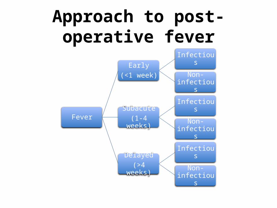
Approach to post-operative fever
Fever
Early(<1 week)
Infectious
Non-infectious
Subacute(1-4 weeks)
Infectious
Non-infectious
Delayed(>4 weeks)
Infectious
Non-infectious
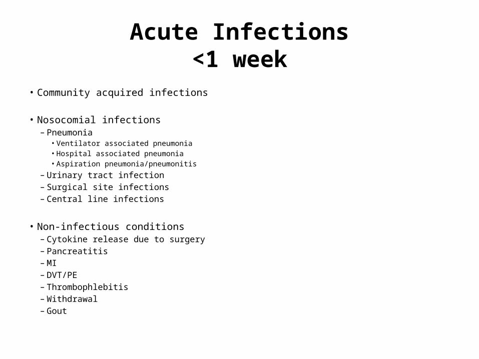
Acute Infections<1 week
• Community acquired infections
• Nosocomial infections– Pneumonia
• Ventilator associated pneumonia• Hospital associated pneumonia• Aspiration pneumonia/pneumonitis
– Urinary tract infection– Surgical site infections– Central line infections
• Non-infectious conditions– Cytokine release due to surgery– Pancreatitis– MI– DVT/PE– Thrombophlebitis– Withdrawal– Gout

Subacute Infections
• Nosocomial infections– Surgical site infections– C. difficile– Central line infections– VAP/HAP– UTIs
• Non-infectious causes– Drug reactions– DVT/PE
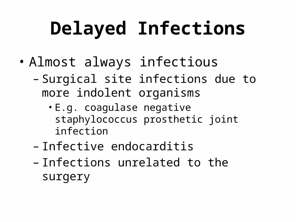
Delayed Infections
• Almost always infectious– Surgical site infections due to more indolent
organisms• E.g. coagulase negative staphylococcus prosthetic joint
infection
– Infective endocarditis– Infections unrelated to the surgery
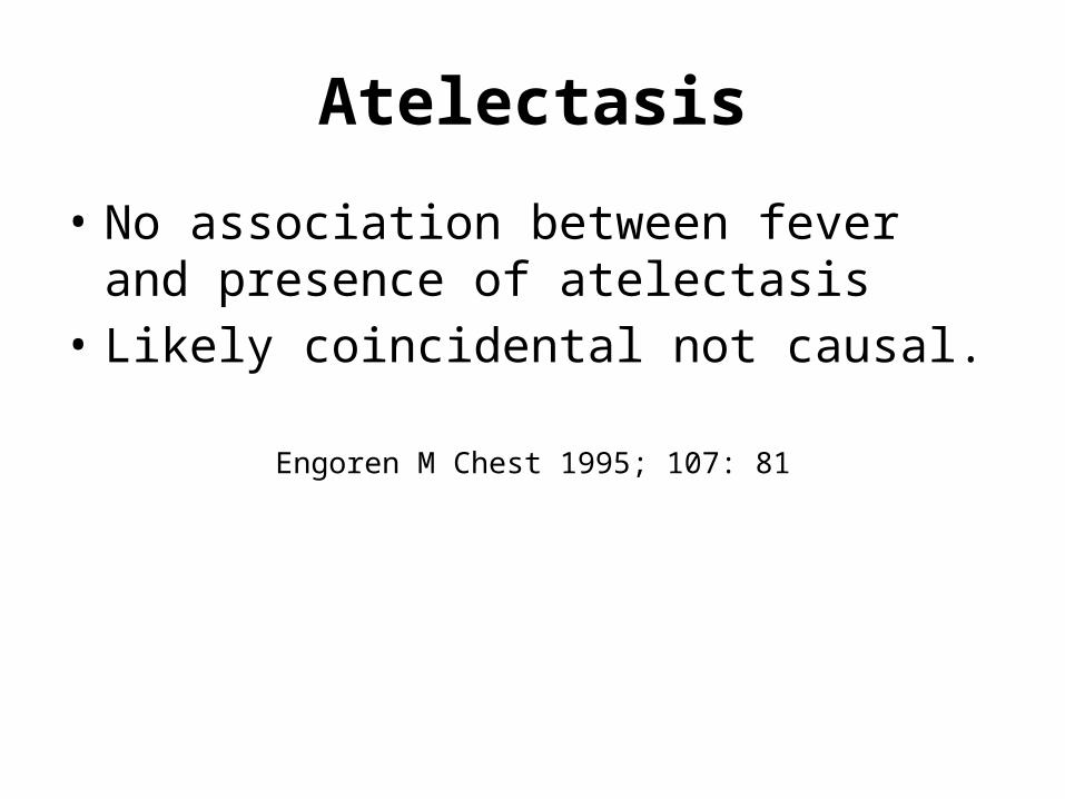
Atelectasis
• No association between fever and presence of atelectasis
• Likely coincidental not causal.
Engoren M Chest 1995; 107: 81
SURGICAL SITE INFECTIONS (SSI)
Case
• 68 year old M with known DM and CAD
• Requires CABG
• Does well post-operatively
• To the ward, but after several days complains of increasing pain in the sternum
• Notable cellulitis around lower incision site and drainage from lower aspect of wound
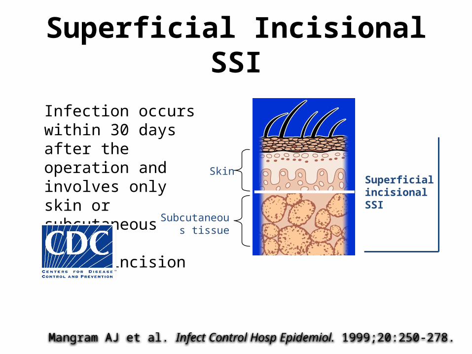
Superficial Incisional SSI
Infection occurs within 30 days after the operation and involves only skin or subcutaneous tissue of the incision
Mangram AJ et al. Infect Control Hosp Epidemiol. 1999;20:250-278.
Subcutaneous tissue
SkinSuperficial incisional SSI
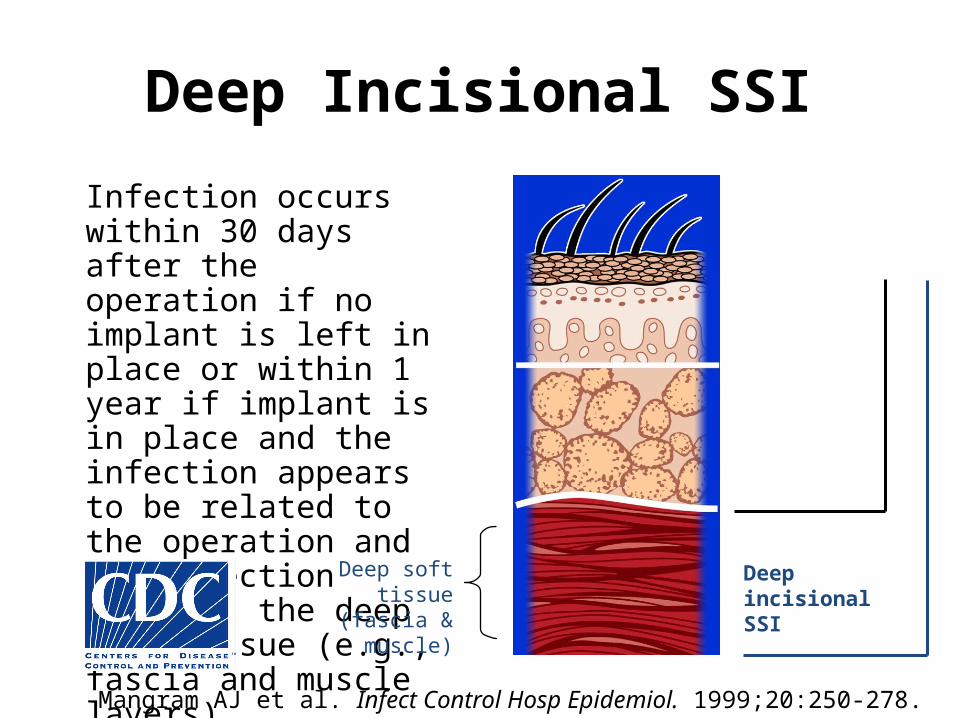
Deep Incisional SSI
Infection occurs within 30 days after the operation if no implant is left in place or within 1 year if implant is in place and the infection appears to be related to the operation and the infection involves the deep soft tissue (e.g., fascia and muscle layers)
Deep soft tissue (fascia & muscle)
Deep incisional SSI
Superficial incisional SSI
Mangram AJ et al. Infect Control Hosp Epidemiol. 1999;20:250-278.

Organ/Space SSI
Infection occurs within 30 days after the operation if no implant is left in place or within 1 year if implant is in place and the infection appears to be related to the operation and the infection involves any part of the anatomy, other than the incision, which was opened or manipulated during the operation
Deep incisional SSI
Superficial incisional SSI
Organ/space SSIOrgan/space
Mangram AJ et al. Infect Control Hosp Epidemiol. 1999;20:250-278.
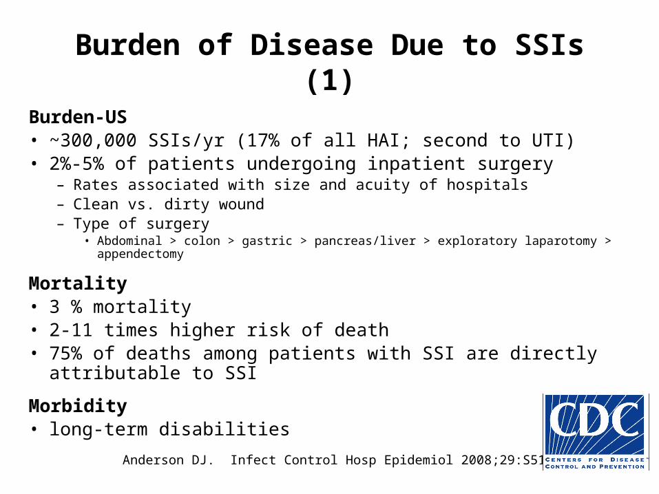
Burden of Disease Due to SSIs (1)
Burden-US• ~300,000 SSIs/yr (17% of all HAI; second to UTI) • 2%-5% of patients undergoing inpatient surgery
– Rates associated with size and acuity of hospitals– Clean vs. dirty wound– Type of surgery
• Abdominal > colon > gastric > pancreas/liver > exploratory laparotomy > appendectomy
Mortality• 3 % mortality • 2-11 times higher risk of death • 75% of deaths among patients with SSI are directly attributable to SSI
Morbidity• long-term disabilities
Anderson DJ. Infect Control Hosp Epidemiol 2008;29:S51-S61
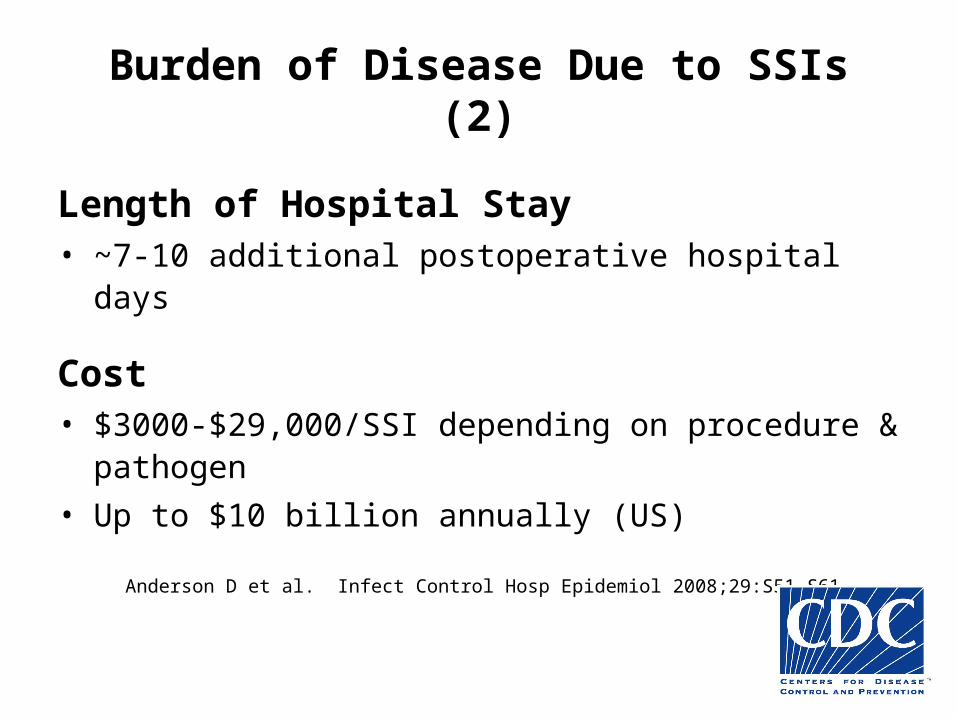
Burden of Disease Due to SSIs (2)
Length of Hospital Stay• ~7-10 additional postoperative hospital days
Cost• $3000-$29,000/SSI depending on procedure & pathogen• Up to $10 billion annually (US)
Anderson D et al. Infect Control Hosp Epidemiol 2008;29:S51-S61

Risk Factors: Operation Factors
• Duration of surgical scrub• Maintain body temp• Skin antisepsis• Preoperative shaving• Duration of operation• Antimicrobial prophylaxis• Operating room ventilation• Inadequate sterilization of
instruments
Mangram AJ et al. Infect Control Hosp Epidemiol. 1999;20:250-278.
• Foreign material at surgical site
• Surgical drains• Surgical technique
– Poor hemostasis– Failure to obliterate
dead space – Tissue trauma
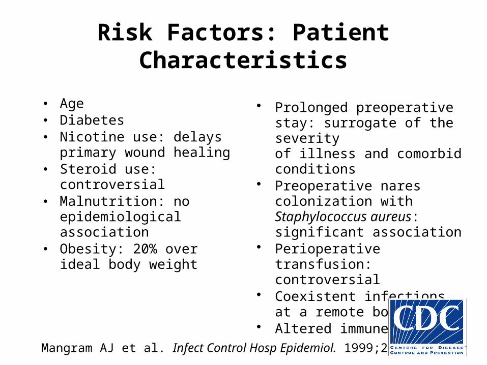
Risk Factors: Patient Characteristics
• Age• Diabetes• Nicotine use: delays primary
wound healing• Steroid use: controversial• Malnutrition: no
epidemiological association• Obesity: 20% over ideal body
weight
Mangram AJ et al. Infect Control Hosp Epidemiol. 1999;20:250-278.
• Prolonged preoperative stay: surrogate of the severity of illness and comorbid conditions
• Preoperative nares colonization with Staphylococcus aureus: significant association
• Perioperative transfusion: controversial
• Coexistent infections at a remote body site
• Altered immune response

PathogenesisEndogenous• Patient flora
– Skin – Mucous membranes– GI tract
• Seeding from a distant focus of infection
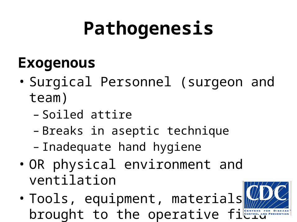
Pathogenesis
Exogenous• Surgical Personnel (surgeon and team)
– Soiled attire– Breaks in aseptic technique– Inadequate hand hygiene
• OR physical environment and ventilation • Tools, equipment, materials brought to the
operative field
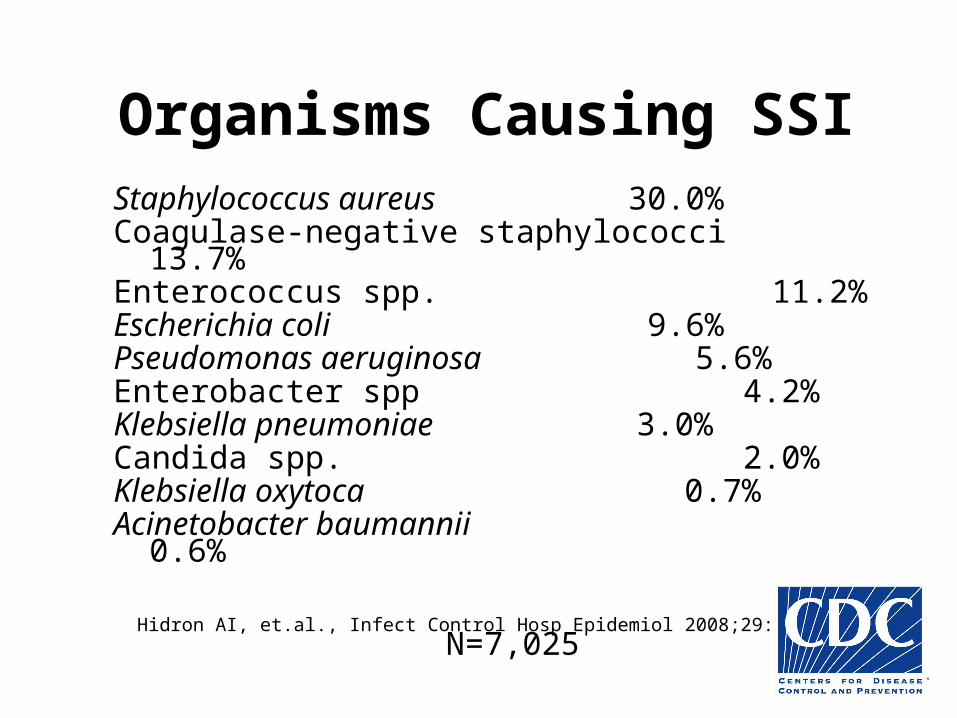
Organisms Causing SSIStaphylococcus aureus
30.0%Coagulase-negative staphylococci 13.7%Enterococcus spp.
11.2%Escherichia coli
9.6%Pseudomonas aeruginosa
5.6%Enterobacter spp
4.2%Klebsiella pneumoniae
3.0%Candida spp.
2.0%Klebsiella oxytoca
0.7%Acinetobacter baumannii
0.6%
N=7,025
Hidron AI, et.al., Infect Control Hosp Epidemiol 2008;29:996-1011

Clinical Manifestations• Localized erythema
• Induration
• Warmth
• Pain at incision site
• Purulent wound drainage and separation of wound can occur
• May be associated with fever and leukocytosis

Treatment• Surgical:
– Suture removal– Explored– Drained– Irrigated– Dressed open
• Antibiotics:– Not routinely needed– Use when there is:
• Presence of cellulitis >5 cm from wound edge• Systemically unwell (temperature >38.5°C, HR >110bpm, or WBC > 12)• Progressing despite drainage
IDSA Guidelines 2014
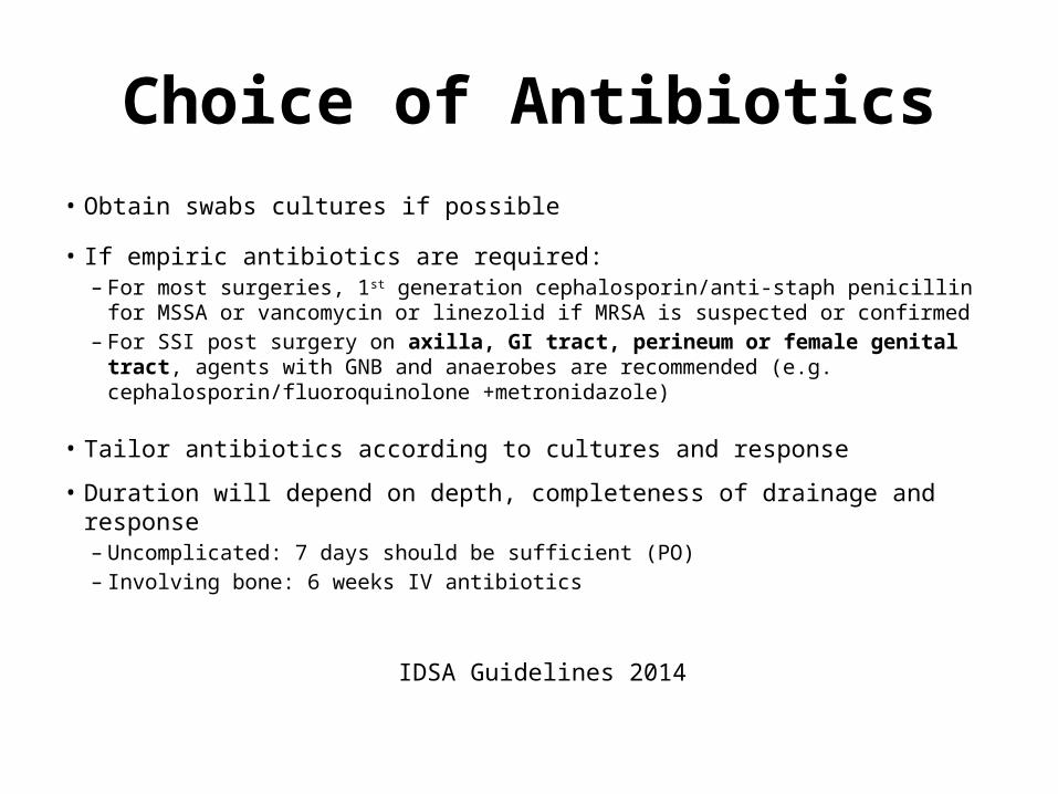
Choice of Antibiotics• Obtain swabs cultures if possible
• If empiric antibiotics are required:– For most surgeries, 1st generation cephalosporin/anti-staph penicillin for MSSA or
vancomycin or linezolid if MRSA is suspected or confirmed – For SSI post surgery on axilla, GI tract, perineum or female genital tract, agents with
GNB and anaerobes are recommended (e.g. cephalosporin/fluoroquinolone +metronidazole)
• Tailor antibiotics according to cultures and response
• Duration will depend on depth, completeness of drainage and response– Uncomplicated: 7 days should be sufficient (PO)– Involving bone: 6 weeks IV antibiotics
IDSA Guidelines 2014
CLOSTRIDIUM DIFFICILE

Case
• 80 year old F admitted from home with hip # after a fall
• ORIF complicated by post-operative delerium due to pain medications
• Slow recovery, transferred to in-patient rehabilitation
• Two weeks post-operatively developed 5 non-bloody BMs in one day, associated with decreased appetite, fatigue
• Labs showed WBC of 14, slight elevation of Cr, otherwise normal
• Stool sent for C. difficile: PCR positive
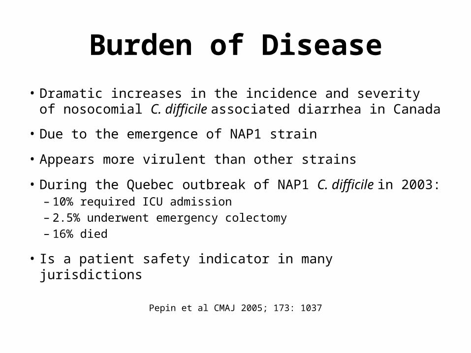
Burden of Disease• Dramatic increases in the incidence and severity of nosocomial C.
difficile associated diarrhea in Canada
• Due to the emergence of NAP1 strain
• Appears more virulent than other strains
• During the Quebec outbreak of NAP1 C. difficile in 2003:– 10% required ICU admission– 2.5% underwent emergency colectomy– 16% died
• Is a patient safety indicator in many jurisdictions
Pepin et al CMAJ 2005; 173: 1037
Pathogenesis
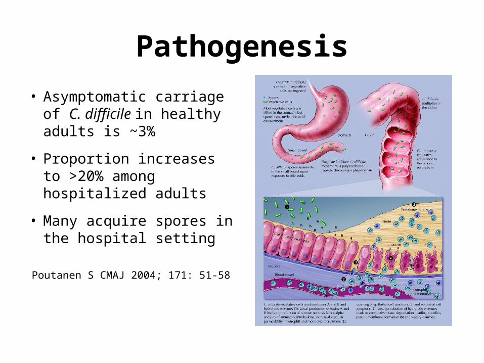
• Asymptomatic carriage of C. difficile in healthy adults is ~3%
• Proportion increases to >20% among hospitalized adults
• Many acquire spores in the hospital setting
Poutanen S CMAJ 2004; 171: 51-58

Risk Factors• Antibiotic use (esp. clindamycin
use, fluoroquinolones and cephalosporins)
• Hospital exposure• Advanced aged• Co-morbid illness• Gastric acid suppression• Enteral feeding• Gastrointestinal surgery• Obesity• Cancer chemotherapy
Poutanen S CMAJ 2004; 171: 51-58
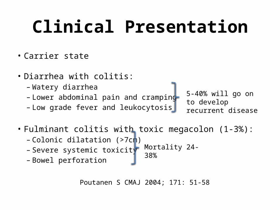
Clinical Presentation• Carrier state
• Diarrhea with colitis:– Watery diarrhea– Lower abdominal pain and cramping– Low grade fever and leukocytosis
• Fulminant colitis with toxic megacolon (1-3%):– Colonic dilatation (>7cm)– Severe systemic toxicity– Bowel perforation
Poutanen S CMAJ 2004; 171: 51-58
Mortality 24-38%
5-40% will go on to develop recurrent disease

Differential Diagnosis
• Laxative use
• Enteral feeds
• Underlying disease (i.e. IBD)
• Medications
• Other causes of diarrhea
Diagnosis
• PCR assays: real-time PCR that detect toxin A and B genes
• EIA for C. difficile toxins
• Colonoscopy
• High sensitivity, high specificity
• Lower sensitivity, high specificity
Treatment
• Cessation of inciting antibiotics where possible
• Supportive care– Avoid anti-motility agents– Discontinue laxatives
• Non-severe disease– Metronidazole 500 mg PO QID x 14 days– Vancomycin 125mg PO QID x 14 days
IDSA Guidelines 2010Zar et al Clin Infect Dis 2007; 45: 302-307
Treatment
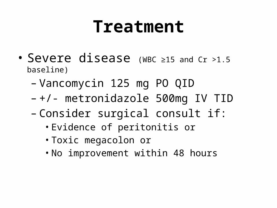
• Severe disease (WBC ≥15 and Cr >1.5 baseline)
– Vancomycin 125 mg PO QID – +/- metronidazole 500mg IV TID– Consider surgical consult if:
• Evidence of peritonitis or• Toxic megacolon or• No improvement within 48 hours
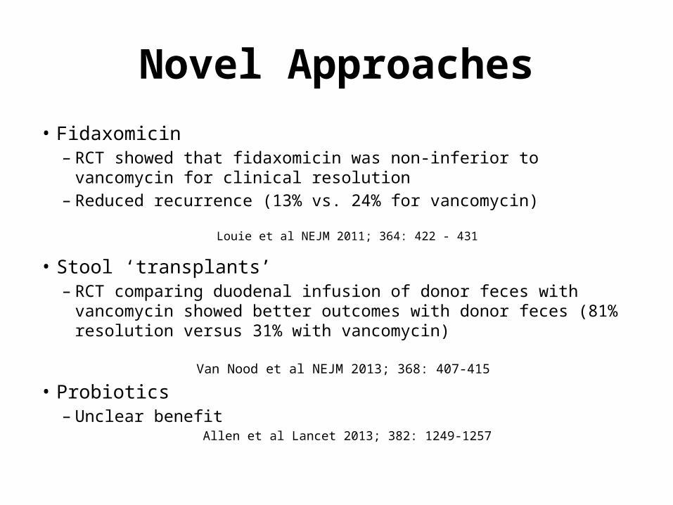
Novel Approaches• Fidaxomicin
– RCT showed that fidaxomicin was non-inferior to vancomycin for clinical resolution
– Reduced recurrence (13% vs. 24% for vancomycin)
Louie et al NEJM 2011; 364: 422 - 431
• Stool ‘transplants’– RCT comparing duodenal infusion of donor feces with vancomycin showed
better outcomes with donor feces (81% resolution versus 31% with vancomycin)
Van Nood et al NEJM 2013; 368: 407-415
• Probiotics– Unclear benefit
Allen et al Lancet 2013; 382: 1249-1257
VENTILATOR ASSOCIATED PNEUMONIA (VAP)
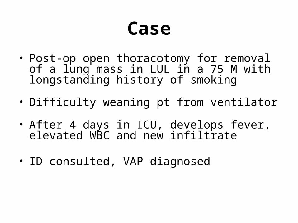
Case• Post-op open thoracotomy for removal of a lung mass
in LUL in a 75 M with longstanding history of smoking
• Difficulty weaning pt from ventilator
• After 4 days in ICU, develops fever, elevated WBC and new infiltrate
• ID consulted, VAP diagnosed

Burden of Disease due to VAP• Incidence of VAP (>48 hours of intubation):
– 51 RCTs – 23%– 38 cohort studies
• Med-surg – 9%• Medical ICUs – 17%
• Mortality of VAP: OR 2
• LOS due to VAP: 6 additional days
• Cost due to VAP: ~$10,000 per episode
Safdar N et al Crit Care Med 2005; 33:2184 - 2193
Pathogenesis
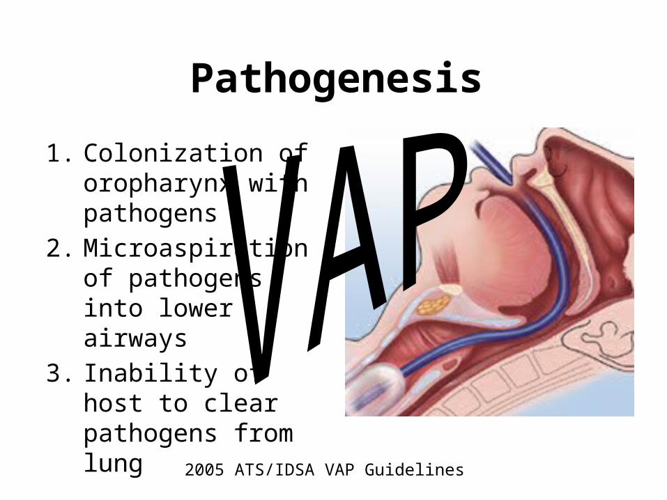
1. Colonization of oropharynx with pathogens
2. Microaspiration of pathogens into lower airways
3. Inability of host to clear pathogens from lung
2005 ATS/IDSA VAP Guidelines
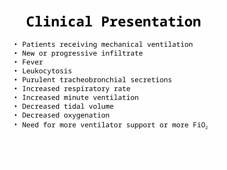
Clinical Presentation• Patients receiving mechanical ventilation• New or progressive infiltrate• Fever• Leukocytosis• Purulent tracheobronchial secretions• Increased respiratory rate• Increased minute ventilation• Decreased tidal volume• Decreased oxygenation• Need for more ventilator support or more FiO2
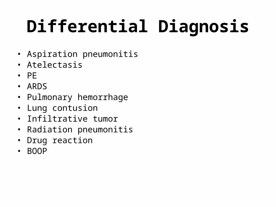
Differential Diagnosis• Aspiration pneumonitis• Atelectasis• PE• ARDS• Pulmonary hemorrhage• Lung contusion• Infiltrative tumor• Radiation pneumonitis• Drug reaction• BOOP

Diagnosis• Diagnosis is challenging
• Generally suspected when there is:– New infiltrate on CXR plus – Two or more symptoms or signs of respiratory infection– No alternate explanation
• Role of invasive vs non-invasive techniques– If empiric therapy for VAP to be initiated, ETT specimen with
non-quantitative culture acceptable as initial dx test
Muscedere J Critical Care 2008; 23: 138-147
Comprehensive Evidence Based CPGs
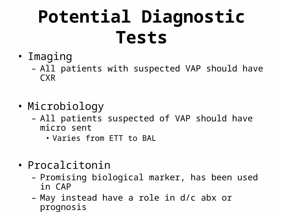
Potential Diagnostic Tests
• Imaging– All patients with suspected VAP should have CXR
• Microbiology– All patients suspected of VAP should have micro sent
• Varies from ETT to BAL
• Procalcitonin– Promising biological marker, has been used in CAP– May instead have a role in d/c abx or prognosis

Therapy
• Empiric therapy is recommended when there is a clinical suspicion of VAP
• No role for combination therapy (for synergy purposes)– Appropriate single agent therapy for each potential pathogen
Muscedere J Critical Care 2008; 23: 138-147Comprehensive Evidence Based CPGs
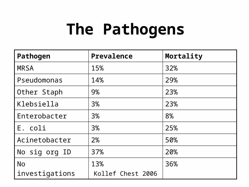
The PathogensPathogen Prevalence Mortality
MRSA 15% 32%
Pseudomonas 14% 29%
Other Staph 9% 23%
Klebsiella 3% 23%
Enterobacter 3% 8%
E. coli 3% 25%
Acinetobacter 2% 50%
No sig org ID 37% 20%
No investigations 13% 36%
Kollef Chest 2006
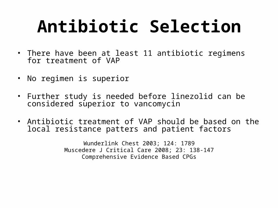
Antibiotic Selection• There have been at least 11 antibiotic regimens for treatment of VAP
• No regimen is superior
• Further study is needed before linezolid can be considered superior to vancomycin
• Antibiotic treatment of VAP should be based on the local resistance patters and patient factors
Wunderlink Chest 2003; 124: 1789Muscedere J Critical Care 2008; 23: 138-147
Comprehensive Evidence Based CPGs

Potential OptionsPotential Pathogens Antibiotic Therapy
Antipseudomonal cephalosporin
Pseudomonas aeruginosa or
Klebsiella pneumoniae (ESBL) Antipseudomonal carbepenem
Acinetobacter species* or
MSSA ß-Lactam/ß-lactamase inhibitor
plus
Legionella pneumophila Antipseudomonal fluoroquinolone
or
Macrolide
plus
Methicillin-resistant Staphylococcus aureus (MRSA) Linezolid or vancomycin
From ATS/IDSA 2005 Guidelines

Duration• Prospective RCT of 401 patients with VAP
– 8 days vs. 15 days
• All treated with anti-pseudomonal beta-lactam plus either an aminoglycoside or fluorquinolone
• No difference in mortality or recurrent infection at 28 days
• In the presence of pseudomonas/acinetobacter, use clinical judgment
Chastre JAMA 2003; 290: 2588-2598

De-escalation of therapy
• If patient with suspected VAP started on abx and no improvement and cultures negative, look for alternative diagnosis– Consider discontinuation of therapy
• If pathogen isolated, can narrow abx
ATS/IDSA 2005 VAP Guidelines

HAND HYGIENE


Healthcare Acquired Infections
• Healthcare acquired infections (HAI’s)– Antibiotic resistant organism infections– Surgical site infections– C. difficile infections
• 5-10% of all patients will acquire an HAI, translates to 8000 deaths each year in hospitals in Canada
• Hand hygiene helps reduce infections– 8 observational studies showing reduction in AROs or HAIs– 10 RCTs in community setting showing reduction in respiratory or
diarrheal illness
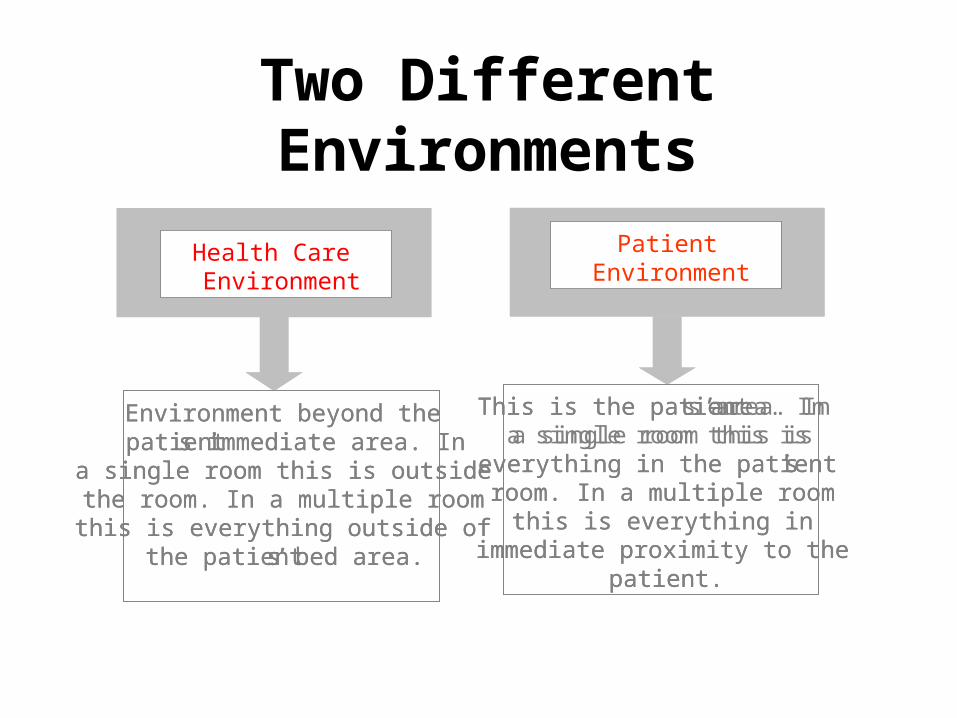
Two Different Environments
Health care Environment
Patient Environment
Environment beyond the patient’s immediate area. In a single room this is outside the room. In a multiple room this is everything outside of
the patient’s bed area.
This is the patient’s area. In a single room this is
everything in the patient’s room. In a multiple room
this is everything in immediate proximity to the
patient.
Health Care Environment
Patient Environment
Environment beyond the patient’s immediate area. In a single room this is outside the room. In a multiple room this is everything outside of
the patient’s bed area.
This is the patient’s area. In a single room this is
everything in the patient’s room. In a multiple room
this is everything in immediate proximity to the
patient.
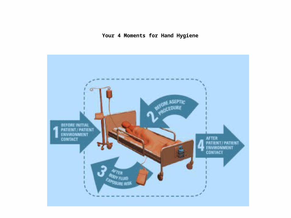
Your 4 Moments for Hand Hygiene
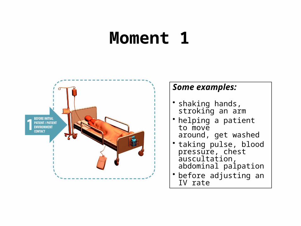
Moment 1
Some examples:
• shaking hands, stroking an arm• helping a patient to move
around, get washed• taking pulse, blood pressure,
chest auscultation, abdominal palpation
• before adjusting an IV rate
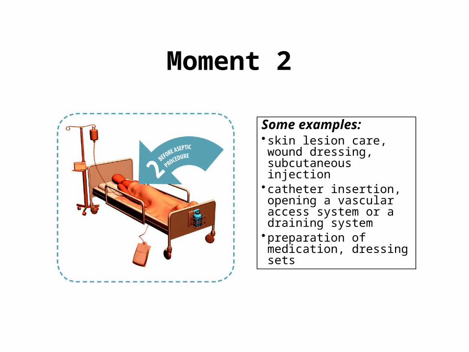
Moment 2
Some examples:• skin lesion care, wound dressing,
subcutaneous injection• catheter insertion, opening a
vascular access system or a draining system
• preparation of medication, dressing sets
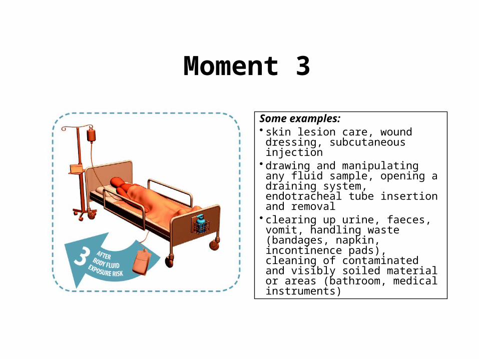
Moment 3
Some examples:• skin lesion care, wound dressing,
subcutaneous injection• drawing and manipulating any fluid sample,
opening a draining system, endotracheal tube insertion and removal
• clearing up urine, faeces, vomit, handling waste (bandages, napkin, incontinence pads), cleaning of contaminated and visibly soiled material or areas (bathroom, medical instruments)
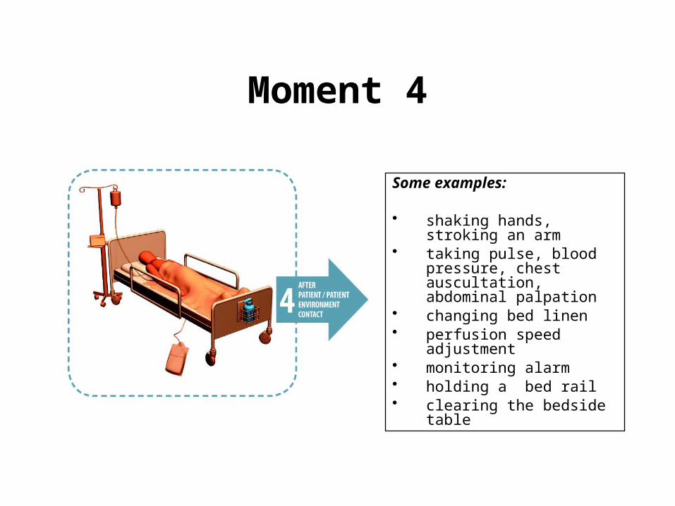
Moment 4
Some examples:
• shaking hands, stroking an arm• taking pulse, blood pressure, chest
auscultation, abdominal palpation• changing bed linen• perfusion speed adjustment• monitoring alarm • holding a bed rail • clearing the bedside table
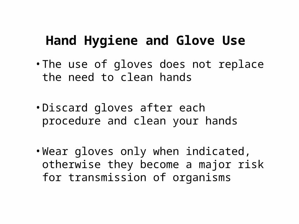
Hand Hygiene and Glove Use• The use of gloves does not replace the need
to clean hands
• Discard gloves after each procedure and clean your hands
• Wear gloves only when indicated, otherwise they become a major risk for transmission of organisms

Hand Hygiene Compliance at SJHC
• One of the key Provincial patient safety indicators
• Publically reported on the Health Quality Ontario website
• Went from >90% to ~50% in 2013/2014.... what happened?
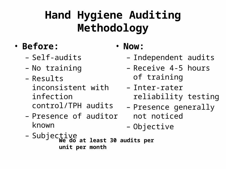
Hand Hygiene Auditing Methodology
• Before:– Self-audits– No training– Results inconsistent with
infection control/TPH audits– Presence of auditor known– Subjective
• Now:– Independent audits– Receive 4-5 hours of
training– Inter-rater reliability testing– Presence generally not
noticed– Objective
We do at least 30 audits per unit per month

Hand Hygiene Adherence at SJHC
Moment Compliance2014-2015
Q1
Compliance 2014-2015
Q2
Compliance 2014-2015
Q3Moment #1 (Before patient/patient environment)
225/594 (38%) 247/559 (44%) 159/267 (60%)
Moment #4 (After patient/patient environment)
657/1101 (60%) 645/1018 (63%) 434/610 (71%)
Moment #1 and #4 882/1695 (52%) 892/1577 (57%) 593/877 (68%)
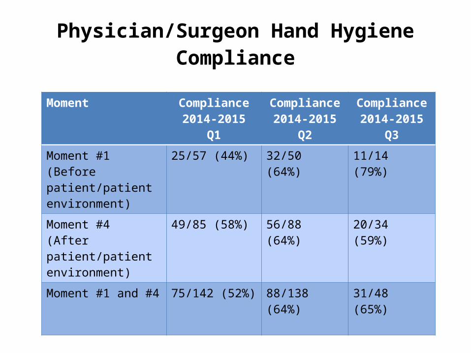
Physician/Surgeon Hand Hygiene Compliance
Moment Compliance2014-2015
Q1
Compliance 2014-2015
Q2
Compliance 2014-2015
Q3Moment #1 (Before patient/patient environment)
25/57 (44%) 32/50 (64%) 11/14 (79%)
Moment #4 (After patient/patient environment)
49/85 (58%) 56/88 (64%) 20/34 (59%)
Moment #1 and #4 75/142 (52%) 88/138 (64%) 31/48 (65%)
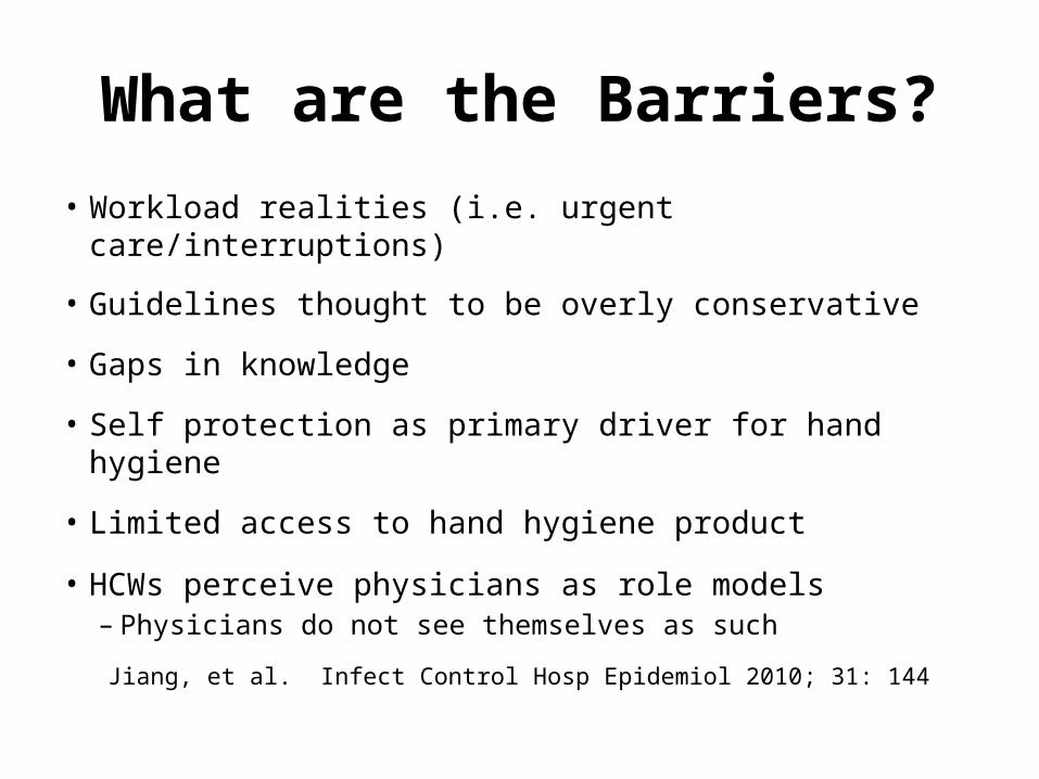
What are the Barriers?
• Workload realities (i.e. urgent care/interruptions)
• Guidelines thought to be overly conservative
• Gaps in knowledge
• Self protection as primary driver for hand hygiene
• Limited access to hand hygiene product
• HCWs perceive physicians as role models– Physicians do not see themselves as such
Jiang, et al. Infect Control Hosp Epidemiol 2010; 31: 144
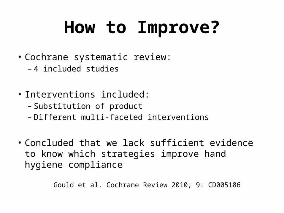
How to Improve?
• Cochrane systematic review:– 4 included studies
• Interventions included:– Substitution of product– Different multi-faceted interventions
• Concluded that we lack sufficient evidence to know which strategies improve hand hygiene compliance
Gould et al. Cochrane Review 2010; 9: CD005186
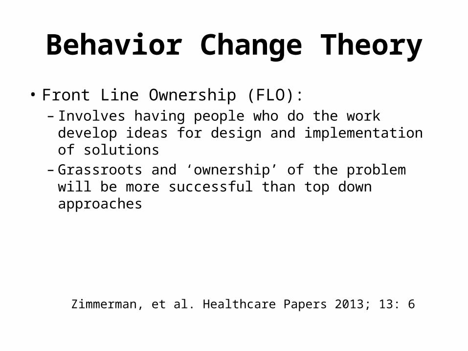
Behavior Change Theory
• Front Line Ownership (FLO):– Involves having people who do the work develop
ideas for design and implementation of solutions– Grassroots and ‘ownership’ of the problem will be
more successful than top down approaches
Zimmerman, et al. Healthcare Papers 2013; 13: 6
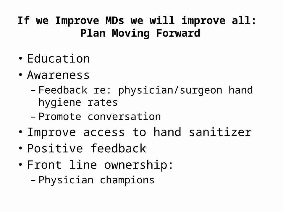
If we Improve MDs we will improve all: Plan Moving Forward
• Education• Awareness
– Feedback re: physician/surgeon hand hygiene rates– Promote conversation
• Improve access to hand sanitizer• Positive feedback• Front line ownership:
– Physician champions

Conclusions
• When approaching post-op fever, consider timing of fever and infectious and non-infectious causes
• Nosocomial infections are common and have associated morbidity and mortality
• Most nosocomial infections are preventable– Hand hygiene is one of the most effective ways to
reduce nosocomial infections