Embed Size (px)
Citation preview

Surgical Freedom in Endoscopic Skull Base Surgery:
Quantitative Analysis for Endoscopic Approaches
by
Ali Elhadi
A Dissertation Presented in Partial Fulfillment
of the Requirements for the Degree
Doctor of Philosophy
Approved April 2014 by the
Graduate Supervisory Committee:
Mark Preul, Co-Chair
Bruce Towe, Co-Chair
Andrew Little
Peter Nakaji
Eric Vu
ARIZONA STATE UNIVERSITY
May 2014

i
ABSTRACT
During the past five decades neurosurgery has made great progress, with marked
improvements in patient outcomes. These noticeable improvements of morbidity and
mortality can be attributed to the advances in innovative technologies used in
neurosurgery. Cutting-edge technologies are essential in most neurosurgical procedures,
and there is no doubt that neurosurgery has become heavily technology dependent. With
the introduction of any new modalities, surgeons must adapt, train, and become
thoroughly familiar with the capabilities and the extent of application of these new
innovations. Within the past decade, endoscopy has become more widely used in
neurosurgery, and this newly adopted technology is being recognized as the new
minimally invasive future of neurosurgery. The use of endoscopy has allowed
neurosurgeons to overcome common challenges, such as limited illumination and
visualization in a very narrow surgical corridor; however, it introduces other challenges,
such as instrument "sword fighting" and limited maneuverability (surgical freedom). The
newly introduced concept of surgical freedom is very essential in surgical planning and
approach selection and can play a role in determining outcome of the procedure, since
limited surgical freedom can cause fatigue or limit the extent of lesion resection. In my
thesis, we develop a consistent objective methodology to quantify and evaluate surgical
freedom, which has been previously evaluated subjectively, and apply this model to the
analysis of various endoscopic techniques. This model is crucial for evaluating different
endoscopic surgical approaches before they are applied in a clinical setting, for
identifying surgical maneuvers that can improve surgical freedom, and for developing

ii
endoscopic training simulators that accurately model the surgical freedom of various
approaches. Quantifying the extent of endoscopic surgical freedom will also provide
developers with valuable data that will help them design improved endoscopes and
endoscopic instrumentation.

iii
DEDICATION
For my Mother and Father, my greatest role models, thank you for your
continuous encouragement, prayers and love.
For my lovely sisters and Yara, thank you for your great love, support and
encouragements.
For my professors and mentors, who were always by my side and provided
guidance and advice.
For my family and friends, who always kept me in a high spirit throughout this
process.
For all neurosurgeons and members of the scientific community who will find this
work relevant and apply it for better patient outcomes.

iv
ACKNOWLEDGMENTS
I would like to express my greatest gratitude to all those who made this thesis possible.
Thank you Dr. Mark C. Preul you have been a great and very thoughtful advisor, Dr.
Andrew S. Little you taught me a lot and was patient with me, Dr. Peter Nakaji for
guiding me and providing me with continuous support, Dr. Eric T. Vu for your creative
suggestions, and for the great support and advice from Dr. Bruce Towe. I would also like
to thank the research fellows in the lab and neurosurgery residents in the neurosurgical
division at the Barrow Neurological Institute for helping me in executing my projects,
thank you for helping me formulate my ideas. To all the co-authors who helped me in
drafting and revising the articles, those were an important portion of my thesis. To all the
professors in the neuroscience program who helped me throughout my study. And to all
my colleagues who were very supportive. To the lab staff, thank you Bill, Ashley, Joshua
and Tad, thank you for your help in providing and preparing all the instruments, space
and specimens with a lovely smile. Special thanks to each of the teams from Storz, (
Tuttlingen, Germany) Medtronic (Minneapolis, MN) and Visionsense (Petach Tikva,
Israel) who were very supportive and supplied me with the needed equipment,
instruments, specimens and tuition coverage. Thank you to all my family, friends and
teachers for everything they have done for me.

v
TABLE OF CONTENTS
Page
LIST OF TABLES ................................................................................................................... vi
LIST OF FIGURES ............................................................................................................... vii
CHAPTER
1. INTRODUCTION IX
2. BACKGROUND XII
3. SURGICAL FREEDOM XIX
4. CHAPTER 1 COMPARISON OF SURGICAL FREEDOM AND AREA OF
EXPOSURE IN THREE ENDOSCOPIC TRANSMAXILLARY APPROACHES TO
THE ANTEROLATERAL CRANIAL BASE 1
5. CHAPTER 2 EVALUATION OF SURGICAL FREEDOM FOR MICROSCOPIC
AND ENDOSCOPIC TRANSSPHENOIDAL APPROACHES TO THE SELLA 24
6. CHAPTER 3 MALLEABLE ENDOSCOPE INCREASES SURGICAL
FREEDOM WHEN COMPARED TO A RIGID ENDOSCOPE IN ENDOSCOPIC
ENDONASAL APPROACHES TO THE PARASELLAR REGION 50
7. CONCLUSIONS 69
8. LIMITATIONS 76
9. BROADER IMPACT AND FUTURE WORK 77
REFERENCES 79

vi
LIST OF TABLES
Table Page
1. Table 1.1: Area of exposure for three transmaxillary approaches ............................15
2. Table 1.2: Surgical freedom for three transmaxillary approaches ............................16
3. Table 2.1: P-values for the four trans-sphenoidal approaches compared to each
other ..........................................................................................................................37
4. Table 3.1: Exposed area surgical freedom for the rigid and malleable endoscopes .61
5. Table 3.2: Anatomical target surgical freedom for the rigid and malleable
endoscopes ................................................................................................................61
6. Table 3.3: Angle of attack for the rigid and malleable endoscopes in sagittal and
axial planes................................................................................................................62

vii
LIST OF FIGURES
Figure Page
1. Figure 1.1: Illustration showing anatomy of the anteriolateral cranial base and
endoscopic trajectories to access this area 1
2. Figure 1.2: Endoscopic images showing the different views of the surgical corridors
for the three types of endoscopic transmaxillary approaches 10
3. Figure 1.3: Endoscopic image from an ipsilateral endonasal approach showing the
area of exposure 12
4. Figure 1.4: Illustration showing the method used to calculate the surgical freedom
for three transmaxillary approaches 13
5. Figure 1.5: Endoscopic images showing the difference in views by each
transmaxillary approach 15
6. Figure 1.6: A diagram showing the importance of the pivot point in the different
transmaxillary approaches 20
7. Figure 2.1: An illustration showing the exposed area surgical freedom for two
endoscopic transsphenoidal approaches 31
8. Figure 2.2: An illustration showing the exposed area surgical freedom for two
microscopic approaches 32
9. Figure 2.3: An illustration showing the point anatomical target surgical freedom and
the angle of attack for four transsphenoidal approaches in an axial plane 34
10. Figure 2.4: An illustration showing the point anatomical target surgical freedom and
the angle of attack for four transsphenoidal approaches in a sagittal plane 35

viii
Figure Page
11. Figure 2.5: Boxplot showing total exposed area surgical freedom for four
transsphenoidal approaches 36
12. Figure 2.6: Boxplot showing anatomical target surgical freedom for four
transsphenoidal approaches 41
13. Figure 2.7: Boxplot showing axial angle of attack for four transsphenoidal
approaches 42
14. Figure 2.8: Boxplot showing the sagittal angle of attack for four transsphenoidal
approaches 43
15. Figure 3.1: Image showing the malleable and the rigid 3D endoscopes 54
16. Figure 3.2: Representave image taken from both the malleable and rigid 3D
endoscopes 55
17. Figure 3.3: Series of photographic images demonstrating the position of the
endoscopic instruments while acquisition of the spatial coordinates 59
ix
INTRODUCTION
Neurosurgical practices may have been performed as early as two millennia B.C.
(Elhadi, 2012). As a discipline neurosurgery is based on a long, slow, and deliberate
history of important developments (Preul MC et al, 1997). It was not until about 150
years ago, however, that neurosurgery began to be considered as an independent field of
medicine. During this period, technology and science allowed and promoted greater
“neurosurgical” intervention although outcomes were disastrous with significant
mortalities. Some authors consider neurosurgery to be one of the youngest fields in
medicine (Barker, 1993), and thus open to significant discoveries and scientific progress.
Significant advancements in neurosurgery have been prominent in the 20th
century and
especially within the last 50 years which have been characterized by the introduction and
development of diagnostic modalities, operative techniques, or surgical tools and
instruments. Neurosurgery is truly a technologically dependent specialty.
Microscopic procedures have been a hallmark of most modern neurosurgery. The
continuous improvements of the surgical microscope and microscopic instruments, as
well as development of microsurgical techniques and proper training, have all played an
important role in shaping neurosurgery as we know it today. Microscopic procedures
made possible operating on lesions or pathologies that were previously deemed
challenging (Yasargil, 1999) or inoperable. Such procedures are routinely performed on
a daily basis resulting in less morbidity and mortality as a result of the increased scope
and application of technology of the procedures performed.
The sensitive and fragile natures of the tissue of the nervous system and the complex
network of the associated arteries and veins, mandates precise approaches that can

x
address anatomical targets or lesions within the central nervous system with the least
amount of retraction, manipulations and neural or vascular injuries (Rhoton, 2003). The
trend in neurosurgery, as with any surgical discipline, is to “minimize” invasion and the
extent of the approach. Minimizing the operative extent such as in large craniotomies
usually means fewer complications for the patient. There is thus a strong tendency
towards development of minimally invasive procedures.
The term minimally invasive is relative. Microscopic neurosurgery was considered
minimally invasive when first applied in the early 1960s and 1970s (Yasargil 1970). Dr.
Theodore Kurze from the university of Southern California was considered to be the first
neurosurgeon to use the microscope in the OR in 1957 (Dounaghy et al, 1979). Today use
of the microscope is considered nearly a required technology for the neurosurgical
procedure, but is no longer considered necessarily associated with minimal invasion.
The surgical endoscope was introduced to neurosurgery in early 1923 with little
success because of technological limitations. Use of the endoscope was significantly
expanded in the 1990s when it was first used for diagnostic purposes, and used as an
adjunct to the microscope to improve visualization of structures. During the late 1990s
and early 2000s neurosurgeons found increased use for the endoscope in removing
pituitary adenomas (Prevedello, 2007). Endoscopic use increased to include lesions in the
middle and posterior cranial fossae (Little, 2013) and more lateral skull base lesions
(Little, 2012). This innovation is due to technological endoscopic improvements, new
instrumentations and the development of different endoscopic approaches for different
anatomical areas.

xi
Endoscopic neurosurgeons tend to determine their preference towards a certain
approach over another for accessing the same anatomical area based on the type of the
lesion, extent, surgeon’s training and confidence performing this approach, previous
experiences and pre-operative planning. Sometimes a single approach can access several
anatomical targets in different surgical planes (Cavallo, 2005), and sometimes several
approaches can be used to access a single target or a determined anatomical area (Van
Rompaey, 2013). Several endoscopic approaches and techniques have been described
that illustrate maneuvers and anatomical landmarks within different endoscopic
approaches. An important factor in determining the right approach for a lesion or an
anatomical area is the degree of ease or ability of the surgeon to maneuver different
surgical instruments within an endoscopic approach, which is critical in decreasing the
surgeons fatigue, frustration and stress. This concept is one of “surgical freedom.” It will
also help determine if the surgeon will be capable of removing a lesion completely or no,
due to technical difficulties.
In this thesis I expand and assess this new concept of surgical freedom to endoscopic
neurosurgery and developed a method to quantify it. Surgical freedom is an important
factor and aspect for each endoscopic approach and contributes to surgical planning,
decision making, and approach selection.

xii
BACKGROUND
The nervous system is still mysterious in many ways, and many neuroscience studies
still have a lot to discover about this system which mandates continuous and thorough
investigations. Neurosurgery is no different from other neuroscience fields and there is
yet a lot to be studied in neurosurgery.
As previously mentioned that advancements in neurosurgery are usually marked by
improvements in certain aspects like; diagnostics, visualization and illumination, surgical
instruments, new surgical techniques, new treatment modalities, and improved
neurosurgical training (Powell, 1999). I would like to briefly discuss few examples.
The role of innovative technologies in neurosurgery
The use of imaging techniques was essential in advancing neurosurgery, the first
systematic use of an X-ray was in 1908 by Fedor Krause (Elhadi, 2012), then in 1947
imaging technology advanced to involve the use of radioisotopes in localizing abnormal
brain tissue, and in 1950 Angiography became an accepted diagnostic modality to
visualize vascular lesions such as aneurysms. During the early 1970, computed
tomography (CT) scan was used to localize pathologies in the brain, five years later
positron emission tomography (PET) scan was developed which shows different signals
for brain cells based on their activity (Xiong, 1997), later in 1980s magnetic resonance
imaging (MRI) was regularly used to diagnose pathologies in the CNS. These important
evolutions in imaging; technologies, techniques and interpretations played an important
role in neurosurgery and its evolution.

xiii
Another example is the use of Electro-encephalogram (EEG) which became an
acceptable diagnostic tool in epileptic lesions by 1935, and the development of Somato-
sensory evoked potential (SSEP) in 1980s which is a way to monitor the integrity of
nerves or neural tracts, this monitoring modality have become an important intra-
operative tool that may help in preventing nerve injuries, especially during spine
procedures.
Basic principles in neurosurgery can change slightly, but a radical change in
neurosurgery has been obvious with the introduction of “power tools” that aid in
magnification and illumination which make a possible “shrinking” of the surgical
working space with sufficient access to the area of interest and minimizing any assault on
brain tissue or other delicate structures along the surgical approach or within the working
area (Setti, 1994).
Microscopy and neurosurgery
The surgical microscope was ideal in providing such advantages, the magnification
power dramatically increased when compared to previously used magnifying methods. It
also provided excellent illumination when compared to older conventional methods. And
the illumination is along the line of entry and enables a direct view of the surgeons’
working space (Rand, 1968). These two fundamental aspects that the microscope offered
revolutionized and expanded the scope of neurosurgery. It is also notable that continuous
innovations in the surgical microscope like; better and lighter microscopes (counter
balance microscope), integrated mouth piece to enable hands free maneuver capabilities
during most of the operative time, integrated neuro-navigation system, higher

xiv
magnification and better focus, less heat emitting illumination… etc. all have contributed
to better and more efficient procedures (Yasargil, 2006).
A new era of micro-neurosurgery started with new micro-neurosurgical techniques
described and used regularly, neuroanatomy has been re-described using the microscope
and different microscopic approaches; micro-neuroanatomy, newer micro-surgical
instruments have been modified (Yasargil, 2006), it has also become essential to train
surgeons and residents on the microscope through micro-neurosurgery laboratories and
courses. Even different classifications for tumors and vascular lesions were described
based on the microscope usage.
Endoscopy introduced to the field
Many were mistaken that the degree of visualization offered by the microscope might
be the best to be offered to neurosurgery, this idea was then well thought out with the
introduction of the endoscope which provides superior visualization, better illumination
and even more angular views (Perneczky, 1999).
The endoscope was first used by otolaryngologists in the nasal cavity then
neurosurgeons started using the endoscope in the early 1990s as an adjunct to
microscopic approaches to provide better view to difficult areas (Perneczky 1998). The
endoscope was then used in the removal of pituitary tumors using a trans-shenoidal
approach and kept on being limited to sellar lesions during the late 1990s and early
2000s.
The idea of the endoscope goes back to the early19th
century when it was first used
for hollow organs inspection like the rectum, bladder, nasal cavity, cervix and pharynx.
Phillip Bozzini (1773-1809) a German Physician devoted his life to develop this new

xv
instrument which was called the Lichtleiter “Light Conductor” which is considered as the
very primitive endoscope. He developed the Bozzini Lichtleiter which consists of
candlelight as the light source, and a long tube with several lenses for magnification and a
number of convex and concave mirrors to reflect light from the source to the distal end of
the tube and then back again to the eye piece. Bozzini devoted himself to developing this
instrument which was revolutionary at that time. But the endoscope was only a diagnostic
tool for the purpose of visualization and curetting or for taking biopsies (Doglietto,
2005).
The early use of the endoscope in neurosurgery was when Walter Dandy used it for
ventriculostomy for the treatment of hydrocephalus with little success and this
technology was abandoned due to its limited visualization and low magnification, no
improvement in outcome, lack of proper instruments (Paine, 1955), relatively large size
of the endoscope, and most importantly the availability of an alternative instrument with
better outcome which is the microscope.
So although the theoretical idea of the endoscope seemed better, there were technical
and logistic limitations and the application of this idea remained difficult and
challenging. By the late 1980s, immense advancement in endoscopic technology like the
introduction of rod lens, fiber optics, coupled cameras, high definition monitors,
malleable scopes (FU, 2007), three dimensional endoscopes and numerous fine
endoscopic tools (straight and curved) has made the endoscope a powerful tool in
neurosurgery and its used expanded significantly in the last decade.
The common old saying “the eye of the obstetrician should be located in his
fingertips” has now changed. In fact, the endoscope today has made possible to have the

xvi
eye of the surgeon beyond the reach of the tip of his fingers or even beyond the tip of his
instruments. Endoscopy has markedly improved visualization to a point that amount of
magnification and illumination is not the main catch for an approach but the technique
and maneuverability became the main concern in these minimally invasive procedures
and how to prevent unwarranted maneuvers (Snyderman, 2009, Kassam, 2008).
Challenges for endoscopy
With the endoscope being a more reliable tool, neurosurgeons explored its use in
various areas of the skull base and numerous new approaches were developed to access
unusual areas of the skull base. Endoscopy provides neurosurgeons with a huge
advantage by being able to access almost every anatomical target within the realm of
neurosurgery. With this ability, endoscopy is now realized to be the new evolving era in
minimally invasive neurosurgery (Oi, 2000).
In contrast to the microscopic techniques (most commonly used magnifying tool in
neurosurgery), the endoscopic approaches are characterized by having narrower corridors
than that of the microscope and the endoscope makes use of longer instruments
(O’Malley, 2008), the endoscope also provides the surgeon with a monocular vision (this
has been overcome with the new 3D endoscope). Robust endoscopic anatomical
knowledge is very essential in all endoscopic approaches (Cavallo, 2005). Endoscopic
surgical training is also significantly different than that of the microscope, and this
necessitates proper training facilities and programs to train surgeons on these new
innovative techniques (Snyderman, 2007) since there is no correlation between being
skilled and experienced in using the microscope and being skilled and experienced in
endoscopy.
xvii
With these significant differences new endoscopic surgical concepts evolved which
need to be further investigated and studied, similar to most diagnostic and operative tools
in surgery. The progression of neurosurgical endoscopy is dependent on, technological
advancements, development of surgical techniques, sufficient training and robust
anatomical knowledge (Hadad, 2006). Technological advancements have been discussed
earlier, and sufficient endoscopic training can be acquired through clinical practice and
cadaveric dissections, there is also a new trend towards developing endoscopic simulator
for training purposes and several projects in our laboratory have been directed to develop
and validate such a training modality.
Anatomy has always been the same throughout history; once a certain anatomy was
described it stayed the same until today (excluding different anatomical variations and
other anomalies). Neuro-anatomy is not any different, but knowing the map is always
different than knowing how to navigate through different routes of the city. Thus in
neurosurgery with the evolving new visualization tools and unusual positioning of the
patient, new anatomical descriptions for different corridors and approaches are essential.
This has been performed for microscopic neuro-anatomy and several endoscopic neuro-
anatomy studies are out there in the literature which are crucial roadmaps for different
approaches.
Endoscopic surgical techniques development can be achieved by either describing
new techniques or through mastering existing ones through appreciating anatomy and
applying surgical concepts of dissection, suction, cutting and several other maneuvering
methods. As I mentioned earlier, endoscopy has brought up new surgical concepts such
as “Surgical Freedom” (Wilson, 2013) which is the main focus of my thesis.
xviii
Being able to have the surgeon’s “eye” at the anatomical target -where dissection is
taking place and where the tip of the instrument is- has several advantages. The operator
can precisely observe the minute movements at the distal end of the instrument. Unlike
conventional methods, endoscopic surgical corridor does not need to be wider or has a
similar area to that of exposed area of interest, so there can be several “pinch points”
along the surgical corridor that can be overcome by the leverage movements of the
endoscope and endoscopic instruments while enabling the operator to keep track of the
distal end of these instruments, these pinch points can significantly limit the view –thus
the exposed area- when using a microscope. However, the ability of the operator to keep
track of the position of the shaft and the proximal part of the instrument and the
endoscope is limited and can produce surgical struggle which can be a source of
distraction, frustration and fatigue and may affect the outcome of the procedure
(Ramakrishnan, 2013). Therefore proper understanding of the available space for hand
movements and endoscopic instruments’ ergonomics are warranted (Paluzzi, 2012), thus
surgical freedom studies.

xix
SURGICAL FREEDOM
Surgical Freedom can be defined as the area in which the surgeon’s hand can freely
move while holding the proximal end of a surgical instrument and maneuvering the distal
end of this instrument in a given surgical approach. Surgical freedom depends on the
type of approach, anatomical target, exposed area (Wilson, 2013) and it also depends on
the type and shape of surgical instruments.
Knowing the surgical freedom prior to operating can be very helpful in surgical
planning, and although ancillary neuroradiology play an important role in surgical
planning and decision making they can only provide the surgeon with the degree of
extension of a lesion and the suitable trajectory for this lesion and might be plane limited
(Ukimura, 2008). While surgical freedom will provide the surgeon with an estimate of
freedom and ease that the surgeon should expect during the procedure which is a vital
piece of information in any surgical procedure and can be essential during surgical
planning. Several studies have reported certain preference for a particular approach or
technique based on the ease and comfort that the surgeon may have while performing this
particular approach while other studies may oppose this opinion (Kassam, 2009), this
might be due to difference in training among institutions or different endoscopy training
schools and experiences, that is why quantifying the surgical freedom can help settle this
debate.
Using surgical freedom to compare between different endoscopic approaches that
have been developed requires a reliable quantifying method that can measure this virtual
area in space, and in my thesis I develop a method to quantify different types of surgical

xx
freedom that can be simply applied for almost all endoscopic approaches based on
methods previously used in measuring potential space and area in our laboratory.
Surgical freedom, angle of attack and the area of exposure are complimentary
surgical concepts that can that can influence approach selection. The angle of attack for a
certain anatomical target represents the degree of maneuverability around this target in a
certain plane (saggittal, axial, coronal)(Wilson 2013), while the exposed area is the
anatomical region that needs to be exposed during the approach.
Another factor that plays a role in determining the surgical freedom is the presence of
a pinch point along the surgical corridor, which is important because at this point a
reversal of movement of the endoscope or the endoscopic surgical instrument occurs due
to pivoting. There can be more than a pinch point along the surgical corridor which can
limit the surgical freedom and change the pivot point along the endoscope with different
maneuvers. Other factors such as the type of instrument and endoscope, degree of
dissection, bone drilling and the presence of vital anatomical structures all can have an
effect on the surgical freedom (Fraser, 2010, Kassam, 2011)).
In my thesis, all these factors are taken into consideration in developing this novel
method of quantifying the surgical freedom, which by its turn can be a powerful tool for
surgical planning and decision making as well as evaluating and comparing different
endoscopic surgical approaches.
The following chapters show the application of the surgical freedom quantifying
method in different endoscopic approaches and the results were then compared with the
literature to validate our methods and to determine the application of knowledge of the
surgical freedom.

xxi
These chapters have been designed and formatted under the direct supervision of Drs
Little, Preul and Nakaji so that these chapters can be presented independently as peer
review articles to professional journals and national / international conferences.

1
Chapter 1
The following chapter has been presented on February 16th
2013 in the Proffered Papers
XII section: Endoscopic Approaches / Anterior Skull Base at the North American Skull
Base Society annual meeting 2013, Miami, FL. Also a complete revised manuscript has
been accepted for publication at the Endoscopy section of the Skull Base Journal.

2
CHAPTER 1
COMPARISON OF SURGICAL FREEDOM AND AREA OF EXPOSURE IN
THREE ENDOSCOPIC TRANSMAXILLARY APPROACHES TO THE
ANTEROLATERAL CRANIAL BASE
Elhadi AM, Mendes GA, Almefty K, Kalani YS, Nakaji P, Dru A, Preul MC,
Little AS
Abstract
Objective: Endoscopic ipsilateral transmaxillary endonasal, contralateral transseptal
transmaxillary and endoscopic Caldwell-Luc approaches can access lesions within the
retromaxillary space and pterygopalatine fossa. We sought to compare the exposure and
surgical freedom of these transmaxillary approaches to assist with surgical decision
making.
Design: Four cadaveric heads were dissected bilaterally using the above three
approaches. Prior to dissection, stereotactic CT scans were obtained on each head to
obtain anatomic measurements. Surgical freedom and area of exposure were determined
by stereotaxis.
Main Outcome Measures: Area of exposure was calculated as the extent of the
orbital floor, maxillary sinus floor, nasal floor, and mandibular ramus exposed through
each approach. Surgical freedom was the area through which the proximal end of the
endoscope could be freely moved while moving the tip of the endoscope to the edges of
the exposed area.
Results: The mean exposed area was similar, 9.9±2.5cm2
(Caldwell-Luc),
10.4±2.6cm2 (ipsilateral endonasal), and 10.1±2.1cm
2 (contralateral transseptal) (p>0.05).
The surgical freedom of the Caldwell-Luc approach (113±7cm2) was greater than for

3
either approaches; 76cm2±15, (p=0.001) (ipsilateral endonasal) and 83 cm
2±15,
(p=0.003) contralateral transseptal.
Conclusions: Our work demonstrates that the Caldwell-Luc approach offers greater
surgical freedom than either approach for anterolateral skull base targets. Although these
approaches offer similar exposure.
Introduction
The infratemporal fossa and pterygopalatine fossa are among the most inaccessible
areas of the anterolateral skull base. Although open approaches have been used to access
these domains,{1, 2, 3} these approaches often require extensive craniofacial resection
associated with a high degree of morbidity. Less invasive endoscopic approaches that
exploit the maxillary sinus have gradually replaced traditional open approaches for
certain anterior and anterolateral skull base lesions.{4, 5, 6, 7, 8, 9, 10} The
armamentarium of endoscopic approaches to this region includes the ipsilateral endonasal
transmaxillary approach, sublabial transmaxillary approach (Caldwell-Luc), and the
contralateral transseptal transmaxillary approach.{8}
The anterolateral skull base is anatomically complex and has been well described.{9,
32, 33} The infratemporal fossa and the pterygopalatine fossa communicate through the
pterygomaxillary fissure. They are connected with the orbit through the inferior orbital
fissure and with the middle cranial fossa through the foramen spinosum, foramen ovale,
and foramen rotundum. The infratemporal fossa and pterygopalatine fossa are bordered
superiorly by the squamous temporal bone, the posterior part of the orbital floor, and the
inferior surface of the greater wing of the sphenoid. Medially they are bordered by the
lateral part of the clivus, first cervical vertebrae, and inferior surface of the petrous bone.

4
Laterally, these areas are bordered by the zygomatic arch, ascending mandibular ramus,
mandibular angle, parotid gland, and masseter and temporalis muscles. Inferiorly, they
are connected with the peripharyngeal space and posteriorly they continue as the
temporal space and, anteriorly, by the posterior wall of the maxillary sinus. Proper
understanding of the different approaches through this corridor is important for surgical
planning, Because of the anatomical relationships of the pterygopalatine fossa and the
presence of neurovascular structures such as the maxillary artery, maxillary (V2) and
mandibular (V3) nerves, pterygopalatine ganglion, and infraorbital nerve and artery.
Anterior endoscopic approaches to the lateral skull base typically cross the
pterygopalatine fossa. A transmaxillary corridor has been used to access the
infratemporal fossa, parapharyngeal space, middle cranial fossa, and anterolateral skull
base.{6, 7, 9} However, detailed anatomical comparisons of endoscopic approaches with
anatomical correlations in these areas are lacking but are necessary for selecting the
optimal approach.
Surgical freedom and area of exposure are important surgical concepts in skull base
surgery that may influence surgical decision making and approach selection. Surgical
freedom describes the working area for a surgeon’s hands and the instruments necessary
to complete the operative goals. Greater working area improves the ease of surgery. Area
of exposure defines the surgical field and what anatomical targets can be reached with a
given exposure.
Previously, we developed a model system based on neuronavigation to study the
surgical freedom and angle of attack of the ipsilateral endonasal transmaxillary approach
and the Caldwell-Luc transmaxillary approach{34} for surgical targets in the

5
anterolateral skull base. In the current study, we extend our previous work by studying
the surgical freedom provided by the contralateral endonasal transseptal transmaxillary
approach and also by studying the area of exposure.
Materials and methods
Three endoscopic transmaxillary approaches were performed bilaterally in four fresh
silicon-injected heads (Fig. 1.1). Dissections were performed using a 0° endoscope and
standard endoscopic techniques, with heads placed in rigid fixation in a supine position.
Burrs, dissector blades, and standard endoscopic instruments (Karl Storz, Tuttlingen,
Germany) were used. Visualization was supplemented with 30° and 45° endoscopes for
lateral visualization. High-resolution computed tomography (CT) scans were performed
on each specimen to document the bony facial and cranial anatomy, and the images were
uploaded to an image guidance platform (StealthStation Treon Plus with FrameLink
Software, Medtronic, Louisville, CO). Image guidance was used to obtain anatomical
measurements and to confirm anatomical structures.
Image guidance still images showing the trajectory, as well as the position of the
registered blunt tip when measuring the area of exposure in a Caldwell Luc approach
(Right Side).
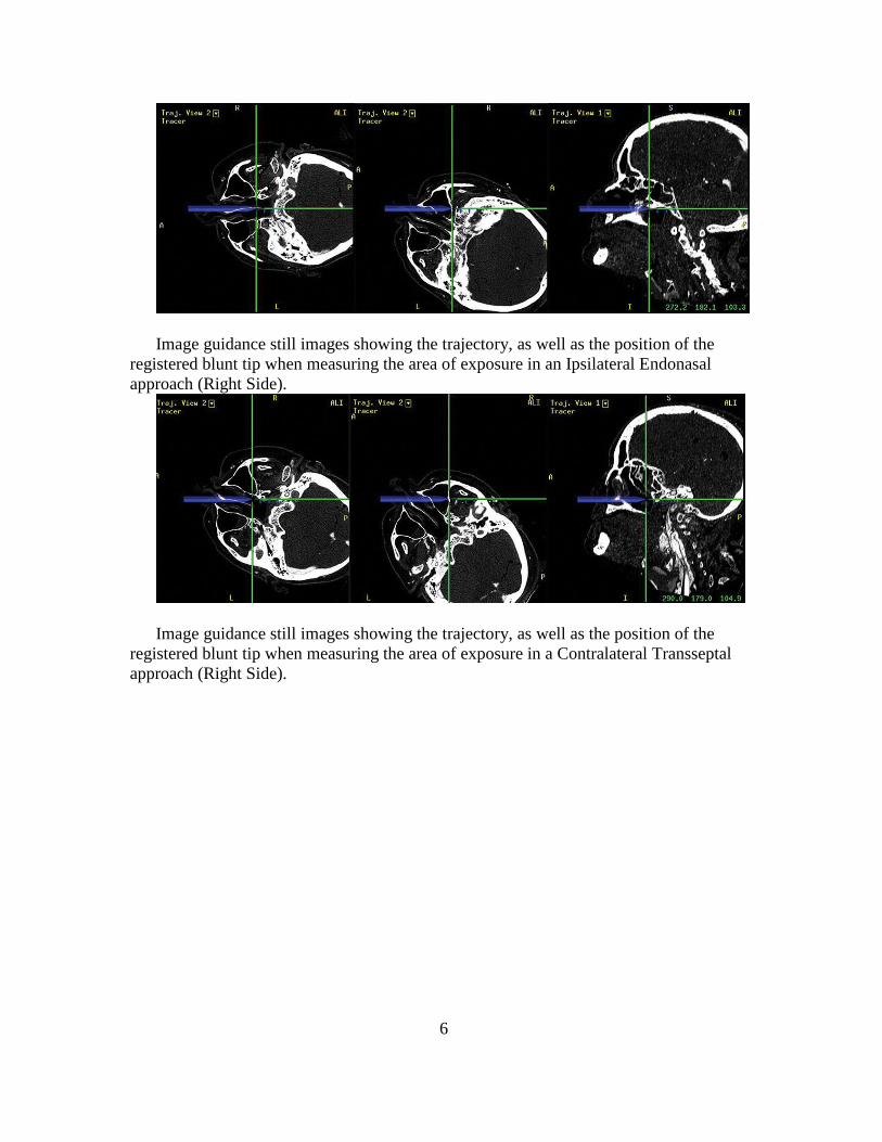
6
Image guidance still images showing the trajectory, as well as the position of the
registered blunt tip when measuring the area of exposure in an Ipsilateral Endonasal
approach (Right Side).
Image guidance still images showing the trajectory, as well as the position of the
registered blunt tip when measuring the area of exposure in a Contralateral Transseptal
approach (Right Side).

7
Figure 1.1
An illustration showing anatomy of the lateral skull base (inferior image). The arrows
show the trajectories used for the three endoscopic approaches. Used with permission
from Barrow Neurological Institute.

8
Ipsilateral sublabial transmaxillary approach: Similar to techniques described
previously,{9, 17, 20, 34} the upper lip is retracted and a transverse incision is made at
the buccogingival sulcus just lateral from the canine and extending laterally to the second
molar. The incision is made through the mucosa and periosteum. A subperiosteal plane is
developed, exposing the anterior wall of the maxilla. Care is taken to protect the
infraorbital nerve, which is the superior limitation of the anterior maxillary wall
exposure. An osteotome is used to perform an anterior maxillotomy, and the opening is
enlarged using Kerrison rongeurs (Integra, Plainsboro, NJ), to create an osteotomy
approximately 15 mm wide and 10 mm high. After entering the maxillary sinus, (Fig.
1.2A) the mucosa is peeled away and the infraorbital nerve is identified at the junction
between the maxillary roof and posterior wall. A second osteotomy is made in the
posterior wall, and the posterior wall is removed using a Kerrison rongeur to expose the
periosteum, which is opened to enter the pterygopalatine fossa and expose its contents.
Ipsilateral endonasal transmaxillary approach: The endonasal transmaxillary
approach has previously been described in detail.{5, 7, 10, 13, 17, 18, 19, 34} In brief,
the middle turbinate bone is removed through the ipsilateral nostril, and the inferior
turbinate bone is reflected inferiorly or removed, allowing the ethmoid bulla to be
identified. An antrostomy is performed using a Kerrison rongeur to allow access to the
maxillary sinus (Fig. 1.2B), and the greater palatine nerve and artery are preserved along
the junction between the maxillary base and posterior maxillary wall. To increase the
intranasal exposure, the ethmoid bulla is removed, exposing the anterior ethmoid artery,
and then the sphenopalatine artery is identified and preserved. Next, the infraorbital nerve

9
is identified in the roof of the maxillary sinus, and the posterior maxillary sinus is
fractured and carefully removed with Kerrison rongeurs. After removing the posterior
maxillary wall, the periosteal membrane is immediately visible and is dissected to expose
the contents of the pterygopalatine fossa, which includes the internal maxillary artery and
a complex network of its branches. The pterygopalatine ganglion is identified posterior to
the sphenopalatine artery and fat tissue; tracing the pterygopalatine ganglion posteriorly
and medially can lead to the vidian canal and the vidian nerve. The approach is extended
along the course of the vidian canal to the medial portion of the internal carotid artery
(ICA) genu by drilling the medial pterygoid plate using a 2-mm diamond bit. The
infraorbital nerve is followed posteriorly to the maxillary branch of the trigeminal nerve
(V2). The lateral plate of the pterygoid is removed to expose the foramen ovale and the
mandibular division of the trigeminal nerve (V3).
Contralateral endonasal transseptal approach: The contralateral endonasal
transeptal approach provides access to the maxillary sinus through the contralateral nasal
cavity {8, 22} with a nasoseptal mucosal flap pedicled posteriorly on the septal branch of
the sphenopalatine artery (Fig. 1.2C). An additional ipsilateral flap may also be
performed, but was not done in this study. Once access is gained to the ipsilateral nasal
cavity, the transmaxillary dissection is performed in a similar manner to the ipsilateral
endonasal transmaxillary approach described previously. The endoscope is advanced
until it reaches the posterior third of the nasal septum of the contralateral nasal cavity and
is then directed through the transseptal window. The endoscope and an instrument are
advanced to the maxillary sinus and retromaxillary space.

10
Figure 1.2
Images for a 0° endoscope from the right side of cadaveric dissections showing the
corridors used to access the maxillary sinus: A) Ipsilateral Caldwell Luc approach, B)
ipsilateral endonasal approach, C) contralateral (from left nostril) transseptal approach.
MS= maxillary Sinus, ST= superior turbinate, MT= middle turbinate, IT= inferior
turbinate, NS= nasal septum. Used with permission from Barrow Neurological Institute.
Area of exposure
To calculate the area of exposure, four points were identified. The first point (ION)
was a fixed anatomical landmark that is the point at which the infraorbital nerve enters
the infraorbital canal and is crossed by the infraorbital artery. The other three points were
determined relative to the infraorbital nerve: 1) a medial point (MP), which was defined
as the point at the junction between the vomer and the sphenoid crest; 2) a lateral point
(LP) which was defined as the point directly lateral to the infraorbital nerve after removal
of the posterior wall of the maxillary sinus and represented the most lateral point of
exposure; and 3) an inferior point (IP) directly inferior to the ION and slightly lateral to
the inferior part of the junction between the base of the maxillary sinus and the posterior
maxillary wall, just lateral to the greater palatine nerve and vessel. The MP, LP, and IP
were used to determine the medial, lateral and inferior extent of the exposure,
respectively. Although the extent of each approach can be increased by using curved
instruments and angled endoscopes or using other maneuvers to increase the angle of

11
attack, in our dissections we used only standardized approaches with a standard
antrostomy, septectomy or medial maxillotomy.
Three other anatomical landmarks were identified and further dissected for
anatomical reference. The first landmark was the Eustachian tube (ET) at the level of the
nasopharynx just anterior to the posterior choana. The second landmark was the second
genu of the internal carotid artery (gICA) which was exposed after drilling the sphenoidal
wall. The third landmark was the second division of the trigeminal nerve (V2) as it exits
the foramen rotundum.
Screen captures from the neuronavigation system were used to measure the area of
exposure The area of exposure was identified as the sum of two areas (Fig. 1.3). The first
is a rectangular area bounded by a line between the ION and IP laterally, by a line
between the ION and MP superiorly, by a line between the the IP and the medial border
of the posterior choana inferiorly, and by the junction between the septum and the vomer
medially. The second area is a triangular area between the ION, IP, and LP.

12
Figure 1.3
An endoscopic image form an ipsilateral endonasal (right side) approach showing the
area of exposure as the sum of two areas; a rectangular area (highlighted in blue) and a
triangular area (highlighted in red). LP: lateral point which represents the lateral extent of
a registered blunt tip. IP: inferior point which represents the inferior extent of a registered
blunt tip. MP: medial point which represents the medial extent of a registered blunt tip.
ION: Infraorbital nerve, as it enters the infraorbital canal. Used with permission from
Barrow Neurological Institute.
Surgical freedom
Surgical freedom was defined as the maximal oval area along which the surgical
(proximal) end of the endoscope can be freely and easily moved. This area was calculated
by measuring the vertical and transverse limits that can be reached by the proximal end of
the endoscope (Fig. 1.4A).
The neuro-navigation system was used to measure the transverse limit (Fig. 1.4B-D)
which was determined by identifying two points in space. The first point corresponded to

13
the position of the proximal end of the endoscope while placing the distal end of the
endoscope as closely as possible to the midpoint of the line between the IP and LP and
moving the proximal end of the endoscope as medially as possible, sometimes even
crossing the midline. The second point was determined at the proximal end of the
endoscope while placing the distal end of the endoscope at the midpoint between the MP
and medial border of the posterior choana and moving the endoscope as far laterally as
possible.

14
Figure 1.4
An illustration demonstrating the method used to calculate the area of surgical
freedom. (A) A cone that represents the volume where the endoscope can be freely
moved. The oval area at the base of the cone is the area of surgical freedom. for an
ipsilateral sublabial approach (B), ipsilateral endonasal approach (C), contralateral
transseptal approach (D). Used with permission from Barrow Neurological Institute.
In a similar fashion, the vertical limit of the surgical freedom was determined by
identifying two points in space. The first point was determined by the position of the
proximal end of the endoscope while placing the distal end of the endoscope at a point
along the ION and IP as superiorly as possible and moving the proximal end of the
endoscope gently as inferiorly as possible. The second point was identified as the
position of the proximal end of the endoscope while placing the distal end of the
endoscope on a point along the line between the ION and IP as inferiorly as possible
while moving the proximal end of the endoscope gently and superiorly. These two points
were considered to determine the vertical limit of the surgical freedom. A series of t-tests
were used to compare the average means of the surgical freedom and area of exposure for
each approach with the other two approaches. Analysis of variance (ANOVA) was also
used to compare the surgical freedom between all three approaches.
Results
The mean area of exposure for the three endoscopic approaches was similar (Fig. 1.5,
Table 1.1). The sublabial approach had an area of 9.92 ± 2.5 cm2, the endoscopic
endonasal approach had an area of 10.47 ± 2.65 cm2, and the transseptal approach had an
area of 10.01 ± 2.16 cm2.
15
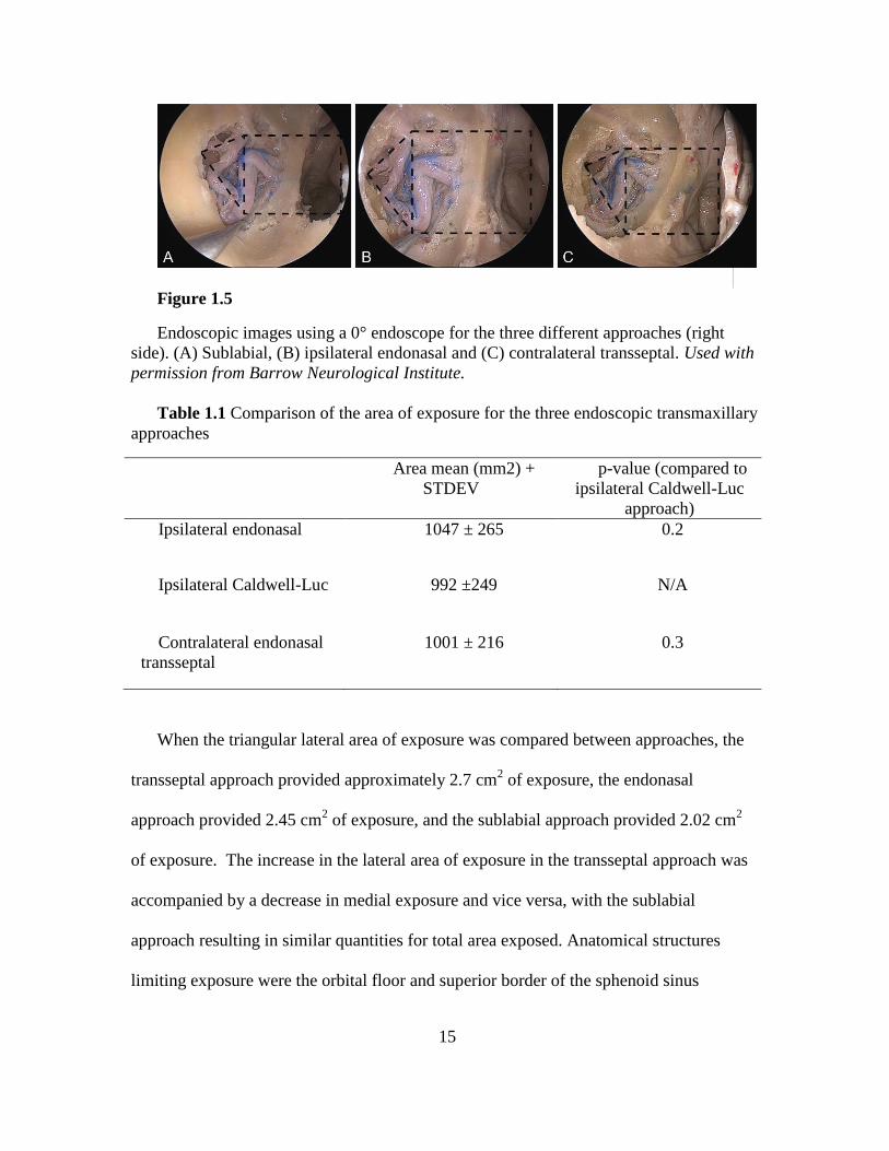
Figure 1.5
Endoscopic images using a 0° endoscope for the three different approaches (right
side). (A) Sublabial, (B) ipsilateral endonasal and (C) contralateral transseptal. Used with
permission from Barrow Neurological Institute.
Table 1.1 Comparison of the area of exposure for the three endoscopic transmaxillary
approaches
Area mean (mm2) +
STDEV
p-value (compared to
ipsilateral Caldwell-Luc
approach)
Ipsilateral endonasal
1047 ± 265 0.2
Ipsilateral Caldwell-Luc
992 ±249 N/A
Contralateral endonasal
transseptal
1001 ± 216 0.3
When the triangular lateral area of exposure was compared between approaches, the
transseptal approach provided approximately 2.7 cm2 of exposure, the endonasal
approach provided 2.45 cm2 of exposure, and the sublabial approach provided 2.02 cm
2
of exposure. The increase in the lateral area of exposure in the transseptal approach was
accompanied by a decrease in medial exposure and vice versa, with the sublabial
approach resulting in similar quantities for total area exposed. Anatomical structures
limiting exposure were the orbital floor and superior border of the sphenoid sinus

16
superiorly, the nasal floor and maxillary sinus floor inferiorly, the nasal septum medially,
and the lateral wall of the maxillary sinus laterally.
The mean areas of surgical freedom were 112.82 ± 7 cm2 for the sublabial approach,
76.3 ± 14.5 cm2 for the ipsilateral endonasal approach, and 83.51 ± 15.13 cm
2 for the
contralateral transseptal approach. The sublabial approach provided significantly more
surgical freedom when compared to the ipsilateral endonasal approach (p <.01) and the
transeptal approach (p <.01, Table 1.2). No significant difference was found in the
surgical freedom afforded between the endonasal ipsilateral and transeptal approaches
(p=0.20). The mean transverse (T) and vertical (V) axis of the three approaches were
12.8 cm ± 1.2(T) and 11 cm ± 0.6 (V) for the sublabial, 8.9 cm ± 1 (T) and 10.5 ± 1.1 (V)
for the endoscopic endonasal, and 10.6 cm ± 1.2(T) and 9.7cm ± 1(V) for the transseptal.
Table 1.2 Comparison of surgical freedom for three endoscopic transmaxillary
approaches.
Area mean (mm2) +
STDEV
p-value
(compared to
ipsilateral Caldwell-
Luc approach)
Ipsilateral endonasal
7630 ± 1454 0.0005
Ipsilateral Caldwell-Luc
11282 ± 696 N/A
Contralateral endonasal
transseptal
8351 ± 1513 0.001
N/A, not applicable

17
Discussion
Endonasal endoscopic approaches have been used with good results to access midline
lesions of the pituitary, and suprasellar and clival lesions.{16, 23, 24, 25, 26} They have
been used widely for benign tumors{17, 28, 29} and have recently been applied to
malignant lesions.{17, 30} The morbidity of open surgical approaches to this region have
allowed for a natural expansion of the endoscopic technique to the lateral anterior skull
base regions.{3, 27} Although they were initially used only for diagnostic and palliative
treatment, endoscopic techniques are now routinely being used in the primary treatment
of anterolateral skull base lesions such as inverted papillomas and juvenile
angiofibromas.{4, 7} The maxillary sinus has been used as a corridor to access the lateral
skull base. The sinus is a natural route and its large volume permits a great deal of
surgical freedom and access to critical neurovascular structures. Several transmaxillary
approaches has been used in the treatment of retromaxillary lesions.{9, 17}
Our data show that the sublabial approach provides the best horizontal working space,
while the endonasal approach has the least horizontal working space; therefore, a
sublabial approach may be superior in the case of tumors that extend in the same axial
plane. In addition, we found that the transseptal approach has the least vertical working
space; as a result, it may not be the best option in the case of tumors or lesions that extend
in the same sagittal plane. The approaches that we describe in this study have been
previously described with a detailed anatomical overview,{9, 22} but an analysis of the
exposed working space, including the surgical freedom and the area of exposure, has not
been previously described. These concepts are important in planning the appropriate
surgical approach for a specific lesion and for understanding the limitations of each of the

18
approaches. Theodospolous et al.{9} concluded in an anatomical study that a combined
ipsilateral sublabial and ipsilateral endonasal approach can provide a full exposure to the
infratemporal fossa and pterygopalatine fossa, but the lateral aspect of the infratemporal
fossa was challenging to access and required traumatic traction to the nose. Eloy et
al.{22} demonstrated that the transseptal approach provided more posterolateral exposure
for the infratemporal fossa. Our study confirms this: combining any of these approaches
with a contralateral endonasal transseptal approach will provide a lateral shift of the area
of exposure. This shift in exposure will assist in the removal of lesions that extend as far
laterally as the mandibular ramus and temporalis muscle. In our study, the quantitative
exposed area for each approach was similar; however, the areas exposed were not the
same. Therefore, combining any two approaches will allow for a larger exposure.
Combining the ipsilateral sublabial approach with the transseptal approach provides an
additional 1.2 cm2 of exposure and adds increased maneuverability, permitting a four-
handed technique to be used. Adding an ipsilateral endonasal approach increases the
exposed area by 0.6 cm2, which may be of great value in approaching large, challenging
lesions.
In their cadaveric study, Harvey et al.{8} determined that surgical access was
increased 14.7 ± 2.5 % when a transseptal approach is used compared to ipsilateral
approaches. According to our data, the contralateral transseptal exposure will lead to an
increase of approximately 12% and 11% in the area of exposure when compared to an
ipsilateral endonasal and ipsilateral sublabial approaches, respectively.
In cadaveric studies, Hartnick et al.{31} and Eloy et al.{22} approached the
infratemporal fossa via a temporal hairline incision and concluded that it is a limited

19
approach that can be used only for targeted CSF leak treatment or lesion biopsy. Eloy et
al. concluded that adding this approach to any of the previously described approaches
may lead to an increased surgical freedom to the superior portion of the infratemporal
fossa.{22} With the increased experience of the surgical team, the use of angled
endoscopes and angled instruments can be helpful in increasing the size of the accessible
operative field and leading to improved tumor resections.{35, 36} While these angled
instruments and endoscopes would greatly increase the extent and application for each
approach, in our comparison we used only straight instruments and a 0° endoscope, so
that we could study the approaches in a standard manner.
Other maneuvers can be used to increase surgical freedom. For example, in the
sublabial approach, the anterior maxillary antrostomy can be widened, but care should be
taken with the superior extension of the antrostomy so as not to injure the infraorbital
nerve and artery.{9} For the endonasal approach, the medial maxillotomy can be
enlarged and the piriform aperture drilled (Denker's approach), but the lacrimal duct
should be identified and spared to prevent post-operative complications. For the
contralateral endonasal approach, a larger septectomy will allow easier introduction of
other endoscopic instruments.{8}
The difference in surgical freedom among the three approaches, with similar exposed
areas, is attributed to the pivot point (Pinch point, Fig. 1.6). The pivot point is the fixed
point between the tip of the endoscope and the base of the endoscope where the direction
of movement is changed. The movement of the proximal end of the endoscope to one
direction results in a movement of the distal end to the opposite direction. The pivot point
was closer to the tip in the sublabial approach (1-3 cm), thus a larger movement of the

20
proximal end results in a smaller and finer movement at the distal end of the endoscope.
The pivot point ranged from 4.5-7 cm in the ipsilateral endonasal approach; thus, a
movement of the proximal end would lead to a larger movement of the distal end when
compared to the sublabial approach.
Figure 1.6
A diagram showing the importance of the pivot point in the three different
approaches, which when changed (depending on the approach) with a fixed area of
exposure, affect the degree of surgical freedom. Used with permission from Barrow
Neurological Institute.
Conclusion
The sublabial, ipsilateral endonasal, and contralateral transseptal endoscopic
transmaxillary approaches provide excellent exposure to the retromaxillary area. The
quantity of area exposed is similar for the three approaches, but the transseptal approach
offers greater lateral exposure. Surgical freedom is greatest with the sublabial approach

21
and is least in the ipsilateral endonasal approach. This information may benefit
practitioners in surgical planning and decision making for lesions of the infratemporal
fossa and pterygopalatine fossa.
References
1- Browne JD, Jacob SL. Temporal approach for resection of juvenile nasopharyngeal
angiofibromas. Laryngoscope 2000;110:1287-1293.
2- Cass SP, Hirsch BE, Stechison MT. Evolution and advances of the lateral surgical
approaches to cranial base neoplasms. J Neurooncol 1994;20:337-361.
3- Zhang M, Garvis W, Linder T, Fisch U. Update on the infratemporal fossa approaches
to nasopharyngeal angiofibroma. Laryngoscope 1998;108:1717-1723.
4- Onerci TM, Yucel OT, Ogretmenoglu O. Endoscopic surgery in treatment of juvenile
nasopharyngeal angiofibroma. Int J Pediatr Otorhinolaryngol 2003;67:1219-1225.
5- Nicolai P, Berlucchi M, Tomenzoli D et al. Endoscopic surgery for juvenile
angiofibroma: when and how. Laryngoscope 2003;113:775-782.
6- Nicolai P, Villaret AB, Farina D et al. Endoscopic surgery for juvenile angiofibroma: a
critical review of indications after 46 cases. Am J Rhinol Allergy 2010;24:e67-e72.
7- Robinson S, Patel N, Wormald PJ. Endoscopic management of benign tumors
extending into the infratemporal fossa: a two-surgeon transnasal approach.
Laryngoscope 2005;115:1818-1822.
8- Harvey RJ, Sheehan PO, Debnath NI, Schlosser RJ. Transseptal approach for extended
endoscopic resections of the maxilla and infratemporal fossa. Am J Rhinol Allergy
2009;23:426-432.
9- Theodosopoulos PV, Guthikonda B, Brescia A, Keller JT, Zimmer LA. Endoscopic
approach to the infratemporal fossa: anatomic study. Neurosurgery 2010;66:196-202.
10- Herzallah IR, Germani R, Casiano RR. Endoscopic transnasal study of the
infratemporal fossa: a new orientation. Otolaryngol Head Neck Surg 2009;140:861-
865.
11- Tosun F, Ozer C, Gerek M, Yetiser S. Surgical approaches for nasopharyngeal
angiofibroma: comparative analysis and current trends. J Craniofac Surg 2006;17:15-
20.

22
13- Wang QY, Chen HH, Lu YY. Comparison of two approaches to the surgical
management of juvenile nasopharyngeal angiofibroma stages I and II. J Otolaryngol
Head Neck Surg 2011;40:14-18.
14- Wormald PJ, Van HA. Endoscopic removal of juvenile angiofibromas. Otolaryngol
Head Neck Surg 2003;129:684-691.
15- Dehdashti AR, Karabatsou K, Ganna A, Witterick I, Gentili F. Expanded endoscopic
endonasal approach for treatment of clival chordomas: early results in 12 patients.
Neurosurgery 2008;63:299-307.
16- Fisch U, Fagan P, Valavanis A. The infratemporal fossa approach for the lateral skull
base. Otolaryngol Clin North Am 1984;17:513-552.
17- Hong JW, Ping ZS, Hai XZ, Zhang H, Zhang J, Yun XJ. Endoscopic resection of
chordomas in different clival regions. Acta Otolaryngol 2009;129:71-83.
18- Little AS, Nakaji P, Milligan J. Endoscopic Endonasal Transmaxillary Approach and
Endoscopic Sublabial Transmaxillary Approach: Surgical Decision Making and
Implications of the Nasolacrimal Duct. World Neurosurg 2012.
19- Alfieri A, Jho HD, Schettino R, Tschabitscher M. Endoscopic endonasal approach to
the pterygopalatine fossa: anatomic study. Neurosurgery 2003;52:374-378.
20- Cavallo LM, Messina A, Gardner P et al. Extended endoscopic endonasal approach to
the pterygopalatine fossa: anatomical study and clinical considerations. Neurosurg
Focus 2005;19:E5.
21- Ong BC, Gore PA, Donnellan MB, Kertesz T, Teo C. Endoscopic sublabial
transmaxillary approach to the rostral middle fossa. Neurosurgery 2008;62:30-36.
22- Eloy JA, Murray KP, Friedel ME, Tessema B, Liu JK. Graduated endoscopic
multiangle approach for access to the infratemporal fossa: a cadaveric study with
clinical correlates. Otolaryngol Head Neck Surg 2012;147:369-378.
23- Schwartz TH, Stieg PE, Anand VK. Endoscopic transsphenoidal pituitary surgery
with intraoperative magnetic resonance imaging. Neurosurgery 2006;58:ONS44-
ONS51.
24- Esposito F, Cappabianca P, Del Basso De CM, Cavallo LM, Rinaldi C, de DE.
Endoscopic endonasal transsphenoidal removal of an intra-suprasellar schwannoma
mimicking a pituitary adenoma. Minim Invasive Neurosurg 2004;47:230-234.
25- Aydin S, Cavallo LM, Messina A et al. The endoscopic endonasal trans-sphenoidal
approach to the sellar and suprasellar area. Anatomic study. J Neurosurg Sci
2007;51:129-138.

23
26- Laufer I, Anand VK, Schwartz TH. Endoscopic, endonasal extended transsphenoidal,
transplanum transtuberculum approach for resection of suprasellar lesions. J
Neurosurg 2007;106:400-406.
27- Lund VJ. Extended applications of endoscopic sinus surgery--the territorial
imperative. J Laryngol Otol 1997;111:313-315.
28- Liu JK, Christiano LD, Patel SK, Tubbs RS, Eloy JA. Surgical nuances for removal
of olfactory groove meningiomas using the endoscopic endonasal transcribriform
approach. Neurosurg Focus 2011;30:E3.
29- Liu JK, Christiano LD, Patel SK, Eloy JA. Surgical nuances for removal of
retrochiasmatic craniopharyngioma via the endoscopic endonasal extended
transsphenoidal transplanum transtuberculum approach. Neurosurg Focus
2011;30:E14.
30- Hanna E, DeMonte F, Ibrahim S, Roberts D, Levine N, Kupferman M. Endoscopic
resection of sinonasal cancers with and without craniotomy: oncologic results. Arch
Otolaryngol Head Neck Surg 2009;135:1219-1224.
31- Hartnick CJ, Myseros JS, Myer CM, III. Endoscopic access to the infratemporal fossa
and skull base: a cadaveric study. Arch Otolaryngol Head Neck Surg 2001;127:1325-
1327.
32- Falcon RT, Rivera-Serrano CM, Miranda JF et al. Endoscopic endonasal dissection
of the infratemporal fossa: Anatomic relationships and importance of eustachian tube
in the endoscopic skull base surgery. Laryngoscope 2011;121:31-41.
33- Rivera-Serrano CM, Terre-Falcon R, Fernandez-Miranda J et al. Endoscopic
endonasal dissection of the pterygopalatine fossa, infratemporal fossa, and post-
styloid compartment. Anatomical relationships and importance of eustachian tube in
the endoscopic skull base surgery. Laryngoscope 2010;120 Suppl 4:S244.
34- Wilson DA, Williamson RW, Preul MC, Little AS. Comparative Analysis of Surgical
Freedom and Angle of Attack of Two Minimal-Access Endoscopic Transmaxillary
Approaches to the Anterolateral Skull Base. World Neurosurg 2013.
35- Cappabianca P, Cavallo LM, de DO, Solari D, Esposito F, Colao A. Endoscopic
pituitary surgery. Pituitary 2008;11:385-390.
36- Tang CT, Kurozumi K, Pillai P, Filipce V, Chiocca EA, Ammirati M. Quantitative
analysis of surgical exposure and maneuverability associated with the endoscope and
the microscope in the retrosigmoid and various posterior petrosectomy approaches to
the petroclival region using computer tomograpy-based frameless stereotaxy. A
cadaveric study. Clin Neurol Neurosurg 2013;115:1058-1062.

CHAPTER 2
The following chapter has been presented on February 15th
2014 in the Proffered Papers
IX section: Endonasal approaches 3, at the North American Skull Base Society annual
meeting 2014, San Diego, CA. Also a complete and revised manuscript has been
submitted for publication at the Journal of Operative Neurosurgery.

25
CHAPTER 2
Evaluation of Surgical Freedom for Microscopic and Endoscopic
Transsphenoidal Approaches to the Sella
Elhadi AM, Hardesty H, Zaidi H, Kalani YS, Nakaji P, White WL, Preul MC,
Little AS.
ABSTRACT
Background. Microscopic and endoscopic transsphenoidal approaches to the sellar
are well-established. Surgical freedom is an important skull base principle that can be
measured objectively and compare approaches.
Objective. In this study, we compared the surgical freedom of four transsphenoidal
approaches to the sella turcica.
Methods. Four transsphenoidal approaches to the sella (microscopic sublabial,
microscopic endonasal, endoscopic binostril, and endoscopic uninostril) were performed
on eight silicon-injected cadaveric heads. Surgical freedom was determined with
stereotactic image guidance using previously established techniques. The results are
presented as the area of surgical freedom and angular surgical freedom (angle of attack)
in the axial and sagittal planes.
Results. Mean total exposed area surgical freedom for the microscopic sublabial,
endoscopic binostril, endoscopic uninostril, and microscopic endonasal approaches were
102±13cm2, 89±6cm2, 81±4cm2, and 69±10cm2, respectively. The endoscopic binostril
approach had the greatest surgical freedom at the pituitary gland, ipsilateral and
contralateral ICAs (25.7±5.4, 28.0±4.0, and 23.0±3.0 cm2) compared to microscopic

26
sublabial (21.8±3.5, 21.3±2.4, and 19.5±6.3 cm2), microscopic endonasal (14.2±2.7,
14.1±3.2, and 16.3±4.0 cm2), and endoscopic uninostril (19.7±4.8, 22.4±2.3, and
19.5±2.9 cm2). Axial angle of attack was greatest for the microscopic sublabial approach
to the same targets (14.7±1.3, 11.0±1.5, and 11.8±1.1 degrees). For the sagittal angle of
attack, the endoscopic binostril approach was superior for all three targets (16.6±1.7,
17.2±0.70, and 15.5±1.2 degrees).
Conclusions. The microscopic sublabial and endoscopic binostril approaches
provided superior surgical freedom compared to the endonasal microscopic and uninostril
endoscopic approaches. This work provides objective, baseline values for the
quantification and evaluating future refinements in surgical technique or instrumentation.
INTRODUCTION
Surgical approaches to sellar region pathology have challenged neurosurgeons since
the inception of the field. Microscope-based transsphenoidal approaches using either a
sublabial or transnasal passageway are the mainstay of the neurosurgical armamentarium
for sellar lesions with excellent results.{6,14,18} In the last two decades, progressive
technological advances in the field of neurosurgical endoscopy have ushered in the
endoscopic, endonasal, transsphenoidal approach as a viable alternative to microscope-
based approaches. Excellent clinical results for a wide variety of sellar pathology have
been published using purely endoscopic surgical techniques.{2,4,6,8,12,19} A significant
volume of literature has been published regarding the clinical outcomes and
complications of microscope- and endoscope-based approaches to the sella. However,
there remains a paucity of head-to-head comparisons of the strategies from a technical
standpoint. Spencer and colleagues performed a variety of microsurgical and endoscopic

27
transsphenoidal approaches on cadavers and found a significantly improved volume of
exposure with endoscopy-based approaches, especially in visualization superiorly (above
the dorsum sella) but also in lateral and anterior bony exposure.{20} Catapano and
colleagues also demonstrated greater bony exposure using an endoscopic approach
compared to a microsurgical approach to the sella.{3} Yet, no anatomical study has
examined the surgeon’s ability to manipulate instruments at the sella with these
approaches, nor determined if one approach provides a superior working corridor. Our
laboratory and others have previously established a method of assessing the surgical
freedom and angles of attack provided by various microsurgical and endoscopic
exposures using stereotaxy.{7,9,17,21} This provides a quantitative analysis of the
surgeon’s ability to move instruments in space during surgery through the operative
corridor, and permits a more rigorous and objective comparison of skull base approaches.
Herein we apply the same anatomic analyses to the microscopic and endoscopic
transsphenoidal approaches to the sella.
METHODS
We dissected eight silicon-injected, formalin-fixed cadaveric heads using four
transsphenoidal approaches. Two endoscopic approaches were used: a uninostril
endonasal transsphenoidal and a binostril endonasal transsphenoidal approach. Two
microscopic transsphenoidal approaches were also used: a microscopic endonasal
transsphenoidal and a microscopic sublabial transsphenoidal approach. Details of each
approach are described below.
Endoscopic approaches were performed using a 0° endoscope and standard
endoscopic techniques, burrs, dissector blades, and standard endoscopic instruments

28
(Karl Storz, Tuttlingen, Germany) with heads placed in rigid fixation in a supine position.
Microscopic approaches were performed using a standard surgical microscope (Pentero,
Zeiss, Germany) and standard micro-surgical instruments, with the heads placed in rigid
fixation in a supine position. High-resolution computed tomography (CT) scans were
performed on each specimen to document the bony facial and cranial anatomy, and the
images were uploaded to an image guidance platform (StealthStation Treon Plus with
FrameLink Software, Medtronic, Louisville, CO). Image guidance was used to obtain
anatomical measurements and to confirm anatomical structures. For endoscopic
measurements, the endoscope was parked in the superior aspect of the right nares.
Statistical analysis was performed by comparing the data collected from the each
approach with the other approaches using two-tailed t-tests, and significance was
determined when p-value was less than 0.05, and analysis of variance (ANOVA) was
used to further compare between the means of surgical freedom and angle of attack for
the four different approaches.
Uninostril endoscopic endonasal transsphenoidal approach
This approach has been described previously.{1} In brief, we used the right
nostril to approach the nasal cavity and the middle turbinate was out-fractured. The
sphenoid ostia were identified bilaterally and opened widely using a mushroom punch or
Kerrison rongeurs. The posterior third of the bony septum was resected along with a
piece of the vomer. The sphenoid rostrum was then opened wide using a drill or punch,
and bilateral posterior ethmoidectomies were performed. The posterior wall of the
sphenoid sinus was then removed to expose the anterior pituitary, the cavernous internal

29
carotid artery (cICA) and a part of the petrous internal carotid artery (pICA). In a
unilateral approach, the contralateral nasal mucosa is preserved. For all measurements,
the endoscope and the endoscopic dissector instrument were both inserted through the
right naris.
Binostril endoscopic endonasal transsphenoidal approach
In this approach dissections were performed similar to the previous approach but the
contralateral posterior septal mucosa was removed and the left middle turbinate
outfractured,{13,22} such that the endoscope can be inserted through the right naris and
the dissector inserted through the left naris. To remain consistent with the other
approaches, the terms ipsilateral and contralateral in reference to the carotid arteries, etc,
for this approach are named by the side of endoscope insertion. An advantage to the
binostril approach that we did not attempt to quantify in this study is that the dissector
can be placed through whichever nares provides the best working angle for the surgeon.
Microscopic endonasal transsphenoidal approach
The classic approach has been well-described by Griffith in 1987, and several
modifications have been reported.{10} The technique was performed as follows. A
vertical incision was made at the mucodermal junction of the nasal septum, and the
incision was then extended to the nasal floor. The mucosa was then dissected from the
septal cartilage and elevated from the nasal floor, whereafter dissection was extended to
the anterior wall of the sphenoid sinus. The posterior part of the septal cartilage was
disarticulated from the plate of the ethmoid and vomer, and the 80 mm nasal speculum
was inserted to retract the mucosa and expose the anterior wall of the sphenoid sinus. The
anterior wall of the sphenoid was removed and bilateral ethmoidectomies were performed

30
to expose the clivus and sellar floor. The pituitary gland, cICA, and pICA were exposed
by bony removal of the posterior sphenoid wall and the carotid prominence. The right
naris was used for all measurements.
Sublabial microscopic transsphenoidal approach
Similar to the classic technique by Jules Hardy, a horizontal incision was made under
the upper lip at the junction of the gum.{11} This incision was made deep enough to
incise the periosteum then elevated using a Cushing periosteal elevator to expose the
nasal cavum which was enlarged using rongeurs. The mucosal elevator was introduced
along the nasal septum to detach the mucosa from the cartilage to the deepest part of the
septum to the vomer. A Fukoshima nasal speculum was used to hold the mucosa out of
the field, and the nasal cartilage was removed creating a new submucosal cavity. The
vomer was detached and further resection of the sphenoid wall performed to expose the
whole sphenoid sinus cavity. The sphenoid mucosa was removed, exposing the sellar
floor along with the carotid prominence on both sides.
Exposed area surgical freedom
This variable is calculated using four points in space and represents the available
area of maneuverability that can be offered for the proximal (surgeon’s) end of an
endoscopic instrument (2 mm dissector, 23 cm in length) while moving the distal end of
this instrument along the borders of the exposed area (holding the endoscope within the
nasal vestibule, in the endoscopic approaches). The four points were determined using the
neuro-navigation system. Each point corresponded to the position (outside the patient) of
the proximal end of the dissector while placing the distal end of the dissector at an
anatomic target. The four anatomic targets for the distal dissector were as follows: first
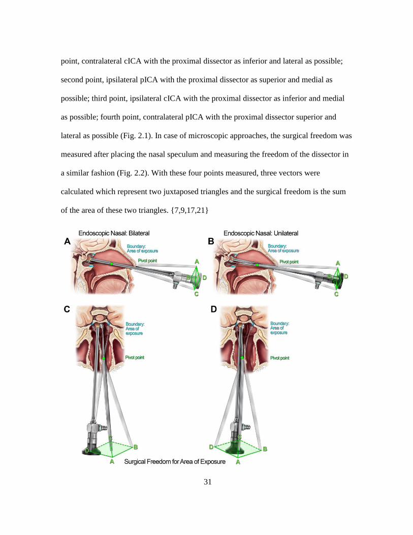
31
point, contralateral cICA with the proximal dissector as inferior and lateral as possible;
second point, ipsilateral pICA with the proximal dissector as superior and medial as
possible; third point, ipsilateral cICA with the proximal dissector as inferior and medial
as possible; fourth point, contralateral pICA with the proximal dissector superior and
lateral as possible (Fig. 2.1). In case of microscopic approaches, the surgical freedom was
measured after placing the nasal speculum and measuring the freedom of the dissector in
a similar fashion (Fig. 2.2). With these four points measured, three vectors were
calculated which represent two juxtaposed triangles and the surgical freedom is the sum
of the area of these two triangles. {7,9,17,21}

32
Figure 2.1
An illustration showing the exposed area surgical freedom for the two endoscopic
approaches; Endoscopic Endonasal Binostril approach (A) sagittal, (C) axial, and the
Endoscopic Endonasal Uninostril approach (B) sagittal, (D) axial.
Figure 2.2
An illustration showing the exposed area surgical freedom for the two microscopic
approaches; microscopic sublabial approach (using a fukushima retractor) (A) sagittal,
(C) axial, and the microscopic endonasal approach (B) sagittal, (D) axial.
Anatomic target surgical freedom
This variable represents the maneuverability of the proximal end of the dissector
while fixing the distal end of the dissector on a specific anatomic target and the

33
endoscope placed within the vestibule (in case of endoscopic approaches), or after
placing the nasal speculum in case of the microscopic approaches.
Four points were again determined using the neuro-navigation system, which
represent the four positions of the proximal end of the dissector outside the patient while
fixing the distal end on an anatomical target and placing the proximal end as inferiorly,
superiorly, medially and laterally as possible. As above, after these four points are
calculated three vectors can be measured which represent two juxtaposed triangles and
the surgical freedom is the sum of the area of these two triangles(Fig. 2.3, 2.4). We
measured anatomic target surgical freedom for the center of the pituitary gland and the
two cavernous ICAs.
Angle of attack
The angular surgical freedom (“angles of attack”) in two planes was determined for
three targets: the pituitary gland and both cICAs. This was measured, as we have
described previously, by fixing the distal end of the dissector on the anatomic target and
moving the proximal end of the dissector as far left and right as possible to determine the
maximum angle of attack within the axial plane.{21}(Fig. 2.3) The angle of attack in the
sagittal plane was calculated by measuring the maximum angle of movement when fixing
the distal end of the dissector on the anatomic target and moving the proximal end as
superior and inferior as possible (Fig. 2.4). These measurements were taken while
positioning the endoscope against the nasal vestibule and providing a full view of the
exposed area for the endoscopic techniques, and after placing the microscopic nasal
speculum in case of the microscopic approaches.

34
Figure 2.3
Illustration showing the point anatomical target surgical freedom and angle of attack
for the pituitary gland in the axial plane for the endoscopic endonasal binostril approach
(A), endoscopic endonasal uninostril approach (B), microscopic sublabial approach (C),
microscopic endonasal approach (D).

35
Figure 2.4
Illustration showing the point anatomical target surgical freedom and angle of attack
for the pituitary gland in the sagittal plan for the endoscopic endonasal binostril approach

36
(A), endoscopic endonasal uninostril approach (B), microscopic sublabial approach (C),
microscopic endonasal approach (D).
RESULTS
Exposed area surgical freedom
The microscopic sublabial approach provided the greatest exposed area surgical
freedom (102.3 ± 12.6 cm2, Fig. 2.5), followed by the endoscopic binostril approach
(88.9 ± 5.5 cm2; two-tailed t-test compared to microscopic sublabial, p=0.02), the
endoscopic uninostril approach (80.9 ± 4.5 cm2; two-tailed t-test compared to
microscopic sublabial, p=0.004), and the least exposed area surgical freedom was
provided by the microscopic endonasal approach (68.7 ±9.6 cm2; two-tailed t-test
compared to microscopic sublabial, p=0.0008). Statistical significance of each approach
compared to every other approach is summarized in Table 2.1.

37
Figure 2.5
Total exposed area surgical freedom by approach. MSL, microscopic sublabial. ME,
microscopic endonasal. EBN, endoscopic binostril. EUN, endoscopic uninostril. Every
measurement reported is statistically significant when compared to all other values by
two-tailed t-test.
Table 2.1
Two-tailed t-test p values for each approach compared to A) Microscopic Sublabial
Approach, B) Endoscopc Binostril Approach, C) Microscopic Endonasal Approach, D)
Endoscopic Uninostril approach.
Table 2.1-a
Microscopic Sublabial Approach
Surgi
cal
appro
ach
Angle of Attack Surgical freedom
Axial Sagittal Anatomic target Expos
ed
area Pit I-
cICA
C-
cICA
Pit I-
cICA
C-
cIC
A
Pit I-
cICA
C-
cIC
A
ME 0.000
2**
0.2
0.001
**
0.008
**
0.005
**
0.06
0.005
**
0.001
**
0.08
**
0.0008
**
EBN 0.02*
*
0.02*
*
0.04*
*
0.03*
0.005
*
0.2
0.01*
0.003
*
0.3
0.02**
EUN 0.001
**
0.007
**
0.02*
*
0.8
0.5
0.9
0.1
0.4
1
0.004*
*
Table 2.1-b
Endoscopic Binostril Approach
Surgic
al
appro
ach
Angle of Attack Surgical freedom

38
Axial Sagittal Anatomic target Expose
d area Pit I-
cIC
A
C-
cIC
A
Pit I-
cICA
C-
cIC
A
Pit I-
cIC
A
C-
cIC
A
MSL 0.02*
0.02
*
0.04
*
0.03** 0.005
**
0.2
0.0
1**
0.00
3**
0.3
0.02*
ME 0.002
**
0.6
0.6
0.002*
*
0.000
07**
0.01
**
0.0
01*
*
0.00
004
**
0.01
**
0.0009*
*
EUN 0.003
**
0.09
0.3
0.0003
**
0.000
0003*
*
0.00
2**
0.0
003
**
0.00
2**
0.02
**
0.0002*
*
Table 2.1-c
Microscopic Endonasal Approach
Surgi
cal
appr
oach
Angle of Attack Surgical freedom
Axial Sagittal Anatomic target Expose
d area Pit I-
cIC
A
C-
cICA
Pit I-
cIC
A
C-
cIC
A
Pit I-
cICA
C-
cIC
A
MSL 0.000
2*
0.2
0.001
*
0.008
*
0.005
*
0.06
0.005
*
0.001
*
0.0
8*
0.0008*
EBN 0.002
*
0.6
0.6
0.002
*
0.000
07**
0.01
**
0.001
*
0.000
04*
0.0
1*
0.0009*
EUN 0.7
0.08
0.2
0.02*
0.004
*
0.08
*
0.03*
0.001
*
0.0
3*
0.009*
Table 2.1-d
Endoscopic Uninostril Approach

39
Surgi
cal
appr
oach
Angle of Attack Surgical freedom
Axial Sagittal Anatomic target Expos
ed
area
Pit I-
cICA
C-
cIC
A
Pit I-
cICA
C-
cIC
A
Pit I-
cICA
C-
cICA
MSL 0.00
1*
0.007
*
0.02
*
0.8 0.5
0.9 0.1 0.4
1 0.004*
ME 0.7 0.08 0.2 0.0
2**
0.004*
*
0.08 0.03
*
0.001*
*
0.03*
*
0.009*
*
EBN 0.00
3*
0.09 0.3 0.0
003
**
0.0000
003*
0.00
2*
0.00
03*
0.002* 0.02* 0.0002
*
(**) represent a statisically significant superiority. (*) represent a statistically
significant inferiority.
MSL, microscopic sublabial
ME, microscopic endonasal
EBN, endoscopic binostril
EUN, endoscopic uninostril
Pit, pituitary gland
I-cICA Ipsilateral cavernous internal carotid artery
C-cICA Contralateral cavernous internal carotid artery
Anatomic target surgical freedom
The largest anatomic target surgical freedom for the pituitary gland was provided by
the endoscopic binostril approach (27.7 ± 5.4 cm2) followed by the microscopic sublabial
approach (21.8 ± 3.5 cm2; two-tailed t-test compared to endoscopic binostril, p=0.01), the
endoscopic uninostril approach (19.7 ± 4.8 cm2; two-tailed t-test compared to endoscopic
binostril, p=0.0002), and the least surgical freedom was provided by the microscopic
endonasal approach (14.1 ± 2.7 cm2; two-tailed t-test compared to endoscopic binostril,

40
p=0.001, Fig. 2.6). The surgical freedom for the ipsilateral cICA (right cICA) was
greatest using the endoscopic binostril approach (27.0 ± 4.0 cm2), followed by the
endoscopic uninostril approach (22.4 ± 2.32 cm2; two-tailed t-test compared to
endoscopic binostril, p=0.002), the microscopic sublabial approach (21.3 ± 2.4 cm2; two-
tailed t-test compared to endoscopic binostril, p=0.01) and the microscopic endonasal
approach (14.1 ±3.17 cm2; two-tailed t-test compared to endoscopic binostril, p =
0.0004). For the contralateral cICA (left cICA) the endoscopic binostril approach had the
greatest anatomic target surgical freedom (23.0 ± 3 cm2), followed by the endoscopic
uninostril approach (19.5 ± 2.9 cm2; two-tailed t-test compared to endoscopic binostril,
p=0.014), the microscopic sublabial approach (19.5 ± 6.2 cm2; two-tailed t-test compared
to endoscopic binostril, p>0.05, non-significant), and lastly the microscopic endonasal
approach (16.3 ± 4.0 cm2; two-tailed t-test compared to endoscopic binostril, p=0.02).
Statistical significance of each approach compared to every other approach is
summarized in Table 2.1.

41
Figure 2.6
Anatomic target surgical freedom by approach. MSL, microscopic sublabial. ME,
microscopic endonasal. EBN, endoscopic binostril. EUN, endoscopic uninostril.
Statistical comparisons are reported separately in Table 2.1.
Angle of attack
The axial plane angle of attack for the pituitary gland was greatest for the microscopic
sublabial approach (14.7° ± 1.3, Fig. 2.7), followed by the endoscopic binostril approach
(12.8° ± 1.7; two-tailed t-test compared to microscopic sublabial, p=0.02), the
microscopic endonasal approach (9.5° ± 1; two-tailed t-test compared to microscopic
sublabial, p=0.0002), and the endonasal uninostril approach (9.2° ± 2; two-tailed t-test
compared to microscopic sublabial, p=0.001). The angle of attack for the pituitary gland
in the sagittal plane was greatest for the endoscopic binostril approach (16.5° ± 1.7, Fig.
2.8), followed by the microscopic sublabial approach (14.9° ±1.9; two-tailed t-test
compared to endoscopic binostril, p=0.03), the endoscopic uninostril approach (14.7°

42
±1.3; two-tailed t-test compared to endoscopic binostril, p=0.0003), and the microscopic
endonasal approach (12.4° ± 2; two-tailed t-test compared to endoscopic binostril,
p=0.002). The axial and sagittal plane angles of attack for the ipsilateral (right) and
contralateral (left) cICAs by approach are summarized in Figures 2.7 and 2.8; in short,
the microscopic sublabial approach had the greatest axial angle of attack for both cICAs,
while the endoscopic binostril approach had the greatest sagittal angle of attack for both
cICAs. Statistical significance of each approach compared to every other approach is
summarized in Table 2.1.

43
Figure 2.7
Axial angle of attack by approach. MSL, microscopic sublabial. ME, microscopic
endonasal. EBN, endoscopic binostril. EUN, endoscopic uninostril. Statistical
comparisons are reported separately in Table 2.1.
Figure 2.8
. Sagittal angle of attack by approach. MSL, microscopic sublabial. ME, microscopic
endonasal. EBN, endoscopic binostril. EUN, endoscopic uninostril. Statistical
comparisons are reported separately in Table 2.1.
DISCUSSION
Microscopic transsphenoidal surgery represents the gold-standard for addressing
lesions of the sella turcica.{6,14,18} The two most commonly used microsurgical
approaches applied in practice are the uninostril direct endonasal approach and the
sublabial transsphenoidal approach, but recent endoscopic technological advances and the
development of effective closure techniques have led to the adoption of purely

44
endoscopic, endonasal approaches to the sella.{2,4,5,8,12,19} The two most commonly
applied endoscopic approaches are uninostril and binostril transsphenoidal techniques.
While most recent literature has focused on the technical nuances of individual
approaches and preliminary patient outcomes, few objective technical comparisons of the
approaches exist. Surgical freedom is an important skull base principle that describes the
extent to which a surgeon can moves his hands in the operative field. Increased surgical
freedom and angle of attack limit sword fighting and instrument collisions, reduce
surgeon frustration, improve delicate microdissection, and improve target visualization.
Numerous impediments to surgical freedom in the crowded nasal corridor exist, such as
the nasal septum, turbinates, nares, sphenoid sinus bone, endoscope, and retractors. In
this study, we present the first objective comparison of surgical freedom of the four most
commonly performed transsphenoidal approaches as one might use to remove a pituitary
tumor. We estimated surgical freedom using four measurements (exposed are surgical
freedom, target surgical freedom, axial angular freedom, sagittal angular freedom). These
complementary measurements allow us to determine not only the total area of freedom,
but also in which plane one approach may be superior to another.
We demonstrated that the sublabial microscopic and the binostril endoscopic
approaches were superior to the uninostril microscopic and uninostril endoscopic
approaches in the examined variables. The sublabial approach provided the greatest
surgical freedom in the exposed area and axial angular freedom, whereas the endoscopic
binostril approach provided the greatest target surgical freedom and sagittal angular
freedom. The microscopic endonasal approach provided the least surgical freedom in
three of the four measurements in our model. The surgical freedom results can be

45
explained by the anatomical structures that limit movement in each approach. For
example, in the sublabial approach, the retractor is placed in a horizontal plane thus
providing a wide but short orifice at the distal end of the exposure. In contrast, in the
endoscopic approaches, the axial angular freedom is limited by the nares, nasal septum,
middle turbinate and maxillary sinus wall. However, sagittal angular freedom is excellent
because one can elevate the soft tissue of the nares to generate more freedom. The
uninostril microscopic approach provided the least surgical freedom because axial plane
freedom was limited by the nasal speculum, and the sagittal freedom was reduced by the
skin and cartilage of the nares made tight by the expanded retractor. The other interesting
observation was the noted superiority of the binostril endoscopic approach compared to
uninostril approach. This confirms our clinical impression. The uninostril approach
surgical freedom was more impacted by the presence of the endoscope and the conflicts
between the endoscope and dissecting instruments. In a standard binostril approach, the
endoscope is parked in the right nostril, and the dissectors are placed in the left nostril
thus limiting collisions.
These results provide practical information for surgeons choosing a surgical approach
and help quantify clinical impressions. For example, the senior author [author name
blinded for review] will choose a sublabial microscopic approach over a microscopic
uninostril approach for complex sellar tumors, such as craniopharyngioma, because of the
greater ease of dissection and shorter operative distance. Regarding the endoscopic
approaches, the authors now utilize an exclusively binostril approach instead of a
uninostril approach for all sellar lesions. This improves the ease of tumor dissection,
limits endoscope-instrument conflict, and significantly eases the hassle of sellar

46
reconstruction. However, surgical freedom is only one of several factors that a surgeon
considers when choosing an approach. Other factors include approach-related
morbidity{15} and surgeon experience/preference.
Our study utilized cadaveric heads fixed in standard preservatives, and this process
decreases the elasticity of tissue. This is a drawback inherent to any anatomical study
performed in cadavers, but naturally should still be considered a limitation to the present
study. We tried to address this variable by performing the measurements in the same
specimens for all four approaches, thus having each specimen serve as its own internal
control. To standardize the methodology, we chose to use only straight instruments and 0
degree endoscopes. Different surgical freedom areas could have been obtained with
angled instruments or endoscopes. Lastly, our study examines a standardized dissection
using each approach. Individual patient anatomy and surgical pathology is highly variable
and each surgical approach in the living patient is tailored to that unique anatomy.
Therefore, surgical freedom and angles of attack may differ somewhat when these
approaches are used in the operating room.
The choice to approach a lesion from either an endoscopic, microscopic, or combined
approach has many deciding factors and can yield excellent clinical results. We view the
present results not as an unqualified endorsement of the endoscopic binostril approach or
sublabial approach, but more as the early steps towards a rigorous and objective
anatomical comparison of surgical approaches in sellar surgery. With these baselines now
established for routine approaches, we can utilize the same principles to evaluate
expanded exposures, new instrumentation, and other technical modifications. Innovations

47
in surgical approach will in the future have standardized quantitative, rather than simply
qualitative, data to support their adoption.
CONCLUSIONS
The microscopic, sublabial approach to the sella provides the greatest surgical
freedom in the axial plane, and the greatest total surgical area freedom. The endoscopic,
binostril approach provides the greatest degree of sagittal surgical freedom and freedom
at common anatomic targets within the sella. Microscopic endonasal and endoscopic
uninostril approaches yielded significantly less surgical freedom in most examined
variables. This research provides a foundation for the quantitative measurement of
endoscopic skull base approaches.
References
1. Berhouma M, Messerer M, Jouanneau E. Occam's razor in minimally invasive
pituitary surgery: tailoring the endoscopic endonasal uninostril trans-sphenoidal
approach to sella turcica. Acta Neurochir (Wien ). 2012; 154:2257-2265.
2. Cappabianca P, Cavallo LM, Colao A et al. Endoscopic endonasal transsphenoidal
approach: outcome analysis of 100 consecutive procedures. Minim Invasive
Neurosurg. 2002; 45:193-200.
3. Catapano D, Sloffer CA, Frank G, Pasquini E, D'Angelo VA, Lanzino G. Comparison
between the microscope and endoscope in the direct endonasal extended
transsphenoidal approach: anatomical study. J Neurosurg. 2006; 104:419-425.
4. Charalampaki P, Reisch R, Ayad A et al. Endoscopic endonasal pituitary surgery:
surgical and outcome analysis of 50 cases. J Clin Neurosci. 2007; 14:410-415.
5. Dehdashti AR, Ganna A, Karabatsou K, Gentili F. Pure endoscopic endonasal
approach for pituitary adenomas: early surgical results in 200 patients and
comparison with previous microsurgical series. Neurosurgery. 2008; 62:1006-1015.

48
6. Fatemi N, Dusick JR, de Paiva Neto MA, Kelly DF. The endonasal microscopic
approach for pituitary adenomas and other parasellar tumors: a 10-year experience.
Neurosurgery. 2008; 63:244-256.
7. Figueiredo EG, Deshmukh V, Nakaji P et al. An anatomical evaluation of the mini-
supraorbital approach and comparison with standard craniotomies. Neurosurgery.
2006; 59:ONS212-ONS220.
8. Gondim JA, Schops M, de Almeida JP et al. Endoscopic endonasal transsphenoidal
surgery: surgical results of 228 pituitary adenomas treated in a pituitary center.
Pituitary. 2010; 13:68-77.
9. Gonzalez LF, Crawford NR, Horgan MA, Deshmukh P, Zabramski JM, Spetzler RF.
Working area and angle of attack in three cranial base approaches: pterional,
orbitozygomatic, and maxillary extension of the orbitozygomatic approach.
Neurosurgery. 2002; 50:550-555.
10. Griffith HB, Veerapen R. A direct transnasal approach to the sphenoid sinus.
Technical note. J Neurosurg. 1987; 66:140-142.
11. Hardy J. Transsphenoidal hypophysectomy. J Neurosurg. 1971; 34:582-594.
12. Jho HD. Endoscopic pituitary surgery. Pituitary. 1999; 2:139-154.
13. Kassam A, Snyderman CH, Mintz A, Gardner P, Carrau RL. Expanded endonasal
approach: the rostrocaudal axis. Part I. Crista galli to the sella turcica. Neurosurg
Focus. 2005; 19:E3.
14. Kerr PB, Oldfield EH. Sublabial-endonasal approach to the sella turcica. J Neurosurg.
2008; 109:153-155.
15. Little AS, Jittapiromsak P, Crawford NR et al. Quantitative analysis of exposure of
staged orbitozygomatic and retrosigmoid craniotomies for lesions of the clivus with
supratentorial extension. Neurosurgery. 2008; 62:ONS318-ONS323.
16. Little AS, Kelly D, Milligan J et al. Prospective validation of a patient-reported nasal
quality-of-life tool for endonasal skull base surgery: The Anterior Skull Base Nasal
Inventory-12. J Neurosurg. 2013; 119:1068-1074.
17. Little AS, Nakaji P, Milligan J. Endoscopic endonasal transmaxillary approach and
endoscopic sublabial transmaxillary approach: surgical decision-making and
implications of the nasolacrimal duct. World Neurosurg. 2013; 80:583-590.
18. Loyo-Varela M, Herrada-Pineda T, Revilla-Pacheco F, Manrique-Guzman S.
Pituitary tumor surgery: review of 3004 cases. World Neurosurg. 2013; 79:331-336.

49
19. Mamelak AN, Carmichael J, Bonert VH, Cooper O, Melmed S. Single-surgeon fully
endoscopic endonasal transsphenoidal surgery: outcomes in three-hundred
consecutive cases. Pituitary. 2013; 16:393-401.
20. Spencer WR, Das K, Nwagu C et al. Approaches to the sellar and parasellar region:
anatomic comparison of the microscope versus endoscope. Laryngoscope. 1999;
109:791-794.
21. Wilson DA, Williamson RW, Preul MC, Little AS. Comparative Analysis of Surgical
Freedom and Angle of Attack of Two Minimal-Access Endoscopic Transmaxillary
Approaches to the Anterolateral Skull Base. World Neurosurg. 2013.
22. Zador Z, Gnanalingham K. Endoscopic transnasal approach to the pituitary--operative
technique and nuances. Br J Neurosurg. 2013; 27:718-726

CHAPTER 3
The following chapter has been presented on February 14th
2014 at the Proffered Papers
III section: Innovative Technology, at the North American Skull Base Society annual
meeting 2014, San Diego, CA. Also a complete and revised manuscript has been
accepted for publication in the Journal of Operative Neurosurgery.

51
CHAPTER 3:
MALLEABLE ENDOSCOPE INCREASES SURGICAL FREEDOM WHEN
COMPARED TO A RIGID ENDOSCOPE IN ENDOSCOPIC ENDONASAL
APPROACHES TO THE PARASELLAR REGION
Elhadi AM, Zaidi H, Hardesty H, Cavallo C, Preul MC, Nakaji P, Little AS.
ABSTRACT
Background: One challenge performing endoscopic endonasal approaches is the
surgical conflict that occurs between the surgical instruments and endoscope in the
crowded nasal corridor. This conflict decreases surgical freedom, increases surgeon
frustration, and lengthens the learning curve for trainees.
Objective: In this study, we evaluated the impact a malleable endoscope has on
surgical freedom for endoscopic approaches to the parasellar region.
Methods: Uninostril and binostril endoscopic transsphenoidal approaches to the
pituitary gland and cavernous carotid arteries were performed on eight silicon-injected,
formalin-fixed cadaveric heads using both rigid and flexible 3D endoscopes. Surgical
freedom to targets in the parasellar region was assessed using an established technique
based on image guidance. Results are presented as three measurements: area of surgical
freedom for a point target, area for the surgical field (cavernous carotids and sella), and
angular surgical freedom (angle of attack).
Results: Point target surgical freedom, exposed area surgical freedom, and angle of
attack were all significantly greater in approaches using the malleable endoscope
compared to the rigid endoscope (p values 0.06 to <0.001) between 17 and 28%. The

52
improved surgical freedom noted with the malleable endoscope was due to the
minimization of instrument-endoscope conflict at the back-end (camera) and front-end
(tip) of the endoscope.
Conclusions: This study demonstrates that application of a malleable endoscope to
transsphenoidal approaches to the parasellar region decreases instrument-endoscope
conflict and improves surgical freedom.
INTRODUCTION
Endoscopic transsphenoidal surgery is an increasingly popular surgical technique
to address pituitary and parasellar lesions.{15,16,17,18,19} The approach presents a
technical challenge and causes surgeon frustration because the long, crowded, narrow
working corridor promotes instrument collisions and “sword fighting”. Instrument
conflict occurs at several locations within the operative corridor. At the back-end of the
endoscope, the camera and cable interfere with the surgeon’s hands on the dissection
instruments and suction. The movement of the endoscope shaft and instruments can be
limited by the nasal vestibule, middle turbinate, and amount of boney removal of the
sphenoid ostium, posterior ethmoids, and nasal septum.{20} Finally, the tip of the
endoscope can interfere with dissection instruments and scissors because the endoscope
takes up valuable space near the surgical target. To date, the standard endoscopic
approach is performed with a rigid endoscope, which contributes to the difficulty by
redirecting or impeding instruments with which it interacts.
Surgical freedom is an important topic in skull base surgery and describes the ease
and extent the surgeon can move his/her hands in the operative field. Limited surgical

53
freedom causes increased surgeon frustration, lengthens the learning curve for trainees,
increases operative time, and impairs the ease and perhaps the safety of conducting
delicate dissections. Surgical freedom is one factor that a surgeon may consider when
choosing a surgical approach. We have modeled surgical freedom in various open and
endoscopic skull base approaches and demonstrated that it can be a useful objective
measure to compare approaches.{3,7,8,9} This concept is brought into sharp relief in
endoscopic endonasal surgery because of the tight anatomical corridor puts a premium on
space.{3,21}
One possible solution to improving instrument conflict in endoscopic endonasal
surgery is the development of a malleable endoscope that can be contoured to minimize
endoscope-instrument collisions. In this study, we compare the surgical freedom attained
in endoscopic transsphenoidal approaches to the parasellar region using a rigid 3D
endoscope and a malleable 3D endoscope
METHODS
Endoscopes
Two 3D endoscopes manufactured by Visionsense (Petach Tikva, Israel) were
utilized in this study. The rigid endoscope (VSII, 4.9 mm diameter and 180 mm length) is
commercially available, while the malleable endoscope (Cobra, 4.7 mm diameter and 180
mm length) is not yet available (Fig 3.1). The malleable endoscope retains its shape after
it is bent. Both endoscopes provide high-quality standard definition images. According to
the manufacturer, the malleable and rigid 3D endoscopes use the same optic technology
and software. Therefore, the luminosity and image quality are identical (Fig 3.2). Both

54
endoscopes also provide a 70 degree field of view. The malleable endoscope has a rigid
portion about 1 cm long at the tip. The malleable endoscope is not available in angled or
fisheye lenses.
Figure 3.1
Image showing the malleable 3D endoscope and the rigid 3D endoscope developed
by Visionsense. The malleable endoscope (Cobra) is not yet cleared for use in the United
States

55
A
B
Figure 3.2
Representative images taken from the malleable (A) and rigid 3D (B) endoscopes
used in this study. According to the manufacturer, the image definition and luminosity
are the same.
Anatomical Dissections
Uninostril endonasal transsphenoidal approaches and binostril endonasal
transsphenoidal approaches were performed on eight silicone-injected, formalin-fixed
cadaveric heads. Dissections were performed using a single-surgeon technique with a
rigid 3D 0-degree endoscope. Once the surgical corridor was exposed, the endoscope’s
position (either rigid or malleable) was fixed using an endoscope holder (Karl, Storzz,

56
Germany) against the nasal vestibule. Right-sided unilateral transsphenoidal approaches
were performed using established techniques according to the method published by
Berhouma et al.,{4} In brief, this included out-fracture of the right middle turbinate, wide
bilateral sphenoidotomies, posterior septectomy, and removal of the sellar bone to expose
the pituitary gland and cavernous internal carotid arteries (ICA). In the binostril
approach, the left middle turbinate was out-fractured and the contralateral posterior septal
mucosa was removed as described by Kassam.{5}
Prior to dissection, stereotactic imaging using high resolution computed tomography
(CT) scans were performed on each head to document bone and cranial anatomy. Images
were uploaded to an image guidance platform (StealthStation Treon Plus with FrameLink
Software, Medtronic, Minneapolis, MN). Image guidance was then used to obtain
anatomical measurements and confirm anatomical structures, and assist with anatomical
dissections.
Surgical Freedom
Surgical freedom was defined as the maximum allowable working area or angle at the
proximal (surgeon’s) end of a 23 cm endonasal dissecting instrument.{3} This definition
was chosen to reflect the working space available at the level of the surgeon’s hand while
holding an instrument. In this study, surgical freedom was estimated using three
measurements. First, we measured the maximum area through which a surgeon could
move his hand holding a 2 mm tip dissector with the tip of the instrument held on a
designated surgical target (“point target surgical freedom”). We chose the center of the
anterior face of the pituitary gland and bilateral midsegment cavernous ICAs as the

57
targets. To estimate surgical freedom for point anatomical targets, the area between four
points representing the extreme positions (i.e., as far medially, superiorly, inferiorly, and
laterally as possible) of the proximal end of the dissector was calculated using the vector
cross-product method.{3,9,6,8} The spatial coordinates of the four points were
determined using neuronavigation (Stealth System, Medtronic, Minneapolis, MN).
Next, we measured the maximal area through which a surgeon could move his hand
holding a dissector while moving the distal end (tip) of the instrument along the borders
of the surgical field (“exposed area surgical freedom”). This area was calculated by
identifying four points in space that represents the position of the proximal end of the
dissector. The first point represents the position of the proximal end of the dissector while
placing the distal end at the contralateral cavernous ICA and moving the proximal end as
inferiorly and laterally as possible. The second point was represents the proximal end of
the dissector while placing the distal end at the ipsilateral cavernous ICA and moving the
dissector as superiorly and medially as possible. The third point represents the proximal
end of the dissector while placing the distal end at the ipsilateral cavernous ICA and
placing the proximal end as inferiorly and medially as possible. The fourth point
represents the proximal end of the dissector while placing the distal end at the
contralateral cavernous ICA and placing the proximal end as superiorly and laterally as
possible.
Third, we measured the angle through which the surgeon could move his hands while
holding a dissector (angular surgical freedom or “angle of attack”). The angle of attack
was determined for the center of the anterior face of the pituitary gland. The axial angle
of attack was measured by fixing the distal end of the dissector on the target and moving

58
the proximal end of the dissector as far left and right as possible. The angle of attack on
the sagittal plane was calculated by measuring the maximum angle of movement when
fixing the distal end of the dissector on the anatomical target and moving the proximal
end as superiorly and inferiorly as possible.
To standardize the procedure, the endoscopes were placed in the superior aspect of
the right nares and positioned with an endoscope holder (Karl Storz, Germany) to see the
entire surgical target (bilateral cavernous ICA and pituitary fossa. The malleable
endoscope was contoured such that the proximal camera end moved away from the
operative corridor (Fig. 3.3). Spatial coordinates were obtained on the proximal end of
the dissector placed in the right nostril for the uninostril approach and in the left nostril
for the binostril approach. Measurements for the uninostril and binostril technique were
made on the same eight specimens to eliminate the extent of boney removal as a
confounding variable and so the specimens could serve as their own internal controls.
Surgical freedom data were calculated for both binostril and uninostril approaches using
both a rigid and malleable endoscope. Statistical analysis was performed using paired,
independent t-tests and analysis of variance (ANOVA) was used to compare the surgical
freedom and angle of attack for the two approaches using the two different systems. A p
value of less than 0.05 was considered significant.

59
A
B
C

60
D
Figure 3.3
A series of photographs demonstrating the position of the dissector and endoscope
during the acquisition of spatial coordinates. (A) and (B) illustrate how the camera of the
malleable endoscope has been contoured out of the operative corridor and the dissector is
moved (A) inferiorly and (B) superiorly. (C) and (D) demonstrate the position of the rigid
endoscope as the dissector is moved (C) inferiorly and (D) superiorly, where it collides
with the endoscope camera restricting surgical freedom.
RESULTS
In all three estimates of surgical freedom, use of the malleable endoscope was
superior to the rigid endoscope for parasellar targets (Tables 3.1, 3.2, and 3.3). For
example, in the uninostril approach, use of the malleable endoscope improved surgical
field freedom by 17% (91.85 ± vs 107.84 ±, p <0.001) and by 17% in the binostril
approach (115.46 ± vs 135.00 ± , p<0.001) (Table 3.1). When surgical freedom was
calculated to point anatomical targets such as the center of the face of the pituitary gland
and cavernous carotid arteries (Table 3.2), use of the malleable endoscope improved
surgical freedom by 26% in the uninostril approach (21.7 ± vs 27.4 ±, p = 0.02) and by
28% in the binostril approach (26.72 ± vs 34.56 ±, p = 0.001). Use of the malleable

61
endoscope also improved the angular surgical freedom to the pituitary gland (angle of
attack) in the axial plane for the binostril approach (13.34 ± vs 18.1 ±, p < 0.001) and in
the sagittal plane in both the uninostril (16.34 ± vs 18.9 ±, p = 0.001) and binostril
approaches (17.51 ± vs. 20.25 ±, p = 0.002) (Table 3). There was a trend towards
improved surgical freedom in axial angular freedom in the uninostril approach (p=0.06).
Table 3.1
Comparison of mean area of surgical freedom (cm2) for the operative field (including
parasellar carotid ICA and sella turcica) for a rigid endoscope and malleable endoscope.
Surgical Freedom (cm2)
Rigid Endoscope (SD) Malleable endoscope (SD) p-value
Uninostril
Transsphenoidal
91.9 (6.2) 107.8 (7.3) 0.0003
Binostril
Transsphenoidal
115.5 (10.4) 135.0 (2.7) 0.0001
SD, standard deviation
Table 3.2
Comparison of mean area of surgical freedom (cm2) for parasellar targets for a rigid
endoscope and malleable endoscope.
Surgical Freedom (cm2)
Endoscopic
approach
Anatomical
target
Rigid
Endoscope (SD)
Malleable
endoscope (SD)
p-value
Uninostril
Transsphenoidal
Pituitary gland 21.7 (4.9) 27.4 (5.3) 0.02
Ipsilateral ICA 24.3 (2.5) 30.4 (2.5) 0.0001
Contralateral 20.7 (3.5) 26.1 (4.2) 0.007

62
ICA
Binostril
Transsphenoidal
Pituitary gland 29.3 (5.8) 37.5 (5.9) 0.007
Ipsilateral ICA 31.9 (4.6) 39.8 (4.8) 0.004
Contralateral
ICA
26.7 (4.0) 34.6 (4.8) 0.001
ICA, internal carotid artery; SD, standard deviation
Table 3.3
Comparison of angle of attack to the pituitary gland in the axial and sagittal planes
using a rigid and malleable endoscope.
Angle of attack for the pituitary gland
(degrees) (SD)
Surgical plane
Endoscopic
approach
Rigid
Endoscope (SD)
Malleable
endoscope (SD)
p-value
Axial Uninostril
Transsphenoidal
9.2 (1.7) 10.6 (1.7) 0.06
Binostril
Transsphenoidal
13.3 (2.2) 18.1 (1.7) 0.0001
Sagittal Uninostril
Transsphenoidal
16.3 (1.5) 18.9 (1.2) 0.001
Binostril
Transsphenoidal
17.5 (1.7) 20.3 (1.6) 0.002
SD, standard deviation
Experimental observations revealed that the malleable endoscope decreased
endoscope-instrument collisions at two locations. First, there was less conflict at the
back-end of the endoscope where a surgeon’s hands holding a dissector would collide
with the endoscope camera and cords. The malleable endoscope camera could be
positioned out of the operative corridor to avoid this conflict (Fig. 3.3). The second
location was at the tip of the endoscope because the malleable nature of the scope
63
allowed for the dissectors to easily push the tip out of the way to reach the surgical target.
Because of the memory properties of the malleable endoscope, the tip returned to its
original position after the dissectors were moved.
DISCUSSION
Improvements in endoscopic endonasal surgery are in part driven by
technological advancements. Pioneers in the 1990’s adopted the rigid endoscope since it
provided superior image quality, illumination, and magnification when compared to the
malleable fiberoptic endoscope.{11} The rigid endoscope was originally developed for
general surgery, and was ideally suited for abdominal pathology: a CO2 insufflated
abdominal cavity allows for a large working space to navigate rigid instruments.
However, when applied to endonasal neurosurgical procedures, frequent instrument
conflict in a narrow nasal corridor during delicate microsurgical dissection steepens the
learning curve and increases surgeon frustration. Recent technological advances in digital
optics have created a new generation of endoscopes which permit for malleability without
compromising image quality or illumination. In this cadaveric model of an endoscopic
transsphenoidal approach to the sella turcica, we demonstrate that using a malleable 3D
endoscope improves surgical ergonomics and reduces instrument conflict when compared
to a rigid 3D endoscope. Experimental observations suggest that this is because of
decreased instrument conflict at the both the front-end (endoscope tip) and back-end
(endoscope camera) of the malleable endoscope. The malleable nature of the endoscope
allows the camera to be positioned away from the surgical corridor and allows the tip that
is obstructing access to the surgical target to be temporarily displaced by dissecting

64
instruments as they approach the target. One limitation we noted with the malleable
endoscope was in conducting the initial dissection of the nasal cavity. The malleable
endoscope was more difficult to navigate through the nasal cavity because precise
movements with the surgeon’s hand did not always translate directly into tip movement.
However, once the sphenoid sinus was opened, the advantage of being able to contour the
back-end out of the operative field became apparent.
In addition to using a malleable endoscope, there are other techniques that can be
used to improve surgical freedom in an endonasal exposure. These include increasing the
amount of tissue removal in the nasal cavity, such as resecting the middle turbinate or
widening the posterior septectomy, ethmoidectomy, or sphenoidotomy. The choice of
surgical approach can also make a difference as demonstrated in other endoscopic
approaches.{3,12} The data presented here along with clinical observations suggests that
a binostril approach offers improved freedom compared to a uninostril approach because
of the increase number of potential instrument corridors. Next, smaller endoscopes may
be of benefit, as they occupy less space in the surgical field. In our study, the endoscope
diameters differed by 0.2 mm, so we hypothesized that the impact of this difference was
negligible. However, a rigorous analysis of endoscope diameter on surgical freedom is an
intriguing future direction. For example, the influence of smaller diameter endoscopes,
such as pediatric endonasal scopes, may be a useful next step. Finally, in order to
standardize the study methodology we affixed the endoscopes with a holder in the upper
aspect of the right nostril, which is the most common location to park an endoscope.{2}
However, in two-handed team endoscopic surgery, an experienced endoscopist can

65
continuously move the endoscope to maximize visualization and limit instrument
conflict.{1}
Given the minimal access nature of endonasal surgery which puts a premium on
optimizing surgical ergonomics in a narrow corridor, the development of standardized
models to objectively compare technologies may be beneficial. In order to compare
surgical freedom between various endoscopic procedures, other investigators have
analyzed computer tomography (CT) scans to calculate angle of exposure,{12}
millimeter scale rulers to measure area of exposure,{22} performed 3D virtual dissection
studies,{14} or simply compared subjective data.{13} As an alternative objective
approach to analyzing degree of freedom, our group previously described a method of
using stereotaxy to measure surgical freedom and angle of attack for both traditional
cranial approaches.{7} as well as extended endonasal approaches.{3} This model
provides rigorous, quantitative, and practical data to compare surgical methodologies,
and establishes a framework by which surgeons can study the merits of new tools and
approaches.
The study methodology also deserves further discussion. First, the study was
conducted in cadaveric specimens, which have decreased tissue elasticity compared to
living specimens. Therefore, the data presented herein likely represent an underestimate
of the surgical freedom obtainable in a living specimen. Second, uninostril and binostril
measurements were made in the same specimens, and, therefore, each specimen served as
its own control for tissue elasticity and degree of boney removal, thus limiting the impact
of these potential confounders on the results. Third, our study does not address how much
surgical freedom a surgeon actually needs to successfully carry out a procedure, which

66
may depend on surgeon experience. We propose that, in general, more surgical freedom
is better in order to minimize surgeon struggle and fatigue, but whether the additional
surgical freedom provided by the malleable endoscope is beneficial will be up to the
surgeon. Finally, the purpose of our study was not to evaluate the benefits and
disadvantages of a 3D imaging platform, nor to compare image quality with currently
available 2D platforms.
CONCLUSIONS
In this study, we performed an analysis of endoscopic surgical freedom comparing
the use of a rigid and malleable 3D endoscope for approaches to the parasellar region. We
used three measurements of surgical freedom and found that the application of a
malleable endoscope significantly improved surgical freedom in all three measures from
17% to 29%. The improvement in surgical freedom was a result of limiting endoscope-
instrument conflicts that occur at the front-end (tip) and back-end (camera) of the
endoscope.
References
1. Castelnuova P, Locatelli D. The endoscopic surgical technique: "two nostrils-four
hands". Tuttlingen: Endo-Press; 2007.
2. Mamelak AN, Carmichael J, Bonert VH, Cooper O, Melmed S. Single-surgeon fully
endoscopic endonasal transsphenoidal surgery: outcomes in three-hundred
consecutive cases. Pituitary. 2013; 16:393-401.
3. Wilson DA, Williamson RW, Preul MC, Little AS. Comparative Analysis of Surgical
Freedom and Angle of Attack of Two Minimal-Access Endoscopic Transmaxillary
Approaches to the Anterolateral Skull Base. World Neurosurg. 2013.

67
4. Berhouma M, Messerer M, Jouanneau E. Occam's razor in minimally invasive
pituitary surgery: tailoring the endoscopic endonasal uninostril trans-sphenoidal
approach to sella turcica. Acta Neurochir (Wien ). 2012; 154:2257-2265.
5. Kassam AB, Prevedello DM, Carrau RL et al. Endoscopic endonasal skull base
surgery: analysis of complications in the authors' initial 800 patients. J Neurosurg.
2011; 114:1544-1568.
6. Little AS, Nakaji P, Milligan J. Endoscopic endonasal transmaxillary approach and
endoscopic sublabial transmaxillary approach: surgical decision-making and
implications of the nasolacrimal duct. World Neurosurg. 2013; 80:583-590.
7. Little AS, Jittapiromsak P, Crawford NR et al. Quantitative analysis of exposure of
staged orbitozygomatic and retrosigmoid craniotomies for lesions of the clivus with
supratentorial extension. Neurosurgery. 2008; 62:ONS318-ONS323.
8. Figueiredo EG, Zabramski JM, Deshmukh P, Crawford NR, Preul MC, Spetzler RF.
Anatomical and quantitative description of the transcavernous approach to
interpeduncular and prepontine cisterns. Technical note. J Neurosurg. 2006; 104:957-
964.
9. Gonzalez LF, Crawford NR, Horgan MA, Deshmukh P, Zabramski JM, Spetzler RF.
Working area and angle of attack in three cranial base approaches: pterional,
orbitozygomatic, and maxillary extension of the orbitozygomatic approach.
Neurosurgery. 2002; 50:550-555.
10. Jane JA, Jr., Han J, Prevedello DM, Jagannathan J, Dumont AS, Laws ER, Jr.
Perspectives on endoscopic transsphenoidal surgery. Neurosurg Focus. 2005; 19:E2.
11. Edmonson JM. History of the instruments for gastrointestinal endoscopy.
Gastrointest Endosc. 1991; 37:S27-S56.
12. Prosser JD, Figueroa R, Carrau RI, Ong YK, Solares CA. Quantitative analysis of
endoscopic endonasal approaches to the infratemporal fossa. Laryngoscope. 2011;
121:1601-1605.
13. Chowdhury F, Haque M, Kawsar K et al. Transcranial microsurgical and endoscopic
endonasal cavernous sinus (CS) anatomy: a cadaveric study. J Neurol Surg A Cent
Eur Neurosurg. 2012; 73:296-306.
14. de NM, Topczewski T, de AM et al. Anatomic skull base education using advanced
neuroimaging techniques. World Neurosurg. 2013; 79:S16-13.
15. Dehdashti AR, Ganna A, Karabatsou K, Gentili F. Pure endoscopic endonasal
approach for pituitary adenomas: early surgical results in 200 patients and
comparison with previous microsurgical series. Neurosurgery. 2008; 62:1006-1015.
68
16. Frank G, Pasquini E, Farneti G et al. The endoscopic versus the traditional approach
in pituitary surgery. Neuroendocrinology. 2006; 83:240-248.
17. Sheehan MT, Atkinson JL, Kasperbauer JL, Erickson BJ, Nippoldt TB. Preliminary
comparison of the endoscopic transnasal vs the sublabial transseptal approach for
clinically nonfunctioning pituitary macroadenomas. Mayo Clin Proc. 1999; 74:661-
670.
18. Alfieri A, Jho HD. Endoscopic endonasal approaches to the cavernous sinus: surgical
approaches. Neurosurgery. 2001; 49:354-360.
19. Jho HD, Carrau RL. Endoscopic endonasal transsphenoidal surgery: experience with
50 patients. J Neurosurg. 1997; 87:44-51.
20. Catapano D, Sloffer CA, Frank G, Pasquini E, D'Angelo VA, Lanzino G. Comparison
between the microscope and endoscope in the direct endonasal extended
transsphenoidal approach: anatomical study. J Neurosurg. 2006; 104:419-425.
21. Little AS. Trends and Challenges in Endoscopic Endonasal Skull Base Surgery.
Barrow Quarterly. 2013; 25:4-10.
22. Silva D, Attia M, Kandasamy J, Alimi M, Anand VK, Schwartz TH. Endoscopic
Endonasal Transsphenoidal "Above and Below" Approach to the Retroinfundibular
Area and Interpeduncular Cistern-Cadaveric Study and Case Illustrations. World
Neurosurg. 2012.

69
CONCLUSIONS
Clinical decision making
Appropriate clinical decision making and proper approach selection are key in
endoscopic neurosurgery, and have a great role in surgical outcome. Depending on the
type of the lesion, site of the lesion, its extent and its relation to the surrounding
anatomical structures, surgeons evaluate their patients clinically and select the approach
that may seem appropriate, which can significantly vary for individual cases.
The literature is replete with articles describing the angle of attack or surgical
trajectories based on imaging, or other descriptive articles (qualitative) without
quantitative data, while our methods provided both quantitative data as well as
quantitated surgical trajectory description. Surgeons mainly depend on neuro-imaging in
determining the trajectory for the approach to be used, without addressing the degree of
maneuverability and ability to manipulate certain anatomical targets. The surgical
freedom data from the studies in this thesis, which is based on comparative analyses
between approaches, provided a more realistic evaluation for the degree of
maneuverability that can be offered in a certain approach rather than just determining a
simple or mere surgical trajectory. Knowing the surgical freedom that is offered by
different endoscopes or using different instruments can be important even within the
surgical procedure itself Aa surgeon can then easily change to an endoscope or an
endoscopic instrument with a better surgical freedom which enables the operator to
perform with less struggle.

70
Our data suggest technical maneuvers that may increase the surgical freedom. For
example in the first chapter; a sublabial transmaxillary approach can have a rather larger
surgical freedom when the anterior maxillary antrostomy is increased in size while trying
not to injure other structures like the infra-orbital nerve and artery. The concept of the
pivot point is also an interesting point, our data shows that the more the pivot point is
closer to the distal end of the endoscope or endoscopic instrument the larger the surgical
freedom can be provided. So if there is a struggle in the endoscopic field the surgeon can
simply pull the endoscope slightly outwards and increase the magnification power, this
will move the distal end of the endoscope closer to the pivot point while maintaining the
view of the surgical field using the increased magnification.
In the second chapter our data showed that the microscopic sublabial approach has
the greatest surgical freedom for the exposed area when compared to the other
approaches, while the bilateral endoscopic endonasal approach provided the greatest
surgical freedom for the anatomical target. These fundamental differences may explain
why this particular endoscopic approach is more widely used, in addition to other factors
related to the latter being less invasive and affording better visualization.
These studies are different from previous studies in that most of the articles in the
literature provide subjective opinions for different endoscopic approaches based on either
anatomical description, surgeons opinion (Domenico Catapano et al, 2006, Pillai, P. et al
, 2009), or comparisons of clinical outcomes for different approaches ( Sheehan, MT et
al, 1999., Cho, DY et al, 2002., Kawamata, T et al, 2002). While many descriptions of
new approaches or maneuvers claim to increase the surgical freedom, there have been no
quantitive comparisons or evaluations of this crucial aspect of neurosurgical endoscopy.

71
The quantifying method used for the studies in this thesis can provide a numerical value
for changes in the operative approach that in turn alter surgical freedom. Only in this way
can reliable comparisons be made employing validated data resulting in objective
assessments of surgical maneuvers.
Approach selection
A better understanding of the surgical freedom for a given approach can greatly
influence approach selection, for example; if there is a large lesion that needs to be
ressected, an approach with a larger surgical freedom for this anatomical area would be
preferred than other approaches with less surgical freedom. On the other hand, if there is
a rather smaller lesion that compresses on a vital neural or vascular structure, an approach
with a larger surgical freedom for this anatomical target will be warranted since this
means that this approach provides better maneuverability around this anatomical target
which enables better dissection between the lesion and the anatomical structure.
Although our data is based on comparing approaches, the absolute surgical freedom
value will be more and more appreciated with the increased application of this
quantifying method to different surgical approaches (both endoscopic and microscopic)
enabling surgeons to determine their ability to operate within a given surgical freedom
limits. So as the surgeon’s experience increase along with his surgical skills he will be
able to better operate within approaches that have less surgical freedom.
Our data is also important in the assessments of uncommon approaches amenable to
endoscopy these approaches may be unfamiliar (chapter one). A newly described
approach should be evaluated for its surgical freedom in addition to its anatomical and
technical description, this evaluation should be done before applying this new approach

72
clinically. The surgical freedom evaluation is not only for the approach by itself it can be
applied for different instruments that may aid in changing an approach from being with a
limited surgical freedom to being an approach with adequate surgical freedom (discussed
later).
Since most newly introduced spine instrumentations are evaluated biomechanically, I
believe that all newly introduced endoscopic kits, instruments, approaches and maneuvers
should be evaluated for the surgical freedom provided by them.
Training application
Although not mentioned in my thesis, I believe that the series of studies that were
performed can be a valuable tool in residents training, in our laboratory we have been
working on developing and endoscopic simulator that may help improve surgical skills
for novices, with the surgical freedom data in hand a more accurate and significant
system can be designed. And when designing a simulator with surgical freedom in mind
we can have a simulator the has a certain surgical freedom which can be manipulated,
then novices can be scored and evaluated with different surgical freedom provided, we
can even monitor the progress of surgical skills acquisition through providing the same
endoscopic tasks and gradually restricting the surgical freedom.
Cadaveric dissections that are performed during these studies are very crucial and can
provide an excellent material for training for young residents, so the expanding of these
studies and its application on a wider scale will enable not only anatomical appreciation
during cadaveric dissections but a more in-depth analysis and understanding for the
whole surgical corridor and awareness to the surrounding anatomical structures, not only

73
to prevent injury to these structures but to realize maneuvers that can be performed to
avoid vital structures and increase surgical freedom as well.
Pre-operative imaging which is the main measure used by surgeons in providing the
appropriate trajectory and determining the surgical approach cannot give an idea about
the surgical freedom that will be provided because the surgical freedom estimates the
available space within the whole approach in all dimensions. While imaging would only
provide a certain anatomical cut in a given plane (sagittal, coronal, axial), which is not
adequate to provide information on the degree of freedom available. And with data like
this available for more and more endoscopic approaches immediate and more accurate
surgical planning and decision making can be obtained.
Effect on endoscopic and endoscopic instruments design
In the first chapter of my thesis I was able to determine that the difference in the
approach itself can lead to a 25-30% change in the surgical freedom, the second and third
chapters, in addition to comparing the type of approach, addressed the effect of difference
in instruments and type of the endoscope.
During my dissection work I noticed that most of the limitations and struggle that
arise -and may affect the surgical freedom- usually comes from the shaft, proximal and
distal ends of the endoscope and the instruments while they “sword” against each other.
The type of instruments used as well as type of the endoscope also played an important
role and our results showed that these factors if optimized will have an effect on surgical
freedom to a point that will shape a new era of endoscopy in neurosurgery (which is
actually taking place every day with new innovative technologies).

74
The results shown from these studies enables developers to focus on certain aspects
that directly influence surgical freedom so if I was to submit a design for an endoscope I
would think about the length, diameter, material, shape, malleability or flexibility and the
tip of the endoscope. The length of the endoscope will depend on the purpose for which
the endoscope is used, deeper structures will need longer endoscopes, but I think that 18-
22 cm is a reasonable length, most importantly that the longer the endoscope the better it
is for the surgical freedom because it keeps the camera along with the other cables away
from the surgical field, but the control of a longer endoscope is more challenging due to
amplification of the movement at the distal end.
The diameter of the endoscope would also matter, right now the common diameters
of the endoscopes range between 4-5 mm with 2-3 mm more when an irrigation sheath is
added to the endoscope increasing the endoscope’s diameter to around 7-8 mm, and this
can be significant especially for narrow corridors that may have pinch points to almost 1-
3 cm2. (The thinner the better).
The images in the endoscopic telescope is usually transmitted through a series of
glass rod lenses, which mandated the endoscope to be rigid, less durable and limited to
the power of these lenses, although this is the most commonly used system until today,
but this system is being replaced by a new technology in which the image is transmitted
digitally from an minute chip at the distal tip of the endoscopic telescope through fiber
optics and this made a malleable endoscope possible. This can also make a thinner
endoscope possible, I think that a flexible endoscope is a great advancements in
endoscopic neurosurgery, it takes away the whole proximal end of the endoscope out of
the surgeon’s working space and enables better surgical freedom, I doubt that a malleable

75
endoscope can be used in the initial phase of an endoscopic procedure, I think a rigid
endoscope will be better used in this phase followed by a malleable endoscope during
tissue manipulation or resection. That is why it will be interesting to have an endoscope
that is flexible and can be locked in a certain position similar to the Mizuho self-retaining
retractor (Hongo Buykyo-ku, Japan).
Also one of the advantages of the endoscope is that its ability to look around the
corners using 30, 40, 70 degrees endoscopes. With the new technology, I believe that it is
possible to have the distal chip placed on a rotator head that can be controlled from a
control unit placed next to the camera so that the distal end can be moved to get angled
views without having to change the endoscopic telescope, or maybe have a rather half
sphere lens at the tip of the endoscope that can give a “peephole” effect.
The increase in the viewing ability of the endoscope without having to move the
endoscope itself or by having the camera and cables all the way out of the surgical field
will improve the surgical freedom for the other instruments used. I also think that a
flexible device that goes through a rather rigid catheter and then becomes directable can
be a practical solution because I would assume that it will be easier to sterilize.
Manipulating the shapes of the endoscopic instruments will greatly influence the
surgical freedom, and this may be tested using our methods. Curved instruments with 30
or 45 degrees at the distal end enables the surgeon to operate without having to be in line
with the endoscope, also bent instruments at the proximal end enables the operator’s
hands to be away from each other, thus a larger surgical freedom (chapter three).

76
LIMITATIONS
Dissections are performed on chemically fixed heads which change the properties of
the tissue when compared to tissue in vivo.
The studies are performed on specimens without intracranial pathologies, some
lesions may displace normal anatomical structures and distort normal anatomical
relationships.
In a clinical setting, hemostasis is an important issue and can be time consuming; this
cannot be replicated in a cadaveric study.

77
BROADER IMPACT AND FUTURE STUDIES
Applying this methodology for measuring the surgical freedom can greatly impact the
future of endoscopic neurosurgery. It provides a powerful tool in comparing different
endoscopic approaches which is very essential for surgeons for surgical planning and
understanding the working space limitations which can be expected during the procedure.
Certain endoscopic maneuvers that have been described to increase the exposed area
or the working space, these maneuvers can be tested to quantify exactly what is the rate
of increase of surgical freedom.
Surgical freedom can also be as important to endoscopic instruments developers, and
by measuring the newly introduced instruments or endoscopes a better evaluation for
these tools can be achieved.
With surgical freedom better evaluated, endoscopic approaches can be optimized as
well as endoscopic instruments leading to better steady fast improvement of endoscopy in
neurosurgery.
Minimally invasive procedures have shown better outcome clinically, and the main
limiting factor was the visual limitation due to smaller incisions or smaller surgical
corridors as well as limited working space. Surgical freedom studies can help provide
minimally invasive procedures with better working space while preserving other
minimally invasive characters.
Future studies
Being able to quantify the novel concept of surgical freedom will open a new realm
of studies for evaluating neurosurgical endoscopy.

78
Evaluation of surgical freedom for other commonly used endoscopic approaches like:
extended trans-sphenoidal approaches, interventricular approaches, transcribriform,
transclival, transodontoid… etc
Comparing different endoscopic approaches that can be used to access similar
anatomical targets.
Evaluating surgical freedom for newly evolving endoscopic techniques that may
replace microscopic procedures.
Determining the least surgical freedom that can be sufficient for removal of certain
lesions with specific dimensions.
Evaluating surgical freedom for endoscopic instruments that can be used for the same
purpose but may have different surgical freedom.
Evaluating newly advanced technologies that may affect the surgical freedom like,
decreased number of cables, longer endoscopic telescope, slimmer cameras…etc.

REFERENCES
Elhadi AM, Kalb S, Perez-Orribo L, Little AS, Spetzler RF, Preul MC. The journey of
discovering skull base anatomy in ancient Egypt and the special influence of Alexandria.
Neurosurg Focus. 2012 Aug;33(2):E2. doi: 10.3171/2012.6.FOCUS12128
Epstein, MH A History of Neurosurgery, Greenblant, SH., Dagi TF.,.
Brain (1999) 122 (11): 2197-2199.doi: 10.1093/brain/122.11.2197-a
Barker FG 2nd The Massachusetts General Hospital. Early history and neurosurgery to 1939. J
Neurosurg. 1993 Dec;79(6):948-59.
Samuel H. Greenblatt, T. Forcht Dagi, Mel H. Epstein A History of Neurosurgery: In Its
Scientific and Professional Contexts
Tew, John M. Jr. M.D M. Gazi Yasargil: Neurosurgery’s Man of the Century.
Neurosurgery. 2003 Oct;53(4):907-34; discussion 934.
Rhoton AL Jr Operative techniques and instrumentation for neurosurgery..
Jannetta PJ The surgical binocular microscope in neurological surgery. Am Surg. 1968
Jan;34(1):31-4..
Yaşargil MG, Krayenbühl H. The use of the binocular microscope in neurosurgery. Bibl
Ophthalmol. 1970;81:62-5.
Hellwig D., Tirakotai W., Riegel T., Heinze S., Bertalanffy H. Endoscopy in Neurosurgery Dtsch
Arztebl 2007; 104(4): A 185–91
Prevedello DM, Doglietto F, Jane JA Jr, Jagannathan J, Han J, Laws ER Jr.
History of endoscopic skull base surgery: its evolution and current reality. J Neurosurg. 2007
Jul;107(1):206-13.
Cavallo LM, Messina A, Gardner P, Esposito F, Kassam AB, Cappabianca P, de Divitiis
E, Tschabitscher M. Extended endoscopic endonasal approach to the pterygopalatine fossa:
anatomical study and clinical considerations. Neurosurg Focus. 2005 Jul 15;19(1):E5.
Van Rompaey J, Suruliraj A, Carrau R, Panizza B, Solares CA.Meckel's cave access: anatomic
study comparing the endoscopic transantral and endonasal approaches. Eur Arch
Otorhinolaryngol. 2013 Jun 15
Powell M. Recent advances: neurosurgery. BMJ. 1999 Jan 2;318(7175):35-8.Neurosurg Clin N
Am. 1997 Jul;8(3):293-306.

Xiong J, Nickerson LD, Downs JH 3rd, Fox PT. Basic principles and neurosurgical applications
of positron emission tomography.
Setti S. Rengachary and Robert H. Wilkins. Principles of Neurosurgery. N Engl J Med 1994;
331:1597December 8, 1994DOI: 10.1056/NEJM199412083312323. illustrated. London, Wolfe,
1994.. ISBN: 1-56375-022-8
Rand RW, Jannetta PJ. Microneurosurgery: application of the binocular surgical microscope in
brain tumors, intracranial aneurysms, spinal cord disease, and nerve reconstruction. Clin
Neurosurg. 1968;15:319-42.
Eljamel MSFrameless stereotactic neurosurgery: two steps towards the Holy Grail of surgical
navigation. Stereotact Funct Neurosurg. 1999;72(2-4):125-8.
Yasargil, Mahmut Gazi. Microsurgery: applied to neurosurgery. Thieme, 2006.
L. FU, M. GU. Fibre-optic nonlinear optical microscopy and endoscopy. Article first published
online: 23 MAY 2007 DOI: 10.1111/j.1365-2818.2007.01777.x
Perneczky, Axel, ed. Keyhole Concept in Neurosurgery.: With Endoscopic-Assisted
Microsurgery and Case Studies. Thieme, 1999.p.3.
Perneczky, Axel, and Georg Fries. "Endoscope-assisted brain surgery: Part 1-Evolution, basic
concept, and current technique." Neurosurgery 42.2 (1998): 219-224.
Doglietto F, Prevedello DM, Jane JA Jr, Han J, Laws ER Jr
Brief history of endoscopic transsphenoidal surgery--from Philipp Bozzini to
the First World Congress ofEndoscopic Skull Base Surgery. Neurosurg Focus. 2005 Dec
15;19(6):E3.
PAINE KW, McKISSOCK W. Aqueduct stenosis; clinical aspects, and results of treatment by
ventriculocisternostomy (Torkildsen's operation). J Neurosurg. 1955 Mar;12(2):127-45.
Snyderman, CH., Harshita P, Ricardo L. Carrau, Prevedello D, Gardner P., and Kassam. AB.
"What are the limits of endoscopic sinus surgery?: the expanded endonasal approach to the skull
base." The Keio journal of medicine 58, no. 3 (2009): 152-160.
Kassam, AB., Carrau RL, Snyderman CH, Thomas A., Vescan A., Prevedello D., Mintz A., and
Gardner P., "Endoscopic reconstruction of the cranial base using a pedicled nasoseptal
flap."Neurosurgery 63, no. 1 (2008): ONS44-ONS53.
Oi, Shizuo, Masayoshi Shibata, Jiro Tominaga, Yumie Honda, Masaki Shinoda, Futoshi Takei,
Ryuichi Tsugane, Kazuhito Matsuzawa, and Osamu Sato. "Efficacy of neuroendoscopic
procedures in minimally invasive preferential management of pineal region tumors: a
prospective study." Journal of neurosurgery 93, no. 2 (2000): 245-253.

O'Malley Jr, Bert W., M. Sean Grady, Brandon C. Gabel, Marc A. Cohen, Gregory G. Heuer,
Jared Pisapia, Leif-Erik Bohman, and Jason M. Leibowitz. "Comparison of endoscopic and
microscopic removal of pituitary adenomas: single-surgeon experience and the learning
curve." Neurosurgical focus 25, no. 6 (2008): E10.
Little AS, Almefty KK, Spetzler RF.Endoscopic Surgery of the Posterior Fossa: Strengths and
Limitations. World Neurosurg. 2013 Sep 5. pii: S1878-8750(13)01075-9. doi:
10.1016/j.wneu.2013.08.044.
Wilson DA, Williamson RW, Preul MC, Little ASComparative Analysis of Surgical Freedom
and Angle of Attack of Two Minimal-Access Endoscopic Transmaxillary Approaches to the
Anterolateral Skull Base. World Neurosurg. 2013 Feb 6. pii: S1878-8750(13)00268-4. doi:
10.1016/j.wneu.2013.02.003.
Little AS, Nakaji P, Milligan J.Endoscopic Endonasal Transmaxillary Approach and Endoscopic
Sublabial Transmaxillary Approach: Surgical Decision-Making and Implications of the
Nasolacrimal Duct. World Neurosurg. 2012 Feb 10. pii: S1878-8750(12)00132-5. doi:
10.1016/j.wneu.2012.01.059.
Ramakrishnan, Vijay R., and Paul N. Montero. "Ergonomic considerations in endoscopic sinus
surgery: Lessons learned from laparoscopic surgeons."American journal of rhinology &
allergy 27, no. 3 (2013): 245-250.
Paluzzi, Alessandro, Paul Gardner, Juan Carlos Fernandez-Miranda, and Carl Snyderman. "The
expanding role of endoscopic skull base surgery." British Journal of Neurosurgery 26, no. 5
(2012): 649-661.
Snyderman, Carl, Amin Kassam, Ricardo Carrau, Arlan Mintz, Paul Gardner, and Daniel M.
Prevedello. "Acquisition of surgical skills for endonasal skull base surgery: a training
program." The Laryngoscope 117, no. 4 (2007): 699-705.
Cavallo, Luigi M., Andrea Messina, Paolo Cappabianca, Felice Esposito, Enrico de Divitiis, Paul
Gardner, and Manfred Tschabitscher. "Endoscopic endonasal surgery of the midline skull base:
anatomical study and clinical considerations."Neurosurg Focus 19, no. 1 (2005): E2.
Hadad, Gustavo, Luis Bassagasteguy, Ricardo L. Carrau, Juan C. Mataza, Amin Kassam, Carl H.
Snyderman, and Arlan Mintz. "A novel reconstructive technique after endoscopic expanded
endonasal approaches: vascular pedicle nasoseptal flap." The Laryngoscope 116, no. 10 (2006):
1882-1886.
Ukimura, Osamu, and Inderbir S. Gill. "Imaging-assisted endoscopic surgery: Cleveland Clinic
experience." Journal of Endourology 22, no. 4 (2008): 803-810.

Fraser, Justin F., Gurston G. Nyquist, Nicholas Moore, Vijay K. Anand, and Theodore H.
Schwartz. "Endoscopic endonasal transclival resection of chordomas: operative technique,
clinical outcome, and review of the literature: Clinical article." Journal of neurosurgery 112, no.
5 (2010): 1061-1069.
Kassam, Amin B., Daniel M. Prevedello, Paul A. Gardner, Juan C. Fernandez-Miranda, Ricardo
L. Carrau, and Carl H. Snyderman. "17 Endoscopic Endonasal Approach for Anterior Skull Base
Meningiomas." Transnasal Endoscopic Skull Base and Brain Surgery: Tips and Pearls (2011).
Falcon, Ramon Terre, Carlos M. Rivera‐Serrano, Juan Fernandez Miranda, Daniel M.
Prevedello, Carl H. Snyderman, Amin B. Kassam, and Ricardo L. Carrau. "Endoscopic
endonasal dissection of the infratemporal fossa: anatomic relationships and importance of
eustachian tube in the endoscopic skull base surgery." The Laryngoscope 121, no. 1 (2011): 31-
41.
Theodosopoulos, Philip V., Bharat Guthikonda, Aaron Brescia, Jeffrey T. Keller, and Lee A.
Zimmer. "Endoscopic approach to the infratemporal fossa: anatomic study." Neurosurgery 66,
no. 1 (2010): 196-203.
Ong BC, Gore PA, Donnellan MB, Kertesz T, Teo C. Endoscopic sublabial transmaxillary
approach to the rostral middle fossa. Neurosurgery 2008;62:30-36.
Nicolai P, Berlucchi M, Tomenzoli D et al. Endoscopic surgery for juvenile angiofibroma: when
and how. Laryngoscope 2003;113:775-782.
Robinson S, Patel N, Wormald PJ. Endoscopic management of benign tumors extending into the
infratemporal fossa: a two-surgeon transnasal approach. Laryngoscope 2005;115:1818-1822.
Herzallah IR, Germani R, Casiano RR. Endoscopic transnasal study of the infratemporal fossa: a
new orientation. Otolaryngol Head Neck Surg 2009;140:861-865.
Harvey RJ, Sheehan PO, Debnath NI, Schlosser RJ. Transseptal approach for extended
endoscopic resections of the maxilla and infratemporal fossa. Am J Rhinol Allergy 2009;23:426-
432.
Schwartz TH, Stieg PE, Anand VK. Endoscopic transsphenoidal pituitary surgery with
intraoperative magnetic resonance imaging. Neurosurgery 2006;58:ONS44-ONS51.
Berhouma, M., Messerer, M., & Jouanneau, E. (2012). Occam’s razor in minimally invasive
pituitary surgery: tailoring the endoscopic endonasal uninostril trans-sphenoidal approach to sella
turcica. Acta neurochirurgica,154(12), 2257-2265.
Gonzalez, L. F., Crawford, N. R., Horgan, M. A., Deshmukh, P., Zabramski, J. M., & Spetzler,
R. F. (2002). Working area and angle of attack in three cranial base approaches: pterional,

orbitozygomatic, and maxillary extension of the orbitozygomatic approach. Neurosurgery, 50(3),
550-557.
Little, A. S., Jittapiromsak, P., Crawford, N. R., Deshmukh, P., Preul, M. C., Spetzler, R. F., &
Bambakidis, N. C. (2008). Quantitative analysis of exposure of staged orbitozygomatic and
retrosigmoid craniotomies for lesions of the clivus with supratentorial
extension. Neurosurgery, 62(5), ONS318-
ONS324.
Figueiredo, E. G., Deshmukh, V., Nakaji, P., Deshmukh, P., Crusius, M. U., Crawford, N., ... &
Preul, M. C. (2006). An anatomical evaluation of the mini-supraorbital approach and comparison
with standard craniotomies.Neurosurgery, 59(4), ONS-212.
Elhadi AM, Kalb S, Martirosyan NL, Agrawal A, Preul MC. Fedor Krause: the first systematic
use of x-rays in neurosurgery. Neurosurg Focus. 2012 Aug;33(2):E4 10.3171/2012.6.FOCUS12.
Preul MC, A History of Neuroscience from Galen to Gall, in A History of Neurosurgery in its
Scientific and Professional Contexts. SH Greenblatt, TF Dagi, Epstein MH, eds. The American
Association of Neurological Surgeons: Park Ridge, IL, 1997, pp. 99-130.
Donaghy RMP: The history of microsurgery in neurosurgery. Clin Neurosurg 26:619-625, 1979.