Embed Size (px)
Citation preview

Surgical Neonatal Vomiting
Jonathan WellsBristol Children’s Hospital
2nd August 2013

• Interactive• Case studies• Summary of specific surgical conditions

• What is a neonate?• What is preterm?• What is term?
Definitions
• Neonate – premature and term babies less that 44 weeks post-conceptional age
• Premature neonate <37 weeks post-conceptional age
• Term neonate 37-40 weeks post-conceptional age
• Post term >40 weeks post-conceptional age

History and Symptoms

History and Symptoms
• Gestation• Weight• Antenatal history• Colour of vomit• Frequency of vomit• Bowel opening• Saliva?• Associated co-morbidities

Physical Findings

Physical findings• Observations• Erythema and bruising • Distended• Scaphoid abdomen• Mass• Anus – site, size and patency• Tenderness• External genitalia – normal? Palpable testes?• Inguinal hernia

Investigations
• Plain AXR/CXR• Upper/Lower GI contrast• Abdominal USS

Case 1
• Term neonate• 1 day old• Vomiting• Relevant points in history• Relevant examination findings• Differential diagnosis


Oesophageal atresia and tracheo-oesophageal fistula
• 1 in 3500 liveborn births• Antenatal
– Polyhydramnios, absent stomach, associated anomalies• Salivation, cyanosis on feeding• Inability to pass NGT• Associated anomalies:
– Vertebral – butterfly vertebra, rib anomalies– Anorectal– Cardiac – Tetralogy of Fallot, AVSD, ASD, VSD etc– Tracheo-oesophageal fistula– Esophageal atresia– Renal – dyeplasia, agenesis and other defects– Limb – radial ray defects

Classification
• Type A: 8%, Type B: 1%, Type C: 86%, Type D 1%, Type E: 4%

Repair• Right thoracotomy (usually)• 4th or 5th intercoastal space• Extrapleural approach• +/- division of azygous vein• Identification of TOF• Transfixion and division• Identification of upper pouch• End to end full thickness anastomosis• Transanastomotic tube• +/- post op contrast study


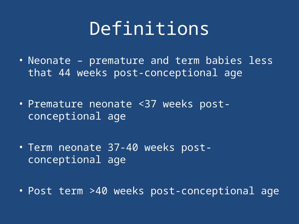
Duodenal Atresia
• 1 in 5000• Antenatal diagnosis – ‘double bubble’• Associated with Trisomy 21 - 30%, malrotation• Milky or bilious vomiting depending on level
of obstruction in relation to bile duct• 85% obstruction distal to bile duct
• Side to side duodenoduodenostomy


Malrotation +/- volvulus

Malrotation• 1 in 6000 present in
babies– 0.5% of autopsies show
degree of malrotation
• Abnormal duodenal loop• Narrow mesentery• Peritoneal band ‘Ladds’
bands from caecum to lateral abdominal wall
• Clockwise torsion of entire midgut

Malrotation + volvulus
• SURGICAL EMERGENCY• Bilious vomiting in neonate• Upper GI contrast to diagnose• Emergency laparotomy to devolve bowel– counterclockwise
• Total gut necrosis – life threatening

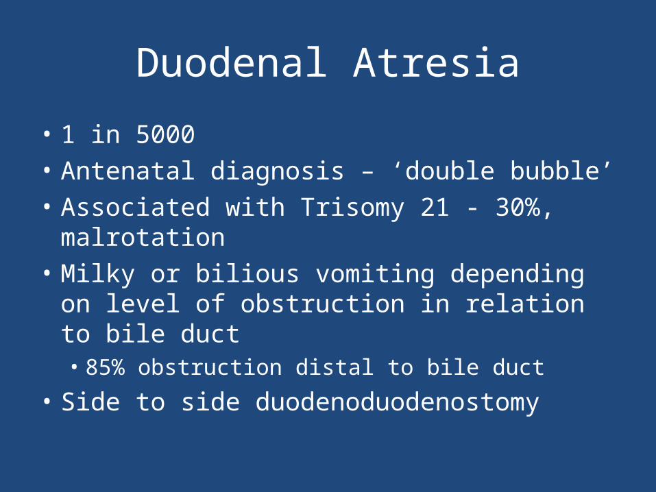
Jejunal/Ileal atresia
• Stenosis – 11%• Type 1 – 23% • Type 2 – 10%• Type 3 – 35%• Type 4 – 21%
Jejunal/ileal atresia
• 1 in 5000 births• Aetiology – antenatal vascular compromise• May have short bowel• Resection and anastomosis– May be multiple– May require tapering– May be end to end or end to side depending on
discrepancy

Meconium Ileus
• CF – 1 in 2500 births• ~16% of babies with CF• Inspissated sticky meconium– Distal small bowel obstruction– May be complicated
• Microcolon on contrast enema– may be therapeutic
• Contrast enema• Laparotomy and washout of bowel +/- stoma

Microcolon in Meconium ileus

Hirschsprung Disease

Hirschsprung Disease
• 1 in 5000 births• M:F 4:1• Associated with Trisomy 21• Delayed passage of meconium >48hours• Abdominal distension• Vomiting – may be bilious• Diagnosis – rectal biopsy– Aganglionosis, thickened nerve trunks, increased
acetylcholinesterase

Hirschsprung Disease• Aganglionosis of bowel• Variable failure of neural crest cell migration– Rectosigmoid – 75%– Long (colonic) segment – 15%– Total colonic – 5-7%– Total interstinal – <5%
• Spastic bowel – failure to relax• Requires decompression – rectal washouts• Definitive surgery – pullthrough of ganglionic bowel

Anorectal malformation
Anorectal malformation
• 1 in 4000 births• Management depends on level of ARM• Primary anoplasty for low• Stoma and delayed reconstruction for high– Recto-urethral fistula most common in boys– Recto-vestibular fistula most common in girls

Case 2
• 3 week old term baby• Relevant points in history• Relevant examination findings• Differential diagnosis

Infantile Hypertrophic Pyloric Stenosis
• 1-4:1000, M:F 4:1• Overgrowth of pyloric muscle• Gastric outlet obstruction• Increasing non-bilious vomiting• Metabolic derangement– Hypochloremic– Hypokalaemic– Metabolic alkalosis
• Medical emergency - rehydration

Pyloric stenosis

Infantile Hypertrophic Pyloric Stenosis
• Diagnosis – palpable mass on ‘test feed’• USS– Pyloric length >16mm– Single muscle thickness >4mm
• Pyloromyotomy– Open – supraumbilical or RUQ– Laparoscopic

Inguinal hernia

Inguinal hernia
• Usually can reduce• If truly incacerated – emergency exploration• Otherwise if premature baby or younger than
4 weeks post birth – repair urgent basis

Case 3
• Preterm neonate – bilious vomiting• Born 27 weeks gestation• Weight 1 kg• 1 week post birth• Relevant points in history• Relevant examination findings• Differential diagnosis


Necrotising Enterocolitis
• 90% in preterm 10% in term babies• ~5% of all babies admitted to Neonatal Unit• Multifactorial pathogenesis– Inflammation and coagulative necrosis
• 20-40% require surgery– Up to 50% mortality reported in those requiring
surgery
• Worst outcome extremely low weight preterm babies

Necrotising Enterocolitis
• Surgery indicated for:– Worsening clinical condition despite maximal
supportive therapy– Perforation
• Laparotomy– Assess extent of disease - may be total gut necrosis– Resection anastomosis – if appropriate– Resection and stomas– ‘Clip and drop’

Summary
• Many surgical causes of surgical neonatal vomiting
• Congenital obstructive and functional anomalies throughout entire gut
• Green vomiting is malrotation and volvulus until proven otherwise – Emergency