Embed Size (px)
Citation preview
IDEAS AND TECHNICAL INNOVATIONS
Surgical planning, manufacturing and implantationof an individualized cervical fusion titanium cageusing patient-specific data
Uwe Spetzger1 • Miles Frasca2 • Stefan Alexander Konig1
Received: 31 July 2015 / Revised: 18 February 2016 / Accepted: 19 February 2016
� Springer-Verlag Berlin Heidelberg 2016
Abstract
Background Most cervical fusion cages imperfectly
mimic the anatomy of the intervertebral disc space. The
production of individualized cages might be the next step
to further improve spinal implants due to their enhanced
load-bearing surface.
Objective To evaluate the planning, manufacturing, and
implantation of an individualized cervical cage in co-op-
eration with EIT and 3D Systems.
Methods A digital 3D model of the patient’s cervical
spine was rendered from the patients CT data. It was then
possible to correct degenerative deformities by digitally
repositioning the vertebrae and virtually resecting the
osteophytes. The implantation of the cage can be simulated
to check the accuracy of the fit. The cage is made of tra-
becular titanium and manufactured by Direct Metal
Printing.
Results The pilot project for the implantation of the first
individualized cervical cage ever, resulted in a highly
accurate fit. During surgery, the cage self-located into the
correct position after suspending distraction due to the
implants unique end plate design. Furthermore, it was
impossible to move the cage in any direction with the
inserting instrument after suspending distraction for the
same reason. Thus, it can be assumed that an individualized
cervical implant provides excellent primary stability.
Conclusion Preconditions for the manufacturing of indi-
vidualized cervical fusion cages using specific patient data
are given. The implantation is uncomplicated. The
improved load-bearing surface will lower the rate of
implant dislocation and subsidence. The production of
individualized cages at a reasonable price has to be eval-
uated by spine surgeons and the industry.
Keywords Titanium cage � Cervical spine � Fusion �Patient data � Computer-aided design � 3D modeling �Manufacturing
Introduction
The implantation of intervertebral fusion cages made of
polyether-etherketone or titanium with or without anterior
plating has been the standard surgical therapy for the
treatment of spondylotic cervical myelopathy and/or
radiculopathy for many years [1–4].
The numerous cervical cages offered by the industry
mimic the anatomy of the intervertebral disc space more or
less whereat size and design of the cages are adapted to
average shapes and sizes of intervertebral discs.
For the reconstruction of several bony defects in the
human body such as skull defects individualized implants
based on the patient’s CT data have been used on a routine
base for many years [5–8]. Thus, it was a logical step to
evaluate the technical possibilities for the manufacturing of
individualized fusion implants for the cervical spine toge-
ther with our industrial partners EIT (Emerging Implant
Technologies GmbH, Tuttlingen, Germany) and 3D Sys-
tems (Rock Hill, SC, USA).
Electronic supplementary material The online version of thisarticle (doi:10.1007/s00586-016-4473-9) contains supplementarymaterial, which is available to authorized users.
& Stefan Alexander Konig
1 Neurochirurgische Klinik, Klinikum Karlsruhe, Moltkestr.
90, 76133 Karlsruhe, Germany
2 3D Systems Corporation, Rock Hill, SC, USA
123
Eur Spine J
DOI 10.1007/s00586-016-4473-9
This development aims to create an implant that per-
fectly fits to the endplates of the adjacent vertebral bodies
to avoid fusion complications such as secondary disloca-
tion or subsidence of the cage into the bone [9–11].
Methods
Surgical planning
Using a DICOM CT dataset, a 3D model of the patient’s
cervical spine is rendered (‘as-scanned anatomy’, Fig. 1).
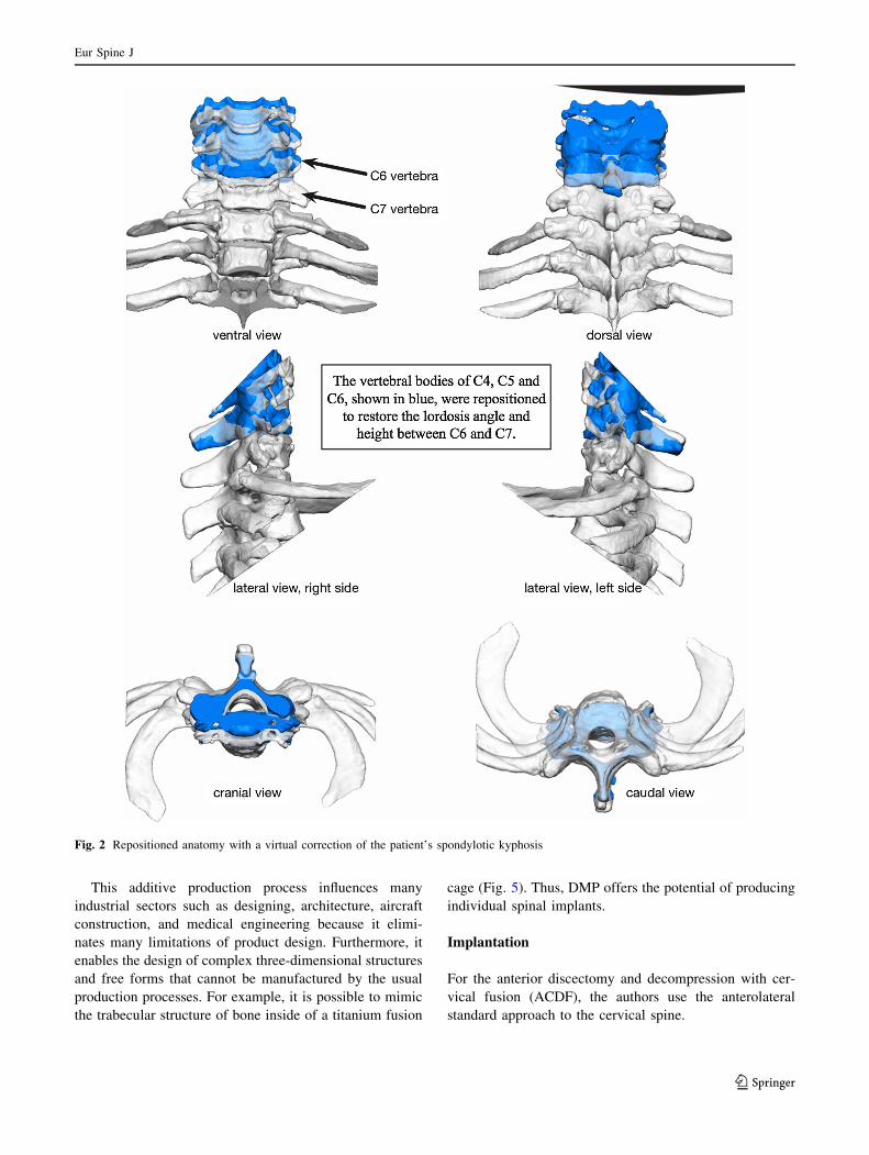
After analyzing the 3D model with emphasis on
deformities such as kyphosis, it is possible to virtually
correct these deformities by repositioning vertebrae. By
this procedure, the individualized cage obtains the ideal
lordotic angle for restoring the sagittal balance of the
cervical spine.
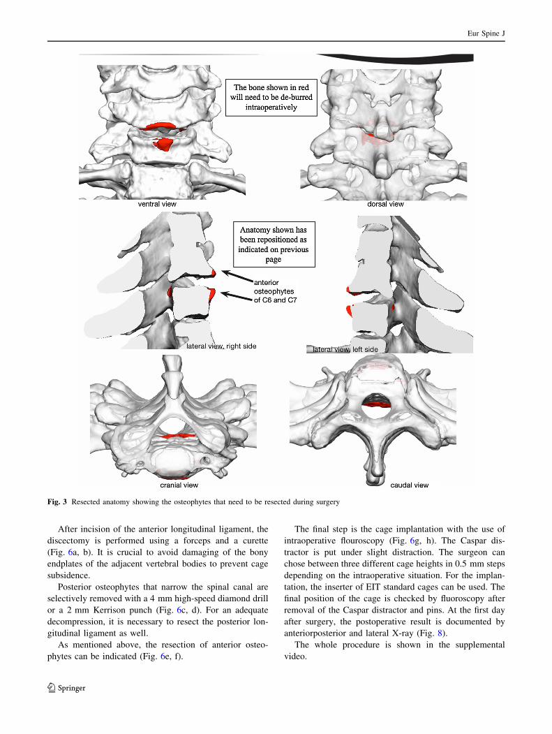
The next planning step is the virtual resection of
osteophytes (‘resected anatomy’, Fig. 2). The resection of
posterior osteophytes is often necessary for the decom-
pression of the spinal cord and nerve roots. The resection of
anterior osteophytes should be considered if they obstruct
the entrance into the disc space or in case of symptomatic
dysphagia.
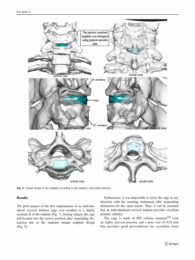
Afterwards the cage implantation can be simulated to
check the implants accuracy of fit (‘implant placement’,
Fig. 3). The determination of the optimum height of the
implant takes the height and facet joint orientation of
adjacent levels into consideration. The planning starts with
the data of an EIT standard titanium cage which is modified
according to the patients individual anatomy.
After the rendering of an implant that matches the
individual shape of the patients endplate anatomy, it is
possible to simulate different implant heights (Fig. 4).
Beside the height of the intervertebral disc space as
determined in the 3D CT model the design and manufac-
turing process includes the production of two more cages
with a height that is lower by 0.5 and 1.0 mm, respectively,
than the original height to have two more options during
surgery (Fig. 4).
Manufacturing
The EIT titanium cages are manufactured slice by slice
using the modern additive production process DMP (direct
metal printing). During this process, a very thin powder
layer of the titanium alloy Ti6AL 4V is applied to a base
plate. The titanium alloy powder is completely melted by a
laser beam and makes up a tight layer after consolidation.
After this process, the base plate is lowered by 30–50 lm,
and the next layer is applied. This procedure is repeated
until all layers are completed and the cage reached its final
shape.
The guidance of the laser beam is carried out by a 3D
CAD software that divides the device into several layers
and calculates the lanes of the laser.
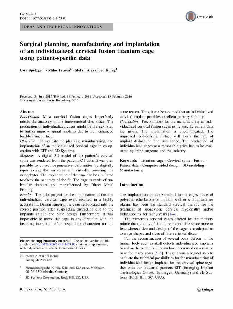
Fig. 1 Flow diagram showing
the process from planning to
implantation
Eur Spine J
123
This additive production process influences many
industrial sectors such as designing, architecture, aircraft
construction, and medical engineering because it elimi-
nates many limitations of product design. Furthermore, it
enables the design of complex three-dimensional structures
and free forms that cannot be manufactured by the usual
production processes. For example, it is possible to mimic
the trabecular structure of bone inside of a titanium fusion
cage (Fig. 5). Thus, DMP offers the potential of producing
individual spinal implants.
Implantation
For the anterior discectomy and decompression with cer-
vical fusion (ACDF), the authors use the anterolateral
standard approach to the cervical spine.
Fig. 2 Repositioned anatomy with a virtual correction of the patient’s spondylotic kyphosis
Eur Spine J
123
After incision of the anterior longitudinal ligament, the
discectomy is performed using a forceps and a curette
(Fig. 6a, b). It is crucial to avoid damaging of the bony
endplates of the adjacent vertebral bodies to prevent cage
subsidence.
Posterior osteophytes that narrow the spinal canal are
selectively removed with a 4 mm high-speed diamond drill
or a 2 mm Kerrison punch (Fig. 6c, d). For an adequate
decompression, it is necessary to resect the posterior lon-
gitudinal ligament as well.
As mentioned above, the resection of anterior osteo-
phytes can be indicated (Fig. 6e, f).
The final step is the cage implantation with the use of
intraoperative flouroscopy (Fig. 6g, h). The Caspar dis-
tractor is put under slight distraction. The surgeon can
chose between three different cage heights in 0.5 mm steps
depending on the intraoperative situation. For the implan-
tation, the inserter of EIT standard cages can be used. The
final position of the cage is checked by fluoroscopy after
removal of the Caspar distractor and pins. At the first day
after surgery, the postoperative result is documented by
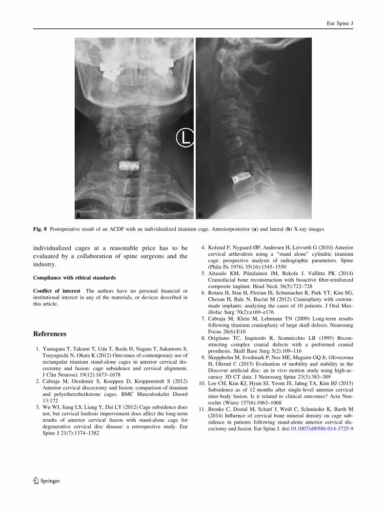
anteriorposterior and lateral X-ray (Fig. 8).
The whole procedure is shown in the supplemental
video.
Fig. 3 Resected anatomy showing the osteophytes that need to be resected during surgery
Eur Spine J
123
Results
The pilot project of the first implantation of an individu-
alized cervical titatium cage ever resulted in a highly
accurate fit of the implant (Fig. 7). During surgery the cage
self-located into the correct position after suspending dis-
traction due to the implants unique endplate design
(Fig. 5).
Furthermore, it was impossible to move the cage in any
direction with the inserting instrument after suspending
distraction for the same reason. Thus, it can be assumed
that an individualized cervical implant provides excellent
primary stability.
The cage is made of EIT cellular titaniumTM with
an eighty percent porosity and a pore size of 0.65 mm
that provides good preconditions for secondary bony
Fig. 4 Virtual design of the implant according to the patient’s individual anatomy
Eur Spine J
123
fusion without an additional synthetic bone graft
(Fig. 5).
Discussion
Individualized cages for cervical fusion for the surgical
treatment of spondylotic cervical myelopathy and/or radicu-
lopathy have the potential to be the next step in the develop-
ment of up-to-date spinal implants [1, 2]. Especially in
neurosurgery there is an analogy to computer-aided design
cranial implants for the reconstruction of skull defects [5–8].
Nevertheless, the manufacturing process of individual-
ized spinal implants is much more complex because of the
spine’s contour and function. Thus, the production process
takes much more time, effort, and costs compared to a
standard cage especially when considering the planning
and manufacturing procedures. Beside these disadvantages,
Fig. 5 Finished design of the individual implant with three different heights in 0.5 mm steps
Fig. 6 The actual implant with a macro- and microcellular trabecular structure for improved osseointegration. Cranial endplate (a) and lateral
view (b)
Eur Spine J
123

there are some potential advantages in avoiding implant-
related complications: a better load-bearing surface, and a
lower rate of implant dislocation and cage subsidence into
the bony endplates of adjacent vertebral bodies. As a result
of this, there will be a lower rate of revision surgeries.
It is likely that the production of a higher number of
individualized cages will lower the implant costs. The
biggest challenge will be the process optimization to
minimize the additional cost.
Conclusion
The technical preconditions for the planning and manu-
facturing of individualized cervical fusion cages using
specific patient data are given. The implantation of these
cages is as uncomplicated as the implantation of standard
cages. The improved load-bearing surface will be proba-
bly able to reduce the rate of implant dislocation and cage
subsidence. If it will be possible to produce such
Fig. 7 Surgical steps of the implantation. Discectomy (a) with
removal of the cartilage attached to the bony endplates (b). Selective
removal of posterior osteophytes using a diamond drill (c). Resection
of the thickened posterior longitudinal ligament (d). Removal of
anterior osteophytes (e, f). Checking the height of the disc space to
estimate the appropriate height for the final implant (g). Individual-
ized titanium cage in situ (h)
Eur Spine J
123
individualized cages at a reasonable price has to be
evaluated by a collaboration of spine surgeons and the
industry.
Compliance with ethical standards
Conflict of interest The authors have no personal financial or
institutional interest in any of the materials, or devices described in
this article.
References
1. Yamagata T, Takami T, Uda T, Ikeda H, Nagata T, Sakamoto S,
Tsuyuguchi N, Ohata K (2012) Outcomes of contemporary use of
rectangular titanium stand-alone cages in anterior cervical dis-
cectomy and fusion: cage subsidence and cervical alignment.
J Clin Neurosci 19(12):1673–1678
2. Cabraja M, Oezdemir S, Koeppen D, Kroppenstedt S (2012)
Anterior cervical discectomy and fusion: comparison of titanium
and polyetheretherketone cages. BMC Musculoskelet Disord
13:172
3. Wu WJ, Jiang LS, Liang Y, Dai LY (2012) Cage subsidence does
not, but cervical lordosis improvement does affect the long-term
results of anterior cervical fusion with stand-alone cage for
degenerative cervical disc disease: a retrospective study. Eur
Spine J 21(7):1374–1382
4. Kolstad F, Nygaard ØP, Andresen H, Leivseth G (2010) Anterior
cervical arthrodesis using a ‘‘stand alone’’ cylindric titanium
cage: prospective analysis of radiographic parameters. Spine
(Phila Pa 1976) 35(16):1545–1550
5. Aitasalo KM, Piitulainen JM, Rekola J, Vallittu PK (2014)
Craniofacial bone reconstruction with bioactive fiber-reinforced
composite implant. Head Neck 36(5):722–728
6. Rotaru H, Stan H, Florian IS, Schumacher R, Park YT, Kim SG,
Chezan H, Balc N, Baciut M (2012) Cranioplasty with custom-
made implants: analyzing the cases of 10 patients. J Oral Max-
illofac Surg 70(2):e169–e176
7. Cabraja M, Klein M, Lehmann TN (2009) Long-term results
following titanium cranioplasty of large skull defects. Neurosurg
Focus 26(6):E10
8. Origitano TC, Izquierdo R, Scannicchio LB (1995) Recon-
structing complex cranial defects with a preformed cranial
prosthesis. Skull Base Surg 5(2):109–116
9. Skeppholm M, Svedmark P, Noz ME, Maguire GQ Jr, Olivecrona
H, Olerud C (2015) Evaluation of mobility and stability in the
Discover artificial disc: an in vivo motion study using high-ac-
curacy 3D CT data. J Neurosurg Spine 23(3):383–389
10. Lee CH, Kim KJ, Hyun SJ, Yeom JS, Jahng TA, Kim HJ (2015)
Subsidence as of 12 months after single-level anterior cervical
inter-body fusion. Is it related to clinical outcomes? Acta Neu-
rochir (Wien) 157(6):1063–1068
11. Brenke C, Dostal M, Scharf J, Weiß C, Schmieder K, Barth M
(2014) Influence of cervical bone mineral density on cage sub-
sidence in patients following stand-alone anterior cervical dis-
cectomy and fusion. Eur Spine J. doi:10.1007/s00586-014-3725-9
Fig. 8 Postoperative result of an ACDF with an individualized titanium cage. Anteriorposterior (a) and lateral (b) X-ray images
Eur Spine J
123

![3H]Thymidine-Labeled Cells in the Rat Utricular Maculadepts.washington.edu/rubelab/personnel/popoff.pdf · Surgical procedure for minipump implantation Surgical procedures described](https://img.pdfslide.net/doc/110x75/5eb64936c4181619b96005e9/3hthymidine-labeled-cells-in-the-rat-utricular-surgical-procedure-for-minipump.jpg)