Embed Size (px)
Citation preview
146 Volume 39, Number 3compendium march 2018
Abstract: Comorbidities that negatively impact orthodontic (malocclusion), periodontal (periodontitis, deficient dentoalveolar bone volume, mucogingival), and prosthetic (structural integrity compromise from caries, attrition, and erosion) conditions can affect the general health of the patient. In addition, emerging data highlights the importance of undiagnosed airway volume deficiencies and sleep-disordered breathing conditions in the adult and pediatric population. Deficiencies in dentoalveolar bone and discrepancies in skeletal relationships can impact the volume of hard- and soft-tissue structures of the periodontium and decrease oral cavity volume. Contemporary interdisciplinary dentofacial therapy (IDT) is a key process for addressing the comprehensive problems of patients based on etiology, homeostasis, and sustainability of physiologically sound outcomes. These
continuing education 1InterdIscIplInary dentofacIal therapy
T he late Dr. Morton Amsterdam astutely wrote, “There may be different ways of treating a disease, but there can only be but one correct diagnosis.”1 The changing culture and landscape of interdisciplinary dentofa-cial therapy (IDT) suggests the need for a symbio-
sis of the primary care provider and specialist for the effective
management of dental, periodontal, and craniofacial issues. The goal of such collaborative work is to accurately and comprehen-sively diagnose and prognosticate craniofacial deficiencies. The unique perspectives and knowledge of each medico-dental pro-fessional is incorporated into an ideal plan for each individual patient. This collaborative IDT requires the leadership of the
Surgically Facilitated Orthodontic Therapy: Optimizing Dentoalveolar Bone and Space Appropriation for Facially Prioritized Interdisciplinary Dentofacial TherapyGeorge a. Mandelaris, dds, Ms; Bradley s. deGroot, dds, Ms; robert relle, dds; Brian shah, Md, dds; Iwei huang, dMd, Ms; and Brian s. Vence, dds
learnInG oBjectIVes
• Discusstheuseofsurgically
facilitatedorthodontic
therapy(SFOT)forfacially
prioritizedinterdisciplinary
dentofacialtreatment
• Reviewthebenefitsof
expandedorthodontic
approachesformanaging
thenaturaldentition
• Explaintheimportance
ofSFOTwhentreatment
planningwithafacially
prioritizedperspectivethat
involvesCBCTimaging
DISCLOSURE:Theauthorshadnodisclosurestoreport.
provide the patient with sustainable esthetics and function. Surgically facilitated orthodontic therapy (SFOT) uses corticotomies and dentoalveolar bone decortication to stimulate the regional acceleratory phenomenon and upregulate bone remodeling and tooth movement as a part of orthodontic decompensation. It also generally includes guided periodontal tissue regeneration and/or dentoalveolar bone augmentation. SFOT as a part of IDT is demanding and requires extensive attentiveness and communication among all team members. This article focuses on the role of SFOT as an integral component of contemporary IDT to facilitate highly predictable and sustainable outcomes.
147www.compendiumlive.com March 2018 coMpendiuM
patient’s restorative dentist to coordinate and amalgamate input from each specialist involved.
Evolutionary changes over the past three centuries have contrib-uted to the current prevalence of the phenomenon of “facial reces-sion.”2 This progressively retrognathic maxillary and mandibular positioning is the result of the evolutionary and cultural demands to develop a more pronounced frontal lobe of the brain. The end-point of IDT is to re-establish the homeostatic balance between the craniofacial structures and the periodontium that they sup-port. This is necessary to obtain oral health that can be sustained over a lifetime but cannot be achieved without a thorough under-standing of the embryologic and developmental processes leading to craniofacial anatomy.
Contemporary IDT begins with a facially prioritized approach and often requires the interdisciplinary team to re-establish tooth position and proportions. Space appropriation of the teeth is critical to the dentofacial management of the stomatognathic system and often requires optimizing root position within a sound periodontal foundation. This becomes especially crucial when wear (through attrition and erosion) and compensatory tooth movement (dental compensations) have occurred.
Dental compensations occur as a result of a skeletal disharmony and are commonly seen whenever anterior-posterior or trans-verse maxilla-mandibular disharmonies are present.3,4 Dental crowding is often an arch length deficiency, which corresponds to a deficiency in dentoalveolar bone volume. This limits the oppor-tunities for dental expansion and often necessitates extractions to reconcile the existing tooth mass to the available arch length. Imaging-based software programs such as Digital Smile Design (DSD) (digitalsmiledesign.com), Suresmile® (suresmile.com), or Nemotec (nemotec.com) can help bridge the gap between a facially prioritized treatment plan and optimal, patient-centered results. Such software provides greater clarity, precision, and accuracy when transitioning from patient expectations to realized endpoints.
The 3-dimensional (3D) cone-beam com-puted tomography (CBCT) imaging compo-nent of these planning software programs represents a paradigm shift in IDT. Incor-porating 3D regional anatomy into virtual planning allows a biologic conscience to guide the clinician during dental and orth-odontic planning.5 If the facially prioritized treatment plan calls for the tooth position outside of the dentoalveolar bone volume limits, alternative approaches to orthodon-tic tooth movement must be considered. Treatment such as surgically facilitated orthodontic therapy (SFOT) may allow the IDT team to accomplish outcome goals and avoid iatrogenic complications. Collabora-tive treatment planning by a cohesive IDT team must occur prior to embarking on a restorative rehabilitation or orthodontic treatment that may exceed the boundar-ies of the “orthodontic walls.”6 Failure to
respect these boundaries may lead to unstable and potentially harmful results.
The ever-important but often deficient (especially in the anterior sextant) facial bone thickness must be considered during treatment planning.7,8 The use of CBCT is critical in assessing dentoalveolar, alveoloskeletal, and skeletal relationships (as well as the anatomic structures of the temporomandibular joint) during the comprehen-sive treatment planning process.5
The aim of this article is to highlight the importance of con-temporary IDT and expatiate on the benefits of expanded orth-odontic approaches such as SFOT in the context of managing the natural dentition.
Mouth, Bone, and Airway Volumes: Historical and Evolutionary PerspectivesThe prevalence of an ideal dentofacial condition currently may be decreasing among some populations.9 Conversely, tooth crowding, retrognathia, deficient dentoalveolar bone, and other dentofacial abnormalities in both maxillary and mandibular osseous struc-tures are widespread.10,11 These anomalies may be quite prevalent, but they cannot necessarily be considered a variation of normal. Although these maladies have always existed, new tools are now available in the dental armamentarium to deal with them. Man-delaris et al have described case type patterns of common maloc-clusions that can be useful in identifying situations where SFOT can be helpful in IDT.5
The exact reason for the increase in incidences of malocclusion patterns is not known for certain. However, their rise has been cor-related with the increased consumption of highly processed foods12 and decreased breastfeeding.13-15 The imposition of a uniformly soft diet upon infants and toddlers, as well as the resulting failure to develop forward tongue and lip muscular habits, may reduce oral cavity volume (OCV). Subsequently, the impaired development of craniofacial-respiratory structures may manifest as deficient
dentoalveolar bone16,17 as well as impaired development of the maxilla and mandible (ie, hypoplasia and/or retrognathia). There has been an increasing incidence of these is-sues for approximately 250 years.18,19
The position of teeth within the mouth represents a homeostatic relationship be-tween the opposing forces of the lips, tongue, oral musculature, and alveolar bone. These tissue structures define a certain OCV. The teeth develop independently of the soft-tissue structures and require a specific vol-ume in order to properly align within the dental arch. When the volume within the oral cavity is deficient compared to the vol-ume required by the teeth, significant mal-occlusions can occur. In these cases, intra-oral forces may move teeth into positions to compensate (dental compensations) for the skeletal imbalance, and dental crowding may occur.20 Crowding of the natural dentition
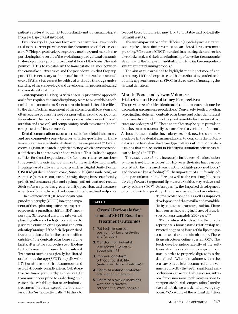
taBle 1
Overall Rationale for/ Goals of SFOT Based on
Treatment Outcomes1. Putteethincorrectpositionforfacialestheticsandfunction
2.Transformperiodontalphenotypeinordertoaccomplish#1
3.Improvelong-termorthodonticstability(reduceincidenceofrelapse)29
4.Optimizeanteriorprotectedarticulationparameters
5.Optimizeairwaydimensionswithnon-retractiveorthodontia,whenpossible
148 Volume 39, Number 3compendium march 2018
contInuInG educatIon 1 | InTERDISCIPLInaRyDEnTOFaCIaLThERaPy
is frequently accompanied by suboptimal alveolar bone thickness, resulting in dehiscences and fenestrations,21 which then limit the extent to which teeth can be safely decompensated (ie, orthodonti-cally moved).
The combined impact of these abnormalities on oropharyn-geal airway volume is an emerging focus of contemporary IDT. The most common manifestation of a suboptimal airway space is obstructive sleep apnea (OSA), which has reached epidemic pro-portions in both adults22,23 and children.24,25 This has resulted in controversy, as some research demonstrates that a reduced OCV (ie, increased tongue volume:OCV ratio) may trend patients to-ward sleep-disordered breathing conditions,26 yet other publica-tions suggest that the extraction of four bicuspids does not influ-ence sleep apnea conditions or esthetic perceptions of changes in facial profile.27,28
Orthodontic Decompensation: Re-establishing Homeostasis by Re-envisioning OutcomesAs a member of the IDT team, the orthodontist has the critical job of engineering tooth movement and architecting a facially prioritized plan. He or she is responsible for correcting the dental compensa-tions that nature facilitated in response to space misappropriation. This process, known as decompensation, includes alignment of the arches in preparation for orthognathic surgery (OGS) when a true alveoloskeletal or skeletal discrepancy exists.
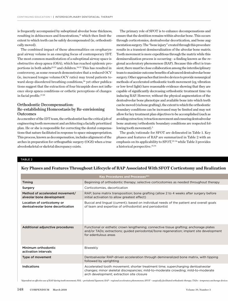
taBle 2
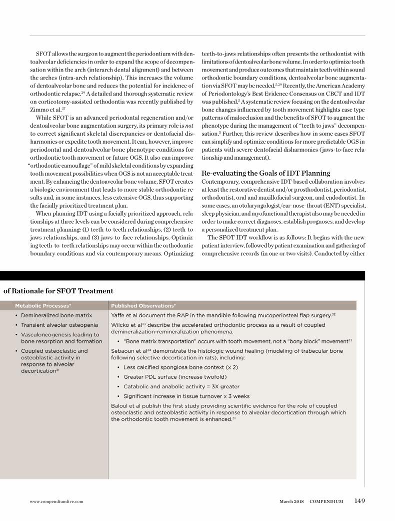
Key Phases and Features Throughout Lifecycle of RAP Associated With SFOT Corticotomy and Realization of Rationale for SFOT Treatment
Key Procedures and Processes30* Metabolic Processes* Published Observations*
timing Beginningoforthodontictherapy;selectivecorticotomiesasneededthroughouttherapy • Demineralizedbonematrix
• Transientalveolarosteopenia
• Vasculoneogenesisleadingtoboneresorptionandformation
• Coupledosteoclasticandosteoblasticactivityinresponsetoalveolardecortication31
yaffeetaldocumenttheRaPinthemandiblefollowingmucoperiostealflapsurgery.32
Wilckoetal33describetheacceleratedorthodonticprocessasaresultofcoupleddemineralization-remineralizationphenomena.
• “Bonematrixtransportation”occurswithtoothmovement,nota“bonyblock”movement33
Sebaounetal34demonstratethehistologicwoundhealing(modelingoftrabecularbonefollowingselectivedecorticationinrats),including:
• Lesscalcifiedspongiosabonecontext(x2)
• GreaterPDLsurface(increasetwofold)
• Catabolicandanabolicactivity=3Xgreater
• Significantincreaseintissueturnoverx3weeks
Balouletalpublishthefirststudyprovidingscientificevidencefortheroleofcoupledosteoclasticandosteoblasticactivityinresponsetoalveolardecorticationthroughwhichtheorthodontictoothmovementisenhanced.31
surgery Corticotomies,decortication
Method of accelerated movement/ alveolar bone development
RaP;bonematrixtransposition;bonegrafting(allow2to4weeksaftersurgerybeforeinitialactivationtoallowgreatesteffect)
location of corticotomy or dentoalveolar bone decortication
Buccalandlingual(current);basedonindividualneedsofthepatientandoverallgoalsofteamandexpertiseoforthodontistandperiodontist
additional adjunctive procedures Functionalorestheticcrownlengthening;connectivetissuegrafting;anchorageplatesand/orTaDs;extractions;guidedperiodontal/boneregeneration;implantsitedevelopmentforedentulousareas
Minimum orthodontic activation intervals
Biweekly
type of movement DentoalveolarRaP-drivenaccelerationthroughdemineralizedbonematrix,withtippingfollowedbyuprighting
Indications acceleratedtoothmovement;shortertreatmenttime;superchargingdentoalveolarchanges;minorskeletaldiscrepancies;mild-to-moderatecrowding;mild-to-moderatearchdevelopment;extractionsiteclosure
*dependent on effective use of RAP during tooth movement; PDL = periodontal ligament; RAP = regional acceleratory phenomenon; SFOT = surgically facilitated orthodontic therapy; TADs = temporary anchorage devices
The primary role of SFOT is to enhance decompensations and ensure that the dentition remains within alveolar bone. This occurs through corticotomies, dentoalveolar decortication, and bone aug-mentation surgery. The “bone injury” created through this procedure results in a transient demineralization of the alveolar bone matrix. Tooth movement is more expeditious through the matrix while this demineralization process is occurring—a finding known as the re-gional acceleratory phenomenon (RAP). Because this effect is tran-sient, there must be close collaboration among the interdisciplinary team to maximize outcome benefits of advanced dentoalveolar bone surgery. Other approaches that involve devices to provide nonsurgical methods of accelerated orthodontic tooth movement (eg, vibration or low-level light) have reasonable evidence showing that they are capable of significantly decreasing orthodontic treatment time via inducing RAP. However, without the physical augmentation of the dentoalveolar bone phenotype and available bone into which teeth can be moved (via bone grafting), the extent to which the orthodontic boundary conditions can be increased may be limited and may not allow for key treatment plan objectives to be accomplished (such as avoiding extraction/retraction movement and ensuring dentoalveolar bone anatomy/orthodontic boundary conditions are respected fol-lowing tooth movement).5
The goals/rationale for SFOT are delineated in Table 1. Key phases and features of RAP are summarized in Table 2 with an emphasis on its applicability to SFOT,29-34 while Table 3 provides a historical perspective.35,36
149www.compendiumlive.com March 2018 coMpendiuM
teeth-to-jaws relationships often presents the orthodontist with limitations of dentoalveolar bone volume. In order to optimize tooth movement and produce outcomes that maintain teeth within sound orthodontic boundary conditions, dentoalveolar bone augmenta-tion via SFOT may be needed.5,29 Recently, the American Academy of Periodontology’s Best Evidence Consensus on CBCT and IDT was published.5 A systematic review focusing on the dentoalveolar bone changes influenced by tooth movement highlights case type patterns of malocclusion and the benefits of SFOT to augment the phenotype during the management of “teeth to jaws” decompen-sation.5 Further, this review describes how in some cases SFOT can simplify and optimize conditions for more predictable OGS in patients with severe dentofacial disharmonies ( jaws-to-face rela-tionship and management).
Re-evaluating the Goals of IDT PlanningContemporary, comprehensive IDT-based collaboration involves at least the restorative dentist and/or prosthodontist, periodontist, orthodontist, oral and maxillofacial surgeon, and endodontist. In some cases, an otolaryngologist/ear-nose-throat (ENT) specialist, sleep physician, and myofunctional therapist also may be needed in order to make correct diagnoses, establish prognoses, and develop a personalized treatment plan.
The SFOT IDT workflow is as follows: It begins with the new-patient interview, followed by patient examination and gathering of comprehensive records (in one or two visits). Conducted by either
SFOT allows the surgeon to augment the periodontium with den-toalveolar deficiencies in order to expand the scope of decompen-sation within the arch (interarch dental alignment) and between the arches (intra-arch relationship). This increases the volume of dentoalveolar bone and reduces the potential for incidence of orthodontic relapse.29 A detailed and thorough systematic review on corticotomy-assisted orthodontia was recently published by Zimmo et al.37
While SFOT is an advanced periodontal regeneration and/or dentoalveolar bone augmentation surgery, its primary role is not to correct significant skeletal discrepancies or dentofacial dis-harmonies or expedite tooth movement. It can, however, improve periodontal and dentoalveolar bone phenotype conditions for orthodontic tooth movement or future OGS. It also can improve
“orthodontic camouflage” of mild skeletal conditions by expanding tooth movement possibilities when OGS is not an acceptable treat-ment. By enhancing the dentoaveolar bone volume, SFOT creates a biologic environment that leads to more stable orthodontic re-sults and, in some instances, less extensive OGS, thus supporting the facially prioritized treatment plan.
When planning IDT using a facially prioritized approach, rela-tionships at three levels can be considered during comprehensive treatment planning: (1) teeth-to-teeth relationships, (2) teeth-to-jaws relationships, and (3) jaws-to-face relationships. Optimiz-ing teeth-to-teeth relationships may occur within the orthodontic boundary conditions and via contemporary means. Optimizing
Key Phases and Features Throughout Lifecycle of RAP Associated With SFOT Corticotomy and Realization of Rationale for SFOT Treatment
Key Procedures and Processes30* Metabolic Processes* Published Observations*
timing Beginningoforthodontictherapy;selectivecorticotomiesasneededthroughouttherapy • Demineralizedbonematrix
• Transientalveolarosteopenia
• Vasculoneogenesisleadingtoboneresorptionandformation
• Coupledosteoclasticandosteoblasticactivityinresponsetoalveolardecortication31
yaffeetaldocumenttheRaPinthemandiblefollowingmucoperiostealflapsurgery.32
Wilckoetal33describetheacceleratedorthodonticprocessasaresultofcoupleddemineralization-remineralizationphenomena.
• “Bonematrixtransportation”occurswithtoothmovement,nota“bonyblock”movement33
Sebaounetal34demonstratethehistologicwoundhealing(modelingoftrabecularbonefollowingselectivedecorticationinrats),including:
• Lesscalcifiedspongiosabonecontext(x2)
• GreaterPDLsurface(increasetwofold)
• Catabolicandanabolicactivity=3Xgreater
• Significantincreaseintissueturnoverx3weeks
Balouletalpublishthefirststudyprovidingscientificevidencefortheroleofcoupledosteoclasticandosteoblasticactivityinresponsetoalveolardecorticationthroughwhichtheorthodontictoothmovementisenhanced.31
surgery Corticotomies,decortication
Method of accelerated movement/ alveolar bone development
RaP;bonematrixtransposition;bonegrafting(allow2to4weeksaftersurgerybeforeinitialactivationtoallowgreatesteffect)
location of corticotomy or dentoalveolar bone decortication
Buccalandlingual(current);basedonindividualneedsofthepatientandoverallgoalsofteamandexpertiseoforthodontistandperiodontist
additional adjunctive procedures Functionalorestheticcrownlengthening;connectivetissuegrafting;anchorageplatesand/orTaDs;extractions;guidedperiodontal/boneregeneration;implantsitedevelopmentforedentulousareas
Minimum orthodontic activation intervals
Biweekly
type of movement DentoalveolarRaP-drivenaccelerationthroughdemineralizedbonematrix,withtippingfollowedbyuprighting
Indications acceleratedtoothmovement;shortertreatmenttime;superchargingdentoalveolarchanges;minorskeletaldiscrepancies;mild-to-moderatecrowding;mild-to-moderatearchdevelopment;extractionsiteclosure
*dependent on effective use of RAP during tooth movement; PDL = periodontal ligament; RAP = regional acceleratory phenomenon; SFOT = surgically facilitated orthodontic therapy; TADs = temporary anchorage devices
150 Volume 39, Number 3compendium march 2018
the general dentist or prosthodontist (whomever is responsible for restorative oversight and leadership), this includes obtaining clini-cal data through comprehensive exam, photographs, high-resolution pulse oximetry (HRPO)/home sleep study, intraoral scanning, and mounted study models, and imaging diagnostics via full-mouth digital radiographs, CBCT imaging, and magnetic resonance imaging (MRI) when needed. The next step in the workflow is co-discovery and comprehensive treatment planning, with input from team members representing periodontics, orthodontics, oral and maxillofacial surgery, and any others needed, such as primary care physician, ENT, pediatrics, endodontics, and the dental laboratory. In grand rounds, all the IDT team members evaluate the gathered patient data, and an action plan, steered by re-storative goals, is generated. The following steps are then: (1) disease control and pro-visionalization (pre-SFOT restorative therapy), (2) pre-SFOT orthodontics, (3) SFOT surgery, (4) SFOT orthodon-tics, (5) interim orthodontic transitional restorations, (6) orthodontic refinement and finishing, (7) orthognathic surgery (if required), (8) pros-thetic phase completion as determined by esthetic and functional goals, and, finally, (9) supportive periodontal maintenance.
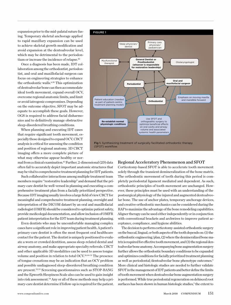
The treatment synthesis of the SFOT workflow used in contem-porary IDT is exhibited in Figure 1.
IDT has to consider the aforementioned limitations of the exist-ing dentoalveolar bone, the volume of which will dictate the extent to
which tooth movement is biologically safe. This determinant of tooth movement has been termed the “orthodontic walls”6 and is often a limiting factor in treatment due to inadequate bone thickness.38 In 2013, Mandelaris et al published a CBCT-based classification system that can be utilized prior to orthodontic therapy to help determine the relative safety and risk of tooth movement.39 This system evalu-ates the thickness of both the crestal and radicular dentoalveolar bone (thick, >1 mm; or thin, <1 mm) for the classification of patients into four distinct phenotypical presentations. The use of such a sys-tem may help not only in determining but also in conveying the im-portance of alternative orthodontic approaches, such as SFOT.
Restorative LeadershipFrequently, aberrant tooth position and structural compromise of the dentition (from attrition and erosion) make ideal dentistry impossible. In such cases, orthodontic treatment becomes neces-sary to re-appropriate space in order to facilitate anatomically cor-rect prosthetic dentistry. However, the benefits of idealizing tooth form and position are not limited to restorative outcomes. In the contemporary IDT paradigm, the recognition of airway problems (frequently accompanied by retrognathia40,41) as a component of dental-related treatment planning is critically important. This emerging scope of the restorative dental practice should empha-size early and, of course, accurate diagnosis and prevention. From a dental perspective, the outcome goal of airway management can be envisioned in terms of expanding OCV in conjunction with hard-tissue space appropriation to allow for ideal functional and esthetic prosthodontics.
Sleep-disordered breathing conditions are not unlike other sys-temic health problems in that if they are diagnosed and treated early, fewer negative sequelae and more favorable outcomes will likely result. Even mild forms of these conditions, such as upper airway resistance syndrome or respiratory effort related arousals, can be addressed before they begin to harm the patient. Because of the ability to influence growth in skeletally immature patients, orthodontic and dentofacial intervention can be instrumental in idealizing space appropriation and increasing OCV. However, this requires enlisting alternative orthodontic approaches such as SFOT and/or using temporary skeletal anchorage in rapid palatal
contInuInG educatIon 1 | InTERDISCIPLInaRyDEnTOFaCIaLThERaPy
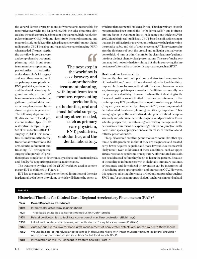
Year Event/Procedure Introduced
1893 Interalveolarosteotomy(Cunningham)
1921 Threebasicstrategiestocorrectmalocclusion(Cohn-Stock)
1931 Palatalcorticotomiestofacilitatecorrectionofmaxillaryprotrusion(Bichlmayr)
1959 Labialandpalatalcorticotomies,withorthodontic“bonyblockmovement”(Köle)
1968 autogenoushipmarrowforbone-graftmanagementofbonycraterdefectsaroundnaturalteeth(Schallhorn)
1969 Woundhealingofinteralveolarosteotomiesinrhesusmonkeyswithintactmucoperiosteum;collateralcirculationplusvascularanastomosespreservebone/pulpbloodsupply(Bell)
1983 IntroductionoftheRaPconceptinfracturehealing(Frost)36
taBle 3
Historical Timeline for Clinical Use of Regional Acceleratory Phenomenon (RAP)35
The next step in the workflow is
co-discovery and comprehensive
treatment planning, with input from team
members representing periodontics,
orthodontics, oral and maxillofacial surgery,
and any others needed, such as primary
care physician, ENT, pediatrics,
endodontics, and the dental laboratory.
151www.compendiumlive.com March 2018 coMpendiuM
expansion prior to the mid-palatal suture fus-ing. Temporary skeletal anchorage applied to rapid maxillary expansion can be used to achieve skeletal growth modification and avoid expansion at the dentoalveolar level, which may be detrimental to the periodon-tium or increase the incidence of relapse.42
Once a diagnosis has been made, IDT col-laboration among the orthodontist, periodon-tist, and oral and maxillofacial surgeon can focus on engineering strategies to enhance the orthodontic walls.6,38 This optimization of dentoalveolar bone can then accommodate ideal tooth movement, expand overall OCV, overcome regional anatomic limits, and limit or avoid iatrogenic compromises. Depending on the outcome objective, SFOT may be ad-equate to accomplish these goals. However, OGS is required to address facial disharmo-nies and to definitively manage obstructive sleep-disordered breathing conditions.
When planning and executing IDT cases that require significant tooth movement, es-pecially those designed to expand OCV, CBCT analysis is critical for assessing the condition and position of regional anatomy. 3D CBCT imaging offers a more complete picture of what may otherwise appear healthy or nor-mal from a clinical examination.43 Further, 2-dimensional (2D) data often fail to accurately depict important anatomic structures that may be vital to comprehensive treatment planning for IDT patients.
Such collaborative interactions among multiple treatment team members require “restorative leadership” and demand that the pri-mary-care dentist be well-versed in planning and executing a com-prehensive treatment plan from a facially prioritized perspective. Because IDT imaging usually involves a large field of view CBCT for meaningful and comprehensive treatment planning, oversight and interpretation of the DICOM dataset by an oral and maxillofacial radiologist (OMFR) should be considered to optimize patient safety, provide medicolegal documentation, and allow inclusion of OMFR patient interpretation for the IDT team during treatment planning.
Even dentists who may be uncomfortable managing complex cases have a significant role in improving patient health. A patient’s primary-care dentist is often the most frequent oral healthcare contact for the patient. The dentist is uniquely positioned to evalu-ate a worn or crowded dentition, assess sleep-related dental and airway anatomy, and make appropriate specialty referrals. CBCT and other applicable 3D modalities can be used to assess tongue volume and position in relation to total OCV.26,44-51 The presence of tongue crenations may be an indication that an OCV problem and possible undiagnosed sleep-disordered breathing condition are present.52,53 Screening questionnaires such as STOP-BANG and the Epworth Sleepiness Scale also can be used to gain insight into risk assessment.54 Any or all of these methods may help a pri-mary-care dentist determine if follow-up is required for the patient.
Regional Acceleratory Phenomenon and SFOTCorticotomy-based SFOT is able to accelerate tooth movement solely through the transient demineralization of the bone matrix. The orthodontic movement of teeth during this period is com-pletely periodontal ligament mediated and dependent. As such, orthodontic principles of tooth movement are unchanged. How-ever, these principles must be used with an understanding of the postsurgical physiology of the injured and augmented dentoalveo-lar bone. The use of anchor plates, temporary anchorage devices, and creative orthodontic mechanics can be considered during the RAP to maximize the advantage of the bone remodeling capabilities. Aligner therapy can be used either independently or in conjunction with conventional brackets and archwires to improve patient ac-ceptance, compliance, and hygiene abilities.
The decision to perform corticotomy-assisted orthodontic surgery on the buccal, lingual, or both aspects of the tooth depends on: (1) the orthodontic engineering/plan, (2) where the demineralized bone ma-trix is required for effective tooth movement, and (3) the regional den-toalveolar bone anatomy. Accompanying bone augmentation surgery further allows the orthodontic boundary conditions to be expanded and optimizes conditions for facially prioritized treatment planning as well as periodontal/dentoalveolar bone phenotype outcomes.5 More clinical and histologic studies are needed to further validate SFOT in the management of IDT patients and better define the limits of tooth movement when dentoalveolar bone augmentation surgery is performed. While true periodontal regeneration on dehisced root surfaces has been shown in human histologic studies,5 the extent to
fig 1. Synthesizingtreatmentofsurgicallyfacilitatedorthodontictherapy(SFOT)workflow.
fIGure 1
Periodontist
Orthodontist
Oral and maxillofacial surgeon
Patient education (neededas part of patient-centric
treatment planning model)
Re-establish normal physiologic conditions
Emphasis on moving maxillaand mandible forward
Myofunctionaltherapist Otolaryngologist
Primary care physician/
pediatrician
Sleep physician/dentist
General Dentist or Prosthodontist
(whoever is responsible for restorative leadership)
Use SFOT and
optimize beneficial e�ectsorthognathic surgery to
on oral cavity and airway volume and associated
systemic health parameters
152 Volume 39, Number 3compendium march 2018
which bone augmentation volume is gained and then maintained over time via a SFOT treatment model remains to be shown.
Completing the “Restoratively Driven Circle” in Comprehensive IDTThe outcome goal of SFOT and OGS is to achieve ideal root and tooth position in biologically sound locations within the orthodon-tic walls/boundary conditions, mange skeletal discrepancies, and achieve facial harmony. This sets the stage for creation of the most occlusally stable and proportionally correct dental rehabilitation.
When used for IDT in this manner, the primary objective of SFOT is health and stability as opposed to expeditious tooth movement. However, the overall reduction in orthodontic treat-ment time can indeed be critical for the patient who requires OGS in the context of general health management, especially in the presence of airway compromise such as OSA. Here, it can become urgent to expedite decompensations. Timely manage-ment of OSA is important to reduce the impact of its myriad systemic effects.55 SFOT can reduce the time required to pre-pare the patient for OGS without sacrificing the periodontium
contInuInG educatIon 1 | InTERDISCIPLInaRyDEnTOFaCIaLThERaPy
fig 3.
fig 5. fig 4.
fig 7.
fig 2.
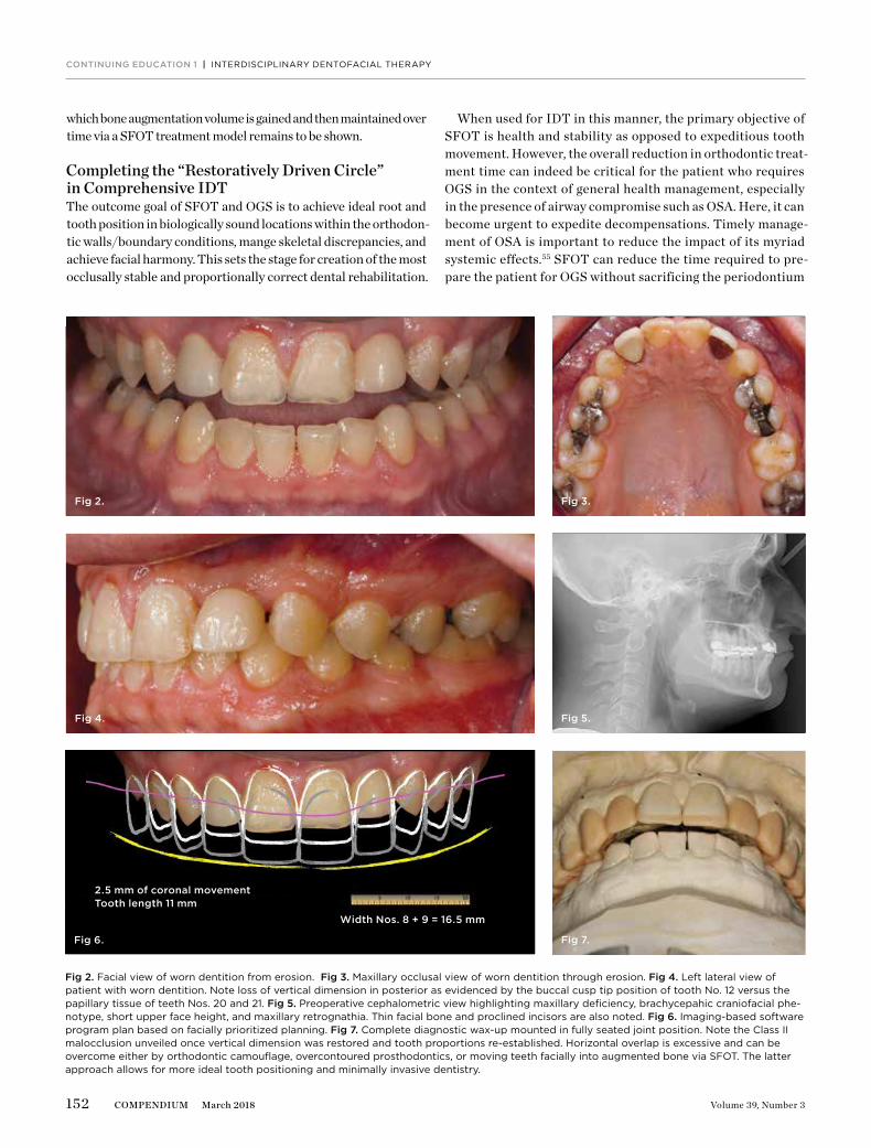
fig 2.Facialviewofworndentitionfromerosion.fig 3.Maxillaryocclusalviewofworndentitionthrougherosion.fig 4.Leftlateralviewofpatientwithworndentition.notelossofverticaldimensioninposteriorasevidencedbythebuccalcusptippositionoftoothno.12versusthepapillarytissueofteethnos.20and21.fig 5.Preoperativecephalometricviewhighlightingmaxillarydeficiency,brachycepahiccraniofacialphe-notype,shortupperfaceheight,andmaxillaryretrognathia.Thinfacialboneandproclinedincisorsarealsonoted.fig 6.Imaging-basedsoftwareprogramplanbasedonfaciallyprioritizedplanning.fig 7.Completediagnosticwax-upmountedinfullyseatedjointposition.notetheClassIImalocclusionunveiledonceverticaldimensionwasrestoredandtoothproportionsre-established.horizontaloverlapisexcessiveandcanbeovercomeeitherbyorthodonticcamouflage,overcontouredprosthodontics,ormovingteethfaciallyintoaugmentedboneviaSFOT.Thelatterapproachallowsformoreidealtoothpositioningandminimallyinvasivedentistry.
fig 6.
Width nos. 8 + 9 = 16.5 mm
2.5 mm of coronal movementtooth length 11 mm
153www.compendiumlive.com March 2018 coMpendiuM
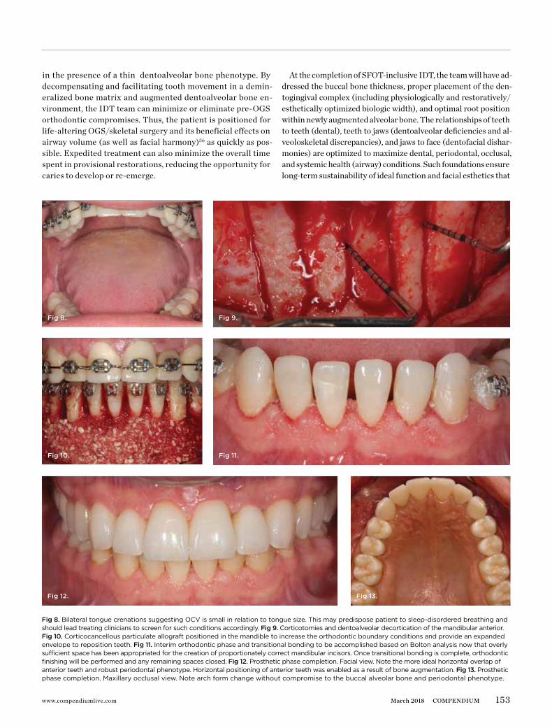
in the presence of a thin dentoalveolar bone phenotype. By decompensating and facilitating tooth movement in a demin-eralized bone matrix and augmented dentoalveolar bone en-vironment, the IDT team can minimize or eliminate pre-OGS orthodontic compromises. Thus, the patient is positioned for life-altering OGS/skeletal surgery and its beneficial effects on airway volume (as well as facial harmony)56 as quickly as pos-sible. Expedited treatment can also minimize the overall time spent in provisional restorations, reducing the opportunity for caries to develop or re-emerge.
At the completion of SFOT-inclusive IDT, the team will have ad-dressed the buccal bone thickness, proper placement of the den-togingival complex (including physiologically and restoratively/esthetically optimized biologic width), and optimal root position within newly augmented alveolar bone. The relationships of teeth to teeth (dental), teeth to jaws (dentoalveolar deficiencies and al-veoloskeletal discrepancies), and jaws to face (dentofacial dishar-monies) are optimized to maximize dental, periodontal, occlusal, and systemic health (airway) conditions. Such foundations ensure long-term sustainability of ideal function and facial esthetics that
fig 8. fig 4.
fig 10.
fig 13.
fig 11.
fig 12.
fig 9.
fig 8.BilateraltonguecrenationssuggestingOCVissmallinrelationtotonguesize.Thismaypredisposepatienttosleep-disorderedbreathingandshouldleadtreatingclinicianstoscreenforsuchconditionsaccordingly.fig 9.Corticotomiesanddentoalveolardecorticationofthemandibularanterior.fig 10.Corticocancellousparticulateallograftpositionedinthemandibletoincreasetheorthodonticboundaryconditionsandprovideanexpandedenvelopetorepositionteeth.fig 11.InterimorthodonticphaseandtransitionalbondingtobeaccomplishedbasedonBoltonanalysisnowthatoverlysufficientspacehasbeenappropriatedforthecreationofproportionatelycorrectmandibularincisors.Oncetransitionalbondingiscomplete,orthodonticfinishingwillbeperformedandanyremainingspacesclosed.fig 12.Prostheticphasecompletion.Facialview.notethemoreidealhorizontaloverlapofanteriorteethandrobustperiodontalphenotype.horizontalpositioningofanteriorteethwasenabledasaresultofboneaugmentation.fig 13.Prostheticphasecompletion.Maxillaryocclusalview.notearchformchangewithoutcompromisetothebuccalalveolarboneandperiodontalphenotype.
154 Volume 39, Number 3compendium march 2018
will meet the patient’s expectations during the subsequent restor-ative phase. The teeth and smile can be rehabilitated using the most minimally invasive/bonded restorative therapies possible. Mini-mally invasive restorative/rehabilitative approaches can improve long-term outcomes and prognoses, offering the best opportunity to sustain the greatest number of vital teeth over the longest time.
Figure 2 through Figure 17 demonstrate a case that was managed with SFOT-based IDT therapy under the conditions and with the rationale discussed in this article.
ConclusionsThe paradigm shift from treatment planning that is occlusally based to a facially prioritized treatment plan requires that clinicians fo-cus on the tooth, root, and soft-tissue positions. When treatment planning with a facially prioritized perspective that involves CBCT imaging and diagnoses, SFOT becomes an increasingly important instrument for clinicians. The IDT team needs to manage the dy-namic craniomandibular system with greater totality. Through further research, the exact roles, indications, and efficacy of SFOT for this treatment may be elucidated.
Emphasis must be placed on changing the way that dentistry iden-tifies and manages dentofacial deformities and dentoalveolar bone
phenotypes so that patient results can be optimized. Effective com-munication among all team members is the initial step toward a mean-ingful cultural shift in the direction of a facially prioritized IDT collab-oration. Education in IDT planning will likely require more training (or sub-specialization) as the scope of dentoalveolar surgical therapy expands. Finally, while technology has greatly improved dentistry’s diagnostic acumen, it is not a substitute for experience or judgment and cannot replace the principles of biology or wound healing.
contInuInG educatIon 1 | InTERDISCIPLInaRyDEnTOFaCIaLThERaPy
fig 15. fig 14.
fig 16. fig 17.
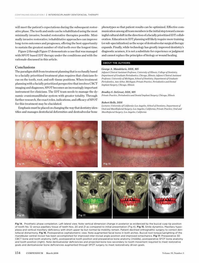
fig 14.Prostheticphasecompletion.Leftlateralview.noteverticaldimensionchangeinposteriorasevidencedbythebuccalcusptippositionoftoothno.12versuspapillarytissueofteethnos.20and21ascomparedtoinitialpresentation(Fig4).fig 15.Smiledynamics.Maxillaryhypo-plasiaandverticalmaxillarydeficiencywithshortupperlipbutnormallipmobilityremain.Patientdeclinedorthognathicsurgerytocorrectden-tofacialdisharmony.fig 16.Postoperativecephalometricview.noteaugmentedfacialboneinbotharches.Buccalroottorque/uprightingofthemandibularcentralincisorhasbeenaccomplishedforimprovedinter-incisalanglepositionandimprovediomechanics.fig 17.Preoperative3DCBCTboneandtoothanatomy(left),postoperativetoothpositionandpreoperativeboneanatomy(middle),postoperativeSFOTboneanatomyandtoothposition(right).notedentoalveolardeficienciesandprojectedbonelosssecondarytotoothmovementrequiredtomeetrestorativegoalsanddentoalveolarbonedeficienciesaugmentedthroughSFOTsurgerytomeetrestorativelydrivengoals.
aBout the authors
George A. Mandelaris, DDS, MSAdjunct Clinical Assistant Professor, University of Illinois, College of Dentistry, Department of Graduate Periodontics, Chicago, Illinois; Adjunct Clinical Assistant Professor, University of Michigan, School of Dentistry, Department of Graduate Periodontics, Ann Arbor, Michigan; Private Practice, Periodontics and Dental Implant Surgery, Chicago, Illinois
Bradley S. DeGroot, DDS, MSPrivate Practice, Periodontics and Dental Implant Surgery, Chicago, Illinois
Robert Relle, DDSLecturer, University of California-Los Angeles, School of Dentistry, Department of Oral and Maxillofacial Surgery, Los Angeles, California; Private Practice, Oral and Maxillofacial Surgery, Los Angeles, California
155www.compendiumlive.com March 2018 coMpendiuM
Brian Shah, MD, DDSDirector of Research, Department of Oral and Maxillofacial Surgery, Yale New Haven Hospital, New Haven, Connecticut; Private Practice, Oral and Maxillofacial Surgery, Chicago, Illinois
Iwei Huang, DMD, MSPrivate Practice, Orthodontics and Dentofacial Orthopedics, Chicago, Illinois
Brian S. Vence, DDSPrivate Practice, Restorative Dentistry, Oakbrook Terrace, Illinois
Queries to the author regarding this course may be submitted to [email protected].
references
1. amsterdamM.Periodontalprosthesis.Twenty-fiveyearsinretro-spect.Alpha Omegan.1974;67(3):8-52.2. EnlowDh,MoyersRE.Growthandarchitectureoftheface.J Am Dent Assoc.1971;82(4):763-774.3. ahnJ,KimSJ,LeeJy,etal.Transversedentalcompensationinrela-tiontosagittalandtransverseskeletaldiscrepanciesinskeletalClassIIIpatients.Am J Orthod Dentofacial Orthop.2017;151(1):148-156.4. StaudtCB,KiliaridisS.DifferentskeletaltypesunderlyingClassIIImalocclusioninarandompopulation.Am J Orthod Dentofacial Or-thop.2009;136(5):715-721.5. MandelarisGa,neivaR,ChambroneL.Cone-beamcomputedto-mographyandinterdisciplinarydentofacialtherapy:anamericanacademyofPeriodontologybestevidencereviewfocusingonriskassessmentofthedentoalveolarbonechangesinfluencedbytoothmovement.J Periodontol.2017;88(10):960-977.6. handelmanCS.Theanterioralveolus:itsimportanceinlimitingorth-odontictreatmentanditsinfluenceontheoccurrenceofiatrogenicsequelae.Angle Orthod.1996;66(2):95-109.7. BrautV,BornsteinMM,BelserU,BuserD.Thicknessoftheanteriormaxillaryfacialbonewall—aretrospectiveradiographicstudyusingconebeamcomputedtomography.Int J Periodontics Restorative Dent.2011;31(2):125-131.8. nowzarih,MolayemS,ChiuCh,RichSK.Conebeamcomputedtomographicmeasurementofmaxillarycentralincisorstodetermineprevalenceoffacialalveolarbonewidth≥2mm.Clin Implant Dent Relat Res.2012;14(4):595-602.9. EvensenJP,OgaardB.aremalocclusionsmoreprevalentandse-verenow?acomparativestudyofmedievalskullsfromnorway.Am J Orthod Dentofacial Orthop.2007;131(6):710-716.10.KrnetaB,Zhurova,RichmondS,OvsenikM.DiagnosisofClassIIImalocclusionin7-to8-year-oldchildren—a3Devaluation.Eur J Or-thod.2015;37(4):379-385.11.Joshin,hamdanaM,FakhouriWD.Skeletalmalocclusion:ade-velopmentaldisorderwithalife-longmorbidity.J Clin Med Res.2014;6(6):399-408.12.BoydK.Darwiniandentistrypart2:earlychildhoodnutrition,den-tofacialdevelopmentandchronicdisease.J Am Orthodontia Soc.2012;12(2):28-32.13.agarwalSS,SharmaM,nehraK,etal.Validationofassociationbe-tweenbreastfeedingduration,facialprofile,occlusion,andspacing:across-sectionalstudy.Int J Clin Pediatr Dent.2016;9(2):162-166.14.KohliMV,PatilGB,KulkarninB,etal.achangingtrendineruptionageandpatternoffirstdeciduoustooth:correlationtofeedingpat-tern.J Clin Diagn Res.2014;8(3):199-201.15.ThomazEB,CangussuMC,assisaM.Maternalbreastfeeding,para-functionaloralhabitsandmalocclusioninadolescents:amultivariateanalysis.Int J Pediatr Otorhinolaryngol.2012;76(4):500-506.16.KimSJ,KimKh,yuhS,BaikhS.Dentoalveolarcompensationac-cordingtoskeletaldiscrepancyandoverjetinskeletalClassIIIpatients.Am J Orthod Dentofacial Orthop.2014;145(3):317-324.
17.QuX,LiuZ,Wangy,etal.Dentofacialtraitsinassociationwithlowerincisoralveolarcancellousbonethickness:amultipleregressionanalysis.Angle Orthod.2017;87(3):409-415.18.RoseJC,RobleeRD.Originsofdentalcrowdingandmalocclusions:ananthropologicalperspective.Compend Contin Educ Dent.2009;30(5):292-300.19.GilbertSF.Ecologicaldevelopmentalbiology:developmentalbiol-ogymeetstherealworld.Dev Biol.2001;233(1):1-12.20.EstelitaS,JansonG,ChiquetoK.ExtremedentalcompensationinanadultskeletalclassIIImalocclusion:3-yearfollow-upofasuccess-fullycompromisedtreatment.Int J Orthod Milwaukee.2015;26(2):69-76.21.EvangelistaK,VasconcelosKdeF,Bumanna,etal.DehiscenceandfenestrationinpatientswithClassIandClassIIDivision1malocclusionassessedwithcone-beamcomputedtomography.Am J Orthod Dento-facial Orthop.2010;138(2):133.e1-e7.22.FranklinKa,LindbergE.Obstructivesleepapneaisacommondis-orderinthepopulation—areviewontheepidemiologyofsleepapnea.J Thorac Dis.2015;7(8):1311-1322.23.PeppardPE,youngT,BarnetJh,etal.Increasedprevalenceofsleep-disorderedbreathinginadults.Am J Epidemiol.2013;177(9):1006-1014.24.PatinoM,SadhasivamS,MahmoudM.Obstructivesleepapnoeainchildren:perioperativeconsiderations.Br J Anaesth.2013;111(suppl1):i83-i95.25.nespoliL,Caprioglioa,BrunettiL,nosettiL.Obstructivesleepap-neasyndromeinchildhood.Early Hum Dev.2013;89(suppl3):S33-S37.26.Iida-KondoC,yoshinon,KurabayashiT,etal.Comparisonoftonguevolume/oralcavityvolumeratiobetweenobstructivesleepap-neasyndromepatientsandnormaladultsusingmagneticresonanceimaging.J Med Dent Sci.2006;53(2):119-126.27.IaredW,KogadaSilvaEM,RufinoMacedoC.Estheticpercep-tionofchangesinfacialprofileresultingfromorthodontictreatmentwithextractionofpremolars:asystematicreview.J Am Dent Assoc.2017;148(1):9-16.28.PliskaBT,TamIT,Loweaa,etal.Effectoforthodontictreatmentontheupperairwayvolumeinadults.Am J Orthod Dentofacial Or-thop.2016;150(6):937-944.29.FergusonDJ,MakkiL,StapelbergR,etal.Stabilityoftheman-dibulardentalarchfollowingperiodontallyacceleratedosteogenicorthodonticstherapy:preliminarystudies.Semin Orthod.2014;20(3):239-246.30.RobleeRD,BoldingSL,LandersJM.Surgicallyfacilitatedorth-odontictherapy:anewtoolforoptimalinterdisciplinaryresults.Com-pend Contin Educ Dent.2009;30(5):264-275.31.BaloulSS,GerstenfeldLC,MorganEF,etal.Mechanismofactionandmorphologicchangesinthealveolarboneinresponsetoselectivealveolardecortication-facilitatedtoothmovement.Am J Orthod Den-tofacial Orthop.2011;139(suppl4):S83-S101.32.yaffea,Finen,BindermanI.Regionalacceleratedphenomenoninthemandiblefollowingmucoperiostealflapsurgery.J Periodontol.1994;65(1):79-83.33.WilckoWM,WilckoT,BouquotJE,FergusonDJ.Rapidorthodon-ticswithalveolarreshaping:twocasereportsofdecrowding.Int J Periodontics Restorative Dent.2001;21(1):9-19.34.SebaounJD,Kantarcia,TurnerJW,etal.Modelingoftrabecularboneandlaminadurafollowingselectivealveolardecorticationinrats.J Periodontol.2008;79(9):1679-1688.35.MerrillRG,PedersenGW.Interdentalosteotomyforimmediatere-positioningofdental-osseouselements.J Oral Surg.1976;34(2):118-125.36.FrosthM.Theregionalacceleratoryphenomenon:areview.Henry Ford Hosp Med J.1983;31(1):3-9.37.Zimmon,SalehMh,MandelarisGa,etal.Corticotomy-acceleratedorthodontics:acomprehensivereviewandupdate.Compend Contin Educ Dent.2017;38(1):17-25.
156 Volume 39, Number 3compendium march 2018
38.BrautV,BornsteinMM,LauberR,BuserD.Bonedimensionsintheposteriormandible:aretrospectiveradiographicstudyusingconebeamcomputedtomography.Part1—analysisofdentatesites.Int J Periodontics Restorative Dent.2012;32(2):175-184.39.MandelarisGa,VenceBS,RosenfeldaL,ForbesDP.aclassifica-tionsystemforcrestalandradiculardentoalveolarbonephenotypes.Int J Periodontics Restorative Dent.2013;33(3):289-296.30.yilmazBS,Kucukkelesn.Skeletal,softtissue,andairwaychangesfollowingthealternatemaxillaryexpansionsandconstrictionsproto-col.Angle Orthod.2014;84(5):868-877.41.DenolfPL,VandervekenOM,MarklundME,BraemMJ.Thestatusofcephalometryinthepredictionofnon-CPaPtreatmentoutcomeinob-structivesleepapneapatients.Sleep Med Rev.2016;27:56-73.42.EvansM.Three-dimensionalcontrolwithTaD-tissuesupportedrapidpalatalexpander:anoverviewofclinicalapplicationsandbiolog-icaladvantages.Orthodontic Clinical Review.2013:22-31.https://www.rmortho.com/wp-content/uploads/2013/08/Clinical_Final_LR.pdf.ac-cessedJanuary24,2018.43.GauthierC,VoyerR,PaquetteM,etal.Periodontaleffectsofsur-gicallyassistedrapidpalatalexpansionevaluatedclinicallyandwithcone-beamcomputerizedtomography:6-monthpreliminaryresults.Am J Orthod Dentofacial Orthop.2011;139(suppl4):S117-S128.44.XiongG,huJ,ChenW,etal.Theanalysisofcorrelationbe-tweentonguebodyMRIandupperairwaypressuremeasurementsofblockedlingualregioninpatientswithmoderateandsevereOSahS.Lin Chung Er Bi Yan Hou Tou Jing Wai Ke Za Zhi.2015;29(21):1853-1856.45.KimaM,KeenanBT,Jacksonn,etal.Tonguefatanditsrelation-shiptoobstructivesleepapnea.Sleep.2014;37(10):1639-1648.46.FregosiRF.Influenceoftonguemusclecontractionandtransmuralpressureonnasopharyngealgeometryintherat.J Appl Physiol (1985).2011;111(3):766-774.47.BabademezMa,yorubulutM,yurekliMF,etal.Comparisonofminimallyinvasivetechniquesintonguebasesurgeryinpatientswithobstructivesleepapnea.Otolaryngol Head Neck Surg.2011;145(5):858-864.48.BaileyEF,huangyh,FregosiRF.anatomicconsequencesofintrinsictonguemuscleactivation.J Appl Physiol (1985).2006;101(5):13771385.49.StuckBa,KopkeJ,hormannK,etal.Volumetrictissuereductioninradiofrequencysurgeryofthetonguebase.Otolaryngol Head Neck Surg.2005;132(1):132-135.50.DoKL,Ferreyrah,healyJF,DavidsonTM.Doestonguesizedifferbetweenpatientswithandwithoutsleep-disorderedbreathing?La-ryngoscope.2000;110(9):1552-1555.51.PowellnB,RileyRW,GuilleminaultC.Radiofrequencytonguebasereductioninsleep-disorderedbreathing:apilotstudy.Otolaryngol Head Neck Surg.1999;120(5):656-664.52.ZonatoaI,BittencourtLR,MartinhoFL,etal.associationofsys-tematicheadandneckphysicalexaminationwithseverityofobstruc-tivesleepapnea-hypopneasyndrome.Laryngoscope.2003;113(6):973-980.53.ZonatoaI,MartinhoFL,BittencourtLR,etal.headandneckphysi-calexamination:comparisonbetweennonapneicandobstructivesleepapneapatients.Laryngoscope.2005;115(6):1030-1034.54.Chiuhy,ChenPy,ChuangLP,etal.DiagnosticaccuracyoftheBerlinquestionnaire,STOP-BanG,STOP,andEpworthsleepinessscaleindetectingobstructivesleepapnea:abivariatemeta-analysis.Sleep Med Rev.2017;36:57-70.55.KhayatR,Pleistera.Consequencesofobstructivesleepapnea:cardiovascularriskofobstructivesleepapneaandwhethercon-tinuouspositiveairwaypressurereducesthatrisk.Sleep Med Clin.2016;11(3):273-286.56.UesugiT,KobayashiT,hasebeD,etal.Effectsoforthognathicsurgeryonpharyngealairwayandrespiratoryfunctionduringsleepinpatientswithmandibularprognathism.Int J Oral Maxillofac Surg.2014;43(9):1082-1090.
contInuInG educatIon 1 | InTERDISCIPLInaRyDEnTOFaCIaLThERaPy
24 CE
Credits
May 17th-19th, 2018 | Hilton TorontoRegister now, at www.ardii.org
or call 847-677-7777
157www.compendiumlive.com March 2018 coMpendiuM
1. collaborative interdisciplinary dentofacial therapy (Idt) requires the leadership of whom to coordinate input from each specialist involved? a. thepatient B. thepatient’sorthodontist C. thepatient’srestorativedentist D. theoralandmaxillofacialsurgeon
2. What occur as a result of skeletal disharmony and are common when anterior-posterior or transverse maxilla-mandibular disharmonies are present? a. dentalcompensations B. dentaldecompensations C. iatrogeniccomplications D. orthodonticwalls
3. What alternative approach to orthodontic tooth movement may allow the Idt team to accomplish outcome goals and avoid iatrogenic complications? a. surgicallyfacilitatedorthodontictherapy(SFOT) B. immediateimplantplacement/provisionalization(IIPP) C. continuouspositiveairwaypressure(CPaP) D. guidedboneregeneration(GBR)
4. a uniformly soft diet among infants and toddlers and the resulting failure to develop forward tongue and lip muscular habits may reduce: a. theamountoforthodontictreatmenttime. B. theneedforestheticcrownlengthening. C. theoralcavityvolume. D. regionalacceleratoryphenomenon.
5. Which member of the Idt team is responsible for architecting a facially prioritized plan and correcting dental compensations? a. prosthodontist B. orthodontist C. periodontist D. myofunctionaltherapist
6. sfot creates a biologic environment that leads to more stable orthodontic results and sometimes less extensive orthognathic surgery, thus supporting: a. oralappliancetherapy. B. afaciallyprioritizedtreatmentplan. C. dentalcompensations. D. alloftheabove
7. Which of the following is a step included in the sfot Idt workflow? a. diseasecontrolandprovisionalization B. interimorthodontictransitionalrestorations C. orthodonticrefinementandfinishing D. alloftheabove
8. once a diagnosis has been made, Idt collaboration among the orthodontist, periodontist, and oral and maxillofacial surgeon can focus on: a. haltingbonyblockmovement. B. facilitatingregionalacceleratoryphenomenon. C. usingtemporaryanchoragedevices. D. enhancingtheorthodonticwalls.
9. Whether to perform corticotomy-assisted orthodontic surgery on the buccal and/or lingual aspects of a tooth is dependent partly on the: a. numberoftonguecrenationspresent. B. flexibilityoftheperiodontalligament. C. regionaldentoalveolarboneanatomy. D. noneoftheabove
10. at the completion of sfot-inclusive Idt, the team will have addressed: a. buccalbonethickness. B. properplacementofthedentogingivalcomplex. C. optimalrootpositionwithinnewlyaugmentedalveolarbone. D. alloftheabove
continuing education 1quIz
Course is valid from March 1, 2018, to March 31, 2021. Participants must attain a score of 70% on each quiz to re-ceive credit. Participants receiving a failing grade on any exam will be notified and permitted to take one re-examina-tion. Participants will receive an annual report documenting their accumulated credits, and are urged to contact their own state registry boards for special CE requirements.
aEGISPublications,LLC,isanaDaCERPRecognizedProvider.aDaCERPisaserviceoftheamericanDentalassociationtoassistdentalprofessionalsinidentifyingqualityprovidersofcontinuingdentaleducation.aDaCERPdoesnotapproveorendorseindividualcoursesorinstructors,nordoesitimplyacceptanceofcredithoursbyboardsofdentistry.ConcernsorcomplaintsaboutaCEprovidermaybedirectedtotheproviderortoaDaCERPatwww.ada.org/cerp.
Surgically Facilitated Orthodontic Therapy: Optimizing Dentoalveolar Bone and Space Appropriation for Facially Prioritized Interdisciplinary Dentofacial TherapyGeorge a. Mandelaris, dds, Ms; Bradley s. deGroot, dds, Ms; robert relle, dds; Brian shah, Md, dds; Iwei huang, dMd, Ms; and Brian s. Vence, dds
This article provides 2 hours of CE credit from AEGIS Publications, LLC. Record your answers on the enclosed Answer Form or submit them on a separate sheet of paper. You may also phone your answers in to 877-423-4471 or fax them to 215-504-1502 or log on to compendiumce.com/go/1806. Be sure to include your name, address, telephone number, and last 4 digits of your Social Security number.
please complete answer form on page 166, including your name and payment information.you can also take thIs course onlIne at coMpendIuMce.coM/Go/1806.
quIz
Program Approval forContinuing Education
Approved PACE Program ProviderFAGD/MAGD CreditApproval does not imply acceptanceby a state or provincial board ofdentistry or AGD endorsement1/1/2017 to 12/31/2022Provider ID# 209722