Embed Size (px)
Citation preview

RESEARCH ARTICLE Open Access
Survey of bluetongue virus infection in free-rangingwild ruminants in SwitzerlandJulien Casaubon1, Valérie Chaignat2, Hans-Rudolf Vogt3, Adam O Michel1, Barbara Thür2
and Marie-Pierre Ryser-Degiorgis1*
Abstract
Background: In 2006, bluetongue virus serotype 8 (BTV-8) was detected for the first time in central Europe.Measures to control the infection in livestock were implemented in Switzerland but the question was raisedwhether free-ranging wildlife could be a maintenance host for BTV-8. Furthermore Toggenburg orbivirus (TOV),considered as a potential 25th BTV serotype, was detected in 2007 in domestic goats in Switzerland and wildruminants were considered a potential source of infection. To assess prevalences of BTV-8 and TOV infections inwildlife, we conducted a serological and virological survey in red deer, roe deer, Alpine chamois and Alpine ibexbetween 2009 and 2011. Because samples originating from wildlife carcasses are often of poor quality, we alsodocumented the influence of hemolysis on test results, and evaluated the usefulness of confirmatory tests.
Results: Ten out of 1,898 animals (0.5%, 95% confidence interval 0.3-1.0%) had detectable antibodies against BTV-8and BTV-8 RNA was found in two chamois and one roe deer (0.3%, 0.1-0.8%). Seroprevalence was highest amongred deer, and the majority of positive wild animals were sampled close to areas where outbreaks had beenreported in livestock. Most samples were hemolytic and the range of the optical density percentage valuesobtained in the screening test increased with increasing hemolysis. Confirmatory tests significantly increasedspecificity of the testing procedure and proved to be applicable even on poor quality samples. Nearly all samplesconfirmed as positive had an optical density percentage value greater than 50% in the ELISA screening.
Conclusions: Prevalence of BTV-8 infection was low, and none of the tested animals were positive for TOV.Currently, wild ruminants are apparently not a reservoir for these viruses in Switzerland. However, we report for thefirst time BTV-8 RNA in Alpine chamois. This animal was found at high altitude and far from a domestic outbreak,which suggests that the virus could spread into/through the Alps. Regarding testing procedures, hemolysis did notsignificantly affect test results but confirmatory tests proved to be necessary to obtain reliable prevalence estimates.The cut-off value recommended by the manufacturer for the screening test was applicable for wildlife samples.
Keywords: Bluetongue virus, Cross-sectional study, Hemolysis, Switzerland, Toggenburg orbivirus, Wildlife samples
BackgroundBluetongue (BT) is a disease of economic importance [1]caused by the bluetongue virus (BTV), a RNA-virus thatbelongs to the genus Orbivirus of the family Reoviridae.Twenty-six serotypes have been reported around theworld so far [2]. Although other infection pathways havebeen described [3,4], BTV is generally transmitted by bit-ing midges (Culicoides spp.) [5]. The virus may cause ahemorrhagic disease with high morbidity rates, especially
in sheep, while cattle mostly act as a reservoir. As an ex-ception, a high morbidity was observed in this species dur-ing the recent epidemic due to BTV serotype 8 (BTV-8) inEurope [6]. Observations during previous BT outbreaksand experimental infections have shown that indigenouswild ruminant species may become infected with andwithout clinical manifestations and may therefore act as avirus reservoir [7-12].Indigenous Swiss cattle and sheep breeds are highly
susceptible to BTV infection and develop clinical signs[13,14]. The first BTV-8 infection in a domestic animalin Switzerland was diagnosed at the end of October2007 [13] and in 2008, like in most European countries
* Correspondence: [email protected] for Fish and Wildlife Health (FIWI), Vetsuisse Faculty, University ofBern, Bern, SwitzerlandFull list of author information is available at the end of the article
© 2013 Casaubon et al.; licensee BioMed Central Ltd. This is an Open Access article distributed under the terms of the CreativeCommons Attribution License (http://creativecommons.org/licenses/by/2.0), which permits unrestricted use, distribution, andreproduction in any medium, provided the original work is properly cited.
Casaubon et al. BMC Veterinary Research 2013, 9:166http://www.biomedcentral.com/1746-6148/9/166
confronted to the BT epidemic, a large scale compulsoryvaccination campaign was initiated to limit the expan-sion of the virus [15]. From 2007 to 2010, 76 local out-breaks have been reported in Swiss livestock [16]. Astudy in wildlife prior to the 2007 epidemic reported noevidence of BTV infection in Swiss red deer [17]. How-ever, considering recent data from other European coun-tries [8,18-20], an update of the situation including morespecies from the whole country was necessary to evalu-ate the potential role of wild ruminants in the BT epi-demiology in Switzerland and assess whether they mayrepresent a threat to the success of the control programin livestock. Furthermore, a new orbivirus named Toggen-burg orbivirus (TOV) was detected in 2007 in healthygoats in Switzerland [21]. Since then, it has been shownthat the TOV circulates in small domestic ruminants, es-pecially in the southern Swiss canton of Ticino (TI), andthe question was raised as to whether wildlife may be areservoir for this virus [22,23]. A study addressing risk fac-tors for TOV infection revealed a possible association withalpine pastures [24].In this study, we estimated the prevalence of infections
with BTV and TOV in roe deer (Capreolus c. capreolus),red deer (Cervus e. elaphus), Alpine chamois (Rupicaprar. rupicara) and Alpine ibex (Capra i. ibex) over two
years (2009 to 2011). We considered potential risk fac-tors for infection such as geographical location, altitude,animal species, age and sex, and we compared our re-sults with data on domestic ruminants. Given that bloodsamples from hunted wildlife are often of poor quality(e.g. hemolysis), which may affect reactions in serologicaltests [25], we investigated the influence of hemolysis onour testing protocol. Our data show that wild ruminantsare currently not a reservoir for BTV and TOV inSwitzerland. We also confirm that recording serum qualityof samples from hunted wildlife is essential to correctly in-terpret test results, and that the use of confirmatory testsis crucial to identify false positive results.
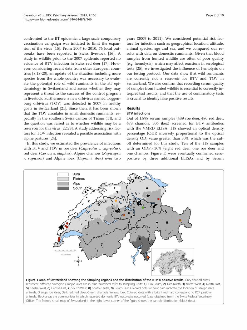
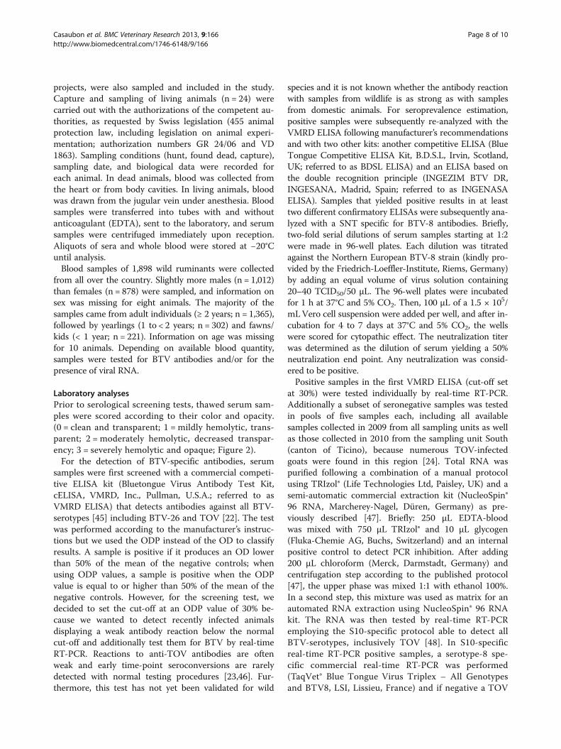
ResultsBTV infectionsOut of 1,898 serum samples (439 roe deer, 480 red deer,473 chamois, 506 ibex) screened for BTV antibodieswith the VMRD ELISA, 118 showed an optical densitypercentage (ODP, inversely proportional to the opticaldensity OD) value greater than 30%, which was the cut-off determined for this study. Ten of the 118 sampleswith an ODP > 30% (eight red deer, one roe deer andone chamois; Figure 1) were eventually confirmed sero-positive by three additional ELISAs and by Serum
Figure 1 Map of Switzerland showing the sampling regions and the distribution of the BTV-8 positive results. Grey shaded areasrepresent different bioregions, major lakes are in blue. Numbers refer to sampling units: 1) Jura-South, 2) Jura-North, 3) North-West, 4) North-East,5) Centre-West, 6) Centre-East, 7) South-West, 8) South-Centre, 9) South-East. Colored dots without halo indicate the location of seropositiveanimals: Orange: roe deer; Dark red: red deer; Green: chamois; Yellow: ibex. Colored dots with a bright red halo correspond to PCR positiveanimals. Black areas are communities in which reported domestic BTV outbreaks occurred (data obtained from the Swiss Federal VeterinaryOffice). The framed small map of Switzerland in the right lower corner of the figure shows the sample distribution (black dots).
Casaubon et al. BMC Veterinary Research 2013, 9:166 Page 2 of 10http://www.biomedcentral.com/1746-6148/9/166

Neutralization Test (SNT), with titers against BTV-8ranging from 1:6 to 1:108 (Table 1 and 2). Only theseconfirmed positive samples were used for seroprevalencecalculation. Overall, estimated seroprevalence was 0.5%(95% confidence interval [CI] 0.3-1%) (Table 1).With the exception of one roe deer (fawn), all seroposi-
tive animals were adults (no statistically significant differ-ence between age classes or sexes). There were significantlymore seropositive red deer than roe deer (p = 0.039), cham-ois (p = 0.038) or ibex (p = 0.003). Among the samplingunits, a significantly higher seroprevalence was recorded inJura-South (9.3%, Table 1; p < 0.001 to p = 0.014), andwithin this unit, more red deer than roe deer and chamois(p = 0.004) were seropositive. The elevation above sea level(a.s.l.) of the seropositive animals did not differ from thespecies-specific elevation range for each ruminant species(Table 3). There was also no difference between the twosampling periods (2009 and 2010).None of the 10 seropositive samples was positive by S10
BTV real-time RT-PCR. In contrast, BTV-8 RNA wasdetected in 3 out of 1,070 seronegative samples (Ct-valuesbetween 28 and 31). These three samples were from twoadult chamois out of the 118 samples with initial positivereaction in the VMRD ELISA screening (ODP > 30%) andfrom one adult roe deer from the canton of Ticino (sam-pling unit South Centre, Figure 1) that tested negative inthe VMRD ELISA screening. Virus isolation from thesethree samples was not successful. Overall, BTV-8 virusprevalence was 0.3%, (95% CI 0.1-0.8%) and no animalwas found positive for TOV.
The comparison of our results with documented BToutbreaks in domestic livestock (virus positive animals,data from the Swiss Federal Veterinary Office; Figure 1)shows that seropositive wild ruminants were found in arange from 2.0 to 7.3 km (average of 5.8 km, linear dis-tance) around communities with domestic outbreaks. Incontrast, two virus positive animals (one chamois andone roe deer) originated from regions where no domes-tic BT outbreak has been reported so far [16], at a dis-tance of 38.0 km and 85.5 km, respectively, to the nextcommunity with a documented outbreak.
Serum quality and testing protocolThe time span between sample collection in the fieldand arrival at laboratory was in average 3.2 days with arange of 0 to 21 days. Of all obtained serum samples,1,842 were scored (0 to 3) according to their degree ofhemolysis (Figure 2). Only 5% were scored as clean (0)and 18% as mildly hemolytic (1). Most of the samples(45%) were classified as moderately hemolytic with de-creased transparency (2), while 31% were severelyhemolytic and opaque (3). As expected, the proportionof clean samples was higher in animals sampled alive(55%) than after death (4%; p < 0.001), while the propor-tion of moderately to severely hemolytic samples wassimilar in hunted animals (77%) and animals found dead(70%). The distribution of the different hemolysis scoreswas generally similar in all four species. However, theproportion of clean samples was significantly higher inhunted ibex (7%) than hunted chamois (2%, p = 0.001),
Table 1 Prevalences of antibodies against BTV-8 in four species of wild ruminants from Switzerland, 2009-2011
Sampling units
Jura Jura North North Centre Centre South South South Total
South North West East West East West Centre East
Roe deer Seroprevalence 0 0 0 0 0 1.4 0 0 0 0.2
(95% CI) (0–13.7) (0–5.3) (0–6) (0–4.5) (0–9.5) (0–7.5) (0–8) (0–20.6) (0–9.7) (0.01-1.3)
positive / tested 0 /25 0 / 68 0 / 60 0 / 81 0 /37 1 / 72 0 / 44 0 / 16 0 / 36 1 / 439
Red deer Seroprevalence 29.2 - - 1 0 0 0 0 0 1.7
(95% CI) (12.6-51.1) (0–5.3) (0–7.4) (0–8.4) (0–6.5) (0–4.5) (0–2.8) (0.7-3.3)
positive / tested 7 / 24 - - 1 /103 0 / 48 0 / 42 0 / 55 0 / 80 0 / 128 8 / 480
Chamois Seroprevalence 0 0 0 0 0 1.5 0 0 0 0.2
(95% CI) (0–13.2) (0–5.8) (0–36.9) (0–4.9) (0–12.8) (0.04-8.3) (0–6.5) (0–9.5) (0–3) (0.01-1.2)
positive / tested 0 / 26 0 / 62 0 / 8 0 / 73 0 / 27 1 / 65 0 / 55 0 / 37 0 / 120 1 / 473
Ibex Seroprevalence - - - 0 0 0 0 0 0 0
(95% CI) (0–6.1) (0–4.5) (0–7.4) (0–3.9) (0–33.6) (0–1.7) (0–0.7)
positive / tested - - - 0 / 59 0 / 81 0 / 48 0 / 92 0 / 9 0 / 217 0 / 506
Total Seroprevalence 9.3 0 0 0.3 0 0.9 0 0 0 0.5
(95% CI) (3.8-18.3) (0–2.8) (0–5.3) (0.01-1.8) (0–1.9) (0.1-3.1) (0–1.5) (0–2.6) (0–0.7) (0.3-1)
positive / tested 7 / 75 0 / 130 0 / 68 1 / 316 0 / 193 2 / 227 0 / 246 0 / 142 0 / 501 10 / 1,898
Samples are classified according to the nine sampling units. Confidence intervals (95% CI) are indicated in parentheses.
Casaubon et al. BMC Veterinary Research 2013, 9:166 Page 3 of 10http://www.biomedcentral.com/1746-6148/9/166

despite the fact that the time span (in days) between thedate of sampling and the date of arrival at the Centre forFish and Wildlife Health (FIWI) was longer for samples ofhunted ibex (3.3 days, range of 1 to 19 days) than of huntedchamois (3.1 days, range of 0 to 21 days) (p = 0.012). Never-theless, when the animal species was not considered,hemolysis generally increased with the time between
sampling and sample processing (2.8 days for score 0, and3.6 for score 3).In the screening VMRD ELISA, severely hemolytic sam-
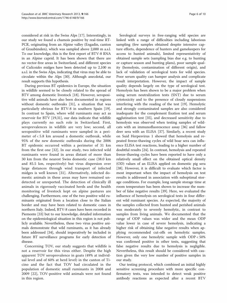
ples (score 3) showed a significantly lower ODP meanvalue than samples with lower scores (p < 0.001; Figure 3),and the overall range of ODP values was wider with in-creasing hemolysis. Positive results were confirmed both
Table 2 Relationship between results of the serological tests and sample hemolysis
Score 0 Score 1 Score 2 Score 3 Total
n = 99 n = 338 n = 829 n = 576 n = 1842
ODP ODP ODP ODP ODP ODP ODP ODP ODP ODP
30-49% 50-100% 30-49% 50-100% 30-49% 50-100% 30-49% 50-100% 30-49% 50-100%
Screening ELISA
VMRDa 6 3 18 2 40 13 22 14 86 32
Confirmatory ELISAs
VMRDb 0 3 0 0 0 3 1c 6 1c 12
BDSL 0 2 0 0 0 3 1c 4 1c 9
INGENASA 0 2 0 0 0 3 1c 3 1c 8
Serum Neutralization Test
Titer≥ 1:2 0 2 0 0 0 3 1 4 1 9
Total seropositive 2 0 3 5 10
Confirmed positive / screeningpositive (ODP ≥ 30%)
22.2% 0% 5.7% 13.9% 8.5%
Seroprevalence 2.0% 0% 0.4% 0.9% 0.5%a Cut-off at ODP 30%, b Cut-off at ODP 50%, c Unclear.Number of positive samples obtained with the screening test (VMRD ELISA) and following confirmatory tests are indicated in relation to the hemolysis serumscores (0–3) and the optical density percentage (ODP) of the screening test. One sample with ODP > 30% yielded an unclear result in the confirmatory ELISAs butto a positive result in the SNT (cut-off ≥ 1:2).
Table 3 Sample size and population data for the four wild ruminant species sampled in the study
Species Geographical data* Population data 2010**
Bioregion Min Mean altitudem.a.s.l. (SD)
Max Estimated populationsize
Hunting bag
Roe deer Jura 393 783 (231) 1,228 112,975 39,664
Plateau 355 553 (126) 938
Alps 374 1,195 (445) 2,504
South 272 731 (337) 1,417
Red deer Jura 474 764 (375) 1,557 28,483 9,016
Alps 402 1,254 (399) 2,604
South 238 940 (465) 2,094
Alpine chamois Jura 434 940 (279) 1,581 91,390 13,339
Plateau 532 650 (160) 1,007
Alps 431 1,840 (547) 3,103
South 312 1,164 (540) 2,087
Alpine ibex Alps 673 2,294 (392) 3,154 15,553 1,074
* Data from the animals sampled in this study.** Data obtained from the Swiss Federal Office of the Environment [55].Population size is based on counts and estimates from wildlife managers and biologists.Mean altitudes differed significantly between species (p < 0.05): Ibex were found higher than all other species (Alps), and chamois were higher than roe deer andred deer (all bioregions).
Casaubon et al. BMC Veterinary Research 2013, 9:166 Page 4 of 10http://www.biomedcentral.com/1746-6148/9/166
for clean and hemolytic sera (Figure 3). Final seropreva-lence was 2.0% for clean samples (score 0) and 0.6% formarkedly hemolytic samples (score 2 and 3), without stat-istical difference between the two groups, and with even alower prevalence for only slightly hemolytic samples(0%, score 1; Table 2).Ten out of 118 samples (8.5%) were eventually con-
firmed seropositive by repeating the VMRD ELISA in adifferent laboratory by other technicians and by twoother ELISAs and SNT (Table 2). The percentage ofconfirmed positive among screening-positive sampleswas higher for score 0 (22.2%) than for scores 2 and 3(5.7% and 13.9%, respectively) but these differences werenot significant. Overall, final seroprevalence (10/1,898,0.5%) was significantly lower than the initial one (screen-ing test: 118/1,898, 6.2%; p < 0.001) even if consideringonly samples with ODP > 50% (9/1,898 confirmed versus32/1,898 positive in the screening test, p < 0.001).Except for one out of the 10 samples confirmed as posi-
tive after running confirmatory tests, all of these had aninitial ODP > 50% (Table 2, Figure 3). Thus, the estimatedseroprevalence using 50% as cut-off (9/1,898, 0.47%)
would have not significantly differed from the oneobtained with the cut-off at 30% (10/1,898, 0.52%; p = 1).
DiscussionFollowing the recent BT epidemic in central Europe,data on BTV circulation in Swiss free-ranging wild rumi-nants were urgently needed for the future planning andsuccessful achievement of the implemented BT controlprogram in livestock. This study provides an overview ofthe situation in the four indigenous wild ruminant spe-cies in Switzerland, and it additionally addresses import-ant questions regarding testing protocols for samplescollected from wildlife carcasses.The low estimated seroprevalence for BTV in all in-
vestigated species suggests that sporadic infection canoccur but that BTV-8 is not circulating within wild pop-ulations in Switzerland at a large scale. Also, the preva-lence in red deer of 1.7% in the present study was notsignificantly higher (p = 0.058) than reported before theEuropean epidemic (0%, CI 0–1.54%) [17].The fact that the prevalences in our study were lower
than the ones previously documented in other Europeancountries [8,19,20] can be due to two different factors.First, albeit 76 BT outbreaks have been recorded inSwiss livestock between 2007 and 2010 [16], this numberis very low compared to the situation observed in othercountries [26], e.g. 26,500 herds with clinical cases in2008 in France [27]. The virus has apparently not spreadas widely in Switzerland as in other European regions.Second, our sampling rounds took place after two yearsof mandatory vaccination of Swiss domestic ruminantspecies [15], a measure that limited the incidence ofBTV-8 infections in domestic and possibly also in wildspecies [8,28].In our study, red deer were more often infected by
BTV-8 than other wild ruminant species, similarly to ob-servations in Belgium [8], Spain [19], and France [20].This supports the hypothesis that, although they live inthe same habitat, red deer are more susceptible to infec-tion than roe deer. It is interesting to note that red deerare abundant in the southern part of the Swiss Jurawhile only few individuals are present in its northernpart [29], where most of the domestic Swiss BT out-breaks occurred [16]. In contrast, roe deer and chamoisare abundant in the northern Jura [29] but none of theindividuals from this region (133 samples) were positive.This further suggests that ruminants other than red deerhave a lower susceptibility to infection or that BTV-infected midges do not feed on them as often. In conclu-sion, it indicates that outbreaks in livestock were likelynot related to wildlife infections.It has been formerly of concern that BTV may expand
from southern Europe through the Alps but data from2004–2005 did not indicate virus presence in regions
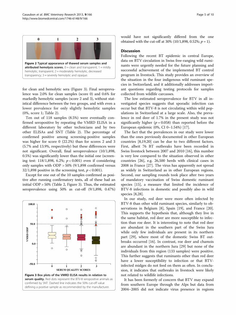
Figure 2 Typical appearance of thawed serum samples andattributed hemolysis scores. 0 = clean and transparent; 1 = mildlyhemolytic, transparent; 2 = moderately hemolytic, decreasedtransparency; 3 = severely hemolytic and opaque.
Figure 3 Box plots of the VMRD ELISA results in relation toserum quality. Red dots represent the BTV-8 seropositive animals asconfirmed by SNT. Dashed line indicates the 50% cut-off valuedefining a positive sample as recommended by the manufacturer.
Casaubon et al. BMC Veterinary Research 2013, 9:166 Page 5 of 10http://www.biomedcentral.com/1746-6148/9/166
considered at risk in the Swiss Alps [17]. Interestingly, inour study we found a chamois positive by real-time RT-PCR, originating from an Alpine valley (Engadin, cantonof Graubünden), which was sampled above 2,000 m a.s.l.To our knowledge, this is the first report of BTV-8 RNAin an Alpine caprid. It has been shown that there areno vector-free areas in Switzerland, and different speciesof Culicoides midges have been detected up to 2,100 ma.s.l. in the Swiss Alps, indicating that virus may be able tocirculate within the Alps [30]. Although anecdotal, ourresult supports this hypothesis.During previous BT epidemics in Europe, the situation
in wildlife seemed to be closely related to the spread ofBTV among domestic livestock [18]. However, seroposi-tive wild animals have also been documented in regionswithout domestic outbreaks [31], a situation that wasparticularly obvious for BTV-8 in southern Spain [19].In contrast to Spain, where wild ruminants may act asreservoir for BTV [19,31], our data indicate that wildlifeplays currently no such role in Switzerland. First,seroprevalences in wildlife are very low; second, allseropositive wild ruminants were sampled in a peri-meter of < 5.8 km around a domestic outbreak, while95% of the new domestic outbreaks during the 2006BT epidemic occurred within a perimeter of 31 kmfrom the first one [32]. In our study, two infected wildruminants were found in areas distant of more than30 km from the nearest Swiss domestic case (38.0 kmand 85.5 km, respectively) but virus dispersion overlarge distances through wind transport of infectedmidges is well known [31]. Alternatively, infected do-mestic animals in these areas may have remained un-detected or unreported. The detection of clinically illanimals in rigorously vaccinated herds and the healthmonitoring of livestock kept on alpine pastures arechallenging. Furthermore, one of the two positive wild ru-minants originated from a location close to the Italianborder and may have been related to domestic cases innorthern Italy. Indeed, BTV-8 cases have been recorded inPiemonte [33] but to our knowledge, detailed informationon the epidemiological situation in this region is not pub-licly available. Nevertheless, these two virus positive ani-mals demonstrate that wild ruminants, as it has alreadybeen addressed [34], should imperatively be included infuture BT surveillance programs for early detection ofdisease.Concerning TOV, our study suggests that wildlife is
not a reservoir for this virus either. Despite the highapparent TOV seroprevalence in goats (49% at individ-ual level and of 60% at herd level) in the canton of Ti-cino and the fact that TOV had circulated in thepopulation of domestic small ruminants in 2008 and2009 [22], TOV-positive wild animals were not foundin this region.
Serological surveys in free-ranging wild species arelinked with a range of difficulties including laborioussampling (few samples obtained despite intensive cap-ture efforts, dependence of hunters and gamekeepers foraccess to hunted animals), limited representativity ofobtained sample sets (sampling bias due e.g. to huntingor capture season and hunting plans), poor sample qual-ity (hemolysis, contamination of different origin), andlack of validation of serological tests for wild species.Poor serum quality can hamper analysis and complicateresult interpretation. However, the impact of samplequality depends largely on the type of serological test.Hemolysis has been shown to be a major problem whenusing serum neutralization tests (SNT) due to serumcytotoxicity and to the presence of cloudy suspensionsinterfering with the reading of the test [19]. Hemolyticand strongly contaminated samples are also consideredinadequate for the complement fixation test and serumagglutination test [35], and decreased sensitivity due tohemolysis was observed when testing samples of wild-cats with an immunofluorescence assay [36] and fallowdeer sera with an ELISA [37]. Similarly, a recent studyon Suid Herpesvirus 1 showed that hemolysis and re-peated freeze-thawing cycles of wild boar sera can influ-ence ELISA test reactions, leading to a higher number ofdoubtful results [24]. In contrast, hemolysis and repeatedfreeze-thawing cycles have been reported to have only arelatively small effect on the obtained optical density(OD) values of an ELISA applied on domestic pig sera[38]. However, it is difficult to evaluate which factor ismost important when the impact of hemolysis on testresults is addressed in association with suboptimal stor-age conditions. For example, long sample storage time atroom temperature has been shown to increase the num-ber of false negative results [39]. Here, we evaluated theinfluence of hemolysis on serological tests in four differ-ent wild ruminant species. As expected, the majority ofthe samples collected from hunted and perished animalswas moderately to severely hemolytic, in contrast tosamples from living animals. We documented that therange of ODP values was wider and the mean ODPvalue lower in case of severe hemolysis, indicating ahigher risk of obtaining false negative results when ap-plying recommended cut-offs on hemolytic samples.However, only one hemolytic sample with ODP < 50%was confirmed positive in other tests, suggesting thatfalse negative results due to hemolysis is negligible.Nevertheless, this result should be considered with cau-tion given the very low number of positive samples inour study.Our testing protocol, which combined an initial highly
sensitive screening procedure with more specific con-firmatory tests, was intended to detect weak positiveantibody reactions as expected after a recent BTV
Casaubon et al. BMC Veterinary Research 2013, 9:166 Page 6 of 10http://www.biomedcentral.com/1746-6148/9/166

infection or in TOV-infected animals. This procedureallowed us to identify such samples to test them individu-ally for virus RNA, while seronegative samples were testedin pools. Independently of the degree of hemolysis, a highnumber of samples identified as positive in the screeningtest turned out to be negative in the confirmation tests,which was due in large part to the low cut-off value set inthe screening test. However, even when considering onlysamples with an ODP > 50%, the use of confirmatory testsproved to be essential to minimize the number of falsepositive reactions. Based on our experience, we recom-mend re-testing samples showing a weak positive or ques-tionable result. If samples are truly only weakly positive,they will produce the same reaction in the second run; incontrast, false positive results are mostly not repeatable.Despite the known limitations of the SNT when appliedon hemolytic samples, we obtained acceptable results inall cases. This success is probably due to our use of alower TCID50 than the one recommended by the OIE,namely 20–40 TCID50 instead of 100 TCID50. This proto-col modification has been shown to be more sensitivewithout a loss of specificity for the detection of antibodiesagainst BTV [40]. The serial serology testing procedureprivileged specificity because only samples that scoredpositive in at least two out of three confirmatory ELISAsas well as in the SNT were considered positive. However,sample selection for investigation by real-time RT-PCRwas based on the results of the screening test only, in-creasing the chances of detecting viremic animals.Three samples determined as seronegative were positive
for BTV RNA by real-time RT-PCR. In the case of BTV in-fection, viremia can be detected up to a couple of monthsafter infection in domestic and wild species, usually inseropositive animals [12]. However, the virus may be occa-sionally found in seronegative individuals, as it has beenreported in red deer from Belgium [8]. This could be eitherrelated to a recent infection (before seroconversion) or dueto a failure in antibody detection (false seronegative).Pooling seronegative samples for PCR analyses may
have decreased sensitivity of the test. However, we werelooking for epidemiologically relevant virus amounts as-sociated with a clearly positive reaction; even in the caseof a low virus load, we would have expected the animalsto be seropositive. Therefore, the pooling procedureallowed saving resources without affecting result quality.While the storage of blood samples at −20°C is not con-sidered problematic for the real-time RT-PCR, virus iso-lation could have been negatively influenced by thistemperature. This may explain the failure of isolationfrom the three positive RT-PCR samples.
ConclusionsOur aim was to assess the role of wild ruminants in theepidemiology of BTV infections in domestic livestock in
Switzerland. Our data suggest that BTV infections inwild ruminants are only sporadic and that this virus doesnot circulate among wild populations. We therefore con-clude that wildlife is currently an incidental spill-overand not a maintenance host in Switzerland and does notrepresent a threat for the BTV control program in live-stock. Similarly, we found no evidence of TOV infec-tions in wild hosts. However, the presence of BTV-8 inareas distant from reported cases in domestic speciessuggests that the BTV-8 situation may evolve in the nearfuture and that wild ruminants could be used as senti-nels for BT surveillance. Additionally, our data suggestthat hemolytic samples can be used for serosurveys, butthey underline the importance of running confirmatorytests to refine results obtained by sensitive screeningprocedures.
MethodsStudy design and sampling strategyThis study was based on a cross-sectional convenientsampling strategy aiming also at estimating the apparentprevalence of infections with the bovine viral diarrheavirus in Swiss wild ruminants [41]. The whole territoryof Switzerland (41,285 km2) was divided in nine sam-pling units (Figure 1) based on definitions for the BTmonitoring program in Swiss livestock [42], politicalunits (cantons), environmental factors influencing theprobability of vector occurrence [43], the occurrence ofwild ruminants, and documented BTV and TOV infec-tions in livestock. Additionally, the altitude range (AR) ofthe main four Swiss bioregions was determined to assessthe potential role of altitude as a risk factor for infection:Jura: 273–1,679 m a.s.l., Plateau: 244–1,290 m a.s.l., Alps:263–4,634 m a.s.l., South: 193–2,887 m a.s.l. We also cal-culated the mean altitude of sampling locations for eachspecies in each bioregion (Table 3).Sampling was carried out from August 2009 to April
2011, and the required sample size for “detection of dis-ease” was calculated with the WinEpiscope 2.0 softwarepackage [44], assuming an expected maximal prevalenceof 1% for virus positive animals, with a confidence inter-val of 95% and an accepted error of 5%. We aimed at atotal of 300 animals per species and year [41].
Sample collection and animalsBlood samples were collected between 2009 and 2011.This study did not involve purposeful killing of animalsand was exempt from ethical approval according toSwiss legislation. Samples originated mainly from deadwildlife legally hunted during the hunting season (922.0hunting law; for details on sampling organization, see[41]). Few animals submitted for necropsy as carcassesto the Centre for Fish and Wildlife Health (FIWI) orcaptured in the fields in the frame of other wildlife
Casaubon et al. BMC Veterinary Research 2013, 9:166 Page 7 of 10http://www.biomedcentral.com/1746-6148/9/166
projects, were also sampled and included in the study.Capture and sampling of living animals (n = 24) werecarried out with the authorizations of the competent au-thorities, as requested by Swiss legislation (455 animalprotection law, including legislation on animal experi-mentation; authorization numbers GR 24/06 and VD1863). Sampling conditions (hunt, found dead, capture),sampling date, and biological data were recorded foreach animal. In dead animals, blood was collected fromthe heart or from body cavities. In living animals, bloodwas drawn from the jugular vein under anesthesia. Bloodsamples were transferred into tubes with and withoutanticoagulant (EDTA), sent to the laboratory, and serumsamples were centrifuged immediately upon reception.Aliquots of sera and whole blood were stored at −20°Cuntil analysis.Blood samples of 1,898 wild ruminants were collected
from all over the country. Slightly more males (n = 1,012)than females (n = 878) were sampled, and information onsex was missing for eight animals. The majority of thesamples came from adult individuals (≥ 2 years; n = 1,365),followed by yearlings (1 to < 2 years; n = 302) and fawns/kids (< 1 year; n = 221). Information on age was missingfor 10 animals. Depending on available blood quantity,samples were tested for BTV antibodies and/or for thepresence of viral RNA.
Laboratory analysesPrior to serological screening tests, thawed serum sam-ples were scored according to their color and opacity.(0 = clean and transparent; 1 = mildly hemolytic, trans-parent; 2 = moderately hemolytic, decreased transpar-ency; 3 = severely hemolytic and opaque; Figure 2).For the detection of BTV-specific antibodies, serum
samples were first screened with a commercial competi-tive ELISA kit (Bluetongue Virus Antibody Test Kit,cELISA, VMRD, Inc., Pullman, U.S.A.; referred to asVMRD ELISA) that detects antibodies against all BTV-serotypes [45] including BTV-26 and TOV [22]. The testwas performed according to the manufacturer’s instruc-tions but we used the ODP instead of the OD to classifyresults. A sample is positive if it produces an OD lowerthan 50% of the mean of the negative controls; whenusing ODP values, a sample is positive when the ODPvalue is equal to or higher than 50% of the mean of thenegative controls. However, for the screening test, wedecided to set the cut-off at an ODP value of 30% be-cause we wanted to detect recently infected animalsdisplaying a weak antibody reaction below the normalcut-off and additionally test them for BTV by real-timeRT-PCR. Reactions to anti-TOV antibodies are oftenweak and early time-point seroconversions are rarelydetected with normal testing procedures [23,46]. Fur-thermore, this test has not yet been validated for wild
species and it is not known whether the antibody reactionwith samples from wildlife is as strong as with samplesfrom domestic animals. For seroprevalence estimation,positive samples were subsequently re-analyzed with theVMRD ELISA following manufacturer’s recommendationsand with two other kits: another competitive ELISA (BlueTongue Competitive ELISA Kit, B.D.S.L, Irvin, Scotland,UK; referred to as BDSL ELISA) and an ELISA based onthe double recognition principle (INGEZIM BTV DR,INGESANA, Madrid, Spain; referred to as INGENASAELISA). Samples that yielded positive results in at leasttwo different confirmatory ELISAs were subsequently ana-lyzed with a SNT specific for BTV-8 antibodies. Briefly,two-fold serial dilutions of serum samples starting at 1:2were made in 96-well plates. Each dilution was titratedagainst the Northern European BTV-8 strain (kindly pro-vided by the Friedrich-Loeffler-Institute, Riems, Germany)by adding an equal volume of virus solution containing20–40 TCID50/50 μL. The 96-well plates were incubatedfor 1 h at 37°C and 5% CO2. Then, 100 μL of a 1.5 × 10⁵/mLVero cell suspension were added per well, and after in-cubation for 4 to 7 days at 37°C and 5% CO2, the wellswere scored for cytopathic effect. The neutralization titerwas determined as the dilution of serum yielding a 50%neutralization end point. Any neutralization was consid-ered to be positive.Positive samples in the first VMRD ELISA (cut-off set
at 30%) were tested individually by real-time RT-PCR.Additionally a subset of seronegative samples was testedin pools of five samples each, including all availablesamples collected in 2009 from all sampling units as wellas those collected in 2010 from the sampling unit South(canton of Ticino), because numerous TOV-infectedgoats were found in this region [24]. Total RNA waspurified following a combination of a manual protocolusing TRIzol® (Life Technologies Ltd, Paisley, UK) and asemi-automatic commercial extraction kit (NucleoSpin®96 RNA, Marcherey-Nagel, Düren, Germany) as pre-viously described [47]. Briefly: 250 μL EDTA-bloodwas mixed with 750 μL TRIzol® and 10 μL glycogen(Fluka-Chemie AG, Buchs, Switzerland) and an internalpositive control to detect PCR inhibition. After adding200 μL chloroform (Merck, Darmstadt, Germany) andcentrifugation step according to the published protocol[47], the upper phase was mixed 1:1 with ethanol 100%.In a second step, this mixture was used as matrix for anautomated RNA extraction using NucleoSpin® 96 RNAkit. The RNA was then tested by real-time RT-PCRemploying the S10-specific protocol able to detect allBTV-serotypes, inclusively TOV [48]. In S10-specificreal-time RT-PCR positive samples, a serotype-8 spe-cific commercial real-time RT-PCR was performed(TaqVet® Blue Tongue Virus Triplex – All Genotypesand BTV8, LSI, Lissieu, France) and if negative a TOV
Casaubon et al. BMC Veterinary Research 2013, 9:166 Page 8 of 10http://www.biomedcentral.com/1746-6148/9/166

specific real-time RT-PCR was carried out [46]. In the casewhere a pool yielded a positive result, samples within thatpool were additionally tested as single reactions. For virusisolation, washed blood or cell culture supernatant were in-oculated intravenously into 10–12 days old SPF embryo-nated chicken eggs as described by the OIE Guidelines [49].
Data management and statistical analysisData handling, validation, cleaning and coding weredone in MS Excel© spread sheets followed by transfer tothe NCSS 2007 software (Hintze, J. (2007). NCSS 2007.NCSS, LLC. Kaysville, Utah, USA. www.ncss.com) forstatistical analyses. Prevalences were calculated assumingtest sensitivity and specificity of 100%. The two-tailedFischer’s exact test (FET) was used to determine differ-ences in prevalence of infection among age classes,sexes, geographical regions and sampling periods. TheMann–Whitney U test was used to test differences be-tween means of altitude. Both tests were applied to studydifferences between means of optical density percentage(ODP) of the VMRD ELISA among sera with differentscores and among species. Level of significance was setat p < 0.05. Non-interpretable serological and PCR re-sults were not included in the statistical analyses.Maps were designed using the gvSIG software, version
1.11.0 final (© gvSIG Association). Elevation of animallocation at sampling time as well as altitude range of thefour Swiss bioregions (based on the definition of theFederal Office of the Environment [50]) were calculatedwith the ArcView GIS software 3.0a and adapted on thebasis of appropriate literature [51].
Competing interestThe authors declare that they have no competing interest.
Authors’ contributionsJC contributed to sample collection, performed the serological screeningtests, analyzed the data, and drafted the manuscript. VC carried out theserological confirmation tests and supervised the real-time PCR. HRVcontributed to the study design and supervised the serological screenings.AOM organized the sample collection and contributed to serologicalscreening tests. BT contributed to the study design and supervised theconfirmation tests. HRV, BT and VC contributed to the interpretation oflaboratory results. MPRD designed and coordinated the study, contributed tosample collection and data analysis, and drafted the manuscript. All authorscritically read and approved the final manuscript.
AcknowledgmentsWe are grateful to all game wardens, hunters and cantonal hunting officeswho contributed to sample collection. We also thank all students and FIWIcollaborators, especially Fabien Mavrot, Nelson Marreros, Helena Pia Greterand Manuela Weber for processing numerous samples. Many thanks go tothe staff of the Institute of Virology and Immunoprophylaxis, in particularNathalie Renevey and Hansjörg Gobet for their help in the frame of thelaboratory analyses. This study was supported by grants of the Swiss FederalVeterinary Office (reference 1.10.07) and the Swiss Federal Office for theEnvironment (reference I301-1459).
Author details1Centre for Fish and Wildlife Health (FIWI), Vetsuisse Faculty, University ofBern, Bern, Switzerland. 2Institute of Virology and Immunoprophylaxis (IVI),
Mittelhäusern, Switzerland. 3Institute of Veterinary Virology (IVV), VetsuisseFaculty, University of Bern, Bern, Switzerland.
Received: 9 January 2013 Accepted: 17 July 2013Published: 14 August 2013
References1. Velthuis AGJ, Saatkamp HW, Mourits MCM, de Koeijer AA, Elbers ARW:
Financial consequences of the Dutch bluetongue serotype 8 epidemicsof 2006 and 2007. Prev Vet Med 2010, 93:294–304.
2. Maan S, Maan NS, Nomikou K, Batten C, Antony F, Belaganahalli MN, SamyAM, Reda AA, Al-Rashid SA, El Batel M, Oura CAL, Mertens PPC: Novelbluetongue virus serotype from Kuwait. Emerg Infect Dis 2011, 17:886–889.
3. Jauniaux TP, De Clercq KE, Cassart DE, Kennedy S, Vandenbussche FE,Vandemeulebroucke EL, Vanbinst TM, Verheyden BI, Goris NE, Coignoul FL:Bluetongue in Eurasian lynx. Emerg Infect Dis 2008, 14:1496–1498.
4. Menzies FD, McCullough SJ, McKeown IM, Forster JL, Jess S, Batten C,Murchie AK, Gloster J, Fallows JG, Pelgrim W, Mellor PS, Oura CAL: Evidencefor transplacental and contact transmission of bluetongue virus in cattle.Vet Rec 2008, 163:203–209.
5. Mellor PS, Wittmann EJ: Bluetongue virus in the Mediterranean Basin1998–2001. Vet J 2002, 164:20–37.
6. Maclachlan NJ, Drew CP, Darpel KE, Worwa G: The pathology andpathogenesis of bluetongue. J Comp Pathol 2009, 141:1–16.
7. Fernández-Pacheco P, Fernández-Pinero J, Agüero M, Jiménez-Clavero MA:Bluetongue virus serotype 1 in wild mouflons in Spain. Vet Rec 2008,162:659–660.
8. Linden A, Gregoire F, Nahayo A, Hanrez D, Mousset B, Massart AL, DeLeeuw I, Vandemeulebroucke E, Vandenbussche F, De Clercq K: Bluetonguevirus in wild deer, Belgium, 2005–2008. Emerg Infect Dis 2010, 16:833–836.
9. Lorca-Oró C, Pujols J, Arenas A, Gómez-Guillamón F, Zorrilla I, Domingo M,Arenas-Montés A, Ruano MJ, García-Bocanegra I: Epidemiologicalsurveillance of bluetongue virus serotypes 1, 4 and 8 in Spanish ibex(Capra pyrenaica hispanica) in Southern Spain. Vet Microbiol 2011,149:230–235.
10. Rodriguez-Sanchez B, Gortazar C, Ruiz-Fons F, Sanchez-Vizcaino JM:Bluetongue virus serotypes 1 and 4 in red deer, Spain. Emerg Infect Dis2010, 16:518–520.
11. Howerth EW, Greene CE, Prestwood AK: Experimentally induced bluetonguevirus infection in white-tailed deer: coagulation, clinical pathologic, andgross pathologic changes. Am J Vet Res 1988, 49:1906–1913.
12. López-Olvera JR, Falconi C, Férnandez-Pacheco P, Fernández-Pinero J,Sánchez MA, Palma A, Herruzo I, Vicente J, Jiménez-Clavero MA, Arias M,Sánchez-Vizcaíno JM, Gortázar C: Experimental infection of European reddeer (Cervus elaphus) with bluetongue virus serotypes 1 and 8.Vet Microbiol 2010, 145:148–152.
13. Hofmann M, Griot C, Chaignat V, Perler L, Thür B: Bluetongue diseasereaches Switzerland. Schweiz Arch Tierh 2008, 150:49–56.
14. Worwa G, Hilbe M, Chaignat V, Hofmann MA, Griot C, Ehrensperger F,Doherr MG, Thür B: Virological and pathological findings in bluetonguevirus serotype 8 infected sheep. Vet Microbiol 2010, 144:264–273.
15. Bruckner L, Fricker R, Hug M, Hotz R, Muntwyler J, Iten C, Griot C:Vaccination against bluetongue: safety and immune response in thefield. Schweiz Arch Tierh 2009, 151:101–108.
16. FVO’s database infoSM (information system for cases of notifiable diseases).https://www.infosm.bvet.admin.ch/public/?lang=en.
17. Köppel C, Knopf L, Thür B, Vogt H, Meli M, Lutz H, Stärk K: Bovine virusdiarrhea and the vector-borne diseases anaplasmosis and bluetongue: asero-surveillance in free-ranging red deer (Cervus elaphus) in selectedareas of Switzerland. Eur J Wildlife Res 2007, 53:226–230.
18. Ruiz-Fons F, Reyes-García AR, Alcaide V, Gortázar C: Spatial and temporalevolution of bluetongue virus in wild ruminants, Spain. Emerg Infect Dis2008, 14:951–953.
19. García-Bocanegra I, Arenas-Montes A, Lorca-Oró C, Pujols J, González MA,Napp S, Gómez-Guillamón F, Zorrilla I, Miguel ES, Arenas A: Role of wildruminants in the epidemiology of bluetongue virus serotypes 1, 4 and 8in Spain. Vet Res 2011, 42:88.
20. Rossi S, Gibert P, Hars J, Wanner M, Moinet M, Maillard D, Klein F, Mastain O,Mathevet P, Bost F: Surveillance de la fièvre catarrhale ovine (FCO) dansla faune sauvage, quoi de neuf docteur? Office national de la chasse et dela faune sauvage 2010, Lettre nr 165:10–14.
Casaubon et al. BMC Veterinary Research 2013, 9:166 Page 9 of 10http://www.biomedcentral.com/1746-6148/9/166

21. Hofmann MA, Renzullo S, Mader M, Chaignat V, Worwa G, Thuer B: Geneticcharacterization of Toggenburg orbivirus, a new bluetongue virus, fromgoats, Switzerland. Emerg Infect Dis 2008, 14:1855–1861.
22. Chaignat V, Schwermer H, Casati S, Planzer J, Worwa G, Vanzetti T, Batten C,Hofmann M, Thür B: Occurrence and spatial distribution of Toggenburgorbivirus in Switzerland. Small Ruminant Res 2010, 93:157–164.
23. Chaignat V, Worwa G, Scherrer N, Hilbe M, Ehrensperger F, Batten C,Cortyen M, Hofmann M, Thuer B: Toggenburg orbivirus, a newbluetongue virus: initial detection, first observations in field andexperimental infection of goats and sheep. Vet Microbiol 2009, 138:11–19.
24. Reber A, Kreienbrock L, Casati S, Chaignat V, Schwermer H: Putative riskfactors for infections with Toggenburg orbivirus in goat herds inSouthern Switzerland (Canton of Ticino). Vet Microbiol 2012, 160:29–34.
25. Boadella M, Gortázar C: Effect of haemolysis and repeated freeze-thawingcycles on wild boar serum antibody testing by ELISA. BMC Res Notes2011, 4:498.
26. Carpenter S, Wilson A, Mellor PS: Culicoides and the emergence ofbluetongue virus in northern Europe. Trends Microbiol 2009, 17:172–178.
27. Durand B, Zanella G, Biteau-Coroller F, Locatelli C, Baurier F, Simon C, LeDréan E, Delaval J, Prengère E, Beauté V, Guis H: Anatomy of bluetonguevirus serotype 8 epizootic wave, France, 2007–2008. Emerg Infect Dis2010, 16:1861–1868.
28. Corbière F, Nussbaum S, Alzieu J-P, Lemaire M, Meyer G, Foucras G,Schelcher F: Bluetongue virus serotype 1 in wild ruminants, France,2008–10. J Wildl Dis 2012, 48:1047–1051.
29. Eidgenössische Jagdstatistik. http://www.wild.uzh.ch/jagdst/.30. Kaufmann C, Schaffner F, Mathis A: Monitoring of biting midges
(Culicoides spp.), the potential vectors of the bluetongue virus, in the 12climatic regions of Switzerland. Schweiz Arch Tierh 2009, 151:205–213.
31. Garcia I, Napp S, Casal J, Perea A, Allepuz A, Alba A, Carbonero A, Arenas A:Bluetongue epidemiology in wild ruminants from Southern Spain. Eur JWildlife Res 2009, 55:173–178.
32. Hendrickx G, Gilbert M, Staubach C, Elbers A, Mintiens K, Gerbier G,Ducheyne E: A wind density model to quantify the airborne spread ofCulicoides species during north-western Europe bluetongue epidemic,2006. Prev Vet Med 2008, 87:162–181.
33. OIE WAHID interface. [http://web.oie.int/wahis/public.php?page=disease_status_detail]
34. Falconi C, López-Olvera JR, Gortázar C: BTV infection in wild ruminants,with emphasis on red deer: a review. Vet Microbiol 2011, 151:209–219.
35. Köppel C, Knopf L, Ryser M-P, Miserez R, Thuer B, Staerk KDC:Serosurveillance for selected infectious disease agents in wild boars (Susscrofa) and outdoor pigs in Switzerland. Eur J Wildl Res 2007, 53:212–220.
36. Leutenegger CM, Hofmann-Lehmann R, Riols C, Liberek M, Worel G, Lups P,Fehr D, Hartmann M, Weilenmann P, Lutz H: Viral infections in free-livingpopulations of the European wildcat. J Wildl Dis 1999, 35:678–686.
37. Boadella M, Barasona JA, Diaz-Sanchez S, Lyashchenko KP, Greenwald R,Esfandiari J, Gortazar C: Performance of immunochromatographic andELISA tests for detecting fallow deer infected with Mycobacterium bovis.Prev Vet Med 2012, 104:160–164.
38. Neumann EJ, Bonistalli KN: Effect of blood sample handlingpost-collection on Erysipelothrix rhusiopathiae antibody titres. Vet J 2009,180:325–329.
39. Jakubek E-B, Mattsson R, Mörner T, Mattsson JG, Gavier-Widén D: Potentialapplication of serological tests on fluids from carcasses: detection ofantibodies against Toxoplasma gondii and Sarcoptes scabiei in red foxes(Vulpes vulpes). Acta Vet Scand 2012, 54:13.
40. Worwa G, Chaignat V, Feldmann J, Thür B: Detection of neutralizingantibodies against bluetongue virus serotype 8 by an optimizedplasma neutralization test. J Virol Methods 2012. doi:10.1016/j.jviromet.2012.08.027.
41. Casaubon J, Vogt H-R, Stalder H, Hug C, Ryser-Degiorgis M-P: Bovine viraldiarrhea virus in free-ranging wild ruminants in Switzerland: lowprevalence of infection despite regular interactions with domesticlivestock. BMC Vet Res 2012, 8:204.
42. Schwermer H, Chaignat V, Thür B, Hadorn D, Schärrer S, Schaffner F,Breidenbach E: The monitoring program of bluetongue disease inSwitzerland. Schweiz Arch Tierh 2008, 150:129–132.
43. Racloz V, Straver R, Kuhn M, Thur B, Vanzetti T, Stärk KDC, Griot C, CagienardA: Establishment of an early warning system against bluetongue virus inSwitzerland. Schweiz Arch Tierh 2006, 148:593–598.
44. Thrusfield M, Ortega C, de Blas I, Noordhuizen JP, Frankena K: WINEPISCOPE 2.0: improved epidemiological software for veterinarymedicine. Vet Rec 2001, 148:567–572.
45. Batten CA, Bachanek-Bankowska K, Bin-Tarif A, Kgosana L, Swain AJ, CorteynM, Darpel K, Mellor PS, Elliott HG, Oura CAL: Bluetongue virus: EuropeanCommunity inter-laboratory comparison tests to evaluate ELISA andRT-PCR detection methods. Vet Microbiol 2008, 129:80–88.
46. Planzer J, Kaufmann C, Worwa G, Gavier-Widén D, Hofmann MA, Chaignat V,Thür B: In vivo and in vitro propagation and transmission of Toggenburgorbivirus. Res Vet Sci 2011, 91:163–168.
47. Saegerman C, Bolkaerts B, Baricalla C, Raes M, Wiggers L, de Leeuw I,Vandenbussche F, Zimmer J-Y, Haubruge E, Cassart D, De Clercq K,Kirschvink N: The impact of naturally-occurring, trans-placentalbluetongue virus serotype-8 infection on reproductive performance insheep. Vet J 2011, 187:72–80.
48. Hofmann MA, Renzullo S, Planzer J, Mader M, Chaignat V, Thuer B:Detection of Toggenburg orbivirus by a segment 2-specific quantitativeRT-PCR. J Virol Methods 2010, 165:325–329.
49. Anonymous: Manual of diagnostic tests and vaccines for terrestrialanimals OIE. 2009.
50. Gonseth Y, Wohlgemuth T, Sansonnens B, Buttler A: Les régionsbiogéographiques de la Suisse – Explications et division standard. Cahier del’Environnement 2001, 137:1–48. http://www.bafu.admin.ch/publikationen/publikation/00207/index.html?download=NHzLpZig7t,xlnp6I0NTU042l2Z6ln1acy4Zn4Z2qZpnO2Yuq2Z6gpJCGdoF_e2ym162dpYbUzd,Gpd6emK2Oz9aGodetmqaN19XI2IdvoaCVZ,s-.pdf&lang=de.
51. Burri K: Schweiz Suisse Svizzera Svizra: Geografische Betrachtungen. 4thedition. Lehrmittelverlag des Kantons Zürich: Zürich; 2006.
doi:10.1186/1746-6148-9-166Cite this article as: Casaubon et al.: Survey of bluetongue virus infection infree-ranging wild ruminants in Switzerland. BMC Veterinary Research 2013 9:166.
Submit your next manuscript to BioMed Centraland take full advantage of:
• Convenient online submission
• Thorough peer review
• No space constraints or color figure charges
• Immediate publication on acceptance
• Inclusion in PubMed, CAS, Scopus and Google Scholar
• Research which is freely available for redistribution
Submit your manuscript at www.biomedcentral.com/submit
Casaubon et al. BMC Veterinary Research 2013, 9:166 Page 10 of 10http://www.biomedcentral.com/1746-6148/9/166