Embed Size (px)
Citation preview
Letters to the Editor 375
Sir,
Survey of viruses of the gut in 10,477 children admitted to general paediatic wards from 1981 to 1986
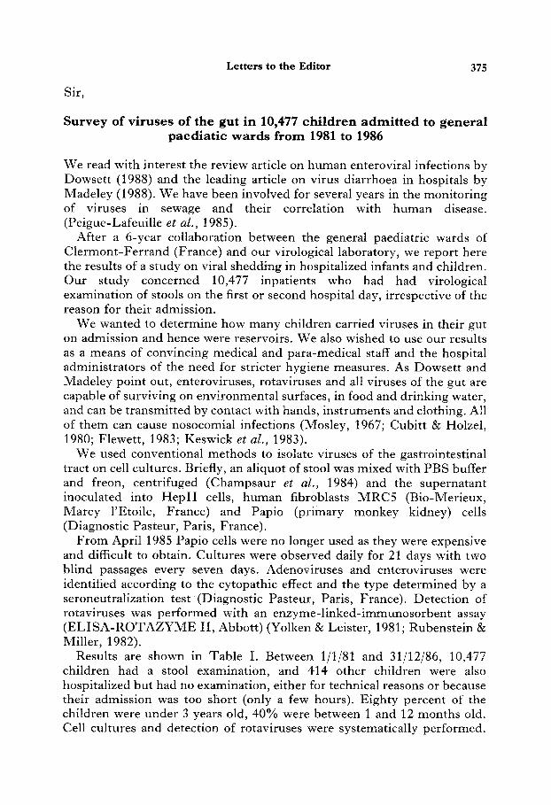
We read with interest the review article on human enteroviral infections by Dowsett (1988) and the leading article on virus diarrhoea in hospitals by Madeley (1988). We have been involved for several years in the monitoring of viruses in sewage and their correlation with human disease. (Peigue-Lafeuille et al., 1985).
After a &year collaboration between the general paediatric wards of Clermont-Ferrand (France) and our virological laboratory, we report here the results of a study on viral shedding in hospitalized infants and children. Our study concerned 10,477 inpatients who had had virological examination of stools on the first or second hospital day, irrespective of the reason for their admission.
We wanted to determine how many children carried viruses in their gut on admission and hence were reservoirs. We also wished to use our results as a means of convincing medical and para-medical staff and the hospital administrators of the need for stricter hygiene measures. As Dowsett and Madeley point out, enteroviruses, rotaviruses and all viruses of the gut are capable of surviving on environmental surfaces, in food and drinking water, and can be transmitted by contact with hands, instruments and clothing. All of them can cause nosocomial infections (Mosley, 1967; Cubitt & Holzel, 1980; Flewett, 1983; Keswick et al., 1983).
We used conventional methods to isolate viruses of the gastrointestinal tract on cell cultures. Briefly, an aliquot of stool was mixed with PBS buffer and freon, centrifuged (Champsaur et al., 1984) and the supernatant inoculated into HepII cells, human fibroblasts MRC5 (Bio-Merieux, Marcy l’Etoile, France) and Papio (primary monkey kidney) cells (Diagnostic Pasteur, Paris, France).
From April 1985 Papio cells were no longer used as they were expensive and difficult to obtain. Cultures were observed daily for 21 days with two blind passages every seven days. Adenoviruses and enteroviruses were identified according to the cytopathic effect and the type determined by a seroneutralization test (Diagnostic Pasteur, Paris, France). Detection of rotaviruses was performed with an enzyme-linked-immunosorbent assay (ELISA-ROTAZYME II, Abbott) (Yolken & Leister, 1981; Rubenstein & Miller, 1982).
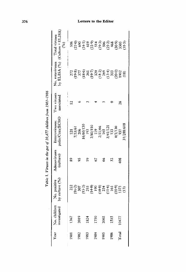
Results are shown in Table I. Between l/l/S1 and 31/12/86, 10,477 children had a stool examination, and 414 other children were also hospitalized but had no examination, either for technical reasons or because their admission was too short (only a few hours). Eighty percent of the children were under 3 years old, 40% were between 1 and 12 months old. Cell cultures and detection of rotaviruses were systematically performed.
Tabl
e I.
Viru
ses
in
the
gut
of
10,4
77
child
ren
from
19
81-1
986
Year
N
o.
child
ren
No.
po
sitiv
e Ad
enov
iruse
s En
tero
viru
ses:
Tw
o vi
ruse
s N
o.
rota
viru
ses
Tota
l vi
ruse
s in
vest
igat
ed
by c
ultu
re
(%)
(cul
ture
) po
lio/C
oxs/
ECH
O
asso
ciat
ed
by E
LISA
(%
) (C
ultu
re
f EL
ISA)
(%
)
1981
13
67
222
89
121
12
272
506
(16-
2)
7153
161
(19.
8)
(37.
0)
1982
20
19
307
9.5
206
6 37
7 69
0 (1
5.2)
16
/60/
l 30
(1
8.6)
(3
4.1)
19
83
1824
25
5 59
19
3 3
361
619
(14.
0)
3/80
/l 10
(1
9.7)
(3
3.9)
19
84
1750
19
0 67
11
9 4
320
514
(10.
8)
2151
166
(18.
2)
(29.
3)
1985
20
02
234
66
165
3 26
9 50
6 (1
1.6)
3/
41/1
21
(13.
4)
(25.
2)
1986
15
15
165
32
133
0 30
3 46
8 (1
0.9)
o/
3/13
0 (2
0.0)
(3
0.9)
To
tal
1047
7 13
73
408
937
28
1902
33
03
(13)
31
1288
1618
(1
8)
(31.
5)
Letters to the Editor 377
Combining the results of the two methods showed that viruses were present in the gut of 31.5% of children. Two percent of positive cultures revealed the presence of two viruses.
Cell lines used routinely had a definite influence on the types of viruses isolated. Since April 1985, when the use of primary monkey kidney cells was abandoned, the number of coxsackieviruses isolated has decreased significantly. Thus, epidemiological studies are inevitably distorted by the cell culture used.
The methods we used showed that one child in three carried viruses in the gut when admitted. This number would probably have been greater if we had considered virus types that were not systematically looked for: coxsackieviruses A (other than A9), astrovirus, calicivirus and other parvoviruses and parvovirus-like agents (Dolin, Treanor & Madore, 1987). We observed such non-cultivable viruses in stools of children with gastroenteritis by electron microscopy (Peigue et al., 1978). However, as Madeley (1988) b o served, neither electron microscopy nor polyacrylamide gel electrophoresis can be used to screen large numbers. Our numbers may therefore be an underestimate.
This study highlights the need for the development of rapid, sensitive and specific metho”ds of detection. Once commercially available they should be routinely used.
Moreover rapid detection of viruses, especially enteroviruses, in this age group might reduce the prescription and duration of antibiotic treatment and thereby indirectly contribute to reducing nosocomial infection.
H. H. Peigue-Lafeuille C. De Champs H. Laveran A. Labbe M. Chambon D. Beytout R. Cluzel
Laboratoire de Bacthiologie- Virologie, Faculte’ de Me’decine,
28 place Henri Dunant, 63001 Clermont-Ferrand, France
Champsaur, H., Questiaux, E., Prevot, J., Henry-Aymar, M., Goldzmidt, D., Bourjouane, M. & Bach, C. (1984). Rotavirus carriage, asymptomatic infection and disease in the first two years of live virus shedding. Journal of Infectious Diseases 149, 667-674.
Cubitt, W. D. & Holzel, H. (1980). Hospital acquired rotavirus infection in adults. Who is at risk? Journal of Hospital Infection 1, 327-331.
Dolin R., Treanor, J. J. & Madore, H. P. (1987). Novel agents of viral enteritis in humans. Journal of Infectious Diseases 155, 365-376.
Dowsett, E. G. (1988). Human enteroviral infections. Journal of Hospital Infection 11, 103-11s.
References
Flewett, T. H. (1983). Rotavirus in the home and hospital nursery. British Medical Journal 287, 568-569.
Keswick, B. H., Pickering, L. K., DuPont, H. L. & Woodward, W. E. (1983). Survival detection of rotaviruses on environmental surfaces in day care centers. Applied and Environmental Microbiology 10, 813-816.
378 Letters to the Editor
Madeley, C. R. (1988). Virus diarrhoea in hospital.Journal of Hospital Infection 12,145-149. Mosley, J. W. (1967). Transmission of viral diseases by drinking water. In Transmission of
viruses by the mater route, (Berg, ed.), pp. 1-9. New York: Wiley-Interscience. Peigue, H., Beytout-Monghal, M., Laveran, H. & Bourges, M. (1978). Coronavirus and
“astrovirus” observed in stools of children with gastroenteritis. Annals of Microbiology (Institut Pasteur) 129, 101-106.
Peigue-Lafeuille, H., Laveran, H., Auberger, M., Chambon, M., Trimolet, M. & Beytout, D. (1985). A four-year survey of enteroviruses in sewage. Correlation with human diseases. Revue &Epidt?miologie et de Sante’ Publique 33,445-451.
Rubenstein, A. S. & Miller, M. F. (1982). Comparison of an enzyme immunoassay with electron microscopic procedures for detecting rotavirus. Journal of Clinical Microbiology 15, 938-944.
Yolken, R. H. & Leister, F. J. (1981). Evaluation of enzyme immunoassays for the detection of human rotavirus. Journal of Infectious Diseases 144, 379.
Sir,
Environmental hazards from a hospital incinerator
We were interested to read the recent article by Blenkharn and Oakland, ‘Emission of viable bacteria in the exhaust flue gases from a hospital incinerator’ but feel that there are some more important environmental hazards which need be addressed.
The amount of liquid present in hospital waste which is to be incinerated was mentioned as one of the possible reasons for inadequate combustion. We have witnessed van loads of yellow sacks containing disposable incontinence pads which are literally dripping with urine as they have been collected from local nursing homes. Engineers have also expressed concern that this amount of liquid would reduce the temperature of the incinerator chamber leading to poor combustion and an increase in toxic exhaust.
The recent introduction of disposable suction equipment adds to this problem. When this product first came on the market in the early 1980s the plastic inner bag, containing up to two litres of fluid, was usually disposed of inside a yellow plastic bag. The manufacturers did later supply a cardboard box, bearing a ‘Hazard’ warning, for transport to the incinerator, but these are intended to hold five full containers, i.e. up to ten litres of fluid containing blood and human tissue. The hazard of storing this quantity is obvious. Two more manufacturers have recently brought similar products on to the market.
Even without taking into account the considerable expense of these products, any advantage of having a disposable system would not appear to outweigh the much safer practice of disposing of liquids which may contain harmful organisms into the sewage system.
Glenys Griffiths Martin Sheppard Timothy Kelly
Department of Microbiology, Mayday University Hospital,
Thornton Heath, Surrey CR4 7YE, UK