Embed Size (px)
Citation preview

Subscriber access provided by READING UNIV
Molecular Pharmaceutics is published by the American Chemical Society. 1155Sixteenth Street N.W., Washington, DC 20036Published by American Chemical Society. Copyright © American Chemical Society.However, no copyright claim is made to original U.S. Government works, or worksproduced by employees of any Commonwealth realm Crown government in the courseof their duties.
Article
Sustained Delivery of Doxorubicin via Acetalated Dextran ScaffoldPrevents Glioblastoma Recurrence after Surgical ResectionElizabeth G Graham-Gurysh, Kathryn M Moore, Andrew B Satterlee, Kevin T Sheets,
Feng-Chang Lin, Eric M. Bachelder, C. Ryan Miller, Shawn Hingtgen, and Kristy M. AinslieMol. Pharmaceutics, Just Accepted Manuscript • DOI: 10.1021/acs.molpharmaceut.7b01114 • Publication Date (Web): 17 Jan 2018
Downloaded from http://pubs.acs.org on January 18, 2018
Just Accepted
“Just Accepted” manuscripts have been peer-reviewed and accepted for publication. They are postedonline prior to technical editing, formatting for publication and author proofing. The American ChemicalSociety provides “Just Accepted” as a free service to the research community to expedite thedissemination of scientific material as soon as possible after acceptance. “Just Accepted” manuscriptsappear in full in PDF format accompanied by an HTML abstract. “Just Accepted” manuscripts have beenfully peer reviewed, but should not be considered the official version of record. They are accessible to allreaders and citable by the Digital Object Identifier (DOI®). “Just Accepted” is an optional service offeredto authors. Therefore, the “Just Accepted” Web site may not include all articles that will be publishedin the journal. After a manuscript is technically edited and formatted, it will be removed from the “JustAccepted” Web site and published as an ASAP article. Note that technical editing may introduce minorchanges to the manuscript text and/or graphics which could affect content, and all legal disclaimersand ethical guidelines that apply to the journal pertain. ACS cannot be held responsible for errorsor consequences arising from the use of information contained in these “Just Accepted” manuscripts.

1
Sustained Delivery of Doxorubicin via Acetalated
Dextran Scaffold Prevents Glioblastoma Recurrence
after Surgical Resection
Elizabeth Graham-Gurysh1, Kathryn M. Moore
2, Andrew B. Satterlee
1, Kevin T. Sheets
1, Feng-
Chang Lin3, Eric M. Bachelder
1, C. Ryan Miller
4, Shawn D. Hingtgen
1, Kristy M. Ainslie
1*
1Division of Pharmacoengineering and Molecular Pharmaceutics, Eshelman School of
Pharmacy, University of North Carolina at Chapel Hill, USA.
2Joint Department of Biomedical Engineering, University of North Carolina at Chapel Hill and
North Carolina State University, USA.
3 Department of Biostatistics and North Carolina Translational and Clinical Sciences Institute,
University of North Carolina at Chapel Hill, USA
4Division of Neuropathology, Department of Pathology and Laboratory Medicine, Departments
of Neurology and Pharmacology, Lineberger Comprehensive Cancer Center, and Neuroscience
Center, School of Medicine, University of North Carolina at Chapel Hill, USA
KEYWORDS
Ace-DEX, poly-lactide (PLA), polyanhidride, carmustine, drug delivery, nanofiber
Page 1 of 39
ACS Paragon Plus Environment
Molecular Pharmaceutics
123456789101112131415161718192021222324252627282930313233343536373839404142434445464748495051525354555657585960

2
ABSTRACT
The primary cause of mortality for glioblastoma (GBM) is local tumor recurrence following
standard-of-care therapies, including surgical resection. With most tumors recurring near the site
of surgical resection, local delivery of chemotherapy at the time of surgery is a promising
strategy. Herein drug loaded polymer scaffolds with two distinct degradation profiles were
fabricated to investigate the effect of local drug delivery rate on GBM recurrence following
surgical resection. The novel biopolymer, acetalated dextran (Ace-DEX), was compared to
commercially available polyester, poly(L-lactide) (PLA). Steady state doxorubicin (DXR)
release from Ace-DEX scaffolds was found to be faster when compared to scaffolds comprised
of PLA, in vitro. This increased drug release rate translated to improved therapeutic outcomes in
a novel surgical model of orthotopic glioblastoma resection and recurrence. Mice treated with
DXR loaded Ace-DEX scaffolds (Ace-DEX/10DXR) resulted in 57% long term survival out to
study completion at 120 days compared to 20% survival following treatment with DXR loaded
PLA scaffolds (PLA/10DXR). Additionally, all mice treated with PLA/10DXR scaffolds
exhibited disease progression by day 38, as defined by a five-fold growth in tumor
bioluminescent signal. In contrast, 57% of mice treated with Ace-DEX/10DXR scaffolds
displayed a reduction in tumor burden, with 43% exhibiting complete remission. These results
underscore the importance of polymer choice and drug release rate when evaluating local drug
delivery strategies to improve prognosis for GBM patients undergoing tumor resection.
Page 2 of 39
ACS Paragon Plus Environment
Molecular Pharmaceutics
123456789101112131415161718192021222324252627282930313233343536373839404142434445464748495051525354555657585960

3
TABLE OF CONTENTS GRAPHIC
INTRODUCTION
Glioblastoma (GBM) is an especially aggressive, grade IV central nervous system tumor.
GBM is the most common primary brain tumor and accounts for 45-50% of all gliomas,1 and
even with surgical resection, radiation, and chemotherapy, the median survival for GBM patients
remains only 12 to 15 months.1-3
The primary cause of mortality for GBM is local tumor
recurrence, with 90 - 95% of tumors recurring within 2 cm of the original tumor.4-7
As such,
patient outcomes could be greatly improved by delaying or preventing local recurrence.
Unfortunately, chemotherapeutic options are limited due to the inability of most drugs to cross
the blood-brain barrier. One strategy to improve drug delivery to GBM and bypass the blood-
brain barrier is interstitial chemotherapy. This can be achieved by lining the resection cavity with
a material that releases drug as it degrades.8 This strategy is applied clinically with Gliadel
®, a
polyester and polyanhydride polymer wafer that is used in some GBM patients to deliver
carmustine (BCNU) directly into the resection cavity.9 Unfortunately, Gliadel
® only extends
median survival by 10-18 weeks over placebo.10, 11
This is likely due to the low tumoricidal
activity of BCNU against GBM and its rapid clearance from the brain after release from the
polymer wafer.12-14
Additionally, tumor cells easily acquire resistance to alkylating agents like
Page 3 of 39
ACS Paragon Plus Environment
Molecular Pharmaceutics
123456789101112131415161718192021222324252627282930313233343536373839404142434445464748495051525354555657585960

4
BCNU through the DNA repair mediated by the enzyme O6-methylguanine-DNA
methyltransferase (MGMT).15, 16
Low tumoricidal activity, easily acquired drug resistance, and
rapid clearance of BCNU from the brain highlight the need for exploration into alternative
anticancer drugs for interstitial therapy.
One promising compound to combat GBM is doxorubicin (DXR). This highly potent
chemotherapeutic agent induces cell death via multiple mechanisms of action, including DNA
intercalation and interaction with topoisomerase II.17, 18
However, DXR is unable to be
administered systemically against GBM due to its inability to pass through the blood-brain
barrier and reach therapeutic levels in the brain.19
Interstitial delivery of DXR against GBM has
been explored previously with some success. Local delivery of DXR was able to extend median
survival and delay tumor growth, but ultimately complete remission was not achieved.20, 21
This
is likely due to the sub-optimal DXR release kinetics from the polymeric devices utilized for
interstitial therapy. Lesniak et al. has shown that interstitial DXR delivered via a polyanhydride
polymer wafer was able to extend median survival of Fisher rats with intracranial glioma from
21 to 45 days, despite the fact that DXR release from the polymer wafer plateaued at
approximately 14% after the first 50 hours.20
Lin et al. also found that DXR released from
polyester polymer device was able to slow tumor growth in a subcutaneous flank model.21
Similar to the polyanhydride wafers, the polyester device displayed an initial release of DXR that
plateaued after the first 2 days. This pattern of stagnant DXR release after an initial period of
rapid release over the first few days is also seen in multiple polymer drug delivery platforms
including polyanhydrides,20
polyesters,21, 22
and polyethylene glycol polyester composites.23, 24
The therapeutic efficacy of interstitial GBM treatment could be greatly improved with sustained
DXR release from an optimal polymer matrix.
Page 4 of 39
ACS Paragon Plus Environment
Molecular Pharmaceutics
123456789101112131415161718192021222324252627282930313233343536373839404142434445464748495051525354555657585960

5
Aggressive cancers, such as GBM, recur in a matter of weeks. As such, polymer drug delivery
platforms capable of rapid and sustained drug release are vital to suppressing local tumor
recurrence. Most commonly used polymers for interstitial drug delivery, such as polyesters and
polyanhydrides, have relatively slow degradation rates on the order of months to years.25, 26
Additionally, polyesters degrade by bulk degradation, which can lead to spontaneous dumping of
drug and potential unforeseen toxicity.27
An example of this can be seen in Manome et al., where
poly(D,L-lactide-co-glycolide) (PLGA) sheets exhibited a sudden burst release of DXR around
30 days in vitro. Finally, polyesters create an acidic microenvironment as they degrade, which
can alter drug activity and cause tissue toxicity.28-30
Taken together, these limitations regarding
effective drug release and adverse polymer by-products highlight the need for a tunable drug
delivery platform to expand the population of drugs that can be utilized for sustained interstitial
drug delivery.
An alternative polymer for drug delivery is acetalated dextran (Ace-DEX). Ace-DEX is a
novel polymer with tunable degradation rates ranging from days to months. It is synthesized by
chemically modifying dextran with 2-ethyoxypropene to render the polymer hydrophobic.31, 32
This reaction generates acyclic and cyclic acetals on the pendent hydroxyl groups of dextran in a
time-dependent manner.33-35
The ratio between cyclic and acyclic acetals can be changed based
on the length of reaction time.33-35
Under aqueous conditions, acetal groups are hydrolyzed,
revealing the parent hydroxyl groups of dextran and resulting in Ace-DEX degradation. Since
cyclic acetals are significantly more stable to hydrolysis than acyclic acetals, the time-dependent
nature of acetal coverage allows for tight control of Ace-DEX degradation rate. The degradation
by-products of Ace-DEX (dextran, acetone, and ethanol) are pH neutral and occur at exceedingly
low concentrations, far below levels that would result in toxicity.33, 34
Our group has previously
Page 5 of 39
ACS Paragon Plus Environment
Molecular Pharmaceutics
123456789101112131415161718192021222324252627282930313233343536373839404142434445464748495051525354555657585960

6
fabricated drug loaded Ace-DEX nanofibrous scaffolds using electrospinning, and demonstrated
tunable drug release and bioactivity.31
The tunability of Ace-DEX degradation rates combined
with the safe, pH-neutral degradation products makes it a promising polymer platform for
interstitial drug delivery against GBM. To advance this potential therapy towards translation to
the clinic, it is imperative that we evaluate GBM treatment in the most relevant animal model.
The therapeutic efficacy of interstitial therapy against GBM is often tested in a simple
subcutaneous flank or orthotopic tumors that fail to incorporate an essential component of
multimodal therapy: surgical resection. As surgery is the standard of care for human patients, a
preclinical animal model of GBM resection and recurrence is extremely beneficial. This is
particularly important because surgical resection induces changes in the peri-tumoral
microenvironment and influences tumor biology, growth, and invasion.36
We previously
developed a fluorescent-guided surgical mouse model of GBM resection and recurrence.37, 38
With this model, GBM xenografts are implanted orthotopically and allowed to establish for
several days. Under fluorescent guidance, approximately 90% of the tumor is removed by
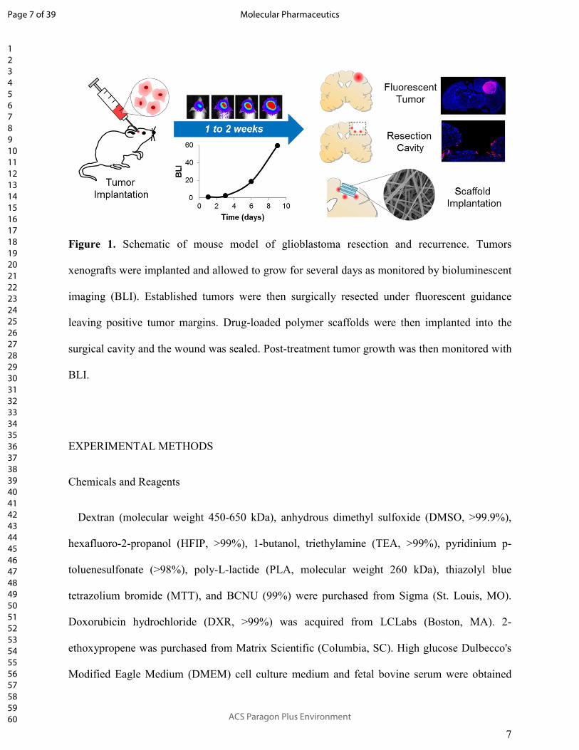
surgical resection and interstitial therapy can then be applied at the time of surgery (Figure 1).
Here we describe the fabrication and utilization of electrospun DXR loaded polymer scaffolds
comprised of Ace-DEX or poly(L-lactide) (PLA), a polyester commonly used for interstitial drug
delivery. We then investigated the effect of polymer on DXR release from scaffolds in vitro.
DXR release rates were then evaluated in a clinically relevant murine model of local GBM
recurrence following partial surgical resection.
Page 6 of 39
ACS Paragon Plus Environment
Molecular Pharmaceutics
123456789101112131415161718192021222324252627282930313233343536373839404142434445464748495051525354555657585960
7
Figure 1. Schematic of mouse model of glioblastoma resection and recurrence. Tumors
xenografts were implanted and allowed to grow for several days as monitored by bioluminescent
imaging (BLI). Established tumors were then surgically resected under fluorescent guidance
leaving positive tumor margins. Drug-loaded polymer scaffolds were then implanted into the
surgical cavity and the wound was sealed. Post-treatment tumor growth was then monitored with
BLI.
EXPERIMENTAL METHODS
Chemicals and Reagents
Dextran (molecular weight 450-650 kDa), anhydrous dimethyl sulfoxide (DMSO, >99.9%),
hexafluoro-2-propanol (HFIP, >99%), 1-butanol, triethylamine (TEA, >99%), pyridinium p-
toluenesulfonate (>98%), poly-L-lactide (PLA, molecular weight 260 kDa), thiazolyl blue
tetrazolium bromide (MTT), and BCNU (99%) were purchased from Sigma (St. Louis, MO).
Doxorubicin hydrochloride (DXR, >99%) was acquired from LCLabs (Boston, MA). 2-
ethoxypropene was purchased from Matrix Scientific (Columbia, SC). High glucose Dulbecco's
Modified Eagle Medium (DMEM) cell culture medium and fetal bovine serum were obtained
Page 7 of 39
ACS Paragon Plus Environment
Molecular Pharmaceutics
123456789101112131415161718192021222324252627282930313233343536373839404142434445464748495051525354555657585960

8
from Corning (Corning, NY). Penicillin/streptomycin was purchased from Hyclone (Pittsburgh,
PA). Rat monoclonal antibodies against CD45 (clone I3/2.3, ab25386), F4/80 (clone BM8, #14-
4801-82), and ImmPRESS HRP Anti-Rat Ig (#MP-7444-15) were acquired from Abcam
(Cambridge, MA), eBioscience Inc. (San Diego, CA), and Vector Labs (Burlingame, CA),
respectively. Rabbit polyclonal antibody against glial fibrillary acidic protein (GFAP, #Z0334)
and protein block (#X0909) were both purchased from DAKO/Agilent Technologies (Santa
Clara, CA). Bond™ Dewax Solution (AR9222), Bond™ Wash Solution (AR9590), Bond™
Epitope Retrieval Solution 1 (pH 6.0, AR9961), Bond™ Enzyme 1 (RE7160-CE), and Bond™
Polymer Refine Detection (DS9900) were all acquired from Leica Biosystems (Buffalo Grove,
IL).
Cell transfection and cytotoxicity assays
Human glioma cell lines U87-MG, LN-18, and LN-229 were purchased from American Type
Culture Collection (ATCC; Manassas, VA; #HTB-14, #CRL-2610, and #CRL-2611,
respectively). Custom vector synthesis services from Invitrogen (Carlsbad, CA) were utilized to
generate mCherry-firefly luciferase (mCh-FL). The construct was packaged as a lentiviral vector
in 293T/17 cells using a helper virus-free packaging system as described previously.39
Cells were cultured in DMEM medium supplemented with 1% penicillin/streptomycin and
10% fetal bovine serum and maintained in the exponential phase of growth at 37°C under 5%
carbon dioxide. Cells were seeded onto 96-well plates at a density of 5 x 10³ cells per well and
allowed to grow to 50% confluency. DXR, at concentrations of 0.005, 0.025, 0.05, 0.1, 0.2, 0.5,
1, 2, 5, 10, 20, or 50 µM, was incubated with the cells for 48 hours. MTT powder was dissolved
in media at a concentration of 0.6 mg/mL and added to treated cells for 3 hours, allowing for
Page 8 of 39
ACS Paragon Plus Environment
Molecular Pharmaceutics
123456789101112131415161718192021222324252627282930313233343536373839404142434445464748495051525354555657585960

9
metabolically active cells to reduce the MTT salt to form purple formazan crystals. Crystals were
dissolved in isopropanol and the absorption at 560 nm was utilized to quantify cell viability
normalizing values to an untreated control, using 670 nm as a reference wavelength.
Experiments were repeated in triplicate and a best fit trend-line was used to determine the dose
required to reduce cell viability by 50%. This procedure was repeated for BCNU at
concentrations of 0.01, 0.05, 0.1, 0.25, 0.5, 0.75, and 1 mM.
Acetalated dextran synthesis
Ace-DEX was synthesized and characterized as described previously.32
Briefly, lyophilized
dextran was dissolved in DMSO in the presence of an acid catalyst, pyridinium p-
toluenesulfonate. Dextran was reacted with 2-ethoxypropene under anhydrous conditions for 2
hours, before the reaction was quenched with TEA. Ace-DEX was precipitated in basic water
and lyophilized. Afterwards, the polymer was dissolved in ethanol and centrifuged to remove
impurities. Ace-DEX was then re-precipitated in basic water, lyophilized, and stored at -20˚C
until it was used. The relative extent of cyclic acetal coverage, which dictates the degradation
rate of the polymer, was calculated via nuclear magnetic resonance (NMR, Varian Inova 400)
based on a previously developed method.32
Scaffold fabrication
Ace-DEX or PLA was dissolved in organic solvents at 200 mg/mL with 5% or 10% (wt/wt)
DXR and loaded into a glass syringe with a blunt 21 gauge needle. A bias of 20 kV (−10 kV to
the collection surface and +10 kV to the needle) was applied over a 13 cm working distance at a
Page 9 of 39
ACS Paragon Plus Environment
Molecular Pharmaceutics
123456789101112131415161718192021222324252627282930313233343536373839404142434445464748495051525354555657585960

10
flow rate of 1 mL/hr. A tri-solvent system consisting of HFIP, butanol, and TEA was evaluated.
Maintaining TEA constant at 1% v/v, HFIP and butanol ratios were varied to create three
conditions: 90% HFIP and 10% butanol, 80% HFIP and 20% butanol, and 60% HFIP and 40%
butanol.
Scaffold characterization
Scanning electron microscopy was performed to assess the morphology of the electrospun
scaffolds. Scaffolds were mounted on aluminum stubs using carbon tape, and imaged at 2 kV on
Hitachi S-4700 Cold Cathode Field Emission Scanning Electron Microscope. DXR
encapsulation efficiency was defined as the ratio of empirical DXR loaded to theoretical loading.
To quantify this, scaffold samples were weighed, dissolved in DMSO, and evaluated by optical
absorption at 480 nm. The amount of DXR loaded was then determined by calibration curve. To
evaluate DXR release in vitro, approximately 1 mg of scaffold was placed into dialysis cups with
a molecular weight cutoff of 7 kDa and added to a sink of phosphate buffered saline (PBS, pH
7.4) stirring at 37°C. At predetermined time points, scaffolds were removed and stored at -20°C.
Samples were then lyophilized, dissolved in DMSO, and evaluated by optical absorption at 480
nm. The amount of DXR retained in each sample was determined by a calibration curve.
Animals
Nude BALB/c mice were housed in groups of five in a vivarium maintained on a 12 hour
light/dark schedule at 30°C and 50% relative humidity. Food and water were available ad
libitum. For all surgical procedures, mice were anesthetized by vapor isoflurane and immobilized
on a three point stereotactic frame (Stoelting, Kiel, WA). For all surgeries, subcutaneous
Page 10 of 39
ACS Paragon Plus Environment
Molecular Pharmaceutics
123456789101112131415161718192021222324252627282930313233343536373839404142434445464748495051525354555657585960

11
carpofen (5 mg/kg) was administered prior to surgery and twice daily for 3 days following
surgery for pain management.
All experimental protocols were approved by the Animal Care and Use Committees at The
University of North Carolina at Chapel Hill, and care of the mice was in accordance with the
standards set forth by the National Institutes of Health Guide for the Care and Use of Laboratory
Animals, USDA regulations, and the American Veterinary Medical Association.
Maximum tolerated dose
An incision was made in the skin to expose the skull of the mouse. A small circular portion of
the skull over the right frontal lobe, approximately 3 mm in diameter, was surgically removed
using a bone drill (Ideal Microdrill, Harvard Apparatus, Holliston, MA) and forceps. A cranial
window was created by leaving this portion of the skull open. The skin was closed with Vetbond
Tissue Adhesive (3M, Maplewood, MN). Several days after cranial windows were established,
the dura over the cranial window was cut away, and a section of the right frontal lobe was
removed by aspiration creating a pseudo resection cavity approximately 1 to 2 mm deep. DXR
loaded Ace-DEX scaffolds were placed in the resection cavity and the skin was closed with
tissue adhesive. Mice were evaluated daily for two weeks to assess for gross toxicity as
evidenced by weight loss or gait abnormalities. After 14 days, mice were euthanized by cardiac
perfusion. Brains were extracted and fixed in formalin in preparation for histology.
Therapeutic efficacy against recurrent GBM
The therapeutic efficacy of drug loaded scaffolds against local GBM recurrence was evaluated
in an image-guided surgical model of GBM resection and recurrence as described previously
Page 11 of 39
ACS Paragon Plus Environment
Molecular Pharmaceutics
123456789101112131415161718192021222324252627282930313233343536373839404142434445464748495051525354555657585960

12
(Figure 1).36, 37
After establishment of a cranial window as detailed above, mice were again
anesthetized by vapor isoflurane and immobilized on a stereotactic frame. Firefly-luciferase and
mCherry transfected U87-MG cells were implanted stereotactically (1 x 10⁵ cells in 3 µL of
phosphate buffered saline at a rate of 1 µL per minute) in the right frontal lobe 2 mm lateral to
the bregma and 0.5 mm below the dura. Tumor growth was monitored by bioluminescent
imaging (BLI, Perkin Elmer IVIS Lumina In Vivo Imaging System). Once tumors were
visualized as being well established (1 to 2 weeks after tumor implantation), mice were
anesthetized and immobilized. The cranial window was exposed and dura was removed to
expose the tumor. Under fluorescent guidance, the tumor was removed by aspiration. Following
tumor removal, scaffolds were placed in the surgical cavity and the skin was closed with tissue
adhesive. For Ace-DEX/10DXR, this required a total scaffold mass of 1.8 mg (which was
equivalent to six, 3 mm circular hole punches). For PLA/10DXR this required a total scaffold
mass of 1.65 mg (which was equivalent to four, 3 mm circular hole punches). Equivalent mass
for blank polymer scaffolds was implanted. Serial BLI imaging was used to noninvasively
quantify tumor growth starting the day after surgical resection and scaffold implantation (Day 1).
A total of 42 mice were implanted with U87-MG tumors. Mice were excluded from the study on
Day 1 if there was no evidence of tumor by BLI (“complete resection”, n=3), or if the BLI signal
was outside two standard deviations of the average (“insufficient resection”, n=3). Mice were
also excluded for extra-cranial tumor location, either in the initial tumor placement (n=2), or
when the tumors recurred (n=5). Two other mice were also excluded; one began having seizures
41 days after tumor resection despite having no evidence of tumor (statistical significance
remained the same regardless of inclusion); another contracted Corynebacterium bovis, which
has been shown to affect tumor xenografts and drug sensitivity in nude mice.40
Excluded mice
Page 12 of 39
ACS Paragon Plus Environment
Molecular Pharmaceutics
123456789101112131415161718192021222324252627282930313233343536373839404142434445464748495051525354555657585960

13
are detailed in Supporting Information Table S1. This resulted in 27 mice being included in
the study with the following breakdown for groups: no treatment control (n=5), Ace-DEX/blank
(n=5), PLA/blank (n=6), Ace-DEX/10DXR (n=7), PLA/10DXR (n=4). Prior to resection, Day -
1, established tumors had an average BLI signal of 4.1x10 ± 2.7x10⁹ ρ/sec/cm
2/sr. The average
BLI signal after resection, Day 1, was 1.8x10⁸ ± 3.4 x10
8 ρ/sec/cm
2/sr. Percent of tumor resected
was determined by the following equation: PercentResected = 100 × (1 −�������
��������). The
average percent of tumor resected for all groups was 95.4% ± 9%. A box plot illustrating pre-
resection BLI values for each mouse and a table detailing average BLI prior to resection (Day -
1), percent tumor resection, and BLI after resection (Day 1) for each treatment group can be
found in Supporting Information Figure S1, Table. Mice were evaluated daily and euthanized
if they lost more than 15% body weight. The study was conducted for 120 days. After euthanasia
by cardiac perfusion, the brain was extracted, formalin fixed and paraffin embedded, and saved
for histological evaluation.
Statistical Analysis
Statistical analysis of normalized mouse weights after scaffold implantation was performed by
two way analysis of variance with post-hoc Bonferroni test using GraphPad Prism (La Jolla,
CA).
Statistical analysis of overall and progression free survival rates was performed by
proportional hazards regression with adjustment for the initial tumor size after surgical resection
using IBM SPSS Statistics 24 (Chicago, IL).
Histology
Page 13 of 39
ACS Paragon Plus Environment
Molecular Pharmaceutics
123456789101112131415161718192021222324252627282930313233343536373839404142434445464748495051525354555657585960

14
Whole mouse brains were fixed in formalin, processed and embedded in paraffin, and step
sectioned at 4 µm thickness using 300 µm gaps. Sections collected at each level were stained for
histopathology using hematoxylin and eosin (H&E) and immunohistochemistry (IHC) to
evaluate for signs of toxicity. H&E staining was performed using an Autostainer XL from Leica
Biosystems. For IHC, rat monoclonal antibodies against CD45 and F4/80 and rabbit polyclonal
antibody against glial fibrillary acidic protein (GFAP) were utilized. IHC was carried out in the
fully automated Bond™ Immunostainer (Leica Biosystems Inc). Slides were dewaxed in Bond™
Dewax Solution and hydrated in Bond™ Wash Solution. Antigen retrieval for CD45 and GFAP
was performed for 20 min at 100ºC in Bond™ Epitope Retrieval Solution 1 (pH 6.0) and in
Bond™ Enzyme 1 for 5 minutes followed by a 10 minute protein block. After pre-treatment,
slides were incubated for 30 minutes with CD45 (1:100) and GFAP (1:2500), and for 1 hour with
F4/80 (1:100). Detection of all antibodies was performed using Bond™ Polymer Refine
Detection. For CD45 and F4/80, the secondary antibody was replaced with ImmPRESS HRP
Anti-Rat Ig. Stained slides were dehydrated and sealed with a glass coverslip. Positive and
negative controls (no primary antibody) were included for each antibody. H&E and IHC stained
slides were digitally imaged using an Aperio ScanScope XT (Leica Biosystems) with a 20x
objective.
RESULTS
GBM sensitivity to DXR
Sensitivity to DXR was tested in three GBM cell lines: U87-MG, LN-229, and LN-18, and
then compared to BCNU (see Supporting Information Figure S2). The concentration required
to reduce cell viability by 50% (IC50) after 48 hour incubation is listed in Table 1. All three
Page 14 of 39
ACS Paragon Plus Environment
Molecular Pharmaceutics
123456789101112131415161718192021222324252627282930313233343536373839404142434445464748495051525354555657585960

15
GBM cell lines were more sensitive to DXR than BCNU by at least 200 fold. These are
comparable to literature values.41, 42
Table 1. Sensitivity of Glioblastoma lines to chemotherapies. Concentration required to reduce
cell viability by 50% (IC50) after 48 hour incubation with DXR or BCNU for glioblastoma cell
lines U87-MG, LN-18, and LN-229, as measured by MTT assay.
Cell Line IC50 (µM) DXR BCNU
U87-MG 0.13 380 LN-18 0.80 225 LN-229 1.10 280
Scaffold fabrication and characterization
Drug-eluting polymer scaffolds were fabricated by electrospinning. The organic solvent
system and drug loading were varied to determine their respective role in DXR release. First,
Ace-DEX scaffolds containing a fixed loading of 5% (wt/wt) DXR (Ace-DEX/5DXR) were
electrospun varying the solvent system ratio of HFIP to butanol to 90:10, 80:20, and 60:40. The
solvent system affected both fiber width and burst release of DXR. Increasing the volume
fraction of HFIP generated wider fibers (Figure 2a-c), which correlated with a faster burst
release of DXR (Figure 2e), and the scaffolds exhibiting a more vibrant hue of orange, the color
of DXR.
Page 15 of 39
ACS Paragon Plus Environment
Molecular Pharmaceutics
123456789101112131415161718192021222324252627282930313233343536373839404142434445464748495051525354555657585960

16
Figure 2. Solvent System Effect on Ace-DEX Scaffolds. Scanning electron micrographs of
acetalated dextran 5% (wt/wt) doxorubicin (DXR) (Ace-DEX/5DXR) scaffolds electrospun in a
solvent system of hexafluoroisopropanol (HFIP) and butanol with ratios of a) 90:10, b) 80:20,
and c) 60:40. d) Picture of 5% wt/wt loaded DXR scaffolds electrospun with HFIP and butanol
with ratios of 90:10, 80:20, and 60:40 (from left to right). e) Burst release of DXR from Ace-
DEX/5DXR scaffolds electrospun with different HFIP to butanol ratios of 90:10 (black square),
80:20 (gray triangle), and 60:40 (white circle). Data points for all graphs are the mean ± standard
error of the mean. Scale bar is the same for all images and represents 5 µm.
The effect of DXR loading on release rate was also investigated. Figure 3a-c illustrates that
increasing DXR loading from 5% to 10% (wt/wt) had no effect on fiber morphology. Increasing
DXR loading did result in a higher burst release of DXR (Figure 3f); however, afterwards the
rate of DXR release is less pronounced (data not shown). To directly compare Ace-DEX to a
Page 16 of 39
ACS Paragon Plus Environment
Molecular Pharmaceutics
123456789101112131415161718192021222324252627282930313233343536373839404142434445464748495051525354555657585960

17
polyester, one of the most commonly used polymers for drug delivery, we electrospun a scaffold
composed of PLA under identical electrospinning conditions with 10% DXR loaded (wt/wt)
(PLA/10DXR). Ace-DEX and PLA scaffolds had similar morphology (Figure 3a-e) with high
encapsulation efficiencies of 85 ± 5% and 94 ± 12%, respectively. Evaluating DXR release in
vitro over time, PLA/10DXR scaffolds had a higher burst release compared to Ace-
DEX/10DXR, with 46 ± 7.1% and 28 ± 1.6% of DXR released in the first 24 hours respectively.
However, PLA/10DXR scaffolds only released 3% of DXR over the next 34 days. By contrast,
Ace-DEX/10DXR released 27% of DXR over the same extended time frame. Although the two
scaffolds ultimately released similar amounts of DXR (approximately 50% over 35 days), the
steady-state release from Ace-DEX scaffolds is faster than that from PLA, which releases the
majority of the DXR within the first 24 hours (Figure 3g, Table).
Page 17 of 39
ACS Paragon Plus Environment
Molecular Pharmaceutics
123456789101112131415161718192021222324252627282930313233343536373839404142434445464748495051525354555657585960

18
Figure 3. Morphology and Drug Release from Scaffolds. Scanning electron micrographs of a)
Acetalated dextran blank (Ace-DEX/blank), b) 5% (wt/wt) doxorubicin (DXR) (Ace-
DEX/5DXR), c) 10% (wt/wt) DXR (Ace-DEX/10DXR), d) poly lactic acid blank (PLA/blank),
and e) 10% (wt/wt) DXR PLA (PLA/10DXR) scaffolds. Scale bar is the same for all images and
represents 5 µm. f) Burst release of DXR from Ace-DEX/5DXR (○) and Ace-DEX/10DXR (■)
electrospun with 60:40 ratio of HFIP to butanol. g) DXR release from Ace-DEX/10DXR (■) and
PLA/10DXR scaffolds (♦). Table describes DXR release kinetics from Ace-DEX/10DXR and
PLA/10DXR scaffolds. Burst release = DXR released over the first 24 hours. Data points for all
graphs are the mean ± standard error of the mean.
Page 18 of 39
ACS Paragon Plus Environment
Molecular Pharmaceutics
123456789101112131415161718192021222324252627282930313233343536373839404142434445464748495051525354555657585960

19
Maximum tolerated dose
Prior to testing the efficacy of DXR loaded Ace-DEX scaffolds against GBM, in vivo toxicity
was evaluated. Mice were weighed and evaluated daily for gross signs of toxicity. Scaffold mass,
DXR dose, and mouse weight are detailed in Supporting Information Table S2. Mice
implanted with Ace-DEX/10DXR scaffolds at 200 µg per mouse exhibited a 5% decrease in
weight (see Supporting Information Figure S3). This was notable given that other groups
gained approximately 5% weight over this same time period. Mice implanted with the highest
DXR dose, 200 µg, also displayed delayed surgical wound healing compared to other mice,
including the second highest dose, 100 µg (data not shown). When evaluated by histology, dose-
dependent toxicity at doses as low as 100 µg were seen with Ace-DEX/10DXR scaffolds after 14
days (see Supporting Information Figure S4). The effect of Ace-DEX/blank scaffold
compared to Ace-DEX/10DXR at the 200 µg dose was also evaluated by IHC staining.
Concurrent coronal sections were stained for F4/80, GFAP, and CD45 to show presence of
macrophages, activated microglia, and immune cell infiltrates, respectively, at the site of scaffold
implantation (Figure 4). As demonstrated by IHC staining, higher levels of DXR led to
increased glial cell activation and immune cell infiltrates, primarily macrophages, compared to
unloaded Ace-DEX scaffolds.
Page 19 of 39
ACS Paragon Plus Environment
Molecular Pharmaceutics
123456789101112131415161718192021222324252627282930313233343536373839404142434445464748495051525354555657585960

20
Figure 4. Histological Toxicity Comparison of unloaded Ace-DEX and Ace-DEX/10DXR
Scaffolds. Coronal sections of mice euthanized 14 days after scaffold implantation with Left)
Unloaded acetalated dextran (Ace-DEX) scaffold (total scaffold mass of 2.2 mg) and Right)
10% w/w Ace-DEX/10DXR scaffold (total scaffold mass of 2.2 mg) with total amount of
doxorubicin (DXR) equal to 200 µg. Images show sections stained with hematoxylin and eosin
(H&E), glial fibrillary acidic protein (GFAP, indicating glial reaction), CD45, (immune cell
infiltrates), and F4/80 (macrophages). Scale bars are the equivalent for all images at the same
magnification. Black boxes indicate location of magnified region of interest presented in
subsequent column.
Efficacy against recurrent GBM
Page 20 of 39
ACS Paragon Plus Environment
Molecular Pharmaceutics
123456789101112131415161718192021222324252627282930313233343536373839404142434445464748495051525354555657585960
21
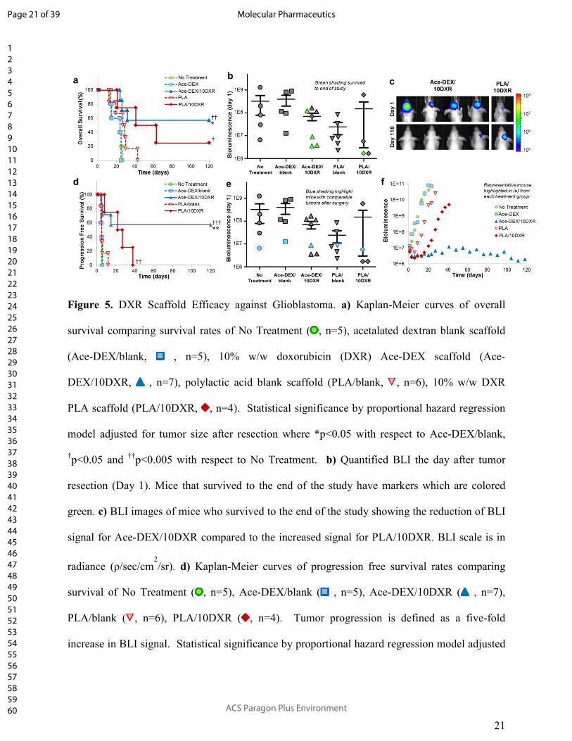
Figure 5. DXR Scaffold Efficacy against Glioblastoma. a) Kaplan-Meier curves of overall
survival comparing survival rates of No Treatment ( , n=5), acetalated dextran blank scaffold
(Ace-DEX/blank, , n=5), 10% w/w doxorubicin (DXR) Ace-DEX scaffold (Ace-
DEX/10DXR, , n=7), polylactic acid blank scaffold (PLA/blank, , n=6), 10% w/w DXR
PLA scaffold (PLA/10DXR, , n=4). Statistical significance by proportional hazard regression
model adjusted for tumor size after resection where *p<0.05 with respect to Ace-DEX/blank,
†p<0.05 and
††p<0.005 with respect to No Treatment. b) Quantified BLI the day after tumor
resection (Day 1). Mice that survived to the end of the study have markers which are colored
green. c) BLI images of mice who survived to the end of the study showing the reduction of BLI
signal for Ace-DEX/10DXR compared to the increased signal for PLA/10DXR. BLI scale is in
radiance (ρ/sec/cm2/sr). d) Kaplan-Meier curves of progression free survival rates comparing
survival of No Treatment ( , n=5), Ace-DEX/blank ( , n=5), Ace-DEX/10DXR ( , n=7),
PLA/blank ( , n=6), PLA/10DXR ( , n=4). Tumor progression is defined as a five-fold
increase in BLI signal. Statistical significance by proportional hazard regression model adjusted
Page 21 of 39
ACS Paragon Plus Environment
Molecular Pharmaceutics
123456789101112131415161718192021222324252627282930313233343536373839404142434445464748495051525354555657585960

22
for tumor size after resection where **p<0.01 with respect to Ace-DEX/blank, ††
p<0.01 and
†††p<0.001 with respect to No Treatment. e) Quantified BLI the day after tumor resection (Day
1) Markers indicating representative mice with comparable starting tumor sizes are highlighted
blue. f) Quantified BLI of a representative mouse from each treatment group illustrating tumor
growth over time (highlighted in blue in e). No Treatment ( ), Ace-DEX/blank ( ), Ace-
DEX/10DXR ( ), PLA/blank ( ), PLA/10DXR ( ).
Treatment with Ace-DEX/10DXR scaffolds statistically improved survival rates compared to
Ace-DEX/blank (p < 0.005) and ‘no treatment’ (p < 0.005) with four mice surviving to the end
of the study (Day 120) (Figure 5a, 5b). Of these four surviving mice, only one had BLI evidence
of tumor at the end of the study. Notably, the quantified BLI for this mouse decreased to just
20% of its original signal the day after resection (Day 1) over the length of the study (120 days)
(Figure 5c). In contrast, only one mouse treated with PLA/10DXR survived to the end of the
study, and over this time, the tumor BLI signal increased by more than eight fold (Figure 5b,
5c). Treatment with PLA/10DXR scaffolds statistically improved overall survival rates
compared to ‘no treatment’ control group (p < 0.05), but was not significant compared to
PLA/blank (Figure 5a). Tumor recurrence occurred for all mice in the ‘no treatment’ control
group. As expected, unloaded polymer scaffolds, Ace-DEX/blank and PLA/blank, had no effect
on GBM recurrence compared to ‘no treatment’ controls. U87-MG tumors regrew at exponential
rates for all control mice. BLI measurements throughout the study for each mouse can be found
in Supporting Information Figure S5.
Non-invasive imaging with BLI offered the opportunity to track tumor growth in real time.
Figure 5d illustrates progression free survival, defining ‘disease progression’ as a five-fold
Page 22 of 39
ACS Paragon Plus Environment
Molecular Pharmaceutics
123456789101112131415161718192021222324252627282930313233343536373839404142434445464748495051525354555657585960
23
growth in tumor as measured by BLI. Treatment with Ace-DEX/10DXR scaffolds statistically
improved progression-free survival rates compared to Ace-DEX/blank (p < 0.01) and ‘no
treatment’ (p < 0.001) with four mice exhibiting no disease progression for the duration of the
study. Treatment with PLA/10DXR scaffolds also statistically improved progression-free
survival rates compared to ‘no treatment’ (p < 0.01), however all mice exhibited disease
progression by day 38.
As shown in Figure 5e, tumor size after resection was quite variable. To attempt a more
uniform comparison between treatment groups, mice with similar tumor sizes are highlighted in
blue in Figure 5e and their individual tumor growth is graphed in Figure 5f. In this direct
comparison, treatment with Ace-DEX/10DXR led to robust tumor suppression. By comparison,
treatment with PLA/10DXR led to an initial decrease in tumor size and control of tumor
regrowth for the first 2 weeks, but ultimately the tumor growth rate reached exponential rates
similar to untreated control mice.
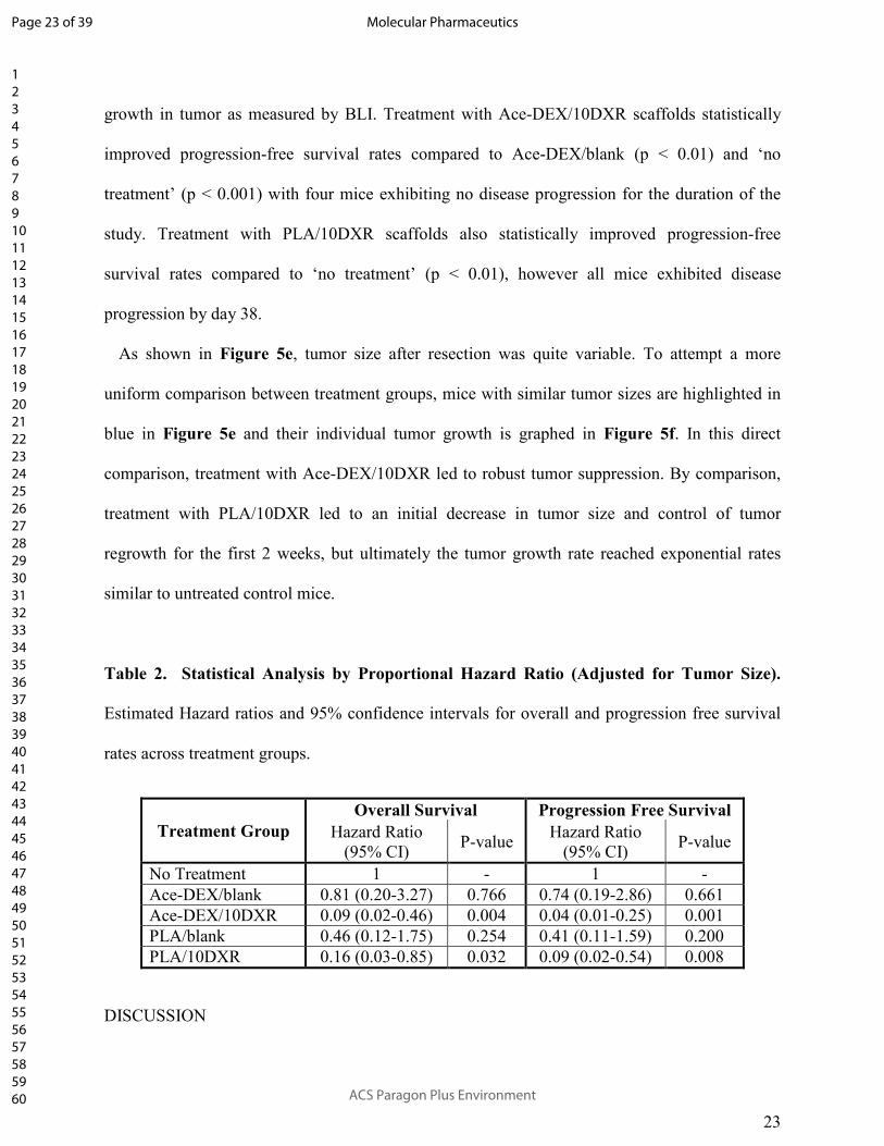
Table 2. Statistical Analysis by Proportional Hazard Ratio (Adjusted for Tumor Size).
Estimated Hazard ratios and 95% confidence intervals for overall and progression free survival
rates across treatment groups.
Treatment Group
Overall Survival Progression Free Survival
Hazard Ratio
(95% CI) P-value
Hazard Ratio
(95% CI) P-value
No Treatment 1 - 1 -
Ace-DEX/blank 0.81 (0.20-3.27) 0.766 0.74 (0.19-2.86) 0.661
Ace-DEX/10DXR 0.09 (0.02-0.46) 0.004 0.04 (0.01-0.25) 0.001
PLA/blank 0.46 (0.12-1.75) 0.254 0.41 (0.11-1.59) 0.200
PLA/10DXR 0.16 (0.03-0.85) 0.032 0.09 (0.02-0.54) 0.008
DISCUSSION
Page 23 of 39
ACS Paragon Plus Environment
Molecular Pharmaceutics
123456789101112131415161718192021222324252627282930313233343536373839404142434445464748495051525354555657585960

24
GBM sensitivity to DXR
Sensitivity to DXR was compared to BCNU, the drug currently used for interstitial GBM
therapy in Gliadel®
wafers, in three established human GBM cell lines: U87-MG, LN-229, and
LN-18. GBM cells lines were found to be approximately 200 fold more sensitive to DXR than
BCNU. This illustrates that DXR is highly potent against GBM and could be an enormously
beneficial option for GBM patients. However, because systemic treatment with DXR is
associated with severe peripheral toxicity, including irreversible cardiotoxicity, combined with
the inability of DXR to cross the blood-brain barrier at therapeutic levels, DXR remains an
untapped potential for GBM therapy.19, 43
Electrospinning and scaffold characterization
We next fabricated drug-eluting polymer scaffolds for interstitial DXR therapy. Scaffolds were
fabricated by electrospinning, a technique that generates a thin, flexible, fibrous scaffold which is
ideal for implantation in the brain.31
Scaffold flexibility allows the implant to contour to the
shape of the resection cavity, maximizing contact surface area, while the thin nature of the
scaffold reduces the risk of mass effects. This is in contrast to Gliadel®
, which is a hard disk that
cannot easily cover contoured surfaces. Importantly, electrospinning is an inexpensive and
scalable technique that can be used to control drug delivery kinetics by varying parameters such
as solvent system, drug loading, and polymer platform. These variables were separately
investigated to determine their role in DXR release.
To ensure sustained DXR release over an extended period of time, a slowly degrading Ace-
DEX polymer with 60% relative cyclic acetal coverage was utilized for scaffold generation.
First, the organic solvent system and drug loading was varied to determine their respective
Page 24 of 39
ACS Paragon Plus Environment
Molecular Pharmaceutics
123456789101112131415161718192021222324252627282930313233343536373839404142434445464748495051525354555657585960

25
effects on DXR release from Ace-DEX. Varying the organic solvent system affected both fiber
width and burst release of DXR. Increased volume fractions of HFIP generated wider fibers, a
faster burst release of DXR, and brighter orange scaffolds. These effects are likely from two
separate phenomena. The increase in fiber diameter is likely due to the higher volatility of HFIP,
leading to more rapid solvent evaporation. The increase in burst release is likely dependent on
drug and solvent system compatibility and indicates that DXR is concentrated at the surface of
the fiber rather than well distributed throughout.22, 44
This is confirmed visually, as scaffolds with
higher burst releases were a more vibrant hue of orange, the color of DXR. Increasing DXR
loading from 5% to 10% weight loading led to a higher burst release, likely due to the
hydrophilicity of DXR. Since high DXR burst release reduces the overall drug reservoir for
controlled and sustained release, the solvent system with a lower volume fraction of HFIP, 60%
HFIP and 40% butanol, was used for the remainder of the studies.
Polyesters are one of the most commonly used polymers for drug delivery,21, 22, 45-52
however
their slow degradation rates can be a limiting factor for effective drug delivery. To investigate
the role that polymer platform has on DXR release a scaffold composed of PLA was fabricated
under identical electrospinning conditions to Ace-DEX. Controlling for fabrication parameters
(polymer concentration, DXR loading, solvent system, and flow rate) allows for a direct
comparison between Ace-DEX and PLA. Both scaffolds had nearly identical morphologies as
visualized by electron microscopy, however, the release rate of DXR varied quite drastically.
PLA/10DXR scaffolds were found to exhibit a high burst release followed by almost no release
over the rest of the 5 week study. This is consistent with the literature, where Zeng et al. showed
a rapid release of DXR from an electrospun PLA scaffold that stagnated over time.22
By contrast,
Ace-DEX/10DXR released approximately 25% as a burst release and then another 27% of DXR
Page 25 of 39
ACS Paragon Plus Environment
Molecular Pharmaceutics
123456789101112131415161718192021222324252627282930313233343536373839404142434445464748495051525354555657585960

26
in a controlled manner over the remaining 5 weeks. Although the two scaffolds ultimately
released similar amounts of DXR (approximately 50% over 35 days), the steady-state release
from Ace-DEX scaffolds is faster than that from PLA, which releases the majority of the DXR
within the first 24 hours. This differential release rate will allow investigation into the role of
DXR release kinetics as determined by polymer platform in controlling GBM recurrence.
Maximum tolerated dose
Prior to testing the efficacy of DXR loaded Ace-DEX scaffolds against GBM, in vivo toxicity
was evaluated. To mimic tumor treatment, cranial windows and a resection cavity approximately
3mm in diameter and 1 to 2 mm deep were created in the parenchyma of nude mice. DXR loaded
Ace-DEX scaffolds were then implanted into the surgical cavity. To increase the total dose of
DXR administered, the scaffold drug loading was maintained and overall mass of scaffold
implanted was increased. To achieve the highest DXR dose tested (200 µg), 2.2 mg of total Ace-
DEX/10DXR scaffold mass was required. Interestingly, all but the highest dose of DXR, 200 µg,
was well tolerated as measured by mouse weight loss. The tolerated dose was higher than
expected based on literature where Kooistra et al. found that the highest non-toxic tolerated dose
of intrathecal DXR for Sprague Dawley rats was 20 µg.53
This discrepancy may be due to the
fact that the dose is not entirely contained intra-cranially (as the cranial window is left open),
differences between rats and mice, and the controlled release of drug from the scaffold,
compared to bolus delivery. Although by histology, dose-dependent toxicity was seen at doses as
low as 100 µg, to maximize therapeutic dose within tolerable toxicity, 150 µg DXR was tested
for therapeutic efficacy.
Page 26 of 39
ACS Paragon Plus Environment
Molecular Pharmaceutics
123456789101112131415161718192021222324252627282930313233343536373839404142434445464748495051525354555657585960

27
Efficacy against recurrent GBM
Surgical resection is part of the standard-of-care for GBM patients. However, the majority of
preclinical studies are conducted in mouse or rat models that lack surgical resection of the
primary tumor.20, 45, 54-56
To mimic clinical conditions, tumor cells are implanted into the
parenchyma of mice. Once tumors are well established, the established primary GBM mass is
resected using fluorescent-guided microsurgery. The surgical resection greatly transforms the
brain microenvironment with studies showing tumors re-grow more aggressively than the
original primary tumor.36
These findings underscore the significance of utilizing this clinically
relevant mouse model for pre-clinical GBM therapies.
As expected, tumors for control mice (untreated and blank scaffolds) regrew rapidly. However,
treatment with DXR loaded scaffolds composed of both Ace-DEX and PLA led statistically
improved survival rates compared ‘no treatment’ controls. Despite the slow steady state release
of DXR from PLA/10DXR scaffolds, treatment with PLA/10DXR extended median survival
from 29 to 63 days over PLA/blank. However, the lack of statistical significance differs from
previous reports in the literature where Lesniak et al.20
and Lin et al.21
found that DXR loaded
polyester or polyanhydride devices were able to significantly extend median survival and delay
tumor growth over empty polymer devices. Since overall and progression free survival rates of
PLA/10DXR were significant when compared to the ‘no treatment’ control group, the low
sample size of our study may be a contributing factor in the lack of statistical significance when
compared to PLA/blank. Additionally, the surgical murine model used in this study is unique in
its clinical significance, with more aggressive tumors recurrence than primary GBM models. The
complexity of this model may have highlighted limitations with PLA delivery of DXR.
Moreover, the total therapeutic dose of DXR implanted for interstitial therapy in this work was
Page 27 of 39
ACS Paragon Plus Environment
Molecular Pharmaceutics
123456789101112131415161718192021222324252627282930313233343536373839404142434445464748495051525354555657585960

28
lower than previous studies.20, 21
This suggests that the ratio of drug to tumor size may be an
important factor in interstitial therapy.
As evidences in Figure 5e, residual tumor size after resection was quite variable.
Unfortunately, because tumors do not grow homogenously and surgical conditions are not
equivalent, it is difficult to ensure identical residual tumor size after resection. Although this
variation is difficult in a research setting, this model closely mimics the clinical setting where
incomplete or insufficient tumor resection outcomes are often a reality. In this way, the range of
residual tumors at the start of treatment allows some insight into the robustness of each therapy.
Not surprisingly, residual tumor size after surgical resection played a significant role in
therapeutic efficacy. For the PLA/10DXR group, mice with larger residual tumors after surgical
resection succumbed to tumors faster than those with smaller residual tumors. This is likely due
to the slow steady state release of DXR from PLA scaffolds which allowed tumor growth to
outpace DXR release. The success of mice treated with Ace-DEX/10DXR was also dependent on
residual tumor size. Mice with smaller residual tumors were more responsive to therapy than
those with larger tumors. However, in contrast with PLA/10DXR, the size threshold for tumor
recurrence was much higher for Ace-DEX/10DXR.
Due to this residual tumor size discrepancy, a more uniform comparison between therapies can
be made by comparing representative mice with similar sized tumors after resection (highlighted
in blue in Figure 5e). In this case, PLA/10DXR resulted in an initial decrease in tumor burden,
possibly due to the burst release of DXR from the scaffold; however, tumor regrowth ultimately
outpaced the slow steady state DXR release from the PLA scaffold (Figure 5f). In contrast, the
release of DXR from Ace-DEX/10DXR scaffolds was high enough to result in complete
Page 28 of 39
ACS Paragon Plus Environment
Molecular Pharmaceutics
123456789101112131415161718192021222324252627282930313233343536373839404142434445464748495051525354555657585960

29
remission for a residual tumor of the same size. This direct comparison helps to highlight the
benefits of the higher steady state DXR release from Ace-DEX scaffolds.
CONCLUSIONS
Despite current treatments, local GBM recurrence and associated mortality is almost 100%.
This is complicated by the fact that the blood-brain barrier limits efficacy of systemic
chemotherapy. To combat recurrence and overcome the blood-brain barrier, local drug delivery
to the tumor resection site is highly advantageous. Judicious selection of a polymer platform is
essential to ensure sustained drug delivery. The most commonly used polymers for drug delivery
are limited by slow degradation rates. Previous reports of polymeric delivery of DXR exhibited
similar trends: rapid release of DXR over the first few days followed by slow steady state
release. Although this led to improved median survival or tumor growth delays, to our
knowledge no complete remissions have been achieved.
Here we addressed the need for an improved polymer platform for sustained interstitial drug
delivery to prevent GBM recurrence. DXR was incorporated into nanofibrous scaffolds
composed of PLA or Ace-DEX to directly compare the effect of polymer on DXR release rate.
Although both DXR loaded scaffolds ultimately release approximately the same amount of
DXR, PLA/10DXR scaffolds released the full payload within the first 24 hours, whereas Ace-
DEX/10DXR scaffolds exhibited controlled and sustained release of DXR over the same period.
PLA/10DXR scaffolds extended progression free survival over ‘no treatment’ control; however,
all mice in this treatment group exhibited tumor growth by 38 days after treatment. The higher
sustained DXR release from Ace-DEX scaffolds led to more robust suppression of tumor
recurrence, leading to complete remission in 43% of mice. Future studies will further explore the
Page 29 of 39
ACS Paragon Plus Environment
Molecular Pharmaceutics
123456789101112131415161718192021222324252627282930313233343536373839404142434445464748495051525354555657585960

30
tunability of the Ace-DEX polymer platform, to determine the optimal degradation rate to
maximally suppress GBM recurrence.
ASSOCIATED CONTENT
Supporting Information
Table S1. Mice Excluded from Therapeutic Efficacy Study
Figure S1. Tumor Size Prior to Treatment
Figure S2. Sensitivity of Glioblastoma Cell Lines to DXR
Figure S3. Toxicity of Scaffolds In Vivo
Table S2. Toxicity of Scaffolds In Vivo
Figure S4. Histological Toxicity Increasing Doses of DXR in Ace-DEX scaffolds
Figure S5. Tumor growth after tumor resection
AUTHOR INFORMATION
Corresponding Author
* Email: [email protected]
Author Contributions
The manuscript was written through contributions of all authors. All authors have given approval
to the final version of the manuscript.
Funding Sources
Page 30 of 39
ACS Paragon Plus Environment
Molecular Pharmaceutics
123456789101112131415161718192021222324252627282930313233343536373839404142434445464748495051525354555657585960

31
This work was supported by the PhRMA Foundation, internal funds from University of North
Carolina Chapel Hill, and funding from the NIH (R01NS097507-02). CRM was supported by the
UNC University Cancer Research Fund (UCRF), NIH National Center for Advancing
Translational Sciences (550KR61332), and NIH (R01CA204136). The UNC Translational and
Clinical Sciences Institute (TraCS) is supported by the NCATS and NIH, through grant award
number UL1TR001111. The UNC Translational Pathology Laboratory is supported in part, by
grants from the NCI (2-P30-CA016086-40), NIEHS (2-P30ES010126-15A1), UCRF, and NCBT
(2015-IDG-1007). The UNC Lineberger Comprehensive Cancer Center (LCCC) Animal Studies
Core is supported in part by an NCI Center Core Support Grant (CA16086) to the UNC LCCC.
ACKNOWLEDGMENT
We thank the UNC LCCC Animal Studies Core for assistance with animal studies. We thank
Xia Yongjuan in the UNC Translational Pathology Laboratory (TPL) and the Animal
Histopathology Core Lab for expert technical assistance. We thank the Chapel Hill Analytical
and Nanofabrication Laboratory (CHANL) for SEM usage, the Biomedical Research Imaging
Core (BRIC) for IVIS usage, the School of Pharmacy’s NMR Core, and the UNC-Olympus
Imaging Research Center for microscope usage.
ABBREVIATIONS
AceDEX, acetalated dextran; Ace-DEX/5DXR, 5% wt/wt doxorubicin loaded acetalated dextran
scaffold; Ace-DEX/10DXR, 10% wt/wt doxorubicin loaded acetalated dextran scaffold; BCNU,
carmustine; BLI, bioluminescent imaging; DNA, deoxyribonucleic acid; DMSO, dimethyl
sulfoxide; DXR, doxorubicin; DMEM, Dulbecco's Modified Eagle Medium; GBM,
glioblastoma; GFAP, glial fibrillary acidic protein; H&E, hematoxylin and eosin; HFIP,
Page 31 of 39
ACS Paragon Plus Environment
Molecular Pharmaceutics
123456789101112131415161718192021222324252627282930313233343536373839404142434445464748495051525354555657585960

32
hexafluoro-2-propanol; IHC, immunohistochemistry; mCh-FL, mCherry-firefly luciferase;
MGMT, O6-methylguanine-DNA methyltransferase; MTT, thiazolyl blue tetrazolium bromide;
NMR, nuclear magnetic resonance; PLGA, poly(D,L-lactide-co-glycolide); PLA, poly(L-
lactide); PLA/10DXR, 10% wt/wt doxorubicin loaded poly(L-lactide) scaffold; TEA,
triethylamine.
REFERENCES
1. Erpolat, O. P.; Akmansu, M.; Goksel, F.; Bora, H.; Yaman, E.; Buyukberber, S.
Outcome of newly diagnosed glioblastoma patients treated by radiotherapy plus concomitant and
adjuvant temozolomide: a long-term analysis. Tumori 2009, 95, (2), 191-7.
2. Stupp, R.; Hegi, M. E.; Mason, W. P.; van den Bent, M. J.; Taphoorn, M. J.; Janzer, R.
C.; Ludwin, S. K.; Allgeier, A.; Fisher, B.; Belanger, K.; Hau, P.; Brandes, A. A.; Gijtenbeek, J.;
Marosi, C.; Vecht, C. J.; Mokhtari, K.; Wesseling, P.; Villa, S.; Eisenhauer, E.; Gorlia, T.;
Weller, M.; Lacombe, D.; Cairncross, J. G.; Mirimanoff, R. O. Effects of radiotherapy with
concomitant and adjuvant temozolomide versus radiotherapy alone on survival in glioblastoma
in a randomised phase III study: 5-year analysis of the EORTC-NCIC trial. The lancet oncology
2009, 10, (5), 459-66.
3. Kauer, T. M.; Figueiredo, J.-L.; Hingtgen, S.; Shah, K. Encapsulated therapeutic stem
cells implanted in the tumor resection cavity induce cell death in gliomas. Nat Neurosci 2012,
15, (2), 197-204.
4. Gaspar, L. E.; Fisher, B. J.; Macdonald, D. R.; Leber, D. V.; Halperin, E. C.; Schold, S.
C.; Cairncross, J. G. Supratentorial malignant glioma: Patterns of recurrence and implications
for external beam local treatment. International Journal of Radiation Oncology*Biology*Physics
1992, 24, (1), 55-57.
Page 32 of 39
ACS Paragon Plus Environment
Molecular Pharmaceutics
123456789101112131415161718192021222324252627282930313233343536373839404142434445464748495051525354555657585960

33
5. Lee, S. W.; Fraass, B. A.; Marsh, L. H.; Herbort, K.; Gebarski, S. S.; Martel, M. K.;
Radany, E. H.; Lichter, A. S.; Sandler, H. M. Patterns of failure following high-dose 3-D
conformal radiotherapy for high-grade astrocytomas: a quantitative dosimetric study.
International Journal of Radiation Oncology*Biology*Physics 1999, 43, (1), 79-88.
6. Peter C. Burger; Philip J. Dubois; S. Clifford Schold, J.; Kenneth R. Smith, J.; Guy L.
Odom; David C. Crafts; Felice Giangaspero. Computerized tomographic and pathologic studies
of the untreated, quiescent, and recurrent glioblastoma multiforme. Journal of Neurosurgery
1983, 58, (2), 159-169.
7. De Bonis, P.; Anile, C.; Pompucci, A.; Fiorentino, A.; Balducci, M.; Chiesa, S.; Lauriola,
L.; Maira, G.; Mangiola, A. The influence of surgery on recurrence pattern of glioblastoma.
Clinical Neurology and Neurosurgery 2013, 115, (1), 37-43.
8. Adamson, C.; Kanu, O. O.; Mehta, A. I.; Di, C.; Lin, N.; Mattox, A. K.; Bigner, D. D.
Glioblastoma multiforme: a review of where we have been and where we are going. Expert Opin
Investig Drugs 2009, 18, (8), 1061-83.
9. Bregy, A.; Shah, A. H.; Diaz, M. V.; Pierce, H. E.; Ames, P. L.; Diaz, D.; Komotar, R. J.
The role of Gliadel wafers in the treatment of high-grade gliomas. Expert Rev Anticancer Ther
2013, 13, (12), 1453-61.
10. Brem, H.; Gabikian, P. Biodegradable polymer implants to treat brain tumors. J Control
Release 2001, 74, (1-3), 63-7.
11. Brem, H.; Piantadosi, S.; Burger, P. C.; Walker, M.; Selker, R.; Vick, N. A.; Black, K.;
Sisti, M.; Brem, S.; Mohr, G.; et al. Placebo-controlled trial of safety and efficacy of
intraoperative controlled delivery by biodegradable polymers of chemotherapy for recurrent
gliomas. The Polymer-brain Tumor Treatment Group. Lancet 1995, 345, (8956), 1008-12.
Page 33 of 39
ACS Paragon Plus Environment
Molecular Pharmaceutics
123456789101112131415161718192021222324252627282930313233343536373839404142434445464748495051525354555657585960

34
12. Grossman, S. A.; Reinhard, C.; Colvin, O. M.; Chasin, M.; Brundrett, R.; Tamargo, R. J.;
Brem, H. The intracerebral distribution of BCNU delivered by surgically implanted
biodegradable polymers. J Neurosurg 1992, 76, (4), 640-7.
13. Arifin, D. Y.; Lee, K. Y.; Wang, C. H. Chemotherapeutic drug transport to brain tumor.
J Control Release 2009, 137, (3), 203-10.
14. Arifin, D. Y.; Lee, K. Y.; Wang, C. H.; Smith, K. A. Role of convective flow in
carmustine delivery to a brain tumor. Pharm Res 2009, 26, (10), 2289-302.
15. Bobola, M. S.; Silber, J. R.; Ellenbogen, R. G.; Geyer, J. R.; Blank, A.; Goff, R. D.
O<sup>6</sup>-Methylguanine-DNA Methyltransferase, O<sup>6</sup>-Benzylguanine, and
Resistance to Clinical Alkylators in Pediatric Primary Brain Tumor Cell Lines. Clin. Cancer Res.
2005, 11, (7), 2747-2755.
16. Hermisson, M.; Klumpp, A.; Wick, W.; Wischhusen, J.; Nagel, G.; Roos, W.; Kaina, B.;
Weller, M. O6-methylguanine DNA methyltransferase and p53 status predict temozolomide
sensitivity in human malignant glioma cells. Journal of Neurochemistry 2006, 96, (3), 766-776.
17. Eom, Y.-W.; Kim, M. A.; Park, S. S.; Goo, M. J.; Kwon, H. J.; Sohn, S.; Kim, W.-H.;
Yoon, G.; Choi, K. S. Two distinct modes of cell death induced by doxorubicin: apoptosis and
cell death through mitotic catastrophe accompanied by senescence-like phenotype. Oncogene
2005, 24, (30), 4765-4777.
18. Gewirtz, D. A critical evaluation of the mechanisms of action proposed for the antitumor
effects of the anthracycline antibiotics adriamycin and daunorubicin. Biochemical Pharmacology
1999, 57, (7), 727-741.
Page 34 of 39
ACS Paragon Plus Environment
Molecular Pharmaceutics
123456789101112131415161718192021222324252627282930313233343536373839404142434445464748495051525354555657585960

35
19. Ohnishi, T.; Tamai, I.; Sakanaka, K.; Sakata, A.; Yamashima, T.; Yamashita, J.; Tsuji, A.
In vivo and in vitro evidence for ATP-dependency of P-glycoprotein-mediated efflux of
doxorubicin at the blood-brain barrier. Biochemical Pharmacology 1995, 49, (10), 1541-1544.
20. Lesniak, M. S.; Upadhyay, U.; Goodwin, R.; Tyler, B.; Brem, H. Local delivery of
doxorubicin for the treatment of malignant brain tumors in rats. Anticancer Res 2005, 25, (6B),
3825-31.
21. Lin, S. Y.; Cheng, L. F.; Lui, W. Y.; Chen, C. F.; Han, S. H. Tumoricidal Effect of
Controlled-release Polymeric Needle Devices Containing Adriamycin Hc1 in Tumor-bearing
Mice. Biomaterials, Artificial Cells and Artificial Organs 1989, 17, (2), 189-203.
22. Zeng, J.; Yang, L.; Liang, Q.; Zhang, X.; Guan, H.; Xu, X.; Chen, X.; Jing, X. Influence
of the drug compatibility with polymer solution on the release kinetics of electrospun fiber
formulation. J Control Release 2005, 105, (1-2), 43-51.
23. Lu, T.; Jing, X.; Song, X.; Wang, X. Doxorubicin-loaded ultrafine PEG-PLA fiber mats
against hepatocarcinoma. J Appl Polym Sci 2012, 123, (1), 209-217.
24. Xu, X.; Yang, L.; Xu, X.; Wang, X.; Chen, X.; Liang, Q.; Zeng, J.; Jing, X. Ultrafine
medicated fibers electrospun from W/O emulsions. J Control Release 2005, 108, (1), 33-42.
25. Li, S.; Garreau, H.; Vert, M. Structure-property relationships in the case of the
degradation of massive poly(α-hydroxy acids) in aqueous media. Journal of Materials Science:
Materials in Medicine 1990, 1, (4), 198-206.
26. Li, S. Hydrolytic degradation characteristics of aliphatic polyesters derived from lactic
and glycolic acids. Journal of Biomedical Materials Research 1999, 48, (3), 342-353.
27. Langer, R. New Methods of Drug Delivery. Science 1990, 249, (4976), 1527-1533.
Page 35 of 39
ACS Paragon Plus Environment
Molecular Pharmaceutics
123456789101112131415161718192021222324252627282930313233343536373839404142434445464748495051525354555657585960

36
28. Sung, H. J.; Meredith, C.; Johnson, C.; Galis, Z. S. The effect of scaffold degradation
rate on three-dimensional cell growth and angiogenesis. Biomaterials 2004, 25, (26), 5735-5742.
29. Ding, A. G.; Shenderova, A.; Schwendeman, S. P. Prediction of Microclimate pH in
Poly(lactic-co-glycolic Acid) Films. Journal of the American Chemical Society 2006, 128, (16),
5384-5390.
30. Fu, K.; Pack, D.; Klibanov, A.; Langer, R. Visual Evidence of Acidic Environment
Within Degrading Poly(lactic-co-glycolic acid) (PLGA) Microspheres. Pharmaceutical Research
2000, 17, (1), 100-106.
31. Borteh, H. M.; Gallovic, M. D.; Sharma, S.; Peine, K. J.; Miao, S.; Brackman, D. J.;
Gregg, K.; Xu, Y.; Guo, X.; Guan, J.; Bachelder, E. M.; Ainslie, K. M. Electrospun acetalated
dextran scaffolds for temporal release of therapeutics. Langmuir 2013, 29, (25), 7957-65.
32. Bachelder, E. M.; Beaudette, T. T.; Broaders, K. E.; Dashe, J.; Fréchet, J. M. J. Acetal-
Derivatized Dextran: An Acid-Responsive Biodegradable Material for Therapeutic Applications.
Journal of the American Chemical Society 2008, 130, (32), 10494-10495.
33. Kauffman, K. J.; Do, C.; Sharma, S.; Gallovic, M. D.; Bachelder, E. M.; Ainslie, K. M.
Synthesis and characterization of acetalated dextran polymer and microparticles with ethanol as a
degradation product. ACS Appl Mater Interfaces 2012, 4, (8), 4149-55.
34. Broaders, K. E.; Cohen, J. A.; Beaudette, T. T.; Bachelder, E. M.; Frechet, J. M.
Acetalated dextran is a chemically and biologically tunable material for particulate
immunotherapy. Proc Natl Acad Sci U S A 2009, 106, (14), 5497-502.
35. Bachelder, E. M.; Beaudette, T. T.; Broaders, K. E.; Paramonov, S. E.; Dashe, J.; Frechet,
J. M. Acid-degradable polyurethane particles for protein-based vaccines: biological evaluation
and in vitro analysis of particle degradation products. Mol Pharm 2008, 5, (5), 876-84.
Page 36 of 39
ACS Paragon Plus Environment
Molecular Pharmaceutics
123456789101112131415161718192021222324252627282930313233343536373839404142434445464748495051525354555657585960

37
36. Okolie, O.; Bago, J. R.; Schmid, R. S.; Irvin, D. M.; Bash, R. E.; Miller, C. R.; Hingtgen,
S. D. Reactive astrocytes potentiate tumor aggressiveness in a murine glioma resection and
recurrence model. Neuro Oncol 2016, 18, (12), 1622-1633.
37. Hingtgen, S.; Figueiredo, J. L.; Farrar, C.; Duebgen, M.; Martinez-Quintanilla, J.; Bhere,
D.; Shah, K. Real-time multi-modality imaging of glioblastoma tumor resection and recurrence.
J Neurooncol 2013, 111, (2), 153-61.
38. Kauer, T. M.; Figueiredo, J. L.; Hingtgen, S.; Shah, K. Encapsulated therapeutic stem
cells implanted in the tumor resection cavity induce cell death in gliomas. Nat Neurosci 2011,
15, (2), 197-204.
39. Sena-Esteves, M.; Tebbets, J. C.; Steffens, S.; Crombleholme, T.; Flake, A. W.
Optimized large-scale production of high titer lentivirus vector pseudotypes. J Virol Methods
2004, 122, (2), 131-9.
40. Burr, H. N.; Lipman, N. S.; White, J. R.; Zheng, J.; Wolf, F. R. Strategies to Prevent,
Treat, and Provoke Corynebacterium-Associated Hyperkeratosis in Athymic Nude Mice. Journal
of the American Association for Laboratory Animal Science : JAALAS 2011, 50, (3), 378-388.
41. Zhang, S.; Han, L.; Wei, J.; Shi, Z.; Pu, P.; Zhang, J.; Yuan, X.; Kang, C. Combination
treatment with doxorubicin and microRNA-21 inhibitor synergistically augments anticancer
activity through upregulation of tumor suppressing genes. Int J Oncol 2015, 46, (4), 1589-600.
42. Weller, M.; Rieger, J.; Grimmel, C.; Van Meir, E. G.; De Tribolet, N.; Krajewski, S.;
Reed, J. C.; von Deimling, A.; Dichgans, J. Predicting chemoresistance in human malignant
glioma cells: the role of molecular genetic analyses. Int J Cancer 1998, 79, (6), 640-4.
43. Volkova, M.; Russell, R. Anthracycline Cardiotoxicity: Prevalence, Pathogenesis and
Treatment. Current Cardiology Reviews 2011, 7, (4), 214-220.
Page 37 of 39
ACS Paragon Plus Environment
Molecular Pharmaceutics
123456789101112131415161718192021222324252627282930313233343536373839404142434445464748495051525354555657585960

38
44. Seif, S.; Franzen, L.; Windbergs, M. Overcoming drug crystallization in electrospun
fibers--Elucidating key parameters and developing strategies for drug delivery. Int J Pharm
2015, 478, (1), 390-7.
45. Ranganath, S. H.; Fu, Y.; Arifin, D. Y.; Kee, I.; Zheng, L.; Lee, H. S.; Chow, P. K.;
Wang, C. H. The use of submicron/nanoscale PLGA implants to deliver paclitaxel with
enhanced pharmacokinetics and therapeutic efficacy in intracranial glioblastoma in mice.
Biomaterials 2010, 31, (19), 5199-207.
46. Ranganath, S. H.; Wang, C. H. Biodegradable microfiber implants delivering paclitaxel
for post-surgical chemotherapy against malignant glioma. Biomaterials 2008, 29, (20), 2996-
3003.
47. Manome, Y.; Kobayashi, T.; Mori, M.; Suzuki, R.; Funamizu, N.; Akiyama, N.; Inoue,
S.; Tabata, Y.; Watanabe, M. Local delivery of doxorubicin for malignant glioma by a
biodegradable PLGA polymer sheet. Anticancer Res 2006, 26, (5A), 3317-26.
48. Lin, S. Y.; Cheng, L. F.; Lui, W. Y.; Wu, L. H.; Kao, S. J.; Han, S. H. Controlled release
of adriamycin HCl from polymeric needle devices. Biomater Artif Cells Artif Organs 1988, 16,
(4), 801-14.
49. Lee, L. Y.; Ranganath, S. H.; Fu, Y.; Zheng, J. L.; Lee, H. S.; Wang, C.-H.; Smith, K. A.
Paclitaxel release from micro-porous PLGA disks. Chemical Engineering Science 2009, 64, (21),
4341-4349.
50. Ong, B. Y.; Ranganath, S. H.; Lee, L. Y.; Lu, F.; Lee, H. S.; Sahinidis, N. V.; Wang, C.
H. Paclitaxel delivery from PLGA foams for controlled release in post-surgical chemotherapy
against glioblastoma multiforme. Biomaterials 2009, 30, (18), 3189-96.
Page 38 of 39
ACS Paragon Plus Environment
Molecular Pharmaceutics
123456789101112131415161718192021222324252627282930313233343536373839404142434445464748495051525354555657585960

39
51. Xie, J.; Wang, C. H. Electrospun micro- and nanofibers for sustained delivery of
paclitaxel to treat C6 glioma in vitro. Pharm Res 2006, 23, (8), 1817-26.
52. Huang, H. H.; He, C. L.; Wang, H. S.; Mo, X. M. Preparation of core-shell
biodegradable microfibers for long-term drug delivery. J Biomed Mater Res A 2009, 90, (4),
1243-51.
53. Kooistra, K. L.; Rodriguez, M.; Powis, G. Toxicity of intrathecally administered
cytotoxic drugs and their antitumor activity against an intrathecal Walker 256 carcinosarcoma
model for meningeal carcinomatosis in the rat. Cancer Res 1989, 49, (4), 977-82.
54. Walter, K. A.; Cahan, M. A.; Gur, A.; Tyler, B.; Hilton, J.; Colvin, O. M.; Burger, P. C.;
Domb, A.; Brem, H. Interstitial Taxol Delivered from a Biodegradable Polymer Implant against
Experimental Malignant Glioma. Cancer Res. 1994, 54, (8), 2207-2212.
55. Ruan, S.; Yuan, M.; Zhang, L.; Hu, G.; Chen, J.; Cun, X.; Zhang, Q.; Yang, Y.; He, Q.;
Gao, H. Tumor microenvironment sensitive doxorubicin delivery and release to glioma using
angiopep-2 decorated gold nanoparticles. Biomaterials 2015, 37, 425-35.
56. Cui, Y.; Xu, Q.; Chow, P. K.; Wang, D.; Wang, C. H. Transferrin-conjugated magnetic
silica PLGA nanoparticles loaded with doxorubicin and paclitaxel for brain glioma treatment.
Biomaterials 2013, 34, (33), 8511-20.
Page 39 of 39
ACS Paragon Plus Environment
Molecular Pharmaceutics
123456789101112131415161718192021222324252627282930313233343536373839404142434445464748495051525354555657585960