Embed Size (px)
Citation preview
Swimming into the Future of Drug Discovery:In Vivo Chemical Screens in ZebrafishTeresa V. Bowman and Leonard I. Zon*Stem Cell Program and Division of Hematology and Oncology, Children’s Hospital Boston and Howard Hughes MedicalInstitute, 300 Longwood Avenue, Boston, Massachusetts 02115
T he target-centric drug discovery para-digm predominantly followed overthe past 50 years entails multiple it-
erations of in vitro biochemical and cell-based assays followed by in vivo studies inanimal models and then ultimately trials inhumans (Figure 1). This process typicallytakes 12�15 years before drugs reach themarket. Less than 1% of developing drugsresult in success, but the pursuit of manypromising “failed” drugs can cost a com-pany millions of dollars in R&D (1, 2). As aresult, the discovery of new drugs from ma-jor pharmaceutical companies has declinedwhile production costs have steadily in-creased. Prospective drugs can be termi-nated at any point during development dueto lack of efficacy, adverse side effects, orexcessive toxicity. Much of the failure comesat the level of animal testing where prob-lems with in vivo absorption, distribution,metabolism, excretion, and toxicity (ADMET)are first assessed. Using in vivo animal mod-els at the initial stages of screening can im-prove determination of ADMET from the startrather than after years of research and mil-lions of dollars down the line. In vivo screen-ing also simultaneously assesses drug se-lectivity and specificity in the context of aliving organism. In an article in this issue,Hao and colleagues describe one of the firstlarge-scale in vivo structure�activity rela-tionship (SAR) studies (3). Dorsomorphin, apromiscuous hit previously identified in aphenotype-based small molecule screen forBMP (bone morphogenetic protein) inhibi-
tors, was found not only to antagonize BMPsignaling but also to abrogate angiogenesisin zebrafish embryos via VEGF (vascular en-dothelial growth factor) inhibition (4). Usingin vivo SAR studies, Hao et al. generatedtwo selective and potent inhibitors for BMPand VEGF signaling. This work demonstratesthe ability to use an in vivo screening modelfor lead compound discovery and subse-quent optimization.
Over the past decade the zebrafish hasemerged as a vertebrate model amendableto large-scale forward genetic and chemicalscreens (5). Similar to classic invertebratemodels, zebrafish develop extra-uterine, al-lowing for facile visualization of early em-bryogenesis and organogenesis. In contrast,zebrafish can be used to study the regula-tion of vertebrate-specific processes that af-fect disease and development. Many otheradvantages make zebrafish a particularlygood model for high-throughput screening.High fecundity and small size permit thegeneration and storage of thousands of fishin a small space. Optical clarity of the em-bryo makes it possible to visualize a widevariety of phenotypes without killing or ma-nipulating the embryos. Gross morphologi-cal changes are easily viewed with lightmicroscopy, and the use of fluorescenttransgenic fish allows for cell-type orpathway-specific visualization.
The process of embryogenesis involvesthe convergence of multiple signaling path-ways. Forward genetic screens in zebrafishprovide an array of mutants with specific
*Corresponding author,[email protected].
Published online February 19, 2010
10.1021/cb100029t
© 2010 American Chemical Society
ABSTRACT In recent years in vivo chemicalscreening in zebrafish has emerged as a rapidand efficient method to identify lead compoundsthat modulate specific biological processes. Byperforming primary screening in vivo, the bioactiv-ity, toxicity, and off-target side effects are deter-mined from the onset of drug development. A re-cent study demonstrates that in vivo screeningcan be used successfully to perform structure�
activity relationship (SAR) studies. This work vali-dates the zebrafish as an effective model for notonly drug discovery but also drug optimization.
Point ofVIEW
www.acschemicalbiology.org VOL.5 NO.2 • ACS CHEMICAL BIOLOGY 159
phenotypes that correspond to perturba-tions in defined pathways. The signalingpathways utilized during embryogenesis areoften defective in disease states, thus ze-brafish embryogenesis provides an easytool to interrogate a multitude of pathway-specific chemical modifiers that have thera-peutic potential. For example, BMP signal-ing is defective in diseases affecting manytissues including bone, kidney, and a multi-tude of cancers and is essential for embryo-genesis (6, 7). In development, the BMPsignaling pathway controls the proper es-tablishment of the dorsal-ventral (D-V) axis.Mutant zebrafish with defective BMP signal-ing display altered D-V axis formation withexcessive dorsal tissues like the brain at theexpense of ventral tissues like blood andmuscle (6). An initial study executed by Yu
and colleagues screened small moleculesfor those that could mimic the effects ofBMP-defective zebrafish mutants (4). Over7500 compounds with known bioactivitywere screened, and one hit induced a repro-ducible dorsalized D-V axis phenotype. Thiscompound, named dorsomorphin, is thefirst identified chemical inhibitor of BMP sig-naling. Its mechanism of action is inhibi-tion of the serine-threonine kinase activityof Type I BMP receptors (BMPR-I), as mea-sured by phosphorylation of the target sub-strates smads 1, 5, and 8.
As with most initial hits from a drugscreen, dorsomorphin was a good inhibitorof BMP signaling but upon closer inspectionwas not the most specific or robust. In vitrokinase assays showed dorsomorphin onlymoderately inhibited the phosphorylation of
smads 1, 5, and 8. Cuny et al. per-formed a SAR study using in vitrobiochemical assays and identi-fied LDN-193189, a derivativethat showed higher potency forBMPR-I kinase inhibition and bet-ter pharmacokinetics in mice (8).This optimized compound wassuccessfully used to treat amouse model of fibrodysplasiaossificans progressiva, a congeni-tal disorder of progressive andwidespread postnatal ossifica-tion of soft tissue induced by con-stitutive activation of BMP recep-tor signaling, demonstrating thepromise of combining zebrafishlead compound discovery, SARfunctional studies, and mamma-lian modeling for drug optimiza-tion (4).
While this in vitro SAR studydid uncover a better BMPR inhibi-tor, the experiments performeddid not fully address off-target ef-fects and bioavailability of alarger number of compounds,which could result in abandon-ing a potentially great in vivo drug
that performs less than ideal in a biochemi-cal assay. Many drugs that show great prom-ise in biochemical assays often fall shortduring ADMET testing, while those that mayhave moderate biochemical activity aremore specific and potent in vivo (5). In thisissue, Hao et al. describe a large-scale invivo SAR study on dorsomorpin, in whichthey identified specific and potent inhibi-tors of BMP and VEGF signaling (3). Usingparallel library synthesis centered aroundthe 3,6-disubstituted pyrazolo[1,5-a]pyrimidine core of dorsomorphin, 63 dis-tinct compounds were generated, isolated,and tested for effects on D-V axis formation,angiogenesis, and overall toxicity. Three de-rivatives were highly selective for D-V axisdefects, and one was specific for inhibitingangiogenesis.
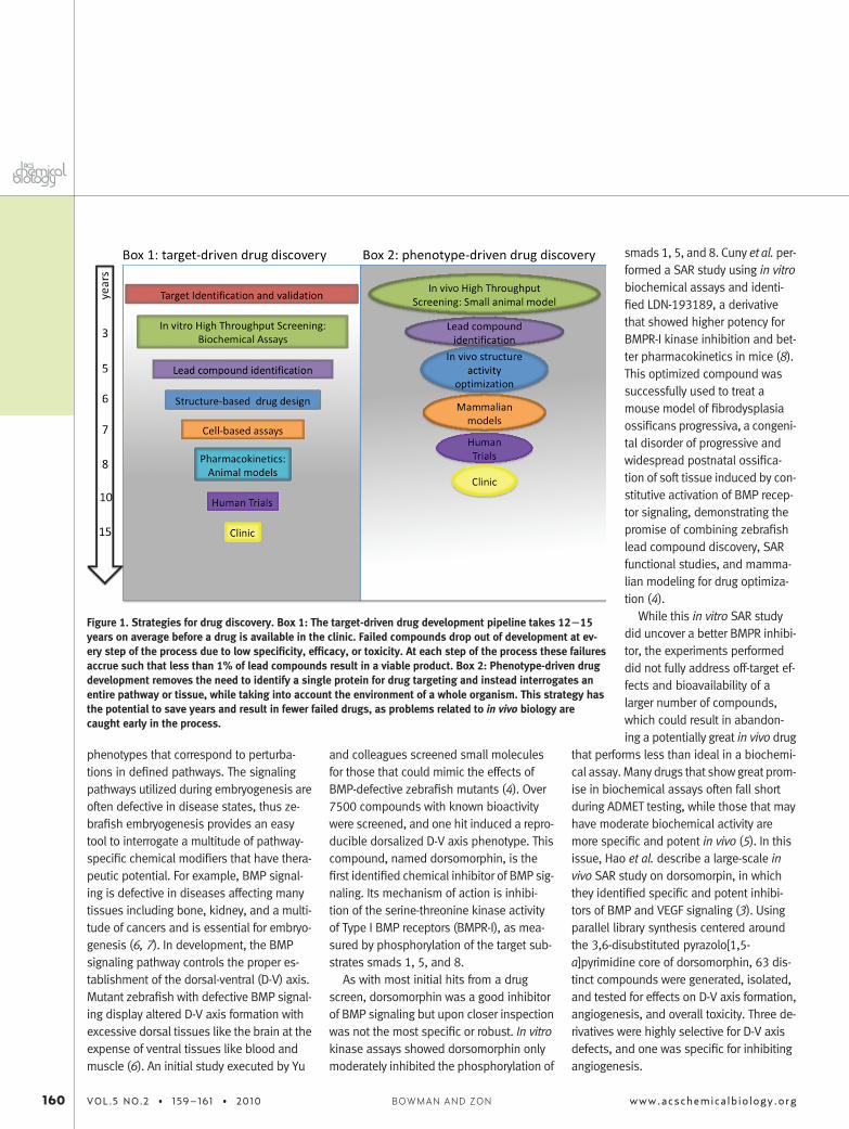
Figure 1. Strategies for drug discovery. Box 1: The target-driven drug development pipeline takes 12�15years on average before a drug is available in the clinic. Failed compounds drop out of development at ev-ery step of the process due to low specificity, efficacy, or toxicity. At each step of the process these failuresaccrue such that less than 1% of lead compounds result in a viable product. Box 2: Phenotype-driven drugdevelopment removes the need to identify a single protein for drug targeting and instead interrogates anentire pathway or tissue, while taking into account the environment of a whole organism. This strategy hasthe potential to save years and result in fewer failed drugs, as problems related to in vivo biology arecaught early in the process.
160 VOL.5 NO.2 • 159–161 • 2010 www.acschemicalbiology.orgBOWMAN AND ZON
The most promising BMP inhibitor wasDMH1. This derivative was more potent thandorsomorphin or LDN-193189 with an effec-tive concentration resulting in 100% of ex-posed embryos displaying the phenotype(EC100) of 0.2 �M compared to 2.5 or 3 �M,respectively. DMH1 was also more selec-tive, displaying negligible toxicity comparedto dorsomorphin or LDN-193189. DMH1was completely selective for BMPR-I inhibi-tion, showing no activity against TGF�R-I,AMPK, VEGFR, or PDGFR. In agreement withthese in vitro results, DMH1 showed no ef-fect on in vivo angiogenesis, even at highconcentrations. The effect of DMH1 trans-lated to human cell culture, blocking BMP-induced phosphorylation of smads 1, 5, and8 and transcription in human HEK293 cells.
In contrast, DMH4 showed no effect onD-V axis formation but had an EC50 for dis-ruption of embryonic angiogensis of 1 �Mcompared to dorsomorpin (5 �M) and LDN-193189 (20 �M). This compound had indis-cernible toxicity regardless of dose, show-ing its high degree of specificity forangiogenesis. This effect was not restrictedto fish embryos, as VEGF-induced endothe-lial tube formation of human venous endo-thelial cells in culture was abrogated byDMH4 exposure, suggesting the mecha-nism of action is likely via inhibition ofVEGFR signaling. Supporting this notion,DMH4 was shown to have a low inhibitoryconcentration 50 (IC50) for VEGFR inhibitionof 161 nM, compared to 3.5 �M for BMPR-Iand 8 �M for AMPK using in vitro kinase as-says. Of note, dorsomorpin, LDN-193189,and 6LP (another tested derivative) werefound to be potent VEGFR inhibitors withIC50 of 25.1, 214.7, and 37 nM, respec-tively, but the in vivo EC50 for disruption ofembryonic angiogenesis was 5, 20, and0 �M, respectively, indicating reduced bio-activity of these analogues compared toDMH4. These results highlight the impor-tance of performing in vivo screens to findthe most relevant analogues for drugdevelopment.
The approach described by Hao et al. ex-emplifies the potential advantages of in vivophenotype-driven screens in small verte-brates, like the zebrafish, at multiple stepsof the early drug development stages inidentifying the most bioactive and relevantcompounds for subsequent investment oftime and money. Chemical screening in ze-brafish is an emerging field but is still in itsinfancy. A full understanding of the physi-ological and pharmacological similaritiesand differences between zebrafish and hu-mans is not fully appreciated, and thus thepredictive power of zebrafish ADMET intohuman ADMET is unclear. Several drugs uti-lized in human patients have been provento work in zebrafish, indicating at leastsome degree of conservation (9). To circum-vent this possibility of differences in ADMETin fish and humans, many zebrafish re-searchers use chemical libraries that arecomposed of small molecules with knownbioactivity and mainly comprise alreadyFDA-approved drugs (3, 10). Using these li-braries, scientists can identify new thera-peutic usage for old drugs, which can leadto a shortened time to human trials savingprecious time and money. One example of anew use for an old drug is the newly uncov-ered ability of prostaglandin E2 (PGE2) to ex-pand hematopoietic stem cells (HSC) ex vivo(10). Our lab observed that 16,16-dimethylPGE2 (dmPGE2) can enhance HSC formationin zebrafish embryos. Murine marrow en-graftment and human cord blood stem cellengraftment in NOD-SCID mice were bothenhanced by pretreatment of transplantedcells with dmPGE2. A clinical trial for leuke-mic patients who are receiving a cord bloodstem cell transplant is now being done onthe basis of this work. This study demon-strates the value of zebrafish screening ofknown small molecules to expedite drug de-velopment. As many already FDA-approveddrugs are used for off-label purposes, thisstrategy holds great potential. These posi-tive “side” effects can be due to unknownusage of the target protein in other biologi-
cal contexts or due to previously unknownoff-target effects of the drug. In vivo SARstudies in genetically amendable modelscan distinguish these scenarios better andimprove drug targeting.
REFERENCES1. van der Greef, J., and McBurney, R. N. (2005) Innova-
tion: Rescuing drug discovery: in vivo systems pa-thology and systems pharmacology, Nat. Rev. DrugDiscovery 4, 961–967.
2. Vernon, J. A., Golec, J. H., and Dimasi, J. A. (2009)Drug development costs when financial risk is mea-sured using the Fama-French three-factor model,Health Econ. Epub ahead of print Aug 4, 2009; DOI:10.1002/hec.1538.
3. Hao, J., Ho, J., Lewis, J., Karim, K., Daniels, R., Gen-try, P., Hopkins, C., Lindsley, C., and Hong, C. (2010)In vivo structure�activity relationship study of dor-somorphin analogues identifies selective VEGF andBMP inhibitors, ACS Chem. Biol. 4, DOI: 10.1021/cb9002865.
4. Yu, P. B., Deng, D. Y., Lai, C. S., Hong, C. C., Cuny,G. D., Bouxsein, M. L., Hong, D. W., McManus, P. M.,Katagiri, T., Sachidanandan, C., Kamiya, N., Fukuda,T., Mishina, Y., Peterson, R. T., and Bloch, K. D.(2008) BMP type I receptor inhibition reduces heter-otopic [corrected] ossification, Nat. Med. 14,1363–1369.
5. Zon, L. I., and Peterson, R. T. (2005) In vivo drug dis-covery in the zebrafish, Nat. Rev. Drug Discovery 4,35–44.
6. Hammerschmidt, M., Serbedzija, G. N., and McMa-hon, A. P. (1996) Genetic analysis of dorsoventralpattern formation in the zebrafish: requirement of aBMP-like ventralizing activity and its dorsal repres-sor, Genes Dev. 10, 2452–2461.
7. Klahr, S. (2003) The bone morphogenetic proteins(BMPs). Their role in renal fibrosis and renal func-tion, J. Nephrol. 16, 179–185.
8. Cuny, G. D., Yu, P. B., Laha, J. K., Xing, X., Liu, J. F., Lai,C. S., Deng, D. Y., Sachidanandan, C., Bloch, K. D.,and Peterson, R. T. (2008) Structure�activity rela-tionship study of bone morphogenetic protein(BMP) signaling inhibitors, Bioorg. Med. Chem. Lett.18, 4388–4392.
9. Milan, D. J., Peterson, T. A., Ruskin, J. N., Peterson,R. T., and MacRae, C. A. (2003) Drugs that induce re-polarization abnormalities cause bradycardia in ze-brafish, Circulation 107, 1355–1358.
10. North, T. E., Goessling, W., Walkley, C. R., Lengerke,C., Kopani, K. R., Lord, A. M., Weber, G. J., Bowman,T. V., Jang, I. H., Grosser, T., Fitzgerald, G. A., Da-ley, G. Q., Orkin, S. H., and Zon, L. I. (2007) Prostag-landin E2 regulates vertebrate haematopoieticstem cell homeostasis, Nature 447, 1007–1011.
www.acschemicalbiology.org VOL.5 NO.2 • 159–161 • 2010 161
Point ofVIEW