Embed Size (px)
Citation preview

a LANGE medical book
SYMPTOM TO DIAGNOSIS
An Evidence-Based Guide
Scott D. C. Stern, MDAssociate Professor of Medicine
Co-Director, Junior Clerkship in MedicineClinical Director of Clinical Pathophysiology and Therapeutics
University of ChicagoPritzker School of Medicine
Chicago, Illinois
Adam S. Cifu, MDAssociate Professor of Medicine
Co-Director, Junior Clerkship in MedicineUniversity of Chicago
Pritzker School of MedicineChicago, Illinois
Diane Altkorn, MDAssociate Professor of Medicine
Director, Senior Student Clerkships in MedicineUniversity of Chicago
Pritzker School of MedicineChicago, Illinois
Lange Medical Books/McGraw-HillMedical Publishing Division
New York St. Louis San Francisco Auckland Bogota Caracas Lisbon LondonMadrid Mexico City Milan Montreal New Delhi San Juan
Singapore Sydney Tokyo Toronto
9310_Stern front matter 7/22/05 2:15 PM Page i
Symptom to Diagnosis: An Evidence-Based Guide
Copyright © 2006 by The McGraw-Hill Companies, Inc. All rights reserved. Printed in the United States of America. Exceptas permitted under the United States Copyright Act of 1976, no part of this publication my be reproduced or distributed inany form or by any means, or stored in a data base or retrieval system, without prior written permission of the publisher.
1234567890 DOC/DOC 09876543210
ISBN 0-07-146389-5
ISSN 1556-2719
NoticeMedicine is an ever-changing science. As new research and clinical experience broaden our knowledge, changes intreatment and drug therapy are required. The authors and the publisher of this work have checked with sourcesbelieved to be reliable in their efforts to provide information that is complete and generally in accord with the stan-dards accepted at the time of publication. However, in view of the possibility of human error or changes in medicalsciences, neither the authors nor the publisher nor any other party who has been involved in the preparation or pub-lication of this work warrants that the information contained herein is in every respect accurate or complete, and theydisclaim all responsibility for any errors or omissions or for the results obtained from use of the information containedin this work. Readers are encourage to confirm the information contained herein with other sources. For example andin particular, readers are advised to check the product information sheet included in the package of each drug they planto administer to be certain that the information contained in this work is accurate and that changes have not beenmade in the recommended dose or in the contraindications for administration. This recommendation is of particularimportance in connection with new or infrequently used drugs.
This book was set in Adobe Garamond by Rainbow Graphics.The editors were Janet Foltin, Harriet Lebowitz, and Karen Davis.The production supervisor was Sherri Souffrance.The text designer was Eve Siegel.The illustrator was Keyword House.The cover designer was Aimée Nordin.The index was prepared by Andover Publishing Services.R.R. Donnelley and Sons, Inc. was printer and binder.
This book is printed on acid-free paper
INTERNATIONAL EDITION ISBN 0-07-110506-9Copyright © 2006. Exclusive right by The McGraw-Hill Companies, Inc. for manufacture and export. This book cannot bere-exported from the country to which it is consigned by McGraw-Hill. The International Edition is not available in NorthAmerica.
9310_Stern front matter 7/22/05 2:15 PM Page ii
ON-LINE ONLY
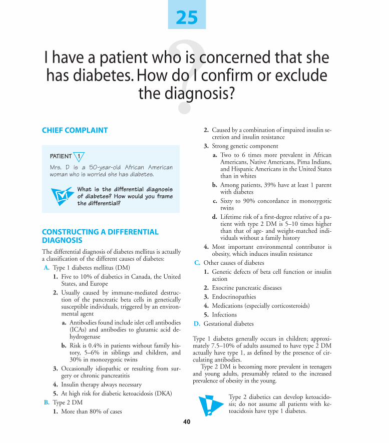
?25
I have a patient who is concerned that shehas diabetes. How do I confirm or exclude
the diagnosis?
CHIEF COMPLAINT
PATIENT 1
Mrs. D is a 50-year-old African Americanwoman who is worried she has diabetes.
What is the differential diagnosisof diabetes? How would you framethe differential?
CONSTRUCTING A DIFFERENTIALDIAGNOSISThe differential diagnosis of diabetes mellitus is actuallya classification of the different causes of diabetes:A. Type 1 diabetes mellitus (DM)
1. Five to 10% of diabetics in Canada, the UnitedStates, and Europe
2. Usually caused by immune-mediated destruc-tion of the pancreatic beta cells in geneticallysusceptible individuals, triggered by an environ-mental agenta. Antibodies found include islet cell antibodies
(ICAs) and antibodies to glutamic acid de-hydrogenase
b. Risk is 0.4% in patients without family his-tory, 5–6% in siblings and children, and30% in monozygotic twins
3. Occasionally idiopathic or resulting from sur-gery or chronic pancreatitis
4. Insulin therapy always necessary5. At high risk for diabetic ketoacidosis (DKA)
B. Type 2 DM1. More than 80% of cases
2. Caused by a combination of impaired insulin se-cretion and insulin resistance
3. Strong genetic componenta. Two to 6 times more prevalent in African
Americans, Native Americans, Pima Indians,and Hispanic Americans in the United Statesthan in whites
b. Among patients, 39% have at least 1 parentwith diabetes
c. Sixty to 90% concordance in monozygotictwins
d. Lifetime risk of a first-degree relative of a pa-tient with type 2 DM is 5–10 times higherthan that of age- and weight-matched indi-viduals without a family history
4. Most important environmental contributor isobesity, which induces insulin resistance
C. Other causes of diabetes1. Genetic defects of beta cell function or insulin
action2. Exocrine pancreatic diseases3. Endocrinopathies4. Medications (especially corticosteroids)5. Infections
D. Gestational diabetes
Type 1 diabetes generally occurs in children; approxi-mately 7.5–10% of adults assumed to have type 2 DMactually have type 1, as defined by the presence of cir-culating antibodies.
Type 2 DM is becoming more prevalent in teenagersand young adults, presumably related to the increasedprevalence of obesity in the young.
Type 2 diabetics can develop ketoacido-sis; do not assume all patients with ke-toacidosis have type 1 diabetes.
40
9310_Stern_C25 9/2/05 1:48 PM Page 40
DIABETES / 41
In most patients, the distinction between type 1 andtype 2 diabetes is clear. Thus, the primary tasks of theinternist are to determine who should be tested for dia-betes, who has diabetes, which complications to moni-tor, and how to treat the patient. Because of its relativepredominance in adults, this chapter focuses on type 2diabetes. Additionally, selected data on outcomes intype 1 diabetes are included.
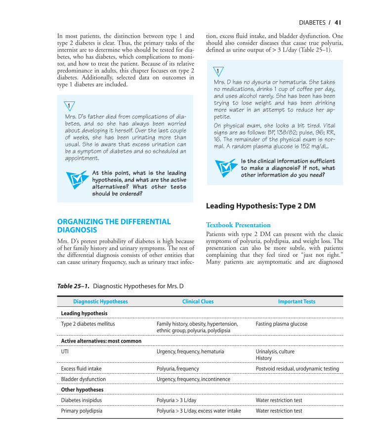
1
Mrs. D’s father died from complications of dia-betes, and so she has always been worriedabout developing it herself. Over the last coupleof weeks, she has been urinating more thanusual. She is aware that excess urination canbe a symptom of diabetes and so scheduled anappointment.
At this point, what is the leadinghypothesis, and what are the activealternatives? What other testsshould be ordered?
ORGANIZING THE DIFFERENTIALDIAGNOSISMrs. D’s pretest probability of diabetes is high becauseof her family history and urinary symptoms. The rest ofthe differential diagnosis consists of other entities thatcan cause urinary frequency, such as urinary tract infec-
tion, excess fluid intake, and bladder dysfunction. Oneshould also consider diseases that cause true polyuria,defined as urine output of > 3 L/day (Table 25–1).
1
Mrs. D has no dysuria or hematuria. She takesno medications, drinks 1 cup of coffee per day,and uses alcohol rarely. She has been has beentrying to lose weight and has been drinkingmore water in an attempt to reduce her ap-petite.On physical exam, she looks a bit tired. Vitalsigns are as follows: BP, 138/82; pulse, 96; RR,16. The remainder of the physical exam is nor-mal. A random plasma glucose is 152 mg/dL.
Is the clinical information sufficientto make a diagnosis? If not, whatother information do you need?
Leading Hypothesis: Type 2 DM
Textbook PresentationPatients with type 2 DM can present with the classicsymptoms of polyuria, polydipsia, and weight loss. Thepresentation can also be more subtle, with patientscomplaining that they feel tired or “just not right.”Many patients are asymptomatic and are diagnosed
ON-LINE ONLY
Table 25–1. Diagnostic Hypotheses for Mrs. D
Diagnostic Hypotheses Clinical Clues Important Tests
Leading hypothesis
Type 2 diabetes mellitus Family history, obesity, hypertension, Fasting plasma glucoseethnic group, polyuria, polydipsia
Active alternatives: most common
UTI Urgency, frequency, hematuria Urinalysis, cultureHistory
Excess fluid intake Polyuria, frequency Postvoid residual, urodynamic testing
Bladder dysfunction Urgency, frequency, incontinence
Other hypotheses
Diabetes insipidus Polyuria > 3 L/day Water restriction test
Primary polydipsia Polyuria > 3 L/day, excess water intake Water restriction test
9310_Stern_C25 9/2/05 1:48 PM Page 41
42 / CHAPTER 25
through plasma glucose testing. Patients can also presentwith complications of diabetes.
Disease HighlightsA. Prevalence in the United States is about 13–14%;
up to half of patients are unaware of the diagnosis.B. The lifetime risk of developing diabetes for individ-
uals born in 2000 is estimated to be 32.8% formales and 38.5% for females; rates are as high as50% for African American and Hispanic women.
C. Risk factors include1. Age ≥ 452. Body mass index (BMI) ≥ 25 kg/m2
3. A first-degree relative with diabetes4. Physical inactivity5. Being a member of a high-risk ethnic group
(African American, Hispanic American, NativeAmerican, Asian American, Pacific Islander)
6. Having delivered a baby weighing > 9 pounds orhaving had gestational DM
7. Hypertension8. Metabolic syndrome (high-density lipoprotein
[HDL] cholesterol < 35 mg/dL and/or triglyc-erides > 250 mg/dL)
9. Polycystic ovary syndrome10. History of impaired glucose tolerance or im-
paired fasting glucose (see Table 25–2 for defini-tions)
11. Vascular diseaseD. Impaired fasting glucose (IFG) and impaired glu-
cose tolerance (IGT)1. Metabolic stage between normal glucose home-
ostasis and diabetes, sometimes called “predia-betes”
2. Patients with IFG or IGT have normal or nearnormal HbA1c levels
3. Both are risk factors for the development of di-abetes and cardiovascular disease
4. Both are associated with the metabolic syn-drome (insulin resistance, compensatory hyper-insulinemia, obesity, hypertension, and dyslipi-demia consisting of high triglycerides and lowHDL)
E. Screening for diabetes1. American Diabetes Association (ADA) recom-
mends screening patients every 3 years begin-ning at age 45, especially those with a BMI ≥ 25kg/m2; those with ≥ 2 risk factors should bescreened earlier and more often
2. US Preventive Services Task Force recommendsscreening patients with hypertension or hyper-lipidemia but not asymptomatic individuals
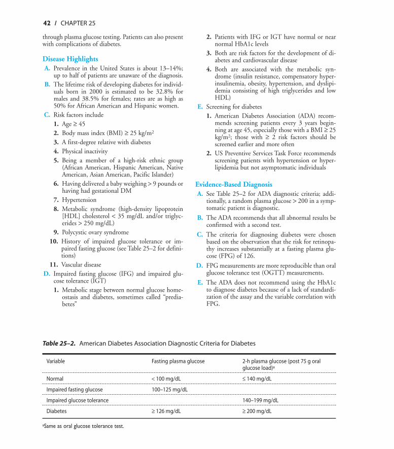
Evidence-Based DiagnosisA. See Table 25–2 for ADA diagnostic criteria; addi-
tionally, a random plasma glucose > 200 in a symp-tomatic patient is diagnostic.
B. The ADA recommends that all abnormal results beconfirmed with a second test.
C. The criteria for diagnosing diabetes were chosenbased on the observation that the risk for retinopa-thy increases substantially at a fasting plasma glu-cose (FPG) of 126.
D. FPG measurements are more reproducible than oralglucose tolerance test (OGTT) measurements.
E. The ADA does not recommend using the HbA1cto diagnose diabetes because of a lack of standardi-zation of the assay and the variable correlation withFPG.
ON-LINE ONLY
Table 25–2. American Diabetes Association Diagnostic Criteria for Diabetes
Variable Fasting plasma glucose 2-h plasma glucose (post 75 g oral glucose load)a
Normal < 100 mg/dL ≤ 140 mg/dL
Impaired fasting glucose 100–125 mg/dL
Impaired glucose tolerance 140–199 mg/dL
Diabetes ≥ 126 mg/dL ≥ 200 mg/dL
aSame as oral glucose tolerance test.
9310_Stern_C25 9/2/05 1:48 PM Page 42
DIABETES / 43
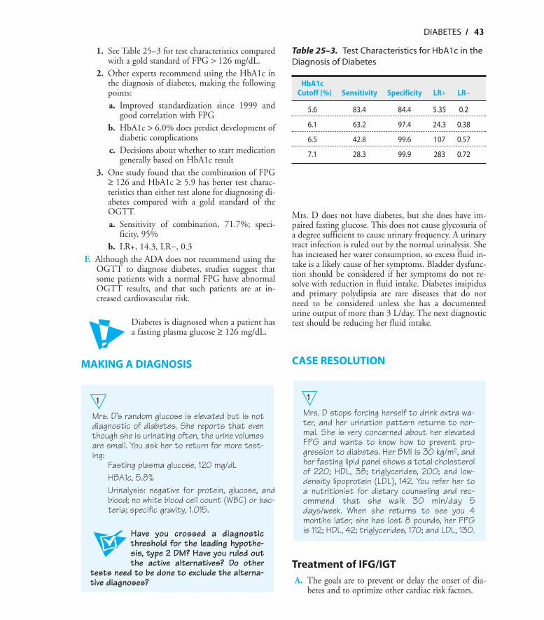
1. See Table 25–3 for test characteristics comparedwith a gold standard of FPG > 126 mg/dL.
2. Other experts recommend using the HbA1c inthe diagnosis of diabetes, making the followingpoints:a. Improved standardization since 1999 and
good correlation with FPGb. HbA1c > 6.0% does predict development of
diabetic complicationsc. Decisions about whether to start medication
generally based on HbA1c result3. One study found that the combination of FPG
≥ 126 and HbA1c ≥ 5.9 has better test charac-teristics than either test alone for diagnosing di-abetes compared with a gold standard of theOGTT.a. Sensitivity of combination, 71.7%; speci-
ficity, 95%b. LR+, 14.3, LR−, 0.3
F. Although the ADA does not recommend using theOGTT to diagnose diabetes, studies suggest thatsome patients with a normal FPG have abnormalOGTT results, and that such patients are at in-creased cardiovascular risk.
Diabetes is diagnosed when a patient hasa fasting plasma glucose ≥ 126 mg/dL.
MAKING A DIAGNOSIS
1
Mrs. D’s random glucose is elevated but is notdiagnostic of diabetes. She reports that eventhough she is urinating often, the urine volumesare small. You ask her to return for more test-ing:
Fasting plasma glucose, 120 mg/dLHBA1c, 5.8%Urinalysis: negative for protein, glucose, andblood; no white blood cell count (WBC) or bac-teria; specific gravity, 1.015.
Have you crossed a diagnosticthreshold for the leading hypothe-sis, type 2 DM? Have you ruled outthe active alternatives? Do other
tests need to be done to exclude the alterna-tive diagnoses?
Mrs. D does not have diabetes, but she does have im-paired fasting glucose. This does not cause glycosuria ofa degree sufficient to cause urinary frequency. A urinarytract infection is ruled out by the normal urinalysis. Shehas increased her water consumption, so excess fluid in-take is a likely cause of her symptoms. Bladder dysfunc-tion should be considered if her symptoms do not re-solve with reduction in fluid intake. Diabetes insipidusand primary polydipsia are rare diseases that do notneed to be considered unless she has a documentedurine output of more than 3 L/day. The next diagnostictest should be reducing her fluid intake.
CASE RESOLUTION
1
Mrs. D stops forcing herself to drink extra wa-ter, and her urination pattern returns to nor-mal. She is very concerned about her elevatedFPG and wants to know how to prevent pro-gression to diabetes. Her BMI is 30 kg/m2, andher fasting lipid panel shows a total cholesterolof 220; HDL, 38; triglycerides, 200; and low-density lipoprotein (LDL), 142. You refer her toa nutritionist for dietary counseling and rec-ommend that she walk 30 min/day 5days/week. When she returns to see you 4months later, she has lost 8 pounds, her FPGis 112; HDL, 42; triglycerides, 170; and LDL, 130.
Treatment of IFG/IGTA. The goals are to prevent or delay the onset of dia-
betes and to optimize other cardiac risk factors.
ON-LINE ONLY
Table 25–3. Test Characteristics for HbA1c in theDiagnosis of Diabetes
HbA1c Cutoff (%) Sensitivity Specificity LR+ LR−
5.6 83.4 84.4 5.35 0.2
6.1 63.2 97.4 24.3 0.38
6.5 42.8 99.6 107 0.57
7.1 28.3 99.9 283 0.72
9310_Stern_C25 9/2/05 1:48 PM Page 43
44 / CHAPTER 25
B. Large randomized trials have shown that lifestylemodification or medication can prevent or delay di-abetes.1. Finnish patients with IGT randomized to brief
diet/exercise counseling or intensive individual-ized instructiona. There was a 58% relative reduction in the
development of diabetes in the intensivegroup.
b. Number needed to treat (NNT) for 1 year toprevent 1 case of DM was 22, with an NNTof 5 over 5 years.
2. US patients (45% African American or His-panic) randomized to intensive diet/exerciseprogram, metformin, or placeboa. There was a 58% relative reduction in the
development of DM in the intensive diet/ex-ercise group and a 31% relative reduction inmetformin group.
b. NNT over 3 years to prevent 1 case of dia-betes was 7 for the intensive diet/exercisegroup and 14 for the metformin group.
3. Patients with IGT randomized to acarbose orplacebo had a 36% relative reduction in the de-velopment of diabetes in the acarbose group.
Lifestyle modification is the best way toprevent or delay the onset of diabetes.
C. Recommended lifestyle modification goals are 30min of modest physical activity daily and loss of5–10% of body weight.
D. Hypertension should be treated to a goal of <140/90.
E. Lipids should be treated according to NationalCholesterol Education Program (NCEP) guidelinesfor nondiabetic patients (see Chapter 28).
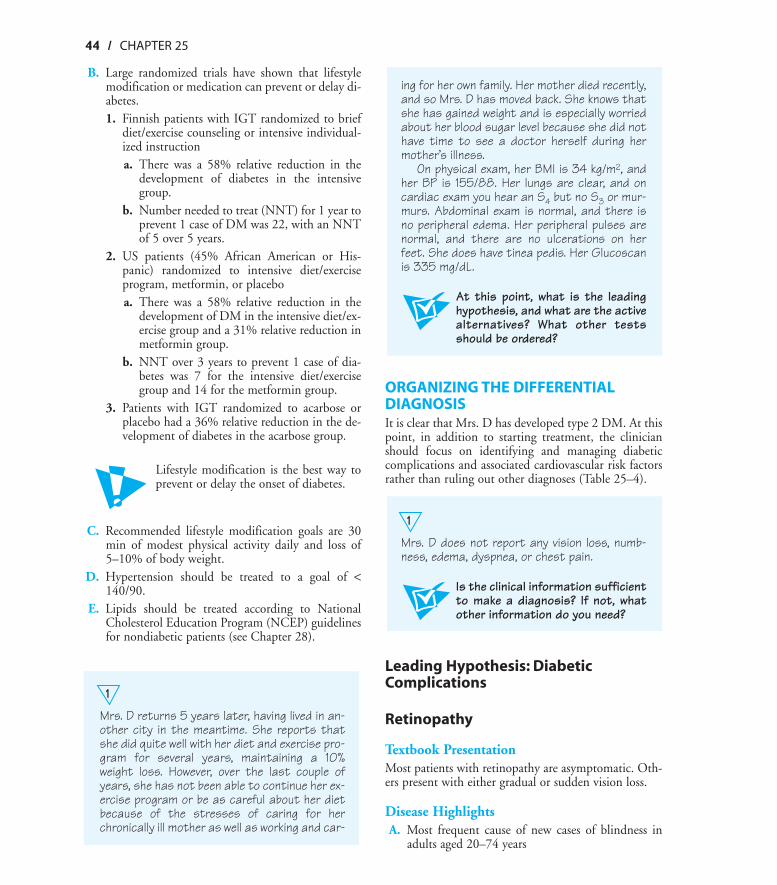
1
Mrs. D returns 5 years later, having lived in an-other city in the meantime. She reports thatshe did quite well with her diet and exercise pro-gram for several years, maintaining a 10%weight loss. However, over the last couple ofyears, she has not been able to continue her ex-ercise program or be as careful about her dietbecause of the stresses of caring for herchronically ill mother as well as working and car-
ing for her own family. Her mother died recently,and so Mrs. D has moved back. She knows thatshe has gained weight and is especially worriedabout her blood sugar level because she did nothave time to see a doctor herself during hermother’s illness.
On physical exam, her BMI is 34 kg/m2, andher BP is 155/88. Her lungs are clear, and oncardiac exam you hear an S4 but no S3 or mur-murs. Abdominal exam is normal, and there isno peripheral edema. Her peripheral pulses arenormal, and there are no ulcerations on herfeet. She does have tinea pedis. Her Glucoscanis 335 mg/dL.
At this point, what is the leadinghypothesis, and what are the activealternatives? What other testsshould be ordered?
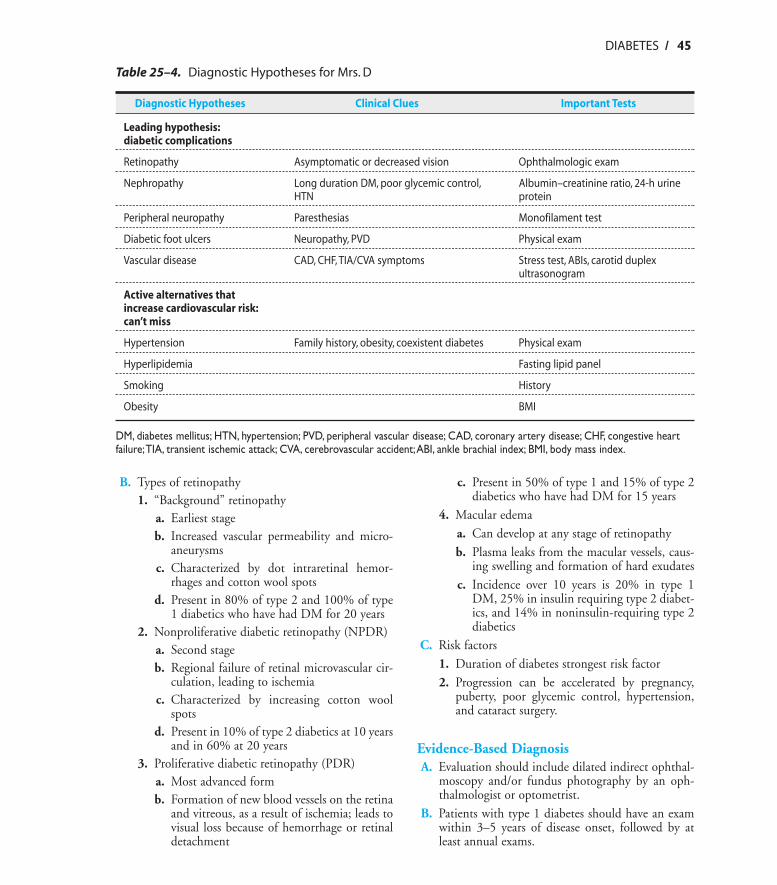
ORGANIZING THE DIFFERENTIALDIAGNOSISIt is clear that Mrs. D has developed type 2 DM. At thispoint, in addition to starting treatment, the clinicianshould focus on identifying and managing diabeticcomplications and associated cardiovascular risk factorsrather than ruling out other diagnoses (Table 25–4).
1
Mrs. D does not report any vision loss, numb-ness, edema, dyspnea, or chest pain.
Is the clinical information sufficientto make a diagnosis? If not, whatother information do you need?
Leading Hypothesis: DiabeticComplications
Retinopathy
Textbook PresentationMost patients with retinopathy are asymptomatic. Oth-ers present with either gradual or sudden vision loss.
Disease HighlightsA. Most frequent cause of new cases of blindness in
adults aged 20–74 years
ON-LINE ONLY
9310_Stern_C25 9/2/05 1:48 PM Page 44
DIABETES / 45
B. Types of retinopathy1. “Background” retinopathy
a. Earliest stageb. Increased vascular permeability and micro-
aneurysmsc. Characterized by dot intraretinal hemor-
rhages and cotton wool spotsd. Present in 80% of type 2 and 100% of type
1 diabetics who have had DM for 20 years2. Nonproliferative diabetic retinopathy (NPDR)
a. Second stageb. Regional failure of retinal microvascular cir-
culation, leading to ischemiac. Characterized by increasing cotton wool
spotsd. Present in 10% of type 2 diabetics at 10 years
and in 60% at 20 years3. Proliferative diabetic retinopathy (PDR)
a. Most advanced formb. Formation of new blood vessels on the retina
and vitreous, as a result of ischemia; leads tovisual loss because of hemorrhage or retinaldetachment
c. Present in 50% of type 1 and 15% of type 2diabetics who have had DM for 15 years
4. Macular edemaa. Can develop at any stage of retinopathyb. Plasma leaks from the macular vessels, caus-
ing swelling and formation of hard exudatesc. Incidence over 10 years is 20% in type 1
DM, 25% in insulin requiring type 2 diabet-ics, and 14% in noninsulin-requiring type 2diabetics
C. Risk factors1. Duration of diabetes strongest risk factor2. Progression can be accelerated by pregnancy,
puberty, poor glycemic control, hypertension,and cataract surgery.
Evidence-Based DiagnosisA. Evaluation should include dilated indirect ophthal-
moscopy and/or fundus photography by an oph-thalmologist or optometrist.
B. Patients with type 1 diabetes should have an examwithin 3–5 years of disease onset, followed by atleast annual exams.
ON-LINE ONLY
Table 25–4. Diagnostic Hypotheses for Mrs. D
Diagnostic Hypotheses Clinical Clues Important Tests
Leading hypothesis:diabetic complications
Retinopathy Asymptomatic or decreased vision Ophthalmologic exam
Nephropathy Long duration DM, poor glycemic control, Albumin–creatinine ratio, 24-h urine HTN protein
Peripheral neuropathy Paresthesias Monofilament test
Diabetic foot ulcers Neuropathy, PVD Physical exam
Vascular disease CAD, CHF, TIA/CVA symptoms Stress test, ABIs, carotid duplex ultrasonogram
Active alternatives that increase cardiovascular risk:can’t miss
Hypertension Family history, obesity, coexistent diabetes Physical exam
Hyperlipidemia Fasting lipid panel
Smoking History
Obesity BMI
DM, diabetes mellitus; HTN, hypertension; PVD, peripheral vascular disease; CAD, coronary artery disease; CHF, congestive heartfailure;TIA, transient ischemic attack; CVA, cerebrovascular accident;ABI, ankle brachial index; BMI, body mass index.
9310_Stern_C25 9/2/05 1:48 PM Page 45
46 / CHAPTER 25
C. Patients with type 2 diabetes should have an examat the time of diagnosis, followed by at least annualexams.
All type 2 diabetics need eye exams by aneye specialist at least annually.
TreatmentA. Glycemic control
1. In type 1 diabetics without retinopathy, the riskof developing it is reduced 76% by tight control.
2. In type 1 diabetics with retinopathy, the risk ofprogression is reduced by 54% with tight con-trol.
3. In type 2 diabetics, better control reduces therisk of microvascular complications (retinopathyand nephropathy) by 25%.
4. In type 2 diabetics, for every percentage pointdecrease in HbA1c, there is a 35% reduction inthe risk of microvascular complications.
B. Better BP control reduces the risk of progression ofretinopathy.
C. Aspirin neither improves nor worsens retinopathy.D. Photocoagulation reduces the rate of developing se-
vere vision loss in patients with either proliferativeretinopathy or macular edema.
Neuropathy
Textbook PresentationDiabetic peripheral neuropathy classically presents asparesthesias or burning pain in a “glove-stocking” distri-bution. Diabetic autonomic neuropathy can manifest ina variety of ways, including orthostatic dizziness, diar-rhea, urinary incontinence, and gastroparesis.
Disease HighlightsA. Diabetic peripheral neuropathy
1. Typesa. Symmetric distal polyneuropathy (DPN)b. Focal neuropathies
(1) Cranial(a) Usually cranial nerve III or VI(b) Usually acute and transient(c) Caused by ischemia
(2) Thoracolumbar(3) Limb
(a) Median nerve most common site(b) Ulnar, femoral, and peroneal also af-
fectedc. Diabetic amyotrophy (pain, severe asymmet-
ric muscle weakness, and wasting of the il-iopsoas and quadriceps).
2. Epidemiology of DPNa. Affects up to 50% of diabetics, with chronic
neuropathic pain in 20% of patients with di-abetes for over 10 years
b. Severity related to duration of disease, degreeof glycemic control, presence of hyperten-sion and hyperlipidemia
c. Independent risk factor for foot ulcerationand amputation; patients with neuropathyhave a 15% lifetime risk of amputation
3. Clinical manifestations of DPNa. History
(1) Burning, shooting, or lancinating pain(2) Paresthesias, hyperesthesias(3) Often worse at night(4) When symptoms ascend to the knees,
upper extremity symptoms startb. Physical exam
(1) Decreased sensation (see Evidence-BasedDiagnosis section)
(2) Loss of DTRs(3) Distal muscle atrophy late in the course
c. Ten percent of patients develop Charcotjoints, usually in the tarsometatarsal region.
4. Differential diagnosisa. Consider other causes of neuropathy if
(1) Neuropathy develops before or early inthe course of the diabetes
(2) Patient has a history of excellentglycemic control
(3) Neuropathy is asymmetric(4) There is proximal or upper extremity in-
volvement disproportionate to distallower extremity involvement
b. Be sure to check for other treatable causes(hypothyroidism and vitamin B12 defi-ciency), even in patients with long-standingdiabetes.
Think about other causes of neuropathyin diabetic patients with atypical presen-tations.
ON-LINE ONLY
9310_Stern_C25 9/2/05 1:48 PM Page 46
DIABETES / 47
B. Diabetic autonomic neuropathy: can affect any or-gan innervated by the autonomic nervous system1. Cardiovascular autonomic neuropathy: many
possible manifestationsa. Reduced heart rate variability, fixed heart
rate, sinus tachycardiab. Inadequate increase in heart rate/BP with ex-
ercisec. Postural hypotension with systolic BP drop
of > 29 mm Hgd. Intraoperative cardiac instability
2. Gustatory sweatinga. Facial sweating, often accompanied by flush-
ing, that occurs after eatingb. Generally occurs in patients with nephropa-
thy or peripheral neuropathyc. Cause unknown
3. GI dysfunctiona. Reduced esophageal motilityb. Gastroparesis
(1) Abnormality of gastric motility leadingto delayed gastric emptying
(2) Symptoms include nausea, vomiting,anorexia, postprandial fullness, earlysatiety.
(3) Poor correlation between demonstratedmotility abnormalities and symptoms
c. Diabetic diarrhea(1) Characterized by intermittent, brown
watery, voluminous stools, occasionallyaccompanied by tenesmus
(2) Can be episodic, separated by periods ofnormal bowel movements or constipa-tion
(3) Rare in the absence of other manifesta-tions of neuropathy, either peripheral orautonomic
d. Constipation(1) Constipation specifically resulting from
autonomic neuropathy occurs in 20% oftype 2 diabetics
(2) Caused by abnormality in autonomicneural control of colonic motility
e. Anorectal dysfunction(1) Results in fecal incontinence, even in the
absence of diarrhea(2) Patients can generally sense the presence
of stool, but cannot prevent passage
4. Genitourinary dysfunctiona. Bladder dysfunction
(1) Initially motor function normal, butsensation of bladder distention impaired
(2) Then detrusor muscle hypocontractilityoccurs, leading to urinary retention andoverflow incontinence
b. Erectile dysfunction(1) Present in 28–45% of diabetic men(2) Most common organic cause of erectile
dysfunction(3) Risk factors include duration of DM,
glycemic control, smoking, other dia-betic complications.
Evidence-Based DiagnosisA. Diabetic peripheral neuropathy (DPN)
1. Nerve conduction studies are the gold standard.2. Several physical exam maneuvers have been
compared with nerve conduction studies.a. Semmes-Weinstein monofilament examina-
tion(1) Apply a 5.07/10-g monofilament to a
noncallused site on the dorsum of thefirst toe just proximal to the nail bed.
(2) Repeat 4 times on both feet in an ar-rhythmic manner.
(3) Add up the total number of times themonofilament is not perceived by thepatient (score range = 0–8).
b. On–off vibration testing(1) Apply a vibrating 128-Hz tuning fork to
the bony prominence at the dorsum ofthe first toe just proximal to the nail bed.
(2) Repeat twice on each foot.(3) Add up the total number of times the pa-
tient did not perceive the application ofthe vibrating tuning fork or the dampen-ing of the vibration (score range = 0–8).
c. Timed vibration testing(1) Apply a vibrating 128-Hz tuning fork to
the same location used for the on–off vi-bration test.
(2) Ask the patient to report the time atwhich vibration diminished beyond per-ception, and compare with the numberof seconds perceived by the examinerwhen the tuning fork is applied to theexaminer’s thumb.
ON-LINE ONLY
9310_Stern_C25 9/2/05 1:48 PM Page 47
48 / CHAPTER 25
(3) Record number of times patient’s per-ception time less than examiner’s (scorerange = 0–8).
d. Superficial pain sensation(1) Apply a sterile Neurotip to the same sites
used for the monofilament.(2) Repeat 4 times on each foot.(3) Add up the total number of times the
patient did not perceive the painfulstimulus (score range = 0–8).
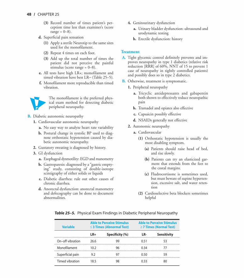
e. All tests have high LR+; monofilament andtimed vibration have best LR− (Table 25–5).
f. Monofilament more reproducible than timedvibration.
The monofilament is the preferred phys-ical exam method for detecting diabeticperipheral neuropathy.
B. Diabetic autonomic neuropathy1. Cardiovascular autonomic neuropathy
a. No easy way to analyze heart rate variabilityb. Postural change in systolic BP used to diag-
nose orthostatic hypotension caused by dia-betic autonomic neuropathy.
2. Gustatory sweating is diagnosed by history.3. GI dysfunction
a. Esophageal dysmotility: EGD and manometryb. Gastroparesis: diagnosed by a “gastric empty-
ing” study, consisting of double-isotopescintigraphy of either solids or liquids
c. Diabetic diarrhea: rule out other causes ofchronic diarrhea.
d. Anorectal dysfunction: anorectal manometryand defecography can be done to documentabnormalities.
4. Genitourinary dysfunctiona. Urinary bladder dysfunction: ultrasound and
urodynamic testingb. Erectile dysfunction: history
TreatmentA. Tight glycemic control definitely prevents and im-
proves neuropathy in type 1 diabetics (relative riskreduction [RRR] of 60%, NNT of 15 to prevent 1case of neuropathy in tightly controlled patients)and possibly does so in type 2 diabetics.
B. Otherwise, treatment is symptomatic.
1. Peripheral neuropathy
a. Tricyclic antidepressants and gabapentinboth shown to effectively reduce neuropathicpain
b. Tramadol and opiates also effective
c. Capsaicin possibly effective
d. NSAIDs generally not effective
2. Autonomic neuropathy
a. Cardiovascular
(1) Orthostatic hypotension is usually themost disabling symptom.
(a) Patients should raise head of bed,and rise slowly.
(b) Patients can try an elasticized gar-ment that extends from the feet tothe costal margins.
(c) Fludrocortisone is sometimes used,but must beware of supine hyperten-sion, excessive salt, and water reten-tion
(2) Cardioselective beta blockers sometimeshelpful
ON-LINE ONLY
Table 25–5. Physical Exam Findings in Diabetic Peripheral Neuropathy
Able to Perceive Stimulus Able to Perceive Stimulus Variable ≤ 3 Times (Abnormal Test) ≥ 7 Times (Normal Test)
LR+ Specificity (%) LR- Sensitivity
On–off vibration 26.6 99 0.51 53
Monofilament 10.2 96 0.34 77
Superficial pain 9.2 97 0.50 59
Timed vibration 18.5 98 0.33 80
9310_Stern_C25 9/2/05 1:48 PM Page 48
DIABETES / 49
b. Sweating: no specific treatment available; cantry clonidine
c. Esophageal dysmotility: can try prokineticagents such as metoclopramide
d. Gastroparesis(1) Severe gastroparesis is very difficult to
manage.(2) Small meals sometimes help.(3) Can try metoclopramide or erythromy-
cin(4) Gastric electrical stimulation being stud-
ied for refractory casese. Constipation
(1) Increase fiber(2) Drug choices include lactulose, polyeth-
ylene glycol, stool softeners.(3) Avoid senna, cascara.
f. Urinary bladder dysfunction(1) Bethanecol(2) Intermittent self-catheterization
g. Erectile dysfunction: sildenafil and othersimilar agents
Nephropathy
Textbook PresentationDiabetic nephropathy is asymptomatic until it is so ad-vanced that the patient has symptoms of renal failure.
Disease HighlightsA. The most common cause of end-stage renal disease
(ESRD) in the United States and Europe, account-ing for about 40% of new cases of ESRD.
B. Definitions (Table 25–6)
C. Natural history: much better defined for type 1than for type 2 DM1. Type 1 DM
a. Renal enlargement and hyperfunction at on-set of diabetes; continues for 5–15 years
b. Microalbuminuria appears 10–15 years afteronset of DM; GFR and BP initially normal.
c. Over the ensuing 10–15 years, 80% of pa-tients progress to macroalbuminuria;glomerular filtration rate (GFR) declines andhypertension develops.
d. ESRD develops in 50% of patients withovert nephropathy within 10 years and in75% by 20 years.
2. Type 2 DMa. Natural history less well defined because on-
set of type 2 DM usually not well defined,and other causes of renal insufficiency (suchas hypertension and vascular disease) aremore common
b. 20–40% of patients with microalbuminuriaprogress to overt nephropathy.
c. 20% have ESRD within 20 years of the on-set of overt nephropathy.
D. Risk factors for development of nephropathy1. Poor glycemic control2. Hypertension3. Long duration of diabetes4. Male sex5. Ethnic predisposition (Native American,
African American, Hispanic [especially MexicanAmerican])
Evidence-Based DiagnosisA. ADA recommends annual screening for microalbu-
minuria beginning at the time of diagnosis for type2 diabetics and at year 5 for type 1 diabetics.
Diabetic patients who are not already re-ceiving angiotensin-converting enzymeinhibitors or angiotensin receptor block-ers (ARBs) should be screened annuallyfor microalbuminuria.
B. ADA recommended screening tests include 24-hurine collections, 4-h urine collection, or spot albu-min/creatinine ratio.1. 24-h urine collection is the gold standard but is
inconvenient to obtain.2. 4-h collections also inconvenient
ON-LINE ONLY
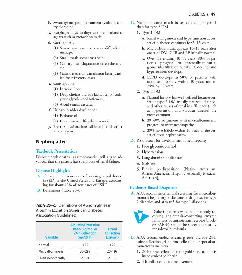
Table 25–6. Definitions of Abnormalities inAlbumin Excretion (American DiabetesAssociation Guidelines)
Albumin/Creatinine Ratio (µg/mg) or Timed 24-h Collection Collection
Variable (mg/24 h) (µg/min)
Normal < 30 < 20
Microalbuminuria 30–299 20–199
Overt nephropathy ≥ 300 ≥ 200
9310_Stern_C25 9/2/05 1:48 PM Page 49
50 / CHAPTER 25
3. Albumin–creatinine ratio in a random spot col-lection easiest to obtaina. There is diurnal variation, so first-void or
early-morning specimens best; otherwise, tryto obtain confirmatory specimen at sametime of day as initial specimen.
b. Short-term hyperglycemia, exercise, urinarytract infection, marked hypertension, heartfailure, and acute febrile illness can causetransient elevations in albumin excretion.
c. All abnormal tests should be confirmed by asecond test.
d. Sensitivity ranges from 70–100%, specificityfrom 91–98% for morning specimens (sensi-tivity 56–97%, specificity 81–92% for ran-dom specimens).
e. Specificity might decrease with age: 1 studyfound a stable sensitivity of about 95%, butdifferent specificities depending on age andsex (men: 84% for ages < 65 years, 72% for> 65 years; women: 89% for ages < 65 years,82% for > 65 years).
TreatmentA. Tight glycemic control reduces nephropathy.
1. Type 1 DMa. Incidence of microalbuminuria reduced by
34% (NNT = 83) in patients withoutretinopathy and by 43% (NNT = 47) in pa-tients with retinopathy
b. Incidence of macroalbuminuria reduced by56% (NNT = 125) in patients with retinopa-thy
2. Type 2 DMa. Microvascular complications (retinopathy
plus nephropathy) reduced by 25% (NNT =36 over 10 years)
b. The microvascular complication rate was58% for patients with a HbA1c ≥ 10 and6.1% for patients with an HbA1c < 6.0.
c. Microvascular complication rate decreasesby 37% for every 1% reduction in HbA1c.
B. BP control and choice of agents1. BP should be less than 130/80.2. See Table 25–7 for agents shown to delay pro-
gression of nephropathy.C. Protein restriction to about 10% of daily calories
may reduce progression of overt nephropathy.D. It is not clear whether or how often albuminuria
should be monitored in patients on ACE inhibitorsor ARBs.
E. Refer to a nephrologist if the creatinine clearance is< 60 mL/min or hypertension cannot be con-trolled.
Diabetic Foot Ulcers
Textbook PresentationA patient with peripheral neuropathy is unaware of mi-nor trauma and the beginning of plantar ulceration. Bythe time the ulcer is discovered incidentally, it is oftenadvanced, sometimes with associated osteomyelitis.
Disease HighlightsA. Lifetime risk of developing an ulcer is about 15%.B. Ninety percent of patients with ulcers have neuropa-
thy, and 15–20% have peripheral vascular disease.C. Tend to occur at pressure points, so plantar surface,
sites of calluses are common locationsD. Risk factors
1. Duration of diabetes > 10 years2. Male sex3. Poor glycemic control4. Coexisting cardiovascular, renal, or retinal com-
plication5. Peripheral neuropathy6. Altered biomechanics
ON-LINE ONLY
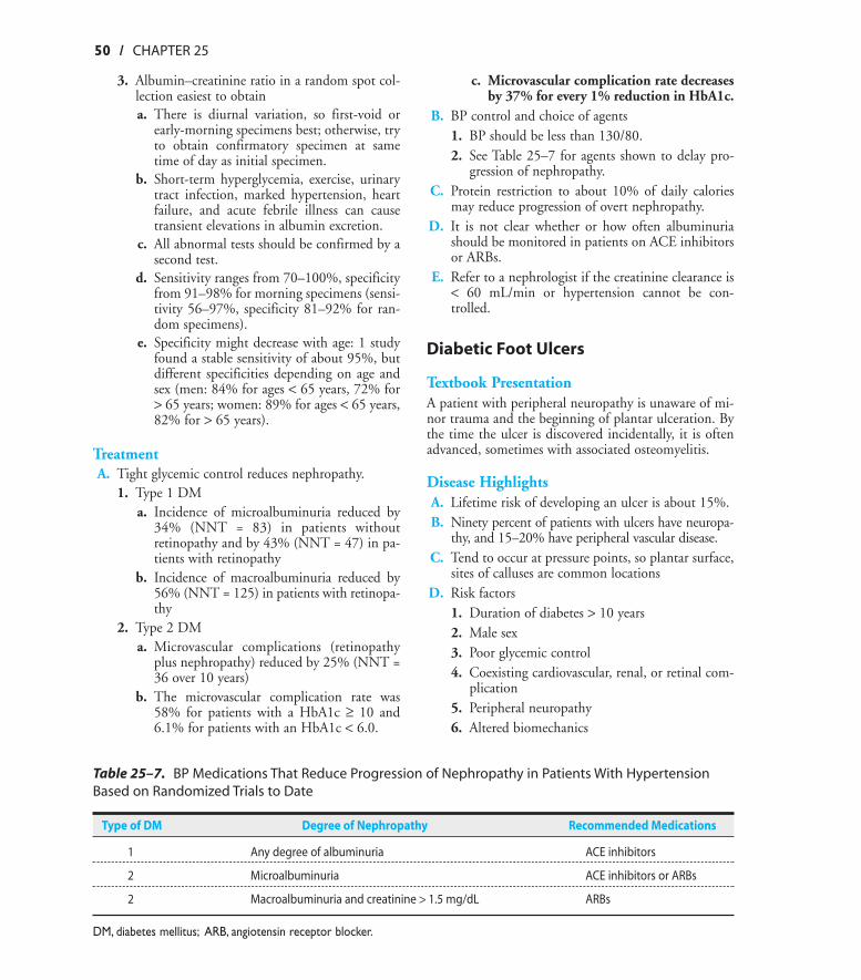
Table 25–7. BP Medications That Reduce Progression of Nephropathy in Patients With HypertensionBased on Randomized Trials to Date
Type of DM Degree of Nephropathy Recommended Medications
1 Any degree of albuminuria ACE inhibitors
2 Microalbuminuria ACE inhibitors or ARBs
2 Macroalbuminuria and creatinine > 1.5 mg/dL ARBs
DM, diabetes mellitus; ARB, angiotensin receptor blocker.
9310_Stern_C25 9/2/05 1:48 PM Page 50
DIABETES / 51
7. Evidence of increased pressure on the foot8. Bony deformity of the foot or ankle9. Peripheral vascular disease
10. A history of ulcers or amputation11. Severe nail pathology
E. Pathophysiology1. Repetitive mechanical stress occurs as a result of
altered biomechanics, foot deformities, ill-fit-ting shoes.
2. Peripheral neuropathy causes loss of protectivesensation, so the patient is unaware of the incip-ient ulceration.
3. Ischemia, resulting from macrovascular disease(commonly in the tibioperoneal vessels) or mi-crovascular dysfunction from autonomic neu-ropathy, inhibits healing and promotes progres-sion.
F. Classification1. Non-limb-threatening
a. Superficial infection, purulent discharge,and minimal (< 2 cm extension from the ul-cer) or absent cellulitis
b. No systemic toxicity (fever, leukocytosis, se-vere hyperglycemia, or osteomyelitis)
2. Limb threateninga. Ulceration to deep tissues, extensive puru-
lent drainage, cellulitis extending more than2 cm from the ulcer, and lymphangitis
b. Systemic toxicity and significant ischemia,with or without gangrene, present
3. Life threateninga. Ulceration to deep tissues, extensive puru-
lent drainage, cellulitis, necrosis, gangrene,osteomyelitis
b. Marked systemic toxicity, including septicshock
G. Microbiology1. Non-limb-threatening infections average 2
species/ulcer, but are often monomicrobial2. Limb-threatening and life-threatening infec-
tions generally polymicrobial3. Staphylococcus aureus most common organism,
present in 50% of infections4. Streptococci present in one third of cases5. Gram-negative organisms, especially Proteus,
Klebsiella, Escherichia coli, and Pseudomonas,present in polymicrobial infections
6. Anaerobic gram-positive cocci and Bacteroidespresent in up to 80% of polymicrobial infections
Evidence-Based DiagnosisA. ADA recommendations include at least annual foot
examinations that should include screening forneuropathy and assessing foot structure, biome-chanics, vascular status, and skin integrity.
You cannot examine the feet of your dia-betic patients too often!
B. Culturing ulcers1. Can be difficult to distinguish between coloniz-
ing organisms and true pathogens2. Deep cultures of the ulcer or the bone thought
to be more reliable3. If the patient is responding to empiric therapy,
it is not necessary to culture.C. Diagnosing complications
1. Cellulitis: clinical diagnosis (see Chapter 12)2. Osteomyelitis
a. Open bone biopsy with culture is the goldstandard.
b. Needle bone biopsy subject to sampling er-ror (sensitivity, 87%; specificity, 93%; LR+,12.4; LR−, 0.14)
c. Being able to probe the ulcer down to bonehas a sensitivity of 66% and specificity of89% for the presence of osteomyelitis (LR+,6; LR−, 0.38).
d. C-reactive protein (CRP), erythrocyte sedi-mentation rate (ESR), complete blood cellcount (CBC), blood cultures not sufficientlysensitive or specific to diagnose osteomyelitis.
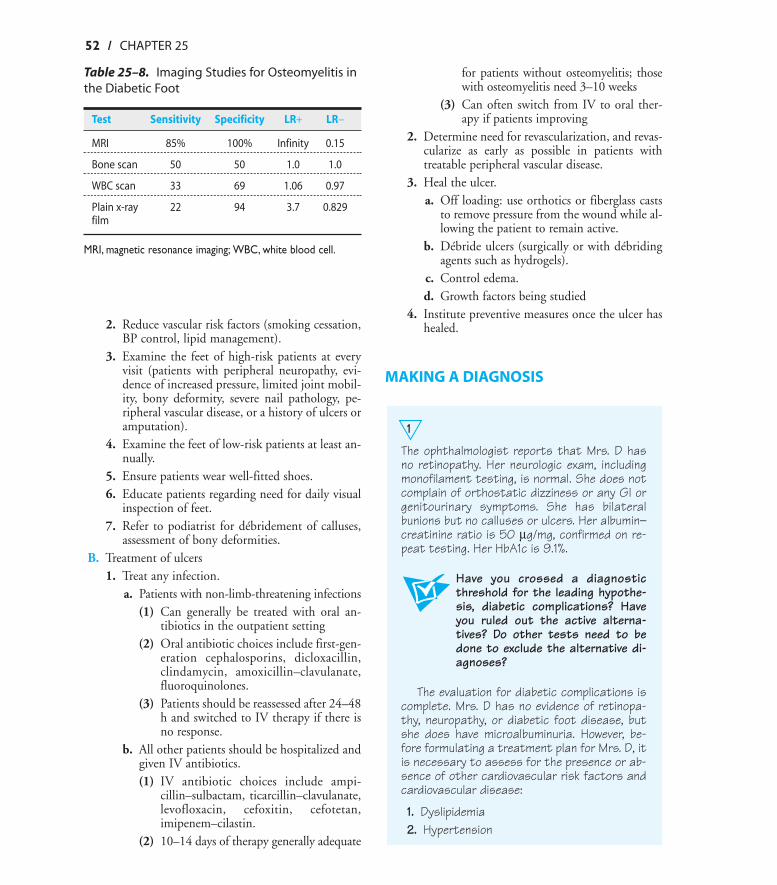
e. Magnetic resonance imaging (MRI) is theimaging procedure with the best test charac-teristics (Table 25–8).
MRI scan is the best imaging procedureto diagnose osteomyelitis in a patientwith a diabetic foot ulcer.
A normal CBC, CRP, or ESR does notrule out osteomyelitis.
TreatmentA. Preventive foot care
1. Improve glycemic control to reduce risk of neu-ropathy.
ON-LINE ONLY
9310_Stern_C25 9/2/05 1:48 PM Page 51
52 / CHAPTER 25
2. Reduce vascular risk factors (smoking cessation,BP control, lipid management).
3. Examine the feet of high-risk patients at everyvisit (patients with peripheral neuropathy, evi-dence of increased pressure, limited joint mobil-ity, bony deformity, severe nail pathology, pe-ripheral vascular disease, or a history of ulcers oramputation).
4. Examine the feet of low-risk patients at least an-nually.
5. Ensure patients wear well-fitted shoes.6. Educate patients regarding need for daily visual
inspection of feet.7. Refer to podiatrist for débridement of calluses,
assessment of bony deformities.B. Treatment of ulcers
1. Treat any infection.a. Patients with non-limb-threatening infections
(1) Can generally be treated with oral an-tibiotics in the outpatient setting
(2) Oral antibiotic choices include first-gen-eration cephalosporins, dicloxacillin,clindamycin, amoxicillin–clavulanate,fluoroquinolones.
(3) Patients should be reassessed after 24–48h and switched to IV therapy if there isno response.
b. All other patients should be hospitalized andgiven IV antibiotics.(1) IV antibiotic choices include ampi-
cillin–sulbactam, ticarcillin–clavulanate,levofloxacin, cefoxitin, cefotetan,imipenem–cilastin.
(2) 10–14 days of therapy generally adequate
for patients without osteomyelitis; thosewith osteomyelitis need 3–10 weeks
(3) Can often switch from IV to oral ther-apy if patients improving
2. Determine need for revascularization, and revas-cularize as early as possible in patients withtreatable peripheral vascular disease.
3. Heal the ulcer.a. Off loading: use orthotics or fiberglass casts
to remove pressure from the wound while al-lowing the patient to remain active.
b. Débride ulcers (surgically or with débridingagents such as hydrogels).
c. Control edema.d. Growth factors being studied
4. Institute preventive measures once the ulcer hashealed.
MAKING A DIAGNOSIS
1
The ophthalmologist reports that Mrs. D hasno retinopathy. Her neurologic exam, includingmonofilament testing, is normal. She does notcomplain of orthostatic dizziness or any GI orgenitourinary symptoms. She has bilateralbunions but no calluses or ulcers. Her albumin–creatinine ratio is 50 µg/mg, confirmed on re-peat testing. Her HbA1c is 9.1%.
Have you crossed a diagnosticthreshold for the leading hypothe-sis, diabetic complications? Haveyou ruled out the active alterna-tives? Do other tests need to bedone to exclude the alternative di-agnoses?
The evaluation for diabetic complications iscomplete. Mrs. D has no evidence of retinopa-thy, neuropathy, or diabetic foot disease, butshe does have microalbuminuria. However, be-fore formulating a treatment plan for Mrs. D, itis necessary to assess for the presence or ab-sence of other cardiovascular risk factors andcardiovascular disease:
1. Dyslipidemia2. Hypertension
ON-LINE ONLY
Table 25–8. Imaging Studies for Osteomyelitis inthe Diabetic Foot
Test Sensitivity Specificity LR+ LR−
MRI 85% 100% Infinity 0.15
Bone scan 50 50 1.0 1.0
WBC scan 33 69 1.06 0.97
Plain x-ray 22 94 3.7 0.829film
MRI, magnetic resonance imaging; WBC, white blood cell.
9310_Stern_C25 9/2/05 1:48 PM Page 52
DIABETES / 53
3. Obesity4. Smoking5. Coronary artery disease6. Cerebrovascular disease7. Peripheral vascular disease
See Table 25–9 for a summary of testing that must beperformed on all patients with diabetes.
CASE RESOLUTION
1
Mrs. D has no symptoms of vascular disease oncareful questioning, and her exercise tolerance ismore than 1 mile. Her fasting lipid panel shows atotal cholesterol of 230, HDL of 45, triglycerides
of 200, and LDL of 145. You refer Mrs. D to a di-abetes educator for instruction in home glucosemonitoring and to a nutritionist for instructionabout diet and exercise. You also prescribe met-formin for the diabetes and atorvastatin for thehyperlipidemia. Because she has hypertensionand microalbuminuria, you elect to start an ACEinhibitor, lisinopril, to treat her hypertension. Youalso recommend that she start taking aspirin,81 mg daily. Over the next 12–18 months, Mrs. Dloses 5 pounds. You increase the dose of met-formin and then add glipizide to achieve an HbA1cof 6.9%. You also increase the dose of lisinopriland add hydrochlorothiazide and atenolol toachieve a BP of 128/80. Her LDL is now 85 mg/dL.
Treatment of Type 2 DiabetesThe treatment of type 2 diabetes involves not only thetreatment of the hyperglycemia but the management of
ON-LINE ONLY
Table 25–9. Summary of Testing for Diabetic Complications and Important Associated Conditions
Condition Required Test/Action
Retinopathy Ophthalmologic exama
Peripheral neuropathy Monofilament testinga
Nephropathy Albumin–creatinine ratio, serum creatininea
Diabetic foot ulcers Foot exama
Dyslipidemia Fasting total cholesterol, HDL, triglycerides, LDLa
Hypertension BP measurementa
Smoking Obtain historya
Obesity Measure weight and calculate BMIa,b
Coronary artery disease ADA recommendations: (1) diagnostic stress test in patients with typical or atypical symptoms and an abnormal resting ECG; (2) consider screening asymptomatic patients witha history of other vascular disease, sedentary lifestyle, age > 35 and plans to start a vigorousexercise program, ≥ 2 cardiac risk factorsc
Cerebrovascular disease Carotid duplex ultrasound in patients with symptoms
Peripheral vascular disease ABIs in symptomatic patients; consider in asymptomatic patients also due to high prevalenceof asymptomatic disease
HDL, high-density lipoprotein; LDL, low-density lipoprotein; BMI, body mass index; ECG, electrocardiogram;ABI, ankle brachial in-dex.aShould be performed at least annually and may need to be done more often in patients with abnormalities.bWeight in kilograms/(height in meters)2; normal < 25, overweight 25–30, obese > 30cConsensus opinion by American Diabetes Association, no clinical trial evidence, and not supported by US Preventive Services TaskForce.
9310_Stern_C25 9/2/05 1:48 PM Page 53
54 / CHAPTER 25
associated complications and cardiovascular risk factorsas well. According to survey data, only 37% of partici-pants reached HbA1c goals, 35.8% reached BP goals,and 48% reached cholesterol goals; only 7.3% reachedall 3 goals.
It is common for patients to require 6–7medications to reach the treatment goalsoutlined next.
Treatment of HyperglycemiaA. Treatment goals
1. ADA: HbA1c < 7.0%2. American College of Endocrinology: HbA1c ≤
6.5%3. Goals should be modified for frail elderly, in
whom avoidance of hypoglycemia and opti-mization of functional status may be more im-portant than tight glycemic control.
B. Monitoring1. HbA1c levels every 3–6 months (see Table
25–10 for correlation between plasma glucoseand Hba1c)
2. Home glucose monitoringa. Patients on insulin should test several
times/day if not well controlled and perhapsless often if well controlled.
b. Optimal frequency for patients on oralagents unclear; patients in whom therapy isbeing changed should test more frequently
C. Lifestyle modification1. Weight loss, diet modification, and exercise are
the foundations of all treatment for diabetes.2. Best instituted in conjunction with a certified
diabetes educator or nutritionistD. Oral hypoglycemics
1. Sulfonylureas (SU)a. Examples: glyburide, glipizide, glimepirideb. Increase insulin secretion.c. Average decrease in HbA1c about 1–2%d. Side effects include weight gain (2–5 kg) and
hypoglycemia, especially in the elderly, pa-tients with reduced renal function, and thosewith erratic eating habits.
e. Shown to reduce diabetes-related end pointsand microvascular end points
f. Can be used as monotherapy or in combina-tion with insulin or other oral agents (exceptnon-SU secretagogues)
2. Biguanidesa. Example: metforminb. Reduce hepatic glucose production.c. Average decrease in HbA1c about 1–2%d. Associated with weight loss (or at least no
weight gain); hypoglycemia raree. Most common side effects are GI (abdomi-
nal pain, nausea, diarrhea).f. Because of risk of lactic acidosis, should be
avoided in patients with creatinine ≥ 1.5mg/dL, congestive heart failure (CHF), he-patic dysfunction, metabolic acidosis, and al-coholism.
Metformin should be withheld in pa-tients with acute illness and those under-going surgery or procedures using radio-contrast.
g. Has been shown to decrease diabetes-relatedend points, macrovascular end point, and to-tal mortality in obese type 2 diabetics
h. Can be used as monotherapy or in combina-tion with all other oral agents and insulin
3. Alpha glucosidase inhibitorsa. Example: acarboseb. Delay intestinal carbohydrate absorption,
decreasing postprandial glucose swingsc. About 50% less efficacious than sulfonyl-
ureas and metformin in reducing HbA1c
ON-LINE ONLY
Table 25–10. Correlation Between PlasmaGlucose and HbA1c
HbA1c (%) Mean Plasma Glucose (mg/dL)
6 135
7 170
8 205
9 240
10 275
11 310
12 345
Reproduced, with permission, from American Diabetes Associa-tion. Tests of glycemia in diabetes. Diabetes Care 2004;27:S91–S93.
9310_Stern_C25 9/2/05 1:48 PM Page 54
DIABETES / 55
d. Side effects include flatulence, abdominaldiscomfort, and diarrhea.
e. No studies of effects on diabetes-related endpoints
f. Can be used as monotherapy, but this israrely done because of relatively poor effi-cacy; can also be used in combination withsulfonylureas
4. Thiazolidinediones (TZDs)a. Examples: rosiglitazone, pioglitazoneb. Increase insulin-stimulated glucose uptake
by skeletal muscle cells.c. Average decrease in HbA1c about 1–2%d. Tend to increase HDL and decrease triglyc-
eridese. Can take weeks or months to obtain maxi-
mum effectf. Side effects include weight gain (as great as
or more so than that seen with sulfonylureas)and edema.
g. Have not been associated with liver injury(unlike troglitazone, which is no longeravailable)
h. Should be avoided in patients with CHF andhepatic impairment
i. No long-term studies of effects on diabetes-related end points
j. Can be used as monotherapy or in combina-tion with sulfonylureas, metformin, and in-sulin
5. Non-SU secretagoguesa. Examples: repaglinide, nateglinideb. Because of short half-life, causes brief,
episodic increases in insulin secretionc. Primarily reduces postprandial glucose, with
less risk of hypoglycemia than with SUsd. Efficacy of repaglinide similar to that of SUs
and metformin; nateglinide less efficaciouse. No long-term studies of effects on diabetes-
related end pointsf. Must be dosed with every mealg. Should be used cautiously in patients with
hepatic or renal dysfunctionh. Can be used as monotherapy or in combina-
tion with metforminE. Choosing an initial monotherapy
1. Must take into account cost and dosing schedulea. Medications taken once or twice daily are gen-
erally preferable to those taken more often.
b. Medications available in generic form are lessexpensive.
2. Must consider presence of other diseases, espe-cially advanced liver disease, renal insufficiency,and CHF
3. Sulfonylureas, metformin, repaglinide, andTZDs all have similar efficacy with regard tolowering HbA1c; outcome data are better forSUs and metformin.
4. Metformin generally the best choice for obesepatients because of the lack of weight gain anddata regarding reduction in mortality
F. Combination oral therapy1. Seventy-five percent of patients require more
than 1 drug by 9 years.2. Combinations shown to reduce HbA1c beyond
the reduction seen with single agents includemetformin and SUs, metformin and TZDs, andSUs and TZDs.
3. No evidence that any specific combination isbetter than another
4. Triple therapy with an SU, metformin, and aTZD has been studied in 1 trial but is not cur-rently approved by Food and Drug Administra-tion.
G. Insulin1. Types of insulin (see Table 25–11)2. Adverse effects of insulin
a. Hypoglycemiab. Weight gain (1.4–2.3 kg more than with sul-
fonylureas or metformin)3. Using insulin in type 2 diabetics
a. Beta cell function declines over time in type2 DM, so many patients will eventually needinsulin.
b. Consider starting insulin if the HbA1c is >8.0% despite optimal oral therapy.(1) Initially, will often combine oral agents
and insulin to minimize insulin dose;combinations that have been studied in-clude(a) Bedtime NPH plus metformin(b) Morning or bedtime glargine plus
metformin(c) Sulfonylureas plus once- or twice-
daily NPH(d) Pioglitazone plus insulin(e) There is less weight gain with met-
formin and insulin than with SUs orTZDs and insulin.
ON-LINE ONLY
9310_Stern_C25 9/2/05 1:48 PM Page 55
56 / CHAPTER 25
(f ) There are fewer nocturnal hypo-glycemic episodes with bedtimeglargine than with bedtime NPH.
(2) As beta cell function continues to de-cline, “physiologic” insulin regimensthat provide both basal and prandial in-sulin become more important.(a) Generally use premixed insulins
twice daily (see Table 25–11).(b) Patients may need more than 100
U/day to achieve glycemic control.
Treatment of HypertensionA. BP goal is < 130/80.
1. See Nephropathy section and Hypertensionchapter for details.
Treatment of HypercholesterolemiaA. For type 2 diabetic patients without coronary dis-
ease, the RRR of cardiovascular events with lipid-lowering therapy is 22%, with an NNT over 4 yearsof 35.
B. For type 2 diabetic patients with coronary disease,the RRR is 24%, with an NNT of 14 over 5 years.
C. ADA guidelines1. The LDL goal is < 100 mg/dL; very high risk
patients should have an LDL < 70 mg/dL.a. Patients with an LDL > 130 should start
pharmacologic therapy with a hepatic hy-droxymethylglutaryl coenzyme A (HMGCoA) reductase inhibitor (“statin”).
b. Patients with and LDL between 100 and 129can be given a trial of diet modification.(1) Maximal expected decrease in LDL is
15–25 mg/dL.(2) Start medication if goal not reached in
3–6 months.2. The HDL goal is > 40 mg/dL.
a. Exercise is the best way to raise HDL.b. Fibrates (fenofibrate preferred to gemfi-
brozil) or niacin are modestly effective.3. The triglyceride goal is < 150 mg/dL.
a. Triglycerides often improve with improvedglycemic control.
b. Consider using fenofibrate if fasting triglyc-erides are consistently > 400 mg/dL.
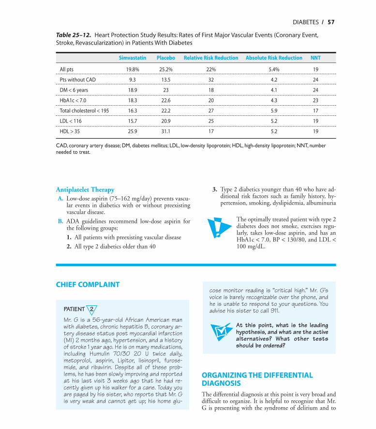
D. Data suggest that diabetic patients older than 40benefit from statin therapy regardless of baselineLDL (Table 25–12).
ON-LINE ONLY
Table 25–11. Types of Insulin
Type of Insulin Onset of Action Peak Effective Duration
Rapid acting
Lispro (Humalog) 5–15 min 30–90 min 5 hAspart (Novolog)
Short acting:
Regular U100 30–60 min 2–3 h 5–8 h
Intermediate acting
Isophane (NPH, Humulin N, Novolin N) 2–4 h 4–10 h 10–16 hInsulin zinc (Lente, Humulin L, Novolin L) 4–12 h 12–18 h
Long acting
Insulin zinc extended (Ultralente) 6–10 h 10–16 h 18–24 hGlargine (Lantus) 2–4 h to reach steady state No peak 20–24 h
Premixed
Humulin 70/30 (70% NPH/ 30% regular)Humalog Mix 75/25 (75% NPL [neutral protamine lispro, similar to NPH]/25% lispro)Novalog Mix 70/30 (70% NPH/30% aspart)
9310_Stern_C25 9/2/05 1:48 PM Page 56
DIABETES / 57
Antiplatelet TherapyA. Low-dose aspirin (75–162 mg/day) prevents vascu-
lar events in diabetics with or without preexistingvascular disease.
B. ADA guidelines recommend low-dose aspirin forthe following groups:
1. All patients with preexisting vascular disease
2. All type 2 diabetics older than 40
3. Type 2 diabetics younger than 40 who have ad-ditional risk factors such as family history, hy-pertension, smoking, dyslipidemia, albuminuria
The optimally treated patient with type 2diabetes does not smoke, exercises regu-larly, takes low-dose aspirin, and has anHbA1c < 7.0, BP < 130/80, and LDL <100 mg/dL.
ON-LINE ONLY
CHIEF COMPLAINT
PATIENT 2
Mr. G is a 56-year-old African American manwith diabetes, chronic hepatitis B, coronary ar-tery disease status post myocardial infarction(MI) 2 months ago, hypertension, and a historyof stroke 1 year ago. He is on many medications,including Humulin 70/30 20 U twice daily,metoprolol, aspirin, Lipitor, lisinopril, furose-mide, and ribavirin. Despite all of these prob-lems, he has been slowly improving and reportedat his last visit 3 weeks ago that he had re-cently given up his walker for a cane. Today youare paged by his sister, who reports that Mr. Gis very weak and cannot get up; his home glu-
cose monitor reading is “critical high.” Mr. G’svoice is barely recognizable over the phone, andhe is unable to respond to your questions. Youadvise his sister to call 911.
At this point, what is the leadinghypothesis, and what are the activealternatives? What other testsshould be ordered?
ORGANIZING THE DIFFERENTIALDIAGNOSISThe differential diagnosis at this point is very broad anddifficult to organize. It is helpful to recognize that Mr.G is presenting with the syndrome of delirium and to
Table 25–12. Heart Protection Study Results: Rates of First Major Vascular Events (Coronary Event,Stroke, Revascularization) in Patients With Diabetes
Simvastatin Placebo Relative Risk Reduction Absolute Risk Reduction NNT
All pts 19.8% 25.2% 22% 5.4% 19
Pts without CAD 9.3 13.5 32 4.2 24
DM < 6 years 18.9 23 18 4.1 24
HbA1c < 7.0 18.3 22.6 20 4.3 23
Total cholesterol < 195 16.3 22.2 27 5.9 17
LDL < 116 15.7 20.9 25 5.2 19
HDL > 35 25.9 31.1 17 5.2 19
CAD, coronary artery disease; DM, diabetes mellitus; LDL, low-density lipoprotein; HDL, high-density lipoprotein; NNT, numberneeded to treat.
9310_Stern_C25 9/2/05 1:48 PM Page 57
58 / CHAPTER 25
use the framework for delirium to organize your think-ing (see Delirium chapter). It is also reasonable to con-sider Mr. G’s underlying chronic medical problems andinitially focus on the serious complications of these con-ditions; in other words, initially focus on diseases forwhich he has a high pretest probability:
1. Diabetes: DKA, hyperosmolar hyperglycemicstate (HHS), infection with or without sepsis.
2. Coronary artery disease (CAD): recurrent MI,possibly with CHF or cardiogenic shock
3. Cerebrovascular disease: recurrent stroke4. Chronic hepatitis B: hepatic encephalopathy
2Mr. G could have any of these conditions ormore than 1 of them. His critical high bloodsugar makes a complication of diabetes theleading hypothesis; all of the other diagnosesare “can’t miss” hypotheses (Table 25–13).When Mr. G arrives in the emergency room, he isbarely responsive but able to move all 4 extrem-ities. His BP is 85/50; pulse, 120; RR, 24; tem-perature, 99 °F. His lungs are clear, and cardiacexam shows an S4 with no S3 or murmurs. Hisabdomen is nontender, and there is no peripheraledema. Initial lab tests include the following:
Sodium, 138; K, 4.9; Cl, 88; HCO3, 37; bloodurea nitrogen, 99; creatinine, 4.3; glucose,1246Arterial blood gases: pH 7.40; PO2, 88;PCO2, 35
ON-LINE ONLY
Table 25–13. Diagnostic Hypotheses for Mr. G
Diagnostic Hypotheses Clinical Clues Important Tests
Leading hypothesis
Hyperosmolar hyperglycemic Delirium/coma, polyuria, polydipsia, Plasma glucose, serum/urine ketonesstate (HHS) dehydration
Active alternatives: can’t miss
Diabetic ketoacidosis Delirium/coma, polyuria, polydipsia, Blood glucose/ bicarbonate, serum/urine dehydration ketones, pH
Sepsis Hypotension, fever Blood cultures, u/a, CXR
Myocardial infarction Chest pain, dyspnea ECG, cardiac enzymes
Cerebrovascular accident Hemiparesis, aphasia Physical exam, head CT
Hepatic encephalopathy Delirium, liver disease Clinical diagnosis
WBC is 8400, with 75 polymorphonuclearneutrophils, 3 bands, 18 lymphocytes, and4 monocytes.Albumin, 4.4; total bilirubin, 0.3; alkalinephosphatase, 175; AST (SGOT), 40; ALT(SGPT), 56; international normalized ratio(INR), 1.1.Serum ketones, negative
Corrected Na (sodium) = measured Na + 1.6 × glucose − 100100
= 138 + 1.6(11)= 155
Urinalysis: 2+ protein, 4+ glucose, no ke-tones, 3–5 WBC/high-power field, occa-sional bacteria
Is the clinical information sufficientto make a diagnosis? If not, whatother information do you need?
Leading Hypothesis: HyperosmolarHyperglycemic State
Textbook PresentationPatients with HHS are usually older type 2 diabeticswho present with the gradual onset of polydipsia,polyuria, and lethargy. They are extremely dehydratedand have very high serum glucose levels.
Disease HighlightsA. Epidemiology
9310_Stern_C25 9/2/05 1:48 PM Page 58
DIABETES / 59
1. Incidence is 1/1000 person-years (DKA inci-dence is 4.6–8.0/1000 person years).
2. Mortality rate about 15%3. Risk factors include older age, nursing home res-
idence, inability to recognize thirst, and lack ofaccess to fluids.
B. Pathogenesis1. A reduction in the effective action of circulating
insulin and a concomitant increase in counter-regulatory hormones leads to increased hepaticand renal glucose production and impaired glu-cose utilization in peripheral tissues.
2. Glycosuria leads to an osmotic diuresis with lossof free water in excess of electrolytes, leading tohyperosmolarity.
3. As volume depletion occurs, urine output drops,and hyperglycemia worsens.
4. The absence of ketoacidosis in HHS is notcompletely understood; possible explanationsare as follows:a. There are higher intraportal insulin levels
than seen in DKA, sufficient to prevent lipol-ysis.
b. The levels of counterregulatory hormonesare lower than in DKA.
c. The hyperosmolar state inhibits lipolysis.C. Precipitating factors
1. The 3 most common precipitants are infection,lack of compliance with insulin, and first pres-entation of diabetes.
2. Other precipitants include postoperative state,cerebrovascular accident (CVA), MI, pancreati-tis, alcohol abuse, trauma, thyrotoxicosis, andmedications (eg, corticosteroids, total parenteralnutrition).
D. Clinical manifestations1. History
a. Symptoms and signs usually evolve over sev-eral days or even weeks.
b. Common findings include polyuria, poly-dipsia, fatigue, and weight loss.
c. Abdominal pain generally does not occur inHHS, as it does in DKA, but there are re-ports of a hypertonicity-induced gastropare-sis leading to abdominal pain, distention,nausea, and vomiting.
d. Neurologic manifestations(1) Lethargy and disorientation common(2) Focal neurologic findings, including
seizures, can occur with hyperglycemia
and resolve with normalization of serumglucose.
(3) Changes in mental status correlate withthe degree of hyperosmolarity.(a) Twenty to 25% present with coma.(b) Coma present in half of patients
with effective serum osmolality of >350 mOsm/L
(c) Must search for another cause of comaif osmolality < 345–350 mOsm/L
2. Physical exama. Hypothermia often seen resulting from pe-
ripheral vasodilationb. Signs of dehydration (see Chapter 20) often
seenc. Tachycardia and hypotension suggest severe
dehydration or underlying sepsis.
Evidence-Based DiagnosisA. Typical total body water deficit is 20–25% (about 9
L).B. See Table 25–14 for laboratory findings in HHS
compared with DKA.
MAKING A DIAGNOSIS
2Mr. G’s glucose is > 600 mg/dL, ketones arenegative, and calculated serum osmolality is345 mOsm/L (effective serum osmolality = 2 ×measured Na + glucose/18). Mr. G’s osmolality= (2 × 138) + 1246/18 = 345.
Have you crossed a diagnosticthreshold for the leading hypothe-sis, hyperglycemic hyperosmolarsyndrome? Have you ruled out theactive alternatives? Do other testsneed to be done to exclude the al-ternative diagnoses?
Mr. G fulfills the diagnostic criteria for HHS. It is notnecessary to consider other diagnoses, but it is essentialto determine the precipitant for this event. ConsideringMr. G’s complicated history, he is at risk for many of theprecipitants of HHS, especially infection, MI, and CVA.
Always look for the precipitant when pa-tients present with either HHS or DKA.
ON-LINE ONLY
9310_Stern_C25 9/2/05 1:49 PM Page 59
60 / CHAPTER 25
CASE RESOLUTION
2Mr. G’s chest x-ray film is clear, his urine andblood cultures are negative, his ECG shows noacute changes, and his cardiac enzymes arenormal. He responds well to IV hydration and in-
sulin therapy. When he becomes more alert, hereports that he had become depressed and hadstopped taking his insulin.
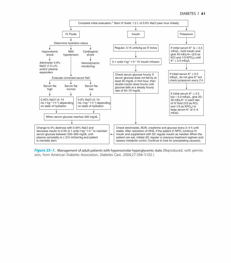
Treatment of HHSA. Patients with HHS generally need more fluid and
less insulin than those with DKAB. See Figure 25–1 for a treatment algorithm.
ON-LINE ONLY
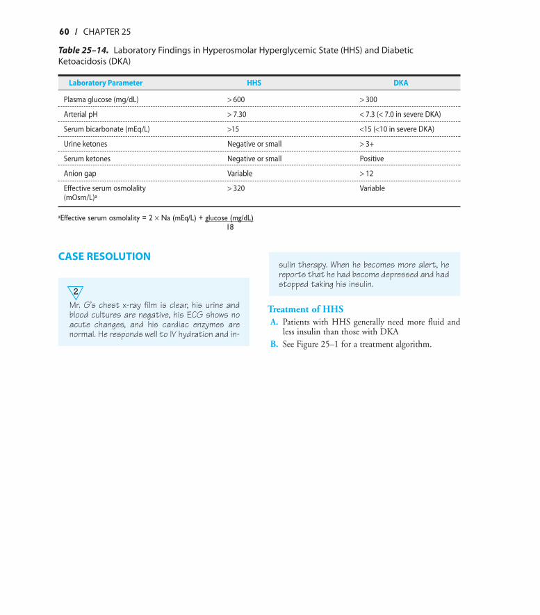
Table 25–14. Laboratory Findings in Hyperosmolar Hyperglycemic State (HHS) and DiabeticKetoacidosis (DKA)
Laboratory Parameter HHS DKA
Plasma glucose (mg/dL) > 600 > 300
Arterial pH > 7.30 < 7.3 (< 7.0 in severe DKA)
Serum bicarbonate (mEq/L) >15 <15 (<10 in severe DKA)
Urine ketones Negative or small > 3+
Serum ketones Negative or small Positive
Anion gap Variable > 12
Effective serum osmolality > 320 Variable(mOsm/L)a
aEffective serum osmolality = 2 × Na (mEq/L) + glucose (mg/dL)18
9310_Stern_C25 9/2/05 1:49 PM Page 60
ON-LINE ONLY
DIABETES / 61
IV Fluids Insulin Potassium
Determine hydration status
Regular, 0.15 units/kg as IV bolus If initial serum K+ is < 3.3mEq/L, hold insulin andgive 40 mEq K+ (2/3 asKCI and 1/3 KPO4) untilK+ ≥ 3.3 mEq/L
Hypovolemicshock
Mildhypotension
Cardiogenicshock
Administer 0.9%NaCI (1.0 L/h)and/or plasmaexpanders
Hemodynamicmonitoring
0.1 units • kg-1 • h-1 IV insulin infusion
Check serum glucose hourly. Ifserum glucose does not fall by atleast 50 mg/dL in first hour, thendouble insulin dose hourly untilglucose falls at a steady hourlyrate of 50–70 mg/dL.
If initail serum K+ ≥ 5.0mEq/L, do not give K+ butcheck potassium every 2 h
If initial serum K+ ≥ 3.3but < 5.0 mEq/L, give 20–30 mEq K+ in each literof IV fluid (2/3 as KCLand 1/3 as KPO4) tokeep serum K+ at 4–5mEq/L
Evaluate corrected serum Na‡
Serum Nahigh
Serum Nanormal
Serum Nalow
0.45% NaCl (4–14mL • kg-1 • h-1) dependingon state of hydration
0.9% NaCl (4–14mL • kg-1 • h-1) dependingon state of hydration
When serum glucose reaches 300 mg/dL
Change to 5% dextrose with 0.45% NaCl anddecrease insulin to 0.05–0.1 units • kg-1 • h-1 to maintainserum glucose between 250–300 mg/dL untilplasma osmolality is ≤ 315 mOsm/kg and patientis mentally alert.
Check electrolytes, BUN, creatinine and glucose every 2–4 h untilstable. After resolution of HHS, if the patient in NPO, continue IVinsulin and supplement with SC regular insulin as needed. When thepatient can eat, initiate SC regular or previous treatment regimen andassess metabolic contol. Continue to look for precipitating cause(s).
Complete initial evaluation.† Start IV fluids: 1.2 L of 0.9% NaCI peer hour initially.
Figure 25–1. Management of adult patients with hyperosmolar hyperglycemic state. (Reproduced, with permis-sion, from American Diabetes Association, Diabetes Care. 2004;27:S94–S102.)
9310_Stern_C25 9/2/05 1:49 PM Page 61