Embed Size (px)
Citation preview
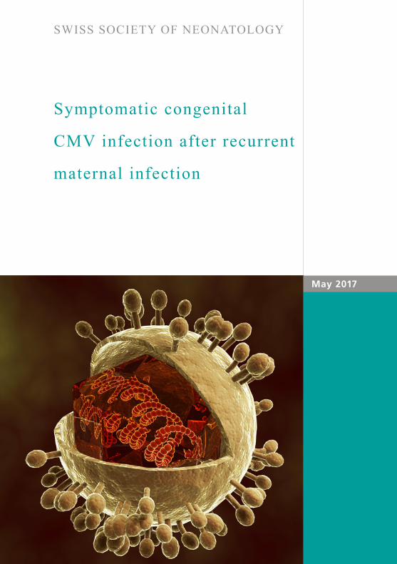
May 2017
Symptomatic congenital
CMV infection after recurrent
maternal infection
SWISS SOCIETY OF NEONATOLOGY
Mack I, Burckhardt MA, Heininger U, Prüfer F, Wellmann
S, Department of Pediatric Infectious Diseases (MI, HU),
Department of Pediatric Radiology (PF), and Department
of Neonatology (WS), University of Basel Children’s Hos-
pital (UKBB), Basel, Switzerland, Department of Pediatric
Endocrinology (BMA), Princess Margaret Hospital, Perth,
Australia
Title figure:
Cytomegalovirus (Source: www.nature.com)
© Swiss Society of Neonatology, Thomas M Berger, Webmaster
Cytomegalovirus (CMV) is probably the most com-
mon congenital viral infection; approximately 10% of
congenitally infected neonates display symptoms at
birth. In the past, symptomatic congenital CMV infec-
tion was thought to occur almost exclusively after
primary infection of the mother during pregnancy,
whereas preexisting maternal CMV immunity was
thought to protect the unborn child from infection in
the case of maternal re-infection. However, this has
been proven to be incorrect by cases of symptomatic
congenital CMV infection in infants of mothers who
had been seropositive before pregnancy. Treatment of
symptomatic infants with intravenous ganciclovir can
decrease the risk of hearing loss, the most common
neurological sequela, and reduce the risk for neuro-
developmental delay in infancy. However, strategies
regarding the route and duration of treatment differ
widely due to a lack of randomized controlled trials.
BACKGROUND
3

CASE REPORT
4
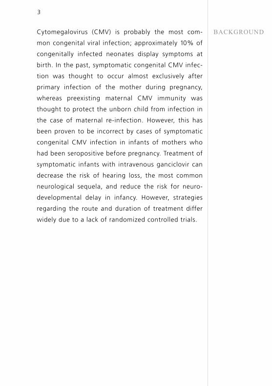
We present the case of a term baby girl born to a
healthy 38-year-old G2/P2 at 40 2/7 weeks of gesta-
tion. Pregnancy had been uneventful until 26 weeks of
gestation, when fetal ultrasound examination revealed
bilateral subependymal cysts. This finding was con-
firmed by magnetic resonance imaging (MRI), which
in addition showed slightly dilated lateral ventricles
(Fig. 1 A). At 36 weeks of gestation, another fetal MRI
examination demonstrated focal white matter lesi-
ons (Fig. 1 B). The mother had been CMV-IgG positive
prior to this pregnancy, but CMV-PCR from amniotic
fluid was positive. Therefore, maternal recurrent CMV
infection during pregnancy was suspected.
The girl adapted well with Apgar scores of 8, 8 and
10 at 1, 5 and 10 minutes, respectively. The umbilical
cord pH values were 7.20 (arterial) and 7.34 (venous).
Birth weight was 3140 g (P19), length was 50 cm (P29),
and head circumference was 33 cm (P8). On physi-
cal examination, extensive petechiae, hepatospleno-
megaly and jaundice were noted.
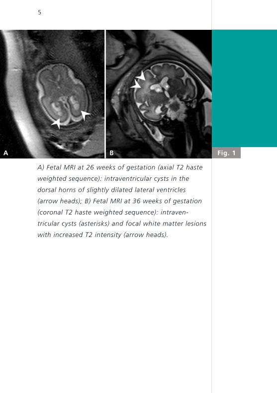
Congenital CMV infection was confirmed by PCR in
infant blood and urine samples, and therapy with
intravenous ganciclovir was initiated. Postnatal MRI
on day of life 5 revealed persistence of the abnor-
malities documented on fetal MRIs; however, neither
calcifications nor polymicrogyria were seen (Fig. 2 A,
B). Severe thrombocytopenia was treated with mul-
tiple platelet transfusions (Fig. 3). Profound hepato-
* *
A B
5
A) Fetal MRI at 26 weeks of gestation (axial T2 haste
weighted sequence): intraventricular cysts in the
dorsal horns of slightly dilated lateral ventricles
(arrow heads); B) Fetal MRI at 36 weeks of gestation
(coronal T2 haste weighted sequence): intraven
tricular cysts (asterisks) and focal white matter lesions
with increased T2 intensity (arrow heads).
Fig. 1
*
* *
Fig. 2 A B
6
Postnatal MRI on day of life 5: A) Axial and B) coro
nal T2weighted sequences demonstrating cysts in
the area of former germinal matrix (asterisks), slight
dilatation of the lateral ventricles and frontal,
as well as occipital white matter hyperintensities
(arrow heads). Neither calcifications nor polymicro
gyria can be seen.
7
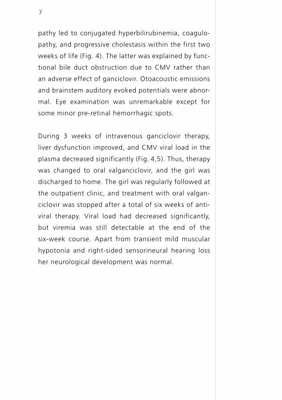
pathy led to conjugated hyperbilirubinemia, coagulo-
pathy, and progressive cholestasis within the first two
weeks of life (Fig. 4). The latter was explained by func-
tional bile duct obstruction due to CMV rather than
an adverse effect of ganciclovir. Otoacoustic emissions
and brainstem auditory evoked potentials were abnor-
mal. Eye examination was unremarkable except for
some minor pre-retinal hemorrhagic spots.
During 3 weeks of intravenous ganciclovir therapy,
liver dysfunction improved, and CMV viral load in the
plasma decreased significantly (Fig. 4,5). Thus, therapy
was changed to oral valganciclovir, and the girl was
discharged to home. The girl was regularly followed at
the outpatient clinic, and treatment with oral valgan-
ciclovir was stopped after a total of six weeks of anti-
viral therapy. Viral load had decreased significantly,
but viremia was still detectable at the end of the
six-week course. Apart from transient mild muscular
hypotonia and right-sided sensorineural hearing loss
her neurological development was normal.
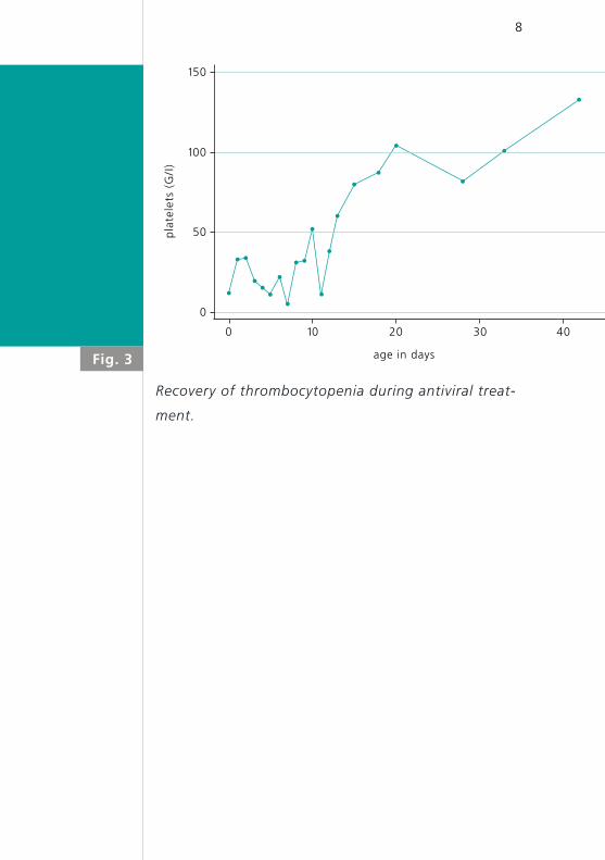
Fig. 3
8
Recovery of thrombocytopenia during antiviral treat
ment.
age in days
pla
tele
ts (
G/I
)
150
100
50
0 10 20 30 40
0

Fig. 4
9
Normalization of liver function tests during antiviral
treatment.
age in days
ASAT (U/L)
ALAT (U/L)
Bili total (μmol/L)
Bili direct (μmol/L)
800
400
600
200
0 10 20 30 40
0
Fig. 5
10
Course of CMV viral load during antiviral treatment.
age in days
CM
Vq
uan
t (G
Eq/m
l)
10 000
8 000
6 000
4 000
2 000
0 10 20 30 40
0

11
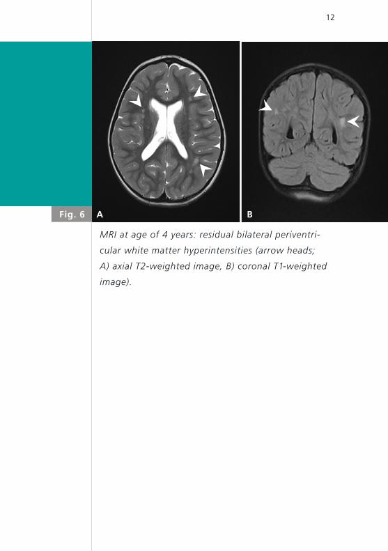
At the age of 4 years, she was seen in the emergency
department because of acute onset of mild ataxia.
Multiple laboratory examinations, including cerebro-
spinal fluid analysis and blood tests for viral or bacte-
rial infections were negative. Cranial MRI showed
regressive cerebral abnormalities with bilateral peri-
ventricular white matter lesions due to demyelination
and gliosis, and intraventricular occipital adhesions
(Fig. 6 A,B) but no signs of hemorrhage, infection
or tumor. When mild signs and symptoms of upper
respiratory tract infection developed, the neurological
symptoms were felt to be parainfectious in origin
rather than being related to her congenital CMV infec-
tion. She was last seen 6 months later: her overall
neurodevelopment continued to be normal but severe
right-sided hearing loss persisted (hearing threshold
level > 90 dB).
A B
12
Fig. 6
MRI at age of 4 years: residual bilateral periventri
cular white matter hyperintensities (arrow heads;
A) axial T2weighted image, B) coronal T1weighted
image).
13
DISCUSSIONFetal infection risk is highest with maternal primary
CMV infection and less likely with recurrent infection
due to the effect of maternal immunity. However, the
observation of highest birth prevalence rates of con-
genital CMV infection in populations with high mater-
nal seroimmunity indicates that CMV re-infections
play an important role (1 – 4). It is unknown whether
CMV reinfections in pregnancy are due to reactiva-
tion or infection with a different strain of CMV. Bop-
pana et al. determined strain-specific IgG and clearly
demonstrated that two-thirds of congenital infections
in sero positive women were caused by exogenous re-
infections (5). In contrast, reactivation as a route for
symptomatic congenital infection has rarely been pro-
ven with molecular evidence (6).
Congenital CMV infection is the leading non-genetic
cause of sensorineural hearing loss in early childhood,
accounting for 21% of children with hearing loss at
birth and 24% of those with hearing loss at 4 years
of age (1). More sensitive screening methods for CMV
have been developed in recent years (7), but treatment
options for congenital CMV infections remain limited.
The current literature suggests that a six-week-course
of ganciclovir, especially when started during the neo-
natal period, is effective in terms of decreasing the
severity of neurological dysfunction and hearing loss
in symptomatic and asymptomatic infants (8 –10). Oral
valganciclovir is more easily administered to infants
14
with congenital CMV-infection and results in similar
plasma concentrations as with intravenous ganciclovir
(11 – 13).
It was suggested that the benefit of a 6-week-course
of ganciclovir therapy could wane over the first years
of life (1). To address this issue, a randomized, pla-
cebo-controlled trial in neonates with symptoma-
tic congenital CMV disease was recently performed,
comparing 6 months with 6 weeks of valganciclovir
therapy. The results showed that long-term treatment
over 6 months had a moderately favorable effect on
long-term audiologic and neurodevelopmental out-
comes with no significant differences in the rate of
adverse events between the two study groups (1).
Whole blood viral loads decreased similarly in the two
study groups during the first 6 weeks of treatment and
then increased again in the 6-week-study group. Red-
uced viral loads correlated with better hearing outco-
mes at 6, 12, and 24 months among participants in the
6-month-group, whereas no such effect was observed
in the 6-week group (1).
15
Symptomatic congenital CMV infection may be caused
not only by re-infection but also by reactivation of
CMV in seropositive mothers. There is still little know-
ledge on the frequency of reactivation-mediated con-
genital infection and its triggers during pregnancy.
Although screening methods are advancing rapidly
have become widely available, there is still limited evi-
dence on treatment efficacy, particularly in cases with
less severe disease. Further studies are needed to inve-
stigate the optimal route and duration of administra-
tion of anti viral drugs and to evaluate the course of
hearing ability after cessation of antiviral treatment.
Recent studies suggest improved outcome after long-
term valganciclovir therapy.
CONCLUSION

1. Kimberlin DW, Jester PM, Sánchez PJ, et al. Valganciclovir for
symptomatic congenital cytomegalovirus disease. Engl J Med
2015;372:933 – 943 (Abstract)
2. De Vries JJ, van Zwet EW, Dekker FW, Kroes AC, Verkerk
PH, Vossen AC. The apparent paradox of maternal seropo-
sitivity as a risk factor for congenital cytomegalovirus infec-
tion: a population-based prediction model. Rev Med Virol
2013;23:241 – 249 (Abstract)
3. Wang C, Zhang X, Bialek S, Cannon MJ. Attribution of con-
genital cytomegalovirus infection to primary versus non-
primary maternal infection. Clin Infect Dis 2011;52:e11 – e13
(Abstract)
4. Boppana SB, Fowler KB, Britt WJ, Stagno S, Pass RF. Sympto-
matic congenital cytomegalovirus infection in infants born
to mothers with preexisting immunity to cytomegalovirus.
Pediatrics 1999;104:55 – 60 (Abstract)
5. Boppana SB, Rivera LB, Fowler KB, Mach M, Britt WJ.
Intrauterine transmission of cytomegalovirus to infants
of women with preconceptional immunity. N Engl J Med.
2001;344(18):1366 – 1371 (Abstract)
6. Nagamori T, Koyano S, Inoue N, et al. Single cytomegalovirus
strain associated with fetal loss and then congenital infection
of a subsequent child born to the same mother. J Clin Virol
2010;49:134 – 136 (Abstract)
7. Boppana SB, Ross SA, Shimamura M, et al. Saliva polymerase-
chain-reaction assay for cytomegalovirus screening in new-
borns. N Engl J Med 2011;364:2111 – 2118 (Abstract)
8. Kimberlin DW, Lin CY, Sanchez PJ, et al. Effect of ganciclovir
therapy on hearing in symptomatic congenital cytomegalovirus
disease involving the central nervous system: a randomized,
controlled trial. J Pediatr 2003;143:16 – 25 (Abstract)
REFERENCES
16
9. Oliver SE, Cloud GA, Sanchez PJ, et al. Neurodevelopmental
outcomes following ganciclovir therapy in symptomatic con-
genital cytomegalovirus infections involving the central nervous
system. J Clin Virol 2009;46:S22 – S26 (Abstract)
10. Lackner A, Acham A, Alborno T, et al. Effect on hearing of
ganciclovir therapy for asymptomatic congenital cytome-
galovirus infection: four to 10-year follow-up. J Laryngol Otol
2009;123:391 – 396 (Abstract)
11. Imamura T, Suzutani T, Ogawa H, et al. Oral valganciclovir
treatment for congenital cytomegalovirus infection. Pediatr Int
53:249 – 252 (no abstract available)
12. Schulzke S, Bührer C. Valganciclovir for treatment of congeni-
tal cytomegalovirus infection. Eur J Pediatr 2006;165:575 – 576
(no abstract available)
13. Acosta EP, Brundage RC, King JR, et al. Ganciclovir population
pharmacokinetics in neonates following intravenous admini-
stration of ganciclovir and oral administration of a liquid val-
ganciclovir formulation. Clin Pharmacol Ther 2007;81:867 – 872
(Abstract)

SUPPORTED BY
CONTACT
Swiss Society of Neonatology
www.neonet.ch
con
cep
t &
des
ign
by
mes
ch.c
h


![Congenital toxoplasmosis kbk-1.ppt [Read-Only]ocw.usu.ac.id/.../tmd175_slide_congenital_toxoplasmosis.pdf · (symptomatic congenital toxoplasmosis infection) PyrimethaminePyrimethamine1](https://img.pdfslide.net/doc/110x75/5e11e77d573e9002e5752212/congenital-toxoplasmosis-kbk-1ppt-read-onlyocwusuacidtmd175slidecongenital.jpg)