Embed Size (px)
Citation preview

Proc. Natl. Acad. Sci. USAVol. 83, pp. 3500-3504, May 1986Medical Sciences
Synaptophysin: A marker protein for neuroendocrine cellsand neoplasms
(synaptic vesicles/neurosecretory granules/pancreatic islets/carcinolds/tumor diagnosis)
BERTRAM WIEDENMANN*t, WERNER W. FRANKE*, CAECILIA KUHN*, ROLAND MOLL*,AND VICTOR E. GOULD§*Division of Membrane Biology and Biochemistry, Institute of Cell and Tumor Biology, German Cancer Research Center, D-6900 Heidelberg, FederalRepublic of Germany; tDepartment of Medicine, University of Heidelberg, Heidelberg, Federal Republic of Germany; *Department of Pathology, Universityof Mainz, D-6500 Mainz, Federal Republic of Germany; and §Department of Pathology, Rush Medical College, Chicago, IL 60612
Communicated by Donald F. Steiner, January 27, 1986
ABSTRACT Synaptophysin is an integral membrane gly-coprotein (Mr 38,000) that occurs in presynaptic vesicles ofneurons and in similar vesicles of the adrenal medulla. By usinga monoclonal antibody to this protein (SY38), we have found,by immunohistochemistry and immunoblotting, that an iden-tical or similar protein is also expressed in neuroendocrinetumors of neural type, such as pheochromocytomas andparagangliomas. In addition, this protein occurs in certainneuroendocrine epithelial cells, such as pancreatic islet cells; ina variety of neuroendocrine epithelial tumors, including islet-cell adenomas and carcinomas and several carcinoids andneureendocrine carcinomas of the gastrointestinal and thebronchial tracts; and in medullary carcinomas of the thyroid.Our results show that synaptophysin, and the vesicles thatcontain it, can occur in normal and neoplastic neuroendocrinecells of neural type, as demonstrated by colocalization withneurofilaments, as well as in those of epithelial type, as shownby colocalization with cytokeratin filaments and desmoplakins.We conclude that synaptophysin is expressed independently ofother neuronal differentiation markers and propose that it beused as a differentiation marker in tumor diagnosis.
Neuroendocrine cells comprise a widely distributed, mor-phologically and embryologically heterogeneous group (1, 2)present not only in the central and peripheral nervoussystems but also in the gastrointestinal and bronchopulmon-ary tracts, the skin, the thyroid, and several other organs(3-5). The common property of these cells is the productionof neurotransmitter substances such as acetylcholine,biogenic neuroamines, and neuropeptides (3-5). Numerousbenign and malignant neoplasms either are entirely com-prised of cells with neuroendocrine differentiation or mayinclude a few such cells as part of complex populations (6).Normal, dysplastic, and neoplastic neuroendocrine cells maybe identified by certain electron microscopic features, suchas neurosecretory granules, or by the cytochemical demon-stration of certain NE products (3-5). Immunocytochemicalmarkers have proven especially important for the study ofneuroendocrine differentiation, notably neuron-specificenolase, which is widely used as a broad marker for normaland neoplastic neuroendocrine cells (7-9), although the ysubunit of neuron-specific enolase is also found in a numberof nonneuroendocrine cells (10). Neuropeptides can also bedemonstrated in normal and neoplastic neuroendocrine cellsand have proven valuable in tumor diagnosis (11, 12).However, as neuropeptides comprise a complex and variablegroup of substances, the application of a single peptide probeas a common marker of neuroendocrine differentiation isprecluded (for discussion, see ref. 13).
The specific complement of cytoskeletal intermediate-filament (IF) proteins of neuroendocrine cells is also variable.Although neurons and certain other neuroendocrine cellsexpress neurofilament proteins (NFP; refs. 14 and 15), manyother neuroendocrine cells are clearly epithelial as defined bytheir expression of cytokeratin IF and desmosomal proteins(12, 16, 17). The corresponding neuroendocrine neoplasmsexpress either NFP or cytokeratin filaments, respectively,but at least a few neuroendocrine neoplasms are capable ofcoexpressing both classes of IFs (13, 17-19).
Recently, an integral membrane glycoprotein (polypeptideMr 38,000) has been identified in presynaptic vesicles (20, 21)and termed synaptophysin (21). By use. of a monoclonalantibody, SY38, it has been shown (21) that this protein isspecifically located in neuronal vesicles with an electronmicroscopically clear content and also occurs in the adrenalmedulla of a number of mammalian species (see also ref. 20).Here we show that synaptophysin is present in a variety ofhuman neuroendocrine cells and neoplasms of both theneural and the epithelial type.
MATERIALS AND METHODSTissues. Table 1 lists the neuroendocrine neoplasms exam-
ined and the pertinent pathologic and clinical data. Forcomparison, metastatic melanomas and a number of carci-nomas of squamous and glandular differentiation lackingneuroendocrine features were studied. Tumor samples werefrozen immediately after surgical removal or within 1 hr afterdeath in the case of autopsy material (12, 21). Samples ofnormal human tissues were obtained from routine biopsies orautopsies. Various bovine tissues were similarly studied,
Light and Electron Microscopy. Samples, were fixed eitherin Bouin's solution or in 10% formalin, embedded in paraffin,and stained with hematoxylin and eosin as well as withvarious special stains. For immunohistochemistry on paraffinsections, tissue fixed in Bouin's solution was preferentiallyused (23). Immunostaining was performed by the peroxidase-antiperoxidase technique (24) or by the avidin-biotin tech-nique (25). Neuroendocrine substances examined are listed inTable 1; sources and dilutions of antibodies as well asmethods and details on positive and negative controls havebeen described (12, 23).Immunofluorescence microscopy on cryostat sections of
frozen tissues (21) used the following antibodies: (i) murinemonoclonal cytokeratin antibody PKK1 (ref. 26; fromLabsystems, Helsinki, Finland); (ii) monoclonal antibody K,18.18, specific for cytokeratin no. 18 (16); (iii) guinea pigantibodies to cytokeratins (27); (iv) guinea pig antibodies tomurine vimentin (28); (v) guinea pig antibodies against the
Abbreviations: IF, intermediate-sized filament; NFP, neurofilamentprotein(s).
3500
The publication costs of this article were defrayed in part by page chargepayment. This article must therefore be hereby marked "advertisement"in accordance with 18 U.S.C. §1734 solely to indicate this fact.
Dow
nloa
ded
by g
uest
on
May
19,
202
0
Proc. Natl. Acad. Sci. USA 83 (1986) 3501
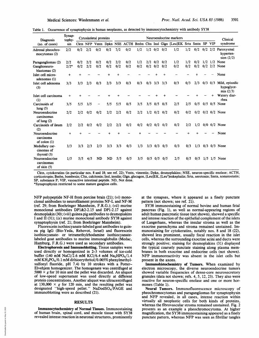
Table 1. Occurrence of synaptophysin in human neoplasms, as detected by immunocytochemistry with antibody SY38
Dgiyap- Cytoskeletal proteins Neuroendocrine markersDiagnosis tophy- Clinical
(no. of cases) sin Cktn NFP Vmtn Dpkn NSE ACTH Bmbn Cltn Insl Glgn [Leu]EK Srtn Smtn SP VIP syndromeAdrenal pheochro- 2/2 0/2 2/2 0/2 0/2 2/2 0/2 1/2 1/2 0/2 0/2 1/2 1/2 0/2 0/2 2/2 Paroxysmalmocytomas (2) hyperten-
sion (2/2)Paragangliomas (2) 2/2 0/2 2/2 0/2 0/2 2/2 0/2 1/2 2/2 0/2 0/2 1/2 1/2 0/2 1/2 1/2 NoneGanglioneuro- 2/2* 0/2 2/2 0/2 0/2 0/2 0/2 0/2 0/2 0/2 0/2 0/2 0/2 0/2 0/2 2/2 None
blastomas (2)Islet cell micro- + + - - + + - - - + + - - + - - Noneadenomas (1)
Islet cell adenoma 3/3 3/3 2/3 0/3 2/3 3/3 0/3 0/3 0/3 3/3 3/3 0/3 0/3 2/3 0/3 0/3 Mild, episodic(3) hypoglyce-
mia (2/3)Islet cell carcinoma + + - - + + - - - + + - - + - + Watery diar-
(1) rheaCarcinoids of 3/5 5/5 3/5 - 5/5 5/5 0/5 3/5 3/5 0/5 0/5 2/5 2/5 0/5 0/5 0/5 None
lung (5)Neuroendocrine 2/2 2/2 0/2 0/2 2/2 2/2 0/2 2/2 1/2 0/2 0/2 0/2 0/2 0/2 0/2 0/2 None
carcinomasof lung (2)
Carcinoids of ileum 2/2 2/2 0/2 0/2 2/2 2/2 0/2 0/2 0/2 0/2 0/2 0/2 2/2 1/2 0/0 0/2 None(2)
Neuroendocrine + + - - + + - + - - - - + + - - Nonecarcinomaof colon (1)
Medullary car- 3/3 3/3 2/3 2/3 3/3 3/3 0/3 1/3 3/3 0/3 0/3 0/3 0/3 1/3 0/3 0/3 Nonecinomas ofthyroid (3)
Neuroendocrine 1/5 5/5 4/5 ND ND 5/5 0/5 3/5 0/5 0/5 0/5 2/5 0/5 0/5 1/5 1/5 Nonecarcinomasof skin (5)Cktn, cytokeratins (in particular nos. 8 and 18; see ref. 22); Vmtn, vimentin; Dpkn, desmoplakins; NSE, neuron-specific enolase; ACTH,
corticotropin; Bmbn, bombesin; Cltn, calcitonin; Insl, insulin; Glgn, glucagon, [Leu]EK, [Leu5]enkephalin; Srtn, serotonin; Smtn, somatostatin;SP, substance P; VIP, vasoactive intestinal peptide. ND, Not done.*Synaptophysin restricted to some mature ganglion cells.
NFP polypeptide NF-H from porcine brain (21); (vi) mono-clonal antibodies to neurofilament proteins NF-L and NF-M(ref. 29; from Boehringer Mannheim, F.R.G.); (vii) murinemonoclonal antibodies DP1&2-2.15 and DP1-2.17 againstdesmoplakin (30); (viii) guinea pig antibodies to desmoplakinsI and II (31); (ix) murine monoclonal antibody SY38 againstsynaptophysin (ref. 21; from Boehringer Mannheim).
Fluorescein isothiocyanate-labeled goat antibodies to guin-ea pig IgG (Bio-Yeda, Rehovot, Israel) and fluoresceinisothiocyanate- or tetramethyirhodamine isothiocyanate-labeled goat antibodies to murine immunoglobulin (Medac,Hamburg, F.R.G.) were used as secondary antibodies.
Electrophoresis and Immunoblotting. Tissue samples wereused directly or homogenized in 2-3 volumes of ice-coldbuffer (140 mM NaCl/2.6 mM KCl/6.4 mM Na2HPO4/1.4mM KH2PO4/0.1mM dithioerythritol/0.005% phenylmethyl-sulfonyl fluoride, pH 7.4) by 10 strokes with a Potter-Elvehjem homogenizer. The homogenate was centrifuged at5000 x g for 10 min and the pellet was discarded. An aliquotof low-speed supernatant was used directly at differentprotein concentrations. Another aliquot was ultracentrifugedat 130,000 x g for 120 min, and the resulting pellet wasdesignated "high-speed pellet." NaDodSO4/PAGE andimmunoblotting were as described (21).
RESULTSImmunocytochemistry of Normal Tissues. Immunostaining
of human brain, spinal cord, and muscle tissue with SY38revealed intense reaction in neuronal structures, prominently
at the synapses, where it appeared as a finely punctatepattern (not shown; see ref. 21).SY38 immunostaining of normal bovine and human fetal
pancreas (Fig. 1), as well as normal-appearing regions ofadult human pancreatic tissue (not shown), showed a specificand intense reaction of the epithelial complement of the isletsof Langerhans, whereas the insular stroma as well as theexocrine parenchyma and stroma remained unstained. Im-munostaining for cytokeratins, notably nos. 8 and 18 (22),showed less prominent, usually focal reaction in the isletcells, whereas the surrounding exocrine acini and ducts werestrongly positive; staining for desmoplakins (31) displayedthe typical coarsely punctate staining along plasma mem-branes in both exocrine and endocrine cells (not shown).NFP immunoreactivity was absent in the islet cells butpresent in the axons.
Immunohistochemistry of Tumors. When examined byelectron microscopy, the diverse neuroendocrine tumorsshowed variable frequencies of dense-core neurosecretorygranules (data not shown; refs. 4, 5, 12, 23). They also werereactive for neuron-specific enolase and one or more hor-mones (Table 1).Neural Tumors. Immunofluorescence microscopy of
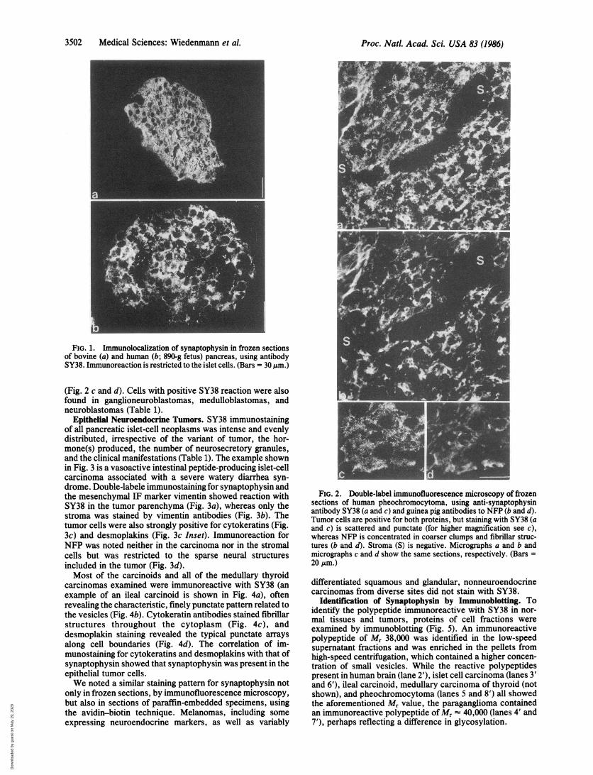
pheochromocytomas and paragangliomas for synaptophysinand NFP revealed, in all cases, intense reaction withinvirtually all neoplastic cells for both kinds of proteins,whereas the fibrovascular stroma remained unstained. Fig. 2presents as an example a pheochromocytoma. At highermagnification, the SY38 immunostaining appeared as a finelypunctate pattern, whereas NFP was seen as fibrillar tangles
Medical Sciences: Wiedenmann et al.
Dow
nloa
ded
by g
uest
on
May
19,
202
0
3502 Medical Sciences: Wiedenmann et al.
FIG. 1. Immunolocalization of synaptophysin in frozen sectionsof bovine (a) and human (b; 890-g fetus) pancreas, using antibodySY38. Immunoreaction is restricted to the islet cells. (Bars = 30 ,gm.)
(Fig. 2 c and d). Cells with positive SY38 reaction were alsofound in ganglioneuroblastomas, medulloblastomas, andneuroblastomas (Table 1).
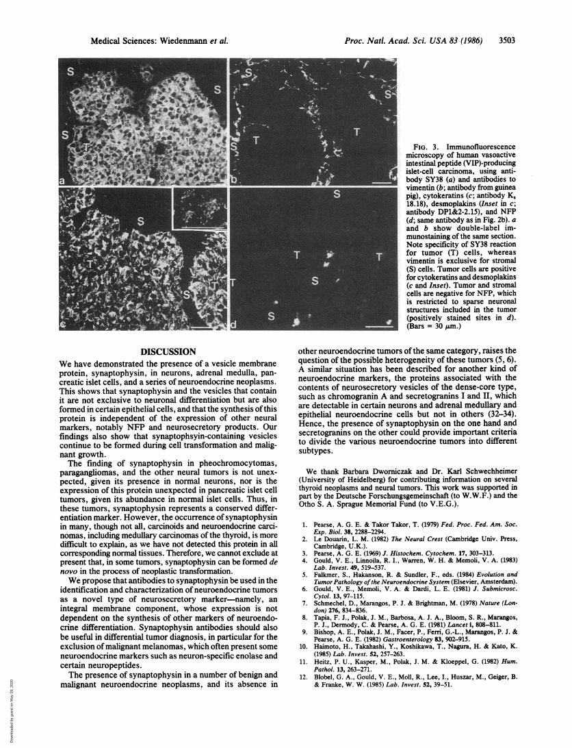
Epithelial Neuroendocrine Tumors. SY38 immunostainingof all pancreatic islet-cell neoplasms was intense and evenlydistributed, irrespective of the variant of tumor, the hor-mone(s) produced, the number of neurosecretory granules,and the clinical manifestations (Table 1). The example shownin Fig. 3 is a vasoactive intestinal peptide-producing islet-cellcarcinoma associated with a severe watery diarrhea syn-drome. Double-labele immunostaining for synaptophysin andthe mesenchymal IF marker vimentin showed reaction withSY38 in the tumor parenchyma (Fig. 3a), whereas only thestroma was stained by vimentin antibodies (Fig. 3b). Thetumor cells were also strongly positive for cytokeratins (Fig.3c) and desmoplakins (Fig. 3c Inset). Immunoreaction forNFP was noted neither in the carcinoma nor in the stromalcells but was restricted to the sparse neural structuresincluded in the tumor (Fig. 3d).Most of the carcinoids and all of the medullary thyroid
carcinomas examined were immunoreactive with SY38 (anexample of an ileal carcinoid is shown in Fig. 4a), oftenrevealing the characteristic, finely punctate pattern related tothe vesicles (Fig. 4b). Cytokeratin antibodies stained fibrillarstructures throughout the cytoplasm (Fig. 4c), anddesmoplakin staining revealed the typical punctate arraysalong cell boundaries (Fig. 4d). The correlation of im-munostaining for cytokeratins and desmoplakins with that ofsynaptophysin showed that synaptophysin was present in theepithelial tumor cells.We noted a similar staining pattern for synaptophysin not
only in frozen sections, by immunofluorescence microscopy,but also in sections of paraffin-embedded specimens, usingthe avidin-biotin technique. Melanomas, including someexpressing neuroendocrine markers, as well as variably
FIG. 2. Double-label immunofluorescence microscopy of frozensections of human pheochromocytoma, using anti-synaptophysinantibody SY38 (a and c) and guinea pig antibodies to NFP (b and d).Tumor cells are positive for both proteins, but staining with SY38 (aand c) is scattered and punctate (for higher magnification see c),whereas NFP is concentrated in coarser clumps and fibrillar struc-tures (b and d). Stroma (S) is negative. Micrographs a and b andmicrographs c and d show the same sections, respectively. (Bars =20 /Am.)
differentiated squamous and glandular, nonneuroendocrinecarcinomas from diverse sites did not stain with SY38.
Identification of Synaptophysin by Immunoblotting. Toidentify the polypeptide immunoreactive with SY38 in nor-mal tissues and tumors, proteins of cell fractions wereexamined by immunoblotting (Fig. 5). An immunoreactivepolypeptide of Mr 38,000 was identified in the low-speedsupernatant fractions and was enriched in the pellets fromhigh-speed centrifugation, which contained a higher concen-tration of small vesicles. While the reactive polypeptidespresent in human brain (lane 2'), islet cell carcinoma (lanes 3'and 6'), ileal carcinoid, medullary carcinoma of thyroid (notshown), and pheochromocytoma (lanes 5 and 8') all showedthe aforementioned Mr value, the paraganglioma containedan immunoreactive polypeptide of Mr 40,000 (lanes 4' and7'), perhaps reflecting a difference in glycosylation.
Proc. Natl. Acad. Sci. USA 83 (1986)
Dow
nloa
ded
by g
uest
on
May
19,
202
0
Proc. NatL. Acad. Sci. USA 83 (1986) 3503
FIG. 3. Immunofluorescencemicroscopy of human vasoactiveintestinal peptide (VIP)-producingislet-cell carcinoma, using anti-body SY38 (a) and antibodies tovimentin (b; antibody from guineapig), cytokeratins (c; antibody K,18.18), desmoplakins (Inset in c;antibody DP1&2-2.15), and NFP(d; same antibody as in Fig. 2b). aand b show double-label im-munostaining of the same section.Note specificity of SY38 reactionfor tumor (T) cells, whereasvimentin is exclusive for stromal(S) cells. Tumor cells are positivefor cytokeratins and desmoplakins(c and Inset). Tumor and stromalcells are negative for NFP, whichis restricted to sparse neuronalstructures included in the tumor(positively stained sites in d).(Bars = 30 gm.)
DISCUSSIONWe have demonstrated the presence of a vesicle membraneprotein, synaptophysin, in neurons, adrenal medulla, pan-creatic islet cells, and a series of neuroendocrine neoplasms.This shows that synaptophysin and the vesicles that containit are not exclusive to neuronal differentiation but are alsoformed in certain epithelial cells, and that the synthesis of thisprotein is independent of the expression of other neuralmarkers, notably NFP and neurosecretory products. Ourfindings also show that synaptophsyin-containing vesiclescontinue to be formed during cell transformation and malig-nant growth.The finding of synaptophysin in pheochromocytomas,
paragangliomas, and the other neural tumors is not unex-pected, given its presence in normal neurons, nor is theexpression of this protein unexpected in pancreatic islet celltumors, given its abundance in normal islet cells. Thus, inthese tumors, synaptophysin represents a conserved differ-entiation marker. However, the occurrence of synaptophysinin many, though not all, carcinoids and neuroendocrine carci-nomas, including medullary carcinomas of the thyroid, is moredifficult to explain, as we have not detected this protein in allcorresponding normal tissues. Therefore, we cannot exclude atpresent that, in some tumors, synaptophysin can be formed denovo in the process of neoplastic transformation.We propose that antibodies to synaptophysin be used in the
identification and characterization of neuroendocrine tumorsas a novel type of neurosecretory marker-namely, an
integral membrane component, whose expression is notdependent on the synthesis of other markers of neuroendo-crine differentiation. Synaptophysin antibodies should alsobe useful in differential tumor diagnosis, in particular for theexclusion of malignant melanomas, which often present someneuroendocrine markers such as neuron-specific enolase andcertain neuropeptides.The presence of synaptophysin in a number of benign and
malignant neuroendocrine neoplasms, and its absence in
other neuroendocrine tumors of the same category, raises thequestion of the possible heterogeneity of these tumors (5, 6).A similar situation has been described for another kind ofneuroendocrine markers, the proteins associated with thecontents of neurosecretory vesicles of the dense-core type,such as chromogranin A and secretogranins I and II, whichare detectable in certain neurons and adrenal medullary andepithelial neuroendocrine cells but not in others (32-34).Hence, the presence of synaptophysin on the one hand andsecretogranins on the other could provide important criteriato divide the various neuroendocrine tumors into differentsubtypes.
We thank Barbara Dworniczak and Dr. Karl Schwechheimer(University of Heidelberg) for contributing information on severalthyroid neoplasms and neural tumors. This work was supported inpart by the Deutsche Forschungsgemeinschaft (to W.W.F.) and theOtho S. A. Sprague Memorial Fund (to V.E.G.).
1. Pearse, A. G. E. & Takor Takor, T. (1979) Fed. Proc. Fed. Am. Soc.Exp. Biol. 38, 2288-2294.
2. Le Douarin, L. M. (1982) The Neural Crest (Cambridge Univ. Press,Cambridge, U.K.).
3. Pearse, A. G. E. (1969) J. Histochem. Cytochem. 17, 303-313.4. Gould, V. E., Linnoila, R. I., Warren, W. H. & Memoli, V. A. (1983)
Lab. Invest. 49, 519-537.5. Falkmer, S., Hakanson, R. & Sundler, F., eds. (1984) Evolution and
Tumor Pathology ofthe Neuroendocrine System (Elsevier, Amsterdam).6. Gould, V. E., Memoli, V. A. & Dardi, L. E. (1981) J. Submicrosc.
Cytol. 13, 97-115.7. Schmechel, D., Marangos, P. J. & Brightman, M. (1978) Nature (Lon-
don) 276, 834-836.8. Tapia, F. J., Polak, J. M., Barbosa, A. J. A., Bloom, S. R., Marangos,
P. J., Dermody, C. & Pearse, A. G. E. (1981) Lancet i, 808-811.9. Bishop, A. E., Polak, J. M., Facer, P., Fern, G.-L., Marangos, P. J. &
Pearse, A. G. E. (1982) Gastroenterology 83, 902-915.10. Haimoto, H., Takahashi, Y., Koshikawa, T., Nagura, H. & Kato, K.
(1985) Lab. Invest. 52, 257-263.11. Heitz, P. U., Kasper, M., Polak, J. M. & Kloeppel, G. (1982) Hum.
Pathol. 13, 263-271.12. Blobel, G. A., Gould, V. E., Moll, R., Lee, I., Huszar, M., Geiger, B.
& Franke, W. W. (1985) Lab. Invest. 52, 39-51.
Medical Sciences: Wiedenmann et al.
Dow
nloa
ded
by g
uest
on
May
19,
202
0

3504 Medical Sciences: Wiedenmann et al.
1' 2' 3 4' 5' 61 7' 8'
FiG. 5. Immunoblot analysis of polypeptides of subcellularfractions from normal (rat and bovine) brain and various neuroen-docrine tumors, using monoclonal antibody against synaptophysin(SY38). Polypeptides separated by parallel NaDodSO4/12.5% PAGEeither were stained with Coomassie blue (a, lanes 1-8) or weretransferred to nitrocellulose paper and treated with antibody SY38and then with 12-I-labeled goat anti-mouse IgG antibodies (b, lanes1'-8', autoradiograph). Both "low-speed supernatants" (lanes 1-5)and "high-speed pellets" (lanes 6-8), which were enriched in smallvesicles, were examined. Tissues used were from rat (lanes 1 and 1')and human (lanes 2 and 2') brain or from human islet-cell carcinoma(lanes 3 and 3' and lanes 5 and 5') pargnloma(anes 4 and 4' andlanes 7 and 7') and pheocbromocytoma (lanes 5 and 5' and lanes 8and 8') Small vertical bars in b denote, from top to bottom, positionof reference proteins analyzed in parallel lanes (Mr 180,000, 96,000,50,000, and 33,000; ref. 21). Arrowheads in a denote the position ofbrain synaptophysin (Mr 38,000). Note that an SY38-reactive poly-peptide band is seen at a similar position in the tumor samples, withthe exception of the paraganglioma samples, in which the reactiveband shows a slightly lower electrophoretic mobility (arrows).
FIG. 4. Immunofluorescence microscopy on frozen sections ofhuman carcinoid of ileum with antibodies against synaptophysin (aand b; SY38), cytokeratins (c; K, 18.18), and desmoplakins (d;DP1&2-2.15). All antibodies stain the tumor but not the stromal (S)cells. Note the finely punctate and dispersed cytoplasmic stainingwith SY38 (b). In contrast, the desmoplakin staining (d) shows acoarser punctate pattern along cell borders. (Bars = 20 gm.)
13. Gould, V. E., Moll, R., Moll, I., Lee, I. & Franke, W. W. (1985) Lab.Invest. 52, 334-353.
14. Osborn, M. & Weber, K. (1983) Lab. Invest. 48, 372-394.15. Miettinen, M., Lehto, V.-P. & Virtanen, I. (1985) Am. J. Pathol. 118,
360-361.16. Moll, R., Moll, I. & Franke, W. W. (1984) Differentiation 28, 136-154.
17. Miettinen, M., Lehto, V.-P., Dahl, D. & Virtanen, I. (1985) Lab. Invest.52, 429-436.
18. Broers, J. L. V., Carney, D. N., de Ley, L., Vooijs, G. P. & Ramaek-ers, F. C. S. (1985) Proc. Natl. Acad. Sci. USA 82, 4409-4413.
19. Lehto, V.-P., Miettinen, M. & Virtanen, I. (1985) let. J. Cancer 35,421-425.
20. Jahn, B., Schiebler, W., Ouimet, C. & Greengard, P. (1985) Proc. Natl.Acad. Sci. USA 82, 4137-4141.
21. Wiedenmann, B. & Franke, W. W. (1985) Cell 45, 1017-1028.22. Moll, R., Franke, W. W., Schiller, D. L., Geiger, B. & Krepler, R.
(1982) Cell 31, 11-24.23. Warren, W. H., Memoli, V. A. & Gould, V. E. (1984) Ultrastruct.
Pathol. 6, 15-27.24. Sternberger, L. A. (1979) Immunocytochemistry, 2nd Ed. (Wiley, New
York).25. Hsu, S.-M., Raine, L. & Fanger, H. (1981) J. Histochem. Cytochem. 29,
577-580.26. Holth6fer, H., Miettinen, A., Paasivuo, R., Lehto, V.-P., Linder, E.,
Alfthan, 0. & Virtanen, I. (1983) Lab. Invest. 49, 317-326.27. Franke, W. W., Schiller, D. L., Moll, R., Winter, S., Schmid, E.,
Engelbrecht, I., Denk, H., Krepler, R. & Platzer, B. (1981) J. Mol. Biol.153, 933-959.
28. Franke, W. W., Schmid, E., Winter, S., Osborn, M. & Weber, K. (1979)Exp. Cell Res. 123, 95-109.
29. Debus, E., Weber, K. & Osborn, M. (1983) Differentiation 25, 193-203.30. Cowin, P., Kapprell, H. P. & Franke, W. W. (1985) J. Cell Biol. 101,
1442-1454.31. Franke, W. W., Moll, R., Mueller, H., Schmid, E., Kuhn, C., Krepler,
R., Artlieb, U. & Denk, H. (1983) Proc. Natl. Acad. Sci. USA 80,543-547.
32. Cohn, D. V., Zangerle, R., Fischer-Colbrie, R., Chu, L. L. H., Etling,J. J., Hamilton, J. W. & Winkler,.H. (1982) Proc. Natl. Acad. Sci. USA79, 6056-6059.
33. Somogyi, P., Hodgson, A. J., DePotter, R. W., Fischer-Colbrie, R.,Schober, M., Winkler, H. & Chubb, I. W. (1984) Brain Res. Rev. 8,193-230.
34. Rosa, P., Hille, A., Lee, R. W. H., Zanini, A., De Camilli, P. &Huttner, W. B. (1985) J. Cell Biol. 101, 1999-2011.
Proc. NatL Acad. Sci. USA 83 (1986)
Dow
nloa
ded
by g
uest
on
May
19,
202
0