Embed Size (px)
Citation preview
306 CASE REPORT
Received for publication 09/07/2012 - Accepted for publication 24/02/2013
The authors declare no conflicts of interest
1 Hospital Universitário Lauro Wanderley - Universidade Federal da Paraíba, João Pessoa, PB, Brazil.
Sínquise cintilante ou colesterolosis bulbide câmara anterior em paciente lactente
Synchisis scintillans or Cholesterolosis bulbiof the anterior chamber in an infant patient
Michelle Rodrigues Gonçalves Dias Chaves1; Isabella Bezerra Wanderley de Queiroga1; Mario Augusto PereiraDias Chaves1; Edivânia Pereira Leite1; Débora Pires Sá de Oliveira1
RESUMO
Este trabalho tem por objetivo relatar um caso de Sínquise cintilante de câmara anterior em lactente sem causa elucidada. Opaciente apresentou desvio no olhar desde o nascimento. Antecedentes pessoais e familiares sem anormalidades. Ao exame: OE:Esotropia constante, nistagmo horizontal, leucocoria e microftalmia. OD: sem anormalidades. Ultrassonografia (USG) OE: reduçãodo comprimento axial, retina aplicada e vítreo hiperecogênico. Aventaram-se hipóteses de persistência de vítreo primário hiperplásico,catarata congênita e retinoblastoma, e solicitou-se tomografia de crânio e cavidade orbitaria (TC). Paciente retornou após 6 meses,trazendo TC cuja única alteração evidenciada era OE com áreas hiperatenuantes e sem sinais de calcificação. Ao exame: OD:Reflexo pupilar direto positivo e consensual negativo. OE: hiperemia conjuntival, análise de reflexo pupilar inviabilizada por pre-sença de sínquises cintilantes de coloração ocre na câmara anterior que não estava presente no exame inicial. Solicitou-se novaUSG, evidenciando: ecos puntiforme na cavidade vítrea, sugerindo hemorragia, espessamento de hialóide posterior, retina aplicadae coróide com espessura aumentada. A sínquise cintilante ou “colesterolosis bulbi” é um processo degenerativo comumente secun-dário a trauma, inflamação ou hemorragia intraocular. Cursa com deposição de cristais de colesterol (provenientes do cristalino emdegeneração ou do próprio vítreo) na cavidade vítrea, espaço sub-retiniano e, mais raramente, na câmara anterior. Sugere-se que ofenômeno decorra de traumas, catarata de longa duração, hifema, glaucoma secundário ou descolamento de retina e mais raramen-te, de uveítes, neoplasias ou vasculopatias. Até o presente, não há relato na literatura de sínquise cintilante de câmara anteriorenvolvendo um lactente.
Descritores: Cristalização; Coleterol/metabolism; Câmara anterior/patologia; Oftalmopatias/patologia; Lactente; Relatos de ca-sos
ABSTRACT
The propose of this article is presenting a case report of Synchisis scintillans of the anterior chamber in an infant patient without anyelucidation. The patient’s initial complaint was “strabismus since birth”. There was not found any other personal or family abnormalities.On examination: OS: Esotropia maintained, horizontal nystagmus, microphthalmia and leukocoria. OD: no abnormalities. Ultrasonography(USG) OS: reduction of the axial length, retinal applied and hyperechoic vitreous. Raised hypotheses were persistence of hyperplasticprimary vitreous, congenital cataract and retinoblastoma, and there was also requested cranial and orbital cavity tomography (TC). Thepatient returned after six months with TC showing as sole evidenced hyperattenuating areas without signs of calcification on OS. Onexamination: OD showing direct pupillary reflex positive and consensual pupillary reflex negative. OS: conjunctival hyperemia and analysisof pupillary reflex frustrated by the presence of sparkling colored ocher in the anterior chamber. We asked for a new USG, which showed:punctate echoes in the vitreous cavity, suggesting hemorrhage; thickening of the posterior hyaloids; choroid and retina were attached, bothwith increased thickness. The synchisis scintillans or “colesterolosis bulbi” is a degenerative process commonly secondary to trauma,inflammation or intraocular hemorrhage. Evolves with deposition of cholesterol crystals (from degeneration of the lens or vitreous itself)in the vitreous cavity, subretinal space and rarely, in the anterior chamber. There are evidences that the phenomenon could arise fromsevere trauma, long-term cataract, hyphema, glaucoma or retinal detachment and, even more rarely, uveitis, neoplasias or vascular disorders.Until this case, there was no report of Synchisis scintillans in the anterior chamber involving an infant.
Keywords: Crystalization; Cholesterol/metabolism; Anterior chamber/pathology; Eye diseases/pathology; Infant; Case reports
Rev Bras Oftalmol. 2015; 74 (5): 306-8
DOI 10.5935/0034-7280.20150063
307
It is known that the normal crystalline is composed 63.5% ofwater and 36.5% of solids. The solids consist of proteins, inorganicsalts of sodium, potassium, calcium, and lipids. Among the lipids arelecithin and cholesterol that increase their concentration with age.Cholesterol is also present in the vitreous, but in a lower concentrationthan in normal serum. Under pathological conditions, the blood /aqueous limit can be changed, allowing the entry into the vitreousof a larger amount of cholesterol. Free insoluble cholesterolcondenses in the form of crystals.
Rev Bras Oftalmol. 2015; 74 (5): 306-8
INTRODUCTION
Synchisis scintillans or cholesterolosis bulbi is a degenerativeprocess characterized by the deposition of cholesterolcrystals in vitreous cavity, subretinal space, and rarely in
the anterior chamber. It is suggested that the phenomenon resultsfrom eye trauma, long-term cataract, recurrent intraocularinflammation or hemorrhage, hyphema, secondary glaucoma orretinal detachment and, more rarely, uveitis, neoplasia or vasculardisorders(1).
This study aims at reporting the case of an infant withcolesterolosis bulbis or synchisis scintillans in the anterior chamber(SSAC) without elucidated cause, emphasizing the rarity of thecase, and commenting on some assumptions about the etiologyof these crystals and their location in anterior chamber.
CASE REPORT
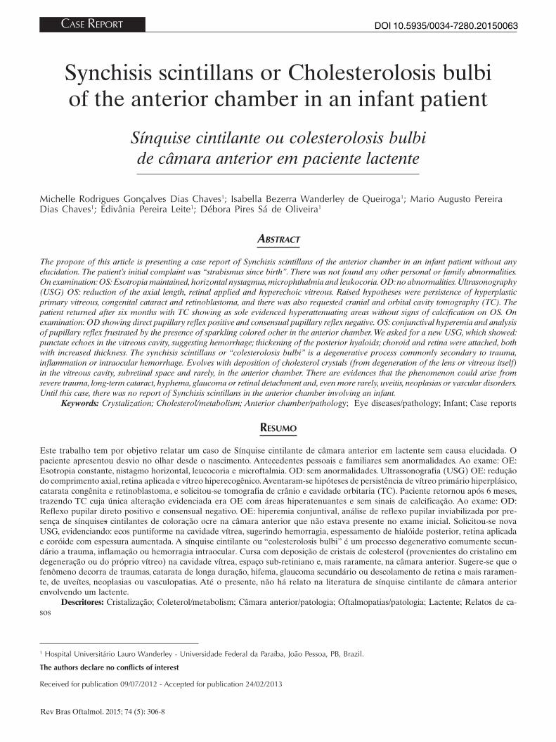
The initial complaint filed by the mother of the 1 year and ahalf infant was on the look deviation since birth. Personal and familyhistory with no abnormalities. The exam showed: Inspection: RE: nochange. LE: constant isotropy, horizontal nystagmus, leukocoria andmicrophthalmia (Figure 1). Indirect binocular ophthalmoscopy(IBO): RE: physiological relationship excavation / disc, characteristicmacular brightness, vascular arcades of habitual compliance, appliedretina. LE: not feasible. Ultrasonography (USG) LE: reduced axiallength applied retina and hyperechoic vitreous.
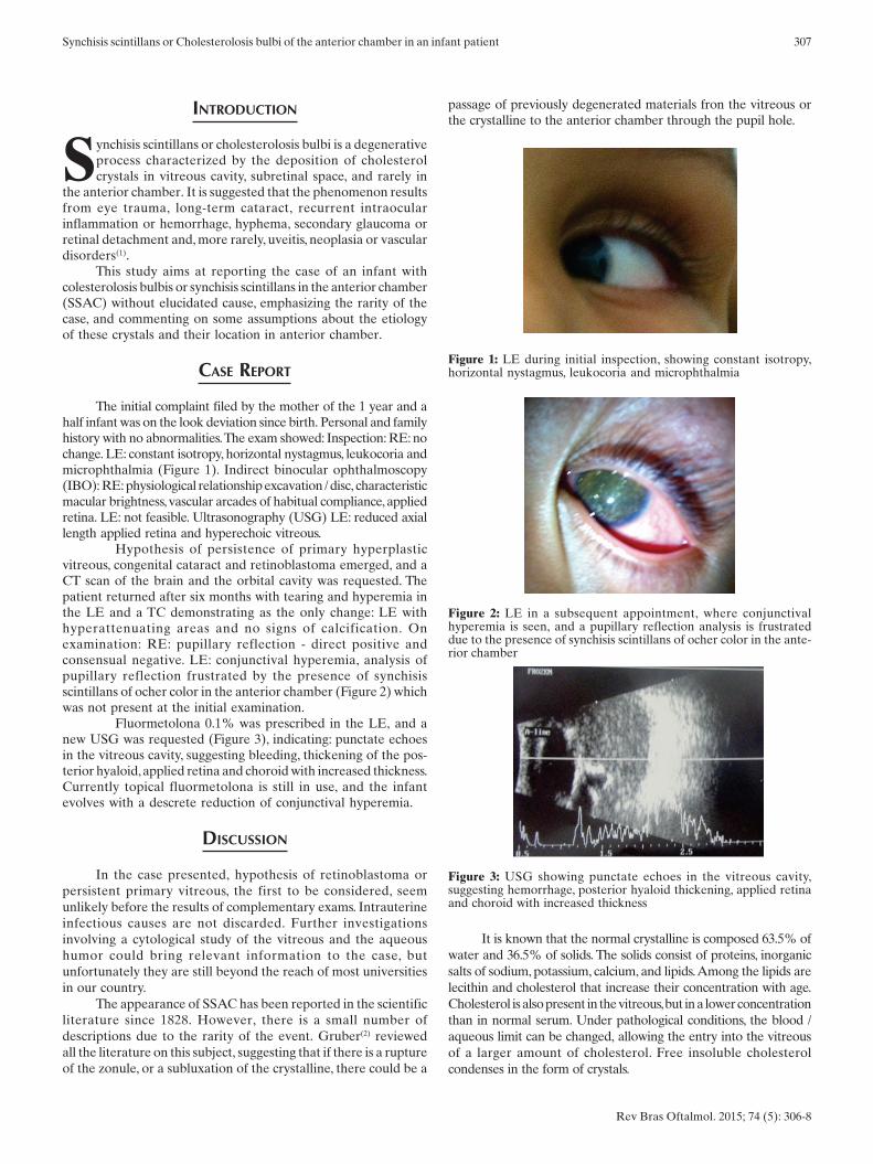
Hypothesis of persistence of primary hyperplasticvitreous, congenital cataract and retinoblastoma emerged, and aCT scan of the brain and the orbital cavity was requested. Thepatient returned after six months with tearing and hyperemia inthe LE and a TC demonstrating as the only change: LE withhyperattenuating areas and no signs of calcification. Onexamination: RE: pupillary reflection - direct positive andconsensual negative. LE: conjunctival hyperemia, analysis ofpupillary reflection frustrated by the presence of synchisisscintillans of ocher color in the anterior chamber (Figure 2) whichwas not present at the initial examination.
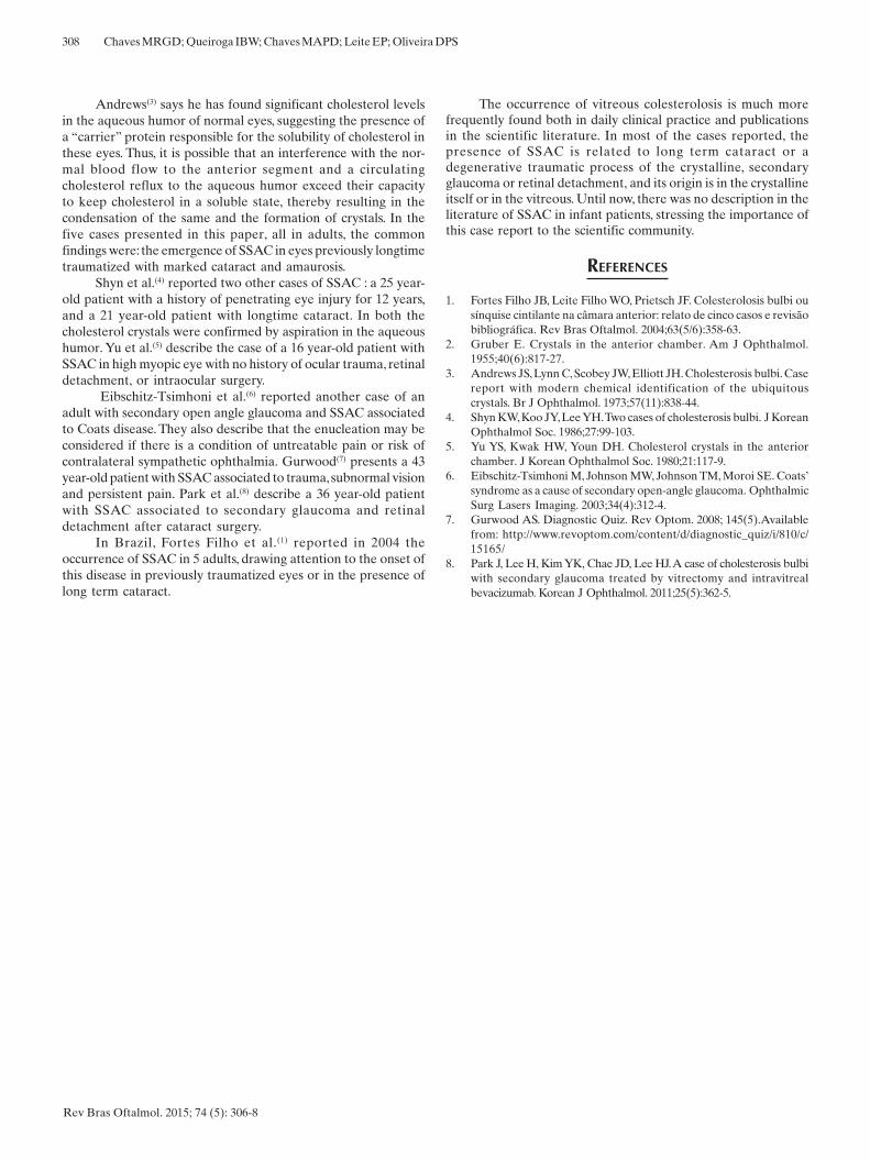
Fluormetolona 0.1% was prescribed in the LE, and anew USG was requested (Figure 3), indicating: punctate echoesin the vitreous cavity, suggesting bleeding, thickening of the pos-terior hyaloid, applied retina and choroid with increased thickness.Currently topical fluormetolona is still in use, and the infantevolves with a descrete reduction of conjunctival hyperemia.
DISCUSSION
In the case presented, hypothesis of retinoblastoma orpersistent primary vitreous, the first to be considered, seemunlikely before the results of complementary exams. Intrauterineinfectious causes are not discarded. Further investigationsinvolving a cytological study of the vitreous and the aqueoushumor could bring relevant information to the case, butunfortunately they are still beyond the reach of most universitiesin our country.
The appearance of SSAC has been reported in the scientificliterature since 1828. However, there is a small number ofdescriptions due to the rarity of the event. Gruber(2) reviewedall the literature on this subject, suggesting that if there is a ruptureof the zonule, or a subluxation of the crystalline, there could be a
passage of previously degenerated materials fron the vitreous orthe crystalline to the anterior chamber through the pupil hole.
Figure 1: LE during initial inspection, showing constant isotropy,horizontal nystagmus, leukocoria and microphthalmia
Figure 2: LE in a subsequent appointment, where conjunctivalhyperemia is seen, and a pupillary reflection analysis is frustrateddue to the presence of synchisis scintillans of ocher color in the ante-rior chamber
Figure 3: USG showing punctate echoes in the vitreous cavity,suggesting hemorrhage, posterior hyaloid thickening, applied retinaand choroid with increased thickness
Synchisis scintillans or Cholesterolosis bulbi of the anterior chamber in an infant patient
308
Andrews(3) says he has found significant cholesterol levelsin the aqueous humor of normal eyes, suggesting the presence ofa “carrier” protein responsible for the solubility of cholesterol inthese eyes. Thus, it is possible that an interference with the nor-mal blood flow to the anterior segment and a circulatingcholesterol reflux to the aqueous humor exceed their capacityto keep cholesterol in a soluble state, thereby resulting in thecondensation of the same and the formation of crystals. In thefive cases presented in this paper, all in adults, the commonfindings were: the emergence of SSAC in eyes previously longtimetraumatized with marked cataract and amaurosis.
Shyn et al.(4) reported two other cases of SSAC : a 25 year-old patient with a history of penetrating eye injury for 12 years,and a 21 year-old patient with longtime cataract. In both thecholesterol crystals were confirmed by aspiration in the aqueoushumor. Yu et al.(5) describe the case of a 16 year-old patient withSSAC in high myopic eye with no history of ocular trauma, retinaldetachment, or intraocular surgery.
Eibschitz-Tsimhoni et al.(6) reported another case of anadult with secondary open angle glaucoma and SSAC associatedto Coats disease. They also describe that the enucleation may beconsidered if there is a condition of untreatable pain or risk ofcontralateral sympathetic ophthalmia. Gurwood(7) presents a 43year-old patient with SSAC associated to trauma, subnormal visionand persistent pain. Park et al.(8) describe a 36 year-old patientwith SSAC associated to secondary glaucoma and retinaldetachment after cataract surgery.
In Brazil, Fortes Filho et al.(1) reported in 2004 theoccurrence of SSAC in 5 adults, drawing attention to the onset ofthis disease in previously traumatized eyes or in the presence oflong term cataract.
Rev Bras Oftalmol. 2015; 74 (5): 306-8
The occurrence of vitreous colesterolosis is much morefrequently found both in daily clinical practice and publicationsin the scientific literature. In most of the cases reported, thepresence of SSAC is related to long term cataract or adegenerative traumatic process of the crystalline, secondaryglaucoma or retinal detachment, and its origin is in the crystallineitself or in the vitreous. Until now, there was no description in theliterature of SSAC in infant patients, stressing the importance ofthis case report to the scientific community.
REFERENCES
1. Fortes Filho JB, Leite Filho WO, Prietsch JF. Colesterolosis bulbi ousínquise cintilante na câmara anterior: relato de cinco casos e revisãobibliográfica. Rev Bras Oftalmol. 2004;63(5/6):358-63.
2. Gruber E. Crystals in the anterior chamber. Am J Ophthalmol.1955;40(6):817-27.
3. Andrews JS, Lynn C, Scobey JW, Elliott JH. Cholesterosis bulbi. Casereport with modern chemical identification of the ubiquitouscrystals. Br J Ophthalmol. 1973;57(11):838-44.
4. Shyn KW, Koo JY, Lee YH. Two cases of cholesterosis bulbi. J KoreanOphthalmol Soc. 1986;27:99-103.
5. Yu YS, Kwak HW, Youn DH. Cholesterol crystals in the anteriorchamber. J Korean Ophthalmol Soc. 1980;21:117-9.
6. Eibschitz-Tsimhoni M, Johnson MW, Johnson TM, Moroi SE. Coats’syndrome as a cause of secondary open-angle glaucoma. OphthalmicSurg Lasers Imaging. 2003;34(4):312-4.
7. Gurwood AS. Diagnostic Quiz. Rev Optom. 2008; 145(5).Availablefrom: http://www.revoptom.com/content/d/diagnostic_quiz/i/810/c/15165/
8. Park J, Lee H, Kim YK, Chae JD, Lee HJ. A case of cholesterosis bulbiwith secondary glaucoma treated by vitrectomy and intravitrealbevacizumab. Korean J Ophthalmol. 2011;25(5):362-5.
Chaves MRGD; Queiroga IBW; Chaves MAPD; Leite EP; Oliveira DPS